Clinical Anatomy of the Aorta
15
Clinical Anatomy of the Aorta Clinical Anatomy of the Aorta Lawrence M. Witmer, PhD Lawrence M. Witmer, PhD Department of Biomedical Sciences College of Osteopathic Medicine Ohio University Athens, Ohio 45701 [email protected] Handout download: http://www.oucom.ohiou.edu/dbms-witmer/gs-rpac.htm
Transcript of Clinical Anatomy of the Aorta

Clinical Anatomy of the AortaClinical Anatomy of the Aorta
Lawrence M. Witmer, PhDLawrence M. Witmer, PhDDepartment of Biomedical SciencesCollege of Osteopathic MedicineOhio UniversityAthens, Ohio [email protected]
Handout download:http://www.oucom.ohiou.edu/dbms-witmer/gs-rpac.htm

Feneis 1994Feneis 1994
General Anatomy of the AortaGeneral Anatomy of the Aorta
Divisions of the Mediastinum
anatomicalanatomicalanatomical surgicalsurgicalsurgical
ascending aorta,arch of aorta
thoracicaorta
• Ascending aorta• Aortic Arch• Thoracic (descending) aorta• Abdominal aorta
from Schwartz et al., 1999from Schwartz et al., 1999from Schwartz et al., 1999

Fawcett 1986
Histology of Arteries and of the AortaHistology of Arteries and of the Aorta
• Layers of CVS: tunica intima, tunica media, tunica adventitia• Aorta is an elastic artery with an expanded, elastic media

Aortic DissectionAortic Dissection
intimalflap
intimalintimalflapflap
MRAMRAMRA
transesoph.echocardiogr.transesoph.transesoph.
echocardiogr.echocardiogr.
From Schwartz et al. 1999From Schwartz et al. 1999From Schwartz et al. 1999
From Blackbourne 1998
intimal tear
truelumen
falselumen
• tear in intima leading to separationof the tunica media & formation ofa “false lumen”
• Re-entry tear leading to a “double-barreled aorta”

DeBakeyDeBakey StanfordStanford
Classification of Aortic DissectionClassification of Aortic DissectionI
II
III
• Types I & II: tear in asc. aorta• Type III: tear in thor. aorta• Type I: asc. & desc. Aorta• Type II: only asc. Aorta• Type III: only desc. aorta
A
B• Type A: asc. aorta
± desc. aorta• Stanford Type A
includes DeBakeyTypes I & II
• Type B: desc. aorta
From Blackbourne 1998

Development of the AortaDevelopment of the Aorta
• Paired endocardial tubesfuse into a single tube
• Endocardial tubeelongates & constricts
• Subdivisions• Sinus venosus• Atrium• Ventricle• Bulbus cordis• Truncus arteriosus
• Truncus arteriosus is partitioned into theaorta and pulmonarytrunk
From Moore and Persaud, 1998

Partitioning of the Truncus ArteriosusPartitioning of the Truncus Arteriosus
• Formation of bulbar & truncal ridges• Ridges spiral 180º as they grow• Ridges fuse to form aortico-
pulmonary septum• Aorticopulmonary septum divides
aorta and pulmonary trunk
From Moore and Persaud, 1998

Defects in Partitioning of the Truncus ArteriosusDefects in Partitioning of the Truncus Arteriosus
From Moore and Persaud, 1998
Transposition of theTransposition of theGreat Arteries (TGA)Great Arteries (TGA)
Tetralogy of FallotTetralogy of Fallot
• Most common cyanotic neonatal heart defect
• Failure of aorticopulmonary septum totake a spiraling course
• Fatal without PDA, ASD, & VSD
• Four co-occurring heart defects• Pulmonary stenosis• Ventricular septal defect• Overriding aorta (dextroposition)• Right ventricular hypertrophy
• Asymmetrical fusion of bulbar & truncalridges
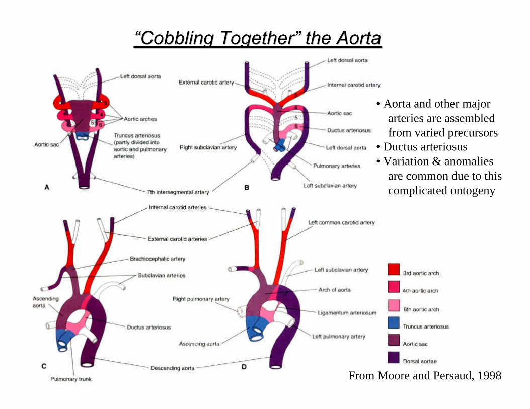
“Cobbling Together” the Aorta“Cobbling Together” the Aorta
• Aorta and other majorarteries are assembledfrom varied precursors
• Ductus arteriosus• Variation & anomalies
are common due to thiscomplicated ontogeny
From Moore and Persaud, 1998

Variation in Branching of the Aortic ArchVariation in Branching of the Aortic Arch
• Based on a study of 1000 cadavers byLiechty et al. 1957
• “Textbook” example (variant I) occursless than two-thirds of the time
• Most are only problematic insofar as they may be initially confusing duringsurgery

Vascular RingsVascular Rings
Anomalous Right Subclavian ArteryAnomalous Right Subclavian ArteryRetroesophageal course compresses trachea & esophagus
Right aortic archRight aortic archCan take a retroesophageal course
From Moore and Persaud, 1998

Vascular RingsVascular Rings
From Moore and Persaud, 1998
Double Aortic ArchDouble Aortic Arch
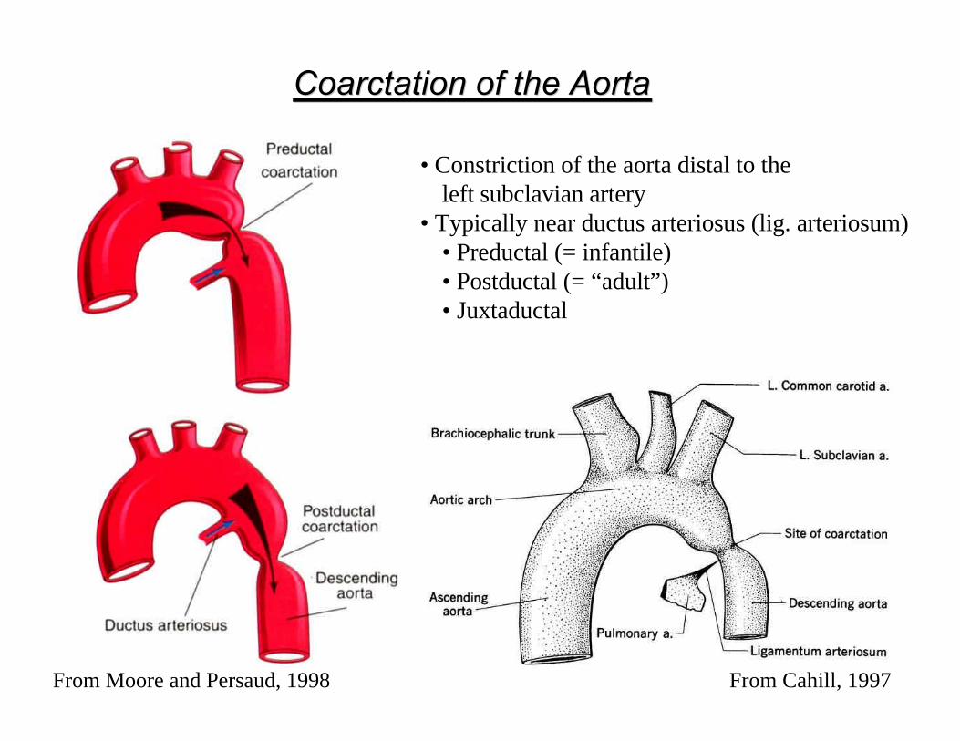
Coarctation of the AortaCoarctation of the Aorta
• Constriction of the aorta distal to the left subclavian artery
• Typically near ductus arteriosus (lig. arteriosum)• Preductal (= infantile)• Postductal (= “adult”) • Juxtaductal
From Moore and Persaud, 1998 From Cahill, 1997

Coarctation of the AortaCoarctation of the Aorta
From Cahill, 1997
• Subclavian → ΙΜΑ →intercostals → aorta
• Subclavian → IMA →sup. epigastr. → inf.epigastr. → iliac → aorta
• Subclavian → cervical & scap. branches → intercostals → aorta
• Subclavian → vertebral→ ant. spinal → inter-costals & lumbars → aorta
Collateral Circulation

ReferencesReferences
Blackbourne, L. H. 1998. Surgical Recall, 2nd Ed. Williams & Wilkins, Baltimore.Cahill, D. R. 1997. Lachman’s Case Studies in Anatomy. Oxford Univ. Press, New York.Fawcett, D. W. 1986. Bloom and Fawcett: A Textbook of Histology, 11th Ed. Saunders,
Philadelphia.Feneis, H. 1994. Pocket Atlas of Human Anatomy. Thieme, New York.Liechty, Shields, and Anson. 1957. Quart. Bull. Northwest. Univ. Med. Sch. 31:136-143.Moore, K. L. and T. V. N. Persaud. 1998. The Developing Human: Clinically Oriented
Embryology, 6th Ed., Saunders, Philadelphia.Schwartz et al. (eds.), Principles of Surgery, 7th Ed., McGraw Hill, New York.