clinic - bsmo.be · clinic 4 the belgian journal of medical oncology is the official journal of the...
34
clinic 4 THE BELGIAN JOURNAL OF MEDICAL ONCOLOGY IS THE OFFICIAL JOURNAL OF THE BELGIAN SOCIETY OF MEDICAL ONCOLOGY (BSMO) AND THE BELGIAN ASSOCIATION FOR CANCER RESEARCH (BACR) Belgian Journal of Medical Oncology VOLUME 13 | ISSUE 4 JUNE 2019 Management of end-stage malignant bowel obstruction: an evidence-based review for clinical practice Q. Binet, L. Duck Interventions in non-metastatic castration-resistant prostate cancer: earlier seems better D. Schrijvers Renal mass in a patient with invasive lobular adenocarcinoma X. Mortiers, T. Adams, H. Vandeursen HER2 overexpression in gastroesophageal adenocarcinoma, prognostic and predictive value and diagnostic approach L. Thijs, P. Peeters, B. Maes, S. Tejpar Highlights of the Gastrointestinal Cancers Symposium 2019 W. Lybaert, H. Prenen, I. Borbath, A. Demols, I. Dero New oncology reimbursements in Belgium P. Specenier
Transcript of clinic - bsmo.be · clinic 4 the belgian journal of medical oncology is the official journal of the...

clinic
4THE BELGIAN JOURNAL OF MEDICAL ONCOLOGY IS THE OFFICIAL JOURNAL OF THE BELGIAN SOCIETY OF MEDICAL ONCOLOGY (BSMO) AND THE BELGIAN ASSOCIATION FOR CANCER RESEARCH (BACR)
Belgian Journal of Medical OncologyVOLUME 13 | ISSUE 4
JUNE 2019
Management of end-stage malignant bowel obstruction: an evidence-based review for clinical practice Q. Binet, L. Duck
Interventions in non-metastatic castration-resistant prostate cancer: earlier seems betterD. Schrijvers
Renal mass in a patient with invasive lobular adenocarcinomaX. Mortiers, T. Adams, H. Vandeursen
HER2 overexpression in gastroesophageal adenocarcinoma, prognostic and predictive value and diagnostic approachL. Thijs, P. Peeters, B. Maes, S. Tejpar
Highlights of the Gastrointestinal Cancers Symposium 2019W. Lybaert, H. Prenen, I. Borbath, A. Demols, I. Dero
New oncology reimbursements in BelgiumP. Specenier

248
VOLUME13 JUNE20194
BELGIAN JOURNAL OF MEDICAL ONCOLOGY
122
123
129
132
135
142
150
153
INTRODUCTION
J. De Grève
REVIEW ONCOLOGY
Management of end-stage malignant bowel obstruction:
an evidence-based review for clinical practice
Q. Binet, L. Duck
PHARMACOTHERAPY
Interventions in non-metastatic castration-resistant
prostate cancer: earlier seems better
D. Schrijvers
ONCOCASE
Renal mass in a patient with invasive lobular adenocarcinoma
X. Mortiers, T. Adams, H. Vandeursen
ONCOTHESIS
HER2 overexpression in gastroesophageal adenocarcinoma,
prognostic and predictive value and diagnostic approach
L. Thijs, P. Peeters, B. Maes, S. Tejpar
CONGRESS NEWS
Highlights of the Gastrointestinal Cancers Symposium 2019
W. Lybaert, H. Prenen, I. Borbath, A. Demols, I. Dero
REIMBURSEMENT NEWS
New oncology reimbursements in Belgium
P. Specenier
CALENDAR OF EVENTS
TABLE OF CONTENTS 121COLOPHON
The Belgian Journal of Medical Oncology (BJMO) is the official scientific journal of the Belgian Society of Medical Oncology (BSMO) and the Belgian Association for Cancer Research (BACR). The BJMO aims to be a peer-reviewed cancer journal covering all aspects of the diagnostic and clinical management of cancer patients. Subscriptions are free for all specialists who are active in the fields of oncolo-gy, radiotherapy and adherent fields.
PUBLISHER AND EDITORIAL OFFICEAriez International B.V.Ms. E. van Zanten, MScc/o Oude Houtlei 118-120, 9000 Gent, BelgiumTel: 0031-75-642 94 20E-mail: [email protected]
FOUNDED BYL. Dirix, MD, PhD, medical oncologist, St Augustinus Hos-pital, Wilrijk.
EDITOR-IN-CHIEFJ. De Grève, MD, PhD, Professor in Oncology, head of de-partment of medical oncology, University Hospital Brus-sels, Brussels.
EDITORIAL BOARDA. Awada, MD, PhD, Professor in Oncology, Jules Bordet Institute, Brussels, J-F. Baurain, MD, PhD, Professor in Oncology, CliniquesUniversitaires Saint-Luc, Brussels, F. Cornelis, MD, medical oncologist, Cliniques Universitaires Saint-Luc, Brussels, H. Denys, MD, PhD, medical oncologist, University HospitalGent, Gent, M. De Ridder, MD, PhD, Professor in Radiation Oncology, UZ Brussel, Brussels, M. Dicato, MD, haematologist-oncologist, Centre Hospitalier de Luxembourg, Luxembourg, P. Dirix, MD, PhD, radiation oncologist, University HospitalsLeuven, Leuven,L. Duck, MD, PhD, medical oncologist, Clinique Saint-Pierre, Ottignies, C. Duhem, MD, haematologist-oncologist, Centre Hospitalier de Luxembourg, Luxembourg, F. Duhoux, MD, PhD, medical oncologist, CliniquesUniversitaires Saint-Luc, Brussels, A. Hendrix, MD, medical oncologist, Gent UniversityHospital, Gent, G. Jerusalem, MD, PhD, medical oncologist, CHU Sart-Tilman, Liège, J. Kerger, MD, PhD, medical oncologist, Jules Bordet Institute,Brussels, J-P. Machiels, MD, PhD, Professor in Oncology, Cliniques Universitaires Saint-Luc, Brussels, J. Mebis, MD, PhD, medical oncologist, Jessa Ziekenhuis, Hasselt, P. Pauwels, MD, PhD, pathologist, University HospitalAntwerp, Edegem, H. Prenen, MD, PhD, Professor in Oncology, UniversityHospitals Leuven, Leuven, S. Rottey, MD, PhD, medical oncologist, Gent UniversityHospital, Gent, D. Schrijvers, MD, PhD, medical oncologist, Ziekenhuis-netwerk Antwerpen, Antwerp,P. Specenier, MD, PhD, Professor in Oncology, University Hospital Antwerp, Edegem, J-L. Van Laethem, MD, PhD, Professor in Gastroenterology,Université Libre de Bruxelles, Brussels, H. Van Poppel, MD, PhD, Professor in Urology, University Hospitals Leuven, Leuven, D. Verhoeven, MD, PhD, medical oncologist, AZ KLINA,Brasschaat, J.B. Vermorken, MD, PhD, Professor in Oncology, University Hospital Antwerp, Edegem, H. Wildiers, MD, PhD, Professor in Oncology, UniversityHospitals Leuven, Leuven.
SUBSCRIPTIONSFor paid subscriptions, please refer to the publisher: [email protected].
Publication Frequency: 8 issues per year.Edition: 3,000 copies.
COPYRIGHT© Ariez International B.V. Zaandam, The Netherlands.No information from this publication may be copied, com-mercialised by third parties without written permission of the publisher in advance. The Editorial Board and the publisher cannot be held responsible for the content of articles or the content of advertisements and cannot be held responsible for or liable to potential claims of third parties regarding damage suffered following publication of this issue.
COVER ILLUSTRATION Science Photo
ISSN-NUMBER 1784-7141

VOLUME13 JUNE2019
DEAR COLLEAGUES,
In this issue, the management of malignant bowel obstruction is reviewed.
Bowel (sub)obstruction severely downgrades the quality of life and should
be managed and relieved as much as possible, even in end-stage disease.
Surgery remains one of the options to be considered, also for patients with
annoying sub-obstructive symptoms.
The Pharmacotherapy section reports on the drastic effects of three newer anti-androgen drugs on the metastasis-free survival
of locoregional castration-resistant prostate cancer, with one of them having less toxicity and survival improvement.
The Oncocase provides yet another example of the erratic metastatic behavior of lobular breast cancer.
The Oncothesis examines HER2-positive gastro-oesophageal cancer, trastuzumab treatment, and secondary resistance.
The Highlights of the Gastrointestinal Cancers Symposium 2019 (San Francisco) focusses on the progress in immunotherapy
and genomic profiling.
Durvalumab in NSCLC and liposomal irinotecan in pancreatic cancer are the most recent reimbursed drugs.
Finally, I would like to call upon you to continue lobbying for broader use of genomics in cancer, both somatic and germline.
Somatic NGS is introduced with the upcoming convention but should be broadened in scope, become agnostic and also
include genes that identify patients for clinical trials running in Belgium or off-label access. One of the ways to go is to
centralise the sophisticated diagnostics needed in platforms that are capable of implementing the various technologies in
high quality and most cost-effective way. Today Belgium is lagging severely behind other countries in the reimbursement
level of NGS. The broad professional co-operation very well co-ordinated by Sciensano is there while the allocated budget
is insufficient.
For the germline testing, the time has arrived to turn genetic testing predictive for high cancer risk from a diagnostic into a
life-saving preventive tool. For example, to prevent 250-500 breast cancer deaths per year. Belgium could be the global front
runner showing the way to go. Why not? The citizens at least support the idea, what about the professionals?
We wish you an enjoyable read,
Jacques De Grève, MD, PhD
4
J. DE GRÈVE
122INTRODUCTION

VOLUME13 JUNE20194
123REVIEW ONCOLOGY
SUMMARYMalignant bowel obstruction is the clinical and imaging evidence of bowel obstruction beyond the ligament of Treitz in the setting of an incurable cancer with intraperitoneal spread. A multi-detector computed tomo-graphy scan with multiplanar reconstructions is the gold standard for diagnosis confirmation and treatment orientation. Treatment is challenging and can either be surgical, endoscopic or most likely medical. In the following manuscript, we discuss the current place of each treatment modality in end-stage malignant bowel obstruction management.(BELG J MED ONCOL 2019;13(4):123-128)
Department of Medical Oncology/Hematology and Palliative Care Unit, Cliniques Saint-Pierre, Ottignies-Louvain-la-Neuve, Belgium.
Please send all correspondence to: L. Duck, MD, Head Department of Medical Oncology and Hematology, Clinique Saint-Pierre, Avenue Reine
Fabiola 9, 1340 Ottignies-Louvain-la-Neuve, Belgium, tel: +32 010437241, email: [email protected].
Conflict of interest: The authors have nothing to disclose and indicate no potential conflict of interest.
Keywords: malignant bowel obstruction, nasogastric tube, palliative care, somatostatin analogues, supportive care
Q. Binet, MD, L. Duck, MD
Management of end-stage malignant bowel obstruction: an evidence-based review for clinical practice
INTRODUCTIONMalignant bowel obstruction (MBO) is described as the clin-
ical and imaging evidence of bowel obstruction beyond the
ligament of Treitz in the setting of an incurable cancer with
intraperitoneal spread.1 Peritoneal carcinomatosis is usual-
ly the result of metastasis from an intra-abdominal primary
cancer (typically ovarian 20-50%, colorectal 10-28%, stom-
ach 6-19%, pancreatic 6-13%, bladder 3-10% or endometrial
3-11%), but extra-abdominal cancers are also reported (e.g.,
breast cancer or melanoma).2
MBO can either be mechanical or functional. Mechanical
aetiologies include intrinsic or extrinsic mass effect. Radi-
ation-induced fibrosis and post-surgical adhesions are dif-
ferent entities and harbour different prognosis. Functional
aetiologies include tumour infiltration of nerves responsible
for intestinal mobility, secondary paralytic ileus (intra-ab-
dominal infection, ascites or pain) and drug-induced neurop-
athy (opioids and anticholinergic drugs).3 In reality, however,
a mixed picture with mechanical and functional elements is
most frequently encountered.2
MBO is of utmost importance in clinical practice since it is
estimated to occur in 3-15% of all patients with advanced
malignancy.4 Hereunder, we synthesize the latest guidelines
in end-stage MBO management, which is known to be chal-
lenging (Figure 1). We will not address upfront MBO, which
treatment may involve chemotherapy, extensive surgery and
more recently hyperthermic intraperitoneal chemothera-
py (HIPEC). Because the prognosis of end-stage MBO is ex-
tremely limited, the goal is highly patient-specific and can
either be obstruction relief, if possible, or symptom man-
agement alone.
CLINICAL MANAGEMENTCLINICAL SUSPICIONCommon clinical signs include (1) abdominal pain and dis-
tension, (2) nausea and vomiting and (3) the absence of gas
VOLUME13 JUNE2019
124
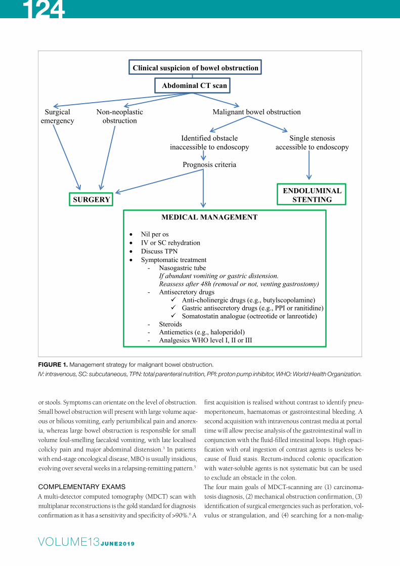
FIGURE 1. Management strategy for malignant bowel obstruction.
IV: intravenous, SC: subcutaneous, TPN: total parenteral nutrition, PPI: proton pump inhibitor, WHO: World Health Organization.
or stools. Symptoms can orientate on the level of obstruction.
Small bowel obstruction will present with large volume aque-
ous or bilious vomiting, early periumbilical pain and anorex-
ia, whereas large bowel obstruction is responsible for small
volume foul-smelling faecaloid vomiting, with late localised
colicky pain and major abdominal distension.3 In patients
with end-stage oncological disease, MBO is usually insidious,
evolving over several weeks in a relapsing-remitting pattern.5
COMPLEMENTARY EXAMSA multi-detector computed tomography (MDCT) scan with
multiplanar reconstructions is the gold standard for diagnosis
confirmation as it has a sensitivity and specificity of >90%.6 A
first acquisition is realised without contrast to identify pneu-
moperitoneum, haematomas or gastrointestinal bleeding. A
second acquisition with intravenous contrast media at portal
time will allow precise analysis of the gastrointestinal wall in
conjunction with the fluid-filled intestinal loops. High opaci-
fication with oral ingestion of contrast agents is useless be-
cause of fluid stasis. Rectum-induced colonic opacification
with water-soluble agents is not systematic but can be used
to exclude an obstacle in the colon.
The four main goals of MDCT-scanning are (1) carcinoma-
tosis diagnosis, (2) mechanical obstruction confirmation, (3)
identification of surgical emergencies such as perforation, vol-
vulus or strangulation, and (4) searching for a non-malig-
Figure 1. Management strategy for malignant bowel obstruction. IV: intravenous, SC:
subcutaneous, TPN: total parenteral nutrition, PPI: proton pump inhibitor, WHO: World
Health Organization.
Clinical suspicion of bowel obstruction
Abdominal CT scan
Surgical emergency
Non-neoplastic obstruction
Malignant bowel obstruction
Identified obstacle inaccessible to endoscopy
Single stenosis accessible to endoscopy
Prognosis criteria
SURGERY
MEDICAL MANAGEMENT
ENDOLUMINAL STENTING
• Nil per os• IV or SC rehydration• Discuss TPN• Symptomatic treatment
- Nasogastric tubeIf abundant vomiting or gastric distension.Reassess after 48h (removal or not, venting gastrostomy)
- Antisecretory drugsü Anti-cholinergic drugs (e.g., butylscopolamine)ü Gastric antisecretory drugs (e.g., PPI or ranitidine)ü Somatostatin analogue (octreotide or lanreotide)
- Steroids- Antiemetics (e.g., haloperidol)- Analgesics WHO level I, II or III
VOLUME13 JUNE20194
125REVIEW ONCOLOGY
nant cause for the obstruction such as adhesions, hernias and
eventration resulting from a previous surgery.3
Other imaging studies are often superfluous. A plain ab-
dominal X-ray is fast, inexpensive and widely available. It
can differentiate upper gastrointestinal, small or large bow-
el obstruction but is unable to accurately identify its loca-
tion, cause and complications and is therefore rather futile.
An ultrasound is intrinsically restrained for the evaluation
of gas-containing structures and is highly operator-depen-
dent. An MRI is accurate in diagnosing MBO but limited by
high costs and an increased acquisition time and has there-
fore few indications such as rectal cancer local staging.7 En-
teroclysis and especially CT and MR enteroclysis enable an
improved evaluation of the small bowel by challenging wall
distensibility. They can help drive the management of inter-
mittent low-grade small bowel obstructions by determin-
ing their number and location. However, the technique is
time and labour intensive and uncomfortable for the patient
(preparation, sedation).8 A FDG-PET scan is expensive and
relatively unavailable but proved useful in refuting the pres-
ence of intra-abdominal malignancy in patients with bowel
obstruction and a prior history of cancer.9
TREATMENT OPTIONSSURGICAL PALLIATIONAfter the above described surgical emergencies have been
ruled out, patients should be selected appropriately for sur-
gery depending on the cancer type (carcinomatosis associ-
ated with ovarian cancer is of better immediate prognosis
than with gastrointestinal cancer, which supports surgical
intervention) and individual prognosis factors for post-op-
erative morbi-mortality (surgery should be avoided in case
of advanced age, comorbidities, poor nutritional state, poor
performance status, ascites, massive infiltration of the mes-
entery or mesocolon and history of radiotherapy)3,10. The
benefit of surgery may be limited to an increase in quality
of life at the cost of high operative risks of morbi-mortality.11
Indications are therefore very rare and should be discussed
in multidisciplinary collaborative meetings. For obvious ob-
stacles with localised peritoneal infiltration, resection plus
anastomosis is preferable to stoma formation or bypass as it
has the greatest operative survival (7.2 months compared to
3.4 and 2.7 months respectively; Level II of evidence, grade
B recommendation).12
INTERVENTIONAL PALLIATIONWhen feasible, endoscopic prosthesis procedures should be
preferred to surgery as they carry a good clinical success rate
and a lower morbi-mortality rate (Level III of evidence, grade
C recommendation).3 The indication is a single stenosis on a
CT scan within reach of either a gastroscope or colonoscope.
Complications rarely occur and consist of migration, obstruc-
tion or perforation.3 Important risk factors for perforation are
current radiotherapy and the use of anti-angiogenic drugs
(e.g., bevacizumab).13,14
MEDICAL PALLIATIONA vicious cycle is entered wherein hypersecretion generates
gut dilatation and vomiting, followed by further secretion
and vomiting, quickly resulting in dehydration and electro-
lyte disturbances, which may prove lethal in absence of a
medical intervention.
1. Nasogastric tube and venting gastrostomy
The placement of a nasogastric tube (NGT) relieves intrac-
table vomiting and gastric distension and thereby decreases
the risk of inhalation. It also enables the patient to drink. It
allows time to determine if medical treatment will work. Af-
ter 48 hours and only if secretions do no exceed 1L/24h, the
removal of the NGT should be discussed to minimise the
patient’s discomfort (nostril ulceration, oesophageal erosion,
pharyngitis and sinusitis; expert consensus).3 Otherwise, a
venting gastrostomy could be discussed as a long-term alter-
native, allowing the patient to eat small amounts of food for
pleasure. Percutaneous endoscopic interventions should be
preferred but there are contra-indications, principally ascites.
Radiological interventions do not require general anaesthe-
sia. Surgical indications remain rare but should be evaluated
during surgical exploration of an obstruction.3
2. Steroids
Steroids may contribute to the treatment of MBO by (1) de-
creasing the oedema around the tumour, (2) their central
antiemetic effect and (3) their indirect analgesic effects by
reducing bowel distension and inflammation. Even though
evidence is lacking, a short course of 5-10 days of 0.5-1 mg/
kg/24h of intravenous methylprednisolone (or equivalent) at
the time of diagnosis may help manage the symptoms and/
or resolve the obstruction (Level II of evidence, grade C rec-
ommendation).3,15 In order to reduce psychotropic effects and
insomnia, total doses should be divided and administered at
breakfast and lunchtime (expert consensus). Even though
side effects for this posology are minimal, it is also worth
bearing in mind the risks of gastrointestinal ulcerations and
immunosuppression.2
3. Antisecretory drugs
Anticholinergic drugs. Scopolamine, butylscopolamine and gly-
copyrronium bromide have antispasmodic, antiemetic and
antisecretory effects and reduce the volume of gastrointesti-

VOLUME13 JUNE2019
126nal secretions. Scopolamine induces central adverse effects
and butylscopolamine or glycopyrronium are therefore pre-
ferred as they hardly cross the blood-brain barrier (expert
consensus).2,3 The mean butylscopolamine dose is 60 to 120
mg/24h administered intravenously (IV) or subcutaneously
(SC). Contra-indications (glaucoma and urinary retention)
and undesirable effects (dry mouth, tachycardia, etc.) are
those of atropinic drugs.
Gastric antisecretory drugs. Proton-pump inhibitors (PPIs)
seem relevant to reduce gastric secretion or bile reflux and
relieve upper digestive symptoms (Level II of evidence, grade
B recommendation). Because oral administration is not feasi-
ble in MBO and half-life in blood is 60 minutes, PPIs should
either be administered IV continuously over 24h or SC (vali-
dated for omeprazole).16 Anti-histamine drugs such as ranit-
idine induce a greater antisecretory effect than PPIs (Level
I of evidence) but conclusions on long-term use cannot be
drawn, especially because H2 antagonists have been asso-
ciated with rapid tolerance (within hours).3,17
Somatostatin analogues. Peripheral actions of somatostatin
include decreasing splanchnic and portal blood flow, small
intestine secretions and gastrointestinal motility, and in-
creasing the gastrointestinal reabsorption of water and
electrolytes. Synthetic analogues of somatostatin such as
octreotide and lanreotide have a longer lasting effect and are
effective in decreasing pain, treating nausea and vomiting,
successfully removing NGT and most importantly increas-
ing the quality of life (Level I of evidence, grade B recom-
mendation).3,18 Both drugs are well tolerated. Possible side
effects include diarrhoea, abdominal pain, change in blood
sugar levels and risk of gallstones.
Recommended posology for octreotide immediate release
(IR) is 600 µg/day SC (continuous or discontinuous) or IV
(continuous) with a reassessment on day 3. Octreotide long-
acting-release (LAR) 30 mg administered intramuscularly
(IM) every month can be considered, but effective concen-
tration is attended on day 7 and therefore requires octreotide
IR for the first six days after IM injection. Posology for lan-
reotide in this indication is only validated in its prolonged
release (PR) form at 30 mg IM every ten days with a reas-
sessment before the second injection. If the treatment does
not prove effective on the reassessment day, it should be
discontinued.3
Some randomised controlled trials proved somatostatin an-
alogues to be superior to anticholinergic drugs regarding
symptomatic relief, but they are much more expensive and
therefore reserved for patients who responded well to these
drugs previously or after failure of standard treatment (ex-
pert consensus).3,17,19,20 In clinical practice, anticholinergic
drugs and somatostatin analogues can be associated.
4. Antiemetics
Antiemetic drugs ensure a decrease in nausea and vomiting,
especially when associated with antisecretory drugs. In pa-
tients with incomplete obstruction, metoclopramide is usu-
ally the first-line treatment. It is contra-indicated in complete
obstructions because of its prokinetic effect that could wors-
en abdominal pain and the risk of perforation (Level III of ev-
idence, grade D recommendation). In patients with complete
obstruction, butyrophenones such as haloperidol (adminis-
tered SC every 8-12 hours or in a continuous infusion) are
therefore considered first-line treatment. Second-line treat-
ments include phenothiazines (e.g., chlorpromazine), since
they have serious sedative and anticholinergic side effects,
and 5-HT3 receptor antagonists (e.g., ondansetron) because
they are expensive and only have a marketing license for che-
motherapy- or radiotherapy-induced emesis.3
5. General measures
Parenteral rehydration and electrolyte replacement should
be promptly initiated and adapted to clinical and biologi-
cal evolution (Level I of evidence, grade A recommendation).
Biochemical imbalance, most commonly hypokalaemia
and hypocalcaemia, may indeed contribute to intestinal
dysmotility.
Parenteral nutrition should be discussed depending on the
life expectancy and the expected risk (e.g., thrombosis, in-
fections) -benefit ratio (Level III of evidence, grade C recom-
mendation). Even though cachexia is a known indicator of
poor prognosis in cancer patients, there is no evidence sup-
porting parenteral nutrition in MBO in order to improve ei-
ther survival or quality of life.21,22 Its only true indication is
to enable adjuvant chemotherapy in patients who are oper-
ated for MBO and have a post-surgical survival likely to be
more than three months.2 Home parenteral nutrition could
be considered in end-stage MBO patients if they have an ac-
ceptable performance status and are expected to die from
starvation prior to tumour spread.21
In terminally ill patients, treatments that prove not reason-
able should be discontinued or never initiated, particularly
venting gastrostomy and artificial nutrition. If obstruction re-
solves (36% of inoperable cases, mostly by Day 7), all treat-
ments described above should gradually been discontinued
and a laxative treatment may be discussed to limit the high
risk of recurrence (72% of cases; Level III of evidence, grade
C recommendation).3,23
CONCLUSIONMalignant bowel obstruction is a frequent and severe condition.
Its management requires prompt collaboration between radiol-
ogists, surgeons and medical oncologists. After abdominal CT

VOLUME13 JUNE20194
127REVIEW ONCOLOGY
scan evaluation, patients should be selected for endoscopic, sur-
gical or most likely symptomatic medical management. The
combination of a NGT for intractable vomiting or gastric dis-
tension with steroids, antisecretory drugs and antiemetics is
important to relieve patient distress and proves in most cases
capable of achieving acceptable patient comfort.
REFERENCES1. Anthony T, Baron T, Mercadante S, et al. Report of the clinical protocol com-
mittee: development of randomized trials for malignant bowel obstruction. J Pain
Symptom Manage. 2007;34(1 Supll):S49-59.
2. Ferguson HJ, Ferguson CI, Speakman J, et al. Management of intestinal ob-
struction in advanced malignancy. Ann Med Surg (Lond). 2015;4(3):264-70.
3. Laval G, Marcelin-Benazech B, Guirimand F, et al. Recommendations for bow-
el obstruction with peritoneal carcinomatosis. J Pain Symptom Manage.
2014;48(1):75-91.
4. Tuca A, Guell E, Martinez-Losada E, et al. Malignant bowel obstruction in ad-
vanced cancer patients: epidemiology, management, and factors influencing
spontaneous resolution. Cancer Manag Res. 2012;4:159-69.
5. Baines MJ. Symptom control in advanced gastrointestinal cancer. Eur J Gas-
troenterol Hepatol. 2000;12(4):375-79.
6. Silva AC, Pimenta M, Guimarães LS. Small bowel obstruction: what to look
for. Radiographics. 2009;29(2):423-39.
7. Beall DP, Fortman BJ, Lawler BC, et al. Imaging bowel obstruction: a compar-
ison between fast magnetic resonance imaging and helical computed tomog-
raphy. Clin Radiol. 2002;57(8):719-24.
8. Kohli MD, Maglinte DD. CT enteroclysis in incomplete small bowel obstruc-
tion. Abdom Imaging. 2009;34(3):321-7.
9. Wu WG, Dong P, Wu XS, et al. Surgical management of patients with bowel
obstructions secondary to gastric cancer. World J Gastroenterol.
2013;19(28):4559-67.
10. Feuer DJ, Broadley KE, Shepherd JH, et al. Surgery for the resolution of
symptoms in malignant bowel obstruction in advanced gynaecological and gas-
trointestinal cancer. Cochrane Database Syst Rev. 2000;(4):CD002764.
11. Hofmann B, Håheim LL, Søreide JA. Ethics of palliative surgery in patients
with cancer. Br J Surg. 2005;92(7):802-9.
12. Shariat-Madar B, Jayakrishnan TT, Gamblin TC, et al. Surgical management
of bowel obstruction in patients with peritoneal carcinomatosis. J Surg Oncol.
2014;110(6):666-9.
13. Datye A, Hersh J. Colonic perforation after stent placement for malignant col-
orectal obstruction - causes and contributing factors. Minim Invasive Ther Allied
Technol. 2011;20(3):133-40.
14. Costamagna G, Tringali A, Spicak J, et al. Treatment of malignant gastrodu-
odenal obstruction with a nitinol self-expanding metal stent: an international pro-
spective multicentre registry. Dig Liver Dis. 2012;44(1):37-43.
15. Feuer DJ, Broadley KE. Corticosteroids for the resolution of malignant bow-
el obstruction in advanced gynaecological and gastrointestinal cancer. Cochrane
Database Syst Rev. 2000;(2):CD001219.
KEY MESSAGES FOR CLINICAL PRACTICE
1. Malignant bowel obstruction is a frequent and challenging situation.
2. Multi-detector computed tomography is the gold standard for (1) carcinomatosis diagnosis, (2) mechanicalobstruction confirmation, (3) identification of surgical emergencies and (4) searching for a non-malignantcause for obstruction.
3. In the treatment of malignant bowel obstruction, surgical indications are rare and will depend on thecancer type and prognosis factors.
4. When feasible, endoscopic prosthesis procedures should be preferred to surgery.
5. The placement of a nasogastric tube is not systematic and is reserved for intractable vomiting and gastricdistension.
6. Medical symptomatic treatment includes steroids, antisecretory drugs and antiemetics.
7. Somatostatin analogues have shown better symptomatic relief than anticholinergic drugs but are secondline in clinical practice because of their price and the lack of marketing license.
8. Treatments should be re-evaluated on a regular basis. Unreasonable treatments should be discontinuedor never initiated in end-stage patients and if obstruction resolves, all treatments should gradually bediscontinued.
VOLUME13 JUNE2019
12816. Agar M, Webster R, Lacey J, et al. The use of subcutaneous omeprazole in
the treatment of dyspepsia in palliative care patients. J Pain Symptom Manage.
2004;28(6):529-30.
17. Clark R, Lam L, Currow D. Reducing gastric secretions - a role for histamine
2 antagonists or proton pump inhibitors in malignant bowel obstruction? Sup-
port Care Cancer. 2009;17(12):1463-8.
18. Mariani P, Blumberg J, Landau A, et al. Symptomatic treatment with lanreotide
microparticles in inoperable bowel obstruction resulting from peritoneal carcino-
matosis: a randomized, double-blind, placebo-controlled phase III study. J Clin
Oncol. 2012;30(35):4337-43.
19. Ripamonti C, Mercadante S, Groff L, et al. Role of octreotide, scopolamine
butylbromide and hydration in symptom control of patients with inoperable bow-
el obstruction and nasogastric tubes: a prospective randomized trial. J Pain
Symptom Manage. 2000;19(1):23-34.
20. Mystakidou K, Tsilika E, Kalaidopoulou O, et al. Comparison of octreotide
administration versus conservative treatment in the management of inoperable
patients with far advanced cancer: a randomized, double blind, controlled clin-
ical trial. Anticancer Res. 2002;22(2B):1187-92.
21. Bozzetti F, Arends J, Lundholm K, et al. ESPEN Guidelines on Parenteral Nu-
trition: non-surgical oncology. Clin Nutr. 2009;28(4):445-54.
22. Sowerbutts AM, Lal S, Sremanakova J, et al. Home parenteral nutrition for
people with inoperable malignant bowel obstruction. Cochrane Database Syst
Rev. 2018;8:CD012812.
23. Tuca A, Guell E, Martinez-Losada E, et al. Malignant bowel obstruction in
advanced cancer patients: epidemiology, management, and factors influencing
spontaneous resolution. Cancer Manag Res. 2012;4:159-69.
VOLUME13 JUNE2019
129
SUMMARYPatients with non-metastatic castration-resistant prostate cancer benefit from an early treatment in terms of metastasis-free survival. Three drugs were compared with placebo in large randomised trials (SPARTAN, PROSPER, ARAMIS) and all showed an improvement in median metastasis-free survival. They differ in some of the secondary endpoints and side effects. This article discusses the results and the impact for patients with non-metastatic castration-resistant prostate cancer.(BELG J MED ONCOL 2019;13(4):129-131)
Department of Medical Oncology, Ziekenhuisnetwerk Antwerpen, Antwerp, Belgium.
Please send all correspondence to: D. Schrijvers, MD, PhD, Department of Medical Oncology, Ziekenhuisnetwerk Antwerpen, Lindendreef 1,
2020 Antwerp, Belgium, tel: 032 802339, email: [email protected].
Conflict of interest: The author has nothing to declare and indicates no potential conflict of interest.
Keywords: apalutamide, darolutamide, enzalutamide, non-metastatic castration-resistant prostate cancer.
D. Schrijvers, MD, PhD
Interventions in non-metastatic castration-resistant prostate cancer: earlier seems better
INTRODUCTIONPatients with a localised prostate cancer are candidates for
a local treatment. This consists of radical surgery or ra-
diotherapy with or without androgen deprivation treat-
ment (ADT).
Unfortunately, a proportion of patients will develop recur-
rent disease and the recurrence rate can be as high as 53%
of patients, depending on prognostic characteristics of the
primary tumour.
In case of overt metastatic disease with lymph node, bone
or visceral metastases, several treatments in patients pro-
gressing after ADT defined as metastatic castration-re-
sistant prostate cancer (mCRPC) have proven to prolong
overall survival.1
In case of prostate-specific antigen (PSA) progression only
– without demonstrable disease on a bone scan, an MRI or
a computed tomography (CT) scan – defined as biochem-
ical recurrence (BCR), the accepted treatment approach in
Europe was to wait until overt clinical disease developed.
However, a number of patients with BCR will be treated
by ADT and may develop non-metastatic castration-resis-
tant prostate cancer (nmCRPC).
The treatment of these nmCRPC patients presented a di-
lemma since many of the newer drugs (e.g., abiraterone
acetate, enzalutamide, radium-223, cabazitaxel) were reg-
istered only for metastatic disease.
Now, data of three randomised trials are available in this
patient population, showing that treatment with newer an-
ti-androgens can postpone the development of overt met-
astatic disease.
RANDOMISED TRIALS WITH NEWER ANTI-ANDROGENSThree randomised trials including patients with nmCRPC
and a high risk of developing metastatic disease have now
been published (Table 1).2-4
High risk for developing metastatic disease was defined
based on the PSA values and its evolution (SPARTAN trial:
PSA doubling time of <10 months or less during continu-
ous ADT; PROSPER trial: minimum of three rising PSA val-

VOLUME13 JUNE20194
130PHARMACOTHERAPY
ues at an interval of at least one week apart, a baseline PSA
level of >2 ng/ml, and a PSA doubling time of <10 months;
ARAMIS: baseline PSA >2 ng/ml and a PSA doubling time
of <10 months).2-4
All studies had as primary endpoint the metastasis-free
survival, while secondary endpoints included progres-
sion-free survival, overall survival, pain and time to the
initiation of cytotoxic chemotherapy.
In the SPARTAN trial, patients were treated with apalut-
amide (240 mg/day). In this study, a significant increase in
median metastasis-free survival was seen from 16.2 months
in the placebo arm to 40.5 months in the apalutamide arm.
Although there was an improvement in progression-free
survival (40.5 vs 14.7 months, hazard ratio [HR] 0.29 [95%
confidence interval (CI) 0.24-0.36], p<0.001), there was no
overall survival benefit.
Side effects considered to be related to apalutamide were
fatigue (30.4% vs 21.1%), rash (23.8% vs 5.5%), falls (15.6%
vs 9%), fractures (11.7% vs 6.5%), hypothyroidism (8.1% vs
2%) and seizures (0.2% vs 0%).2
In the PROSPER study, patients in the experimental arm
were treated with enzalutamide (160 mg/day). Also in this
study, there was an improvement in median metastasis-free
survival (36.6 vs 14.7 months, HR 0.29 [95% CI 0.24-0.35],
p<0.001) and PSA-progression-free survival (37.2 vs 3.9
months, HR 0.07 [95% CI 0.05-0.08], p<0.001), but again
no improvement in overall survival was observed. Side ef-
fects more frequently reported in the enzalutamide group
than the placebo group were hypertension (12% vs 5%),
major adverse cardiovascular events (5% vs 3%) and men-
tal impairment disorders (5% vs 2%).3
The third study, ARAMIS, using darolutamide (2 x 600
mg/day), was also positive in terms of median metasta-
sis-free survival (40.4 vs 18.4 months, HR 0.41 [95% CI
0.34-0.50], p<0.001). This trial showed also a benefit in
progression-free (36.8 vs 14.8 months, HR 0.38 [95% CI
0.32-0.45], p<0.001) and overall survival (HR 0.71 [95% CI
0.50-0.99], p=0.045). The most frequent side effect was fa-
tigue, but its rate was similar to the placebo arm. No neu-
rological symptoms were reported.4
DISCUSSION AND CONCLUSION Three randomised trials showed a beneficial effect in pa-
tients with nmCRPC on median metastatic-free survival
with the use of newer anti-androgens. This shows that early
treatment of this patient population may improve the qual-
ity of life and progression-free survival, while the impact
on overall survival is limited.
Several questions remain to be answered.
The definition of nmCRPC included an evaluation with
classical imaging methods such as a bone scan, an MRI
and a CT scan. The diagnostic yield of these examinations
is low in asymptomatic men and low in PSA levels.
Other more sensitive examinations are introduced into
clinical practice such as a choline positron emission to-
mography (PET)/CT with sensitivities and specificities of
86-89% and 89-93% or a prostate-specific membrane an-
tigen-based PET/CT, which seems more sensitive than cho-
line PET/CT.5,6
Both examinations may upgrade patients classified as
nmCRPC to mCRPC.
We now have three new anti-androgens that all have a ben-
TABLE 1. Results of randomised trials in patients with nm-CRPC.
Trial N° patients
Exp drug Median metastasis-free survival (months)
Overall survival(months)
SPARTAN 1207 Apalutamide 40.5 vs 16.2
HR: 0.28 (95% CI 0.23-0.35)
p<0.001
NR vs 39.0
HR: 0.70 (95% CI 0.47-1.04)
p=0.07
PROSPER 2874 Enzalutamide 36.6 vs 14.7
HR: 0.29 (95% CI 0.24-0.35)
p<0.001
NR vs NR
HR: 0.80 (95% CI 0.58-1.09)
p=0.15
ARAMIS 1509 Darolutamide 40.4 vs 18.4
HR: 0.41 (95% CI 0.34-0.50)
p<0.001
NR vs NR
HR: 0.71 (95% CI 0.50-0.99)
p=0.045
N°: number, Exp: experimental, HR: hazard ratio, CI: confidence interval, NR: not reached.

131
eficial effect on the median metastatic-free survival. As in
the past for mCRPC, the problem of choosing a treatment
drug remains a challenge for the clinician since no ran-
domised trial comparing all three drugs will be done. In
the end, the clinician will have to choose, and the criteria
to help him/her in this choice will be the side effect pat-
tern and secondary endpoints, which are of limited value.
Darolutamide does not penetrate the blood-brain barri-
er and seems to have less neurological complications than
both apalutamide and enzalutamide and is the only drug
with an overall survival benefit, although the remark on
secondary endpoints stands.
In any case, it has been shown that patients with nmCRPC,
benefit from a treatment of these new anti-androgens, and
they should be discussed with them.
REFERENCES1. Ritch C, Cookson M. Recent trends in the management of advanced pros-
tate cancer. F1000Res. 2018;7. pii: F1000 Faculty Rev-1513.
2. Smith MR, Saad F, Chowdhury S, et al. Apalutamide treatment and metasta-
sis-free survival in prostate cancer. N Engl J Med. 2018;378(15):1408-18.
3. Hussain M, Fizazi K, Saad F, et al. Enzalutamide in men with nonmetastatic,
castration-resistant prostate cancer. N Engl J Med. 2018;378(26):2465-74.
4. Fizazi K, Shore N, Tammela TL, et al. Darolutamide in nonmetastatic, castra-
tion-resistant prostate cancer. N Engl J Med 2019, ahead of publication.
5. Calabria F, Rubello D, Schillaci O. The optimal timing to perform 18F/11C-choline
PET/CT in patients with suspicion of relapse of prostate cancer: trigger PSA versus
PSA velocity and PSA doubling time. Int J Biol Markers. 2014;29(4):e423-30.
6. Bluemel C, Krebs M, Polat B, et al. 68Ga-PSMA-PET/CT in patients with bio-
chemical prostate cancer recurrence and negative 18F-Choline-PET/CT. Clin
Nucl Med. 2016;41(7):515-21.
KEY MESSAGES FOR CLINICAL PRACTICE
1. Apalutamide, enzalutamide and darolutamide improve median metastasis-free survival compared toplacebo in patients with non-metastatic castration-resistant prostate cancer.
2. Darolutamide also increases the overall survival, which was studied as secondary endpoint for all threedrugs.
3. The toxicity profile among the three drugs differs.

VOLUME13 JUNE2019
132
SUMMARYBreast cancer often metastasises to bone, lymph nodes, liver and lung. In this case report, we present a 75-year-old woman with a suspicious mammography and ultrasound of the breast who had a synchronous painless renal lesion. On computed tomography, the renal mass was suspected of being a primary lesion of the renal pelvis, but anatomopathological examination of the nephro-ureterectomy specimen revealed that it was a metastatic deposit of invasive lobular adenocarcinoma of the breast.(BELG J MED ONCOL 2019;13(4):132-134)
1University of Antwerp and Department of Urology, GZA Sint-Augustinus hospital, Wilrijk, 2Department of Urology, GZA Sint-Augustinus hospital,
Wilrijk, 3Department of Oncology, Iridium Kankernetwerk, Wilrijk, Belgium.
Please send all correspondence to: X. Mortiers, MD, GZA Ziekenhuizen, campus Sint-Augustinus, Oosterveldlaan 24, 2610 Wilrijk, Belgium,
tel: +32 34433011, email: [email protected].
Conflict of interest: The authors have nothing to disclose and indicate no potential conflict of interest.
Keywords: breast cancer, invasive lobular adenocarcinoma, renal metastasis, urological malignancies.
X. Mortiers, MD1, H. Vandeursen, MD, PhD2, T. Adams, MD2, T. Van den Mooter, MD3
Renal mass in a patient with invasive lobular adenocarcinoma
INTRODUCTIONBreast cancer is the most frequent cancer in women, with
worldwide an estimated two million new cases each year,
and is currently the second most frequent cause of cancer
death.1,2 This type of cancer has a five-year overall relative
survival rate of 90.4%.3 The Belgian screening program
with mammography has increased the number of patients
with breast cancers being diagnosed in an early setting. A
minority of patients with breast cancer is diagnosed with
upfront metastatic disease.4 Renal metastasis of breast can-
cer is uncommon, as Lee described an incidence of 15%.5
Metastases of invasive lobular adenocarcinoma to the kid-
ney have only been described once in medical literature.6
We report a case of a 75-year-old woman with a painless
lump in the right breast, diagnosed as an invasive lobular
adenocarcinoma with an asymptomatic suspicious lesion
in the left kidney during staging examinations.
MAIN SECTIONDESCRIPTION OF THE CASEThe 75-year-old woman consulted her family doctor because
of retraction of both nipples and ‘peau d’orange’ of her right
breast. A mammography and ultrasound were performed
and showed a retroareolar lobulated and dense, though not
well delineated, structure with a spicular appearance, mul-
tiple pathological vessels and enlarged axillary lymph nodes.
A biopsy of the lesion in the right breast showed the presence
of an invasive lobular adenocarcinoma, moderately differen-
tiated (E-Cadherin negative, oestrogen receptor [ER]-posi-
tive, progesterone receptor [PR]-negative, human epidermal
growth factor receptor 2 [HER2 negative]). A computed to-
mography of the thorax and abdomen and a PET scan were
performed and showed a suspicious tumoral mass in the
left kidney (Figure 1), originating from the renal hilus, caus-
ing hydronephrosis. The patient did not show any abdomi-
nal symptoms. After completing urine cytology, this lesion
was suspected of being a second primary tumour originat-
ing from the renal pelvis.
The patient underwent a mastectomy of the right breast with
axillary lymph node dissection that confirmed an invasive
lobular adenocarcinoma (pT4bN3a, ER positive, PR nega-
tive, HER2 negative).
Twenty days later, after recovery of the previous surgery,
a left nephro-ureterectomy was done and anatomopatho-

VOLUME13 JUNE20194
133ONCOCASE
logical examination demonstrated a poorly differentiated
metastatic deposit of the known invasive lobular adenocar-
cinoma of the breast. The tumour (ER positive, PR nega-
tive, HER2 negative) had also expanded into the perirenal
fat and in the adrenal gland. Invasion of the wall of the re-
nal artery and vein was present, as well as invasion of the
wall of the ureter.
Post-operatively, the patient started first-line hormonal
therapy, in particular letrozole and ribociclib.
DISCUSSIONThis is the second report in medical literature of a renal
metastasis originating from an invasive lobular adenocar-
cinoma of the breast. It is an unusual finding that has to be
considered when encountering a renal mass during staging
examinations of this type of invasive breast cancer. Initial
urine cytology was deceptive because it was shown to be
suggestive for a transitional cell carcinoma. Therefore, the
correct diagnosis was made only after surgical resection.
Urine cytology is extensively used for the diagnosis and
follow-up of patients with urothelial malignancies. De-
spite the widespread use, a meta-analysis showed that this
test has a very low sensitivity (34%) and a high specifici-
ty (99%).7 Barkan et al. introduced the first standardised
reporting system for urine cytology, but it was designed
for diagnosing high-grade urothelial carcinoma.8 At pres-
ent, there are no publications in medical literature on di-
agnostic criteria for non-urothelial malignancies in urine
cytology.
In 2017, a study by Chen et al. to identify the differences in
the clinical characteristics and prognoses between invasive
lobular adenocarcinoma (ILC) and invasive ductal carcino-
ma (IDC) was published.9 They analysed nearly 800,000
patients with ILC (10.7%) and IDC (89.3%) and published
the metastatic pattern of these tumours. While bone me-
tastasis was the most common in both breast cancer types
(91.52% in ILC; 76.04% in IDC), the liver, followed by the
lungs and the brain were the most frequent metastatic sites
in ILC compared to the lungs, liver and brain in IDC.
In a retrospective study, an attempt was made to character-
ise the pattern of metastatic disease in patients with breast
cancer.10 This study found that ILC metastasised more to
the bone marrow and the peritoneum compared to IDC
that metastasised more to lung, pleura and bone. Patients
with ILC or IDC who presented initially with bone involve-
ment had the same survival rates.
Other reports show that ILC has the possibility to metas-
tasise to nearly all organs.
In medical literature, different articles on metastases of ILC
can be found, but a literature search on PubMed by the
authors produced no other results than the case report of
Al-Jarrah et al. on the topic of renal metastasis in ILC.6 We
can conclude that this is an unusual metastatic location in
this type of breast cancer.
Invasive lobular adenocarcinoma constitutes 5-15% of all
invasive breast carcinomas and a correct diagnosis pro-
vides the information needed for the initiation of the treat-
FIGURE 1. Suspicious lesion in the left kidney.
TABLE 1. Common primary cancer sites of renal
metastatic lesions.
Primary organ site N %
Lung 66 43.7
Colorectal 16 10.6
ENT 9 6.0
Breast 8 5.3
Soft tissue 8 5.3
Thyroid 8 5.3
Unknown 8 5.3
Gynaecologic 7 4.6
Skin 5 3.3
Pancreas 4 2.7
Haematological 3 2.0
Prostate 3 2.0
Bone 2 1.3
Peritoneum 2 1.3
Small bowel 1 0.7
Thymus 1 0.7
ENT: ear, nose, throat.

VOLUME13 JUNE2019
134
ment.11 The prognosis of an advanced stage ILC is poor,
with a five-year overall survival rate of only 20%.12 Differ-
entiating between invasive ductal adenocarcinoma and in-
vasive lobular adenocarcinoma is important because of the
poorer prognosis of the latter.
Non-renal malignancies have the capability to metastasise
to the kidney. Although the kidneys receive roughly 20%
of the cardiac output, renal metastases are a rare entity.13
The frequency of metastases to the kidney in cancer pa-
tients is 7-13% in large autopsy series.14 Common prima-
ry cancer sites are shown in Table 1 (from Zhou et al. who
analysed 151 patients diagnosed with a primary non-re-
nal malignancy with renal metastasis).15 The most common
primary histology is a carcinoma in 80.8% and a sarcoma
in 11.9% of the study population.
In 10.9-43.7% of lung cancer cases, renal metastases are
detected, compared to 2.7-10.6% in colorectal cancers and
5.3-15% in breast cancer.5,14,16
Most of the renal metastases of non-renal malignancies
are asymptomatic, but some patients present with hae-
maturia or flank pain. Renal function may be unchanged
from baseline. Creatinine and blood urea levels may in-
crease rarely unless a bacterial infection or another com-
plication occurs.
This case report highlights the importance of accurate anato-
mopathological examination in suspicious renal lesions:
urine cytology, in association with computed tomography,
can be deceptive in making a diagnosis of this type of lesion.
CONCLUSIONIn this case report of a 75-year-old woman with a renal me-
tastasis of an invasive lobular adenocarcinoma, we empha-
sised that, when examining renal masses, a consideration of
metastatic deposits of primary breast tumours is important
for a correct diagnosis. Isolated renal metastasis of invasive
lobular adenocarcinoma is not a frequent finding, though
should be kept in mind.
REFERENCES1. Fund WCR. Global cancer incidence in women 2018. Available from: https://
www.wcrf.org/dietandcancer/cancer-trends/worldwide-cancer-data.
2. American Cancer Society. Cancer Facts & Figures 2018. Available from:
h t tps: //w w w.cance r.o rg /con ten t /dam /cance r-o rg /resea rch /can-
cer-facts-and-statistics/annual-cancer-facts-and-figures/2018/cancer-facts-and-
figures-2018.pdf.
3. Kankerregister. Cancer Fact Sheet Breast Cancer 2016. Available from: https://
kankerregister.org/media/docs/CancerFactSheets/2016/Cancer_Fact_Sheet_
FemaleBreastCancer_2016.pdf.
4. Redig AJ, McAllister SS. Breast cancer as a systemic disease: a view of me-
tastasis. J Intern Med. 2013;274(2):113-26.
5. Lee YT. Breast carcinoma: pattern of metastasis at autopsy. J Surg Oncol.
1983;23(3):175-80.
6. Al-Jarrah A, Taranikanti V, Sawhney S, et al. Metastatic invasive lobular carci-
noma of the breast masquerading as a primary renal malignancy. Sultan Qa-
boos Univ Med J. 2013;13(3):460-2.
7. Lotan Y, Roehrborn CG. Sensitivity and specificity of commonly available blad-
der tumor markers versus cytology: results of a comprehensive literature review
and meta-analyses. Urology. 2003;61(1):109-18, discussion 18.
8. Barkan GA, Wojcik EM, Nayar R, et al. The Paris System for Reporting Uri-
nary Cytology: The quest to develop a standardized terminology. Adv Anat Pathol.
2016;23(4):193-201.
9. Chen Z, Yang J, Li S, et al. Invasive lobular carcinoma of the breast: A spe-
cial histological type compared with invasive ductal carcinoma. PLoS One.
2017;12(9):e0182397.
10. Jain S, Fisher C, Smith P, et al. Patterns of metastatic breast cancer in rela-
tion to histological type. Eur J Cancer. 1993;29A(15):2155-7.
11. Makki J. Diversity of breast carcinoma: Histological subtypes and clinical rel-
evance. Clin Med Insights Pathol. 2015;8:23-31.
12. Mathew A, Rajagopal PS, Villgran V, et al. Distinct pattern of metastases in
patients with invasive lobular carcinoma of the breast. Geburtshilfe Frauenheilkd.
2017;77(6):660-6.
13. Dalal R, Sehdev JS. Physiology, renal, blood flow and filtration. Treasure Is-
land (FL): StatPearls Publishing; 2019.
14. Aksu G, Fayda M, Sakar B, et al. Colon cancer with isolated metastasis to
the kidney at the time of initial diagnosis. Int J Gastrointest Cancer. 2003;34(2-
3):73-7.
15. Zhou C, Urbauer DL, Fellman BM, et al. Metastases to the kidney: a com-
prehensive analysis of 151 patients from a tertiary referral centre. BJU Int.
2016;117(5):775-82.
16. Milovanovic IS, Stjepanovic M, Mitrovic D. Distribution patterns of the metas-
tases of the lung carcinoma in relation to histological type of the primary tumor:
An autopsy study. Ann Thorac Med. 2017;12(3):191-8.
KEY MESSAGES FOR CLINICAL PRACTICE
1. A renal mass in a patient with an invasive lobular adenocarcinoma may be a metastasis, although it is aninfrequent finding.
2. Urine cytology and computed tomography may be deceptive in making the diagnosis of such renal masses.

VOLUME13 JUNE20194
135
SUMMARYGastric (including gastroesophageal junction) adenocarcinoma ranks top three in global cancer mortality. Between 4-30% of patients have human epidermal growth factor receptor 2 (HER2) driven disease, and targeting HER2 receptor signalling improved prospects in metastatic setting. HER2 status is assessed by immunohistochemistry and in situ hybridisation. However, determination and interpretation of HER2 status remains challenging due to intra- and intertumoral heterogeneity and lack of data on the biological relevant cut-off. Currently, only trastuzumab is approved for treatment of HER2 amplified advanced gastric cancer. The strength of HER2 amplification at baseline and after progression should be integrated in future prospec-tive randomised trials. HER2 loss occurs predominantly in cases with initial moderate immunostaining for HER2 and can lead to clinical resistance to trastuzumab. We review the use of liquid biopsies as an alterna-tive to traditional tissue biopsies to overcome heterogeneity and to allow monitoring the dynamics of the plasma HER2 status. We believe that early detection of plasma HER2 loss can identify patients at risk for loss of response to anti-HER2 therapy. Based on a clinical case, we tried to define the implications and cli-nical relevance of HER2 positivity. We illustrate the usefulness of re-determination of the HER2 status in metastatic lesions after disease progression and provide the prospects of non-invasive testing.(BELG J MED ONCOL 2019;13(4):135-141)
1Department of Gastroenterology, KU Leuven, Leuven, 2Department of Gastroenterology, Jessa Hospital, Hasselt, 3Department of Clinical
Biology, Jessa Hospital, Hasselt, 4Molecular Digestive Oncology, KU Leuven, Digestive Oncology, UZ Leuven, Leuven, Belgium.
Please send all correspondence to: L. Thijs, MD, Department of Gastroenterology, Herestraat 49, 3000 Leuven, Belgium, tel: +32 16344225,
email: [email protected].
Conflict of interest: The authors have nothing to disclose and indicate no potential conflict of interest.
Keywords: gastric adenocarcinoma, HER2, HER2 status, intra-and intertumoral heterogeneity, liquid biopsy, trastuzumab.
L. Thijs, MD1, P. Peeters, MD2, B. Maes, MD, PhD3, S. Tejpar, MD, PhD4
HER2 overexpression in gastroesophageal adenocarcinoma, prognostic and predictive value and diagnostic approach
ONCOTHESIS
CASEA 38-year-old woman was diagnosed with metastatic Barrett’s
oesophageal adenocarcinoma, clinically staged GxT2-3N2M1
(FDG/PET-positive thoraco-abdominal adenopathies). Hu-
man epidermal growth factor receptor 2 (HER2) status was
determined on four endoscopic biopsies. The pathology re-
port counted 25 viable tumour cells showing strong ampli-
fication of HER2 detected by two-probe chromogenic in situ
hybridization (CISH). Treatment with cisplatin-5-fluoroura-
cil (5FU) and trastuzumab was started for three months.
Re-evaluation by PET/CT scans showed no metabolic active
adenopathies. Hence, radiochemotherapy with curative in-
tent was started (cisplatin-5FU, irradiation of the oesopha-
gus and involved lymph node areas). After completion, an
oesophagectomy with partial gastrectomy and extended
lymphadenectomy was performed, with a definitive ypT-

VOLUME13 JUNE2019
136
1bN0M0R0 staging. The resection specimen showed HER2
positivity (HER2+). No adjuvant therapy was administered.
After three months, novel thoracic adenopathies and a pul-
monary metastasis in the right lower lobe developed. A trans-
bronchial needle aspiration confirmed metastatic disease,
again HER2+. Cisplatin-5FU and trastuzumab was restarted,
with a complete metabolic response after four cycles. Trastu-
zumab was continued in monotherapy. Three months later,
the patient developed a metastasis of the left adrenal gland.
After four months, a left adrenalectomy was performed be-
cause of significant progression under trastuzumab mono-
therapy in absence of other metastatic disease. The pathology
report showed adenocarcinoma with HER2+. Trastuzum-
ab monotherapy was restarted. After six months, PET/CT
scans showed recurrence of the solitary lung lesion. Trastu-
zumab was continued. Four months later, a wedge resection
metastasectomy was performed because of further tumour
progression on trastuzumab. Again, HER2+ adenocarcino-
ma was reported. Therapy with trastuzumab was continued.
PET/CT scans showed a complete metabolic response main-
tained to date, twelve months after surgery.
The disease course of the patient is summarised in Figure
1, including details on HER2 status. The patient developed
new HER2+ lesions despite anti-HER2 therapy. Furthermore,
all lesions showed strong homogenous HER2 amplification,
which points to other unknown resistance mechanisms ac-
quired by these lesions.
INTRODUCTIONAccording to the recently published new GLOBOCAN 2018
data, gastric cancer (including gastroesophageal junction
[GEJ]) is the fifth most common cancer worldwide (5.7% of
all new cases).1 HER2 is a proto-oncogene, located on chro-
mosome 17, encoding for a membrane-bound tyrosine ki-
nase receptor. It is also called NEU, ERBB2 or HER2/neu.
The pathogenesis and progression of several epithelial malig-
nancies is driven by amplification of the HER2 gene leading
to overexpression of the membrane bound HER2 recep-
Figure 1.
FIGURE 1. Disease course.
HER2: human epidermal growth factor receptor 2, IHC: immunohistochemistry, CEP-17: centromeric probe for chromosome
17, 5FU: 5-fluorouracil, TBNA: Transbronchial needle aspiration.
6-2015: metastatic Barrett’s oesophageal adenocarcinoma, GxuT2-3N2M1(PET+ thoraco-abdominaladenopathies). HER2+ (IHC3+, ratio HER2/CEP-17:10, average 20 HER2 signals/cell). 7-2015: cisplatin-5FU + trastuzumab.10-2015: PET/CT: no metabolic active adenopathies. Cisplatin-5FU and irradiation of the oesophagus andlocoregional lymph node areas.
1-2016: oesophagectomy with partial gastrectomy and extended lymphadenectomy, ypT1bN0M0R0. HER2+(IHC3+, ratio HER2/CEP-17:5.48, average 10.76 HER2 signals/cell). No adjuvant therapy.4-2016: novel malign thoracic adenopathies and a suspect nodulus in de right lower lobe of the lung on PET/CT. TBNA: adenocarcinoma with HER2+ (IHC3+, ratio HER2/CEP-17:10, average 20 HER2 signals/cell). 5-2016 - 6-2016: cisplatin-5FU + trastuzumab. 7-2016: PET/CT: metabolic response. Trastuzumab monotherapy.
10- 2016: new metastasis of the left adrenal gland on PET/CT. Trastuzumab monotherapy.2-2017: progression of the metastasis of the adrenal gland.3-2017: left adrenalectomy. Pathology report: adenocarcinoma, HER2+ (IHC3+, Ratio HER2/CEP-17:10, average20 HER2 signals/cell). Trastuzumab monotherapy.7-2017: PET/CT: complete metabolic response.
10-2017: PET/CT: metastatic lesion in the right lower pulmonary lobe. Trastuzumab monotherapy.2-2018. Growth of the metastatic lesion in the lung. Wedge resection of the right lower pulmonary lobe: HER2+adenocarcinoma (IHC3+, ratio HER2/CEP17:5.79, average 10,45 HER2 signal/cell). Trastuzumab monotherapy.4-2018: PET/CT: complete metabolic response, maintained to date, twelve months after pulmonary resection.
Figure 1– Disease course.

VOLUME13 JUNE20194
137
tor. Overexpression is observed in 12-17% of the oesopha-
geal adenocarcinomas and in 13-22% of the gastric tumours.
Expression ranges towards 30% in tumours of the GEJ.2,3
HER2+ is the first target approved for treatment with trastu-
zumab in advanced gastric (including GEJ) cancers. Unfor-
tunately, the interpretation of HER2 molecular tests towards
predicting outcome and therapy response is not yet well de-
fined, and biological variability exists within the HER2 am-
plified population. In this article, we try to provide further
information on these issues.
METHODOLOGYAn electronic search was performed to search literature re-
porting HER2 status in primary gastric adenocarcinoma in
PubMed and ASCO using the keywords gastric, stomach,
gastroesophageal, cancer, tumour, neoplasm, carcinoma,
HER2, neu and heterogeneity. We also manually screened
the reference lists of the retrieved articles to identify other
relevant publications. We collected and analysed clinical da-
ta up to and including October 2018.
DISCUSSIONASSESSING HER2 STATUSHER2 status assessment is performed by using immunohis-
tochemistry (IHC) staining or in situ hybridisation (ISH) tech-
niques, such as CISH, fluorescence ISH (FISH) and silver
ISH. HER2 receptor overexpression is determined by IHC,
whereas HER2 gene amplification is assessed by ISH. There
is a high concordance between biopsies and resected speci-
mens, so both are suitable for HER2 analysis.3-5 The determi-
nation of the HER2 status in gastric carcinoma was based on
the experience gained with HER2 testing in breast carcino-
ma. Initially, HER2 testing in breast carcinoma occurred by
IHC: IHC2+ and IHC3+ were considered positive. Since ret-
rospective analysis showed greater benefit from anti-HER2
therapy in IHC3+, additional testing was necessary to iden-
tify those IHC2+ patients who would benefit most.6 Com-
plementary data confirmed that merely 24% of the IHC2+
showed HER2 amplification when determined by ISH. In
contrast, concordance of IHC0, IHC1+ and ICH3+ is respec-
tively 97, 93 and 89%.7
Since the membranous staining in gastric cancer is often in-
complete with heterogeneous HER2 expression, the guide-
lines for assessment of IHC status in breast carcinoma do not
apply to gastric carcinoma and would lead to false negative
results.8,9 The IHC scores are categorised according to the
Ruschoff/Hofmann method, used in the ToGA trial.4 In gas-
tric carcinoma, the membranous reactivity is assessed in at
least one cluster of ≥5 tumour cells for biopsies and in ≥10%
of tumour cells for surgical specimens.
Immunohistochemistry scores range from IHC0 to IHC3+:
• IHC0: no reactivity;
• IHC1+: scarcely perceptible reactivity;
• IHC2+: moderate/weak complete or basolateral membra-
nous staining;
• IHC3+: strong complete or basolateral membranous
staining.4,8,9
Amplification can be determined using single- or dual-probe
assays. Single probe assays measure the average number of
HER2 copies per cell. This method is prone to bias, since slice
ONCOTHESIS
FIGURE 2. Interpretation of testing tissue HER2 status as proposed by the ASCO/CAP guideline.1
IHC: immunohistochemistry, ISH: in situ hybridisation, HER2: human epidermal growth factor receptor 2, CEP-17: centromeric
probe for chromosome 17.
Resected specimen/ biopsy IHC
IHC0/1+ HER2 negative
IHC2+ Dual probe ISH
HER2 / CEP-17 < 2
Average < 4 signals/ nucleus
Average 4-6 signals per nucleus
Additional scoring of 20 cells by dual
probe ISH
Average > 6 signals per nucleus
HER2 / CEP-17 ≥ 2
IHC3+ HER2 positive
Figure 2- Interpretation of testing tissue HER2 status as proposed by the ASCO/CAP guideline.1
Resectedspecimen/ biopsy
IHC
IHC0/1+
HER2 negative
IHC2+Dual probe
ISH
HER2 / CEP-17 < 2
Average < 4
signals/ nucleus
Average 4-6 signals
per nucleus
Additional scoring
of 20 cells by dual
probe ISH
Average > 6 signals
per nucleus
HER2 / CEP-17 ≥ 2
IHC3+
HER2 positive
Figure 2- Interpretation of testing tissue HER2 status as proposed by the ASCO/CAP guideline.1

VOLUME13 JUNE2019
138
thickness can affect the absolute counts per cell.8 Dual-probe
assays can additionally determine the number of HER2 copies
relative to the centromere 17 copies per nucleus (HER2/centro-
meric probe for chromosome 17 [CEP-17] ratio).10 Guidelines
recommend the use of dual-probe ISH. In a minority of cases,
the interpretation can be challenging, due to anomalies such as
deletion/duplication of subchromosomal regions that include
CEP-17 and/or the HER2 gene, coamplification of HER2/CEP-
17 signals and heterogeneous amplification.8,11 The ASCO/CAP
guideline for HER2 testing in gastric cancer recommends IHC
testing first, since the benefit from the addition of trastuzum-
ab correlated with HER2 protein expression in the ToGA trial.4
Cases with an IHC0 and IHC1+ showed positive HER2 amplifi-
cation by ISH in respectively 11 and 12% of the cases. Because
these patients did not significantly benefit from trastuzumab,
they are considered HER2 negative, and no further ISH assay
is warranted. Patients with a high HER2 protein expression or
positive amplification benefit significantly from HER2 direct-
ed therapy and are considered HER2 positive.4 In the case of
IHC3+, there is a concordance of 94% with ISH positivity, so no
further testing for HER2 amplification is needed.8 In the case of
IHC2+, HER2 amplification is observed in 30-50%, so IHC2+
must be confirmed by ISH.8
In gastric cancer, at least 20 tumoral cell nuclei have to be
evaluated for signal enumeration by ISH, and a HER2/ratio
of ≥2 is positive. Figure 2 shows the scoring of HER2 as pro-
posed by ASCO/CAP.8
The Belgian guideline recommends ISH as the sole assay to
select patients eligible for trastuzumab treatment and con-
sequently to define HER2+. Furthermore, it recommends
starting with IHC, but solely to facilitate the identification of
amplified regions.12 For reimbursement of trastuzumab, am-
plification of HER2 should be demonstrated by ISH, even in
IHC3+ tumours.
PROBLEMS IN ASSESSING HER2 STATUSINTRATUMORAL HETEROGENEITYHeterogeneity is defined by intratumour variation of gen-
otype or gene expression and is common in gastric cancer.
Although the NCCN guideline recommends six to eight en-
doscopic biopsies to provide an adequate-sized material for
histologic interpretation, data about optimum numbers of
biopsies to account for intratumoral heterogeneity of HER2
expression are controversial.13 A recent trial showed that ≥4
containing fragments (tissue fragments with ≥10 viable tu-
mour cells) significantly increase the rate of HER2 IHC3+.14
TABLE 1. Specific tumoral components used as biomarkers.
First author, year
Design (N° patients)
Plasma Component
Technique Tissue HER2 am-plification
+ / - Cut-offPlasma Component
Saito M, 201615
Prospective, multicentre (224)
ECD Direct che-mi-lumines-cence
IHC/FISH Low sensitivity (0.226) High specificity (1.00)
>28 ng/ml
Shoda K, 201718,19
Retrospective (70) ctDNA HER2 ratio
Digital droplet PCR
FISH High specificity (0.933) Sensitivity (0.733)
>2.1
Gao J, 201716
Prospective (70) ctDNA CNV
NGS IHC/DISH High concordance
(91.4%, Kappa index= 0.784, p<0.001)
/
Shi H, 201713
Retrospective (239)
ECD ELISA IHC/FISH Low sensitivity (0.53) High specificity (0.93)
>15 ng/ml
Wang H, 201817
Retrospective (20) + Prospective (36)
ctDNA HER2 copy number
NGS IHC/DISH High concordance (91.0%, Kappa index= 0.820, p<0.001)
>2.2
Bardelli S, 201814
Retrospective (26) ctDNA pCNA
NGS FISH High sensitivity (0.94) Metastatic CRC°
≥3 copies
°These data could probably be extrapolated from gastric cancer. ECD: extracellular domain, ctDNA: circulating tumour DNA, HER2: human epidermal growth factor receptor 2, CNV: copy number variations, pCNA: plasma copy number am-plification, PCR: polymerase chain reaction, NGS: next generation sequencing, IHC: immunohistochemistry, FISH: fluores-cence in situ hybridisation, DISH: dual in situ hybridisation, CRC: colorectal cancer.
VOLUME13 JUNE20194
139Since the IHC3+ positivity rate was unaffected by the amount
of biopsies, a listing of the number of tumour-containing
fragments in the pathology report seems useful. Repeated
biopsies are profitable when the biopsy specimen has ≤4
tumour-containing fragments and a negative IHC.14 In the
GASTHER-1 trial, re-evaluation of HER2 expression through
repeat assessment in primary and metastatic sites led to a
72.2% relative increase of HER2+. Patients with HER2 IHC1+
or IHC2+ primary tumours were 3.1 times as likely as those
with HER2 IHC0 tumours to show HER2+ on repeat biopsy.15
Yagi et al. retrospectively examined the endoscopic biopsies
of 78 patients receiving trastuzumab for HER2+ gastric can-
cer. They found a prolonged median overall survival (29.3
vs 14.4 months, HR 0.352 [95% CI 0.20-0.61], p<0.001) and
prolonged progression-free survival (10.8 vs 6.1 months, HR
0.469 [95% CI 0.29-0.77], p=0.003) in patients with a ho-
mogenous strong HER2 expression in all the cancer cells,
compared to those with a heterogeneous HER2 expression.16
This data suggest that the latter will benefit more from an-
ti-HER2 therapy.
INTERTUMORAL HETEROGENEITY In breast cancer, practice guidelines recommend taking new
biopsies of metastatic disease at first recurrence when the
HER2 status is unknown or not overexpressed.10 Although
trastuzumab-based therapy is used to treat metastatic gastric
cancer, biopsies are usually only taken from the primary le-
sion, assuming that the HER2 status is equal in the primary
tumour and the metastasis. Peng et al. resumed the published
data on HER2 status discordance between paired primary le-
sions and corresponding metastasis. They showed that there
is a discordance of 7% (95% CI 5-10%). Pooled proportions of
HER2 status shifting from positive to negative and vice versa
were 17% (95% CI 7-29%) and 4% (95% CI 2-6%) respective-
ly.17 In the GASTHER-1 trial, reassessment of the HER2 sta-
tus in metastatic or recurrent sites showed an overall positive
conversion rate of 5.7%. Positive conversion occurred about
six times more in liver metastases than at other sites.15 This
finding confirms clonal heterogeneity of receptor status and
changing protein expression in gastric and GEJ carcinoma.
These data correlate with the earlier published data of Peng
et al. and implicate that 4-5.7% of the patients may miss the
potential profit of anti-HER2 treatment, if the HER2 status
is only assessed on the primary tumours. Therefore, HER2+
should be tested in both primary and metastatic sites, and
repeat testing to identify changes in HER2 amplification at
times of progression is recommended.17,18
LIQUID BIOPSIESThe current methods have some limitations. For example,
due to the invasiveness of tissue sampling, it cannot be used
to monitor dynamic changes in HER2 status and therapy re-
sponse. Healthy and tumour tissue release their components
into the bloodstream by apoptosis. Therefore, plasma con-
tains cell-free DNA (cfDNA) and other components derived
from tumour tissue and can be considered as a valuable re-
source of representative material of the tumour (Table 1).18-
25 Tumour-specific genetic alterations, such as copy number
variations, can be analysed in the cfDNA by different next
generation sequencing (NGS) approaches.26
Since HER2 expression in gastric cancer is heterogeneous in
space and time, routine tissue sampling could lead to a mis-
interpretation of the HER2 status. Shoda et al. described that
the plasma HER2 ratio correlates with tumour size. They
found positive HER2 ratios in HER2 negative cases with
larger tumour sizes.24 It is not clear to what extent these tu-
mours benefit from trastuzumab-based therapy. HER2 am-
plification can be acquired or lost in recurrent gastric cancer.
If it is impossible to take biopsies of new lesions, a liquid bi-
opsy could serve as a surrogate for temporal and spatial het-
erogeneity by following the dynamics of the plasma HER2
status during treatment.24,25 Shoda et al. compared pre-and
post-operative HER2 ratios in a small population of 21 pa-
tients with gastric cancer. The post-operative ratios were
significantly lower than the pre-operative ratios. In three
cases with a higher HER2 ratio than the cut-off, an early re-
lapse occurred. Blood samples are easily obtained making
liquid biopsies a perfect tool for monitoring therapeutic ef-
ficacy. Plasma HER2 amplification seems to correlate with
the effects of trastuzumab in tumours with HER2 expres-
sion. This suggests that HER2 plasma ratios could be used
as a sensitive prognostic and predictive biomarker.24,25 How-
ever, NGS techniques on cfDNA are challenging due to the
requirement of high sensitivity, complex bioinformatics and
inter-laboratory standardisation and are at present not acces-
sible to the majority of patients.
RESISTANCE TO TRASTUZUMABUntil now, in trials with HER2 targeting agents, only trastu-
zumab could show significant survival benefit in HER2+ ad-
vanced gastric (including GEJ) adenocarcinomas.3,4
Efficacy of HER2 antagonists is limited by acquired resistance,
due to treatment-induced selective eradication of HER2 over-
expressing cancer clones, and thus progression of the HER2
negative clones. Loss of HER2+ during treatment is known
in breast cancer but may be more pronounced in gastric (in-
cluding GEJ) adenocarcinoma, due to intra- and intertumoral
heterogeneity.27 Pietrantonio et al. published an observational
prospective cohort study in which 22 HER2+ advanced gas-
tric adenocarcinomas with acquired clinical resistance after
ONCOTHESIS

VOLUME13 JUNE2019
140
trastuzumab treatment underwent new tumour sampling for
retesting HER2+. They showed that almost one third of pa-
tients with initially HER2+ adenocarcinoma developed HER2
loss (the likelihood of losing HER2+ was significantly high-
er in patients with initial IHC2+ versus IHC3+ [80 vs 14%,
overall response: 24 (95% CI 1.7-344.8), p=0.008]). Limita-
tions of this study were the small sample size and the possi-
ble influence of intratumoral heterogeneity on the results.27
On the other hand, the mechanisms of resistance are proba-
bly heterogeneous, and not all disease progression is due to
loss of HER2 overexpression (as in our patient).
Based on the experience in HER2+ breast cancer, molecular
mechanisms of trastuzumab resistance have been investigat-
ed in gastric carcinoma.
First, some specific mutations resulting in the activation of al-
ternative tyrosine kinase receptors (FGFR2 and MET/HER3)
or signalling pathways (as PI3K/PTEN, SRC and NOTCH1)
have been associated with resistance to trastuzumab.27, 28 Sec-
ond, in silico studies have shown a coamplification of HER2
with EGFR or MET.29 Pietrantonio et al. showed that the si-
multaneous assessment of various primary resistance mech-
anisms (PTEN mutation, EGFR mutation/amplification, MET
mutation/amplification, KRAS mutation/amplification, BRAF
mutation), together with HER2 IHC, could identify patients
with primary resistance with an accuracy of 84%.28
Intriguingly, liquid biopsy can become a tool for early de-
tection of molecular mechanisms of trastuzumab resistance.
CONCLUSIONHER2+ is found in merely 30% of GEJ tumours and in 13-
22% of gastric tumours. Targeting the HER2 pathway with
trastuzumab showed significant improvement of overall sur-
vival in advanced disease. A major factor of treatment failure
is the intra- and intertumoral heterogeneity, which makes
HER2+ an unstable treatment target.
First, data about the optimal number of biopsies to account
for false negatives are conflicting. Since ≥4 tumour-contain-
ing fragments significantly enhance HER2+, it is recom-
mended to take new biopsies when the specimen contains
≤4 tumour-containing fragments. Second, mainly biopsies
of the primary tumour are tested for determining the HER2
status. In this way, discordances in HER status between pri-
mary tumour and metastasis could misidentify patients with
a potential benefit from anti-HER2 therapy. Moreover, loss
of HER2+ can be induced by HER2 antagonists, and HER2
status can change in recurrent gastric cancer. Recently, some
molecular mechanisms of trastuzumab resistance have be-
come clear in gastric carcinoma.
The challenge is to identify those patients with an unstable
target, upfront or during therapy. Hence, it requires multiple
KEY MESSAGES FOR CLINICAL PRACTICE
1. Human epidermal growth factor receptor 2 (HER2) status is determined by using immunohistochemistry(IHC) staining and in situ hybridisation (ISH) on tissue biopsy or resection specimen.
• HER2 positivity: detection of HER2 amplification by ISH (or IHC3+, which predicts a positive ISH).
2. Inter-/intratumoral heterogeneity:• Heterogeneity is defined by variation of genotype or gene expression, leading to focal positivity by IHC/ISH.• Six to eight endoscopic biopsies are recommended.• ≥4 tumour-containing fragments are desired to account for intertumoral heterogeneity. Prior IHC staining
can detect areas of high HER2 intensity, which indicate areas of HER2 amplification in heterogeneoustumours.
• Repeat biopsies in case of negative HER2 status and ≤4 tumour-containing fragments.• HER2 overexpression should be tested in both primary and metastatic sites to overcome intratumoral
heterogeneity.
3. Components derived from tumour tissue circulate in the plasma and could serve as liquid biopsies. In thefuture, these liquid biopsies could make HER2 assessment in oesophago-gastric cancer less challenging.Liquid biopsies could serve as:• biomarkers;• a prognostic/predictive marker;• a surrogate for temporal and spatial heterogeneity by following the dynamics of the plasma HER2 status
during treatment.
VOLUME13 JUNE20194
141and repeat biopsies in both primary tumour and metastasis
to account for intra- and intertumoral heterogeneity. This ap-
proach is bothersome for the patient and increases the finan-
cial burden. Non-invasive detection of HER2 amplification
by molecular techniques on liquid biopsies is a promising
approach for the dynamic monitoring of therapy response.
REFERENCES1. Bray F, Ferlay J, Soerjomataram I, et al. Global cancer statistics 2018: GLOB-
OCAN estimates of incidence and mortality worldwide for 36 cancers in 185
countries. CA Cancer J Clin. 2018;68(6):394-424.
2. Fusco N, Bosari S. HER2 aberrations and heterogeneity in cancers of the di-
gestive system: Implications for pathologists and gastroenterologists. World J
Gastroenterol. 2016;22(35):7926-37.
3. Fanotto V, Ongaro E, Rihawi K, et al. HER2 inhibition in gastric and colorec-
tal cancers: tangible achievements, novel acquisitions and future perspectives.
Oncotarget. 2016;7(42):69060-74.
4. Bang YJ, Van Cutsem E, Feyereislova A, et al. Trastuzumab in combination
with chemotherapy versus chemotherapy alone for treatment of HER2-positive
advanced gastric or gastro-oesophageal junction cancer (ToGA): a phase 3,
open-label, randomised controlled trial. Lancet. 2010;376(9742):687-97.
5. Okines AF, Thompson LC, Cunningham D, et al. Effect of HER2 on progno-
sis and benefit from peri-operative chemotherapy in early oesophago-gastric
adenocarcinoma in the MAGIC trial. Ann Oncol. 2013;24(5):1253-61.
6. Slamon DJ, Leyland-Jones B, Shak S, et al. Use of chemotherapy plus a
monoclonal antibody against HER2 for metastatic breast cancer that overex-
presses HER2. N Engl J Med. 2000;344(11):783-92.
7. Mass RD, Sanders C, Charlene K, et al. The concordance between the clin-
ical trials assay (CTA) and fluorescence in situ hybridization (FISH) in the Her-
ceptin pivotal trials [abstract 291]. Proc Am Soc Clin Oncol. 2001;20:75a.
8. Bartley AN, Washington MK, Colasacco C, et al. HER2 testing and clinical
decision making in gastroesophageal adenocarcinoma: Guideline from the Col-
lege of American Pathologists, American Society for Clinical Pathology, and the
American Society of Clinical Oncology. J Clin Oncol. 2017;35(4):446-64.
9. Hofmann M, Stoss O, Shi D, et al. Assessment of a HER2 scoring system for
gastric cancer: results from a validation study. Histopathology. 2008;52(7):797-805.
10. Wolff AC, Hammond ME, Hicks DG, et al. Recommendations for human epi-
dermal growth factor receptor 2 testing in breast cancer: American Society of
Clinical Oncology/College of American Pathologists Clinical Practice Guideline
Update. J Clin Oncol. 2013;31(31):3997-4013.
11. Wolff AC, Hammond ME, Allison KH, et al. Human epidermal growth factor
receptor 2 testing in breast cancer: American Society of Clinical Oncology/Col-
lege of American Pathologists Clinical Practice Guideline Focused Update. J
Clin Oncol. 2018;36(20):2105-22.
12. Jouret-Mourin A, Hoorens A, Kockx M, et al. Belgian guidelines for HER2
testing in gastric cancer. Belg J Med Oncol 2011;5(1):14-22.
13. Ajani JA, D’Amico TA, Almhanna K, et al. Gastric Cancer, Version 3.2016,
NCCN Clinical Practice Guidelines in Oncology. J Natl Compr Canc Netw.
2016;14(10):1286-1312.
14. Xu C, Liu Y, Ge X, et al. Tumor containing fragment number influences immu-
nohistochemistry positive rate of HER2 in biopsy specimens of gastric cancer.
Diagn Pathol. 2017;12(1):41.
15. Park SR, Park YS, Ryu MH, et al. Extra-gain of HER2-positive cases trough
HER2 reassessment in primary and metastatic sites in advanced gastric can-
cer with initially HER2-negative primary tumours: results of GASTric cancer HER2
reassessment study 1 (GASTHER1). Eur J Cancer. 2016;53:42-50.
16. Yagi S, Wakatsuki T, Yamamoto N, et al. Clinical significance of intratumoral
HER2 heterogeneity on trastuzumab efficacy using endoscopic biopsy speci-
mens in patients with advanced HER2 positive gastric cancer. Gastric Cancer.
2018 [Epub ahead of print].
17. Peng Z, Zou J, Zhang X, et al. HER2 discordance between paired primary
gastric cancer and metastasis: a meta-analysis. Chin J Cancer Res.
2015;27(2):163-71.
18. Gerson JN, Skariah S, Denlinger CS, et al. Perspectives of HER2-targeting
in gastric and oesophageal cancer. Expert Opin Investig Drugs. 2017;26(5):531-
40.
19. Shi H-Z, Wang Y-N, Huang X-H, et al. Serum HER2 as a predictive biomark-
er for tissue HER2 status and prognosis in patients with gastric cancer. World J
Gastroenterol. 2017;23(10):1836-42.
20. Bardelli A, Siravegna G, Sartore-Bianchi A. et al. Plasma HER2 (ERBB2) copy
number to predict response to HER2-targeted therapy in metastatic colorectal
cancer. ASCO international meeting 2018, J Clin Oncol. 2016;36(15):3506.
21. Saito M, Yamashita K, Arimura Y, et al. Serum HER2 as an adjunct to assess
HER2 status for advanced gastric cancer: A prospective multicenter trial (SHER-
LOCK). Clin Cancer Res. 2018;24(22):5635-44.
22. Gao J, Wang H, Zang W, et al. Circulating tumor DNA functions as an alter-
native for tissue to overcome tumor heterogeneity in advanced gastric cancer.
Cancer Sci. 2017;108(9):1881-7.
23. Wang H, Li B, Liu Z. et al. HER2 copy number of circulating tumour DNA
functions as a biomarker to predict and monitor trastuzumab efficacy in advanced
gastric cancer. Eur J Cancer. 2018;88:92-100.
24. Shoda K, Ichikawa D, Fujita Y, et al. Monitoring the HER2 copy number sta-
tus in circulating tumor DNA by droplet digital PCR in patients with gastric can-
cer. Gastric Cancer. 2017;20(1):126-35.
25. Shoda K, Masuda K, Ichikawa D, et al. HER2 amplification detected in the
circulating DNA of patients with gastric cancer: a retrospective pilot study. Gas-
tric Cancer. 2015;18(4):698-710.
26. Chen X, Chang CW, Spoerke JM, et al. Low-pass whole-genome sequenc-
ing of circulating cell-free DNA demonstrates dynamic changes in genomic copy
number in a squamous lung cancer clinical cohort. Clin Cancer Res. 2019 [Epub
ahead of print].
27. Pietrantonio F, Caporale M, Morano F, et al. HER2 loss in HER2-positive gas-
tric or gastroesophageal cancer after trastuzumab therapy: Implication for fur-
ther clinical research. Int J Cancer. 2016;139(12):2859-64.
28. Pietrantonio F, Fucà G, Morano F, et al. Biomarkers of primary resistance to
trastuzumab in HER2-positive metastatic gastric cancer patients: the AMNESIA
case-control study. Clin Cancer Res. 2018;24(5):1082-9.
29. Ha SY, Lee J, Jang J, et al. HER2-positive gastric cancer with concomitant
MET and/or EGFR overexpression: a distinct subset of patients for dual inhibi-
tion therapy. Int J Cancer 2015;136(7):1629-35.
ONCOTHESIS

VOLUME13 JUNE20194
142CONGRESS NEWS
SUMMARYAt the background of the Golden Gate Bridge with a view on the Pacific Ocean, the annual Gastrointestinal Cancers Symposium was held from 17-19 January 2019 in the Moscone Congress Center in San Francisco, California. The meeting further underlined the invasion of immunotherapy in the treatment landscape of gastrointestinal tumours, both in the upper and lower digestive tract. In addition to immunotherapy, lots of efforts were made in the molecular profiling of gastrointestinal cancers in every possible tumour location, which will hopefully fuel more biomarker-driven medicine in this setting in the near future. In this report, the most important headlines will be discussed, with comments on the clinical relevance of the different studies.(BELG J MED ONCOL 2019;13(4):142-149)
1Department of Medical Oncology, AZ Nikolaas, AZ Lokeren, Sint-Niklaas, Lokeren, 2Multidisciplinary Oncological Center Antwerp (MOCA), UZA,
Edegem, 3Department of Hepatobiliary and Pancreatic Tumours, Cliniques universitaires Saint-Luc/UCL, Brussels, 4Department of
Gastroenterology, Hospital Erasme, Brussels, 5Department of Gastroenterology, GZA, Wilrijk, Belgium.
Please send all correspondence to: W. Lybaert, MD, Department of Medical Oncology, AZ Nikolaas, Moerlandstraat 1, 9100 Sint-Niklaas, Belgium,
tel: +32 37606060, email: [email protected].
Conflict of interest: The authors have nothing to disclose and indicate no potential conflict of interest.
Keywords: adjuvant HIPEC, angiogenesis, biliary tract, BRAF, colorectal, durvalumab, oesophageal, gastrectomy, gastric, GEJ, hepatocellular
cancer, HER2, immunotherapy, lenvatinib, molecular profiling, MSI, neoadjuvant gemcitabine, neuroendocrine, pancreatic, PDAC, pembrolizumab,
rectal, regorafenib, S-1, SBRT, TAS-102, tremelimumab.
Acknowledgement: Special word of thanks to Bayer for delivering contributing articles.
W. Lybaert, MD1, H. Prenen, MD, PhD2, I. Borbath, MD, PhD3, A. Demols, MD, PhD4, I. Dero, MD5
Highlights of the Gastrointestinal Cancers Symposium 2019
OESOPHAGEAL, GASTRIC AND GASTROESOPHAGEAL JUNCTION CANCER According to the KEYNOTE-181 trial, second-line therapy
with pembrolizumab (anti-PD1) reduced the risk of death
by 31% in patients with PD-L1 positive metastatic oesoph-
ageal (adenocarcinoma and squamous cell carcinoma) or
gastroesophageal junction (GEJ) cancer in comparison to
chemotherapy. In this randomised phase III trial, 628 pa-
tients pre-treated with cisplatin and 5-FU were randomised
(1:1) between pembrolizumab and chemotherapy consist-
ing of taxanes or irinotecan. Patients in the study were strat-
ified according to histological subtype, geographical origin
and PD-L1 expression scored by a combined positivity score
(CPS) system. In patients with a PD-L1 CPS of 10 or more,
the investigators reported a significant improvement in over-
all survival (OS) from 6.7 months with second-line chemo-
therapy to 9.3 months with the checkpoint inhibitor (hazard
ratio [HR] 0.69).1
A small clinical trial including 35 patients with human epider-
mal growth factor receptor 2 (HER2)-positive metastatic gas-
tric or GEJ adenocarcinoma evaluated first-line therapy with a
combination of chemotherapy (XELOX/FOLFOX) plus trastu-
zumab and pembrolizumab. The combination led to an over-
all response rate (ORR) of 87% in 32 evaluable patients, which
is very promising. The investigators hypothesised that com-
bining an anti-PD-1 agent and HER2-directed therapy could
induce a T-cell activation, augment antibody-dependent cellu-
lar cytotoxicity and potentiate anti-tumour immune response
in HER2-positive disease.2 A phase III study (KEYNOTE-811)
to validate these findings is ongoing.
Another promising strategy under evaluation in patients with
advanced gastric adenocarcinoma combines nivolumab (an-

VOLUME13 JUNE2019
143ti-PD-1) with ramucirumab (anti-VEGFR2). The combination
showed activity in patients with previously treated advanced
gastric adenocarcinoma (NivoRam study). In this phase I/
II study, the combination resulted in a six-month progres-
sion-free survival (PFS) rate of 37% and a median OS of 17.05
months.3
Finally, results of the TAGS trial suggest that TAS-102 (triflu-
ridine/tipiracil) might have a roll in the management of pre-
viously treated gastric and GEJ cancer. The results of this trial
were already presented at ESMO 2018 and showed that TAS-
102 was associated with a 31% reduction in the risk of death
compared to placebo. Patients in TAGS were randomised (2:1)
to receive TAS-102 (N=337) or placebo (N=170). The median
OS was 5.7 months with TAS-102 as compared to 3.6 months
with placebo. A subgroup analysis presented during the AS-
CO GI 2019 conference showed that patients who underwent
a prior gastrectomy derived a survival benefit that was similar
to what was seen in the overall population. Also, treatment ex-
posure was similar in the gastrectomy subgroup.4
The ASCO GI conference also featured a few negative stud-
ies. One of these studies was the phase III GAMMA trial that
studied the efficacy and safety of the MMP9 inhibitor andeca-
liximab in combination with FOLFOX as first-line treatment
for patients with advanced gastric and GEJ adenocarcinoma.
Unfortunately, the study did not meet its primary endpoint,
as it could not demonstrate an OS benefit with the experi-
mental agent.5
PANCREATIC CANCERTwo notable clinical studies and three molecular projects in
the field of pancreatic cancer were presented at ASCO GI 2019.
When a pancreatic ductal adenocarcinoma (PDAC) is deemed
resectable at diagnosis, resection followed by adjuvant chemo-
therapy is the standard of care anno 2019. In this setting, the
best results were obtained with FOLFIRINOX as the adjuvant
chemotherapy regimen. The phase II/III Prep-02/JSAP-05 tri-
al conducted in Japan is the first study to demonstrate the
efficacy of neoadjuvant therapy in resectable PDAC. In this
randomised study, the oral fluoropyrimidine combination of
tegafur/gimeracil/oteracil (S-1) + gemcitabine prior to surgery
improved the median OS by approximately ten months com-
pared to upfront surgery in patients with resectable PDAC.6
Delivery of adjuvant S-1 following PDAC resection is stan-
dard in East Asia. In the trial, 364 patients with resectable or
borderline resectable PDAC without arterial abutment from
57 Japanese centres were enrolled from January 2013 to Jan-
uary 2015. Patients were randomly assigned to two cycles of
neoadjuvant chemotherapy or upfront surgery, after which all
patients who fully recovered within ten weeks of curative re-
section received adjuvant S-1 for six months. The neoadjuvant
regimen consisted of gemcitabine 1000 mg/m2 on days 1 and
8 + S-1 80 mg/m2 daily for 14 days, followed by a 7-day rest.
The phase III portion of the study met its primary endpoint
of OS. The medians reached 36.72 months with neoadjuvant
gemcitabine + S-1 as compared to 26.65 months with upfront
surgery, translating into a 28% reduction in the risk of death
(p=0.015). The two-year OS rate was 63.7% with neoadjuvant
therapy as compared to 52.5% with upfront surgery. Follow-
ing resection, the incidence of node-positive disease based
on pathology (pN1) was significantly decreased with neoad-
juvant chemotherapy compared to upfront surgery (59.6% vs
81.5%, p<0.01). Recurrence under form of liver metastases
was significantly lower with neoadjuvant gemcitabine + S-1
compared to upfront surgery (30% vs 47.5%, p=0.01). In the
neoadjuvant chemotherapy group, the most commonly re-
ported grade 3/4 adverse events (AEs) were leukopenia and
neutropenia (72.8%). The resection rate, R0 resection rate and
surgical morbidity were equivalent in the two groups. Addi-
tionally, neither group had any perioperative mortality.6
In conclusion, perioperative chemotherapy with neoadjuvant
gemcitabine + S-1 and adjuvant S-1 improves OS for Japa-
nese patients with resectable PDAC, the first study to show
the value of neoadjuvant chemotherapy in this setting. Neo-
adjuvant chemotherapy could be a new standard for patients
with resectable PDAC, at least in Japan. How this study com-
pares to adjuvant and neoadjuvant FOLFIRINOX/gemcitabine
+ nab-paclitaxel in a Western population remains to be seen.
The efficacy of immunotherapy as monotherapy is very limit-
ed in advanced PDAC, and pancreatic cancer is considered as
an immunogenic ‘cold’ tumour type. Stereotactic body radio-
therapy (SBRT) is effective in locally advanced PDAC and en-
hances anti-tumour immunity. Immunotherapy + SBRT can
potentially improve immunomodulatory effects in advanced
PDAC, resulting in a greater clinical benefit.
In the presented phase I/II study, eligible patients with ad-
vanced PDAC were enrolled in one of four treatment cohorts.
• cohort 1: durvalumab 1500 mg every 4 weeks + SBRT 1
fraction x 8 Gy on day 1;
• cohort 2: SBRT 5 fractions x 5 Gy followed by durvalumab;
• cohort 3: durvalumab + tremelimumab 75 mg every 4
weeks + SBRT 1 fraction x 8 Gy on day 1;
• cohort 4: SBRT 5 fractions x 5 Gy followed by durvalum-
ab + tremelimumab.
Immunotherapy was continued until unacceptable toxicity
or disease progression. In total, 51 patients with advanced
PDAC were enrolled, and 31 patients were evaluable for effi-
cacy. Grade 3/4 AEs were lymphopaenia and anaemia. One
patient in cohort 1 and two in cohort 4 achieved a confirmed
partial response (PR). Seven cases of stable disease (SD) were
seen across the four treatment arms. Median PFS and OS were
VOLUME13 JUNE20194
144CONGRESS NEWS
1.7 and 3.4 months in cohort 1; 2.6 and 9.1 months in cohort
2; 1.6 and 3.0 months in cohort 3; and 3.2 and 6.4 months in
cohort 4, respectively.7
In conclusion, the combination of immunotherapy with SBRT
is safe and well tolerated in patients with advanced PDAC. The
ORR of 9.6% including two patients who achieved a dura-
ble PR lasting over twelve months, suggests meaningful clin-
ical activity. As such, immunotherapy + SBRT is a potential
new treatment for advanced PDAC. However, more studies are
needed to position their exact role in the treatment algorithm.
In addition, the combination of chemotherapy and immuno-
therapy also deserves further study.7
PDACs exhibit a wide range of genetic mutations and meta-
bolic aberrations that allow them to survive during chemo-
therapy. Large-scale genetic sequencing to identify mutations
and transcriptional features that could offer the potential for
targeted treatment is underway in the ongoing COMPASS tri-
al.8 In this trial, advanced PDAC patients agreed to submit tu-
mour tissue for whole-genome sequencing (WGS) and RNA
sequencing before the initiation of first-line chemotherapy
with modified FOLFIRINOX or gemcitabine/nab-paclitaxel.
In a second step, treatment outcomes will be linked back to
pre-treatment molecular characteristics. To date, enriched tu-
mour tissue was analysed by WGS and RNA sequencing in
154 and 145 patients, respectively. A median of 39 days be-
tween time of biopsy and the results of the sequencing analy-
ses was reported. The prospective COMPASS data suggest that
response to chemotherapy differs depending on the transcrip-
tional features of the tumour. More specifically, the best PFS
was observed in patients who had the classical subtype and
were treated with FOLFIRINOX (median PFS: 7.17 months).
Conversely, significantly worse outcomes were seen in pa-
tients with a basal-like tumour type who received FOLFIRI-
NOX (median PFS: 2.5 months). Patients with the basal-like
subtype under gemcitabine/nab-paclitaxel had a median
PFS of 5.65 months, which was comparable with the pro-
gression-free interval in patients with the classical subtype
who received the same treatment (median PFS: 4.93 months).
This result suggests that the basal-like subtype confers resis-
tance to FOLFIRINOX.8 The detection of GATA6 expression
through RNA in situ hybridisation was withheld as a surro-
gate biomarker to classify patients into GATA6 high (= classi-
cal PDAC)- and low (= basal-like PDAC)-expressing groups.8
Approximately 40% of the patients had potentially actionable
mutations identified through genomic profiling. For exam-
ple, 25% of patients with KRAS mutations had co-occurring
mutations suitable for targeted therapy (e.g., activating muta-
tions in PIK3CA and ERBB3, or HER2 amplification), as did
8% of patients with KRAS wild-type disease (e.g., BRAF mu-
tation and NTRK3-EML4 fusion). Another 6% of patients had
homologous recombination deficiencies. In addition, a novel
group of patients with duplicator genotypes in which BRCA1
remained intact, but who were characterised by prevalent ge-
nome duplications and genomic instability, was discovered.
These patients tended to overlap with the classical PDAC RNA
subtype, and the disease typically responded well to first-line
chemotherapy.
In other work, performed for the Know Your Tumor Program,
exome sequencing was carried out on tumour samples from
882 patients with PDAC in an attempt to identify outcomes
based on the proficiency or deficiency of DNA damage repair
(DDR) pathways. DDR-deficient PDAC seems to respond well
to platinums and PARP inhibitors. This raises the question to
whether DDR deficiency is just a favourable prognostic mark-
er. Among patients naïve to platinum-based therapy, no differ-
ences in median OS were observed between DDR-proficient
and DDR-deficient patients, regardless of whether patients
had resected or advanced disease.
This suggests that DDR-deficiency is unlikely to be prognos-
tic. In contrast, advanced PDAC with DDR-deficiency demon-
strated improved median OS compared to DDR-proficiency
when treated with platinums (2.37 vs 1.45 years, p<0.0001).9
Because 50% of patients in the advanced setting get first-line
therapy with non-platinum-based treatment, and only 50%
will go on to receive second-line therapy, this is a very rel-
evant finding. In fact, approximately 75% of patients with
DDR-deficient disease might not be receiving optimal plati-
num-based therapy. Multiple genes, not only BRCA1/2, were
included in the analysis of DDR-deficiency. More basic work is
needed to show that these genes are equally relevant in terms
of the biology of DNA damage repair and in influencing treat-
ment outcomes.
Genomic analyses led to the development of targeted thera-
pies in oncology and may enable the discovery of new treat-
ment options. However, biopsy in PDAC often yields limited
tissue, hampering tissue-based profiling opportunities. Cir-
culating tumour DNA (ctDNA) profiling in PDAC in clinical
practice is under study. ctDNA next generation sequencing
(NGS) analysis in patients with advanced PDAC was per-
formed. ctDNA analysis was performed using Guardant 360
(Guardant Health, CA), which detects single nucleotide vari-
ants, amplifications, fusions and specific insertion/deletion
mutations in up to 73 different genes. Among 171 patients
(206 samples in total), ctDNA NGS revealed at least one ge-
nomic alteration in 150 patients (88%). The median number of
alterations per patient was three. The total number of unique
alterations was 450 with the most commonly altered genes be-
ing: TP53 (40%), KRAS (26%), CDKN2A (5%), SMAD4 (3%),
EGFR (2.4%), PIK3CA (2%) and GNAS (1.5%). Amplifications
were noted in sixteen genes, including BRAF, CCND2, CCNE1,

VOLUME13 JUNE2019
145CDK4, CDK6, EGFR, ERBB2, FGFR1, FGFR2, KIT, KRAS, MET,
MYC, PDGFRA, PIK3CA and RAF1. Therapeutically relevant
alterations were seen in 95 of 150 patients (63%). Tissue-based
profiling was performed in 56 of the 171 patients (33%). KRAS
and TP53 were the most common gene mutations found in pa-
tients with both liquid and tissue biopsy results.10
In summary, ctDNA plasma profiling of patients with ad-
vanced PDAC is a feasible alternative method to gather com-
prehensive genomic data and to look for therapeutic targets
HEPATOCELLULAR CARCINOMAHepatocellular carcinoma (HCC) is an active field of clinical
research. Especially the results of trials evaluating the role
of immunotherapeutic compounds (mainly nivolumab and
pembrolizumab) compared with sorafenib in first-line, or to
placebo in second-line, are eagerly awaited. During the ASCO
GI, no practice-changing data were brought to our attention.
Kudo et al. presented an interesting analysis of the REFLECT
trial, recently published in the Lancet, that demonstrated
non-inferiority of lenvatinib versus sorafenib in advanced
HCC. Authors retrospectively investigated the relationship
between ORR assessed by the modified RECIST (mRECIST)
criteria and OS in the whole study population. This land-
mark analysis nicely showed that patients who responded to
treatment (154/954, 16%) had a statistically longer OS than
non-responders (22 vs 11.4 months, HR: 0.61, p<0.001). Ob-
jective response according to mRECIST was an independent
predictor of OS regardless of treatment. Although appealing
and statistically valid, these data are based on a retrospective
analysis, do not differentiate the effect of the drug and need
to be validated prospectively.11
Kaseb et al. reported results of an original strategy evaluat-
ing perioperative immunotherapy for resectable HCC. Using
a combination of nivolumab +/- ipilimumab given pre- and
postoperatively in preliminary 8 surgically resected patients
(of a total of 30 scheduled patients), they could show a patho-
logical complete response (pCR) in 3 of 8 patients. The main
interest of this small cohort was in the translational analy-
sis. Indeed, authors could show an increase in CD3+/CD8+
lymphocytes and in the PD-L1 expression after neoadjuvant
treatment. They also showed a correlation between the level
of immune infiltration after neoadjuvant treatment and patho-
logical response to therapy. Tolerance was good in this very
preliminary, but promising, trial.12
Also, the poster session included interesting new data. A SE-
ER-Medicare database analysis was performed looking at
HCC survival by aetiology. Authors analysed the database to
evaluate whether HCC survival varied by aetiology. A total of
11,522 SEER-Medicare HCC cases met criteria to assess sur-
vival differences among the risk factors for hepatitis C virus
(HCV) infection, hepatitis B virus (HBV) infection, alcohol
disorders and metabolic disorders. HBV-associated cases had
the highest proportion of single nodules (40% vs 33% overall),
localised stage disease (57% vs 49%), treatment (40% vs 27%)
and greatest frequency of resection (18.6% vs 9%). Compared
to cases with HBV infection, the risk of death was highest for
alcohol-related HCC (HR: 1.69) followed by multiple aetiol-
ogies (HR: 1.40), metabolic disorders (HR: 1.32), HCV infec-
tion (HR: 1.30) and HCC of unknown aetiology (HR: 1.22).
As such, patients with HBV-associated HCC had a better OS
than patients with HCC of other aetiologies.13
A few posters derived from the REFLECT trial showed:
• that one third of the patients could receive post-trial treat-
ment. Their OS differed if they received lenvatinib (OS:
21 months) or sorafenib (OS: 17 months) as first-line
treatment.14
• that 8 mg for patients <60 kg or 12 mg for patients ≥60 kg
were very similar in terms of efficacy and tolerance and
support the 8 mg and 12 mg starting doses based on body
weight of <60 kg and ≥60 kg, respectively.15
• that AEs of special interest, such as hypertension (OS: 18 vs
11 months, HR: 0.64, p<0.001) and hypothyroidism (OS:
20 vs 13 months, HR: 0.72, p=0.02) seem to correlate with
a longer OS, although authors acknowledge the possible
role of confounding baseline characteristics.16
BILIARY TRACT CANCERDifferent discussants have insisted on the importance of
mutational analysis in biliary tract cancers (BTC) and the
identification of potential targets for treatment. In intrahe-
patic cholangiocarcinoma, the presence of FGFR2 fusions,
IDH1/2 and BRAF mutations might represent targetable genes.
Similarly, genomic aberrations in HER2 and EGFR might
be targetable in gallbladder tumours, while BRCA1/2 and
KRAS mutations can potentially be targeted in extrahepatic
cholangiocarcinoma.
The results of the biliary tract cancer cohort of the ROAR trial
(NCT02034110) were presented. ROAR is a phase II, open-la-
bel, basket trial in which patients with BRAFV600E mutated can-
cer (nine tumour types) were included. Eligible patients have
advanced or metastatic cancer and have been previously treat-
ed with ≥1 prior systemic therapy. Patients were treated with
dabrafenib (150 mg BID) and trametinib (2mg QD) until pro-
gression or unacceptable toxicity or death, and the primary
endpoint was investigator-assessed ORR. In the BTC cohort,
32 of the 33 included patients were evaluable, and 78% had
received at least two prior lines of therapy. The investigator-as-
sessed ORR was 41% (12/32, 95% confidence interval [CI]
24-59%) with 6 of 13 responses ongoing at data cut-off. The
median PFS was 7.2 months (95% CI 4.6-10.1 months), and a

VOLUME13 JUNE20194
146CONGRESS NEWS
favourable safety profile was reported. Grade 3/4 AEs in ≥3 pa-
tients included increased gamma-glutamyltransferase (N=3)
and a decreased white blood cell count (N=3).17
In conclusion, the combination of dabrafenib and trame-
tinib has demonstrated a promising efficacy in patients with
BRAFV600E mutated BTC, with favourable safety profile.
REACHIN (NCT02162914) is a Belgian academic (BGDO)
multicentre, double-blind, placebo-controlled, randomised
phase II study that evaluates the safety and efficacy of rego-
rafenib (REG) in patients with locally advanced (non-resect-
able) and metastatic histologically proven BTC, progressing
after gemcitabine-platinum chemotherapy. In total, 66 pa-
tients were randomised (1:1) to receive best supportive care
(BSC) plus REG 160 mg/day, 3 weeks on/1 week off (cycle=
4 weeks) or BSC + placebo until progression or unacceptable
toxicity. The primary endpoint is PFS, with ORR, OS and
safety as secondary objectives.18 Of the 66 patients that were
treated (26 females/40 males), 42 tumours were intrahepatic,
6 peri-hilar, 9 extrahepatic and 9 originated in the gallblad-
der. One patient remains on REG treatment, and 9 patients are
still alive at data analysis. The median PFS with REG was 3.0
months (95% CI 2.3-4.9) as compared to 1.5 months (95% CI
1.2-2.0) with placebo resulting in a HR of 0.49 (95% CI 0.29-
0.81, p=0.005). Rates of PR+SD are 23/33 (70%) for REG and
11/33 (33%) for placebo (p=0.002). The median treatment
duration was 11 weeks for REG as compared to 6.3 weeks for
placebo (p=0.002). Dose reductions were applied in 14/33 pa-
tients in the REG arm and in 5/33 patients treated with pla-
cebo. There were no unexpected or new safety signals. After
a median follow-up of 20 months, the median OS was 5.3
months for REG and 5.0 months for placebo (p=0.31).18
In conclusion, regorafenib significantly increases median PFS
and tumour control in patients with previously treated meta-
static/unresectable BTC.
COLORECTAL CANCER For many years, colorectal cancer (CRC) data has been the
core of GI oncology meetings. Since the introduction of im-
munotherapy in the management of GI cancer, a very inter-
esting and promising new landscape is developing.
RECTAL CANCERThe main clinical practice-changing topic at the ASCO GI re-
lated to total neoadjuvant treatment (TNT) in rectal cancer
and the way this approach could change the multimodali-
ty management.
Surgery
With respect to surgery, the need of a very accurate staging of
rectal cancer was stressed. Endoscopic ultrasonography and
MRI are complementary to show depth of invasion, CRM,
mesorectal nodes, extramural vascular invasion and lateral
lymph nodes but also to assess clinical response to neoadju-
vant treatment.
Total mesorectal excision (TME) is still the gold standard, and
this can be performed in an open, laparoscopic (with equal
survival data if performed in the hands of experts) or robotic
(with a higher number of nodes retrieved) manner.19 For tran-
sanal TME, prospective data are still lacking.
What about the lateral lymph nodes (LLN)? If they are larger
than 7 mm, a higher risk for local recurrence is seen. If LLN
are still ≥4 mm after neoadjuvant treatment, this is marked as
a high potential for local recurrence (but there was no impact
on survival). The question remains on how to change the out-
come in this group of patients: should we use a more intensive
neoadjuvant treatment? Could a higher dose of radiation add
benefit? Anyhow, an accurate assessment of the response after
neoadjuvant treatment is crucial and a LLN dissection should
be considered if they are ≥4 mm (as LLN dissection reduces
local recurrence even after chemoradiation).20
With respect to a watch-and-wait strategy after neoadjuvant
treatment, the available data are mainly retrospective. How-
ever, the OPRA trial might reveal a highly selected group of
patients in whom the surgical modality could be skipped. In
this trial, patients with distal rectal cancer were randomised
to neoadjuvant chemotherapy (FOLFOX/CAPOX) for 16-18
weeks, followed by 5.5 weeks chemoradiation or vice versa.
When restaged, patients who did not respond underwent
TME, while patients who did show a clinically significant
response were followed-up with a non-operative approach.
More insight into the potential of a watch-and-wait strategy
could come from the international Watch and Wait Registry.
This registry already includes 880 patients from 47 centres
in 15 countries.22
Radiotherapy: beyond the German Rectal Trial (preopera-
tive versus postoperative chemo-radiotherapy)
A Polish study identified a better schedule of neoadjuvant
chemoradiation for ‘unresectable’ rectal cancer and shows that
a short course of high-dose preoperative radiotherapy (5 x 5
Gy) followed by consolidation chemotherapy yields the same
local efficacy as compared to a conventional chemo-radiother-
apy approach, but it offers improved patient convenience. The
trial included 515 patients with a fixed cT3 lesion or a cT4
rectal tumour involving or abutting adjacent organs or struc-
tures but without distant metastasis. These patients received
either a 5 x 5-Gy radiotherapy schedule followed by three
courses of FOLFOX4 after one week of rest (the experimen-
tal arm) or 50.4 Gy of radiotherapy delivered in 28 fractions
over 5.5 weeks with concurrent 5-fluorouracil, leucovorin and

VOLUME13 JUNE2019
147oxaliplatin (the control arm). The R0 resection rate (i.e., a re-
section margin >1 mm; primary endpoint) turned out to be
comparable between the experimental and control arms (77%
vs 71%, p=0.07). These findings were supported by compa-
rable pathological complete response rates between the re-
spective arms (16% vs 12%, p=0.21). Interestingly, the OS at
three years significantly favoured the experimental arm over
the control arm (73% vs 65%, p=0.046). This finding has yet
to be explained, particularly given that the three-year rates of
disease-free survival (DFS) (53% vs 52%, p=0.85), local fail-
ure (22% vs 21%, p=0.82) and distant metastasis (30% vs 27%,
p=0.26) did not differ between the two respective groups.
A major boon for the 5 x 5-Gy regimen with consolidation
chemotherapy was the improved toxicity profile compared to
standard chemoradiation. The rates of acute events (73% vs
81%) and toxicity-related deaths (1% vs 3%) favoured the ex-
perimental arm over the control arm.23
The phase III GRECCAR-6 randomised controlled trial as-
sessed whether a longer waiting period after neoadjuvant ra-
diochemotherapy improves the oncological prognosis of rectal
cancer (7 or 11 weeks). In this trial, there seemed to be no dif-
ference in the pCR rate between both arms (15% for 7 weeks
vs 17.4% with 11 weeks, p=0.60). However, the 11-week strat-
egy was associated with a higher morbidity mainly due to
medical complications (more fibrosis) and a worse TME qual-
ity (44.4% vs 32%, p=0.04).24
Organ preservation is a concept proposed for patients with
rectal cancer after a good clinical response to neoadjuvant
chemotherapy, to potentially avoid morbidity and side ef-
fects of rectal excision. In this light, the randomised phase
III GRECCAR-2 study compared local excision and TME in
patients with a good response after chemo-radiotherapy for
lower rectal cancer.25 A total of 145 patients with lower rectal
cancer (<8 cm from the anal verge and <4 cm large) under-
went preoperative chemo-radiotherapy. The poor responders
(having a scar >2 cm) underwent TME, while the good re-
sponders (scar <2 cm) were randomised to local excision or
TME. Within the local excision group, pT0-1 patients were
followed-up every 4 months for up to 5 years, while pT2-3 or
R1 patients completed TME. Of the 89 patients who under-
went TME, no patients with pT0-1 had positive lymph nodes,
as compared to 8% in the pT2 group and 40% among pT3 pa-
tients. Remarkably, 40% of patients had a pCR. A take home
message here was that completion surgery probably should be
limited to <10% of patients for ypT2/N1 and ypT3.25
ONCOLOGY: CHEMOTHERAPY FOR RECTAL CANCERCurrent stage II/III rectal cancer patients have a five-year lo-
cal relapse rate of 5-10%. Despite the good local control with
TME and neoadjuvant chemo-radiotherapy, the pCR rate
is <20% and the five-year DFS and OS rates are 65% and
75%, respectively. Extrapolation of clinical data with adju-
vant therapy in colon cancer to the rectal cancer setting has
been controversial due to deletion, delay and dose reduction
in postoperative chemotherapy.
By changing the paradigm to a total neoadjuvant treatment in
phase II trials, results show a safe strategy without excess of
surgical complications and less than 2% of patients who de-
velop disease progression. In the postoperative space, patients
were allowed to recover and undergo stoma reversal. Patients
with a persistently poor prognosis can be included in clinical
trials evaluating novel agents.
Is there a role for chemotherapy alone in the neoadjuvant
treatment for rectal cancer patients? In the phase III FOR-
WARC trial, perioperative modified FOLFOX6 alone had in-
ferior results and a lower pCR rate than chemo-radiotherapy
but led to a similar downstaging rate as fluorouracil-radio-
therapy, with less toxicity and fewer postoperative complica-
tions.26 More insights on the potential of chemotherapy alone
in this setting will come from the recently closed American
PROSPECT trial. In this study, 1180 patients are treated with
FOLFOX once every 2 weeks for 6 cycles total over a period
of 12 weeks. If the tumour has not decreased at least 20% af-
ter completing FOLFOX, the patient will receive chemo-ra-
diotherapy while patients with a tumour decrease of at least
20% proceed directly to surgery without radiation. If all bor-
ders of the tumour are normal post-surgery, the patient re-
ceives six additional cycles of FOLFOX. In case of positive
margins, the patient receives chemoradiation therapy for 5.5
weeks after surgery.
Several trials are also looking into non-operative strategies
for rectal cancer patients. Neoadjuvant chemoradiation and
post-treatment response evaluation will be needed to select
patients where surgery can be avoided. An ongoing trial in
the US, NRG-GI002 TNT, includes different non-comparative
neoadjuvant experimental arms in high-risk locally advanced
rectal cancers to improve delivery of systemic treatment, as-
sess biologic response and develop biomarkers.
COLON CANCERSegal et al. presented safety and efficacy results of durvalum-
ab monotherapy in advanced CRC patients with microsatellite
instability-high tumours. In this trial, 36 CRC patients were
treated with durvalumab at a dose of 10 mg/kg intravenously
(IV) every 2 weeks for 12 months. Durvalumab was associat-
ed with a tolerable safety profile and promising anti-tumour
activity. The ORR in the colorectal cohort was 22% with a
two-year OS rate of 54%. After one year, 38% of durvalumab
treated CRC patients were free of progression.27

VOLUME13 JUNE20194
148CONGRESS NEWS
In the randomised phase II CCTG CO.26 study, durvalum-
ab (D; 1500 mg IV q28 days) + tremelimumab (T; 75 mg IV
q28 days for the first four cycles) was compared to BSC in pa-
tients with advanced refractory CRC. The primary endpoint
of OS was met in the study with a median OS of 6.6 months
with the experimental combination versus 4.1 months with
BSC (HR [95% CI]: 0.72 [0.54-0.97], p=0.07).28 A circulating
DNA analysis was performed which showed that 117 patients
in the D+T arm and 49 patients in the BSC arm were microsat-
ellite stable. The median PFS was short in both arms (1.8 and
1.9 months for D+T and BSC, respectively). The disease con-
trol rate (DCR) was reported at 22.6% for D+T as compared
to 6.6% with BSC (p=0.006). As such, this is the first study in
which immune checkpoint blockade is shown to be effective
in CRC patients who are unselected for DNA mismatch repair.
However, phase III confirmation is warranted.28
Finally, the multicenter randomised COLOPEC trial evaluated
the potential of adjuvant HIPEC in patients with colon cancer
at high risk of peritoneal metastases.29 Despite adjuvant che-
motherapy, patients with T4 or perforated colon cancer are
still at a high risk (~25%) of peritoneal metastases (PM). More-
over, the sensitivity of imaging modalities for PM is limited,
and as a result, the majority of patients is diagnosed in a pal-
liative setting. In the COLOPEC study, 204 patients with T4
(either cT4 or pT4, N0-2, M0) or perforated colon cancer, who
underwent curative resection, were randomised to adjuvant
HIPEC followed by routine adjuvant systemic chemotherapy
or to adjuvant systemic chemotherapy alone (1:1). Adjuvant
HIPEC with oxaliplatin was performed simultaneously (9%)
or within five to eight weeks (91%) after the primary tumour
resection. Patients without evidence of recurrent disease at 18
months based on CT imaging underwent a diagnostic laparos-
copy in both arms. The primary endpoint was PM-free sur-
vival (PMFS) at 18 months. The trial revealed no difference
in 18 months PMFS: 77% (control) versus 81% (experimen-
tal) (HR [95% CI]: 0.836 [0.489-1.428]). Also, no differences
were observed in 18 months DFS (HR: 1.016) and OS rates
(HR: 1.139). Of note, HIPEC was associated with substantial
morbidity in this trial.29
NEUROENDOCRINE TUMOURSTwo relevant studies in patients with neuroendocrine tu-
mours (NETs) were presented: KEYNOTE-158 and – up-
dated results from – the phase II TALENT trial (GETNE
1509).30,31
Earlier findings from the phase I KEYNOTE-028 trial, which
studied pembrolizumab in a number of solid tumours,
showed activity of immunotherapy in some patients with
heavily pre-treated NETs. The phase II basket trial KEY-
NOTE-158 studied the efficacy and safety of pembrolizum-
ab in ten different tumour types, including NETs. At ASCO
GI 2019, Strosberg et al. presented the analysis of 107 patients
including the NET cohort of KEYNOTE-158. This cohort in-
cluded grade 1/2 NETs of the lung, appendix, small intes-
tine, colon, rectum and pancreas, with disease progression
on or intolerance to at least one line of standard therapy. Pa-
tients received 200 mg of pembrolizumab every three weeks
for two years or until progression, intolerable toxicity or phy-
sician or patient decision. Tumour imaging was performed
every nine weeks for the first year and then every twelve
weeks.
The median age of patients enrolled in the trial was 59 years;
67.3% had received two or more prior therapies. Sixteen per-
cent of participants had a PD-L1-positive NET. The primary
endpoint was ORR, while the secondary objectives included
duration of response, PFS, OS and safety. After a median fol-
low-up of 18.6 months, an ORR of 3.7% was reported, with
no complete responses and four PRs (three in patients with
a NET of the pancreas and one in a patient with a gastro-
intestinal [unknown primary] NET). All four patients with
a response to pembrolizumab had PD-L1-negative disease.
In addition to the four responders, 61 patients had stable
disease as their best response. Three of the four responses
were ongoing after nine months of follow-up. The six-month
OS was 84.6%. The median PFS was 4.1 months with a six-
month PFS rate of 38.2%.30 Treatment-related AEs occurred
in approximately three-quarters of patients (most frequently
fatigue), and 20.6% of patients experienced a grade 3/4 AE.
In conclusion, pembrolizumab monotherapy showed on-
ly limited anti-tumour activity in NETs grade 1/2 and will
probably not find its way into routine clinical practice. May-
be other approaches under study like combination therapy
(e.g., immunotherapy + angiogenesis inhibition) or peptide
receptor radionuclide therapy, are more promising.
Capdevila et al. updated the results of the phase II TALENT
trial, which studies the efficacy of lenvatinib 24 mg/day in
metastatic patients with grade 1/2 advanced pancreatic (pan)
NETs and gastrointestinal (gi)NETs. The ORR with current
targeted agents like sunitinib and everolimus in NETs rang-
es from 2-9%. The ORR with lenvatinib in panNETs reached
40.4% and 18.5% in giNETs. The median PFS in panNETs
was 15.8 months, while giNET patients had a median PFS
of 15.4 months. Dose reductions/interruptions were needed
in 91.8% of patients resulting in a median lenvatinib dose of
20 mg/day. No new toxicities were reported.32
In conclusion, lenvatinib showed the highest reported ORR
with a targeted agent in panNETs and giNETs with a prom-
ising PFS in a pre-treated population. The benefit was ob-
served across all investigated subgroups, including patients
who were previously treated with targeted agents.

VOLUME13 JUNE2019
149REFERENCES1. Kojima T, et al. Pembrolizumab versus chemotherapy as second-line therapy
for advanced esophageal cancer: the phase 3 KEYNOTE-181 study. J Clin On-
col. 2019;37 (suppl 4; abstr 2).
2. Janjigian YY, et al. First-line pembrolizumab, trastuzumab, capecitabine and
oxaliplatin in HER2-positive metastatic esophagogastric adenocarcinoma. J Clin
Oncol. 2019;37 (suppl 4; abstr 62).
3. Hara H, et al. Phase I/II study of ramucirumab plus nivolumab in patients in
second-line treatment for advanced gastric adenocarcinoma (NivoRam study).
J Clin Oncol. 2019;37 (suppl 4; abstr 129).
4. Ilson DH, et al. Efficacy and safety of trifluridine/tipiracil in patients with meta-
static gastric cancer with gastrectomy: results from a phase 3 study (TAGS). J
Clin Oncol. 2019;37 (suppl 4; abstr 3).
5. Shah MA, et al. A Phase 3, randomized, double-blind, placebo-controlled
study to evaluate the efficacy and safety of andecaliximab combined with mFOLF-
OX6 as first-line treatment in patients with advanced gastric or gastroeophageal
junction adenocarcinoma (GAMMA-1). J Clin Oncol. 2019;37 (suppl 4; abstr 4).
6. Unno M, et al. Randomized phase II/III trial of neoadjuvant chemotherapy with
gemcitabine and S-1 versus upfront surgery for resectable pancreatic cancer
(Prep-02/JSAP-05). J Clin Oncol. 2019;37 (suppl 4; abstr 189).
7. Brar G, et al. Immune checkpoint inhibition (ICI) in combination with SBRT in
patients with advanced pancreatic adenocarcinoma (aPDAC). J Clin Oncol.
2019;37 (suppl 4; abstr 192).
8. O’Kane GM, et al. Integrative molecular profiling and response to chemother-
apy on the COMPASS trial. J Clin Oncol. 2019;37 (suppl 4; abstr 188).
9. Pishvaian MJ, et al. Outcomes in pancreatic adenocarcinoma (PDA) patients
(pts) with genetic alterations in DNA damage repair (DDR) pathways: Results from
the Know Your Tumor (KYT) program. J Clin Oncol. 2019;37 (suppl 4; abstr 191).
10. Mody K, et al. Landscape of circulating tumor DNA profiling of advanced
pancreatic cancer (PDAC). J Clin Oncol. 2019;37 (suppl 4; abstr 289).
11. Kudo, et al. Analysis of survival and objective response (OR) in patients with
hepatocellular carcinoma in a phase III study of lenvatinib (REFLECT). J Clin On-
col. 2019;37 (suppl 4; abstr 186).
12. Kaseb, et al. Randomized, open-label, perioperative phase II study evaluat-
ing nivolumab alone versus nivolumab plus ipilimumab in patients with resect-
able HCC. J Clin Oncol. 2019;37 (suppl 4; abstr 185).
13. Brar, et al. Hepatocellular carcinoma (HCC) survival by etiology: A SEER-Medi-
care database analysis. J Clin Oncol. 2019;37 (suppl 4; abstr 201).
14. Alsina, et al. Subsequent anticancer medication following first-line lenvatinib:
A posthoc responder analysis from the phase 3 REFLECT study in unresectable
hepatocellular carcinoma. J Clin Oncol. 2019;37 (suppl 4; abstr 371).
15. Okusaka, et al. Safety and efficacy of lenvatinib by starting dose (8 mg or 12
mg) based on body weight in patients with unresectable hepatocellular carcino-
ma in REFLECT. J Clin Oncol. 2019;37 (suppl 4; abstr 316).
16. Sung, et al. Association between overall survival and adverse events with
lenvatinib treatment in patients with hepatocellular carcinoma (REFLECT). J Clin
Oncol. 2019;37 (suppl 4; abstr 317).
17. Zev A, et al. Efficacy and safety of dabrafenib (D) and trametinib (T) in patients
(pts) with BRAF V600E–mutated biliary tract cancer (BTC): A cohort of the ROAR
basket trial. J Clin Oncol. 2019;37 (suppl 4; abstr 187).
18. Demols A, et al. Regorafenib after failure of gemcitabine and platinum-based
chemotherapy for locally advanced (non resectable) and metastatic biliary tu-
mors: a randomized double-blinded placebo-controlled phase II trial. J Clin On-
col. 2019;37 (suppl 4; abstr 345).
19. Fleshman J, et al. Disease-free survival and local recurrence for laparoscop-
ic resection compared with open resection of stage II to III rectal cancer: Fol-
low-up results of the ACOSOG Z6051 randomized controlled trial. Ann Surg.
2019;269(4):589-95.
20. Fujita et al. Mesorectal excision with or without lateral lymph node dissec-
tion for clinical stage II/III lower rectal cancer (JCOG0212): A multicenter, random-
ized controlled, noninferiority trial. Ann Surg. 2017;266(2):201-7.
21. Ogura A, et al. Neoadjuvant (chemo)radiotherapy with total mesorectal exci-
sion only is not sufficient to prevent lateral local recurrence in enlarged nodes:
Results of the multicenter lateral node study of patients with low cT3/4 rectal can-
cer. J Clin Oncol. 2019;37(1):33-43.
22. Van der Valk M, et al. Long-term outcomes of clinical complete responders
after neoadjuvant treatment for rectal cancer in the International Watch & Wait
Database (IWWD): an international multicentre registry study. Lancet.
2018;391(10139):2537-45.
23. Bujko K, et al. Neoadjuvant chemoradiation for fixed cT3 or cT4 rectal can-
cer: Results of a Polish II multicentre phase III study. J Clin Oncol. 2019;37 (sup-
pl 4; abstr 489).
24. Lefevre J, et al. Does a longer waiting period after neoadjuvant radiochem-
otherapy improve the oncological prognosis of rectal cancer? Three-year fol-
low-up results of the GRECCAR-6 randomized multicenter trial. J Clin Oncol.
2019;37 (suppl 4; abstr 483).
25. Rullier E, et al. Organ preservation for rectal cancer (GRECCAR 2): a pro-
spective, randomised, open-label, multicentre, phase 3 trial. Lancet.
2017;390(10093):469-79.
26. Deng Y, et al. Modified FOLFOX6 with or without radiation versus fluoroura-
cil and leucovorin with radiation in neoadjuvant treatment of locally advanced
rectal cancer: initial results of the Chinese FOWARC multicenter, open-label, ran-
domized three-arm phase III trial. J Clin Oncol 2016;34(27):3300-7.
27. Segal N, et al. Safety and clinical activity of durvalumab monotherapy in pa-
tients with microsatellite instability–high (MSI-H) tumors. J Clin Oncol. 2019;37
(suppl 4; abstr 670).
28. Chen E, et al. CCTG CO.26 trial: A phase II randomized study of durvalum-
ab (D) plus tremelimumab (T) and best supportive care (BSC) versus BSC alone
in patients (pts) with advanced refractory colorectal carcinoma (rCRC). J Clin On-
col. 2019;37 (suppl 4; abstr 481).
29. Klaver C, et al. Adjuvant HIPEC in patients with colon cancer at high risk of
peritoneal metastases: Primary outcome of the COLOPEC multicenter random-
ized trial. J Clin Oncol. 2019;37 (suppl 4; abstr 482).
30. Strosberg JR, et al. Pembrolizumab treatment of advanced neuroendocrine
tumors: Results from the phase II KEYNOTE-158 study. J Clin Oncol. 2019;37
(suppl 4; abstr 190).
31. Capdevila J, et al. Progression-free survival (PFS) and subgroups analyses of
lenvatinib in patients (pts) with G1/G2 advanced pancreatic (panNETs) and gas-
trointestinal (giNETs) neuroendocrine tumors (NETs): Updated results from the
phase II TALENT trial (GETNE 1509). J Clin Oncol. 2019;37 (suppl 4; abstr 332).

VOLUME13 JUNE2019
150
Overview of Belgian reimbursement news(BELG J MED ONCOL 2019;13(4):150-151)
1Department of Oncology, University Hospital Antwerp, Edegem, Belgium, 2National Institute for Health and Disability Insurance (INAMI-RIZIV).
Please send all correspondence to: P. Specenier, MD, PhD, Antwerp University Hospital, Department of Oncology, Wilrijkstraat 10, 2650
Edegem, Belgium, tel: +32 3 8214014, email: [email protected].
Conflict of interest: The authors have nothing to disclose and indicate no potential conflict of interest.
P. Specenier, MD, PhD1, A. Waeytens, PhD2
New oncology reimbursements in Belgium
CHAPTER VIII FOR “PERSONALISED DRUGS”From July 1st 2019, a new chapter will be introduced in the
reimbursement of drugs: “chapter VIII”. This chapter col-
lects drugs for which reimbursement is conditioned by the
presence/absence of a molecular biomarker. Chapter VIII
very much resembles chapter IV: the reimbursement con-
ditions of the drugs are listed in different paragraphs and
a priori authorisation by the Insurance Agency is required.
Next to a list of these drugs, chapter VIII also holds a list
of the coupled predictive biomarkers. This makes it possi-
ble to link the reimbursement of both drug and biomarker,
so both can be assessed in the same reimbursement proce-
dure and reimbursement can start simultaneously after a
uniform decision. For the moment, chapter VIII only holds
drugs with a companion molecular biology test. Drugs that
are merely linked to an immunohistochemistry or heredi-
tary test are not in this scope and stay in chapter IV. Some
drugs have an indication linked to a molecular biomark-
er and another indication without a link. These drugs will
have a paragraph in chapter VIII but also a paragraph in
chapter IV. More information on the new chapter and the
linked reimbursement can be found on the RIZIV/INAMI
website.
Drugs that are reimbursed via chapter VIII as of July 1st are:
tretinoin, trastuzumab, imatinib, arsenic trioxide, erlotinib,
cetuximab, panitumumab, lapatinib, gefitinib, nilotinib, ve-
murafenib, crizotinib, bosutinib, dabrafenib, pertuzumab,
afatinib, trastuzumab emtansine, ibrutinib, dasatanib, idelal-
isib, ponatinib, ceritinib, osimertinib, trametinib, venetoclax,
cobimetinib, alectinib, and midostaurine. Reimbursement
demand forms for these drugs can be found at the same lo-
cations as the demand forms for drugs in chapter IV. As a
transitional measure, authorisations for the drugs in chapter
IV that were delivered before the entry into force of chapter
VIII can retain their validity in accordance with the provi-
sions stated on these authorisations.
DURVALUMAB (IMFINZI®) Durvalumab (Imfinzi®) is reimbursed when administered for
any indication approved by the European Medicines Agen-
cy (EMA).
Durvalumab (Imfinzi®) as monotherapy is currently ap-
proved for the treatment of locally advanced, unresectable
non-small cell lung cancer (NSCLC) in adults whose tumours
express PD-L1 on >1% of tumour cells and whose disease
has not progressed following platinum-based chemoradia-
tion therapy.
The efficacy of durvalumab was evaluated in the PACIFIC
study, a randomised, double-blind, placebo-controlled, trial
in 713 patients with locally advanced, unresectable NSCLC
who had completed at least two cycles of platinum based
definitive chemotherapy with radiation within 1 to 42 days
prior to initiation of the study and who had not progressed
following chemoradiation. Patients were randomised 2:1 to
receive durvalumab 10 mg/kg or placebo every two weeks

VOLUME13 JUNE20194
151for up to twelve months or until unacceptable toxicity or con-
firmed disease progression.
The study demonstrated a statistically significant improve-
ment in progression-free survival (PFS) and overall survival
(OS) (co-primary endpoints) (Table 1).
LIPOSOMAL IRINOTECAN (ONIVYDE®) Liposomal irinotecan (Onivyde®) is reimbursed in associ-
ation with 5-fluorouracil and leucovorin for the treatment
of metastatic adenocarcinoma of the pancreas in irinotec-
an-naïve patients with an Eastern Cooperative Oncology
Group performance status <2, progressing after gemcitabine.
In NAPOLI-1, 417 patients with metastatic adenocarcinoma
of the pancreas who had documented progression after gem-
citabine or gemcitabine-containing chemotherapy were ran-
domised to receive liposomal irinotecan 70 mg/m² followed
by folinic acid 400 mg/m² followed by 5-FU 2400 mg/m²
over 46 hours every two weeks (n=117), or folinic acid 200
mg/m² followed by 5-FU 2000 mg/m² over 24 hours admin-
istered on day 1, 8, 15, and 22 of a six week cycle (n=149), or
liposomal irinotecan 100 mg/m² every three weeks (n=151).
Key eligibility criteria included Karnofsky Performance score
(KPS) >70, normal bilirubin, albumin >3 g/dL.
The primary endpoint was OS. Median OS was 6.1 months
(95% confidence interval [CI] 4.8-8.9) in patients assigned
to liposomal irinotecan plus folinic acid/5-FU vs. 4.2 months
(95% CI 3.3-5.3) in patients assigned to folinic acid/5-FU (HR
0.67; 95% CI 0.42-0.92; p=0.012).
There was no difference between patients who received lipo-
somal irinotecan alone and those who received folinic acid/
FU (HR 0.99; 95% CI 0.77-1.28; p=0.94).
OSIMERTINIB (TAGRISSO®)Osimertinib (Tagrisso®) is now also reimbursed as first line
treatment in patients with locally advanced or metastatic NS-
CLC presenting an epidermal growth factor receptor (EGFR)
exon 19 deletion or exon 21 L858R mutation.
TABLE 1. Results of the PACIFIC study.
Durvalumab Placebo
N 476 237
OS
median (months) NR 28.7
95% CI 34.7-NR 22.9-NR
HR 0.68
95% CI 0.53-0.87
p 0.00251
at 24 months 66.3% 55.6%
95% CI 61.7-70.4 48.9-61.3
p 0.005
PFS
median (months) 16.8 5.6
95% CI 13.0-18.1 4.6-7.8
HR 0.52
95% CI 0.42-0.65
p <0.0001
at 12 months 55.9% 35.3%
95% CI 51.0-60.4 29.0-41.7%
at 18 months 44.2% 27.0%
95% CI 37.7-50.5 19.9-34.5
N: number of patiens; OS: overall survival; PFS: progres-sion-free survival; CI confidence interval; HR: hazard ratio; NR: not reached

VOLUME13 JUNE20194
153
2019Congress Calendar
JUNE 31 MAY - 4 JUNEASCO 2019Chicago, Illinois, United States
Website: https://am.asco.org/
5-8 JUNE30th European Society ofGastrointestinal and AbdominalRadiology 2019: Annual MeetingRome, Italy
Website: https://www.esgar.org/
8-11 JUNESIO 2019: World Conference on Interventional Oncology Boston, Massachusetts, United States
Website: http://www.sio-central.org/page/
sio-2019-conference-program
13-16 JUNE 201924th Congress of EHAAmsterdam, The Netherlands
Website: https://ehaweb.org/congress/
eha24/
14-15 JUNE 20196th Gastro Update EuropeBudapest, Hungary
Website: https://gastro-update-europe.eu/
14-16 JUNE 2019 3rd Masterclass on Systematic Reviews in Cancer Care, Guidelines and ResearchBelfast, United Kingdom
Website: http://bit.y/3Masterclass
SystematicReviewsCancerCare
GuidelinesResearch
21-23 JUNE 2019MASCC/ISOO Annual Meeting onSupportive Care in CancerSan Francisco, California, United States
Website: https://masccmeeting.org/2019
22 JUNE 201922nd Post-ASCO MeetingLa Hulpe, Belgium
Website: https://congresscare.com/
congress/22nd-post-asco-meeting/
JULY 3-6 JULY 2019ESMO 21st World Congress onGastrointestinal CancerBarcelona, Spain
Website: https://www.worldgicancer.com/
19-22 JULY 2019Immune Cell Therapies for Cancer:Successes and Challenges of CAR TCells and Other Forms of AdoptiveTherapySan Francisco, California, United States
Website: https://www.aacr.org/Meetings/
Pages/MeetingDetail.aspx?EventItem
ID=178
AUGUST 25-29 AUGUST 2019ICRR 2019Manchester, United Kingdom
Website: http://icrr2019manchester.com
29-31 AUGUST 2019Advanced Prostate cancerconsensus conference: APCCC 2019Basel, Switzerland
Website: https://www.apccc.org/
apccc2019.html
SEPTEMBER 5 SEPTEMBER 2019 ASCO Oncology Practice Conference San Diego, California, United States
Website: https://opc.asco.org/
6-7 SEPTEMBER 2019ASCO Quality Case SymposiumSan Diego, California, United States
Website: https://quality.asco.org
7-10 SEPTEMBER 2019 IASLC 20th World Conference on Lung CancerBarcelona, Spain
Website: https://www.iaslc.org/events
12-14 SEPTEMBER 2019 ECCO 2019 European Cancer SummitBrussels, Belgium
Website: https://www.eccosummit.eu
12-15 SEPTEMBER 2019 21st Annual John Goldman Conference on Chronic Myeloid Leukaemia: Biology and TherapyBordeaux, France
Website: http://www.esh.org/conferences/
15-18 SEPTEMBER 2019ASTRO’s 61st Annual MeetingChicago, IL, United States
Website: http://bit.ly/astro2019
18-22 SEPTEMBER 201914th Meeting of the World Federationof Neuro-Oncology (EANO)Lyon, France
Website: https://www.eano.eu/
19-21 SEPTEMBER 2019IGCS The global meeting of theInternational Gynaecologic CancerSocietyRio de Janeiro, Brazil
Website: https://igcs2019.com/
20-22 SEPTEMBER 2019ILCA 2019 – The International LiverCancer Association’s 13th AnnualConferenceLondon, United Kingdom
Website: http://ilca2019.org/
27 SEPTEMBER - 1 OCTOBER 2019ESMO 2019 CongressBarcelona, Spain
Website: https://www.esmo.org/
Conferences
OCTOBER3-5 OCTOBER 2019ESHNR 2019Palermo, Sicily, Italy
Website: http://www.eshnr.eu/meetings/
future-meetings/

VOLUME13 JUNE2019
2019Congress Calendar
154
9-13 OCTOBER 2019
28th EADV CongressMadrid, Spain
Website: https://www.eadv.org/
calendar/2019
19 OCTOBER13th Respiratory Oncology Update MeetingDolce La Hulpe, La Hulpe, Brussels,
Belgium
Website: https://www.update-
respiratoryonco.be/
23-26 OCTOBER 201951st SIOP 2019Lyon, France
Website: https://siop19.kenes.com/
25-26 OCTOBER 2019Palliative and Supportive Care inOncology SymposiumSan Francisco, California, United States
Website: https://pallonc.org/about/
dates-overview
NOVEMBER2-5 NOVEMBER 201921st ESGO CongressAthens, Greece
Website: https://www.esgo.org/esgo2019/
7-9 NOVEMBER 20197th Trends in Head and NeckOncology (THNO-7)Athens, Greece
Website: https://congresscare.com/
congress/7th-trends-in-head-and-neck-
oncology/
13-15 NOVEMBER 2019 BSIR Annual Meeting Manchester, United Kingdom
Website: https://www.bsir.org/meetings/
14-15 NOVEMBER Oncologiedagen voor Nederland en Vlaanderen 2019 Papendal, Arnhem, The Netherlands
Website: http://www.nvmodagen.nl
14-16 NOVEMBER
19th SIOG Annual ConferenceGeneva, Switzerland
Website: https://www.siogconference.org
14-16 NOVEMBER 2019 Advanced Breast Cancer 5th ESO-EMSO International Consensus Conference Lisbon, Portugal
Website: http://www.abc-lisbon.org/
14-17 NOVEMBER 201911th European Multidisciplinary Congress on Urological Cancers (EMUC19) Location not yet available
Website: http://uroweb.org/events/
calendar/
17-19 NOVEMBER 20193rd International Oncology Leadership Conference (IOLC) Antwerp, Belgium
Website: http://oncologyleadership.org/
register
21-23 NOVEMBER 201927th COGI CongressParis, France
Website: http://cogi-congress.org/
DECEMBER 7-10 DECEMBER 2019 61st ASH Annual MeetingOrlando, Florida, United States
Website: http://www.hematology.org/
10-14 DECEMBER 2019 42nd San Antonio Breast Cancer Symposium San Antonio, Texas, United States
Website: https://www.sabcs.org/