Clin.4.7 Suction March 2012 - St George's Healthcare · clinical area where a patient’s...
45
Page 1 of 45 Adult, Paediatric and Neonatal Airway Suction Policy (All Routes and Methods) The Trust strives to ensure equality of opportunity for all, both as a major employer and as a provider of health care. This procedural document has been equality impact assessed to ensure fairness and consistency for all those covered by it regardless of their individual differences and the results are shown in the first Appendix . Policy Profile Policy Reference: Clin 4.7 Version: 2 Author: Senior Staff Nurse, GICU Executive sponsor: Director of Nursing & Patient Safety Executive sponsor sign off: 03/02/2012 Target audience: Any qualified member of staff performing airway suction procedure Date issued: 26 March 2012 Review date: February 2015 Consultation and approval Health and Safety Manager Dates 12-10-2011 Corporate Risk and Assurance Manager Dates 01-11-2011 Foundation Trust Membership Manager Dates 01- 12-2011 Suction policy review group Dates 14-12-2011 Medical Devices Training Cordinator Dates 03-01-2012 Anaesthetic Department Dates 03-01-2012 Nursing Board Dates 05-01-2012 Infection Control Department Dates 03-01-2012 MDMC Dates 09-01-2012 Resuscitation team Dates 05-01-2012 Medical Devices Committee Dates 03-01-2012 Ratification Ratification Committee: Policy Ratification Group Date: 16 February 2012 Document History Version Date Review date Reason for change 1.0 15 th March 2000 April 2011 New evidence from growing research in the field of airway suctioning Fully updated and revised policy in new trust format 2.0 16 February 2012 16 February 2015 Policy ratified
Transcript of Clin.4.7 Suction March 2012 - St George's Healthcare · clinical area where a patient’s...

Page 1 of 45
Adult, Paediatric and Neonatal Airway Suction Polic y (All Routes and Methods)
The Trust strives to ensure equality of opportunity for all, both as a major employer and as a provider of health care. This procedural document has been equality impact assessed to ensure fairness and consistency for all those covered by it regardless of their individual
differences and the results are shown in the first Appendix .
Policy Profile Policy Reference: Clin 4.7
Version: 2 Author: Senior Staff Nurse, GICU
Executive sponsor: Director of Nursing & Patient Safety Executive sponsor sign off: 03/02/2012
Target audience: Any qualified member of staff performing airway suction procedure
Date issued: 26 March 2012 Review date: February 2015
Consultation and approval Health and Safety Manager Dates 12-10-2011 Corporate Risk and Assurance Manager
Dates 01-11-2011
Foundation Trust Membership Manager
Dates 01- 12-2011
Suction policy review group Dates 14-12-2011 Medical Devices Training Cordinator
Dates 03-01-2012
Anaesthetic Department Dates 03-01-2012 Nursing Board Dates 05-01-2012 Infection Control Department Dates 03-01-2012 MDMC Dates 09-01-2012 Resuscitation team Dates 05-01-2012
Medical Devices Committee Dates 03-01-2012 Ratification
Ratification Committee: Policy Ratification Group Date: 16 February 2012
Document History Version Date Review date Reason for change
1.0 15th March 2000 April 2011 New evidence from growing research in the field of airway
suctioning Fully updated and revised policy in new trust format
2.0 16 February 2012
16 February 2015 Policy ratified

Page 2 of 45
Contents
Paragraph Page
Executive Summary 3
1 Introduction 4
2 Purpose 4
3 Definitions 4
4 Abbreviations 4
5 Scope 4
6 Roles and responsibilities 4
7 Training 5
8 Process 5
9 Dissemination and Implementation 6
10 Monitoring compliance 7
11 Associated documentation 8
Appendices
Equality Impact Assessment 9
Procedural Document Checklist 11
Appendix 1 -Airway suction via an ET tube/ Tracheostomy/ Minitracheostomy/ Laryngeal stoma (open/ closed)
13
Appendix 2 –Procedure for insertion of a nasopharyngeal airway 24
Appendix 3- Sizing of an oropharngeal airway 25
Appendix 4 – Dos and dont’s of airway suction 26
Appendix 5 –Quick Reference Guide – Suctioning the ventilated, intubated patient
28
Appendix 6 - Quick Reference Guide –Nasopharyngeal suction 29
Appendix 7 - Quick Reference Guide –Oropharyngeal suction 30
Appendix 8 - Quick Reference Guide –Suctioning the spontaneously breathing patient with a tracheostomy
31
Appendix 9 – Competency Assessment Document – Airway Suction
32
Appendix 10 –Competency Statements Laerdal LSUand Laerdal Premier
37
Appendix 11- Suction audit 41
Appendix 12 - References 42

Page 3 of 45
Executive Summary This policy was constructed and critically reviewed using an interdisciplinary approach, drawing on the current available evidence in the field of airway suctioning. It covers any clinical area where a patient’s respiratory status, following assessment suggests they require airway suction, i.e. neonates, children and young people and adults in the ward and intensive care setting. The target patient group includes all patients in whom suction is indicated, such as the spontaneously breathing patient, the intubated / ventilated patient, the patient with a tracheostomy in situ and the patient with a laryngeal stoma. The target professional group is any qualified member of staff (medical, nursing or physiotherapy) caring for a patient, who is trained and competent to do the procedure.

Page 4 of 45
1. Introduction The evidence base for this policy is taken from the current literature available on this subject. The reference list accompanying this document contains the recommended reading material used in the construction of this policy. This policy with kind permission has been adapted but is substantively based on the Hull and East Yorkshire Hospitals NHS Trust policy. The aim of this policy is to guide clinical members of staff in all aspects of airway suction, from the decision making process through to practical application.
2. Purpose This policy identifies all patient groups and types of suction a healthcare professional may be called upon to perform. The purpose of this policy is to ensure that airway suction practice is standardised across the St.George’s Healthcare NHS Trust.
3. Definitions Airway suction: - The removal of airway secretions, by artificial means, using an applied negative pressure.
4. Abbreviations ET - Endo Tracheal PPE - Personal Protective Equipment CQC - Care Quality Commission CO2 - Carbon dioxide HR - Heart Rate RR - Respiratory Rate BP - Blood Pressure SpO2 - Pulse Oximetry BTS - British Thoracic Society ABG - Arterial Blood Gas FiO2 - Fraction of inspired Oxygen PEEP - Positive End Expiratory Pressure kPa - Kilo Pascal CSF - Cerebro Spinal Fluid ICP - Intra Cranial Pressure OP - Oropharyngeal NP - Nasopharyngeal CLD - Chronic Lung Disease
5. Scope The target audience of this policy is any qualified member of staff (medical, nursing,
operating department practitioner, theatre recovery practitioner and physiotherapy) caring for a patient, who, following respiratory assessment clinically reasons the need for airway suction. This policy applies to all staff (temporary and permanent) working in any of the “locations” registered by St. George’s Healthcare NHS Trust with the Care Quality Commission to provide regulated activities. “Locations” are not necessarily geographically based or determined. Therefore, the term “locations” does not just refer to Trust buildings; it is the term used by the CQC to describe the hub of operations for a service or range of services and so includes all activities being performed in the course of performing one’s role.
6. Roles and Responsibilities
6.1 Chief Executive: - The Chief Executive is accountable to the Trust Board for ensuring that there are policies and procedures in place for effective patient care. 6.2 Chief Nurse and Medical Director : the Chief Nurse and Medical Director are responsible for ensuring that systems are in place for the health and safety of the trust’s service users.

Page 5 of 45
6.3 Ward Managers: - Ward managers are responsible for ensuring the supply, storage, security, replacement and correct application of all suction devices. They are also responsible for dealing with any untoward incidences, for taking appropriate action, for ensuring that all staff are appropriately trained in suction procedure and follow the policy. 6.4 Practice Educators/ other identified staff memb ers: - Practice educators/ other identified staff members are responsible for ensuring that all staff are adequately trained in suction procedure and that they follow the policy. 6.5 All Medical, Nursing, Operating Department Prac titioner, Theatre Recovery Practitioner and Physiotherapy Staff: - All staff performing the procedure is responsible for checking the function of the suction equipment, reporting the faulty equipment and for using the correct suction procedure in accordance with the medical devices management policy (Clin. 4.1). They are also responsible for seeking training if it is required as per the medical devices training policy (Clin.4.1.1)
7. Training
7.1 Training in Medical Device: - All staff should undertake training to use the suction device prior to undertaking the suction procedure. They should be able to differentiate the suction devices used for high vacuum suction and low vacuum suction for their safe use and demonstrate competence in correctly assembling both wall and portable suction devices. A local inventory will list all suction devices in the wards and departments. Individualised training records should be available and updated annually, identifying staff authorised and trained to use these devices.
7.2 Training in Procedure: - All staff should have had training in suction procedure prior
to carrying out the procedure. They are responsible for reading the policy, undertaking the competency and for updating regularly. Intensive Care Units have suction procedure training as part of the foundation course. In the ward settings ward managers are responsible to ensure that staff are trained, competent and updated on the procedure regularly. A Competency tool is provided as Appendix 9.
8. Process Appendix one describes the process of airway suctioning via an airway, the process
should be applied in both community and in-patient settings. It provides guidance on indications, contraindications, adverse effects, and equipment. Three tables describe the procedure for suctioning, describing the action and rationale. The first table relates to suctioning via an endotracheal tube; tracheostomy tube, minitracheostomy and laryngeal stoma, the second table nasopharyngeal and oropharyngeal suction and the last table suctioning with a yankeur sucker..This information is colour coded black indicating the process for adults and all generic information, blue for information specific to children and purple for information specific to neonates.
Appendices 2 & 3 describe the procedure for insertion and the sizing of nasopharyngeal airway in adults and older children. Appendix 4 provides a brief document highlighting the ‘Do’s and Don’ts of airway suctioning, this should initially be read in conjunction with the complete policy. Appendices 5-8 provide quick reference charts that may be usually displayed in the appropriate clinical areas. Appendices 9 & 10 provide competencies for the process and equipment. Appendix 11 is an audit tool for the correct use and set up of suctioning equipment.
Page 6 of 45
Appendix 12 provides an extensive list of references pertaining to the evidence described within this document.
9. Dissemination and implementation
9.1 Dissemination: As this document replaces the current version, the current policy and associated documents will be removed from the intranet and the same will be archived by the corporate office. This document represents a full revision. The new policy will be made available on the trust intranet for all departments to access and implement. This information will be emailed to all the staff, to notify them of the change. 9.2 Implementation Actions will be taken to embed the policy throughout the trust. The training of all the relevant staff will be achieved by including the policy in local staff induction, yearly update days and by `training on the job’. Practice educators will be mainly responsible for ensuring this training. The flow charts included in the appendix can be kept at bedside for on the spot quick reference.

Page 7 of 45
10. Monitoring compliance The table below outlines the process for monitoring compliance with this document.
Monitoring compliance and effectiveness table –
Element/ Activity being
monitored Lead/role Methodology to be
used for monitoring
Frequency of monitoring and
Reporting arrangements
Acting on recommendations
and Leads
Change in practice and lessons to be
shared
Compliance to the correct suction procedure
Ward managers
Competency tool, local audit
Ward managers monitor the compliance with the policy and feedback to staff with the audit results. Audit results and percentage of staff completing the competencies, to be fed back annually to Divisional Directors of Nursing and Governance.
Required actions will be identified and completed in a specified timeframe by ward managers and escalate as necessary. Also be aware of DATIX incidents
Identified lessons will be shared with all the relevant staff and within the clinical governance framework. Any required changes to practice will be lead by the ward managers.

Page 8 of 45
11. Associated documentation Infection Control Policy (Policy Ref: Clin 2.0 Version 2.1)) Hand Hygiene Policy (Policy Ref: CCP6 Version 2) Glove Policy for Clinical Use (Policy Ref: Clin 2.2 Version 4) Policy for the Management and Use of Medical devices (Policy Ref: Clin 4.1 Version
4) Medical Devices Training Policy (Policy Ref: Clin 4.1.1 Version 3.0) Adult tracheostomy or laryngectomy (neck breathers) Policy (Policy Ref: Clin 4.5
Version 2) Policy for the Care of Neonates, Infants & Children with Tracheostomy Tubes (Policy
Ref: Clin 4.5.1 Version 1) Cardiopulmonary Resuscitation Policy (Policy Ref: Clin 5.5 Version 5) Policy on Obtaining Valid Consent for Treatment (Policy Ref: Clin 5.11 Version 7) Safeguarding Adults Policy (Policy Ref: Clin 5.21 Version 2.22) Safeguarding Children and Young People (Policy Ref: Clin 5.6
Version 3.2)

Page 9 of 45
Appendix BB:
1. EQUALITY IMPACT ASSESSMENT FORM – INITIAL SCREENING
Service/Function/Policy Directorate / Department Assessor(s)
New or Existing Service or
Policy?
Date of Assessment
Suction Policy Nursing Wilfred Carneiri/ Anne Walker
Existing 1st Dec 2011
1.1 Who is responsible for this service / function / policy? All staff who are directly involved in the procedures linked to this policy.
1.2 Describe the purpose of the service / function / po licy?
Patients and staff are intended to benefit from this policy. The intended outcome is safer clinical delivery of patient suction.
1.3 Are there any associated objectives?
Trust strategic objectives. No 1, (National Guidelines) All EDS goals are covered.
1.4 What factors contribute or detract from achievi ng intended outcomes?
Patient consent, availability of resources and staff training are the factors that can contribute or detract from achieving intended outcomes.
1.5 Does the service / policy / function / have a p ositive or negative impact in terms of race, disability, gender, sexual orientation, age, religi on or belief and Human Rights? Details: [see Screening Assessment Guidance] AGE: The policy recognises that there are differences within the procedure for differing age groups. The procedures will have a positive impact to all protected groups, and recognises that extra time and support is given to patients who need it. 1.6 If yes, please describe current or planned acti vities to address the impact. N/A 1.7 Is there any scope for new measures which would promote equality? N/A 1.8 What are your monitoring arrangements for this policy/ service A competency assessment of all staff undertaking the suctioning procedure (Appendix 9) and using the suctioning devices (appendix 10) will be completed on all new staff and as required by the line manager. An audit to assess compliance with the use of the suctioning equipment is attached as Appendix 11 for ward managers to assess the competence within their clinical areas. In addition to this there will be three yearly policy reviews. 1.9 Equality Impact Rating [low, medium, high]- s ee guidance notes 3.1 above Low 2.0. Please give you reasons for this rating This policy has a positive impact on all patients and is established practice in clinical care. If you have rated the policy, service or function as having a high impact for any of these equality dimensions, it is necessary to carry out a detailed assessment and then complete section 2 of this form

Page 10 of 45
2. EQUALITY IMPACT ASSESSMENT FORM – DETAILED ASSES SMENT FOR HIGH IMPACT AREAS
Service/Function/Policy Directorate /
Department Assessor(s) New or Existing
Policy/Service Date of
Assessment 2.1 In which areas is the service, function or poli cy judged to be high priority? Summarise issues raised at the screening stage. Outlined above 2.2 What relevant data is available [e.g. ethnic co ding monitoring, complaints, previous consultation etc]? Does the data indicate there is a differential impact on any groups? 2.3 Is there any national or local guidance on equality issues for this service, policy or function?
2.4 Summarise the consultation. Who are the main stakeholders? What are their view s?
2.5 What are the recommendations for change arising from the assessment? (To consult with key stakeholders before disseminating trust wide)
2.6 What are the costs and benefits to the relevant group and to the Trust?
This policy does not bring any additional costs. It ensures safer clinical delivery of patient suction.
2.7 Details of the action plan to ensure implementation, including how relevant groups will be advised of the changes.
To ensure implementation, the relevant groups will be advised of the new policy via e-bulletin and intranet. This information will also be disseminated across each division including patient safety committee through managers meetings, clinical update days and clinical governance days.
2.8Monitoring arrangements
A competency assessment of all staff undertaking the suctioning procedure (Appendix 9) and using the suctioning devices (appendix 10) will be completed on all new staff and as required by the line manager. An audit to assess compliance with the use of the suctioning equipment is attached as Appendix 11 for ward managers to assess the competence within their clinical areas. In addition to this there will be three yearly policy reviews.

Page 11 of 45
Appendix CC: Checklist for the Review and Approval of Procedural Documents
To be completed and attached to any document submitted to the Policy Ratification Group for ratification.
Title of document being reviewed Yes/No/
Unsure Comments
1. Title
Is the title clear and unambiguous? Yes
Is it clear whether the document is a guideline, policy, protocol or standard? Yes
2. Rationale
Are reasons for development of the document stated? Yes
3. Development Process
Is the method described in brief? Yes
Are individuals involved in the development identified? Yes
Do you feel a reasonable attempt has been made to ensure relevant expertise has been used? Yes
Is there evidence of consultation with stakeholders and users? Yes
4. Content
Is the objective of the document clear? Yes
Is the target population clear and unambiguous? Yes
Are the intended outcomes described? Yes
Are the statements clear and unambiguous? Yes
5. Evidence Base
Is the type of evidence to support the document identified explicitly? Yes
Are key references cited? Yes
Are the references cited in full? Yes
Are local/organisational supporting documents referenced? Yes
6. Approval
Does the document identify which committee/group will approve it? Yes
If appropriate, have human resources/staff side committees (or equivalent) approved the document?

Page 12 of 45
7. Dissemination and Implementation
Is there an outline/plan to identify how this will be done? Yes
e-bulletin
intranet
Managers meetings
Does the plan include the necessary training/support to ensure compliance? Yes
Mandatory training
Local induction
Local annual update
8. Document Control
Does the document identify where it will be held? Yes
Have archiving arrangements for superseded documents been addressed?
Yes
9. Process for Monitoring Compliance
Are there measurable standards or KPIs to support monitoring compliance of the document?
Yes
Is there a plan to review or audit compliance with the document? Yes
By completion of competencies and annual audit.
10. Review Date
Is the review date identified? Yes 2015
Is the frequency of review identified? If so, is it acceptable? Yes Three yearly
11. Overall Responsibility for the Document
Is it clear who will be responsible for coordinating the dissemination, implementation and review of the documentation?
Yes

Page 13 of 45
Appendix: 1 Airway suction via an ET tube/ Tracheostomy/ Minitr acheostomy/ Laryngeal
stoma (open/ closed) Indications • Presence of airway secretions unable to be independently expectorated due to
presence of an artificial airway or an ineffective cough generally with impaired mucociliary action.
• A need to ensure the patency of a tracheostomy or endotracheal tube/ artificial airway.
• Saw tooth pattern on the flow volume loop on the monitor screen of the ventilator, indicating the presence of airway secretions (check that there is no excess water in the ventilator tubing as this can give the same saw tooth pattern on the flow volume loop).
• Presence of coarse crackles over the trachea, sometimes able to be palpated by laying the palm of the hand gently over the trachea or upper anterior chest.
• A rise in peak airway pressures, if intubated on volume controlled ventilation. • A reduction in tidal volumes, if intubated on pressure controlled ventilation. • Deterioration in oxygen saturation or arterial blood gas values. • The need to obtain a sputum/ secretion specimen and independent effort not
possible, to rule out pneumonia or other pulmonary infection or for sputum cytology.
• Acute respiratory distress. • Suspected aspiration of gastric or upper airway secretions. • Patients with permanent stomas/ tracheostomies with acute infection/ inability to
clear secretions independently
Contraindications Most contra indications are relative to the patient’s risk of developing adverse reactions or worsening clinical conditions as a result of the procedure Adverse effects
• Decrease in dynamic lung compliance and functional residual capacity • Atelectasis • Hypoxia/ hypoxaemia • Trauma to the tracheal and/ or bronchial mucosa • Bronchospasm • Increased microbial colonization of lower airways, therefore an infection risk • Changes in cerebral blood flow and increased intracranial pressure • Cardiovascular disturbances – hypotension or hypertension • Cardiac arrhythmias, especially as a result of vagal stimulation • Patient distress and anxiety
Equipment required
• Clean gloves, eye protection, apron and masks for infected patients e.g. TB • Functional suction unit (wall or portable) • Sterile glove (to wear on dominant hand for open suction) • Single use sterile catheters of appropriate size (for open suction) • Closed circuit suction catheters of appropriate size (for closed suction) • Water to flush suction tubing (pour water into a utensil, do not put the end of
the suction catheter directly into a bottle of sterile water as the bottle may become contaminated)
• Sterile sodium chloride (0.9%) for irrigation of closed circuit suction tubing • Oxygen supply, with appropriate delivery device • A port to control suction on/off, usually on the catheter itself

Page 14 of 45
• Suction device with dial to monitor safe yet effective suction pressures • Clinical waste bag
Suction Procedure
Action Rationale
Preparation: Undertake an assessment of need: • Have the physiological parameters changed? • Is the chest moving equally bilaterally or
thoracic expansion markedly changed? • What was the result of the most recent blood
gas, is the CO2 rising suggesting compromise of artificial airway patency or retention of secretions?
• Are breath sounds the same bilaterally or are there added sounds, suggesting the presence of airway secretions?
• Has the tidal or minute volume decreased markedly if it is able to be recorded?
• Have airway pressures increased with no obvious explanation if the patient is on a ventilator?
• Has the oxygen requirement increased? • Ensure emergency airway management
equipment is immediately available If relatives are present explain the reason for suction and inform them of what to expect during the procedure. If the patient can in any way understand, explain the procedure to the patient in simple terms (sensations etc). Obtain informed consent if possible, documenting if consent was not gained but the procedure was performed in the patients best interests. Wash hands with soap and water, use alcohol hand gel, and don gloves, apron and eye protection (and any other PPE deemed necessary by the ICD.) If the patient has a fenestrated tracheostomy tube, ensure a non-fenestrated inner tube is in situ. Determine baseline observations of the patient (HR, RR, BP, SpO2 , last ABG and if ventilated, tidal volume and peak airway pressure). Ensure oxygenation is within recommendations of the BTS Guidelines for oxygen therapy: - 94-98% for most adult patients. 88-92% if risk of hypercapnia/ type II respiratory failure. Pre-
Suctioning should be performed following an assessment of need and not on a routine basis due to the inherent risks of the procedure itself. A rising CO2 may indicate the need to perform ET tube or tracheostomy care (i.e. cleaning, checking the position and performing suction, or in the case of a double lumen tracheostomy, checking the patency of the inner tube) to exclude the presence of any secretions within the internal dimensions of the tube, which if allowed to continue would place the patient at serious risk due to impaired delivery of ventilation. To obtain the relatives cooperation and to prevent unnecessary distress, especially if they wish to be present during the procedure. The procedure can be unpleasant and frightening for the individual undergoing it but can also be distressing when witnessed by a relative, if they are not informed of what to expect. To minimise risk of cross infection. Suction via a fenestrated inner tube may result in the catheter passing through the fenestration and suction of mucosa causing trauma to the tracheal wall. To set baseline measurements to compare to after suctioning to use as outcome measures, detecting improvement or deterioration following the suction episode. To minimise the risk of acute hypoxia and follow the recommendations for controlled oxygen therapy produced by the BTS, whilst at the same time treating each patient as an individual and not generalising.

Page 15 of 45
oxygenation using 100% oxygen should be considered for 30 to 60 seconds, especially if the patient has a ‘clinically important’ reduction in oxygen saturation with suctioning. This can accomplished by:
1. Adjusting the FiO2 on the ventilator/ delivery device
2. Using temporary oxygen enrichment programmes found on many ventilators now
3. Ensuring PEEP is maintained if no other alternative is available to hyper oxygenate (e.g. consider transferring to closed circuit suction)
INFANTS & CHILDREN Preoxygenation & Oxygen Saturations Normal saturations in children are 95-100% however infants and children with chronic lung disease or congenital heart disease will have their own normal acceptable range. These should be discussed with the consultant and documented clearly in the care plan. Preoxygenation is routinely undertaken prior to suctioning the ventilated child. However in the self ventilating child with a tracheostomy this is not routine and only undertaken if they are dependent on oxygen. NEONATES SaO2 born< 32 weeks 88-93% born≥ 32 weeks with oxygen dependent CLD 91-94% (If baby has significant hypoxia once retina vascularised, or ≥ 34 weeks corrected gestational age, saturation limits can be adjusted on case by case basis) SaO2 > 32 weeks, CLD ≥ 93% or Pulmonary Hypertension of the Newborn (aim for 95-99%) Preoxygenation Do not preoxygenate the baby routinely prior to suctioning. If required increase oxygen by 5-10% for one minute prior to suctioning Suction Pressures
Age Group Open suction
Closed Suction
Neonates 7-11kPa
(50-80mm Hg)
11-13kPa (80-100mm
Hg) Infants & Young
children
7-13kPa 50-10 mmHg
7-13kPa 50-100 mmHg
Older 13-16kPa 13-16kPa

Page 16 of 45
Children & Adolescent
100-120mm Hg
100-120mm Hg
Adults 11-16 kPa 11-16 kPa Check the suction pressure is set to correct level (11-16kPa, or up to 20kPa if thick secretions). This is done by occluding the end of the suction tubing and reading the pressure recorded on the manometer of the suction source, prior to attaching a suction catheter to the tubing. For closed suction, ensure the suction control button is unlocked on the closed suction system. Select the correct catheter size for the airway being used: Ideally suction catheters should be no more than half of the inner diameter of the endotracheal tube or tracheostomy, providing an internal to external diameter ratio of 0.5. Larger catheters should be used with caution and clinical justification. Other catheter sizing equations, related to endotracheal / tracheostomy size are: - (Size of ET tube/ tracheostomy) – 2 x 2 The MAXIMUM size of catheter that can be inserted into a certain size of ET tube/ tracheostomy is: - (Size of ET tube/ tracheostomy) -1 x 2. (N.B. This equation has to be used with clinical justification and NOT as a routine catheter insertion). Minitracheostomy = no larger than a size 10 able to be inserted. Open the end of the catheter pack and attach the catheter to the suction tubing, touching the pack or coloured part only. Keep the rest of the catheter in the sterile packet. Place a sterile disposable glove on the dominant hand (for open suction). Support the endotracheal tube or tracheostomy with the clean, non sterile, gloved non - dominant hand. Remove the catheter from the sleeve with the ‘sterile’ hand and inform the patient that suction is about to occur and that it will make them cough. It is advisable to try to disconnect the patient from the ventilator or open the suction port at the end of the catheter mount immediately at the end of the inspiratory phase of respiration.
To reduce the risk of suction induced trauma, atelectasis and hypoxia, yet still ensure effective suction. The larger the catheter size, the less attenuation of the suction pressure throughout the airways. This is the recommended equation to use when sizing an appropriate size of catheter related to the size of the endotracheal tube or tracheostomy, assuming the cuff is inflated This equation must be used with caution. Larger than the recommended ideal size of catheter may increase the risk of adverse effects occurring such as mucosal trauma, hypoxia and atelectasis. To reduce the risk of transferring infection to the catheter and to keep the catheter as clean as possible. To minimise risk of the spread of infection to the patient. Supporting the endotracheal tube or tracheostomy will stabilise the artificial airway during suction, prevent accidental extubation or dislodgement and reduce patient discomfort. Talking to the patient will help to prepare them for the sometimes unpleasant sensations felt during suction. By inserting the catheter at the end of inspiration you are most likely to get the greatest cough strength as the maximum tidal volume will have been achieved and hence the greatest elastic recoil, all important for generation of maximum cough peak flow. It is

Page 17 of 45
Gently introduce the catheter into the airway to the length of the endotracheal tube/tracheostomy tube until the patient coughs, or in adults only resistance is felt. Slowly withdraw the catheter by 1-2cms to reduce tissue grab, then apply negative pressure by occluding the side port of the catheter. Continue to withdraw the catheter applying continuous suction until it is fully removed. Do not suction for more than 15 seconds (from insertion to end of withdrawal). Apply suction only on withdrawal of catheter, not on insertion. Ensure the oxygen/ventilation source is reapplied immediately and attention is paid to oxygen saturations post procedure, considering hyperoxygenation again should oxygen saturations have fallen, using the methods stated above Wrap the catheter around the dominant hand, then pull back the sterile glove over the soiled catheter and discard into the clinical waste bin. Monitor the patient throughout the procedure, and check observations (HR, RR, BP, SpO2 and if ventilated, tidal volume and peak pressure) afterwards. Seek help in the event of patient deterioration. Reassess the patient, including auscultation, to identify whether further suction is needed. Always allow the patient time to recover between each suction. If procedure needs repeating, use new sterile catheter / sterile glove and repeat as above. Flush the suction tubing with water at the end of the session.
also less distressing for the patient to cough after taking a deep breath rather than at the end of expiration. This all increases the likelihood of maximum secretion mobilization and removal and will assist in prevention of suction induced hypoxia. Gentleness is essential; damage to the tracheal mucosa can lead to trauma and infection. The catheter is inserted without applying suction to reduce the risk of trauma to the tracheal mucosa. Prolonged suction may result in acute hypoxia, cardiac arrhythmias, mucosal trauma, infection, and the patient experiencing unpleasant feelings of choking. Suction is used not directly onto the carina to minimise the risk of vagal stimulation which can cause profound bradycardia/ cardiorespiratory arrest. To allow the patient to re-oxygenate post suction thereby controlling for some of the adverse effects. To protect from infection and ensure each catheter is only used once to reduce infection risk. To ensure patient safety and clinical condition is stable/ improving. To review whether further suction is needed and ensure the patient stabilizes if oxygen saturations have fallen. If so, consider further hyperoxygenation. Maximises effectiveness of the procedure and minimises the risk of cross infection. To reduce risk of infection/ contamination of the sterile water bottle if using and to ensure the tubing stays patent and secretions do not dry within the tubing, becoming difficult to remove.

Page 18 of 45
For closed suction, if the suction tubing becomes full of secretions in between repeated passing of suction catheters, use a plastic vial of sodium chloride (0.9%)/ syringe of sodium chloride (0.9%) to flush the catheter. This is achieved by NOT inserting the catheter into the airway at all and depressing the negative pressure control an instant before squeezing the sodium chloride (0.9%) vial or syringe attached to the closed circuit system via the lavage/ instillation port. The sodium chloride (0.9%) will then be directed into the catheter and suction tubing rather than into the patient. Wash hands with soap and water, sanitize with alcohol hand gel. Record in the patient’s care plan the size of catheter used with clinical justification if larger than recommended size of suction catheter used and nature of secretions. Also note any complications/patient’s general response to suctioning; including if pre or post oxygenation was required due to unsatisfactory oxygen saturations. Record if a sample was sent for culture and sensitivity during the suction procedure.
To minimize infection risk, to prevent drying of secretions within the suction tubing shortening its lifespan and to ensure the catheter is ready for the next suction. (Local policy is for closed suction systems to be changed every 72 hours ). NEONATES & Children: Closed suction catheter is changed every 24 hours To minimize risk of spread of infection to other patients under your care. Promotes continuity of care and prevents duplication of tests.
Nasopharyngeal (NP)/ Oropharyngeal (OP) suction Normally infants, children and adults will keep their airway clear by coughing, sneezing and blowing their noses and by the protective mechanism of the gag/cough reflex. NP and OP suction is an invasive procedure, and as such may be traumatic to the adult, child and family; it should therefore be used with care after thorough multidisciplinary assessment, where less invasive interventions have been ineffective and/or there is significant physiological deterioration. It is only performed on the semi conscious patient to decrease the risk of stimulation of the gag reflex and potential aspiration of gastric contents into the respiratory system. Clinical justification is required if it is used as the preferred method in the more conscious patient. Indications The need to maintain a patent airway and remove saliva, pulmonary secretions, blood, vomit or foreign material from the trachea in the presence of: • Inability to clear secretions when audible or visible evidence of secretions in the
large/ central airways persist in spite of the patient making their own efforts to remove them, evidenced by:
• Coarse, audible upper respiratory tract added sounds, or diminished breath sounds on chest auscultation. Secretions can also often be heard by the clinician on approach to the patient.
• Palpable secretions unable to be cleared/ improved by the patients’ own efforts at coughing.
• Suspected aspiration of gastric contents into the respiratory system with associated upper airway audible sounds.

Page 19 of 45
• Clinically apparent increased work of breathing (caution must be practiced to not increase work of breathing further, especially in the absence of audible upper respiratory tract sounds). Thorough assessment recommended if able (i.e. not an emergency procedure to maintain airway patency) and to clinically justify the technique due to its associated potential adverse effects
• Deterioration of arterial blood gases (e.g. hypoxia and hypercarbia) in the presence of audible secretions
• Chest radiographic evidence of atelectasis in the presence of audible secretions • To assist in augmentation of independent cough strength should it be ineffective • To obtain a sputum specimen in the presence of audible or palpable secretions
and patients own cough effort not proving effective Contraindications • Epiglottitis or croup Relative contraindications • Severe coagulopathy or unexplained haemoptysis • Laryngospasm/ stridor or generally known irritable airways • Acute head injury with evidence of CSF leakage, suggestive of specifically basal
skull fracture • Acute facial or neck injury • Severe bronchospasm • Recent/ acute cardiac insult/ event • High anastamotic surgery (tracheal or oesophageal/ gastric) • Occluded nasal passages with a history of nosebleeds • Upper respiratory tract infection (for fear of transfer of infection to lower
respiratory tract by the procedure itself) Adverse effects • Mechanical trauma (nosebleeds, mucosal haemorrhage, tracheitis or perforation
of the pharynx). • Nasal irritation • Uvular oedema which can obstruct the airway • Hypoxia/ hypoxaemia • Cardiovascular disturbances (arrhythmias, hypotension, hypertension). • Respiratory arrest • Uncontrolled coughing/ gagging/ potential aspiration of gastric contents • Discomfort and pain in patients (e.g. post surgical) • Bronchospasm • Nosocomial infection • Atelectasis caused by higher than recommended negative pressures or
prolonged suction time • Misdirection of the catheter into the oesophagus • Laryngospasm • Increased ICP • Intraventricular haemorrhage • Exacerbation of cerebral oedema • Pneumothorax caused by perforation of the bronchi by the catheter tip if the
catheter is inserted beyond the carina and is directed down a main stem bronchus All of the above risks are increased in a patient who cannot cooperate with treatment for some reason

Page 20 of 45
Equipment required As for open suction but also: • Appropriately sized nasopharyngeal/ oropharyngeal airway • Sterile lubricating jelly, preferably from a single use sachet
NEONATES Lubricating gel is not used in neonates.
Suction procedure Steps different from open/ closed suction are as follows:
Action Rationale Undertake an assessment of need to determine if the procedure is clinically indicated. If this is the first time doing NP suction, consider the possibility nasal polyps may be present, which if dislodged can cause hemorrhage. It is especially useful for this technique to check platelet levels/ clotting times if able to and the urgency of the procedure permits, to exclude clotting problems, especially in oncology/ haematology/ hepatology patients. Refer to patient’s care plan to review previous episodes of suctioning i.e. size of catheter used and if NP/OP airway is in situ or has been used, description of secretions cleared and patient’s tolerance of procedure, including noting of adverse events as a result of suctioning e.g. bradycardia or hypoxia. If secretions are known to be dry and difficult to remove, consider some form of humidification prior to the suction procedure (e.g. sodium chloride (0.9%) nebulisers or heated humidification of oxygen therapy). Ensure the patient is adequately hydrated. Aim for an upright / head up position or side lying with a degree of cervical extension if possible and if clinical condition permits . NEONATES Aim for an elevated position of approximately 30degrees and in supine or side lying position. Select the correct catheter size for the
To make the intervention as risk free as possible and cause least distress for the patient. Suctioning should be performed following an assessment of need and not on a routine basis due to the inherent risks of the procedure itself. To promote continuity of care and minimize/ control for the adverse effects of suctioning, especially if they have been encountered on previous occasions. Humidification from heated water humidifiers or sodium chloride (0.9%) nebulisers will assist in more effective removal of airway secretions during the suction process. Systemic hydration is the most reliable method of ensuring secretions is not dry and difficult to expectorate or remove and must always be strived to achieve. To obtain the patient’s cooperation and to help him or her relax. The procedure can be unpleasant and frightening. This positioning minimises the risk of aspiration occurring (more of a risk with oropharyngeal suction if the gag reflex stimulated). In certain clinical situations cervical extension, side lying or upright sitting may not be permitted e.g. spinal trauma/ head injury. To reduce risk of suction induced trauma,

Page 21 of 45
airway being used (usually size 10 or 12 catheter – needs to be able to pass freely through the opening of the airway with no resistance). For infants and children the following guide should be used: Neonate (birth – 28days) 6Fr Infant (1 month – 1 year) 8Fr Toddler (1 year – 3 years) 10Fr Child (3 – 18 years) to adult 12Fr If the child has a gag reflex, the child will cough. If the infant or child does not have a gag reflex, measure the catheter from nose or mouth to the suprasternal notch to estimate length. If repeated suction is envisaged, insert an NP/OP airway (see guide for insertion in the appendix). The correct size NP airway is chosen by measuring the device on the patient: the device should reach from the patient's nostril to the earlobe or the angle of the jaw. The length of the OP airway should correspond to the vertical distance between the patients’ incisors and the mandible (jaw angle). The coloured section of the OP airway should be at the level of the patients’ teeth generally. Lubricate the catheter tip with lubricating gel (e.g. aquagel). Gently introduce the catheter into the nostril on inspiration, directing the catheter towards the opposite eye. Try the right nostril in preference to the left if possible. If obstruction is encountered, withdraw a small amount and gently reinsert, never use any force to push the catheter down. If an NP airway is in situ, the catheter will follow the path of the airway. Remove catheter if misdirection into the oesophagus is suspected (e.g. obvious swallow reflex, gagging, absence of
atelectasis and hypoxia, yet still ensure effective suction, depending on the tenacity of secretions. To reduce the risk of hypoxia, trauma and to ensure effective suction. To limit trauma to the nasal cavity. Ensures minimum risk of eliciting a gag reflex/ aspiration and decreases the risk of laryngospasm, damage to soft structures within the pharynx and other adverse effects. To improve ease of passage of catheter, minimising risk of trauma. Gentleness is essential, damage to the nasal passage or tracheal mucosa can lead to haemorrhage, trauma and infection. The catheter is inserted without applying suction to reduce the risk of trauma. Advancing the catheter during inspiration decreases the risk of the catheter being misplaced and entering the oesophagus as it is more difficult to swallow during inspiration. Nasal anatomy permits the passage of a catheter down the right nostril more easily than the left, hence why this is the preferred nostril for insertion. The presence of nasogastric tubes has to be taken into consideration in determining the efficacy of the suctioning procedure and how to proceed. To ensure effective procedure and prevent infection, ensuring removal of excess airway secretions only.

Page 22 of 45
cough, suctioning of gastric contents). DO NOT PASS THE SAME CATHETER AGAIN. Insert the catheter until the patient coughs, or resistance is felt. If resistance is felt, withdraw the catheter slightly approximately 1 cm before applying negative pressure. Withdraw the catheter applying continuous suction.
To reduce risk of trauma and minimise the risk of stimulation of the vagus nerve by direct stimulation of the carina.
The adult or child who requires nasopharyngeal suct ion in the community setting In situations where an adult or child requires nasopharyngeal suction in the community, a care plan should be agreed between the community and acute care team prior to discharge. The relevant carers taught and assessed using agreed competencies. Yankeur suction
Indication • Removal of oropharyngeal secretions Adverse effects • Damage to delicate oral tissue • Stimulation of gag reflex causing aspiration
Equipment required • As for open suction but also soft plastic catheters. Pressure • Attention should be given to use appropriate suction pressures of 37.5 – 75 mm
Hg or 5 – 10 kPa. Precaution • Catheter should not be moved so far to the back of the mouth to stimulate the
gag reflex.
NEONATES Yankeur sucker must be present on the resuscitators in Delivery Suite. Yankeur sucker is not to be used for suction in the Neonatal Unit.
Collection of a sputum specimen, if airway suction is required (open or closed circuit, nasopharyngeal or oropharyngeal suction). Indications • Request from medical staff following assessment • Routine collection depending on departmental policies • Purulent sputum • Change in colour, quantity or odour of sputum • Temperature increase, increase white cell count in conjunction with suspicion of
chest infection • Chest x ray changes • Infection control screening practices

Page 23 of 45
• Request for sputum cytology to investigate possible malignancy
Equipment required • Sputum trap designed to be connected to suction equipment • Plastic ampoule of sterile 0.9% sodium chloride (not sterile water) to use if
secretions aspirated are very small and difficult to place in the trap due to remaining within the suction catheter. Suction equipment – tubing, suction source, catheters and PPE.
Procedure
Action Rationale If collection of a sputum specimen is required, the sputum trap should be attached between the end of the suction tubing and the suction catheter itself, whether it is open or closed circuit suction being performed. Airway suction is performed thereafter as detailed in the trust policy. Following suction, if the specimen is still within the suction catheter due to thick secretions or small amounts open a sterile plastic vial of sodium chloride (0.9%) (not sterile water) and place the catheter tip at the top, taking care to not contaminate the tip. Apply suction and allow some of the contents of the ampoule to flush the inside of the catheter (only enough to visibly move the secretions). The contents of the catheter, however small, should then move into the sputum trap. Once the specimen becomes visible within the specimen bottle, cease suction. Seal the sputum trap with the screw cap and attach the relevant patient identification. Do not open the screw cap again. Forward for culture and sensitivity or sputum cytology with the relevant request form as soon as possible to prevent degradation. Document in patients notes that specimen has been sent.
This will ensure the aspirated secretions from open or closed circuit suction will not bypass the trap and enter the suction tubing, permitting a sample to be collected and sent for culture and sensitivity. Connection of the sputum trap is generic to ALL types of airway suction. Assists in obtaining a good sample to send for culture and sensitivity. Microorganisms are used to surviving within the body in a ‘salty’ environment. Using sodium chloride (0.9%) will assist in maintaining the bacteria in smaller samples until they can be viewed underneath the microscope. Prevents contamination of the specimen from ambient air, safely contains the specimen for transport to the lab and uniquely identifies the correct patient to whom the specimen belongs to. Ensures timely processing of results and minimal degradation of the sample. Ensures continuity of care and prevents unnecessary duplication.

Page 24 of 45
Appendix: 2 Procedure for insertion of a NP airway for adults, older children and adolescents (Resuscitation Council UK, Intermediate Life Support Manual, 2006)
i. Check for patency of the right nostril. ii. Lubricate the airway thoroughly using sterile lubricating jelly, ideally
from a single use sachet. iii. Insert the airway bevel end first, vertically along the floor of the nose
with a slight twisting action, if any obstruction is felt, remove the tube and try the left nostril. The tip should eventually lie in the pharynx behind the tongue.
iv. When fully inserted the flange should lie at the level of the nostrils. Older style airways have smaller, narrower flanges and come with a safety pin within the package. This should be inserted through the side of the flange prior to insertion to prevent the airway begin lost into the nasal cavity and potentially inhaled. More modern NP airways do not require safety pins as the flanges are much wider and rest at the end of the nostril.
v. Once in place, check the patency of the airway and adequacy of ventilation by looking, listening and feeling.
Reproduced from Resuscitation Council UK (1998) Advanced life support manual. How long can an NP airway be left in situ? One must question the integrity of the delicate mucous membranes if an airway is left in for a number of days. It may be possible that excessive pressure caused by the NP airway in close contact could possibly lead to ulceration of the mucous membranes. An NP airway should not be left in situ for more than 48 hours, but could be left longer if the distress to the patient caused by changing the airway outweighs the risk of potential problems. It should regularly be checked to ensure it is not generally obstructing the patients’ airway by looking, listening and feeling. Occasional encrusted secretions that gather around the device can lead to impairment of the airway. This needs to be monitored. There are no guidelines regarding the best position to insert an NP airway; common practice appears to be inserting these devices with the head and neck slightly extended as in the figures. Consensus opinion appears to be that they are rarely inserted in supine, possibly due to risk of aspiration if the insertion procedure causes the patient to ‘gag’.

Page 25 of 45
Appendix: 3 Sizing of an oropharyngeal airway
Insertion of an oropharyngeal airway
i. Insert appropriately sized airway into mouth. Insert airway curved end upwards (see diagram) and then rotate 180o when about 2/3 of the way in into place. Assistance may be required to hold the airway in place.
ii. Proceed as per policy for oropharyngeal suction.
iii. After suction remove airway and rinse with cold tap water. Store dry in
case needed again by same patient.

Page 26 of 45
Appendix: 4
Dos and Don’ts of Airway Suction
1) Endotracheal suctioning should be performed only when secretions are
present not as a routine.
2) Preoxygenate the patient for 30-60 seconds prior to suction if not
contraindicated. NEONATES: Do not preoxygenate the baby routinely prior to
suction. If required increase FiO2 by 5-10% one minute before suctioning.
3) Closed suction is preferred to open suction in ventilated patients.
4) The size of the suction catheter should be (size of ET tube/ tracheostomy) –
2 x 2. The maximum size that can be used in thick secretions is (size of ET
tube/ tracheostomy) -1 x 2. NEONATES AND PAEDIATRICS : Use the
following guideline for sizing the suction catheter.
ETT tube size Suction Catheter
2.5 5 Ch
3.0 6Ch
3.5 6Ch
4.0 8Ch
5.0 10Ch
6.0 12Ch
It may be necessary to select a larger size suction catheter for very thick and
tenacious secretions for oro and nasopharyngeal suction e.g. size 8 or 10Ch.
5) The suction pressure should be 11-16KPa (80-120 mm Hg) or up to 20KPa
(150 mm Hg) for thick secretions. For yankeur suction, the pressure should
be 5 –10 KPa (37.5-75 mm Hg). NEONATES: Closed Suction use pressures
of 11-13 kPa. (80 –100mmHg) Open suction use pressure 7- 11kPa (50-80
mmHg).
6) Insert the catheter into the airway on inspiration, not on expiration.
NEONATES: The depth of insertion of suction catheter is calculated as
follows. Determine the length of the ETT, add 2.5cms, e.g. if the length of the
ETT is 11 cms add 2.5 cms. Total length is 13.5 cms. The measurement of
13.5cms must be seen at the centre of the cleaning chamber.
7) Apply suction only on withdrawal of catheter, not on insertion.
8) Suction pressure should be applied continuously while withdrawing the
catheter as opposed to intermittently.
9) Do not rotate or twist the suction catheter during withdrawal.

Page 27 of 45
10) The duration of suction should be no more than 15 seconds (from insertion to
end of withdrawal). NEONATES: the duration of suction should be no more
than 10 seconds (from insertion to end of withdrawal).
11) Ensure the visible presence of the black tip of the closed circuit suction
catheter within the dome following suction.
12) The number of suction passes should be kept to a minimum.
13) Closed suction catheters must be flushed with normal saline after each
suction.
14) Sterile technique is encouraged for open suction.
15) Change the closed suction catheter every 72 hours only unless indicated.
NEONATES: Change suction catheter every 24 hours.
16) Normal saline instillation should not be performed for endotracheal suctioning.

Page 28 of 45
Appendix: 5
Quick reference guideline flowchart - Suctioning th e intubated patient via an endotracheal (ET) tube or tracheostomy
Patient's privacy, dignity and respect must be main tained at all times.
Obtain informed consent if possible. If the patient is sedated and suction is felt necessary proceed acting in the ‘patients’ best interests’
Respiratory assessment to identify clinical justification for airway suction
If patient is on high ventilator requirements/ has known infection/ on inhaled agents (e.g. NO or Heliox) or oscillatory ventilation use closed circuit suction
Employ a sterile technique (i.e. wear a sterile glove on the dominant hand) for open suction or a clean technique for closed circuit suction (‘clean’ glove). Ensure hands are clean prior to the procedure and wear personal protective equipment including eye/ mouth protection if risk of spray of respiratory/ bloody secretions
Adverse effects of airway suction
• Mucosal trauma
• Pneumothorax (especially neonates)
• Bronchospasm
• Atelectasis
• Infection
• Cardiac arrhythmias
• Haemodynamic
alterations (e.g. mean arterial pressure)
• Hypoxia/ hypoxaemia
Consider preoxygenation if concerned re Sa02 or Pa02. Exercise caution when considering preoxygenating neonates. Seek advice if necessary It is imperative term babies with a diagnosis of pulmonary hypertension remain with an Sa02 above 95%
Select appropriate size of catheter, no more than ½ internal diameter of the ET tube or tracheostomy or employ the equation: ET tube/ Tracheostomy: (size of internal diameter of artificial airway - 2) x 2 ET tube/Tracheostomy: (size of internal diameter of artificial airway – 1) x 2 (maximum size - clinical justification is required if using this equation)
Neonatal patients to <1 year, ensure the maximum negative pressure does not exceed 74 – 100mmHg (10 -13 kPa) Children > 1 year, aim for suction pressures of 80 – 120mmHg (<16 kPa) 11-16 KPa in adults and adolescents (80-120 mmHg,) or up to 20KPa (150 mmHg) if thick secretions)
Apply negative pressure ONLY ON CATHETER WITHDRAWAL for following age related durations: 5 seconds for neonatal patients to < 1 year No longer than 10 seconds in children/ paediatrics > 1 year No longer than 15 seconds for adults/ adolescents
Consider use of sodium chloride (0.9%) if secretions thick – NOT to be routinely used (amounts per suction) 0.25 – 0.5ml [0.25mls = <2kg 0.5mls = > 2kg infant] into 2ml syringe 2 - 5mls boluses children/ adolescents/ adults
Re assess post suction to check oxygenation or whether further suction is needed

Page 29 of 45
Appendix: 6
Quick reference guideline flowchart – Nasopharyngea l suction
Patient's privacy, dignity and respect must be main tained at all times.
Ineffective cough with audible secretions • Adverse vital
observations (e.g. PaO2,
PaCO or SaO2) • Coarse crackles on
auscultation and palpable secretions under hands
• Increased work of breathing
Obtain informed consent if possible; if unable proceed in patients’ best interests
Employ a sterile technique (i.e. wear a sterile glove on the dominant hand) Ensure hands are clean prior to the procedure and wear personal protective equipment including eye/ mouth protection if risk of spray of respiratory/ bloody secretions
Contraindications • Epiglottitis or croup Relative precautions • Known base of skull fracture • Faciomaxillary surgery or
trauma (discuss with consultant)
• Upper oesophageal/ tracheal
surgery (discuss with consultant)
• Severe coagulopathy or
unexplained haemoptysis • Laryngospasm/
bronchospasm
• Occluded nasal passages with history of heavy nosebleeds
• Upper respiratory tract
infection
• Acute cardiac event
Consider preoxygenation if concerned re Sa02 or Pa02. Exercise caution when considering preoxygenating neonates. Seek advice if necessary It is imperative term babies with a diagnosis of pulmonary hypertension remain with an Sa02 above 95%
Advisable to use a nasopharyngeal airway if repeated suction expected on adults and older children. Measure from nostril to angle of jaw to assess correct size of airway to use. Try right nostril first for insertion, then left if difficulty (refer to page 9 for nasal airway insertion).
Lubricate catheter tip with water based gel (preferably single sachets) to ease passage of catheter through nasal passages. Catheter should glide easily – NEVER FORCE AGAINST ANY BLOCK.
Ensure patients Sa02 and general cardiovascular status can be monitored and know location of resuscitation equipment. Consider supplemental oxygen use if not already in situ and ensure written onto drug chart if not already done so.
Ensure patient is inspiring when inserting catheter to decrease risk of swallowing catheter and ending up in stomach.
Only apply negative pressure ON WITHDRAWAL . If cough is stimulated begin withdrawal from that point, otherwise insert until resistance is felt, withdraw 1cm and apply negative pressure • 5 seconds for neonatal
patients to < 1 year • No longer than 10 seconds in
children/ paediatrics > 1 year • No longer than 15 seconds for
adults/ adolescents
Reassess patient to assess response to treatment and if further suction necessary

Page 30 of 45
Appendix: 7
Quick reference guideline flowchart – Oropharyngeal suction
Patient's privacy, dignity and respect must be main tained at all times.
Ineffective cough with audible secretions and patient semi- conscious • Adverse vital
observations (e.g. PaO2,
PaCO or SaO2) • Coarse crackles on
auscultation and palpable secretions under hands
• Increased work of breathing
• Suspected aspiration
Obtain informed consent if possible; if unable proceed in patients’ best interests
Employ a sterile technique (i.e. wear a sterile glove on the dominant hand) Ensure hands are clean prior to the procedure and wear personal protective equipment including eye/ mouth protection if risk of spray of respiratory/ bloody secretions
Contraindications • Epiglottitis or croup Relative precautions • Raised intracranial pressure
(relative to risk of retained secretions raising PaCO 2 levels raising pressure further)
• Faciomaxillary surgery or
trauma (discuss with consultant)
• Upper oesophageal/ tracheal
surgery (discuss with consultant)
• Severe coagulopathy or
unexplained haemoptysis • Laryngospasm/ bronchospasm
• Upper respiratory tract
infection
• Acute cardiac event
• Gag reflex stimulation
Consider preoxygenation if concerned re Sa02 or Pa02. • Exercise caution when
considering preoxygenating neonates. Seek advice if necessary
• It is imperative term babies with a diagnosis of pulmonary hypertension remain with an Sa02 above 95%
Use a oropharyngeal airway if repeated suction expected on adults and children. Measure from incisors to angle of jaw to assess correct size of airway to use. ( Refer to page 10 for airway insertion)
Lubricate catheter tip with water based gel (preferably single sachets) to ease passage of catheter if difficulty encountered. Catheter should glide easily through mouth and oropharynx or through oral airway – NEVER FORCE AGAINST ANY BLOCK.
Ensure patients Sa02 and general cardiovascular status can be monitored and know location of resuscitation equipment. Consider supplemental oxygen use if not already in situ and ensure written onto drug chart if not already done so.
Ensure patient is inspiring when inserting catheter to decrease risk of swallowing catheter and ending up in stomach.
Only apply negative pressure ON WITHDRAWAL . If cough is stimulated begin withdrawal from that point, otherwise insert until resistance is felt, withdraw 1cm and apply negative pressure • 5 seconds for neonatal patients to < 1
year • No longer than 10 seconds in
children/ paediatrics > 1 year • No longer than 15 seconds for adults/
adolescents
Reassess patient to assess response to treatment and if further suction necessary

Page 31 of 45
Appendix: 8
Quick reference guideline flowchart – Suctioning th e spontaneously breathing patient with a tracheostomy
Patient's privacy, dignity and respect must be main tained at all times.
Respiratory assessment to identify clinical justification for airway suction
Obtain informed consent if possible. If the patient is unable to consent and suction is felt necessary proceed acting in the ‘patients’ best interests’
Adverse effects of airway suction
• Mucosal trauma
• Pneumothorax (especially neonates)
• Bronchospasm
• Atelectasis
• Infection
• Cardiac
arrhythmias
• Haemodynamic alterations (e.g. mean arterial pressure)
• Hypoxia/
hypoxaemia
Employ a sterile technique (i.e. wear a sterile glove on the dominant hand) for open suction or a clean technique for closed circuit suction (‘clean’ glove). Ensure hands are clean prior to the procedure and wear personal protective equipment including eye/ mouth protection if risk of spray of respiratory/ bloody secretions
Consider preoxygenation if concerned re Sa02 or Pa02. Ensure knowledge of a nearby oxygen source/ resuscitation bag/ resuscitation trolley in event of an emergency.
If patient has come from ICU/ HDU they should have an emergency bag by the bedside containing equipment required in event of any problems with tracheostomy patency etc, including a smaller sized tracheostomy
Best practice is that suction should take place through a non fenestrated inner tube (i.e. no window throughout its length)
Select appropriate size of catheter, no more than ½ internal diameter of the tracheostomy or employ the equation: Tracheostomy: (size of internal diameter of artificial airway - 2) x 2 Tracheostomy: (size of internal diameter of artificial airway – 1) x 2 (maximum size - clinical justification is required if using this equation)
Neonatal patients to <1 year, ensure the maximum negative pressure does not exceed 74 – 100mmHg (10 -13 kPa) Children > 1 year, aim for suction pressures of 80 – 120mmHg (<16 kPa) 11-16 KPa in adults and adolescents (80-120 mmHg,) or up to 20KPa (150 mmHg) if thick secretions)
Ensure patients Sa02 and general cardiovascular status can be monitored. Consider supplemental oxygen use if not already in situ and ensure written onto drug chart if not already done so.
No more than size 10 for a minitracheostomy
Only apply negative pressure ON WITHDRAWAL . If cough is stimulated begin withdrawal from that point, otherwise insert until resistance is felt, withdraw 1cm and apply negative pressure • 5 seconds for neonatal patients to < 1 year • No longer than 10 seconds in children/
paediatrics > 1 year • No longer than 15 seconds for adults/
adolescents
Reassess patient to assess response to treatment and if further suction necessary

Page 32 of 45
APPENDIX 9 COMPETENCY ASSESSEMENT DOCUMENT – AIRWA Y SUCTION
Skill Area: Airway Suction Competency Assessment Document (CAD)
Ward area/ Service area:
Candidate name:
Post Title:
Assessor name: Post Title:
Statement of Outcome: The practitioner is able to perform safely airway suction via an artificial airway following appropriate assessment of need with the appropriate performance and knowledge criteria.
• The Assessor must have a mentorship qualification e.g. mentorship for practice module/ NMC Mentor Stage 2 with sign off status or an accredited teaching and assessing qualification and have successfully completed this competency assessment
• All competency standards have been developed from the trust policy for Adult, Paediatric and Neonatal Airway Suction Policy (All Routes and Methods) v2 2012
• All staff are required to work under supervision until deemed “competent” in all performance and knowledge criteria
Definition of competence levels
Not Competent (NC)
Has not fulfilled all knowledge and performance criteria in order to perform the skill competently in an unsupervised setting.
Competent (C)
Has fulfilled all knowledge and performance criteria on this occasion & has been assessed by a practitioner deemed competent in this skill. Practitioner must be assessed as competent on a minimum of 3 occasions before assessment is complete.
Supervisor (S)
Able to guide the practice of others through role modelling, demonstration and critical analysis. Has been assessed as competent in this skill by a practitioner deemed competent in this skill.
Assessor (A)
Able to assess and analyse the practice of others in this skill area. Has been assessed as competent in this skill by a practitioner deemed competent in this skill.

Page 33 of 45
APPENDIX 9 - COMPETENCY ASSESMENT DOCUMENT – AIRWA Y SUCTION
Assessment Criteria 1. Knowledge Criteria Lev
el Date
Level
Date
Level
Date
Level Dat
e Level
Date
Level
Date
Evidence of Competency/
Comments Normal airway
• The practitioner can describe the anatomy and physiology of the normal airway
Assessment • The practitioner can
identify the indications/contra-indications for suction
• The practitioner can describe appropriate assessment of the patient prior to and post suctioning
• The practitioner can demonstrates the ability to access and utilise appropriate equipment
• The practitioner can identify the appropriate infection controls required during airway suction
• The practitioner can demonstrate an understanding of the Trust Policy Adult, Paediatric and Neonatal Airway Suction Policy (All Routes and Methods) v2 2012

Page 34 of 45
• The practitioner can demonstrate an understanding of the risks associated with airway suctioning

Page 35 of 45
APPENDIX 9 - COMPETENCY ASSESMENT DOCUMENT – AIRWA Y SUCTION – CONTD.
Assessment Criteria 2. Performance Criteria Lev
el Date
Level
Date
Level
Date
Level Dat
e Level
Date
Level
Date
Evidence of Competency/
Comments
• The practitioner correctly performs an assessment of the patients needs for suctioning
• The practitioner demonstrates the correct use of the equipment required for suctioning
• The practitioner demonstrates the correct use of infection controls procedures
• The practitioner uses the appropriate sized suction catheter
• The practitioner tests the vacuum to ensure it supplies the correct amount of vacuum
• The practitioner inserts the catheter to the correct length
• The practitioner uses the correct suction procedure
• The practitioner disposes correctly all equipment
• The practitioner reassesses the patients reaction to and need for

Page 36 of 45
further suction • The practitioner
demonstrates the correct technique for obtaining a sputum specimen
• The practitioner correctly documents the procedure
Date for review: ______________________

APPENDIX 10 COMPETENCY STATEMENTS LAERDAL LSU
26-Mar-12
Competency Statement. Self Assessment
Laerdal LSU (Portable Suction Unit) Surname: Forename(s):
Title: (Mr/Mrs/Miss/Dr. etc): Employee Number:
Job Title/Designation
Dept/Directorate & Ward/Unit Extension No:
Self – verification of competence is undertaken by assessment against the following statements : These statements are designed to indicate competence to use this device. Responsibility for use remains with the user, so if you are in any doubt regarding your competence to use the device, you should seek education to bring about improvement. Various methods including, self directed learning, coaching and formal training may be initiated. (Consider local resources, product operating manual & discussion with colleagues or the Medical Devices Training Coordinator.) Carry out an initial assessment. You must be able to answer “yes” to all of the questions before considering yourself to be competent. If you are not competent, instigate learning and then repeat self - verification Questions to ask yourself: Initial assessment
date:
Final assessment date
Are you safe using this device? Can you:
1. Demonstrate the ability to SWITCH ON / OFF suction unit. 2. Demonstrate knowledge of the correct usage of the suction
unit. 3. Demonstrate the ability to perform pre-use equipment checks
including self TEST 4. Identify all components and demonstrate assembly of the
LSU. 5. Demonstrate knowledge of suction tubing required . 6. Identify and demonstrate correct setting of VACUUM
REGULATOR. 7. Demonstrate knowledge of correct decontamination
procedures with LSU. 8. Demonstrate knowledge of correct disposal methods for
SUCTION TUBING and DISPOSABLE consumables. 9. Demonstrate ability to attach mains cable to AC supply to
charge LSU. 10. Demonstrate knowledge of correct storage of LSU. 11. Demonstrate knowledge of correct reporting procedure of
defective equipment. 12. Demonstrate knowledge of correct reporting procedure for
adverse incidents involving the device.
Statement: Having answered “yes” to all the questions above and taken into account my personal assessment of my competence with the product, I declare that:
I am competent to use this product without further training.
M

Signature: Date:
I require further training before I can use this pr oduct in a competent manner. Signature: Date:
Please indicate how you plan to meet your learning needs: Keep this form in your personal portfolio or training record. Ensure the details of your self assessment have been sent to the Medical Devices Training Coordinator to update your training status on the ESR system
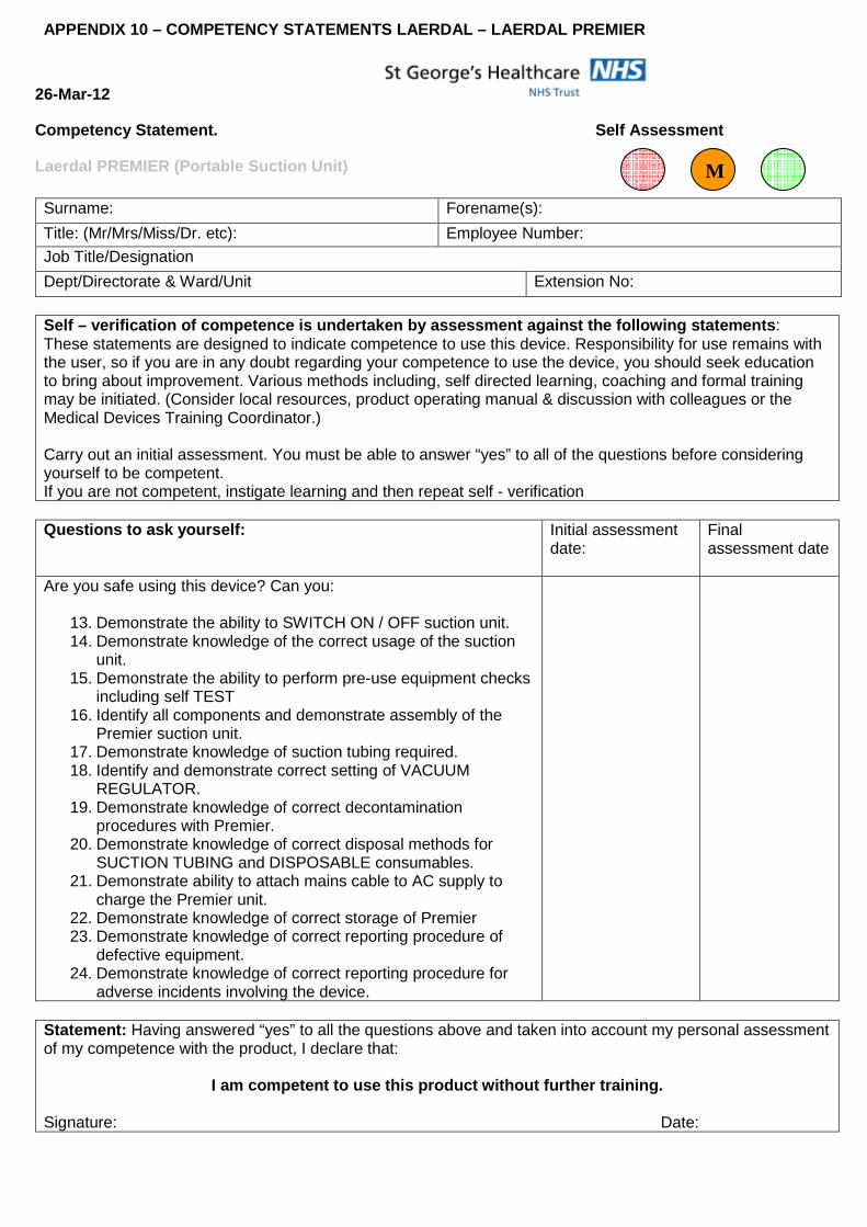
APPENDIX 10 – COMPETENCY STATEMENTS LAERDAL – LAERD AL PREMIER
26-Mar-12
Competency Statement. Self Assessment
Laerdal PREMIER (Portable Suction Unit) Surname: Forename(s):
Title: (Mr/Mrs/Miss/Dr. etc): Employee Number:
Job Title/Designation
Dept/Directorate & Ward/Unit Extension No:
Self – verification of competence is undertaken by assessment against the following statements : These statements are designed to indicate competence to use this device. Responsibility for use remains with the user, so if you are in any doubt regarding your competence to use the device, you should seek education to bring about improvement. Various methods including, self directed learning, coaching and formal training may be initiated. (Consider local resources, product operating manual & discussion with colleagues or the Medical Devices Training Coordinator.) Carry out an initial assessment. You must be able to answer “yes” to all of the questions before considering yourself to be competent. If you are not competent, instigate learning and then repeat self - verification Questions to ask yourself: Initial assessment
date:
Final assessment date
Are you safe using this device? Can you:
13. Demonstrate the ability to SWITCH ON / OFF suction unit. 14. Demonstrate knowledge of the correct usage of the suction
unit. 15. Demonstrate the ability to perform pre-use equipment checks
including self TEST 16. Identify all components and demonstrate assembly of the
Premier suction unit. 17. Demonstrate knowledge of suction tubing required. 18. Identify and demonstrate correct setting of VACUUM
REGULATOR. 19. Demonstrate knowledge of correct decontamination
procedures with Premier. 20. Demonstrate knowledge of correct disposal methods for
SUCTION TUBING and DISPOSABLE consumables. 21. Demonstrate ability to attach mains cable to AC supply to
charge the Premier unit. 22. Demonstrate knowledge of correct storage of Premier 23. Demonstrate knowledge of correct reporting procedure of
defective equipment. 24. Demonstrate knowledge of correct reporting procedure for
adverse incidents involving the device.
Statement: Having answered “yes” to all the questions above and taken into account my personal assessment of my competence with the product, I declare that:
I am competent to use this product without further training. Signature: Date:
M

I require further training before I can use this pr oduct in a competent manner. Signature: Date:
Please indicate how you plan to meet your learning needs: Keep this form in your personal portfolio or training record. Ensure the details of your self assessment have been sent to the Medical Devices Training Coordinator to update your training status on the ESR system

Appendix 11 AUDIT OF SUCTION EQUIPMENT Aims and objectives To measure compliance with the expectation that all bed spaces should have access to functioning suction equipment for use in an emergency. To audit compliance with the expectation that all equipment is checked and signed as checked on a daily basis. To ensure that suction equipment complies with infection control standards. Standard That all bed spaces have access to suction equipment that is clean and in working order. That each crash trolley has portable suction which is clean and in working order. Method All wards/departments using suctioning equipment for each suction unit (portable and wall mounted), must annually identify compliance with the following questions:
• Has the equipment been signed as checked at the beginning of the shift?
• Is suction available at every bed space, if suction unit is shared between two bed spaces is it readily accessible to both bed spaces and does the set up mean that a patient in either bed can be suctioned?
• Is the suction set up correctly (The tubing should be followed visually and the suction canister checked to ensure that the suction is set up correctly.)
• Is the suction working? (An occlusion test should be applied on each suction unit and the inner liner observed. Each unit must be checked to see the maximum amount of suction in kpa that can be obtained and then left set at the standard 20kpa).
• Is it clean? ( Visual assessment of tubing and canister)
• Is the suction on the arrest trolley clean and working?

Appendix 12 References AARC (2010) Endotracheal Suctioning Of Mechanically Ventilated Patients With Artificial Airways. AARC
Clinical Practice Guideline. Respiratory Care 55(6):758-764.
Allen, D (1988) Making Sense of Suctioning. Nursing Times 84(10):46-47.
Almgren, B., Wickerts, CJ., Heinomen, E., and Hogman, M. (2004) Side Effects of Endotracheal Suction in
Pressure and Volume Controlled Ventilation. Chest 125(3):1077-1080.
Anonymous (2000) Supplementaltherapeutic oxygen for prethreshold retinopathy of prematurity (STOP-ROP),
a randomised controlled trial. 1: primary outcomes. Pediatrics 105:295-310
Ashurst, S. (1992) Suction Therapy In The Critically Ill Patient. British Journal of Nursing 1(10):485-489.
Askie, L. Henderson-Smart D. et al (2003) The BOOST Study (Benefits of oxygen saturation targeting trial)
Oxygen saturation targets and outcomes in extremely preterm infants. New England Journal of Medicine 349:
959-967
Baker, PO., Baker, JP., and Koen, PA. (1993) Endotracheal Suctioning Techniques In Hypoxaemic Patients.
Respiratory Care 28:1563-1568.
Baun, MM., Stone, KS., and Rogge, JA. (2002) Endotracheal suctioning: Open Versus Closed with and without
Positive End-Expiratory Pressure. Critical Care Nursing Quarterly 25(2):13-25.
Blackwood, B. (1998) The Practice And Perception Of Intensive Care Staff Using The Closed Suctioning
System. Journal of Advanced Nursing 28(5):1020-1029.
Bould, MD., Thomas, ML., and Stylianou, M. (2006) Variablilty of Guedel type airways. Anaesthesia
61(11):1125-1126.
Bourgault, AM., Brown, CA., Hains, SMJ., and Parlow, JL. (2006) Effects of endotracheal tube suction on
arterial oxygen tension and heart rate variability. Biological Research for Nursing 7(4):268-278.
Branson, RD. (2007) Secretion management in the mechanically ventilated patient. Respiratory Care
52(10):1328-1347.
Brooks, D., Solway, S., Graham, I., Downes, L., Carter, M. (1999) A Survey Of Suctioning Practices Among
Physical Therapists, Respiratory Therapists And Nurses. Canadian Respiratory Journal 6(6):513-520.
Celik, S., Elbas, N. (2000) The Standard of Suction for Patients Undergoing Endotracheal Suction. Intensive
and Critical Care Nursing 16(3):191-198.
Clark, AP., Winslow, EH., Tyler, DO., and White, KM. (1990) Effects Of Endotracheal Suctioning On Mixed
Venous Oxygen Saturation And Heart Rate In Critically Ill Adults. Heart Lung 19:552-557.
Copnell, B., Tingay, DG., Mills, JF., and Dargaville, PA. (2009) Endotracheal suction techniques that effectively
remove secretions do not preserve lung volume. Aud Crit Care 22(1):61.
Copnell, B., Tingay, DG., Kiraly, NJ., Sourial, M., Gordon, MJ., Mills, JF., Morley, CJ., and Dargaville, PA.
(2007) A comparison of the effectiveness of open and closed suction systems. Int Care Med 33:1655-1662.
Czarnick, RE., Stone, KS., Everhart, CC., and Preusser, BA. (1991) Differential Effects Of Continuous Versus
Intermittent Suction On Tracheal Tissue. Heart Lung 20:144-151.
Day, T., Wainwright, S., and Wilson-Barnett, J. (2001) An Evaluation of a Teaching Intervention to Improve the
Practice of Endotracheal Suctioning in Intensive Care Units. Journal of Clinical Nursing 10:682-696.
Department Of Health. (2001) Standard Principles For Preventing Hospital Acquired Infections. Journal of
Hospital Infection 47 (Supplement) S21-S37.

Diaz, E., Rodriguez, AH., and Rello, J. (2005) Ventilator-associated pneumonia: issues related to the artificial
airway. Respir Care 50(7):900-909.
Donald, KJ., Robertson, VJ., and Tsebelis, K. (2000) Setting Safe And Effective Suction Pressure: The Effect
Of Using A Manometer In The Suction Circuit. Intensive Care Medicine 26:15-19.
Dyhr, T., Bonde, J., and Larsson, A. (2003) Lung Recruitment Maneouvres are Effective in regaining Lung
Volume and Oxygenation after Open Endotracheal Suctioning in Acute Respiratory Distress Syndrome. Crit
Care 7(1):55-62.
Fernandez, MdM., Piacentini, E., Blanch, L., and Fernandez, R. (2004) Changes in lung volume with three
systems of endotracheal suctioning with and without pre-oxygenation in patients with mild to moderate lung
failure. Int Care Med 30:2210-2215.
Gemma, M., Tommasino, C., and Cerri, M. (2002) Intracranial Effects of Endotracheal Suctioning in the Acute
Phase of Head Injury. Journal of Neurosurgical Anesthesiology 14(1):50-54.
George, RB. (1983) Duration Of Suction: An Important Variable. Respiratory Care 28(4):457-459.
Glass, CA., and Grap, MJ. (1995) Ten Tips For Safer Suctioning. American Journal of Nursing 5:51-53.
Graff, M., France, J., Hiatt, M.,and Hegyi, T. (1987) Prevention Of Hypoxia And Hyperoxia During Endotracheal
Suctioning. Critical Care Medicine 15:1133-1135.
Guglielminotti, J., Alzieu, M., Maury, E., Guidet, B., and Offenstadt, G. (2000) Bedside Detection Of Retained
Tracheobronchial Secretions In Patients Receiving Mechanical Ventilation. Is It Time For Tracheal Suctioning?
Chest 118(4):1095-1099.
Hagler, DA., and Traver, GA. (1994) Endotracheal Saline and Suction Catheters: Sources of lower Airway
Contamination. American Journal of Critical Care 3:444-447.
Heinze, H., Sedemund-Adib, B., Heringlake, M., Gosch, UW., and Eichler, W. (2008) Functional residual
capacity changes after different endotracheal suctioning methods. Anesh Analg 107:941-944.
Kotecha, S. Allen,J. (2002) Oxygen therapy for infants with chronic lung disease. Archives of Disease in
Childhood 87:F11-14
Lasocki, S., Lu, Q., Sartorius, A., Fouillat, D., Remerand, F., and Rouby, JJ. (2006) Open and closed-circuit
endotracheal suctioning in acute lung injury: efficiency and effects on gas exchange. Anaesthesiology
104(1):39-47.
Lee, ST. (1989) Intracranial Pressure Changes During Positioning Of Patients With Severe Head Injury. Heart
Lung 18:411-414.
Leur, JP., Zwaveling, JH., Loef, BG., and Schans, CP. (2003) Endotracheal Suctioning versus Minimally
Invasive Suctioning in Intubated Patients: a Prospective Randomised Controlled Trial. Int Care Med 29:426-
432.
Leur, JP., Zwaveling, JH., Loef, BG., and Schans, CP. (2003) Patient Recollection of Airway Suctioning in the
ICU: Routine versus a Minimally Invasive Procedure. Int Care Med 29:433-436.
Lindgren, S., Almgren, B., Hogman, M., Lethvall, S., Houltz, E., Lundin, S., and Stenquist, O. (2004)
Effectiveness and side effects of closed and open suctioning: an experimental evaluation. Int Care Med
30:1630-1637.
Lorente, L., Lecuona, M., Martin, MM., Garcia, C., Mora, ML., and Sierra, A. (2005) Ventilator-associated
pneumonia using a closed versus an open tracheal suction system. Crit Care Med 33(1):115-119.

Lorente, L., Lecuona, M., Jimenez, A., Mora, ML., and Sierra, A. (2006) Tracheal suction by closed system
without daily change versus open system. Int Care Med 32:538-544.
Macmillan, C. (1995) Nasopharyngeal Suction Study Reveals Knowledge Deficit. Nursing Times 91(50):28-30.
Maggiore, SM., Lellouche, F., and Pigeot, J. (2003) Prevention of Endotracheal Suctioning Induced Alveolar
Derecruitment in Acute lung Injury. Am J Resp Crit Care Med 167(9):1215-1224.
Oh, H., and Seo, W. (2003) A meta-analysis of the effects of various interventions in preventing endotracheal
suction-induced hypoxaemia. J Clin Nurs 12(6):912-924.
Paratz, JD., and Stockton, KA. (2009) Efficacy and safety of normal saline instillation: A systematic review.
Physio 95:241-250.
Pedersen, CM., Rosendahl-Nielsen, M., Hjermind, J., and Egerod, I. (2009) Endotracheal suctioning of the
adult intubated patient – what is the evidence? Int Crit Care Nurs 25(1):21-30.
Preston, IM., Matthews, HR., and Ready, AR. (1986) Minitracheotomy. A New Technique For Tracheal
Suction. Physiotherapy 72(10):494-497.
Reigal, B., and Forshee, TA. (1985) Review And Critique Of The Literature On Preoxygenation For
Endotracheal Suctioning. Heart Lung 14:507-518.
Resuscitation Council UK (1998) Advanced Life Support Manual. Chapter 5 – Airway Management And
Ventilation p22.
Rogge, JA., Bunde, L., and Baun, MM. (1989) Effectiveness Of Oxygen Concentrations Of Less Than 100%
Before And After Endotracheal Suction In Patients With Chronic Obstructive Pulmonary Disease. Heart Lung
18:64-71.
Safdar, N., Crnich, CJ., and Maki, DG. (2005) The pathogenesis of ventilator-associated pneumonia: its
relevance to developing effective strategies for prevention. Respir care 50(6):725-741.
Shah, S., Fung, K., Brim, S., and Rubin, BK. (2005) An in vitro evaluation of the effectiveness of endotracheal
suction catheters. Chest 128(5):3699-3704.
Siempos, II., Vardakas, KZ., and Falagas, ME. (2008) Closed tracheal suction systems for prevention of
ventilator acquired pneumonia. Brit J Anaesthesia 100(3):299-306.
Sole, ML., Byers, JF., Ludy, JE., and Ostrow, CL. (2002) Suctioning Techniques and Airway management
Practices: Pilot Study and Instrument Evaluation. Am J Crit Care 11(4):363-368.
Stone, KS., Bell, SD., and Pruesser, BA. (1991) The Effect Of Repeated Endotracheal Suctioning On Arterial
Blood Pressure. Appl Nurs Research 4(4):152-158.
Tin, W. et al (2001) Pulse oximetry, severe retinopathy and outcome at 1 year in babies of less than 28wks
gestation. Archives of Disease in Childhood 84:F106-F110
Trigg, E. & Mohammed, T.A. Practices in children’s nursing. Guidelines for hospital and community. Edinburgh:
Elsevier
Vanner, R., and Bick, E. (2008) Tracheal pressures during open suctioning. Anaesthesia 63:313-315.
Walsh, JM., Vandawarf, BS., Hoscheit, D., and Fahey, PJ. (1989) Unsuspected Haemodynamic Alterations
During Endotracheal Suctioning. Chest 95(1):162-165.
Wietl, J., and Betterstetter, H. (1994) Indications for the use of Closed Circuit Suction: artificial respiration with
High Positive End Pressure. Anaesthetist 43:359-363.

Williams, R., Rankin, N., and Smith, T. (1996) Relationship between the Humidity and Temperature of Inspired
gas and the Function of the Airway Mucosa. Crit Care Med 24:1920-1929.
Wood, CJ. (1998) Endotracheal Suctioning: A Literature Review. Int Crit Care Nurs 14:124-136.
Young, CS. (1984) A Review Of The Adverse Effects Of Airway Suction. Physiotherapy 70(3):104-106.
Young, CS. (1984) Recommended Guidelines For Suction. Physiotherapy 70(3):106-108.