Cleft rhinoplasty
71
© Ramaiah University of Applied Sciences 1 Faculty of Dental Sciences University Logo CLEFT RHINOPLASTY
-
Upload
zeeshan-arif -
Category
Health & Medicine
-
view
49 -
download
2
Transcript of Cleft rhinoplasty
© Ramaiah University of Applied Sciences
1
Faculty of Dental Sciences
University Logo
CLEFT RHINOPLASTY
© Ramaiah University of Applied Sciences
2
Faculty of Dental Sciences
University Logo
Contents
• Introduction
• History
• Problem
• Embryology
• Relevant anatomy
• Unilateral cleft lip nose deformity
• Bilateral cleft lip nose deformity
• TreatmentTiming Presurgical naso-alveolar
mouldingPrimary rhinoplast Secondary rhinoplastyDefinative rhinoplasty Surgical techniques
• Summary
© Ramaiah University of Applied Sciences
3
Faculty of Dental Sciences
University Logo
INTRODUCTION
• (Rhinos, "Nose" + Plastikos, "to shape")
• Surgery to alter the shape of the nose sometimes using tissue (
(skin, cartilage or bone) from elsewhere in the body or artificial
implants
• Blair and Brown first described the cleft nose in 1931, critically
identifying the nuances of the pathology.
© Ramaiah University of Applied Sciences
4
Faculty of Dental Sciences
University Logo
• Deformity may be severely asymmetric, making surgical
correction difficult.
• Patients with cleft lip may have been previously subjected to
numerous surgical interventions, leading to significant scar
tissue in the operative site
• Conversely, the surgery may adversely compromise nose
growth
© Ramaiah University of Applied Sciences
5
Faculty of Dental Sciences
University Logo
HISTORY OF THE PROCEDURE
• Cleft lip nasal deformity has received less attention than
primary lip repair or has been ignored altogether.
• Rhinoplasty for cleft lip was introduced in the 20th century.
• Most modern techniques had already been described by the
1920s-1940s, but with less refinement (leaving a conspicuous
scar).
• Currently, the literature offers numerous opinions regarding
the best surgical approach and timing of intervention
© Ramaiah University of Applied Sciences
6
Faculty of Dental Sciences
University Logo
PROBLEM
• cosmetic problems
• impaired nasal airflow
• septal deflections, atretic nostrils, turbinate hypertrophy, and cleft
lips and palates.
• Warren et al showed that children with unilateral cleft deformity
have smaller airways than children with bilateral cleft deformity.
• Although the cleft nose grows as the patient ages, it remains 30%
smaller than that of patients without cleft lip deformity.
© Ramaiah University of Applied Sciences
7
Faculty of Dental Sciences
University Logo
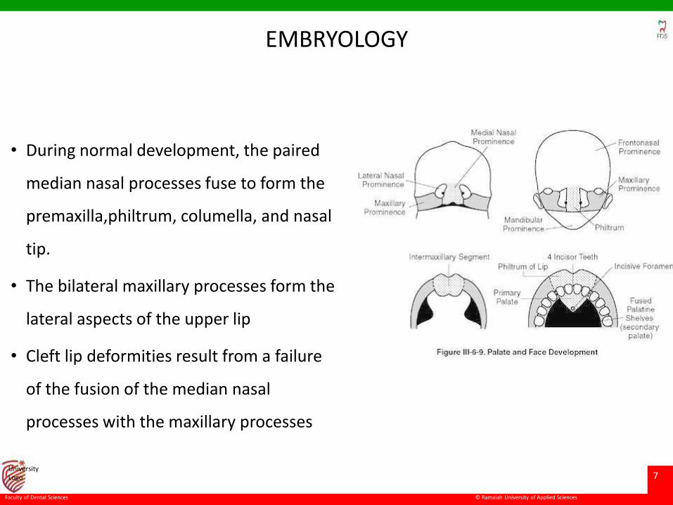
EMBRYOLOGY
• During normal development, the paired
median nasal processes fuse to form the
premaxilla,philtrum, columella, and nasal
tip.
• The bilateral maxillary processes form the
lateral aspects of the upper lip
• Cleft lip deformities result from a failure
of the fusion of the median nasal
processes with the maxillary processes
© Ramaiah University of Applied Sciences
8
Faculty of Dental Sciences
University Logo
• Interuption of this embryonic process creates malformation of
some or all of the upper lip, central alveolus, and primary
palate
• The extent of the associated cleft nasal deformity is related to
the extent of the interruption of the normal developmental
fusion process
• Even though the nasal defects may be subtle there is always a
nasal abnormality associated with cleft lips
© Ramaiah University of Applied Sciences
9
Faculty of Dental Sciences
University Logo
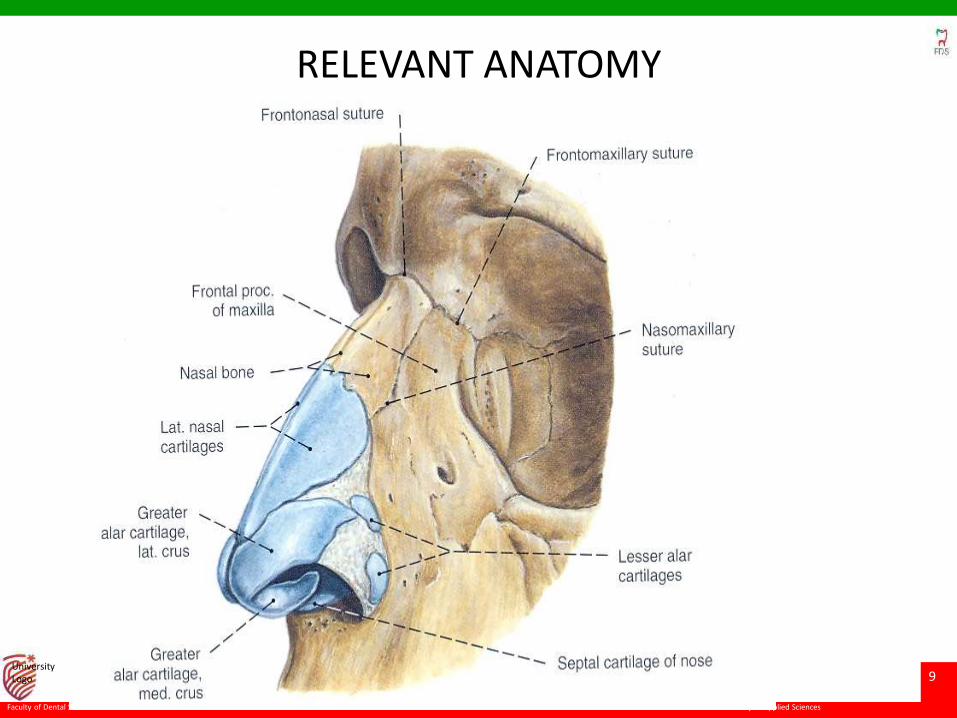
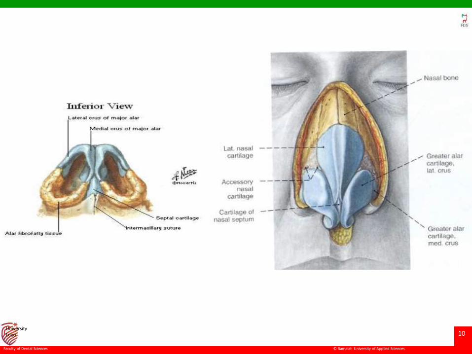
RELEVANT ANATOMY
© Ramaiah University of Applied Sciences
10
Faculty of Dental Sciences
University Logo
© Ramaiah University of Applied Sciences
11
Faculty of Dental Sciences
University Logo
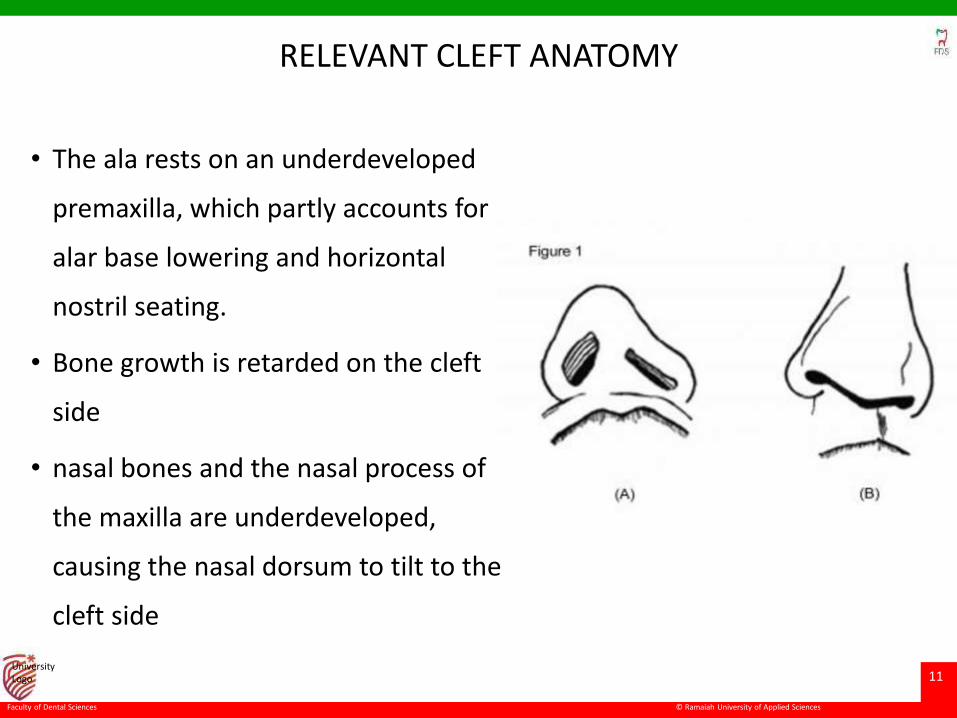
RELEVANT CLEFT ANATOMY
• The ala rests on an underdeveloped
premaxilla, which partly accounts for
alar base lowering and horizontal
nostril seating.
• Bone growth is retarded on the cleft
side
• nasal bones and the nasal process of
the maxilla are underdeveloped,
causing the nasal dorsum to tilt to the
cleft side
© Ramaiah University of Applied Sciences
12
Faculty of Dental Sciences
University Logo
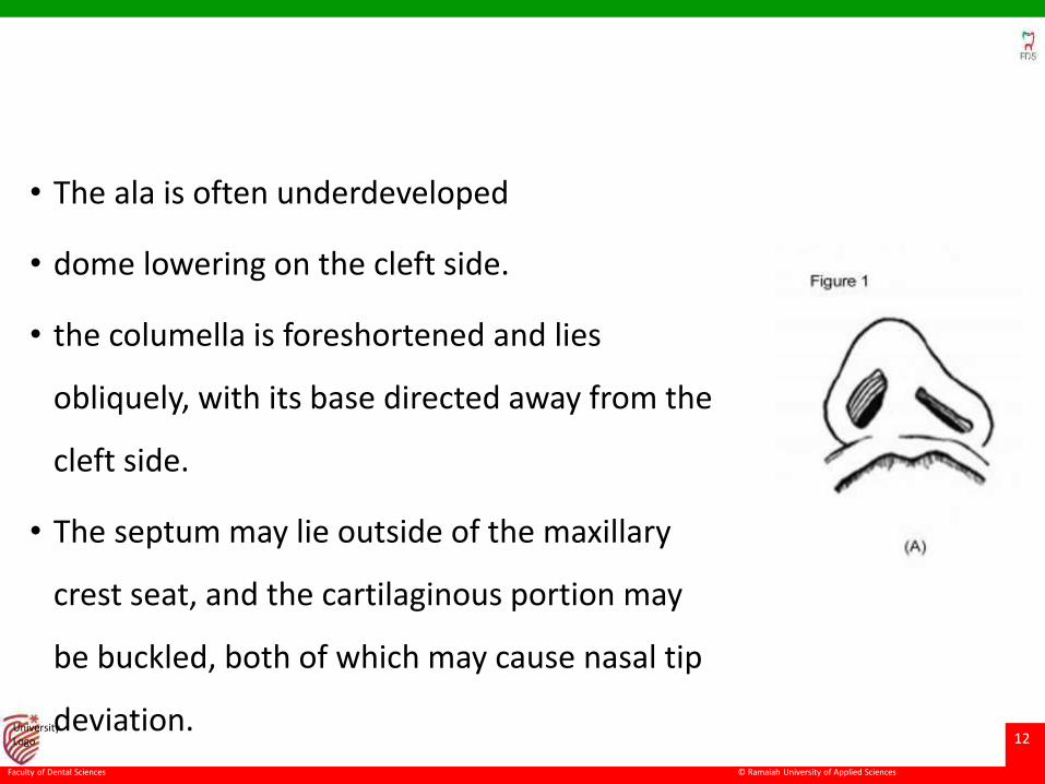
• The ala is often underdeveloped
• dome lowering on the cleft side.
• the columella is foreshortened and lies
obliquely, with its base directed away from the
cleft side.
• The septum may lie outside of the maxillary
crest seat, and the cartilaginous portion may
be buckled, both of which may cause nasal tip
deviation.
© Ramaiah University of Applied Sciences
13
Faculty of Dental Sciences
University Logo
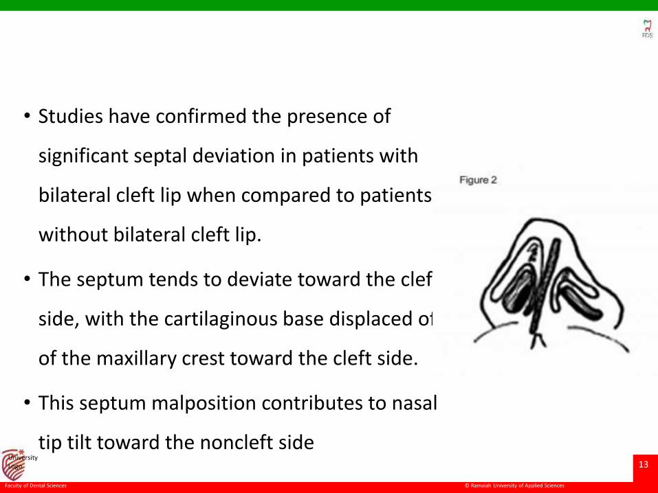
• Studies have confirmed the presence of
significant septal deviation in patients with
bilateral cleft lip when compared to patients
without bilateral cleft lip.
• The septum tends to deviate toward the cleft
side, with the cartilaginous base displaced off
of the maxillary crest toward the cleft side.
• This septum malposition contributes to nasal
tip tilt toward the noncleft side
© Ramaiah University of Applied Sciences
14
Faculty of Dental Sciences
University Logo
Unilateral cleft lip nose deformity
• the maxilla on the cleft side is deficient.
• Because of this, the alar base on the cleft side does not not
fuse in the midline and is positioned more posterior, lateral,
and inferior than the alar base on the noncleft side.
© Ramaiah University of Applied Sciences
15
Faculty of Dental Sciences
University Logo
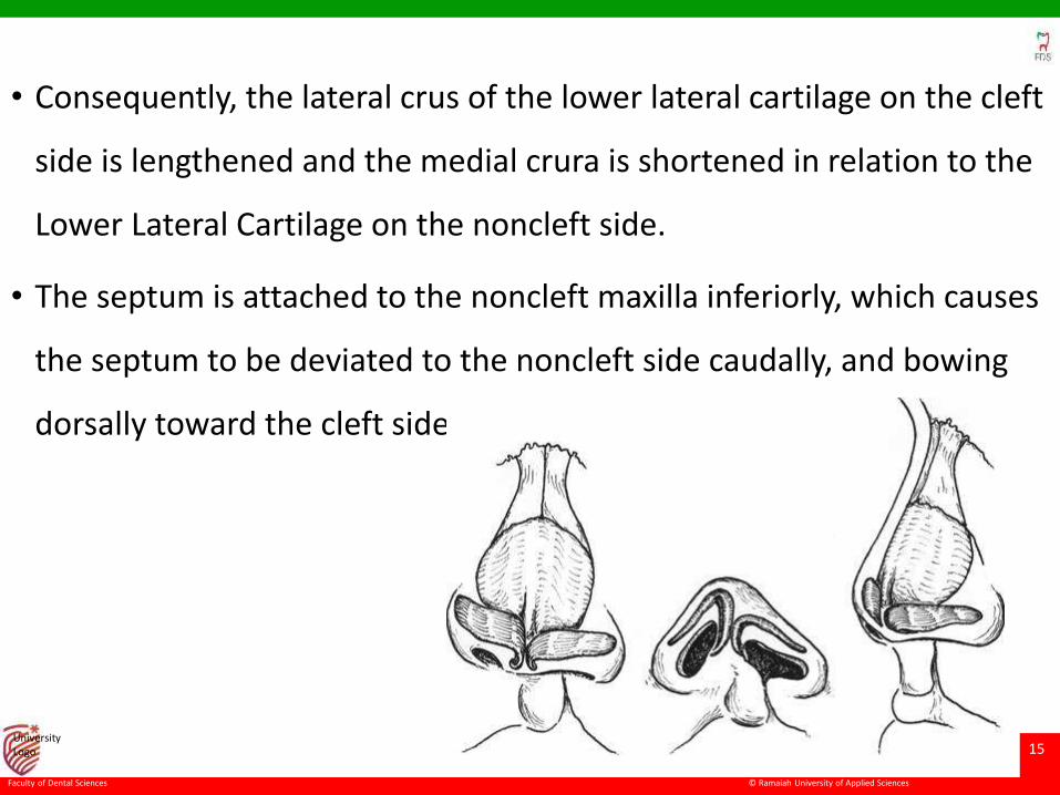
• Consequently, the lateral crus of the lower lateral cartilage on the cleft
side is lengthened and the medial crura is shortened in relation to the
Lower Lateral Cartilage on the noncleft side.
• The septum is attached to the noncleft maxilla inferiorly, which causes
the septum to be deviated to the noncleft side caudally, and bowing
dorsally toward the cleft side.
© Ramaiah University of Applied Sciences
16
Faculty of Dental Sciences
University Logo
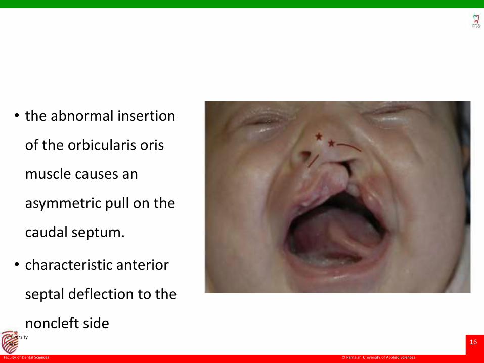
• the abnormal insertion
of the orbicularis oris
muscle causes an
asymmetric pull on the
caudal septum.
• characteristic anterior
septal deflection to the
noncleft side
© Ramaiah University of Applied Sciences
17
Faculty of Dental Sciences
University Logo
Bilateral Cleft Lip Nose Deformity
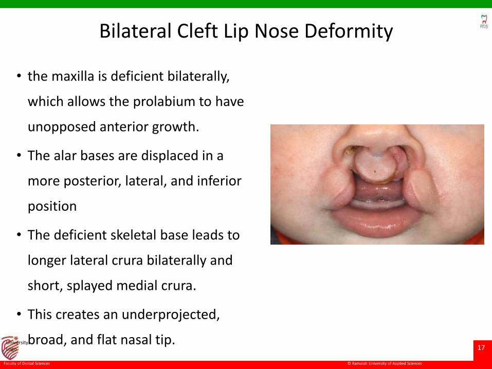
• the maxilla is deficient bilaterally,
which allows the prolabium to have
unopposed anterior growth.
• The alar bases are displaced in a
more posterior, lateral, and inferior
position
• The deficient skeletal base leads to
longer lateral crura bilaterally and
short, splayed medial crura.
• This creates an underprojected,
broad, and flat nasal tip.
© Ramaiah University of Applied Sciences
18
Faculty of Dental Sciences
University Logo
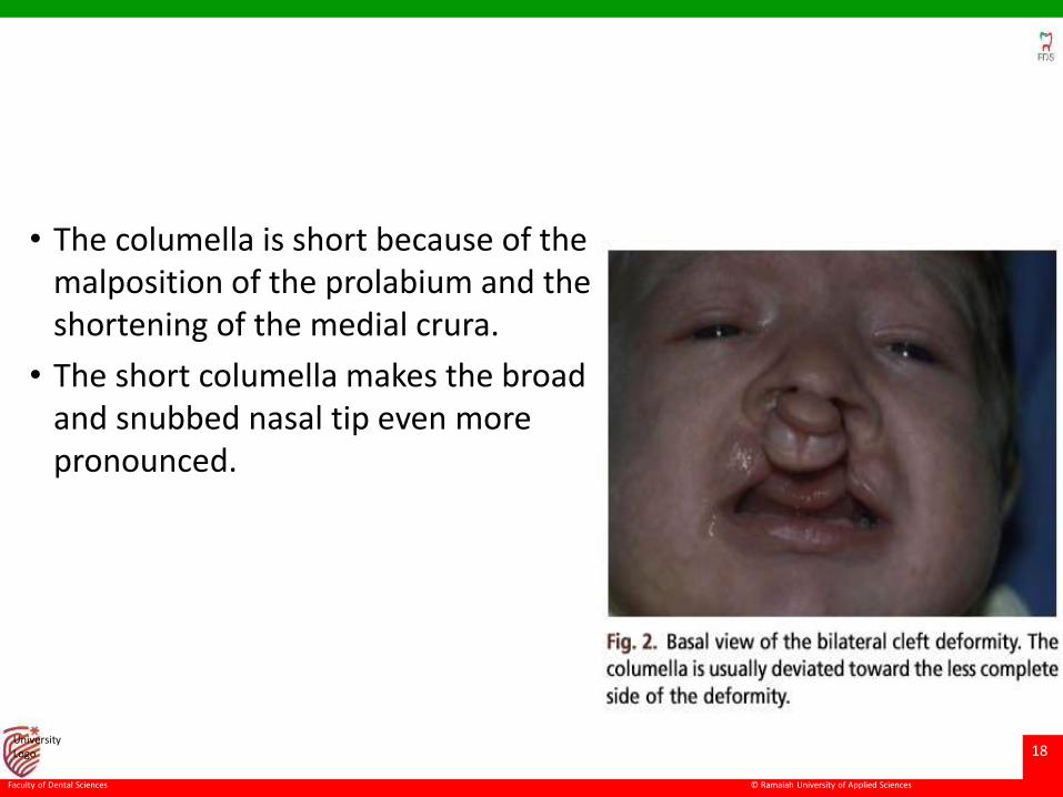
• The columella is short because of the malposition of the prolabium and the shortening of the medial crura.
• The short columella makes the broad and snubbed nasal tip even more pronounced.
© Ramaiah University of Applied Sciences
19
Faculty of Dental Sciences
University Logo
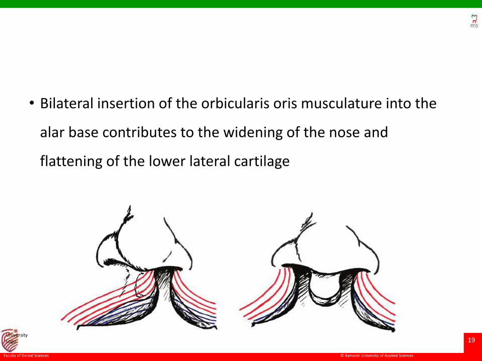
• Bilateral insertion of the orbicularis oris musculature into the
alar base contributes to the widening of the nose and
flattening of the lower lateral cartilage
© Ramaiah University of Applied Sciences
20
Faculty of Dental Sciences
University Logo
Timing of the Cleft Nasal Repair
• The decision to perform early nasal surgery is based on several
factors.
• The extent of the deformity and the potential scarring and
impact of the procedure on nasal growth.
• Advantages of early surgical intervention include minimizing
the deformity as the child grows, lessening asymmetries to
allow optimal nasal growth, and creating favorable conditions
for future surgery.
© Ramaiah University of Applied Sciences
21
Faculty of Dental Sciences
University Logo
• Historically, controversy has existed as to whether primary tip
rhinoplasty was a positive influence on the eventual
appearance of the nose in patients with clefts.
• Major septal work and cartilaginous dissection has been
thought to negatively affect nasal growth.
• However, no experimental or clinical studies have ever proved
that minor manipulations (without resection) of the nasal tip or
nasal base interfere with future nasal growth.
© Ramaiah University of Applied Sciences
22
Faculty of Dental Sciences
University Logo
• Ideal repair of a cleft nasal deformity is performed in 2 stages.
• The first includes alteration in the nose at the time of lip repair
(primary rhinoplasty), delaying a definitive repair until the
patient has completed facial growth (secondary rhinoplasty).
• In female patients, secondary rhinoplasty is generally
performed around 15 to 17 years of age, and in male patients
at approximately 16 to 18 years of age.
© Ramaiah University of Applied Sciences
23
Faculty of Dental Sciences
University Logo
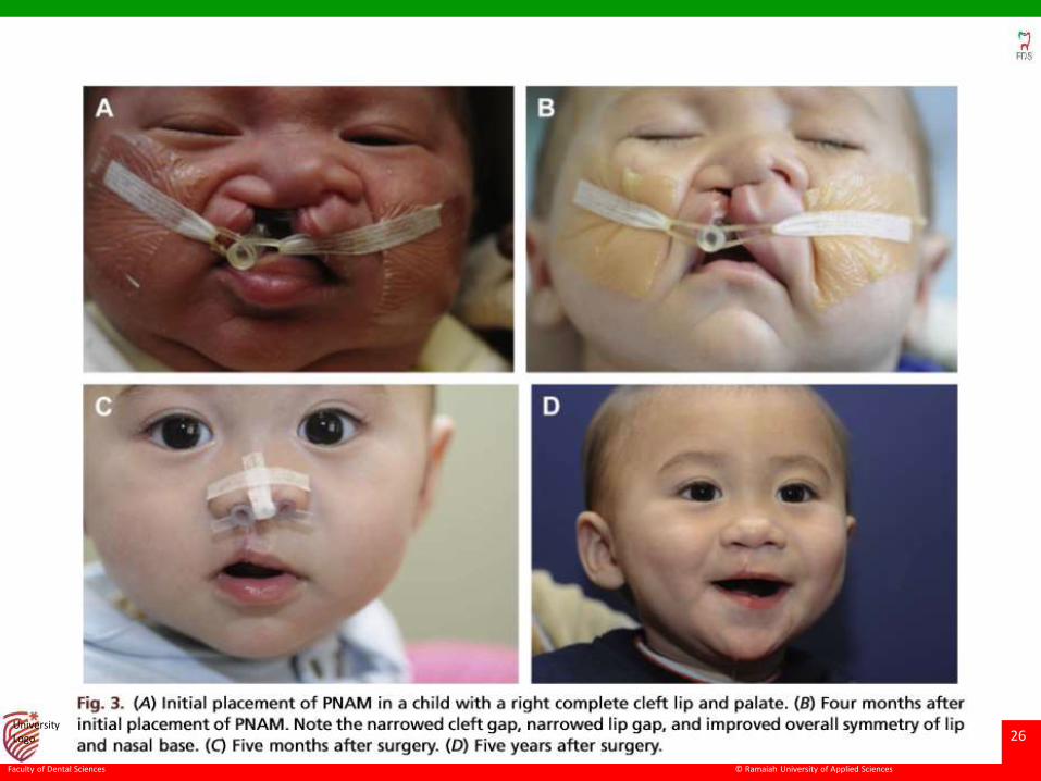
Presurgical Nasoalveolar Molding
• Presurgical nasoalveolar molding (PNAM) can be used in
patients with wide or very asymmetric clefts
• reposition the malaligned alveolar segments
• narrow the cleft gap
• improve nasal tip symmetry in unilateral clefts
• elongate the columella
• expand the nasal soft tissues in bilateral clefts
© Ramaiah University of Applied Sciences
24
Faculty of Dental Sciences
University Logo
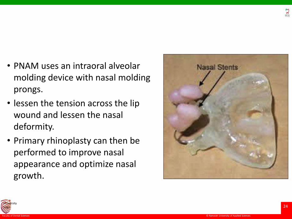
• PNAM uses an intraoral alveolar molding device with nasal moldingprongs.
• lessen the tension across the lip wound and lessen the nasal deformity.
• Primary rhinoplasty can then be performed to improve nasal appearance and optimize nasal growth.
© Ramaiah University of Applied Sciences
25
Faculty of Dental Sciences
University Logo
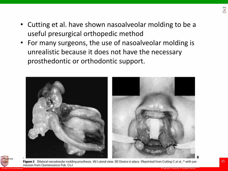
• Cutting et al. have shown nasoalveolar molding to be a useful presurgical orthopedic method
• For many surgeons, the use of nasoalveolar molding is unrealistic because it does not have the necessary prosthedontic or orthodontic support.
© Ramaiah University of Applied Sciences
26
Faculty of Dental Sciences
University Logo
© Ramaiah University of Applied Sciences
27
Faculty of Dental Sciences
University Logo
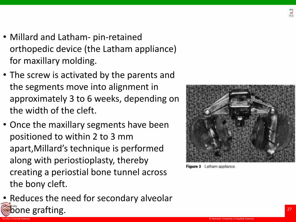
• Millard and Latham- pin-retained orthopedic device (the Latham appliance) for maxillary molding.
• The screw is activated by the parents and the segments move into alignment in approximately 3 to 6 weeks, depending on the width of the cleft.
• Once the maxillary segments have been positioned to within 2 to 3 mm apart,Millard’s technique is performed along with periostioplasty, thereby creating a periostial bone tunnel across the bony cleft.
• Reduces the need for secondary alveolar bone grafting.
© Ramaiah University of Applied Sciences
28
Faculty of Dental Sciences
University Logo
Primary Rhinoplasty
• The purpose of primary rhinoplasty is to close the anterior nasal floor, to relocate the displaced alar base, and to bring early symmetry to the nasal base and tip.
• This approach allows for both a functional and aesthetic improvement without jeopardizing nasal and facial growth.
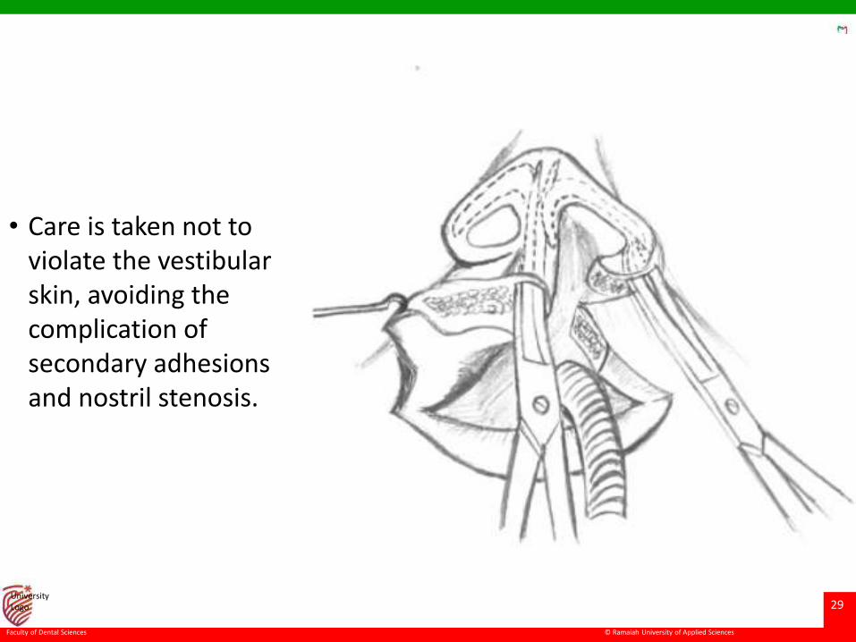
• After the cleft lip incisions are made and the primary lip dissection is completed, the muscle and soft tissues of the alar base are separated from their maxillary attachments.
• The malpositioned alar base is freed by creating an internal alotomy at the anterior head of the inferior turbinate.
• If adequate soft tissue dissection of the alar base is performed, the cleft alar base can be repositioned (during closure) in the optimal three dimensional position
© Ramaiah University of Applied Sciences
29
Faculty of Dental Sciences
University Logo
• Care is taken not to violate the vestibular skin, avoiding the complication of secondary adhesions and nostril stenosis.
© Ramaiah University of Applied Sciences
30
Faculty of Dental Sciences
University Logo
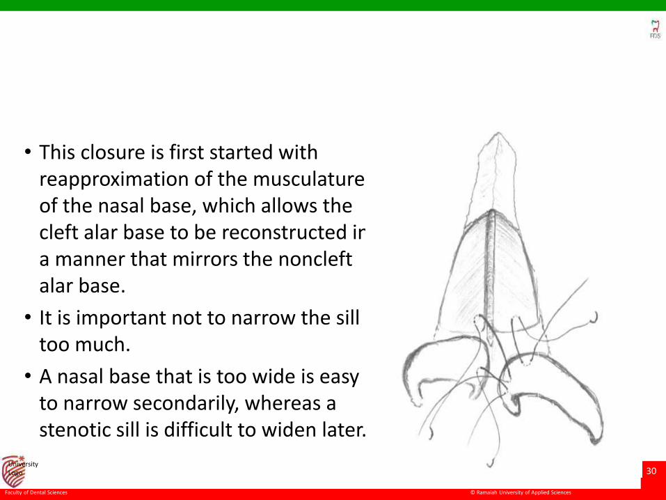
• This closure is first started with reapproximation of the musculature of the nasal base, which allows the cleft alar base to be reconstructed in a manner that mirrors the noncleftalar base.
• It is important not to narrow the sill too much.
• A nasal base that is too wide is easy to narrow secondarily, whereas a stenotic sill is difficult to widen later.
© Ramaiah University of Applied Sciences
31
Faculty of Dental Sciences
University Logo
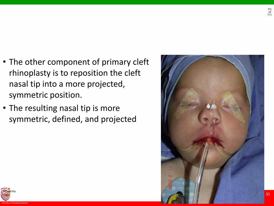
• The other component of primary cleft rhinoplasty is to reposition the cleft nasal tip into a more projected, symmetric position.
• The resulting nasal tip is more symmetric, defined, and projected
© Ramaiah University of Applied Sciences
32
Faculty of Dental Sciences
University Logo
Intermediate Rhinoplasty
• Intermediate rhinoplasty is defined as any nasal surgery performed between the time of initial lip repair and the time of definitive rhinoplasty when the patient reaches facial skeletal maturity.
• Rarely used as surgeons have become more adept at primary rhinoplasty.
• However, many patients have significant nasal deformities that have not been adequately repaired after their initial cleft lip procedures.
• In these cases, performing intermediate rhinoplasty minimizes the social stigmata associated with a more noticeable nasal deformity.
© Ramaiah University of Applied Sciences
33
Faculty of Dental Sciences
University Logo
Secondary (Definitive) Rhinoplasty
• Once the patient has reached facial skeletal maturity, definitive
septorhinoplasty can be performed.
• Structural reconstruction of the cleft nose often requires
cartilage grafting material from the rib or septum to achieve
adequate support.
© Ramaiah University of Applied Sciences
34
Faculty of Dental Sciences
University Logo
The goals of the secondary rhinoplasty are
• the creation of symmetry
• definition of the nasal base and tip
• relief of nasal obstruction
• management of nasal scarring and webbing.
© Ramaiah University of Applied Sciences
35
Faculty of Dental Sciences
University Logo
SURGICAL TECHNIQUES Approaches
• The approach to definitive cleft septorhinoplasty varies
according to surgeon preference and the requirements of the
reconstruction.
• These treatment goals include septal reconstruction and
treatment of the nasal tip, alar rim, alar base, columella, and
nasal sill.
• In most instances, all these goals can be corrected by using
either the external or endonasal approach.
© Ramaiah University of Applied Sciences
36
Faculty of Dental Sciences
University Logo
open or external approach.
• inverted-V columellar incision.
• When there is significant lack of columellar soft tissue in
unilateral clefting, the incision can be modified onto the cleft
lip with a V-to-Y closure to increase columellar soft tissue.
© Ramaiah University of Applied Sciences
37
Faculty of Dental Sciences
University Logo
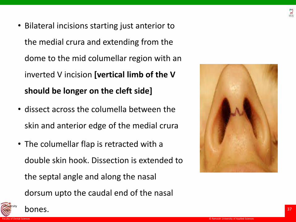
• Bilateral incisions starting just anterior to
the medial crura and extending from the
dome to the mid columellar region with an
inverted V incision [vertical limb of the V
should be longer on the cleft side]
• dissect across the columella between the
skin and anterior edge of the medial crura
• The columellar flap is retracted with a
double skin hook. Dissection is extended to
the septal angle and along the nasal
dorsum upto the caudal end of the nasal
bones.
© Ramaiah University of Applied Sciences
38
Faculty of Dental Sciences
University Logo
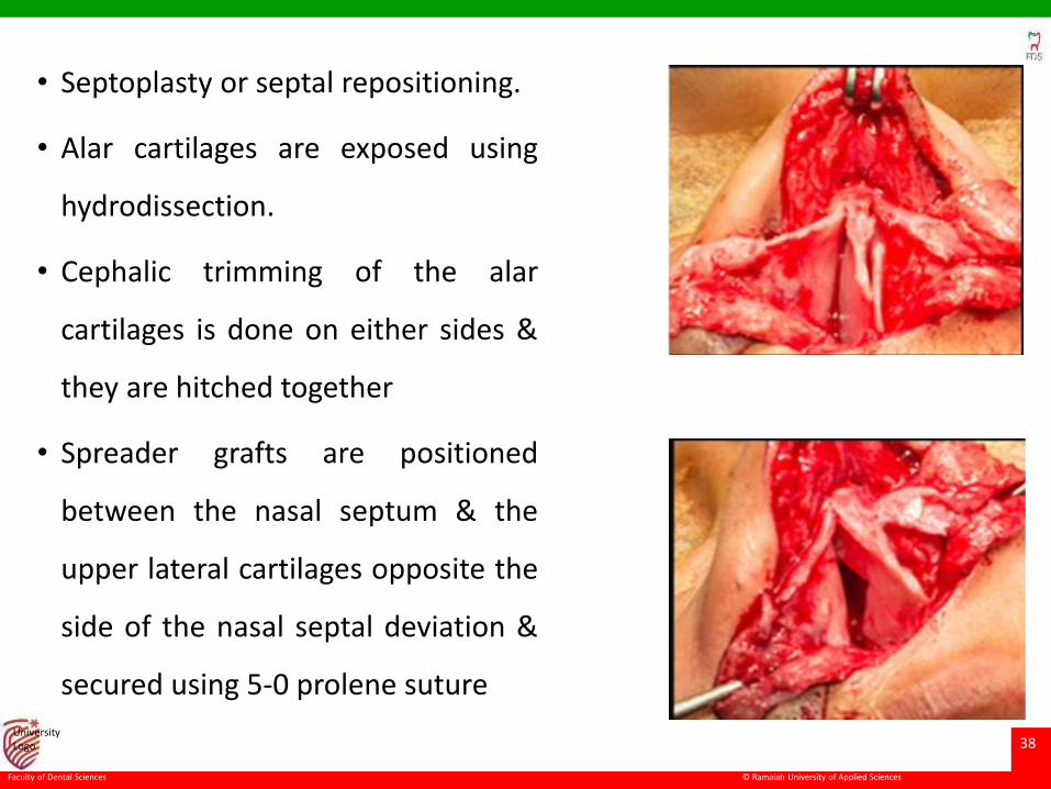
• Septoplasty or septal repositioning.
• Alar cartilages are exposed using
hydrodissection.
• Cephalic trimming of the alar
cartilages is done on either sides &
they are hitched together
• Spreader grafts are positioned
between the nasal septum & the
upper lateral cartilages opposite the
side of the nasal septal deviation &
secured using 5-0 prolene suture
© Ramaiah University of Applied Sciences
39
Faculty of Dental Sciences
University Logo
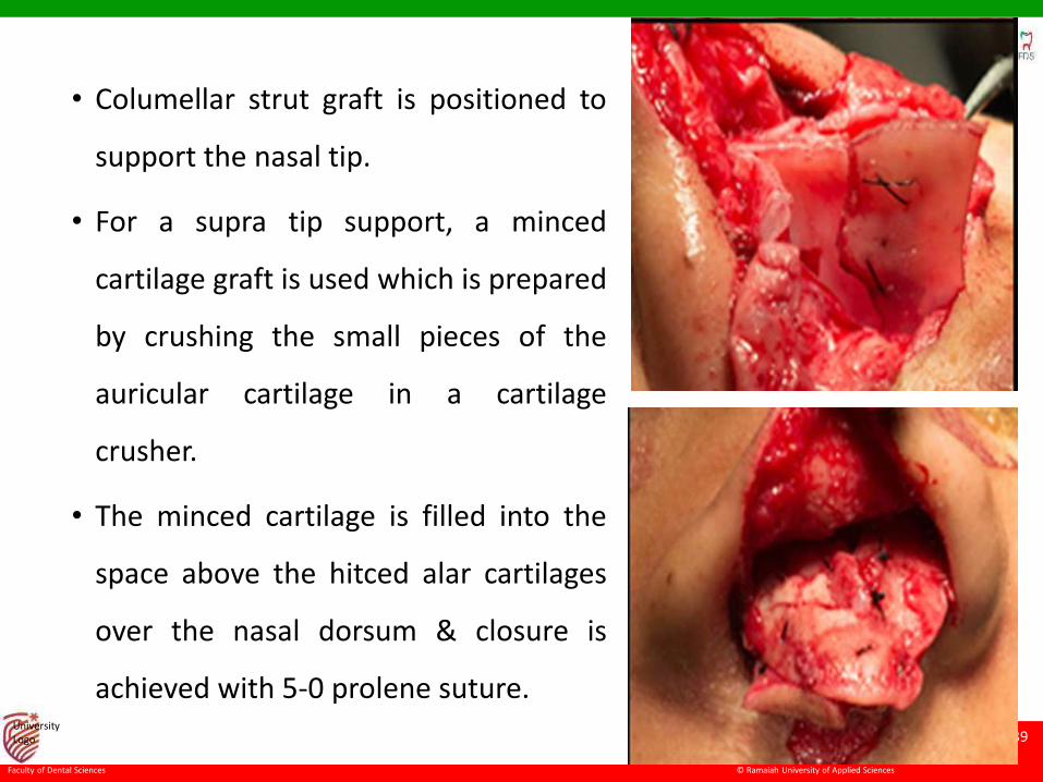
• Columellar strut graft is positioned to
support the nasal tip.
• For a supra tip support, a minced
cartilage graft is used which is prepared
by crushing the small pieces of the
auricular cartilage in a cartilage
crusher.
• The minced cartilage is filled into the
space above the hitced alar cartilages
over the nasal dorsum & closure is
achieved with 5-0 prolene suture.
© Ramaiah University of Applied Sciences
40
Faculty of Dental Sciences
University Logo
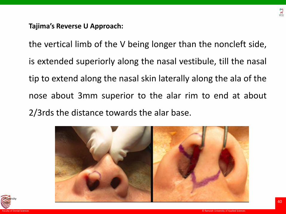
Tajima’s Reverse U Approach:
the vertical limb of the V being longer than the noncleft side,
is extended superiorly along the nasal vestibule, till the nasal
tip to extend along the nasal skin laterally along the ala of the
nose about 3mm superior to the alar rim to end at about
2/3rds the distance towards the alar base.
© Ramaiah University of Applied Sciences
41
Faculty of Dental Sciences
University Logo
Sliding chondrocutaneous flap.
• Wang and Madorsky, called the sliding flap cheliorhinoplasty
• This flap provides additional tissue to augment the vestibular
lining and reduce the alar-columellar web, and, when
combined with the open rhinoplasty, the chondrocutaneous
flap can permit tip stability and lip refinement.
© Ramaiah University of Applied Sciences
42
Faculty of Dental Sciences
University Logo
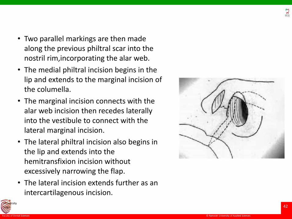
• Two parallel markings are then made along the previous philtral scar into the nostril rim,incorporating the alar web.
• The medial philtral incision begins in the lip and extends to the marginal incision of the columella.
• The marginal incision connects with the alar web incision then recedes laterally into the vestibule to connect with the lateral marginal incision.
• The lateral philtral incision also begins in the lip and extends into the hemitransfixion incision without excessively narrowing the flap.
• The lateral incision extends further as an intercartilagenous incision.
© Ramaiah University of Applied Sciences
43
Faculty of Dental Sciences
University Logo
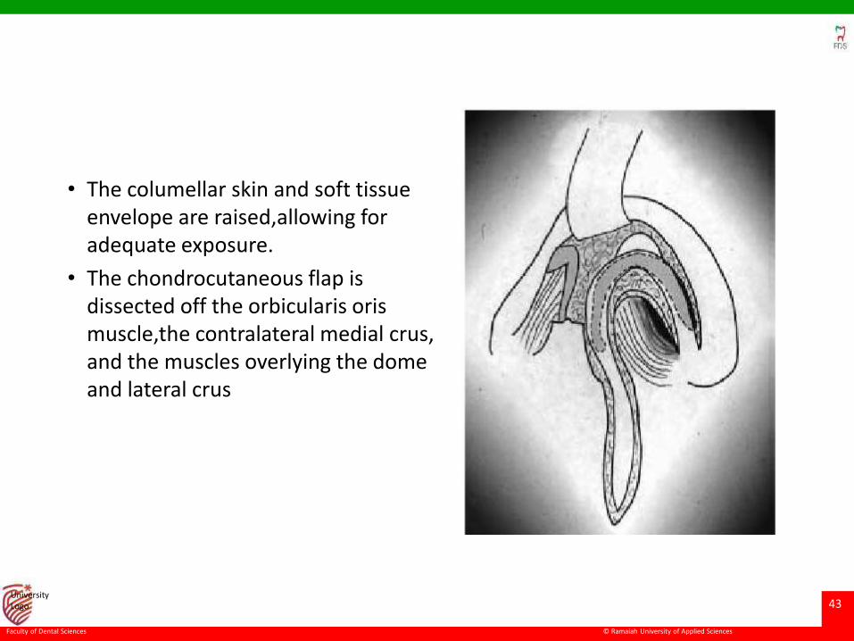
• The columellar skin and soft tissue envelope are raised,allowing for adequate exposure.
• The chondrocutaneous flap is dissected off the orbicularis orismuscle,the contralateral medial crus, and the muscles overlying the dome and lateral crus
© Ramaiah University of Applied Sciences
44
Faculty of Dental Sciences
University Logo
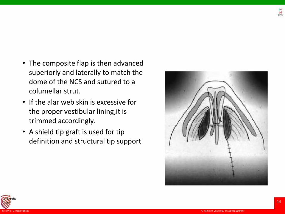
• The composite flap is then advanced superiorly and laterally to match the dome of the NCS and sutured to a columellar strut.
• If the alar web skin is excessive for the proper vestibular lining,it is trimmed accordingly.
• A shield tip graft is used for tip definition and structural tip support
© Ramaiah University of Applied Sciences
45
Faculty of Dental Sciences
University Logo
Closed or endonasal approach
• The advantages of the endonasal approach include maintaining an intact skin–soft tissue envelope.
• When cartilaginous grafts are placed under an intact envelope, the surgeon is better able to immediately visualize the impact of the graft on the eventual nasal contour.
• maintenance of vascular supply to the skin, allowing increased tension when placing structural grafts.
• With the open approach, lengthening or projecting grafts can make eventual wound closure difficult.
• Multiple endonasal incisions (transfixion, infracartilaginous, and intercartilaginous) are often used to obtain adequate exposure to correct the deformity
© Ramaiah University of Applied Sciences
46
Faculty of Dental Sciences
University Logo
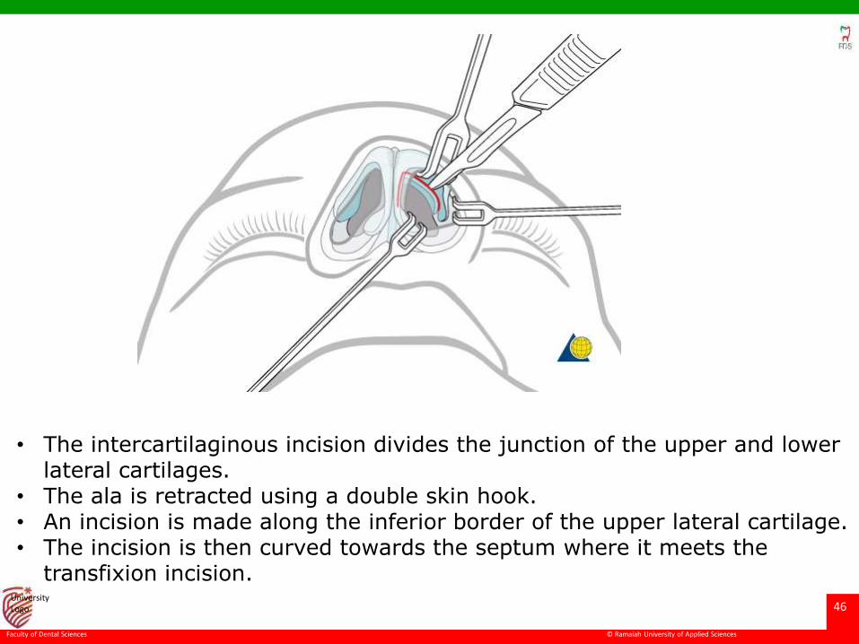
• The intercartilaginous incision divides the junction of the upper and lower lateral cartilages.
• The ala is retracted using a double skin hook. • An incision is made along the inferior border of the upper lateral cartilage. • The incision is then curved towards the septum where it meets the
transfixion incision.
© Ramaiah University of Applied Sciences
47
Faculty of Dental Sciences
University Logo
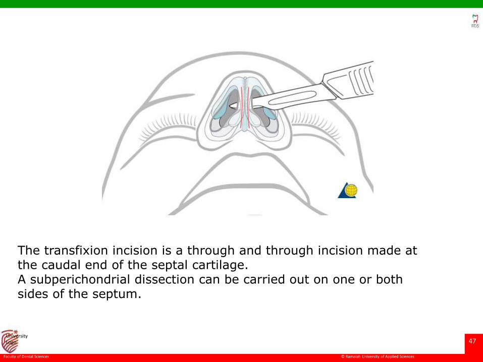
The transfixion incision is a through and through incision made at the caudal end of the septal cartilage. A subperichondrial dissection can be carried out on one or both sides of the septum.
© Ramaiah University of Applied Sciences
48
Faculty of Dental Sciences
University Logo
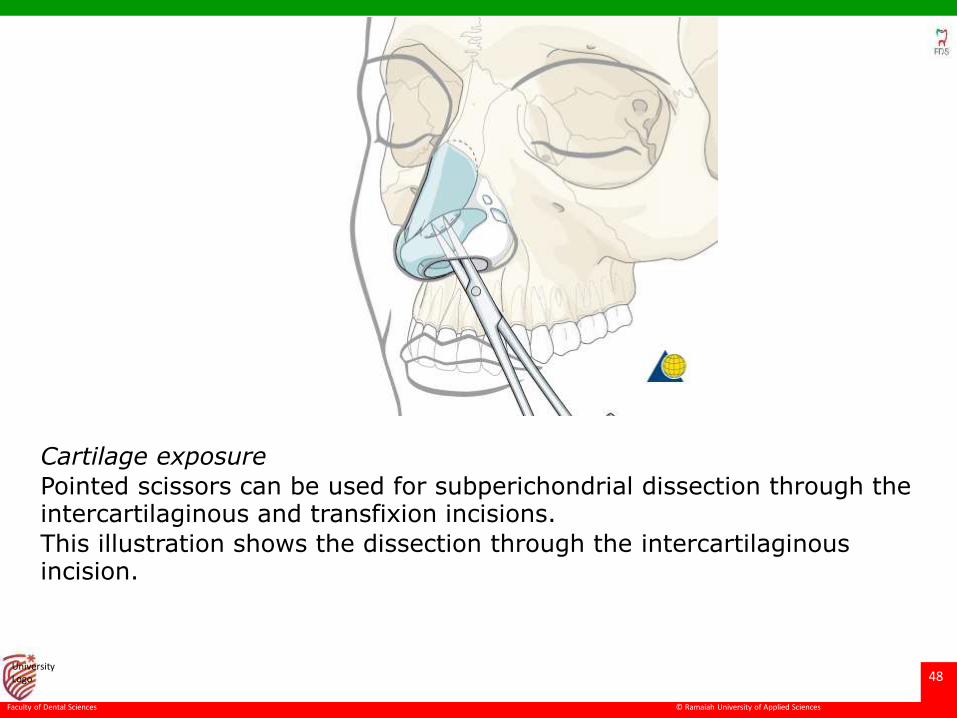
Cartilage exposure
Pointed scissors can be used for subperichondrial dissection through the intercartilaginous and transfixion incisions.
This illustration shows the dissection through the intercartilaginousincision.
© Ramaiah University of Applied Sciences
49
Faculty of Dental Sciences
University Logo
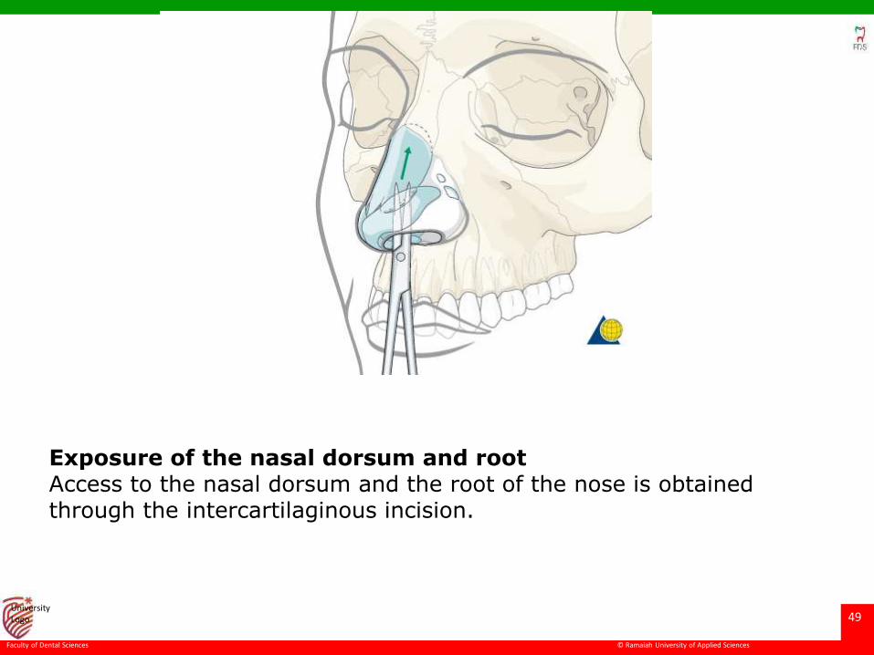
Exposure of the nasal dorsum and rootAccess to the nasal dorsum and the root of the nose is obtained through the intercartilaginous incision.
© Ramaiah University of Applied Sciences
50
Faculty of Dental Sciences
University Logo
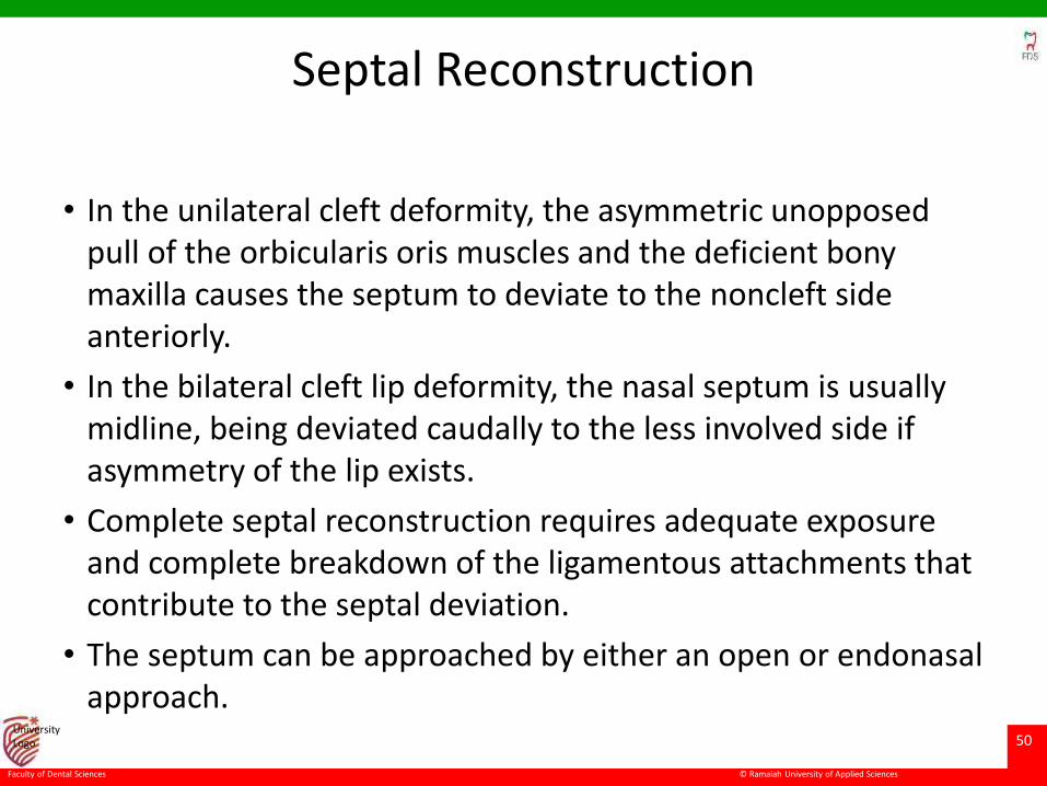
Septal Reconstruction
• In the unilateral cleft deformity, the asymmetric unopposed pull of the orbicularis oris muscles and the deficient bony maxilla causes the septum to deviate to the noncleft side anteriorly.
• In the bilateral cleft lip deformity, the nasal septum is usually midline, being deviated caudally to the less involved side if asymmetry of the lip exists.
• Complete septal reconstruction requires adequate exposure and complete breakdown of the ligamentous attachments that contribute to the septal deviation.
• The septum can be approached by either an open or endonasalapproach.
© Ramaiah University of Applied Sciences
51
Faculty of Dental Sciences
University Logo
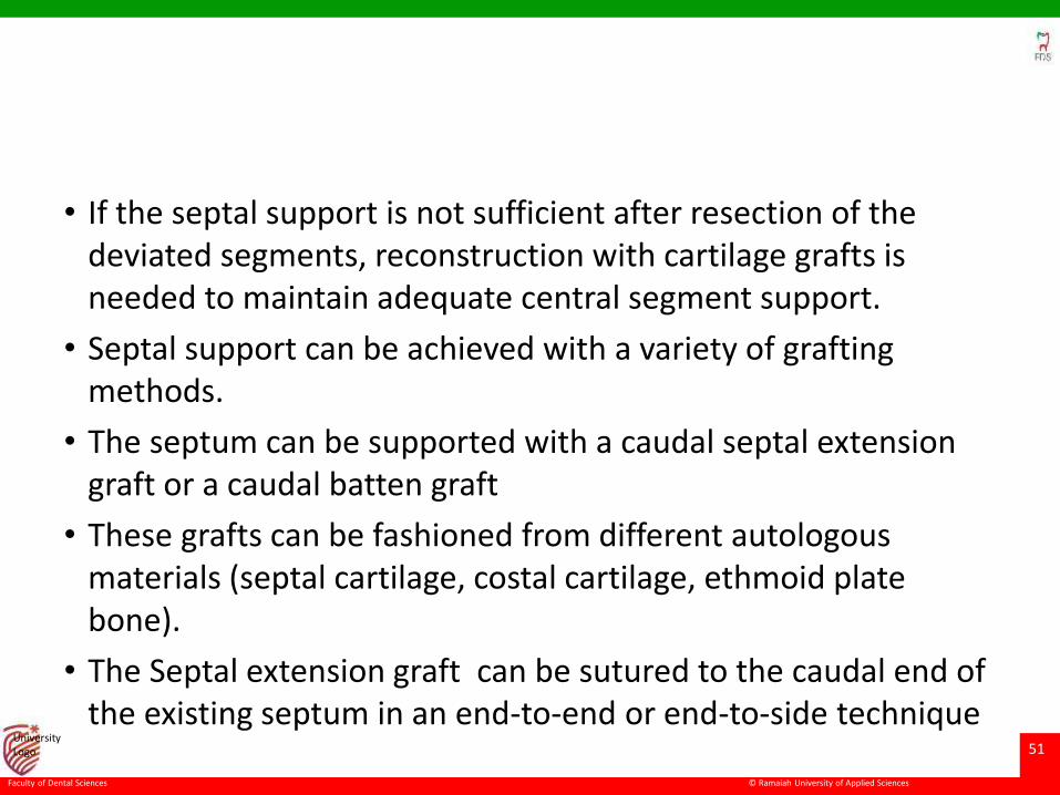
• If the septal support is not sufficient after resection of the deviated segments, reconstruction with cartilage grafts is needed to maintain adequate central segment support.
• Septal support can be achieved with a variety of grafting methods.
• The septum can be supported with a caudal septal extension graft or a caudal batten graft
• These grafts can be fashioned from different autologous materials (septal cartilage, costal cartilage, ethmoid plate bone).
• The Septal extension graft can be sutured to the caudal end of the existing septum in an end-to-end or end-to-side technique
© Ramaiah University of Applied Sciences
52
Faculty of Dental Sciences
University Logo
© Ramaiah University of Applied Sciences
53
Faculty of Dental Sciences
University Logo
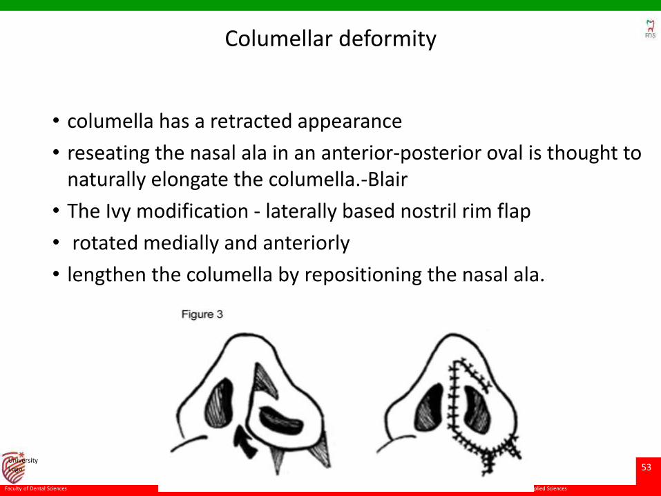
Columellar deformity
• columella has a retracted appearance
• reseating the nasal ala in an anterior-posterior oval is thought to naturally elongate the columella.-Blair
• The Ivy modification - laterally based nostril rim flap
• rotated medially and anteriorly
• lengthen the columella by repositioning the nasal ala.
© Ramaiah University of Applied Sciences
54
Faculty of Dental Sciences
University Logo
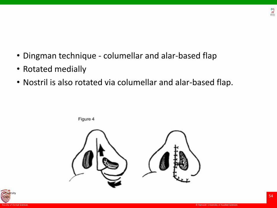
• Dingman technique - columellar and alar-based flap
• Rotated medially
• Nostril is also rotated via columellar and alar-based flap.
© Ramaiah University of Applied Sciences
55
Faculty of Dental Sciences
University Logo
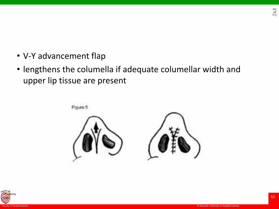
• V-Y advancement flap
• lengthens the columella if adequate columellar width and upper lip tissue are present
© Ramaiah University of Applied Sciences
56
Faculty of Dental Sciences
University Logo
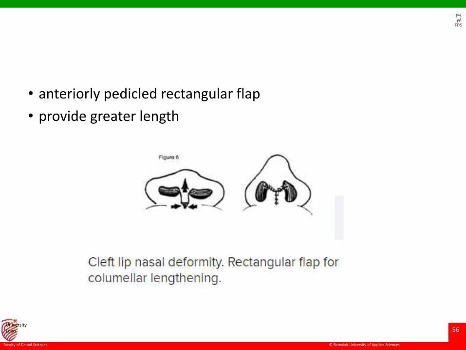
• anteriorly pedicled rectangular flap
• provide greater length
© Ramaiah University of Applied Sciences
57
Faculty of Dental Sciences
University Logo
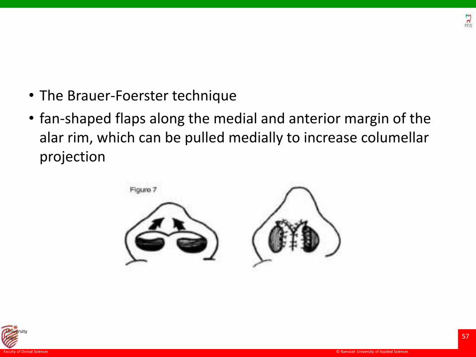
• The Brauer-Foerster technique
• fan-shaped flaps along the medial and anterior margin of the alar rim, which can be pulled medially to increase columellarprojection
© Ramaiah University of Applied Sciences
58
Faculty of Dental Sciences
University Logo
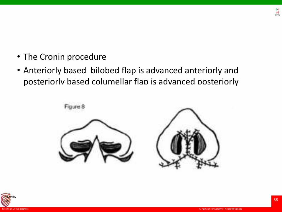
• The Cronin procedure
• Anteriorly based bilobed flap is advanced anteriorly and posteriorly based columellar flap is advanced posteriorly
© Ramaiah University of Applied Sciences
59
Faculty of Dental Sciences
University Logo
• When the columella is completely absent a more radical reconstruction is required .
• The upper lip is the ideal donor site for reconstruction.
• However, the upper lip is often scarred or deficient secondary to cleft lip.
• Lower lip tissue may be transferred to the columella, or nasolabial flaps may be recruited.
• A full-thickness skin graft may be buried below upper lip skin for use in reconstructing the columella (ie, Ferris-Smith technique)
© Ramaiah University of Applied Sciences
60
Faculty of Dental Sciences
University Logo
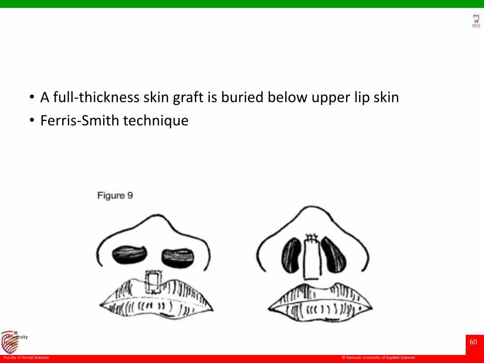
• A full-thickness skin graft is buried below upper lip skin
• Ferris-Smith technique
© Ramaiah University of Applied Sciences
61
Faculty of Dental Sciences
University Logo
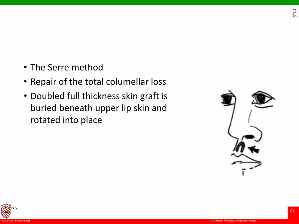
• The Serre method
• Repair of the total columellar loss
• Doubled full thickness skin graft is buried beneath upper lip skin and rotated into place
© Ramaiah University of Applied Sciences
62
Faculty of Dental Sciences
University Logo
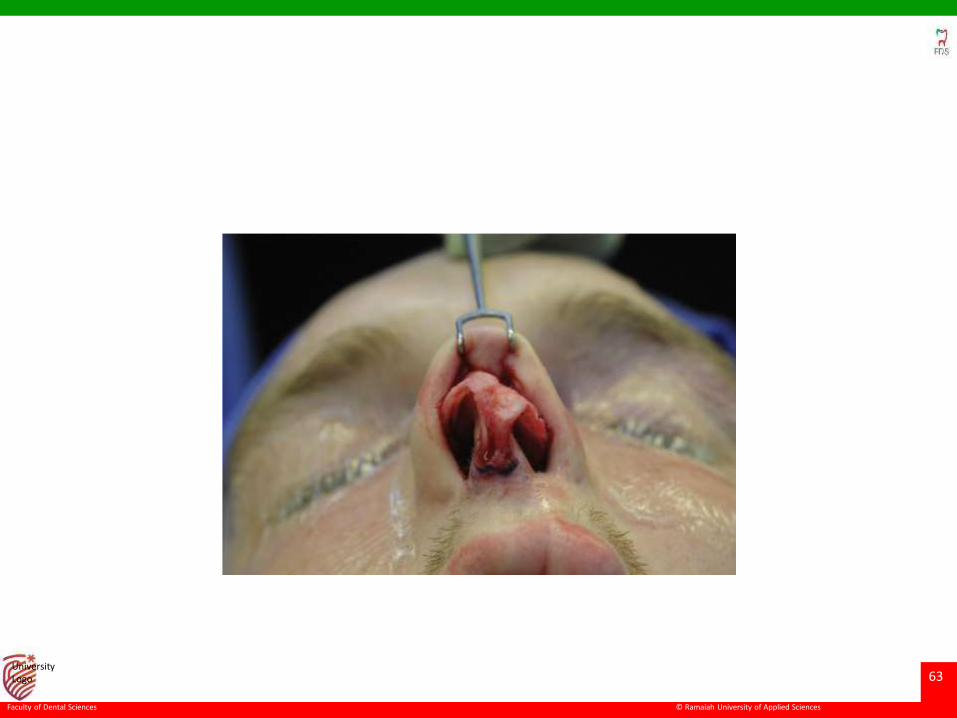
Treatment of the Nasal Tip
• The nasal tip in patients with congenital clefting of the lip is
poorly supported.
• In the unilateral deformity, the tip is asymmetric secondary to
the short medial crus on the cleft side
• In the bilateral deformity, the tip is usually underprojected and
the columella is short.
• Tip techniques are therefore designed to improve tip
symmetry, definition, and projection.
© Ramaiah University of Applied Sciences
63
Faculty of Dental Sciences
University Logo
© Ramaiah University of Applied Sciences
64
Faculty of Dental Sciences
University Logo
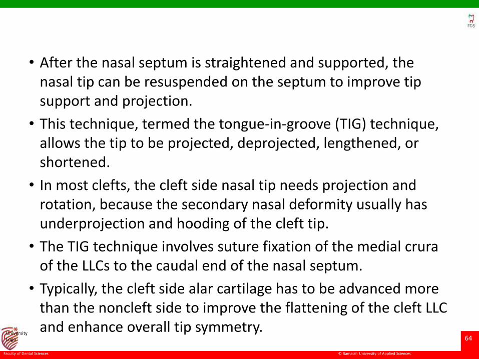
• After the nasal septum is straightened and supported, the nasal tip can be resuspended on the septum to improve tip support and projection.
• This technique, termed the tongue-in-groove (TIG) technique, allows the tip to be projected, deprojected, lengthened, or shortened.
• In most clefts, the cleft side nasal tip needs projection and rotation, because the secondary nasal deformity usually has underprojection and hooding of the cleft tip.
• The TIG technique involves suture fixation of the medial cruraof the LLCs to the caudal end of the nasal septum.
• Typically, the cleft side alar cartilage has to be advanced more than the noncleft side to improve the flattening of the cleft LLC and enhance overall tip symmetry.
© Ramaiah University of Applied Sciences
65
Faculty of Dental Sciences
University Logo
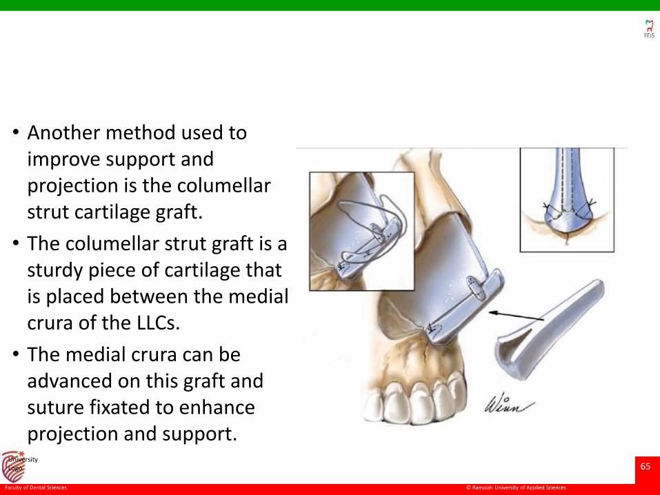
• Another method used to improve support and projection is the columellarstrut cartilage graft.
• The columellar strut graft is a sturdy piece of cartilage that is placed between the medial crura of the LLCs.
• The medial crura can be advanced on this graft and suture fixated to enhance projection and support.
© Ramaiah University of Applied Sciences
66
Faculty of Dental Sciences
University Logo
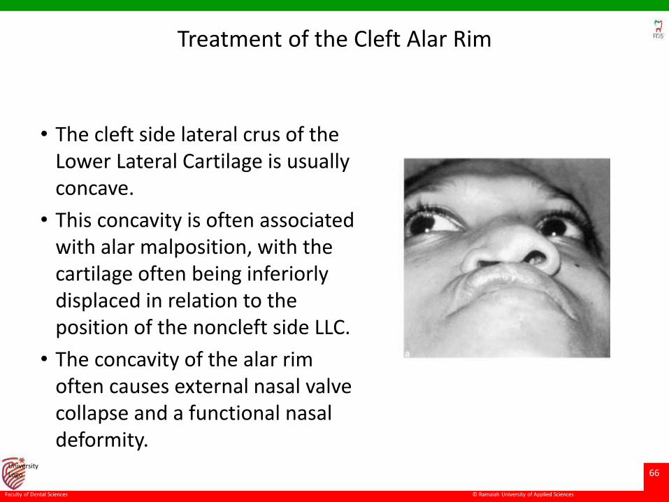
Treatment of the Cleft Alar Rim
• The cleft side lateral crus of the Lower Lateral Cartilage is usually concave.
• This concavity is often associated with alar malposition, with the cartilage often being inferiorly displaced in relation to the position of the noncleft side LLC.
• The concavity of the alar rim often causes external nasal valve collapse and a functional nasal deformity.
© Ramaiah University of Applied Sciences
67
Faculty of Dental Sciences
University Logo
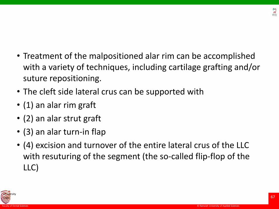
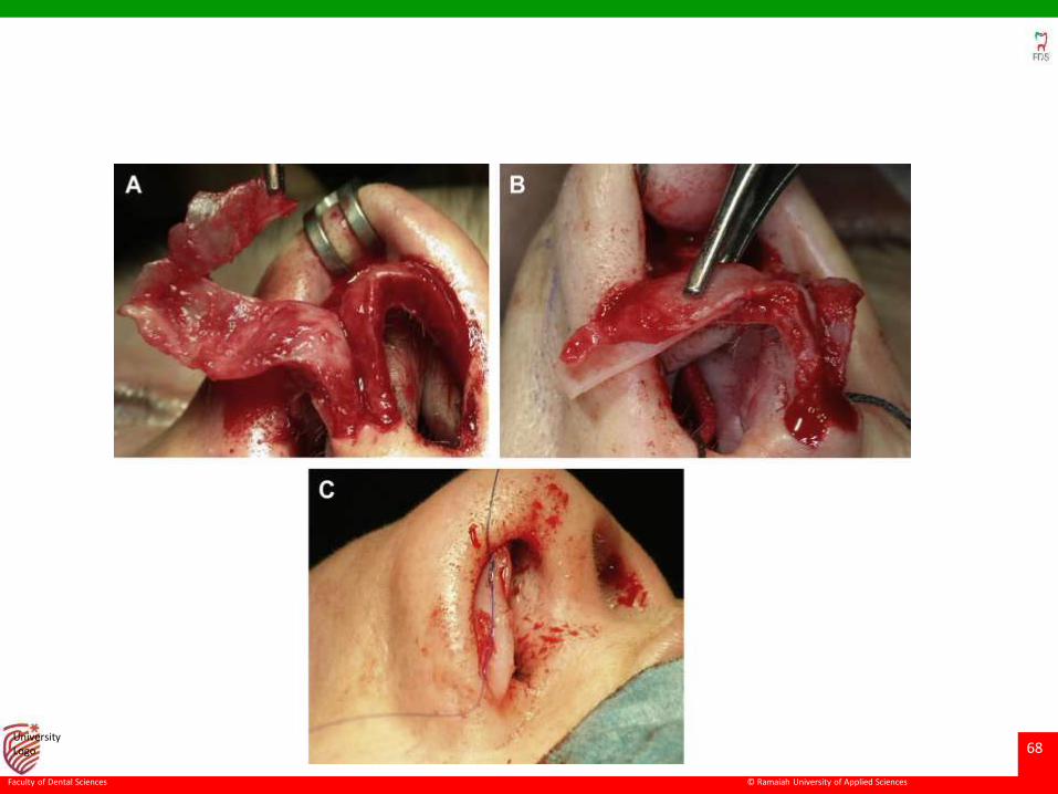
• Treatment of the malpositioned alar rim can be accomplished with a variety of techniques, including cartilage grafting and/or suture repositioning.
• The cleft side lateral crus can be supported with
• (1) an alar rim graft
• (2) an alar strut graft
• (3) an alar turn-in flap
• (4) excision and turnover of the entire lateral crus of the LLC with resuturing of the segment (the so-called flip-flop of the LLC)
© Ramaiah University of Applied Sciences
68
Faculty of Dental Sciences
University Logo
© Ramaiah University of Applied Sciences
69
Faculty of Dental Sciences
University Logo
Incomplete sill closure
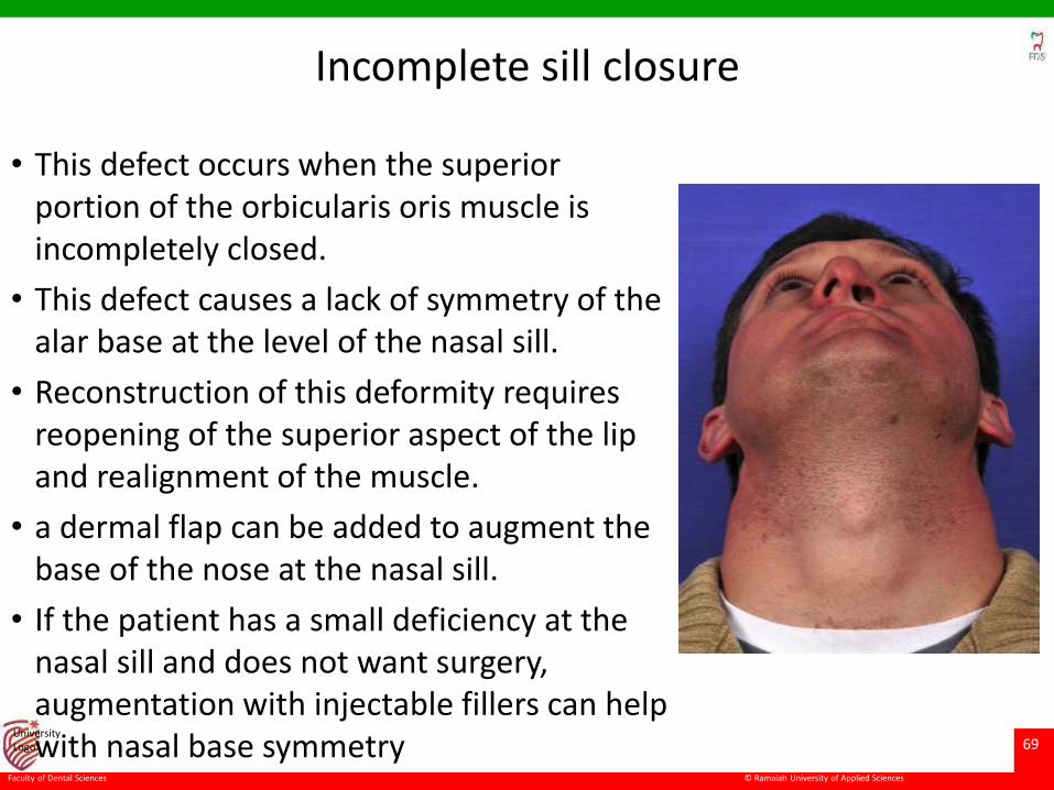
• This defect occurs when the superior portion of the orbicularis oris muscle is incompletely closed.
• This defect causes a lack of symmetry of the alar base at the level of the nasal sill.
• Reconstruction of this deformity requires reopening of the superior aspect of the lip and realignment of the muscle.
• a dermal flap can be added to augment the base of the nose at the nasal sill.
• If the patient has a small deficiency at the nasal sill and does not want surgery, augmentation with injectable fillers can help with nasal base symmetry
© Ramaiah University of Applied Sciences
70
Faculty of Dental Sciences
University Logo
SUMMARY
• The nose in patients with congenital cleft malformations is often the facial feature that is most noticeable to the observer.
• The secondary nasal deformity is variable and is affected by the extent of the original cleft, growth, and by any intervening surgery to correct the lip or nose.
• Repair of secondary cleft nasal deformities is challenging.
• Successful reconstruction requires an understanding of the pathologic anatomy, adequate exposure to perform techniques, and attention to structural grafting to overcome scarring and provide support.
• Often, graft material from the septum, rib, and/or ear is required. Attention must be paid to both function and appearance
© Ramaiah University of Applied Sciences
71
Faculty of Dental Sciences
University Logo
References
• Definitive Cleft Rhinoplasty for Unilateral Cleft Nasal Deformity-MyriamLoyo
• Repair of the Unilateral Cleft Lip/Nose Deformity- J. Madison Clark
• Rhinoplasty in adolescent cleft patients- Deodatta V.
• Cleft lip nose- sykes et al. journal of clinical plastic surgery
• Cleft Nasal Deformity and Rhinoplasty-Yoav Kaufman ; paediatric plastic surgery journal
• SecondaryCleftRhinoplasty- SachinS et al ; JAMA facial plastic surgery
• UNILATERAL CLEET RHINOPLASTY A Review - Simon J. Madorsky ; otorhinolaryngology clinics of north america