
Classification of mitocans, anti-cancer drugs acting on mitochondria · Classification of...
41
Classification of mitocans, anti-cancer drugs acting on mitochondria Jiri Neuzil, Lan-Feng Dong, Jakub Rohlena, Jaroslav Truksa, Stephen J. Ralph PII: S1567-7249(12)00200-0 DOI: doi: 10.1016/j.mito.2012.07.112 Reference: MITOCH 742 To appear in: Mitochondrion Received date: 7 May 2012 Revised date: 15 July 2012 Accepted date: 22 July 2012 Please cite this article as: Neuzil, Jiri, Dong, Lan-Feng, Rohlena, Jakub, Truksa, Jaroslav, Ralph, Stephen J., Classification of mitocans, anti-cancer drugs acting on mi- tochondria, Mitochondrion (2012), doi: 10.1016/j.mito.2012.07.112 This is a PDF file of an unedited manuscript that has been accepted for publication. As a service to our customers we are providing this early version of the manuscript. The manuscript will undergo copyediting, typesetting, and review of the resulting proof before it is published in its final form. Please note that during the production process errors may be discovered which could affect the content, and all legal disclaimers that apply to the journal pertain.
Transcript of Classification of mitocans, anti-cancer drugs acting on mitochondria · Classification of...

�������� ����� ��
Classification of mitocans, anti-cancer drugs acting on mitochondria
Jiri Neuzil, Lan-Feng Dong, Jakub Rohlena, Jaroslav Truksa, Stephen J.Ralph
PII: S1567-7249(12)00200-0DOI: doi: 10.1016/j.mito.2012.07.112Reference: MITOCH 742
To appear in: Mitochondrion
Received date: 7 May 2012Revised date: 15 July 2012Accepted date: 22 July 2012
Please cite this article as: Neuzil, Jiri, Dong, Lan-Feng, Rohlena, Jakub, Truksa,Jaroslav, Ralph, Stephen J., Classification of mitocans, anti-cancer drugs acting on mi-tochondria, Mitochondrion (2012), doi: 10.1016/j.mito.2012.07.112
This is a PDF file of an unedited manuscript that has been accepted for publication.As a service to our customers we are providing this early version of the manuscript.The manuscript will undergo copyediting, typesetting, and review of the resulting proofbefore it is published in its final form. Please note that during the production processerrors may be discovered which could affect the content, and all legal disclaimers thatapply to the journal pertain.
ACC
EPTE
D M
ANU
SCR
IPT
ACCEPTED MANUSCRIPTNeuzil et al.Classification of mitocans 1
Classification of mitocans, anti-cancer drugs acting on mitochondria
Jiri Neuzila,b,*
Lan-Feng Dong,a Jakub Rohlena,
b Jaroslav Truksa,
b Stephen J. Ralph
a
aSchool of Medical Science, Griffith University, Southport, Qld, Australia
bInstitute of Biotechnology, Czech Academy of Sciences, Prague, Czech Republic
Keywords: Mitocans, anti-cancer therapeutics, classification, selectivity
Abbreviations: ANT, adenine nucleotide translocase; BH3, Bcl-2 homology-3; 3BP, 3-
bromopyruvate; CI, complex I; DCA, dichloroacetate; 2DG, 2-deoxyglucose; ECs,
endothelial cells; ETC, electron transport chain; G6P, glucose-6-phosphate; GSAO, 4-[N-(S-
glutathionylacetyl)amino]phenylarsineoxide; HK, hexokinase; MIM, mitochondrial inner
membrane; MOM, mitochondrial outer membrane; mtDNA, mitochondrial DNA; PDK,
pyruvate dehydrogenase kinase; PEITC, phenylethyl isothiocyanate; PTPC, permeability
transition pore complex; Qd, distal UbQ-binding site in CII; Qi, Qo, UbQ-binding sites in CIII;
Qp, proximal UbQ-binding site in CII; SDH, succinate dehydrogenase; SDHA, succinate
dehydrogenase subunit A; SQR, succinate quinone reductase; TCA, tricarboxylic acid cycle;
-TOS, -tocopheryl succinate; TPP+, triphenylphosphonium; UbQ, ubiquinone; UbQH2,
ubiquinol; m,i, mitochondrial inner trans-membrane potential; VDAC, voltage-dependent
anionic channel; VE, vitamin E.
*Corresponding author: Jiri Neuzil, Apoptosis Research Group, School of Medical Science
and the Griffith Health Institute, Griffith University, Southport, 4222 Qld, Australia; phone:
+61-2-55529109; fax: +61-2-555-28444; e-mail: [email protected].

ACC
EPTE
D M
ANU
SCR
IPT
ACCEPTED MANUSCRIPTNeuzil et al.Classification of mitocans 2
Abstract
Mitochondria have recently emerged as an intriguing target for anti-cancer drugs, inherent to
vast majority if not all types of tumours. Drugs that target mitochondria and exert anti-cancer
activity have become a focus of recent research due to their great clinical potential (which has
not been harnessed thus far). The exceptional potential of mitochondria as a target for anti-
cancer agents has been reinforced by the discouraging finding that even tumours of the same
type from individual patients differ in a number of mutations. This is consistent with the idea
of personalised therapy, an elusive goal at this stage, in line with the notion that tumours are
unlikely to be treated by agents that target only a single gene or a single pathway. This
endows mitochondria, an invariant target present in all tumours, with an exceptional
momentum. This train of thoughts inspired us to define a class of anti-cancer drugs acting by
way of mitochondrial ‘destabilisation’, termed ‘mitocans’. In this communication, we define
mitocans (many of which have been known for a long time) and classify them into several
classes based on their molecular mode of action. We chose the targets that are of major
inmportance from the point of view of their role in mitochondrial destabilisation by small
compounds, some of which are now trialed as anti-cancer agents. The classification starts with
targets at the surface of mitochondria and ending up with those in the mitochondrial matrix.
The purpose of this review is to present in a concise manner the classification of compounds
that hold a considerable promise as potential anti-cancer agents.
1. Introduction
In the post-genomic era of the third millennium biomedical research has witnessed resurgence
of some of the ‘yonder-years’ scientific discoveries. It is now clear that some of the processes
that were the focus of research decades ago, are now being exploited as potential targets in
cancer treatment. Interestingly, the products of the same genes whose mutations can promote
ACC
EPTE
D M
ANU
SCR
IPT
ACCEPTED MANUSCRIPTNeuzil et al.Classification of mitocans 3
malignant transformations are also the emerging targets for novel, thus far largely unexploited
anti-cancer agents. For example, the mitochondrial complex II (CII) has recently been
described as a new target for anti-cancer drugs (Albayrak et al., 2005; Dong et al., 2008, 2009,
2011a,b; Rohlena et al., 2011). Intriguingly, mutations in the genes coding for its four
subunits have been classified as tumour suppressors, since mutation in these genes are
positively correlated with the incidence of certain infrequent neoplasias, viz.
pheochromocytoma and paraganglioma (Astuti et al., 2001; Gottlieb and Tomlinson 2005;
Maxwell 2005; Schiavi et al., 2005; Gimenez-Roqueplo et al., 2010; Burnichon et al., 2010).
Therefore, CII is an example of a target for anti-cancer drugs and, at the same time, due to
mutations in the genes coding its subunits it is involved in the mutagenic switch.
Many of the agents with anti-cancer activity that act on mitochondria, mitocans, hold a
substantial promise to be developed into efficient anti-cancer drugs, based on their selectivity
for cancer cells (Wallace et al., 2010; Fulda et al., 2010; Ralph et al., 2010a,b; Wang et al.,
2010; Fulda and Kroemer, 2011; Kepp et al., 2011; Lemarie and Grimm, 2011; Shoshan-
Barmatz and Ben-Hail, 2012). The importance of mitochondria as an emerging and
perspective target for anti-cancer agents is corroborated by the recent findings that tumours
differ in the level of expression of a high number of genes and mutations even amongst
patients with the same type of tumour. This has been documented for pancreatic cancer and
glioblastoma multiforme (Jones et al., 2008; Parsons et al., 2008), and, even worse, there are
differences in mutations within the same tumour (Gerlinger et al., 2012). These findings are
echoed in the somber-sounding editorial in Nature titled 'Cancer complexity slows quest
for cure' (Hayden, 2008). This indicates that it will be unlikely to suppress cancer by targeting
a single gene or a single pathway that may alter amongst cancer patients and that can be
subject to mutations. Rather, it is imperative to search for a target that is invariant and whose

ACC
EPTE
D M
ANU
SCR
IPT
ACCEPTED MANUSCRIPTNeuzil et al.Classification of mitocans 4
exploitation may present a general strategy for efficient treatment across the landscape of
neoplastic pathologies.
It appears clear that such a target is represented by mitochondria that are, at least to
some extent, functional in the vast majority if not all cancers (Ralph et al., 2010a,b).
Mitochondria, while being the ‘powerhouse’ of the cell, are also reservoirs of a number of
apoptosis-promoting proteins that are essential for apoptosis induction and its progression
downstream of these organelles, in order for the cancer cell to go into the commitment phase
and undergo the final demise (Kroemer et al., 2007; Galuzzi et al., 2010). It is also important
to take into consideration the aberrant mitochondrial metabolism in malignant cells (Koppenol
et al., 2011; Ward and Thompson,). Thus, the recent decade or so has witnessed an
unprecedented focus and discovery of novel agents that target mitochondria to induce cancer
cell death. In some cases, ‘old’ compounds have been re-discovered for their propensity to
destabilise mitochondria and kill cancer cells. Similarly and with an undisputable involvement
in the molecular mechanism of the mitochondria-targeting anti-cancer agents, the Warburg’s
hypothesis published in the 1920s (Warburg, 1956) has been recently experiencing a
renaissance of sorts (Vander Heiden et al., 2009; Cairns et al., 2011; Koppenol et al., 2011;
Hannahan and Weinberg, 2000, 2011).
The paramount importance of discovering novel and efficient anti-cancer agents is even
more accentuated by the fact that neoplastic diseases are now the greatest threat of the
Western society and are likely to increase in frequency (Jemal et al., 2011; Siegel et al., 2012;
Simard et al., 2012). We believe that targeting mitochondria, for tumour treatment may lead to
a potential future breakthrough in the management of malignancies. In this review, we
propose the classification of anti-cancer agents that act via mitochondrial destabilisation
(mitocans, an acronym for ‘mitochondria and cancer’) and provide several examples
epitomising the individual classes of mitocans, in particular those that are perceived as
ACC
EPTE
D M
ANU
SCR
IPT
ACCEPTED MANUSCRIPTNeuzil et al.Classification of mitocans 5
clinically relevant anti-cancer agents. The classification is based on the site of action of the
individual agents from the surface of the mitochondrial outer membrane (MOM) to the
mitochondrial matrix. The selection of the sites also stems from their importance as targets for
the development of drugs that hold substantial promise to be utilised in the clinical practice.
2. Class 1 mitocans: Hexokinase inhibitors
This class of agents comprises compounds targeting hexokinase (HK), which is an enzyme
whose main role is to phosphorylate glucose converting it to glucose-6-phosphate (G6P), a
substrate for metabolic pathways ultimately coupled with ATP generation. HK has a very
important function in cancer. Besides converting glucose to G6P, which can then enter the
metabolic machinery to, ultimately, yield ATP, HK is associated with the cytosolic site of the
porin-like voltage-dependent anionic channel (VDAC), a trans-membrane protein in the
MOM (Pedersen, 2008). When expressed at higher levels, as in cancer cells, HK (type II)
binds both ATP and glucose, resulting in the production of G6P. A direct correlation has been
established between the growth of carcinomas and the levels of HK activity (Bustamante et
al., 1981). According to Koobs (1972), mitochondrial-bound HK limits respiration when
tumour cells are utilising glucose (known as the Crabtree effect), even though large amounts
of ADP continue to be produced. The continuous phosphorylation of glucose by ATP
(proceeding by the mitochondrial-bound HK) reduces the level of phosphate available for
oxidative phosphorylation, and thereby prevents attaining maximal rates for state 3 respiration
(Baggetto and Testa-Parussini, 1990). Hence, HKII via its mitochondrial localisation also
helps to stabilise mitochondria, suppressing apoptotic death of cancer cells and promoting
their survival (Mathupala et al., 2006).
Several hexokinase inhibitors have been found to suppress cancer growth and of these,
considerable focus has been on 2-deoxyglucose (2DG) and 3-bromopyruvate (3BP). The basis

ACC
EPTE
D M
ANU
SCR
IPT
ACCEPTED MANUSCRIPTNeuzil et al.Classification of mitocans 6
for the action of 2DG is to inhibit HK activity and, hence, glycolysis with the result that the
binding of HK to VDAC is prevented, promoting the susceptibility of malignant cells to other
forms of treatment (Simons et al., 2007). This finding as well as recent report that 2DG
promotes cancer cell apoptosis when used in combination with another mitocan, the anti-
diabetic drug, metformin, provides the basis for testing these compounds in the clinical setting
(Ben Sahra et al., 2010). 3BP is an alkylating agent that inhibits both HK activity and the
mitochondrial complex II (CII) and consequently is included in the mitocan class 2 and 5
(Figure 1). Recent data indicate that 3BP acts by binding covalently to HKII, causing its
dissociation from VDAC (Chen et al., 2009). 3BP causes cancer cell death due to the rapid
depletion of ATP and suppresses tumour growth in animal models. For these reasons, 3BP is
another candidate for cancer clinical trials (Mathupala et al., 2006).
3. Class 2 mitocans: Compounds targeting Bcl-2 family proteins
This class of mitocans includes compounds acting as mimetics of the Bcl-2 homology-3
(BH3) domains, integral parts of Bcl-2 family of proteins. When the levels of expression of
the pro-apoptotic members of the family is greater, the cell will undergo the demise, whereas
higher levels of expression of the anti-apoptotic Bcl-2 family proteins will provide a pro-
survival ‘environment’. Although recent findings revealed novel functions for the Bcl-2
family members, including a role in the biogenesis of mitochondria (Suen et al., 2008), we
will only briefly discuss these proteins as targets for class 2 mitocans. The basis for the action
of this mitocan class stems from the finding that the anti-apoptotic and pro-apoptotic Bcl-2
family proteins interact via their BH3 domains thereby preventing the BH3 domain-
containing proteins Bak and/or Bax from forming large channels or pores in the MOM (Youle
and Strasser, 2008). Since the MOM channel is made of oligomers of the pro-apoptotic Bax or
Bak proteins, increased expression of the anti-apoptotic BH3-interacting proteins Bcl-2, Bcl-

ACC
EPTE
D M
ANU
SCR
IPT
ACCEPTED MANUSCRIPTNeuzil et al.Classification of mitocans 7
xL or Mcl-1 will protect cancer cells from apoptosis, and anti-apoptotic Bcl-2 family proteins
are often over-expressed in cancer cells (Lessene et al., 2008). Therefore, small molecules or
BH3 mimetics, targeting the interaction between the anti- and pro-apoptotic Bcl-2 protein
members, are of clinical importance (Kang and Reynolds, 2009; Zeitlin et al., 2008).
The BH3 mimetics include the natural, polyphenolic compound gossypol. This agent has
been shown to interact with BH3 binding domains, thereby interfering with the interaction
between Bcl-2, Bcl-xL or Mcl-1 and the pro-apoptotic proteins, Bax or Bak. The result is the
oligomerisation of Bax or Bak to form channels and activation of the post-mitochondrial
apoptotic signalling (Oliver et al., 2005). Gossypol has served as a lead compound for
developing more efficient BH3 mimetics, such as the highly intriguing agent ABT-737 (van
Delft et al., 2006). ABT-737 as well as its orally applicable version, ABT-263 (Tse et al.,
2008) are now being tested in clinical trials. ABT-263 (Navitoclax) has been successfully
tested in phase 1 clinical trial of lymphoid tumour and chronic lymphocytic leukemia,
resulting in the design of phase 2 study (Wilson et al., 2010; Roberts et al., 2012). It has also
been tested on solid tumours. Phase 1 clinical trial with mall-cell lung cancer (SCLC) or
pulmonary carcinoid, patients resulted in good outcome, prompting a phase 2 study (Roberts
et al., 2012).
The apoptogenic compound -tocopheryl succinate (-TOS) has been reported to
interact with the BH3-binding domain of Bcl-2 and Bcl-xL, which suppresses their interaction
with the pro-apoptotic protein Bak, arresting proliferation of prostate cancer cells and
inducing their death by apoptosis (Shiau et al., 2006). This activity of -TOS is interesting in
that it complements its second apoptogenic activity due to its ability to interact directly with
the ubiquinone (UbQ) sites of the mitochondrial CII (see below for details). In addition, the
mitochondrial CIII Qi site inhibitor, antimycin A, also acts as a BH3 mimetic (Tzung et al.,
2001), suggesting that on a more general level, compounds that interact with UbQ-binding

ACC
EPTE
D M
ANU
SCR
IPT
ACCEPTED MANUSCRIPTNeuzil et al.Classification of mitocans 8
sites in the mitochondrial electron transport chain (ETC) may have a tendency to be BH3
mimetics as well, consistent with the possibility that the BH3-binding site in Bcl-2 family
members might also present a UbQ-binding site (Neuzil et al., 2007).
4. Classes 3 and 4 of mitocans: Thiol redox inhibitors plus VDAC/ANT targeting drugs
Classes 3 and 4 comprise thiol redox inhibitors and the VDAC/ANT targeting drugs, and their
activity is linked to the redox environment of cancer cells, which is distinct from that of
normal cells in that cancer cells show higher intrinsic levels of ROS. As a result, it makes
cancer cells more vulnerable to agents that induce further elevations in oxidative stress, since
their anti-oxidant capacity is relatively inferior (Huang et al., 2000; Szatrowski and Nathan,
1991). Therefore, compounds that oxidise thiol group and/or deplete the mitochondrial GSH
pool will cause substantial apoptosis of cancer cells (Fulda et al., 2010; Trachootham et al.,
2009). Agents like arsenic trioxide (Miller, 2002; Pelicano et al., 2003) or isothiocyanates,
represented by phenylethyl isothiocyanate (PEITC) (Trachootham et al., 2006; Xu and
Thornalley, 2001) have been shown to possess relative selectivity in killing cancer cells by
upsetting the normal homeostasis in the cellular redox environment. Intriguingly, PEITC has
been reported to efficiently kill resistant leukemia cells (Trachootham et al., 2008).
The permeability transition pore complex (PTPC) forms as a superchannel comprising
the VDAC/ANT system of proteins embedded in the MOM and MIM, respectively,
interconnecting the mitochondrial matrix with the cytosol, and serves as a mode of transport
for a variety of solutes and small molecules including ATP and ADP (Zhivotovsky et al.,
2009). Deregulation of the VDAC/ANT complex results in apoptosis induction in cancer
cells. Compounds that modulate the PTPC include lonidamine, arsenites and steroid
analogues (represented by CD437) (Belzacq et al., 2001). Interestingly, an arsenite analogue
4-[N-(S-glutathionylacetyl)amino]phenylarsineoxide (GSAO) was shown to inhibit the

ACC
EPTE
D M
ANU
SCR
IPT
ACCEPTED MANUSCRIPTNeuzil et al.Classification of mitocans 9
function of ANT by crosslinking its cysteine residues. This was followed by the generation of
oxidative stress and induction of apoptosis, which was selective for proliferating, angiogenic
endothelial cells (ECs) while being non-toxic to growth-arrested ECs (Don et al., 2003).
These results indicate that GSAO can selectively kill ECs in the context of a growing tumour,
acting in an anti-angiogenic manner. Similar findings were reported forone of the mitocans,
-TOS (see more on its molecular mechanism below).
5. Class 5 mitocans: Electron redox chain targeting drugs
Class 5 mitocans comprises a large group of different compounds that target the
mitochondrial complexes, a part of the ETC (Scheffler, 2008). Some of them were discussed
in recent reviews (Fulda et al., 2010; Galluzzi et al., 2007; Gogvadze et al., 2009; Ralph and
Neuzil, 2009; Wang et al., 2010).
The production of energy is achieved by transporting electrons in a coordinated manner
from NADH or FADH2 (generated from substrates in the TCA cycle) to the final acceptor,
molecular oxygen, to produce water (Figure 1). The five macromolecular protein complexes
of the mitochondrial ETC are embedded in the mitochondrial inner membrane (MIM), and, by
passing electrons from CI and CII via CIII to CIV with the help of electron carriers, UbQ and
cytochrome c, generate energy that is maintained as an electrochemical proton gradient across
the MIM. CV, the F1F0-ATPase, then uses the energy from the proton gradient to generate
ATP from ADP and inorganic phosphate, which provides the cell with its fundamental energy
substrate. The ETC is the major source of mitochondrial reactive oxygen species (ROS) due
to the large electron flows, with CI and CIII identified as prime superoxide-generating sites,
although CII are also implicated in the process (Saraste 1999, Adam-Vizi and Chinopoulos
2006; Ralph et al., 2011). The superoxide by-product is not harmful when ROS production is
controlled within the constraints of the cellular redox system, as the cell must maintain

ACC
EPTE
D M
ANU
SCR
IPT
ACCEPTED MANUSCRIPTNeuzil et al.Classification of mitocans 10
moderate ROS levels necessary to sustain the cellular signalling processes. However,
deregulation leading to permanently higher levels of ROS may predispose, over time, to
carcinogenesis (Murphy, 2009). By contrast, a sudden and substantial increase in ROS levels
may have a much more immediate effect and commit the cell to undergo apoptosis (Fruehauf
and Meyskens, 2007; Kadenbach et al., 2010).
Figure 1 and Table I show several examples of mitocans targeting the mitochondrial
complexes. Due to lack of space, we will only focus here on several representatives of these
compounds. Of high interest is tamoxifen, a routinely used drug against estrogen-positive
breast cancer (Higgins and Stearns, 2011), that has recently been reported to induce apoptosis
via the mitochondrial CI, most likely by interfering with the FAD-binding site (Moreira et al.,
2006). We have been pursuing for a while the anti-cancer agent from the family of VE
analogues, -TOS, that selectively kills cancer cells (Neuzil et al., 2001a,b; Weber et al.,
2002, 2003). The mitochondrial CI (dos Santos et al., 2012) and, in particular CII have been
proposed as targets of -TOS (Dong et al., 2008, 2009). We have documented that -TOS
interferes with the CII’s proximal and distal UbQ-binding sites (Qp and Qd, respectively) that
have been documented recently in the crystallographic study of mammalian CII (Sun et al.,
2005).
Knowing the target for -TOS, we modified the agent by tagging it with the cationic
triphenylphosphonium (TPP+) group (see Figure 2A for structures), in analogy with the earlier
work of Murphy and Smith on UbQ (Murphy and Smith, 2007). This mitochondrially targeted
VE succinate (MitoVES) was found to associate almost exclusively with mitochondria
(Figure 2B) and kill a variety of cancer cell lines in a selective manner more efficiently by the
untargeted parental compound by 1-2 orders of magnitude (Dong et al., 2011a,b). Importantly,
MitoVES showed very high anti-cancer activity in clinically relevant mouse models of HER2-
high breast cancer and colorectal cancer (Dong et al., 2011a,b) (Figure 2C). More detailed
ACC
EPTE
D M
ANU
SCR
IPT
ACCEPTED MANUSCRIPTNeuzil et al.Classification of mitocans 11
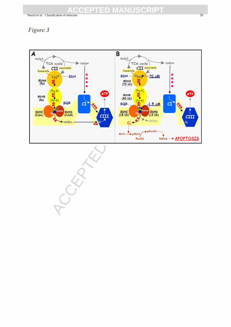
analysis of the molecular mechanism of the interaction of MitoVES with CII revealed that
MitoVES inhibited the SDH activity of CII (associated with the SDHA subunit of CII) with
the IC50 of ~70 M, while it suppressed the succinate quinone reductase (SQR) activity with
the IC50 of ~1.5 M (Figure 3). This indicates that the molecular target of MitoVES is ideally
placed, such that the SDH activity is only mildly depressed, allowing for the Krebs cycle to
proceed, i.e. for succinate to be converted to fumarate with the two electrons channeled via
the SDHB group’s [Fe-S] clusters to the MIM part of CII (Figure 3). Since MitoVES
displaces UbQ bound between SDHC and SDHD, electrons cannot be intercepted and form
superoxide inducing apoptosis in the cells by way of activating the Mst1 kinase that, in turn,
phosphorylates the transcription factor FoxO1, which causes upregulation of the BH3-only
protein Noxa. This protein then provokes the formation of the Bak and/or Bax channel in the
MOM (Prochazka et al., 2010; Valis et al., 2012; Dong et al., 2011a,b) (Figure 3).
Similar to GSAO (see Chapter 3), -TOS as well as MitoVES kill proliferating ECs
while being non-toxic to their arrested counterparts. The mechanism for the resistance for the
confluent ECs to the two agents involves their lack in ROS generation when challenged with
the compounds. Moreover, we also found that MitoVES cannot efficiently enter arrested ECs
due to their lower level of m,i; due to the cationic nature of MitoVES due to the presence
of the TPP+ group, this agent associates better with mitochondria when the m,i is higher
(see below) (Dong et al., 2007; Rohlena et al., 2011).
Addition of the cationic TPP group to mitocans with targets within the mitochondrial
complexes appears a new paradigm of efficient cancer treatment approach, since by tagging
these agents with TPP, directly send them to the proximity of their targets, therefore
increasing considerably their concentration in the compartment where ‘they matter’. We are
now attempting to tag with TPP+ also mitocans that target other mitochondrial complexes than

ACC
EPTE
D M
ANU
SCR
IPT
ACCEPTED MANUSCRIPTNeuzil et al.Classification of mitocans 12
CI. It is worth mentioning that TPP+ tagging has been tested for polyphenolic compounds,
some of which induce apoptosis (Biasutto et al., 2010; Smith et al., 2011; Sassi et al., 2012).
6. Class 6 mitocans: Lipophilic cations targeting the inner membrane
The molecular target of lipophilic cations acting on the MIM is given by the relatively high
trans-membrane potential, m,i that exists across the MIM. It has been documented that
cancer cells have a considerably higher m,i than in non-malignant cells due to altered
mitochondrial bioenergetics (Modica-Napolitano and Aprille, 1997). This feature will dictate
the intracellular targeting of lipophilic cations that as a result of the increased m,i in cancer
cells will make these mitocans relatively more selective for malignant cells. This follows from
the Nernst law defining that with each increase of m,i by -60 mV, a corresponding 10-fold
higher accumulation of cationic compounds in the MIM occurs (Modica-Napolitano and
Aprille, 2001, Wang et al., 2010).
A prime example of this mitocan class targeting the MIM is rhodamine-123, which was
reported to accumulate in mitochondria of src-transformed cells in the 1980s (Johnson et al.
1980), showing selectivity for cancer cells (Lampidis et al., 1983). Following from this, the
selective toxicity of a number of delocalised lipophilic cationic agents, including the peptide
(KLAKKLAK)2 towards cancer cells was found (Ellerby et al., 1999). A compound termed
F16, with a delocalised positive charge, was identified by high-throughput screening and was
found to be selective and effective against breast carcinomas with high level of HER2
expression (Fantin et al., 2002).
7. Class 7 mitocans: Drugs targeting the tricarboxylic acid cycle
The tricarboxylic acid (TCA) cycle, also referred to as the citric acid cycle or Kreb’s cycle, is
a source of electrons that are fed into the ETC and that are used to drive the electrochemical

ACC
EPTE
D M
ANU
SCR
IPT
ACCEPTED MANUSCRIPTNeuzil et al.Classification of mitocans 13
proton gradient required for the generation of ATP and is the target of class 7 mitocans. The
TCA cycle is based on the addition of acetyl-CoA (formed in the mitochondrial matrix by the
conversion of pyruvate (catalysed by pyruvate dehydrogenase) to oxaloacetate to form citrate.
Citric acid is then in a series of reactions converted to oxaloacetate, which again adds a
molecule of acetyl-CoA. During this process, electrons are released to drive the proton
gradient, which is coupled to the generation of ATP. An interesting step in the TCA cycle
involves SDH (or CII). 3BP, mentioned earlier as an inhibitor of HK, is also an inhibitor of
SDH (see Chapter 4) (Sun et al., 2005). Therefore, 3BP acts at the crossroads of the TCA
cycle and the ETC. It suppresses conversion of succinate to fumarate, thereby slowing down
the TCA cycle and, consequently, CI, while also inhibiting the generation of electrons that are
fed into CIII via CII (see Chapter 4). From this point of view, 3BP as an inhibitor of CII may
not be advantageous, while of more importance making it a potential anti-cancer agent may be
its inhibitory activity on HK (Pedersen, 2012).
A number of compounds exist that target the TCA cycle as well as the reaction
converting pyruvate to acetyl-CoA, a prerequisite of pyruvate to enter the mitochondria and
the TCA cycle. The enzyme pyruvate dehydrogenase that catalyses the reaction is regulated
by phosphorylation via the pyruvate dehydrogenase kinase (PDK).The inhibition of PDK
results in increased activity of pyruvate dehydrogenase and higher activity of the TCA cycle.
Dichloroacetate (DCA), a relatively basic compound, is selective for killing cancer cells by
suppressing the activity of PDK (Bonnet et al., 2007). By promoting the activity of pyruvate
dehydrogenase, DCA causes a shift from anaerobic glycolysis to oxidative glycolytic
metabolism accompanied by a decrease in the m,i, ROS generation and activation of the K+
channel, events that are selective for cancer cells (Bonnet et al., 2007). DCA is already in
clinical use to treat patients with mitochondrial deficiencies and, therefore, its development as

ACC
EPTE
D M
ANU
SCR
IPT
ACCEPTED MANUSCRIPTNeuzil et al.Classification of mitocans 14
an anti-cancer drug could be less complicated than when dealing with a completely novel
agent.
8. Class 8 mitocans: Drugs targeting mtDNA
Group 8 of mitocans comprises agents targeting mitochondrial DNA (mtDNA). Mitochondria
are unique organelles because they carry their own genetic information encoded on a small
circular genome, referred to as mitochondrial DNA (mtDNA). The mammalian mitochondrial
genome has the size of over 16 kB, and encodes 13 subunits of the mitochondrial complexes I,
III, IV and V, 24 tRNAs, 12S and 16S rRNA, and also contains a region called the D-loop
sequence, which is important in the regulation of mtDNA replication (Anderson et al., 1981).
To date, several compounds have been reported that interfere with the function and
stability of mtDNA and other drugs that affect the activity of the mitochondrial DNA
polymerase-. For example, vitamin K3 (menadione) targets mtDNA by inhibiting the activity
of DNA polymerase that is specific for replication of mtDNA, with ensuing induction of
apoptosis (Sasaki et al., 2008). Similar effects were reported for fialuridine, which induces
mitochondrial structural defects (Lewis et al., 1996). The Parkinsonian toxin 1-methyl-4-
phenylpyridinium causes a reduction in the copy number of mtDNA by destabilising the
structure of the mitochondrial D-loop (Miyako et al., 1997; Umeda et al., 2000).
We have been studying VE analogues as anti-cancer drugs, epitomised by the redox-
silent -TOS (see Chapters 4 for details on the apoptogenic signalling induced by -TOS).
The agent was made more efficient by tagging with the TPP group (Dong et al., 2008,
2011a,b), as has been done previously for a variety of antioxidants (Murphy and Smith, 2007)
(Figure 2). While the mitochondrially targeted VE analogues are superior to the untargeted -
TOS in its apoptogenic activity, we found another very intriguing feature of MitoVES. It was
observed that the agent modulates mtDNA and more specifically, suppresses the D-loop
ACC
EPTE
D M
ANU
SCR
IPT
ACCEPTED MANUSCRIPTNeuzil et al.Classification of mitocans 15
transcript levels at sub-apoptotic doses in a range of cancer cells, a phenomenon that was not
observed for -TOS. This was accompanied by the inhibition of cancer cell proliferation
(Truksa et al., 2012). It is not clear at this stage, whether modulation of mtDNA by MitoVES
is a direct effect of the drug on the mitochondrial genome; it appears that this is mediated by
the fast dissipation of m,i in response to MitoVES (Figure 4) and rapid ROS generation
(Truksa et al., 2012). Nevertheless, MitoVES provides an intriguing possibility for interfering
with tumour progression by means of suppressing the proliferation of cancer cells without
necessarily inducing apoptosis.
9. Conclusions and future perspectives: Clinical relevance of mitocans
Mitocans are, in quite a few cases, selective for cancer cells, which is a prerequisite for a
potentially clinically relevant anti-cancer agent. Except for a few compounds, mitocans have
not been employed in the clinical setting. One of these exceptions is tamoxifen, one of the
most frequently used drugs against breast cancer, which is now also used as a preventive
agent, albeit thus far largely due to its effect on the ER, competing with its activating ligand
estradiol (Higgins and Stearns, 2011; Cuzick et al., 2011). However, the recent findings that
tamoxifen also acts as a class 5 mitocan via targeting the mitochondrial CI (Moreira et al.,
2006) endows it with a novel translational spin, such that its modification whereby it will
localise primarily to mitochondria will give it an additional, thus far unprecedented
bioactivity. We are currently working on this highly intriguing possibility (Neuzil et al.,
unpublished data).
3BP, a class 1, 5 and 7 mitocan, has been pursued for its possible clinical relevance. A
whole special issue of the Journal of Bioengeneering and Biomemembranes has been devoted
to the compound (Pedersen, 2012). From the clinical point of view, of great interest is a recent
case report documenting that 3BP extended considerably the survival of a patient with

ACC
EPTE
D M
ANU
SCR
IPT
ACCEPTED MANUSCRIPTNeuzil et al.Classification of mitocans 16
fibrolamellar hepatocellular carcinoma with relatively low side effects (Ko et al., 2012). The
patient did succumb to the disease after two years of 3BP therapy, possibly due to the
‘poisoning’ by dead cancer cells that could not be detoxified by the apparently only partially
functional liver. Whether this toxicity is possibly also due to ‘too many’ targets that 3BP acts
on (Shoshan, 2012) is not clear. Notwithstanding the ultimate demise of the patient, 3BP can
be considered as one of the mitocans that can be developed into efficient anti-cancer drugs,
perhaps in combination with other agents.
We tested the class 5 mitocan -TOS in a single case clinical situation. A female
mesothelioma patient was allowed to be transdermally treated with the agent. Daily
administration of -TOS suspended in a cream with a transcutaneous enhancer helped remove
the pain associated with mesothelioma and resulted in the shrinkage of the tumour. However,
the tumour eventually started to progress again and the patient deceased. In spite of the
ultimately fatal outcome, the life of the patient, being of good quality, was extended by
several years, i.e. considerably more than expected. In a very recent in vitro study we have
found that long-term exposure of cancer cells to -TOS renders them resistant to the agent
due to upregulation of a member of the ABC class of transporters. We also discovered that
these resistant cells were, quite surprisingly, considerably susceptible to MitoVES (Neuzil et
al., unpublished observation), possibly due to the altered respiration of the resistant cells, a
condition likely favouring the killing of the cells by the mitochondrially targeted VE
analogue. This is a neat example how modification of mitocans whereby they directly interact
with their target may be utilised for suppression of tumours that are otherwise resistant, and
also indicate the possibility of a prospective intermittent therapy.
While, quite obviously, more needs to be done, it is undisputable that mitocans hold a
substantial promise as future anti-cancer agents, in particular due to mitochondria being a
rather invariant target across the landscape of the variety of types of neoplastic pathologies.

ACC
EPTE
D M
ANU
SCR
IPT
ACCEPTED MANUSCRIPTNeuzil et al.Classification of mitocans 17
The few examples above also document the fact that basic mitocans can be further modified to
accumulate in the mitochondrial compartment, where their presence ‘matters’, as exemplified
by tagging them with the TPP+ cationic group that targets them to the interphase of the MIM
and the mitochondrial matrix. It is proposed that carefully planned clinical trials with selected
mitocans are highly warranted to shift the odds of cancer patients toward their survival and
complete cure. Given the unrelenting increase in the number of cancer patients, this may
appear as a highly daunting task. Notwithstanding this grim perspective, the emerging
knowledge of the importance of mitochondria as clinically relevant targets for anti-cancer
drugs seems to provide the ‘light at the end of the tunnel’.
References
Adam-Vizi, V., Chinopoulos, C., 2006. Bioenergetics and the formation of mitochondrial
reactive oxygen species. Trends Pharmacol. Sci. 27, 639-645.
Albayrak, T., Scherhammer, V., Schoenfeld, N., Braziulis, E., Mund, T., Bauer, M.K.,
Scheffler, I.E., Grimm, S., 2003. The tumor suppressor cybL, a component of the
respiratory chain, mediates apoptosis induction. Mol. Biol. Cell 14, 3082-3096.
Anderson, S., Bankier, A.T., Barrell, B.G., de Bruijn, M.H., Coulson, A.R., Drouin, J.,
Eperon, I.C., Nierlich, D.P., Roe, B.A., Sanger, F., Schreier, P.H., Smith, A.J., Staden, R.,
Young, I.G., 1981. Sequence and organization of the human mitochondrial genome. Nature
290, 457-465.
Astuti, D., Latif, F., Dallol, A., Dahia, P.L., Douglas, F., George, E., Sköldberg, F., Husebye,
E.S., Eng, C., Maher, E.R., 2001. Gene mutations in the succinate dehydrogenase subunit
SDHB cause susceptibility to familial pheochromocytoma and to familial paraganglioma.
Am. J. Hum. Genet. 69, 49-54.

ACC
EPTE
D M
ANU
SCR
IPT
ACCEPTED MANUSCRIPTNeuzil et al.Classification of mitocans 18
Baggetto, L.G., Testa-Parussini, R., 1990. Role of acetoin on the regulation of intermediate
metabolism of Ehrlich ascites tumor mitochondria: its contribution to membrane cholesterol
enrichment modifying passive proton permeability. Arch. Biochem. Biophys. 283, 241-248.
Belzacq, A.S., El Hamel, C., Vieira, H.L., Cohen, I., Haouzi, D., Metivier, D., Marchetti, P.,
Brenner, C., Kroemer, G., 2001. Adenine nucleotide translocator mediates the mitochondrial
membrane permeabilization induced by lonidamine, arsenite and CD437. Oncogene 20,
7579-7587.
Ben Sahra, I., Laurent, K., Giuliano, S., Larbret, F., Ponzio, G., Gounon, P., Le Marchand-
Brustel, Y., Giorgetti-Peraldi, S., Cormont, M., Bertolotto, C., Deckert, M., Auberger, P.,
Tanti, J.F., Bost, F., 2010. Targeting cancer cell metabolism: the combination of metformin
and 2-deoxyglucose induces p53-dependent apoptosis in prostate cancer cells. Cancer Res.
70, 2465-2475.
Bernal, S.D., Lampidis, T.J., McIsaac, R.M., Chen, L.B., 1983. Anticarcinoma activity in vivo
of rhodamine 123, a mitochondrial-specific dye. Science 222, 169-172.
Biassutto, L., Dong, L.F., Neuzil, J., Zoratti, M., 2010. Mitochondrially targeted anti-cancer
drugs. Mitochondrion 10, 670-681.
Bonnet, S., Archer, S.L., Allalunis-Turner, J., Haromy, A., Beaulieu, C., Thompson, R., Lee,
C.T., Lopaschuk, G.D., Puttagunta, L., Harry, G., Hashimoto, K., Porter, C.J., Andrade,
M.A., Thebaud, B., Michelakis, E.D., 2007. A mitochondria-K+ channel axis is suppressed
in cancer and its normalization promotes apoptosis and inhibits cancer growth. Cancer Cell
11, 37-51.
Burnichon, N., Brière, J.J., Libé, R., Vescovo, L., Rivière, J., Tissier, F., Jouanno, E.,
Jeunemaitre, X., Bénit, P., Tzagoloff, A., Rustin, P., Bertherat, J., Favier, J., Gimenez-
Roqueplo, A.P., 2010. SDHA is a tumor suppressor gene causing paraganglioma. Hum.
Mol. Genet. 19, 3011-3020.

ACC
EPTE
D M
ANU
SCR
IPT
ACCEPTED MANUSCRIPTNeuzil et al.Classification of mitocans 19
Bustamante, E., Morris, H.P., Pedersen, P.L., 1981. Energy metabolism of tumor cells.
Requirement for a form of hexokinase with a propensity for mitochondrial binding. J. Biol.
Chem. 256, 8699-8704.
Cairns, R.A., Harris, I.S., Mak, T.W., 2011. Regulation of cancer cell metabolism. Nat. Rev.
Cancer 11, 85-95.
Chen, Z., Zhang, H., Lu, W., Huang, P., 2009. Role of mitochondria-associated hexokinase II
in cancer cell death induced by 3-bromopyruvate. Biochim. Biophys. Acta 1787, 553-560.
Cuzick, J., DeCensi, A., Arun, B., Brown, P.H., Castiglione, M., Dunn, B., Forbes, J.F.,
Glaus., A., Howell, A., von Minckwitz, G., Vogel, V., Zwierzina, H., 2011. Preventive
therapy for breast cancer: a consensus statement. Lancet Oncol. 12, 496-503.
Dell'Antone, P., 2009. Targets of 3-bromopyruvate, a new, energy depleting, anticancer agent.
Med. Chem. 5, 491-496.
Don, A.S., Kisker, O., Dilda, P., Donoghue, N., Zhao, X., Decollogne, S., Creighton, B.,
Flynn, E., Folkman, J., Hogg, P.J., 2003. A peptide trivalent arsenical inhibits tumor
angiogenesis by perturbing mitochondrial function in angiogenic endothelial cells. Cancer
Cell 3, 497-509.
Dong, L.F., Low, P., Dyason, J., Wang, X.F., Prochazka, L., Witting, P.K., Freeman, R.,
Swettenham, E., Valis, K., Liu, J., Zobalova, R., Turanek, J., Spitz, D.R., Domann, F.E.,
Scheffler, I.E., Ralph, S.J., Neuzil, J., 2008. -Tocopheryl succinate induces apoptosis by
targeting ubiquinone-binding sites in mitochondrial respiratory complex II. Oncogene 27,
4324-4335.
Dong, L.F., Freeman, R., Liu, J., Zobalova, R., Marin-Hernandez, A., Stantic, M., Rohlena, J.,
Rodriguez-Enriquez, S., Valis, K., Butcher, B., Goodwin, J., Brunk, U.T., Witting, P.K.,
Moreno-Sanchez, R., Scheffler, I.E., Ralph, S.J., Neuzil, J., 2009. Suppression of tumour
ACC
EPTE
D M
ANU
SCR
IPT
ACCEPTED MANUSCRIPTNeuzil et al.Classification of mitocans 20
growth in vivo by the mitocan -tocopheryl succinate requires respiratory complex II. Clin.
Cancer Res. 15, 1593-1600.
Dong, L.F., Jameson, V.J.A., Tilly, D., Cerny, J., Mahdavian, E., Marín-Hernández, A.,
Hernández-Esquivel, L., Rodríguez-Enríquez, S., Stursa, J., Witting, P.K., Stantic, B.,
Rohlena, J., Truksa, J., Kluckova, K., Dyason, J.C., Ledvina, M., Salvatore, B.A., Moreno-
Sánchez, R., Coster, M.J., Ralph, S.J., Smith, R.A.J., Neuzil, J., 2011a. Mitochondrial
targeting of vitamin E succinate enhances its pro-apoptotic and anti-cancer activity via
mitochondrial complex II. J. Biol. Chem. 286, 3717-3728.
Dong, L.F., Jameson, V.J.A., Tilly, D., Prochazka, L., Rohlena, J., Valis, K., Truksa, J.,
Zobalova, R., Mahdavian, E., Kluckova, K., Stantic, M., Stursa, J., Wang, X.F., Freeman,
R., Witting, P.K., Norberg, E., Goodwin, J., Salvatore, B.A., Novotna, J., Turanek, J.,
Ledvina, M., Hozak, P., Zhivotovsky, B., Coster, M.J., Ralph, S.J., Smith, R.A.J., Neuzil, J.,
2011b. Mitochondrial targeting of -tocopheryl succinate enhances its pro-apoptotic
efficacy: A new paradigm of efficient anti-cancer therapy. Free Radic. Biol. Med. 50, 1546-
1555.
dos Santos, G.A., Abreu e Lima, R.S., Pestana, C.R., Lima, A.S., Scheucher, P.S., Thomé,
C.H., Gimenes-Teixeira, H.L., Santana-Lemos, B.A., Lucena-Araujo, A.R., Rodrigues, F.P.,
Nasr, R., Uyemura, S.A., Falcão, R.P., de Thé, H., Pandolfi, P.P., Curti, C., Rego, E.M.,
2012. (+)-Tocopheryl succinate inhibits the mitochondrial respiratory chain complex I and
is as effective as arsenic trioxide or ATRA against acute promyelocytic leukemia in vivo.
Leukemia 26, 451-460.
Ellerby, H.M., Arap, W., Ellerby, L.M., Kain, R., Andrusiak, R., Rio, G.D., Krajewski, S.,
Lombardo, C.R., Rao, R., Ruoslahti, E., Bredesen, D.E., Pasqualini, R., 1999. Anti-cancer
activity of targeted pro-apoptotic peptides. Nat. Med. 5, 1032-1038.
ACC
EPTE
D M
ANU
SCR
IPT
ACCEPTED MANUSCRIPTNeuzil et al.Classification of mitocans 21
Fantin, V.R., Berardi, M.J., Scorrano, L., Korsmeyer, S.J., Leder, P., 2002. A novel
mitochondriotoxic small molecule that selectively inhibits tumor cell growth. Cancer Cell 2,
29-42.
Fruehauf, J.P., Meyskens, F.L., 2007. Reactive oxygen species: a breath of life or death? Clin.
Cancer Res. 13, 789-794.
Fulda, S., Galluzzi, L., Kroemer, G., 2010. Targeting mitochondria for cancer therapy. Nat.
Rev. Drug Discov. 9, 447-464.
Fulda, S., Kroemer, G., 2011. Mitochondria as therapeutic targets for the treatment of
malignant disease. Antioxid. Redox Signal. 15, 2937-2949.
Fulda, S., Scaffidi, C., Susin, S.A., Krammer, P.H., Kroemer, G., Peter, M.E., Debatin, K.M.,
1998. Activation of mitochondria and release of mitochondrial apoptogenic factors by
betulinic acid. J. Biol. Chem. 273, 33942-33948.
Galluzzi, L., Morselli, E., Kepp, O., Vitale, I., Rigoni, A., Vacchelli, E., Michaud, M.,
Zischka, H., Castedo, M., Kroemer, G., 2010. Mitochondrial gateways to cancer. Mol.
Aspects Med. 31, 1-20.
Gandhi, L., Camidge, D.R., Ribeiro de Oliveira, M., Bonomi, P., Gandara, D., Khaira, D.,
Hann, C.L., McKeegan, E.M., Litvinovich, E., Hemken, P.M., Dive, C., Enschede, S.H.,
Nolan, C., Chiu, Y.L., Busman, T., Xiong, H., Krivoshik, A.P., Humerickhouse, R.,
Shapiro, G.I., Rudin, C.M. 2011. Phase I study of Navitoclax (ABT-263), a novel Bcl-2
family inhibitor, in patients with small-cell lung cancer and other solid tumors. J. Clin.
Oncol. 29, 909-916.
Gerlinger, M., Rowan, A.J., Horswell, S., Larkin, J., Endesfelder, D., Gronroos, E., Martinez,
P., Matthews, N., Stewart, A., Tarpey, P., Varela, I., Phillimore, B., Begum, S., McDonald,
N.Q., Butler, A., Jones, D., Raine, K., Latimer, C., Santos, C.R., Nohadani, M., Eklund,
A.C., Spencer-Dene, B., Clark, G., Pickering, L., Stamp, G., Gore, M., Szallasi, Z.,

ACC
EPTE
D M
ANU
SCR
IPT
ACCEPTED MANUSCRIPTNeuzil et al.Classification of mitocans 22
Downward, J., Futreal, P.A., Swanton, C., 2012. Intratumor heterogeneity and branched
evolution revealed by multiregion sequencing. N. Engl. J. Med. 366, 883-892.
Gimenez-Roqueplo, A.P., Favier, J., Rustin, P., Mourad, J.J., Plouin, P.F., Corvol, P., Rötig,
A., Jeunemaitre, X., 2001. The R22X mutation of the SDHD gene in hereditary
paraganglioma abolishes the enzymatic activity of complex II in the mitochondrial
respiratory chain and activates the hypoxia pathway. Am. J. Hum. Genet. 69, 1186-1197.
Gottlieb, E., Tomlinson, I.P., 2005. Mitochondrial tumour suppressors: a genetic and
biochemical update. Nat. Rev. Cancer 5, 857-866.
Hanahan, D., Weinberg, R.A., 2000. Hallmarks of cancer. Cell 100, 57-70.
Hanahan, D., Weinberg, R.A., 2011. Hallmarks of cancer: the next generation. Cell 144, 646-
674.
Hayden, E.C., 2008. Cancer complexity slows quest for cure. Nature 455, 158.
Higgins, M.J, Stearns, V., 2011. Pharmacogenetics of endocrine therapy for breast cancer.
Annu. Rev. Med. 62, 281-293.
Huang, P., Feng, L., Oldham, E.A., Keating, M.J., Plunkett, W., 2000. Superoxide dismutase
as a target for the selective killing of cancer cells. Nature 407, 390-395.
Jemal, A., Bray, F., Center, M.M., Ferlay, J., Ward, E., Forman, D., 2011. Global cancer
statistics. CA Cancer J. Clin. 61, 69-90.
Johnson, L.V., Walsh, M.L., Chen, L.B., 1980. Localization of mitochondria in living cells
with rhodamine 123. Proc. Nat. Acad. Sci. USA 77, 990-994.
Jones, S., Zhang, X., Parsons, D.W., Lin, J.C., Leary, R.J., Angenendt, P., Mankoo, P., Carter,
H., Kamiyama, H., Jimeno, A., Hong, S.M., Fu, B., Lin, M.T., Calhoun, E.S., Kamiyama,
M., Walter, K., Nikolskaya, T., Nikolsky, Y., Hartigan, J., Smith, D.R., Hidalgo, M., Leach,
S.D., Klein, A.P., Jaffee, E.M., Goggins, M., Maitra, A., Iacobuzio-Donahue, C., Eshleman,
J.R., Kern, S.E., Hruban, R.H., Karchin, R., Papadopoulos, N., Parmigiani, G., Vogelstein,

ACC
EPTE
D M
ANU
SCR
IPT
ACCEPTED MANUSCRIPTNeuzil et al.Classification of mitocans 23
B., Velculescu, V.E., Kinzler, K.W., 2008. Core signaling pathways in human pancreatic
cancers revealed by global genomic analyses. Science 321, 1801-1806.
Kadenbach, B., Ramzan, R., Wen, L.,Vogt, S., 2010. New extension of the Mitchell Theory
for oxidative phosphorylation in mitochondria of living organisms. Biochim. Biophys. Acta
1800, 205-212.
Kang, M.H., Reynolds, C.P., 2009. Bcl-2 inhibitors: targeting mitochondrial apoptotic
pathways in cancer therapy. Clin. Cancer Res. 15, 1126-1132.
Kepp, O., Galluzzi, L., Lipinski, M., Yuan, J., Kroemer, G., 2011. Cell death assays for drug
discovery. Nat. Rev. Drug Discov. 10, 221-237.
Ko, Y.H., Smith, B.L., Wang, Y., Pomper, M.G., Rini, D.A., Torbenson, M.S., Hullihen, J.,
Pedersen, P.L., 2004. Advanced cancers: eradication in all cases using 3-bromopyruvate
therapy to deplete ATP. Biochem. Biophys. Res. Commun. 324, 269-275.
Ko, Y.H., Verhoeven, H.A., Lee, M.J., Corbin, D.J., Vogl, T.J., Pedersen, P.L.., 2012. A
translational study "case report" on the small molecule "energy blocker" 3-bromopyruvate
(3BP) as a potent anticancer agent: from bench side to bedside. J. Bioenerg Biomembr. 44,
163-170.
Koobs, D.H., 1972. Phosphate mediation of the Crabtree and Pasteur effects. Science 178,
127-133.
Koppenol, W.H., Bounds, P.L., Dang, C.V., 2011. Otto Warburg's contributions to current
concepts of cancer metabolism. Nat. Rev. Cancer 11, 325-337.
Kroemer, G., Galluzzi, L., Brenner, C., 2007. Mitochondrial membrane permeabilization in
cell death. Physiol Rev. 87, 99-163.
Lampidis, T.J., Bernal, S.D., Summerhayes, I.C., Chen, L.B., 1983. Selective toxicity of
rhodamine 123 in carcinoma cells in vitro. Cancer Res. 43, 716-720.

ACC
EPTE
D M
ANU
SCR
IPT
ACCEPTED MANUSCRIPTNeuzil et al.Classification of mitocans 24
Le, S.B., Hailer, M.K., Buhrow, S., Wang, Q., Flatten, K., Pediaditakis, P., Bible, K.C.,
Lewis, L.D., Sausville, E.A., Pang, Y.P., Ames, M.M., Lemasters, J.J., Holmuhamedov,
E.L., Kaufmann, S.H., 2007. Inhibition of mitochondrial respiration as a source of
adaphostin-induced reactive oxygen species and cytotoxicity. J. Biol. Chem. 282, 8860-
8872.
Lemarie, A., Grimm, S., 2011. Mitochondrial respiratory chain complexes: apoptosis sensors
mutated in cancer? Oncogene 30, 3985-4003.
Lessene, G., Czabotar, P.E., Colman, P., 2008. Bcl-2 family antagonists for cancer therapy.
Nat. Rev. Drug Discov. 7, 989-1000.
Lewis, W., Levine, E.S., Griniuviene, B., Tankersley, K.O., Colacino, J.M., Sommadossi,
J.P., Watanabe, K.A., Perrino, F.W., 1996. Fialuridine and its metabolites inhibit DNA
polymerase- at sites of multiple adjacent analog incorporation, decrease mtDNA
abundance, and cause mitochondrial structural defects in cultured hepatoblasts. Proc. Nat.
Acad. Sci. USA 93, 3592-3597, 1996.
Mathupala, S.P., Ko, Y.H., Pedersen, P.L., 2006. Hexokinase II: cancer's double-edged sword
acting as both facilitator and gatekeeper of malignancy when bound to mitochondria.
Oncogene 25, 4777-4786.
Maxwell, P.H., 2005. A common pathway for genetic events leading to pheochromocytoma.
Cancer Cell 8, 91-93.
Miller, W.H., Jr., 2002. Molecular targets of arsenic trioxide in malignant cells. Oncologist 7
Suppl 1, 14-19.
Miyako, K., Kai, Y., Irie, T., Takeshige, K., Kang, D., 1997. The content of intracellular
mitochondrial DNA is decreased by 1-methyl-4-phenylpyridinium ion (MPP+). J. Biol.
Chem. 272, 9605-9608.
ACC
EPTE
D M
ANU
SCR
IPT
ACCEPTED MANUSCRIPTNeuzil et al.Classification of mitocans 25
Modica-Napolitano, J.S., Aprille, J.R., 1997. Basis for the selective cytotoxicity of rhodamine
123. Cancer Res. 47, 4361-4365.
Modica-Napolitano, J.S., Aprille, J.R., 2001. Delocalized lipophilic cations selectively target
the mitochondria of carcinoma cells. Adv. Drug Deliv. Rev. 49, 63-70.
Moreira, P.I., Custódio, J., Moreno, A., Oliveira, C.R., Santos, M.S., 2006. Tamoxifen and
estradiol interact with the flavin mononucleotide site of complex I leading to mitochondrial
failure. J. Biol. Chem. 281, 10143-10152.
Murphy, M.P., Smith, R.A., 2007. Targeting antioxidants to mitochondria by conjugation to
lipophilic cations. Annu. Rev. Pharmacol. Toxicol. 47, 629-656.
Murphy, M.P., 2009. How mitochondria produce reactive oxygen species. Biochem. J. 417, 1-
13.
Neuzil, J., Dong, L.F., Ramanathapuram, L., Hahn, T., Chladova, M., Wang, X.F., Zobalova,
R., Prochazka, L., Gold, M., Freeman, R.E., Turanek, J., Akporiaye, E.T., Dyason, J.,
Ralph, S.J., 2007. Vitamin E analogues: a novel group of mitocans, anti-cancer agents that
act by targeting mitochondria. Mol. Aspects Med. 28, 607-645.
Neuzil, J., Weber, T., Gellert, N., Weber, C., 2001a. Selective cancer cell killing by -
tocopheryl succinate. Br. J. Cancer 84, 87-89.
Neuzil, J., Weber, T., Schröder, A., Lu, M., Ostermann, G., Gellert, N., Mayne, G.C.,
Olejnicka, B., Nègre-Salvayre, A., Sticha, M., Coffey, R.J., Weber, C., 2001. Induction of
apoptosis in cancer cells by -tocopheryl succinate: Molecular pathways and structural
requirements. FASEB J 15, 403-415.
Oliver, C.L., Miranda, M.B., Shangary, S., Land, S., Wang, S., Johnson, D.E., 2005. Gossypol
acts directly on the mitochondria to overcome Bcl-2- and Bcl-xL-mediated apoptosis
resistance. Mol. Cancer Ther. 4, 23-31.

ACC
EPTE
D M
ANU
SCR
IPT
ACCEPTED MANUSCRIPTNeuzil et al.Classification of mitocans 26
Pelicano, H., Feng, L., Zhou, Y., Carew, J.S., Hileman, E.O., Plunkett, W., Keating, M.J.,
Huang, P., 2003. Inhibition of mitochondrial respiration: a novel strategy to enhance drug-
induced apoptosis in human leukemia cells by a reactive oxygen species-mediated
mechanism. J. Biol. Chem. 278, 37832-37839.
Parsons, D.W., Jones, S., Zhang, X., Lin, J.C., Leary, R.J., Angenendt, P., Mankoo, P., Carter,
H., Siu, I.M., Gallia, G.L., Olivi, A., McLendon, R., Rasheed, B.A., Keir, S., Nikolskaya,
T., Nikolsky, Y., Busam, D.A., Tekleab, H., Diaz, L.A., Hartigan, J., Smith, D.R.,
Strausberg, R.L., Marie, S.K., Shinjo, S.M., Yan, H., Riggins, G.J., Bigner, D.D., Karchin,
R., Papadopoulos, N., Parmigiani, G., Vogelstein, B., Velculescu, V.E., Kinzler, KW., 2008.
An integrated genomic analysis of human glioblastoma multiforme. Science 321, 1807-
1812.
Pedersen, P.L., 2008. Voltage dependent anion channels (VDACs): a brief introduction with a
focus on the outer mitochondrial compartment's roles together with hexokinase-2 in the
"Warburg effect" in cancer. J. Bioenerg. Biomembr. 40, 123-126.
Pedersen, P.L., 2012. 3-Bromopyruvate (3BP) a fast acting, promising, powerful, specific, and
effective "small molecule" anti-cancer agent taken from labside to bedside: introduction to a
special issue. J. Bioenerg. Biomembr. 44, 1-6.
Pelicano, H., Feng, L., Zhou, Y., Carew, J.S., Hileman, E.O., Plunkett, W., Keating, M.J.,
Huang, P., 2003. Inhibition of mitochondrial respiration: a novel strategy to enhance drug-
induced apoptosis in human leukemia cells by a reactive oxygen species-mediated
mechanism. J. Biol. Chem. 278, 37832-37839.
Pereira da Silva, A.P., El-Bacha, T., Kyaw, N., dos Santos, R.S., da-Silva, W.S., Almeida,
F.C., Da Poian, A.T., Galina, A., 2009. Inhibition of energy-producing pathways of HepG2
cells by 3-bromopyruvate. Biochem. J. 417, 717-726.

ACC
EPTE
D M
ANU
SCR
IPT
ACCEPTED MANUSCRIPTNeuzil et al.Classification of mitocans 27
Prochazka, L., Dong, L.F., Valis, K., Freeman, R., Ralph, S.J., Turanek, J., Neuzil, J., 2010.
-Tocopheryl succinate causes mitochondrial permeabilization by preferential formation of
Bak channel. Apoptosis 15, 782-794.
Ralph, S.J., Neuzil, J., 2009. Mitochondria as targets for cancer therapy. Mol. Nutr. Food Res.
53, 9-28.
Ralph, S.J., Rodríguez-Enríquez, S., Neuzil, J., Moreno-Sánchez, R., 2010a. Bioenergetic
pathways in tumor mitochondria as targets for cancer therapy and the importance of the
ROS-induced apoptotic trigger. Mol. Aspects Med. 31, 29-59.
Ralph, S.J., Rodríguez-Enríquez, S., Neuzil, J., Saavedra, E., Moreno-Sánchez, R., 2010b.
The causes of cancer revisited: "mitochondrial malignancy" and ROS-induced oncogenic
transformation - why mitochondria are targets for cancer therapy. Mol. Aspects Med. 31,
145-170.
Ralph, S.J., Moreno-Sanchez, R., Neuzil, J., Rodriguez-Enriquez, S., 2011. Inhibitors of the
succinate:quinine reductase /complex II regulate production of mitochondrial reactive
oxygen species and protect normal cells from ischemic damage but induce specific cancer
cell death. Pharm. Res. 28, 2695-2730.
Roberts, A.W., Seymour, J.F., Brown, J.R., Wierda, W.G., Kipps, T.J., Khaw, S.L., Carney,
D.A., He, S.Z., Huang, D.C., Xiong, H., Cui, Y., Busman, T.A., McKeegan, E.M.,
Krivoshik, A.P., Enschede, S.H., Humerickhouse, R., 2012. Substantial susceptibility of
chronic lymphocytic leukemia to BCL2 inhibition: results of a phase I study of navitoclax in
patients with relapsed or refractory disease. J. Clin. Oncol. 30, 488-496.
Rodríguez-Enríquez, S., Hernández-Esquivel, L., Marín-Hernández, A., Dong, L.F.,
Akporiaye, E.T., Neuzil, J., Ralph, S.J., Moreno-Sánchez, R. 2012. Molecular mechanism of
the selective effect of vitamin E analogues on cancer cell mitochondrial function. Biochim
Biophys Acta (under revision).
ACC
EPTE
D M
ANU
SCR
IPT
ACCEPTED MANUSCRIPTNeuzil et al.Classification of mitocans 28
Rohlena, J., Dong, L.F., Kluckova, K., Zobalova, R., Goodwin, J., Tilly, D., Stursa, J.,
Pecinova, A., Philimonenko, A., Hozak, P., Banerjee, J., Ledvina, M., Sen, C.K., Houstek,
J., Coster, M.J., Neuzil, J., 2011. Mitochondrially targeted -tocopheryl succinate is
antiangiogenic: Potential benefit against tumor angiogenesis but caution against wound
healing. Antiox. Redox Signal. 15, 2923-2935.
Saraste, M., 1999. Oxidative phosphorylation at the fin de siècle. Science 283, 1488-1493.
Sasaki, R., Suzuki, Y., Yonezawa, Y., Ota, Y., Okamoto, Y., Demizu, Y., Huang, P., Yoshida,
H., Sugimura, K., Mizushina, Y., 2008. DNA polymerase gamma inhibition by vitamin K3
induces mitochondria-mediated cytotoxicity in human cancer cells. Cancer Sci. 99, 1040-
1048.
Sassi, N., Biasutto, L., Mattarei, A., Carraro, M., Giorgio, V., Citta, A., Bernardi, P., Garbisa,
S., Szabò, I., Paradisi, C., Zoratti, M., 2012. Cytotoxicity of a mitochondriotropic quercetin
derivative: Mechanisms. Biochim. Biophys. Acta, in press.
Scheffler, I., 2008. Mitochondria, Second Edition, John Wiley & Sons, Inc., Hoboken, New
Jersey, USA.
Schiavi, F., Boedeker, C.C., Bausch, B., Peçzkowska, M., Gomez, C.F., Strassburg, T.,
Pawlu, C., Buchta, M., Salzmann, M., Hoffmann, M.M., Berlis, A., Brink, I., Cybulla, M.,
Muresan, M., Walter, M.A., Forrer, F., Välimäki, M., Kawecki, A., Szutkowski, Z.,
Schipper, J., Walz, M.K., Pigny, P., Bauters, C., Willet-Brozick, J.E., Baysal, B.E.,
Januszewicz, A., Eng, C., Opocher, G., Neumann, H.P., 2005. Predictors and prevalence of
paraganglioma syndrome associated with mutations of the SDHC gene. JAMA 294, 2057-
2063.
Shiau, C.W., Huang, J.W., Wang, D.S., Weng, J.R., Yang, C.C., Lin, C.H., Li, C., Chen, C.S.,
2006. -Tocopheryl succinate induces apoptosis in prostate cancer cells in part through
inhibition of Bcl-xL/Bcl-2 function. J. Biol. Chem. 281, 11819-11825.

ACC
EPTE
D M
ANU
SCR
IPT
ACCEPTED MANUSCRIPTNeuzil et al.Classification of mitocans 29
Shoshan, M.C., 2012. 3-Bromopyruvate: targets and outcomes. J. Bioenerg. Biomembr. 44, 7-
15.
Shoshan-Barmatz, V., Ben-Hail, D., 2012. VDAC, a multi-functional mitochondrial protein as
a pharmacological target. Mitochondrion 12, 24-34.
Siegel, R., Naishadham, D., Jemal, A., 2012. Cancer statistics, 2012. CA Cancer J. Clin. 62,
10-29.
Simard, E.P., Ward, E.M., Siegel, R., Jemal, A., 2012. Cancers with increasing incidence
trends in the United States: 1999 through 2008. CA Cancer J. Clin. (in press)
Simons, A.L., Ahmad, I.M., Mattson, D.M., Dornfeld, K.J., Spitz, D.R., 2007. 2-Deoxy-D-
glucose combined with cisplatin enhances cytotoxicity via metabolic oxidative stress in
human head and neck cancer cells. Cancer Res. 67, 3364-3370.
Smith, R.A., Hartley, R.C., Murphy, M.P., 2011. Mitochondria-targeted small molecule
therapeutics and probes. Antioxid. Redox Signal. 15, 3021-3038.
Suen, D.F., Norris, K.L., Youle, R.J., 2008. Mitochondrial dynamics and apoptosis. Genes
Dev. 22, 1577-1590.
Sun, F., Huo, X., Zhai, Y., Wang, A., Xu, J., Su, D., Bartlam, M., Rao, Z., 2005. Crystal
structure of mitochondrial respiratory membrane protein complex II. Cell 121, 1043-1057.
Szatrowski, T.P., Nathan, C.F., 1991. Production of large amounts of hydrogen peroxide by
human tumor cells. Cancer Res. 51, 794-798.
Trachootham, D., Alexandre, J., Huang, P., 2009. Targeting cancer cells by ROS-mediated
mechanisms: a radical therapeutic approach? Nat. Rev. Drug Discov. 8, 579-591.
Trachootham, D., Zhang, H., Zhang, W., Feng, L., Du, M., Zhou, Y., Chen, Z., Pelicano, H.,
Plunkett, W., Wierda, W.G., Keating, M.J., Huang, P., 2008. Effective elimination of
fludarabine-resistant CLL cells by PEITC through a redox-mediated mechanism. Blood 112,
1912-1922.
ACC
EPTE
D M
ANU
SCR
IPT
ACCEPTED MANUSCRIPTNeuzil et al.Classification of mitocans 30
Trachootham, D., Zhou, Y., Zhang, H., Demizu, Y., Chen, Z., Pelicano, H., Chiao, P.J.,
Achanta, G., Arlinghaus, R.B., Liu, J., Huang, P., 2006. Selective killing of oncogenically
transformed cells through a ROS-mediated mechanism by -phenylethyl isothiocyanate.
Cancer Cell 10, 241-252.
Truksa, J., Dong, L.F., Stursa, J., Rohlena, J., Goodwin, J., Stapelberg, M., Nguyen, M.,
Vondrusova, M., Kluckova, K., Zoratti, M., Ralph, S.J., Neuzil, J. Mitochondrially targeted
vitamin E succinate modulates the mitochondrial DNA transcripts: the role of complex II.
Cell Res (submitted).
Tse, C., Shoemaker, A.R., Adickes, J., Anderson, M.G., Chen, J., Jin, S., Johnson, E.F.,
Marsh, K.C., Mitten, M.J., Nimmer, P., Roberts, L., Tahir, S.K., Xiao, Y., Yang, X., Zhang,
H., Fesik, S., Rosenberg, S.H., Elmore, S.W., 2008. ABT-263: a potent and orally
bioavailable Bcl-2 family inhibitor. Cancer Res. 68, 3421-3428.
Tzung, S.P., Kim, K.M., Basanez, G., Giedt, C.D., Simon, J., Zimmerberg, J., Zhang, K.Y.,
Hockenbery, D.M., 2001. Antimycin A mimics a cell-death-inducing Bcl-2 homology
domain 3. Nat. Cell Biol. 3, 183-191.
Umeda, S., Muta, T., Ohsato, T., Takamatsu, C., Hamasaki, N., Kang, D., 2000. The D-loop
structure of human mtDNA is destabilized directly by 1-methyl-4-phenylpyridinium ion
(MPP+), a parkinsonism-causing toxin. Eur. J. Biochem. 267, 200-206.
Valis, K., Prochazka, L., Boura, E., Chladova, M., Obsil, T., Rohlena, J., Truksa, J., Dong,
L.F., Ralph, S.J., Neuzil, J., 2011. Hippo/Mst1 stimulates transcription of NOXA in a
FoxO1-dependent manner. Cancer Res 71, 946-954.
van Delft, M.F., Wei, A.H., Mason, K.D., Vandenberg, C.J., Chen, L., Czabotar, P.E., Willis,
S.N., Scott, C.L., Day, C.L., Cory, S., Adams, J.M., Roberts, A.W., Huang, D.C., 2006. The
BH3 mimetic ABT-737 targets selective Bcl-2 proteins and efficiently induces apoptosis via
Bak/Bax if Mcl-1 is neutralized. Cancer Cell 10, 389-399.

ACC
EPTE
D M
ANU
SCR
IPT
ACCEPTED MANUSCRIPTNeuzil et al.Classification of mitocans 31
Vander Heiden, M.G., Cantley, L.C., Thompson, C.B., 2009. Understanding the Warburg
effect: the metabolic requirements of cell proliferation. Science 324, 1029-1033.
Wallace, D.C., Fan, W., Procaccio, V., 2010. Mitochondrial energetics and therapeutics.
Annu. Rev. Pathol. 5, 297-348.
Wang, F., Ogasawara, M.A., Huang, P., 2010. Small mitochondria-targeting molecules as
anti-cancer agents. Mol. Aspects Med. 31, 75-92.
Warburg, O., 1956. On the origin of cancer cells. Science 123, 309-314.
Ward, P.S., Thompson, C.B., 2012. Metabolic reprogramming: a cancer hallmark even
Warburg did not anticipate. Cancer Cell 21, 297-308.
Weber, T., Dalen, H., Andera, L., Nègre-Salvayre, A., Augé, N., Sticha, M., Loret, A.,
Terman, A., Witting, P.K., Higuchi, M., Plasilova, M., Zivny, J., Gellert, N., Weber, C.,
Neuzil, J., 2003. Mitochondria play a central role in apoptosis induced by -tocopheryl
succinate, an agent with anticancer activity. Comparison with receptor-mediated pro-
apoptotic signaling. Biochemistry 42, 4277-4291.
Weber, T., Lu, M., Andera, L., Lahm, H., Gellert, N., Fariss, M.W., Korinek, V., Sattler, W.,
Ucker, D.S., Terman, A., Schröder, A., Erl, W., Brunk, U., Coffey, R.J., Weber, C., Neuzil,
J., 2002. Vitamin E succinate is a potent novel anti-neoplastic agent with high tumor
selectivity and cooperativity with tumor necrosis factor-related apoptosis-inducing ligand
(TRAIL, Apo2L) in vivo. Clin Cancer Res 8, 863-869.
Wilson, W.H., O'Connor, O.A., Czuczman, M.S., LaCasce, A.S., Gerecitano, J.F., Leonard,
J.P., Tulpule, A., Dunleavy, K., Xiong, H., Chiu, Y.L., Cui, Y., Busman, T., Elmore, S.W.,
Rosenberg, S.H., Krivoshik, A.P., Enschede, S.H., Humerickhouse, R.A., 2011. Navitoclax,
a targeted high-affinity inhibitor of BCL-2, in lymphoid malignancies: a phase 1 dose-
escalation study of safety, pharmacokinetics, pharmacodynamics, and antitumour activity.
Lancet Oncol. 11, 1149-1159.
ACC
EPTE
D M
ANU
SCR
IPT
ACCEPTED MANUSCRIPTNeuzil et al.Classification of mitocans 32
Xu, K., Thornalley, P.J., 2001. Involvement of glutathione metabolism in the cytotoxicity of
the phenethyl isothiocyanate and its cysteine conjugate to human leukaemia cells in vitro.
Biochem. Pharmacol. 61 165-177.
Youle, R.J., Strasser, A., 2008. The Bcl-2 protein family: opposing activities that mediate cell
death. Nat. Rev. Mol. Cell Biol. 9, 47-59.
Zeitlin, B.D., Zeitlin, I.J., Nör, J.E., 2008. Expanding circle of inhibition: small-molecule
inhibitors of Bcl-2 as anticancer cell and antiangiogenic agents. J. Clin. Oncol. 26, 4180-
4188.
Zhivotovsky, B., Galluzzi, L., Kepp, O., Kroemer, G., 2009. Adenine nucleotide translocase:
a component of the phylogenetically conserved cell death machinery. Cell Death Differ. 16,
1419-1425.

ACC
EPTE
D M
ANU
SCR
IPT
ACCEPTED MANUSCRIPTNeuzil et al.Classification of mitocans 33
Table I. Classification of mitocans and examples of compounds in individual classes.
Class
number
Type
Examples
References
1 Hexokinase inhibitors 3BP, 2DG Chen et al., 2009; Ko et
al., 2004; Mathupala et
al., 2006; Ben Sahra et
al., 2010; Simons et al.,
2007
2 Compounds targeting
Bcl-2 family proteins
gossypol, ABT-737,
antimycin A, -TOS
Kang et al., 2009; van
Delf et al., 2006; Tzung
et al., 2001; Shiau et al.,
2006; Pelicano et al.,
2003
3 Thiol redox inhibitors Isothiocyanates, arsenic
trioxide
Trachootham et al., 2007;
Xu and Thornalley, 2001;
Millet, 2002; Pelicano et
al., 2003
4 VDAC/ANT targeting
drugs
lonidamine, arsenites,
steroid analogues like
CD437
Belzacq et al., 2001; Don
et al., 2003
5 Electron redox chain
targeting drugs -TOS, MitoVES,
tamoxifen, adaphostin,
3BP
Dong et al., 2008, 2009,
2011a,b; Rohlena et al.,
2011; Moreira et al.,
2006; Le et al., 2007;
Pereira da Silva et al.,
2009
6 Lipophilic cations
targeting inner
membrane
rhodamine-123, F16.
(KLAKKLAK)2
Bernal et al., 1983;
Johnson et al., 1980;
Lampidis et al., 1983;
Fantin et al., 2002;
Ellerby et al., 1999
7 Drugs targeting the
tricarboxylic acid cycle
DCA, 3BP Bonnet et al., 2007;
Dell’Antone, 2011;
Pereira da Silva et al.,
2009
8 Drugs targeting mtDNA vitamin K3, fialuridine,
1-methyl-4-phenyl-
pyridinium, MitoVES
Lewis et al., 1996; Sasaki
et al., 2008; Miyako et
al., 1997; Umeda et al.,
2000; Truksa et al., 2012
9 Drugs targeting other
(unknown) sites
betulinic acid Fulda et al., 1998

ACC
EPTE
D M
ANU
SCR
IPT
ACCEPTED MANUSCRIPTNeuzil et al.Classification of mitocans 34
Legend to Figures
Figure 1. Schematic illustration of the molecular targets of individual classes of
mitocans. The classes of mitocans comprise the following, as enumerated from the outside of
the mitochondria towards the matrix. Class 1: hexokinase inhibitors; Class 2: BH3 mimetics
and related agents that impair the function of the anti-apoptotic Bcl-2 family proteins; Class 3:
thiol redox inhibitors; Class 4: agents targeting VDAC and ANT; Class 5: compounds
targeting the mitochondrial electron transport chain; Class 6: hydrophobic cations targeting
the MIM; Class 7: compounds that affect the TCA; and Class 8: agents that interfere with
mtDNA. Class 9 (not shown) includes agents acting on mitochondria, whose molecular target
has not been thus far described.
Figure 2. Targeting of vitamin E succinate to MIM enhances its anti-cancer activity.
A) Structure of -tocopheryl succinate (-TOS) and mitochondrially targeted vitamin E
succinate (MitoVES) with 11-C aliphatic chain spanning the tocopheryl head-group and the
TPP+ moiety. B) MitoVES associates primarily with mitochondria. Mouse breast cancer cells
NeuTL were incubated with MitoTracker Red, flurorescently tagged MitoVES (MitoVES-F)
or -TOS (-TOS-F), and the nuclei were stained using Hoechst 33342, and were inspected
using confocal microscopy. The overlays document mitochondrial localisation of MitoVES
while -TOS localises to mitochondria as well as to other sites. C) MitoVES is superior in
tumour suppression to -TOS. The transgenic FVB/N202 c-neu mice with spontaneous
formation of HER2-high breast carcinomas (left) and nude mice with human colorectal
HCT116 cell line-derived xenografts (right) were treated with -TOS at 15 or MitoVES at 1-2
mol per animal per injection every 3-4 days. The tumors were regularly visualised and their

ACC
EPTE
D M
ANU
SCR
IPT
ACCEPTED MANUSCRIPTNeuzil et al.Classification of mitocans 35
volume quantified using an ultrasound imaging instrument (Vevo 770 from VisualSonics)
equipped with a 30-m resolution scan head.
Figure 3. Molecular mechanism for the generation of ROS and the induction of
apoptosis by vitamin E analogues via targeting of CII. In the absence of a VE analogue,
the electrons generated during conversion of succinate to fumarate by SDH at the SDHA (or
Fp) subunit are relayed to the [Fe-S] clusters in the SDHB (or Ip) subunit, which direct the
electrons to the Qp (and/or, possibly, Qd) site made up by residues from the SDHC (or CybL)
and SDHD (or CybS) subunits. This results in a two electron reduction of the oxidised form of
UbQ to UbQH2, which has low affinity for its binding site(s) in CII. UbQH2 is then released
from CII to bind to CIII, where it gives up the two electrons to CIII at the Qi site to be
transferred to UbQ at the Qo site. Hence, the incoming UbQH2 is re-oxidised and released to
return as UbQ to CII (A). This ‘electron shuttle’ activity of UbQ becomes disrupted in the
presence of agents like the VE analogues, epitomised in this cartoon by MitoVES (B). In this
case, MitoVES interferes with the function of UbQ, most likely by displacing it. Therefore,
the electrons generated during conversion of succinate to fumarate cannot be intercepted by
their natural acceptor. As a consequence, they interact with molecular oxygen to yield
superoxide. The IC50 values for inhibition of the SDH activity and the CII→CIII electron
transfer (represented by the SQR activity), i.e. ~70 and ~1.5 M, respectively, are also
indicated. The superoxide generated as indicated then causes (auto)phosphorylation of the
Mst1 kinase, which phosphorylates the transcription factor FoxO1. The pFoxO1 protein
translocates to the nucleus, where it triggers expression of the NOXA gene. The resulting
increased level of the Noxa protein then causes the formation of the Bak and/or Bax channel
in the MIM, ultimately resulting in the entry of the cell into the commitment phase of
apoptosis.

ACC
EPTE
D M
ANU
SCR
IPT
ACCEPTED MANUSCRIPTNeuzil et al.Classification of mitocans 36
Figure 4. Mitochondrial targeting of vitamin E succinate causes its fast uptake and
dissipation of m,i. MCF7 cells were incubated with fluorescently tagged MitoVES (green
fluorescence) and labelled for mitochondrial potential using TMRM. The cells were placed on
the platform of a confocal microscope with an incubator-style housing (37°C, 5% CO2) and
images taken at indicated time points. Nuclei were counterstained with DAPI. The graph in
the right bottom corner indicates the intensity of green and red fluorescence at individual time
points taken as an average relative intensity in several fields, each comprising 5-10 cells.

ACC
EPTE
D M
ANU
SCR
IPT
ACCEPTED MANUSCRIPTNeuzil et al.Classification of mitocans 37

ACC
EPTE
D M
ANU
SCR
IPT
ACCEPTED MANUSCRIPTNeuzil et al.Classification of mitocans 38
ACC
EPTE
D M
ANU
SCR
IPT
ACCEPTED MANUSCRIPTNeuzil et al.Classification of mitocans 39

ACC
EPTE
D M
ANU
SCR
IPT
ACCEPTED MANUSCRIPTNeuzil et al.Classification of mitocans 40


