Case Study – Abnormal Appearing Ganglion in the dorsal wrist

_____________________ Received: February 2006; in final form April 2006.
ROMANIAN J. BIOPHYS, Vol. 16, No. 2, P. 77–91, BUCHAREST, 2006
CLASSIFICATION OF DORSAL ROOT GANGLION NEURONS FROM NEWBORN RAT IN ORGANOTYPIC CULTURES
NICOLETA NEACŞU, MARIA-LUISA FLONTA
Department of Biophysics & Physiology, Faculty of Biology, University of Bucharest, 91–95, Splaiul IndependenŃei, Bucharest 050095, Romania
Abstract. This work proposes an in vitro model to study different types of ionic channels in primary sensory neurons involved in nociception. Using dorsal root ganglia (DRG) slices from newborn rats maintained in culture on three types of substrates, we have tested neuronal viability by two distinct methods: a stereological (physical dissector) and an electrophysiological method (patch-clamp technique, whole cell configuration). After applying viability tests, we have chosen to maintain DRG slices in culture on collagen-coated Petri dishes. This substrate preserved a high (80–90%) neuronal viability after 10 days in culture, and was easy to prepare. For subclassification of DRG neurons using current signature protocols, slices were maintained in culture for either 2 or 5 days. In addition, to differentiate neurons belonging to nociceptive classes, we analyzed some characteristics of action potentials (duration at 0 mV, at return to baseline, at –60 mV, and at 80% recovery from afterhyperpolarization (AHP80%)). Following the criteria of Petruska et al. [32], we found after 2 days culture only types 1, 3, 4, 5, 7, 8 and 9 neurons. Cultures at 5 days lacked the types 2, 5, 6, 7 and 8 of cells. The magnitude of hyperpolarization-activated currents (HACs) obtained by us in DRG neurons from newborn rats was small (–94 ± 1.1 pA), confirming that primary sensory neurons at this developmental age feature low levels of HAC expression.
Key words: dorsal root ganglion (DRG) neurons, organotypic slice, neuron viability, physical dissector, patch clamp, current signature.
INTRODUCTION
Fibers that innervate regions of the head and body arise from the cell body in trigeminal and dorsal root ganglia (DRG), respectively [22]. Dorsal root ganglia contain the somata of functionally distinct sensory afferents; therefore numerous in vitro studies attempt to subclassify them according to different criteria. Approximately 25 subgroups have been identified and characterized in vivo [12, 13], and perhaps 12 of these are nociceptive subclasses [2, 3, 5, 6, 11, 27]. Because in vivo methods were difficult to use, primary cultures of DRG neurons and acute DRG slices are commonly employed in experimental investigations, not only because they are simple and accessible, but also because there are some similarities

Nicoleta Neacşu, Maria-Luisa Flonta
78
between the properties of the cell bodies in culture and those of their central and peripheral terminals which are inaccessible to patch-clamping [21, 22]. The neuronal soma can be used as a model of the terminal, because in vivo the channels are synthesized in the soma and are transported along the axon to the terminals. In primary DRG culture, the synthesized channels remain in the soma, which is much more easily accessible to experimentation [1].
Organotypic cultures of nervous tissue provide experimental access to individual neurons, similarly to that provided by dissociated cell cultures, but offer the advantage that the original cytoarchitecture of the explanted tissue is well preserved and cell interactions occur as in vivo [4]. In slices, the cells’ sizes remain unmodified unlike the dissociated cells plated on plastic or glass, which lack supportive tissue and will be flattened. For this reason we consider that acute DRG slices represent a better model for the in vivo conditions in order to functionally classify the DRG neurons. On the other hand, we consider that the DRG slice cultures may represent an in vitro model of axotomy.
In order to use the DRG slice organotypic cultures for electrophysiological studies, we have done a viability test on neurons from newborn rat DRG slices maintained in culture, a classification of these neurons according to patterns of voltage-activated currents (current signatures), method proposed by Petruska et al. [32, 33]. Here we report data showing that DRG slices from newborn rat, maintained in culture on collagen-coated Petri dishes preserve a high neuronal viability after 10 days in culture. However, the 9 different types of neurons found in culture of dissociated neurons [32] were not all found in our slice cultures.
MATERIALS AND METHODS
PREPARATION OF DORSAL ROOT GANGLION ORGANOTYPIC CULTURES
Newborn Wistar rats (1–5 days) were decapitated and dissected on an ice-cold bicarbonate Ringer, containing (in mM): NaCl 115, KCl 5.6, CaCl2 2, MgCl2 1, NaHCO3 25, Na2PO4 1, glucose 11. The solution was continuously bubbled with 95% O2 and 5% CO2 (carbogen gas). The vertebral arches were detached with fine scissors, to open the neural canal. After removal of the spine, DRGs were extracted by pulling the roots with microtweezers. Before immersion of DRG bodies in melted 2% agar, the nerve endings and spinal roots were removed. After solidification of the agar, small blocks containing the ganglia were cut out. 100 µm thick slices were cut in carbogen-bubbled ice-cold Ringer using a tissue slicer (Campden model MA752). In order to test neuronal viability, the DRG slices were maintained in culture using three different methods.

Characterization of neurons from newborn rat dorsal root ganglia (DRG) slices 79
The roller-drum technique
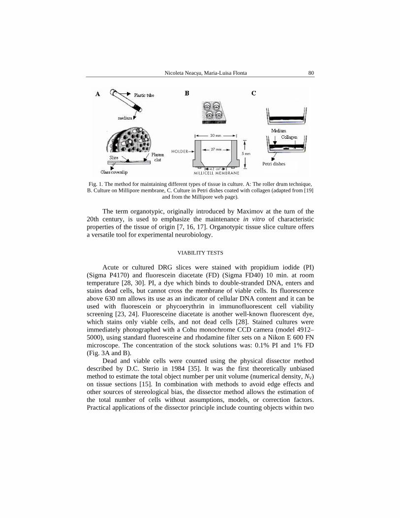
Liophilized plasma was purchased from SIGMA, St. Louis, MO, USA. The powder is reconstituted in sterile deionized water; the solution is centrifuged at 2500 g at 4 °C for 30 min, in order to exclude the resting fibrin in the plasma. This is the working solution for embedding the section. Adding some freshly prepared thrombin will coagulate plasma. Thrombin should be reconstituted with sterile deionized water and centrifuged too. Then the slices were embedded individually on glass coverslips in a plasma clot, transferred to plastic test tubes and cultured by means of the roller-tube technique. The purpose of slow rotation (10 rev/h) is to provide aeration and feeding of the cultures. It also optimizes the flattening and spreading of the slices. The cultures were fed with 1 ml of medium containing horse serum (10%), DMEM medium and penicilin/streptomicin 50 µg/ml. The cultures were fed weekly and survived in vitro for 1 month, in optimal cases up to 3 months (Fig. 1A) [16, 17, 18, 19].
Culture on millipore membrane
A 30 mm diameter, sterile, porous (0.4 µm), transparent and low-protein-binding membrane (Millicell-CM, Millipore) was used as a support for the explant (Fig. 1B). Because the membrane is transparent, it allows frequent observation of the cultures using light microscopy. The membrane has no autofluorescence and can thus be used for immunofluorescence staining procedures. The membranes were placed into a Petri dish containing 1 ml of the same medium used for the first technique. Stoppini first described this method in 1991, and details about it can be found in his article [34].
Culture on Petri dishes coated with collagen
Parsley et al. [31] described in 1998 another simple and inexpensive procedure for explant culture termed ‘thin slice culture’. For coating the plastic dishes and glass coverslips, rat-tail collagen (Sigma, St. Louis, MO, USA, C-8897) is used at a concentration of 50 mg/ml [20, 31]. Twenty-four hours prior to the culture experiment and under sterile conditions, 35 mm culture dishes (Corning) are coated with 0.5 ml of rat-tail collagen solution and allowed to dry in a culture hood for 4–6 h. The dishes are rinsed twice with 1 ml of sterile deionized water, and then soaked overnight in 1 ml of the same medium used for the first technique, in an incubator at 37 °C, in an atmosphere of 5% CO2 and 95% air. On the day of the experiment, the medium is replaced with 0.5 ml of new medium. Culture dishes are then kept in the incubator until tissue slices are ready to be placed in them (Fig. 1C).
Nicoleta Neacşu, Maria-Luisa Flonta
80
Fig. 1. The method for maintaining different types of tissue in culture. A: The roller drum technique, B. Culture on Millipore membrane, C. Culture in Petri dishes coated with collagen (adapted from [19]
and from the Millipore web page).
The term organotypic, originally introduced by Maximov at the turn of the 20th century, is used to emphasize the maintenance in vitro of characteristic properties of the tissue of origin [7, 16, 17]. Organotypic tissue slice culture offers a versatile tool for experimental neurobiology.
VIABILITY TESTS
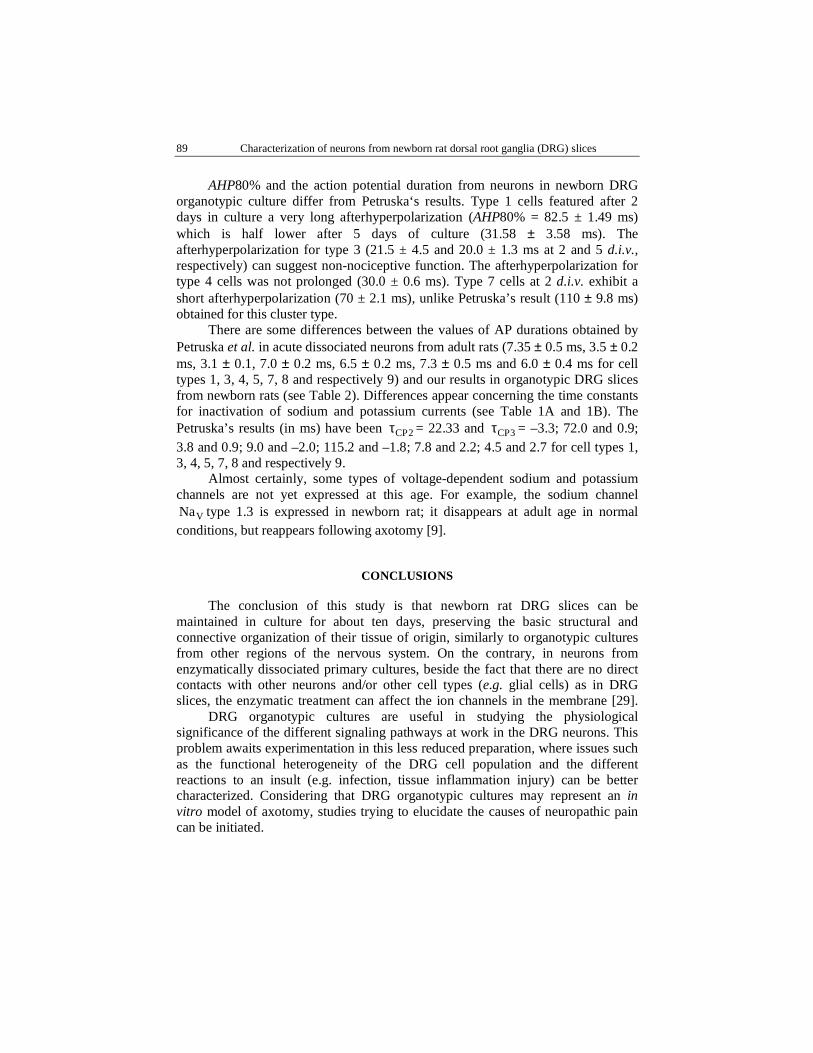
Acute or cultured DRG slices were stained with propidium iodide (PI) (Sigma P4170) and fluorescein diacetate (FD) (Sigma FD40) 10 min. at room temperature [28, 30]. PI, a dye which binds to double-stranded DNA, enters and stains dead cells, but cannot cross the membrane of viable cells. Its fluorescence above 630 nm allows its use as an indicator of cellular DNA content and it can be used with fluorescein or phycoerythrin in immunofluorescent cell viability screening [23, 24]. Fluoresceine diacetate is another well-known fluorescent dye, which stains only viable cells, and not dead cells [28]. Stained cultures were immediately photographed with a Cohu monochrome CCD camera (model 4912–5000), using standard fluoresceine and rhodamine filter sets on a Nikon E 600 FN microscope. The concentration of the stock solutions was: 0.1% PI and 1% FD (Fig. 3A and B).
Dead and viable cells were counted using the physical dissector method described by D.C. Sterio in 1984 [35]. It was the first theoretically unbiased method to estimate the total object number per unit volume (numerical density, NV) on tissue sections [15]. In combination with methods to avoid edge effects and other sources of stereological bias, the dissector method allows the estimation of the total number of cells without assumptions, models, or correction factors. Practical applications of the dissector principle include counting objects within two
Petri dishes
Characterization of neurons from newborn rat dorsal root ganglia (DRG) slices 81
physical planes (physical dissector), two optical planes (optical dissector), and within optical planes in conjunction with the fractionator-sampling scheme (optical fractionator) [26].
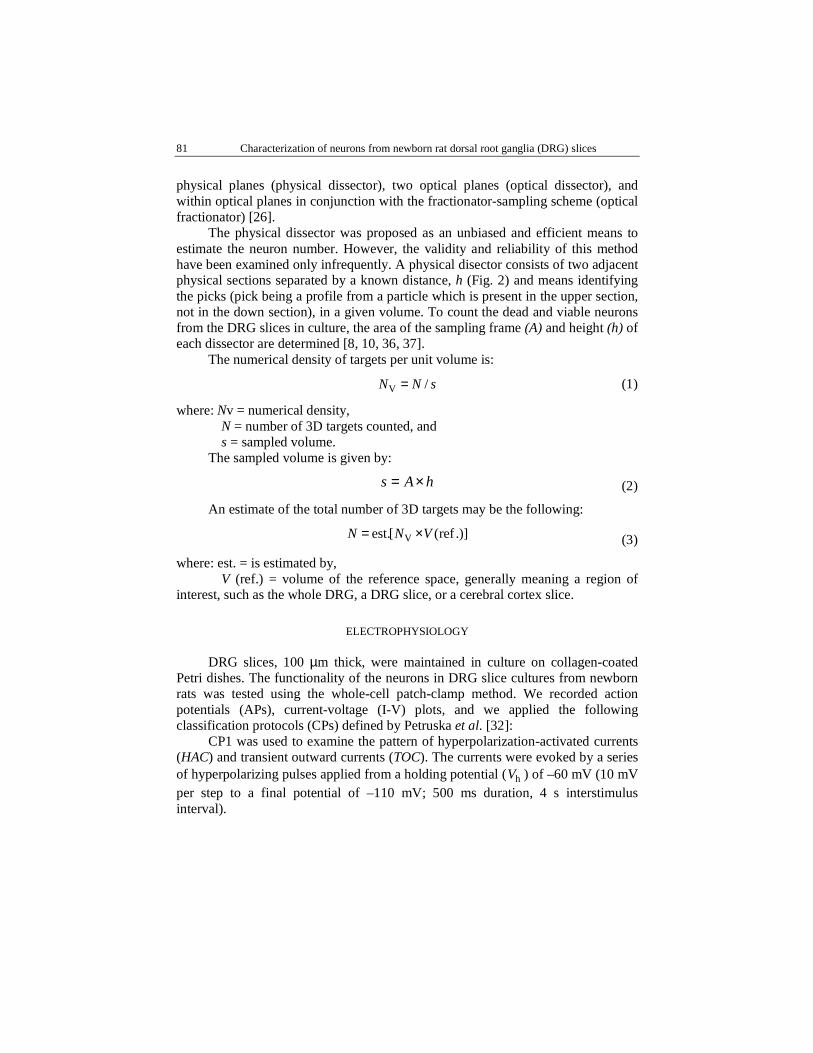
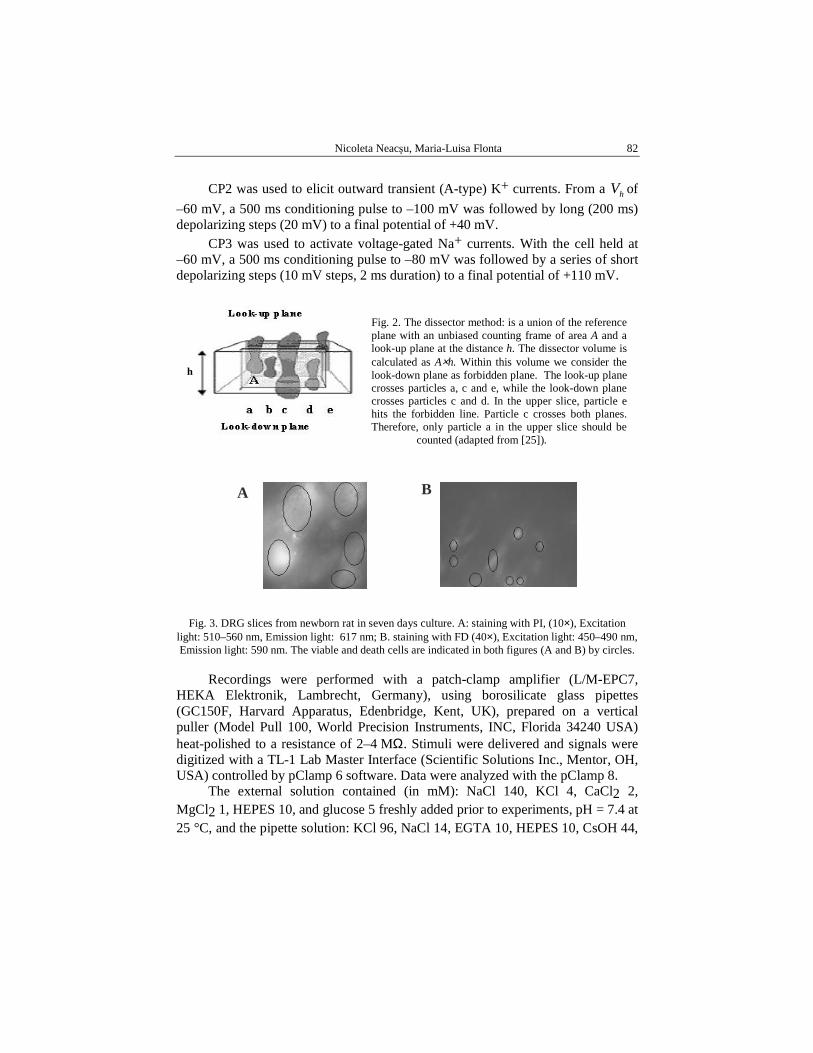
The physical dissector was proposed as an unbiased and efficient means to estimate the neuron number. However, the validity and reliability of this method have been examined only infrequently. A physical disector consists of two adjacent physical sections separated by a known distance, h (Fig. 2) and means identifying the picks (pick being a profile from a particle which is present in the upper section, not in the down section), in a given volume. To count the dead and viable neurons from the DRG slices in culture, the area of the sampling frame (A) and height (h) of each dissector are determined [8, 10, 36, 37].
The numerical density of targets per unit volume is:
V /N N s= (1)
where: Nv = numerical density, N = number of 3D targets counted, and s = sampled volume. The sampled volume is given by:
hAs ×= (2)
An estimate of the total number of 3D targets may be the following:
Vest.[ (ref .)]N N V= × (3)
where: est. = is estimated by, V (ref.) = volume of the reference space, generally meaning a region of
interest, such as the whole DRG, a DRG slice, or a cerebral cortex slice.
ELECTROPHYSIOLOGY
DRG slices, 100 µm thick, were maintained in culture on collagen-coated Petri dishes. The functionality of the neurons in DRG slice cultures from newborn rats was tested using the whole-cell patch-clamp method. We recorded action potentials (APs), current-voltage (I-V) plots, and we applied the following classification protocols (CPs) defined by Petruska et al. [32]:
CP1 was used to examine the pattern of hyperpolarization-activated currents (HAC) and transient outward currents (TOC). The currents were evoked by a series of hyperpolarizing pulses applied from a holding potential ( hV ) of –60 mV (10 mV per step to a final potential of –110 mV; 500 ms duration, 4 s interstimulus interval).
Nicoleta Neacşu, Maria-Luisa Flonta
82
h
A B
CP2 was used to elicit outward transient (A-type) K+ currents. From a hV of
–60 mV, a 500 ms conditioning pulse to –100 mV was followed by long (200 ms) depolarizing steps (20 mV) to a final potential of +40 mV.
CP3 was used to activate voltage-gated Na+ currents. With the cell held at –60 mV, a 500 ms conditioning pulse to –80 mV was followed by a series of short depolarizing steps (10 mV steps, 2 ms duration) to a final potential of +110 mV.
Fig. 3. DRG slices from newborn rat in seven days culture. A: staining with PI, (10×), Excitation light: 510–560 nm, Emission light: 617 nm; B. staining with FD (40×), Excitation light: 450–490 nm, Emission light: 590 nm. The viable and death cells are indicated in both figures (A and B) by circles.
Recordings were performed with a patch-clamp amplifier (L/M-EPC7, HEKA Elektronik, Lambrecht, Germany), using borosilicate glass pipettes (GC150F, Harvard Apparatus, Edenbridge, Kent, UK), prepared on a vertical puller (Model Pull 100, World Precision Instruments, INC, Florida 34240 USA) heat-polished to a resistance of 2–4 MΩ. Stimuli were delivered and signals were digitized with a TL-1 Lab Master Interface (Scientific Solutions Inc., Mentor, OH, USA) controlled by pClamp 6 software. Data were analyzed with the pClamp 8.
The external solution contained (in mM): NaCl 140, KCl 4, CaCl2 2, MgCl2 1, HEPES 10, and glucose 5 freshly added prior to experiments, pH = 7.4 at 25 °C, and the pipette solution: KCl 96, NaCl 14, EGTA 10, HEPES 10, CsOH 44,
Fig. 2. The dissector method: is a union of the reference plane with an unbiased counting frame of area A and a look-up plane at the distance h. The dissector volume is calculated as A×h. Within this volume we consider the look-down plane as forbidden plane. The look-up plane crosses particles a, c and e, while the look-down plane crosses particles c and d. In the upper slice, particle e hits the forbidden line. Particle c crosses both planes. Therefore, only particle a in the upper slice should be counted (adapted from [25]).

Characterization of neurons from newborn rat dorsal root ganglia (DRG) slices 83
pH = 7.2 at 25 °C. The external solution was continuously delivered under hydrostatic pressure via a homemade application system, with miniature solenoid valves into a constant flow chamber, at a rate of approximately 1 ml/min.
RESULTS
NEURONS VIABILITY
DRG slices maintained in culture using the three methods: the roller-drum technique, the culture on Millipore membrane and on Petri dishes coated with collagen, were used to compare them and to establish the best type of organotypic culture for DRG slices. The number of viable cells was counted in five cultures using the physical dissector method. Fig. 4 shows the neurons viability (in percents) depending on the culture age.
Fig. 4. DRG slice cultures viability expressed in percents of living cells mean ± SD of 5 experiments) for each culture method and culture age.
Fig. 4 shows that the number of viable neurons in DRG slices of newborn rat: • is higher on the slices cultured on plastic Petri dishes coated with collagen
(80–85%, after 6–8 days in culture); • is the lowest on the slices kept in culture on Millipore membrane (40–45%,
after 6–8 days in cultures)the neurons from DRG slices maintained in culture using the “Roller-Drum” technique have a 50–60% viability, after 6–7 days in culture.
Consequently, we have chosen for further investigation the culture method, using Petri dishes coated with collagen (Fig. 5).

Nicoleta Neacşu, Maria-Luisa Flonta
84
Fig. 5. DRG slices from newborn rat, after (A) 2 and (B) 5 days in culture on collagen-coated Petri dishes.
ELECTROPHYSIOLOGY
Neuron classification
Whole-cell patch-clamp recordings were performed on very small (diameter <20 µm) and cell capacitance between 20 − 45 pF, (31.65 ± 7.88 pF, mean ± SD), small (diameter 25 − 35 µm) and cell capacitance between 40 − 90 pF, (67 ± 16.28 pF), and medium-sized (diameter 35 − 55 µm) and cell capacitance between 75 − 148 pF, (102 ± 23.63 pF) neurons from DRG slices maintained for 2 or 5 days in culture. Classification of primary sensory neurons using the current signature method [29] used 37 neurons after 2 days in vitro (d.i.v) and 23 neurons after 5 d.i.v. The Petruska protocols 1, 2, and 3 were used to elicit distinct patterns of hyperpolarization- and depolarization-activated inward and outward currents (Fig. 6). Taking into account the cell size, the presence of hyperpolarization-activated current (HAC), type A potassium current, and sodium currents pattern we have done the first sorting of the neurons (Fig. 6).
Subsequently, five parameters of current amplitude and inactivation were determined: hyperpolarization activated current, transient outward current (TOC), decay time constant CP2τ , decay time constant CP3τ , threshold of A-current
activation (AT). The decay time constant CP2τ was derived from single or double exponential fits to the final outward current trace (+40 mV). The decay time constant CP3τ was derived from single exponential fits to the inactivation phase of the first complete inward current trace. When double exponential fits were required, the fastest component was used.
A B

Characterization of neurons from newborn rat dorsal root ganglia (DRG) slices 85
Table 1
A. Analysis of neurons from 2 d.i.v. newborn rat DRG slices (mean ± SD)
Cell Type
n Cell size
HAC (pA)
TOC (pA) CP2τ AT
(mV) CP3τ
1 5 small –12.32±1.4 –32.6±1.9 118.03±2.3 – 1.48±0.23
2 – small – – – – –
3 4 very small
–35±2.5 –209.78±3.5 136.58±4.56
– 1.028±0.3
4 5 medium –94±1.1 –139.8±3.7 2.54±0.2 –20 0.99±1.68
5 7 medium –42.57±3.8 –47±3.4 2.78±0.8 –20 2.09±1.2
6 – medium – – – – –
7 1 very small
–29±1.5 –20±1.5 68.40±3.2 – 0.97±2.2
8 3 medium –30±2.7 –73±0.3 7.3±1.1 –40 1.57±2.3
9 8 medium 6.5±0.5 –49±2.9 5.06±0.4 –40 2.78±0.89
B. Analysis of neurons from 5 d.i.v. newborn rat DRG slices (mean ± SD)
Cell Type
n Cell size HAC (pA)
TOC (pA) CP2τ AT
(mV) CP3τ
1 5 small –6.5±2.5 –43.2±4.2 28.33±2.8 – 1.18±1.2
2 – small – – – – –
3 12 very small –33.15±0.54 –65.8±1.6 196.71±1.46 – 1.36±4.5
4 5 medium –176±1.75 –83.2±2.7 6.25±1.02 –40 1.89±3.2
5 – medium – – – – –
6 – medium – – – – –
7 – very small – – – – –
8 – medium – – – – –
9 9 medium –16.71±2.56 –172±2.87 8.64±2.8 –20 2.24±1.3
Using Petruska’s criteria to classify DRG neurons according to patterns of
voltage-activated currents, neurons from slices after 2 d.i.v. did not feature the type 2, belonging to nociceptors, and the type 6 (Table 1A). Cultures after 5 d.i.v. were lacking the types 2, 5, 6, 7 and 8 of cells, possibly because only 23 neurons were used for recordings during the 5th day of culture (Table 1B).
Medium-sized cells show a large variety of cell types, and A-type potassium currents appeared at depolarization in the range of –40 to –20 mV.

Nicoleta Neacşu, Maria-Luisa Flonta
86
Fig. 6. Current signatures of neurons from dorsal root ganglia (DRG) slices after 2 and 5 days cultures. Cell types 2 and 6 were not identified both in 2 and 5 d.i.v. Types 5, 7 and 8 were lacking
only after 5 d.i.v.
Protocol 1 Protocol 2 Protocol 3 A
– 60 mV
- 100 mV 500 ms
200 ms
+ 40 mV
– 80 mV 500 ms
+ 10 mV
2 ms – 60 mV
– 60 mV
– 110 mV 500 ms CP1 CP2 CP3 B
2 nA Type 1
100 pA
small
400 pA
Type 3 very small
100 pA
2 nA
400 pA
2 nA
500 pA
Type 4 medium
250 pA
500 pA Type 9 medium
100 pA
500 pA
500 pA medium Type 8
250 pA 500 pA
Type 5 medium
250 pA 5 nA
500 pA
200 pA
Type 7 very small
200 pA
500 pA

Characterization of neurons from newborn rat dorsal root ganglia (DRG) slices 87
Cell type 4, belonging to the medium range, presented the largest HAC, but on average it was 6.5 times smaller that the values reported by Petruska et al. for freshly dissociated adult rat DRG neurons. Similar low values were found in neurons from acute newborn rat DRG slices (unpublished data), suggesting that this channel is not very well expressed in newborn rat primary sensory neurons. All time constants for sodium current inactivation (CP3τ ) showed positive values, unlike Petruska’s results.
Action potential duration
Table 2
Estimation of action potential duration in 2 and 5 d.i.v. neurons, at 0 mV (APD 0 mV), when the AP waveform returned to baseline (approximately –60 mV) – (APDb), at threshold (APDt) and the AHP80%, which represents the time (t) required for the afterhyperpolarization to decay to 20% of its peak value (80% recovery). All data are reported as means ± S.D.
2 d.i.v. 5 d.i.v. Cell
type APD 0 mV (ms)
APDb (ms)
APDt (ms)
AHP80% (ms)
APD 0 mV (ms)
APDb (ms)
APDt (ms)
AHP80% (ms)
1 3.8±1.3 5.95±1.32 7.85±2.5 82.5±3.7 3.425±0.5 15.76±3.7 7.2±1.2 31.58±5.4
2 – – – – – – – –
3 2.8±0.5 12±0.65 7.5±1.7 21.05±3.8 3.35±1.2 14.24±1.59 7.4±0.56 20±2.5
4 1.3±4.6 11.6±3.4 10.1±3.1 30±2.6 1.85±0.36 10.36±1.6 6.3±3.4 21±0.58
5 3.05±2.6 13.3±0.4 6±0.25 23.9±0.98 – – – –
6 – – – – – – – –
7 2±1.8 14±2.69 7.9±1.4 70±5.14 – – – –
8 3.3±5.4 10.5±2.9 7.5±1.56 23.5±3.2 – – – –
9 7.26±0.2 12.12±3.5 7.76±0.3 36.8±1.9 3.02±2.1 14.4±4.2 12.28±2.3 29.47±4.1
We have measured the action potential duration at 0 mV (APD 0 mV), when
the AP waveform returned to baseline (approximately –60 mV) (APDb), at threshold (APDt), and the duration of 80% hyperpolarization recovery (AHP80%), for each neuron recorded after 2 and 5 days in culture.
After 2 days in culture, AHP80% for the cell type 1 was markedly prolonged, and it decreased to half after 5 days in culture. The afterhyperpolarization for cell type 3 (21 and 20 ms at 2 and 5 d.i.v., respectively) suggested a nociceptive function of this neuron type. AHP80% for cell type 4 at 2 days in culture was not so long (30 ms), and it was even shorter at 5 days in culture (20 ms). At 2 days in culture, cell type 7 have an AHP80% lasting only 70 ms (Table 2), half the value obtained by Petruska et al. in adult rats.

Nicoleta Neacşu, Maria-Luisa Flonta
88
DISCUSSION
Using sharp microelectrode recordings in vivo, Lawson and colleagues [14, 24] have distinguished nociceptive from non-nociceptive neurons, corresponding to Aδ and C fiber populations, although specific subtypes within nociceptive populations could not be distinguished using AHP80% in vivo. For this reason, Petruska et al. [32, 33] tried to improve nociceptor identification and to find specific linkages between classic nociceptive subpopulations and his subclassified cells.
Starting from the idea that DRG organotypic cultures may represent an in vitro model of axotomy, and aiming to minimize the number of sacrificed animals, we explored the possibility to obtain organotypic DRG cultures that can be maintained for several days, in order to test if the classification procedure of Petruska et al. can be applied to neurons that were not exposed to an enzymatic treatment. Our aim is to find an in vitro model to study the molecular consequences of axotomy and to develop more effective strategies for the treatment of neuropathic pain.
Viability studies were performed on five organotypic DRG cultures for each of the three different techniques: the roller drum technique, culture on Millipore membranes and culture on collagen-coated Petri dishes. Our results show clearly that the largest numbers of viable neurons were present on Petri dishes coated with collagen. In addition, the costs for this type of substrate are lower than for the other two techniques, and the time required for preparation is also shorter. For this reason, the following studies were performed on DRG slices cultured on collagen.
We did not find in 2 d.i.v. cultured slices neurons of the types 2 and 6. Cluster 2, which could not be identified in culture, in a pool of approximately 60 cells, belongs to a nociceptive class, and it was found in newborn rat acute DRG slices. Therefore, it is possible that this cluster disappeared in culture. Another explanation might be a failure to identify it because the number of recorded cells was not large enough to allow the identification of this cell type. The same hypotheses may explain the results after 5 days in culture, when several cell types (2, 5, 6, 7, and 8) were not identified, but the total number of recorded neurons was even smaller.
Because within the present study we performed a cluster analysis according only to the patterns of voltage-activated currents (current signatures) and AP duration, skipping the algesic and immunohistochemical profile, it is possible that this classification is not complete. Another important issue is that the HAC obtained by us in cluster 4 neurons from newborn rat DRG slices is on average 6.5 times smaller than the values reported by Petruska et al. [32, 33]. This suggests that hyperpolarization-activated channels are under expressed in newborn rat primary sensory neurons.
Characterization of neurons from newborn rat dorsal root ganglia (DRG) slices 89
AHP80% and the action potential duration from neurons in newborn DRG organotypic culture differ from Petruska‘s results. Type 1 cells featured after 2 days in culture a very long afterhyperpolarization (AHP80% = 82.5 ± 1.49 ms) which is half lower after 5 days of culture (31.58 ± 3.58 ms). The afterhyperpolarization for type 3 (21.5 ± 4.5 and 20.0 ± 1.3 ms at 2 and 5 d.i.v., respectively) can suggest non-nociceptive function. The afterhyperpolarization for type 4 cells was not prolonged (30.0 ± 0.6 ms). Type 7 cells at 2 d.i.v. exhibit a short afterhyperpolarization (70 ± 2.1 ms), unlike Petruska’s result (110 ± 9.8 ms) obtained for this cluster type.
There are some differences between the values of AP durations obtained by Petruska et al. in acute dissociated neurons from adult rats (7.35 ± 0.5 ms, 3.5 ± 0.2 ms, 3.1 ± 0.1, 7.0 ± 0.2 ms, 6.5 ± 0.2 ms, 7.3 ± 0.5 ms and 6.0 ± 0.4 ms for cell types 1, 3, 4, 5, 7, 8 and respectively 9) and our results in organotypic DRG slices from newborn rats (see Table 2). Differences appear concerning the time constants for inactivation of sodium and potassium currents (see Table 1A and 1B). The Petruska’s results (in ms) have been CP2τ = 22.33 and CP3τ = –3.3; 72.0 and 0.9; 3.8 and 0.9; 9.0 and –2.0; 115.2 and –1.8; 7.8 and 2.2; 4.5 and 2.7 for cell types 1, 3, 4, 5, 7, 8 and respectively 9.
Almost certainly, some types of voltage-dependent sodium and potassium channels are not yet expressed at this age. For example, the sodium channel
VNa type 1.3 is expressed in newborn rat; it disappears at adult age in normal conditions, but reappears following axotomy [9].
CONCLUSIONS
The conclusion of this study is that newborn rat DRG slices can be maintained in culture for about ten days, preserving the basic structural and connective organization of their tissue of origin, similarly to organotypic cultures from other regions of the nervous system. On the contrary, in neurons from enzymatically dissociated primary cultures, beside the fact that there are no direct contacts with other neurons and/or other cell types (e.g. glial cells) as in DRG slices, the enzymatic treatment can affect the ion channels in the membrane [29].
DRG organotypic cultures are useful in studying the physiological significance of the different signaling pathways at work in the DRG neurons. This problem awaits experimentation in this less reduced preparation, where issues such as the functional heterogeneity of the DRG cell population and the different reactions to an insult (e.g. infection, tissue inflammation injury) can be better characterized. Considering that DRG organotypic cultures may represent an in vitro model of axotomy, studies trying to elucidate the causes of neuropathic pain can be initiated.

Nicoleta Neacşu, Maria-Luisa Flonta
90
Acknowledgements. N.N. thanks Dr. Florentina Pena for teaching her the DRG slice protocol, Dr. Bogdan Amuzescu for good advices and Dan Zorzon for technical support. She is also grateful to Dr. Florentina Pluteanu and Dr. Violeta Ristoiu for showing her the meaning of teamwork.
R E F E R E N C E S
1. BACCAGLINI, PAOLA, P.C. HOGAN, Some rat sensory neurons in culture express characteristics of differentiated pain sensory cells, Proc. Natl. Acad. Sci., 1983, 80, 594–598.
2. BELMONTE, C., F. GIRALDES, Responses of cat corneal sensory receptors to mechanical and thermal stimulation, J. Physiol., 1982, 321, 355–378.
3. BELMONTE, C., R. GALLEGO, Membrane properties of cat sensory neurones with chemoreceptor and baroreceptor endings, J. Physiol. (Lond.), 1983, 342, 603–614.
4. BRASCHLER, U.F., C. SPENGER, H.R. LUSCHER, A modified rollertube-technique for organotypic tissue cultures of rat spinal cord, Neurochem. Int., 1988, Suppl. 1, 77, Abstr. R16, 2.
5. CAMPBELL, J.N., S.N. RAJA, R.H. COHEN, C.H. MANNING, R.A. MAJER, Peripheral neural mechanisms of nociception, in: Textbook of Pain, P.D. Wall, R. Melzack eds., Churchill Livingstone, New York, 1990, pp. 22–45.
6. CERVERO, F., Sensory innervation of the viscera: peripheral basis of visceral pain, Physiol. Rev., 1994, 74, 95–134.
7. CRAIN, S.M., Development of "organotypic" bioelectric activities in central nervous tissues during maturation in culture, Int. Rev. Neurobiol., 1966, 9, 1–43.
8. CRUZ-ORIVE, L., E.R. WEIBEL, Recent stereological methods for cell biology: a brief survey, Am. J. Physiol., 1990, 258 (Lung Cell. Mol. Physiol. 2), L148–L156.
9. CUMMINS, T.R., F. AGLIECO, M. RENGANATHAN, R.I. HERZOG, S.D. DIB-HAJJ, S.G. WAXMAN, Nav1.3 sodium channels: rapid repriming and slow closed-state inactivation display quantitative differences after expression in a mammalian cell line and in spinal sensory neurons, J. Neurosci., 2001, 21 (16), 5952–5961.
10. COGGESHALL, R.E., H.A. LENKAN, Methods for determining numbers of cells and synapses: a case for more uniform standards of review, J. Comp. Neurol., 1996, 364, 6–15.
11. COOPER, B.Y., B.J. SESSLE, Anatomy and physiology of the trigeminal system, in: The Headaches, J. Olwson, P. Tfelt-Hansen, K.M.A. Welch eds., Raven, New York, 1993, pp. 87–93.
12. DARIAN-SMITH, I., The sense of touch: performance and peripheral neural processes, in: Handbook of Physiology. The Nervous System. Sensory Processes, Am. Physiol. Soc., Washington, D.C., 1984a., sect.1, vol. III, pp. 739–788.
13. DARIAN-SMITH, I., Thermal sensibility, in: Handbook of Physiology. The nervous system. Sensory processes, Am. Physiol. Soc., Washington, D.C., 1984b., sect.1, vol. III, pp. 879–913.
14. DJOUHRI, L., L. BLEAZARD, S.N. LAWSON, Association of somatic action potential shape with sensory receptive properties in guinea-pig dorsal root ganglion neurons, J. Physiol., 1998, 513, 857–872.
15. GEONISMAN, Y., H.J.G. GUNDERSEN, E. VAN der ZEE, M.J. WEST, Unbiased stereological estimation of the total number of synapses in a brain region, J. Neurocytology, 1996, 25, 805–819.
16. GAHWILER, B.H., Organotypic monolayer cultures of nervous tissue, J. Neurosci. Methods, 1981, 4, 329–342.
17. GAHWILER, B.H., Slice cultures of cerebellar, hippocampal and hypothalamic tissue, Experientia, 1984, 40, 235–244.
18. GAHWILER, B.H., Organotypic cultures of neural tissue, Trends Neurosci., 1988, 11 , 484–489. 19. GAHWILER, B.H., M. COPOGNA, R.A. McKINNEY, S.M. THOMSON, Organotypic slice culture:
a technique has come of age, Trends Neurosci., 1997, 20, 471–477. 20. ELSDALE, T., J. BARD, Collagen substrata for studies on cell behavior, J. Cell Biol., 1972, 54,
626-37.
Characterization of neurons from newborn rat dorsal root ganglia (DRG) slices 91
21. FITZGERALD, S.C., Dissociated spinal cord-dorsal root ganglion cultures on plastic tissue culture dishes and glass coverslips and wells, in: A dissection and tissue culture manual of the nervous system, A. Shahar et al. eds., Alan R. Liss Inc., New York, 1989, pp. 219–22.
22. JULIUS, D., A.I. BASBAUM, Molecular mechanisms of nociception, Nature, 2001, 413, 203–210. 23. LAAKE, J.H., F-M. HAUG, T. WIELOCH, O.P. OTTERSEN, A simple in vitro model of ischemia
based on hippocampal slice cultures and propidium iodide fluorescence, Brain Res. Protocols, 1999, 4, 173–184.
24. LAWSON, S.N., Phenotype and function of somatic afferent nociceptive neurons with C-, A delta- or A alpha/beta-fibres, Exp. Physiol., 2002, 87, 239-244.
25. MACKLIS, J.D., R.D. MADISON, Progressive incorporation of propidium iodide in cultured mouse neurons correlates with declining electrophysiological status: a fluorescence scale of membrane integrity, J. Neurosci. Methods, 1990, 31, 43–46.
26. MANDARIM-DE-LACERDA, C.A., Stereological tools in biomedical research, Annals of the Brazilian Academy of Sciences, 2003, 74 (4), 469–486.
27. MENSE, S., Nociception from skeletal muscle in relation to clinical muscle pain, Pain, 1993, 54, 241–289.
28. MONETTE, R., D.L. SMALL, G. MEALING, P. MORLEY, A fluorescence confocal assay to assess neuronal viability in brain slices, Brain Res. Protocols, 1998, 2, 99–108.
29. NEAGA E., B. AMUZESCU, CRISTINA DINU, BEATRICE MACRI, MARIA-LUISA FLONTA, Extracellular trypsin increases ASIC1a selectivity for monovalent versus divalent cations, J. Neurosci. Methods, 2005, 144, 241–248.
30. NORABERG, J., B.W. KRISTENSEN, J. ZIMMER, Markers for neuronal degeneration in organotypic slice cultures, Brain Res. Protocols, 1999, 3, 278–290.
31. PARSLEY, C.P., K.W. CHENG, L. SONG, S. HOCHMAN, Thin slice CNS explants maintained on collagen-coated culture dishes, J. Neurosci. Methods, 1998, 80, 65–74
32. PETRUSKA, J.C., J. NAPAPORN, R.D. JOHNSON, G.J. GU, B.Y. COOPER, Subclassified acutely dissociated cells of rat DRG: histochemistry and patterns of capsaicin-, proton-, and ATP-activated currents, J. Neurophysiol., 2000, 84, 2365–2379.
33. PETRUSKA, J.C., J. NAPAPORN, R.D. JOHNSON, G.J. GU, B.Y. COOPER, Chemical responsiveness and histochemical phenotype of electrophysiologically classified cells of the adult rat dorsal root ganglion, Neuroscience, 2002, 115, 15–30.
34. STOPPINI, L., P.A. BUCHS, D.A. MULLER, A simple method for organotypic cultures of nervous tissue, J. Neurosci. Methods, 1991, 37, 173–82.
35. STERIO, D.C., The unbiased estimation of number and size of arbitrary particles using the dissector, J. Microsc., 1984, 134, 127–136.
36. WEIBEL, E.R., Stereological methods, in: Practical Methods for Biological Morphometry, vol. 1, Academic Press, London, 1979.
37. WEST, M.J., New stereological methods for counting neurons, Neurobiol. Aging, 1993, 14, 275–285.