Class Nematoda. General characters Cylindrical round worms with tapering ends Separate sex, the...
15
Class Nematoda
-
Upload
cleopatra-goodwin -
Category
Documents
-
view
214 -
download
0
Transcript of Class Nematoda. General characters Cylindrical round worms with tapering ends Separate sex, the...
Class Nematoda
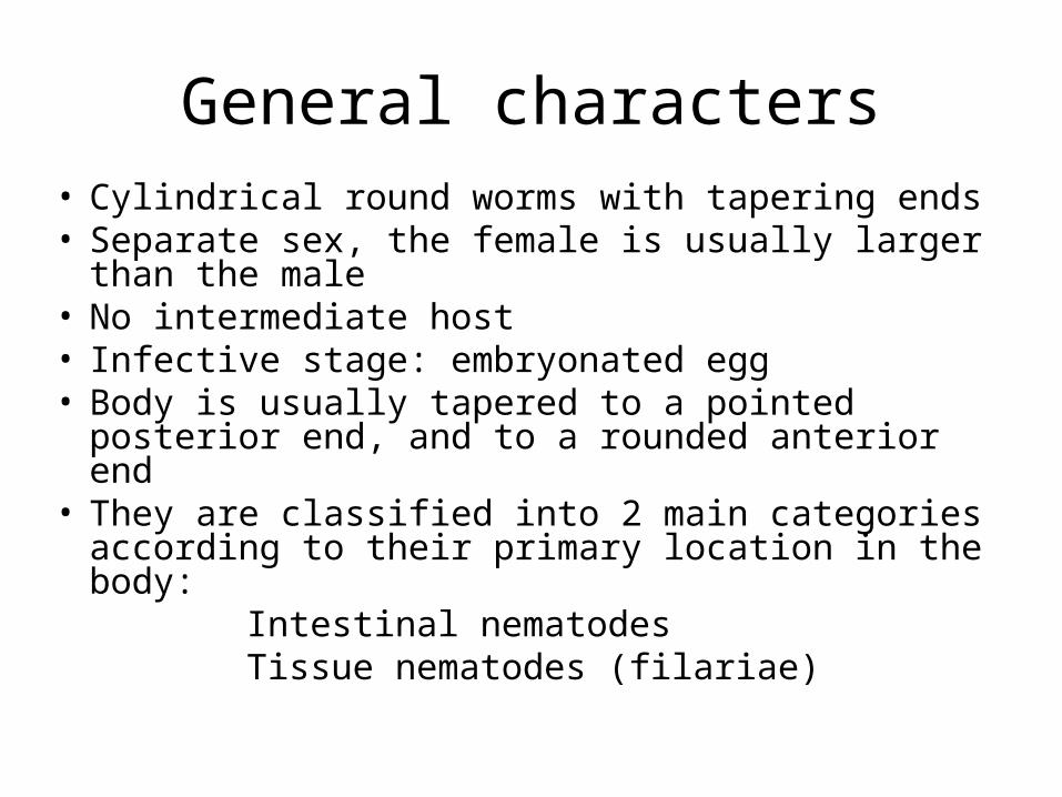
General characters• Cylindrical round worms with tapering ends• Separate sex, the female is usually larger than
the male• No intermediate host• Infective stage: embryonated egg• Body is usually tapered to a pointed posterior
end, and to a rounded anterior end• They are classified into 2 main categories
according to their primary location in the body: Intestinal nematodes Tissue nematodes (filariae)
Examples of Class Nematoda
• Intestinal nematodes:
Trichuris trichiura
Ascaris lumbricoides
Enterobius vermicularis
Anclystoma duodenale
Trichinella spiralis
• Tissue nematodes: Lymphatic filariae
Wuchereria bancrofti
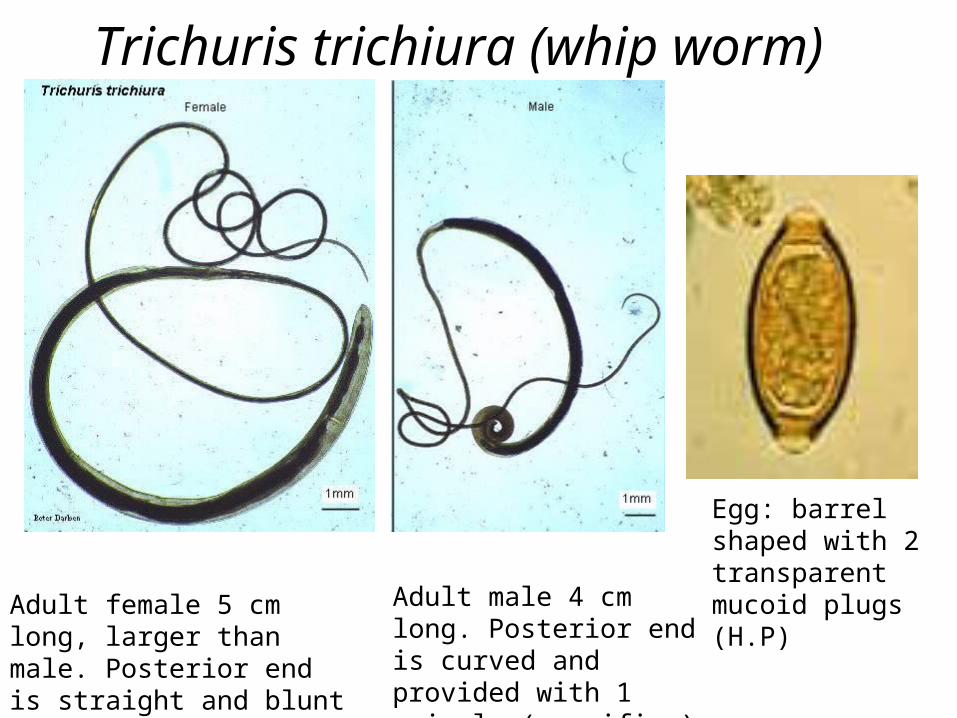
Trichuris trichiura (whip worm)
Adult male 4 cm long. Posterior end is curved and provided with 1 spicule (magnifier)
Adult female 5 cm long, larger than male. Posterior end is straight and blunt . (magnifier)
Egg: barrel shaped with 2 transparent mucoid plugs (H.P)

• Location of adult: large intestine of man
• Infective stage : embryonated egg
• Mode of transmission: ingestion of food contaminated with embryonated eggs
• Diagnosis: eggs in stool
• Disease: Trichuriasis
Trichuris trichiura
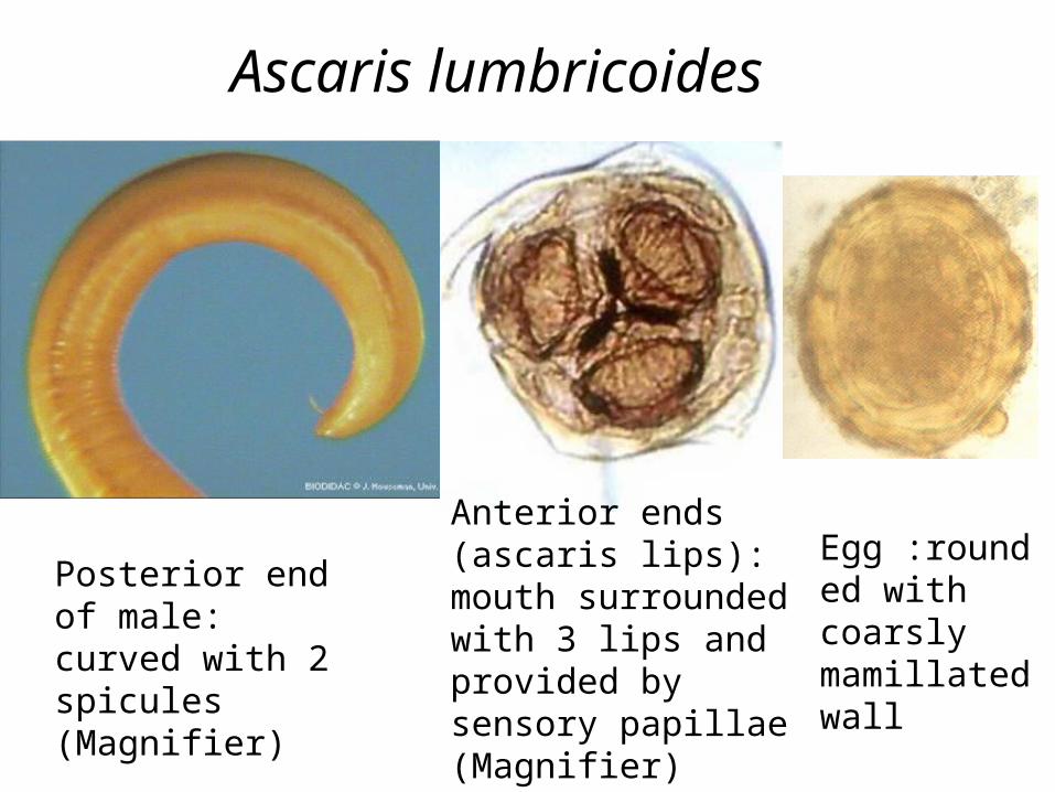
Ascaris lumbricoides
Anterior ends (ascaris lips): mouth surrounded with 3 lips and provided by sensory papillae (Magnifier)
Posterior end of male: curved with 2 spicules (Magnifier)
Egg :rounded with coarsly mamillated wall
Ascaris lumbricoides• Location of adult: Small intestine of
man
• Infective stage: embryonated egg
• Mode of transmission: ingestion of food (green vegetables) contaminated with embryonated egg
• Diagnosis: eggs in stool
• Disease: Ascariasis
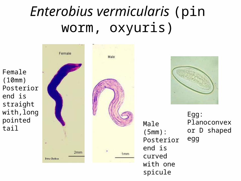
Enterobius vermicularis (pin worm, oxyuris)
Male (5mm): Posterior end is curved with one spicule
Female (10mm) Posterior end is straight with,long pointed tail Egg:
Planoconvex or D shaped egg
Enterobius vermicularis (pin worm, oxyuris)
• Location: large intestine of man• Infective stage: embryonated egg• Mode of transmission: ingestion of food
contaminated with embryonated egg or autoinfection via nails scratching the perianal region
• Diagnosis: eggs in anal or perianal swab collected using transparent adhesive tape. rarely in stool
• Disease: Enterobiasis
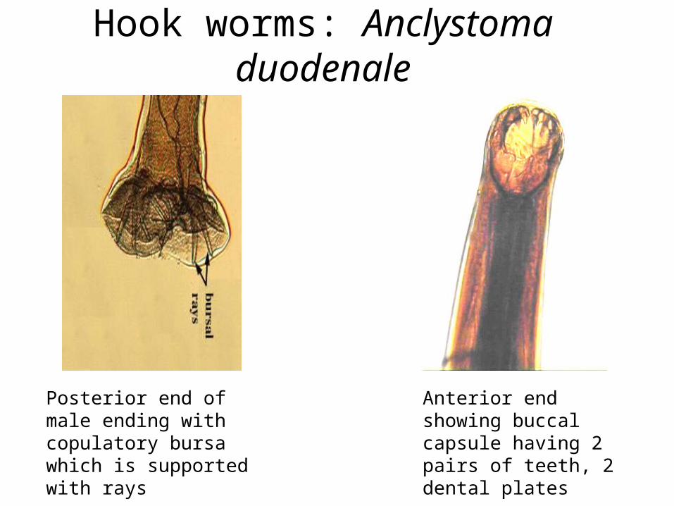
Hook worms: Anclystoma duodenale
Posterior end of male ending with copulatory bursa which is supported with rays
Anterior end showing buccal capsule having 2 pairs of teeth, 2 dental plates
Anclystoma duodenale
• Location of adult: small intestine of man
• Infective stage: filariform larvae
• Mode of transmission: penetration of filariform larvae in skin through bare feet
• Diagnosis: eggs in stool
• Disease: Hook worm infection
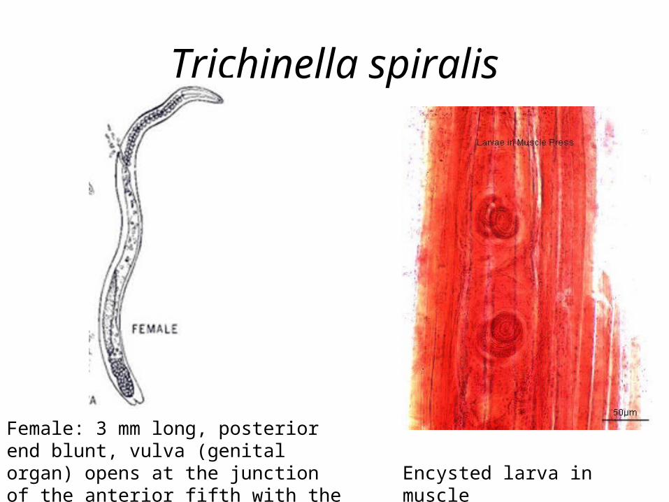
Trichinella spiralis
Female: 3 mm long, posterior end blunt, vulva (genital organ) opens at the junction of the anterior fifth with the rest of the body
Encysted larva in muscle
Trichinella spiralis
• Location of adult: small intestine of man• Location of larvae: encysted in striated muscles• Infective stage: encysted larvae in striated
muscle • Mode of transmission: ingestion of undercooked
pork containing encysted larvae• Diagnosis: muscle biopsy to identify larvae in
striated muscles• Disease: Trichinosis
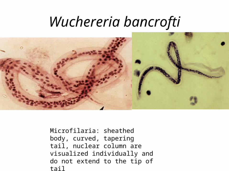
Wuchereria bancrofti
Microfilaria: sheathed body, curved, tapering tail, nuclear column are visualized individually and do not extend to the tip of tail
Wuchereria bancrofti
• Location of adult: lymphatics and lymph nodes• Infective stage: infective filariform larvae in the
mouth parts of mosquito• Vector (intermediate host):
mosquito ( Anopheles or Culex sp). • Mode of transmission: bite of mosquito having
infective filariform larvae • Diagnosis: Microfilaria in blood film• Disease: lymphatic filariasis