Class III b-Tubulin Expression in Sensory and Nonsensory...
16
Class III b-Tubulin Expression in Sensory and Nonsensory Regions of the Developing Avian Inner Ear DAVID MOLEA, 1,2 JENNIFER S. STONE, 1 AND EDWIN W RUBEL 1,2 * 1 Virginia Merrill Bloedel Hearing Research Center, and Department of Otolaryngology-HNS, University of Washington, Seattle, Washington 98195–7923 2 Department of Physiology and Biophysics, University of Washington, Seattle, Washington 98195–7923 ABSTRACT A previous study showed that class III b-tubulin, a widely used neuron-specific marker, is expressed in mature and regenerating hair cells but not the support cells of the avian inner ear. We investigated the expression of this marker in the developing avian inner ear. We found that class III b-tubulin is not neuron-specific in the avian embryo, but appears to accumulate in neuronal cell types, including hair cells, about the time of their differentiation. In the developing inner ear, some degree of class III b-tubulin immunoreactivity is found in all regions of the otic epithelium from its formation as the otic placode (stage 10 [embryonic day, E1.5]) until about stage 21 (E3.5), when the prospective tegmentum vasculosum begins to lose its staining. By stage 35 (E8–9), most of the nonsensory epithelia have lost their class III b-tubulin staining, leaving distinct regions of staining between the morphological compart- ments of the inner ear. Concurrent with the loss of staining from nonsensory regions, the hair cells of the sensory epithelia accumulate class III b-tubulin, whereas the supporting cells decrease their staining. We also observed a similar pattern of development in another hair cell organ, the paratympanic organ. Double labeling with the 275 kD hair cell antigen (HCA) indicated that the majority of hair cells identifiable with class III b-tubulin are HCA-positive. Additionally, presumptive hair cells were identified which were not within defined sensory epithelia. Our findings show that class III b-tubulin can be used as an early marker for hair cell differentiation in all hair cell sensory epithelia in the chicken. J. Comp. Neurol. 406:183–198, 1999. r 1999 Wiley-Liss, Inc. Indexing terms: chicken; cochlea; embryo; paratympanic organ; thyroid; vestibular The vertebrate inner ear develops from a neurogenic ectodermal thickening, the otic placode, which invaginates and separates from the ectoderm to form the otocyst. The otocyst subsequently develops into the morphologically complex and multifunctional membranous labyrinth con- sisting of approximately eight sensory epithelia and at least two specialized nonsensory epithelia (Rubel, 1978; Fritzsch et al., 1998). Several sensory structures, e.g., the cochlea, utricle, and semicircular canals, segregate into distinct spatial domains during development. At least two neuronal cell types are generated by the otic epithelium: the neurons of the acoustic-vestibular ganglion (AVG) which migrate from the otic epithelium to the ganglion (D’Amico-Martel and Noden, 1983), and sensory hair cells. The sensory hair cells appear to be generated within the otic epithelium after the ganglion cell precursors migrate (D’Amico-Martel, 1982; Katayama and Corwin, 1989). We do not know, however, whether the cells migrating to the AVG originate from the same regions in which sensory hair cells subsequently develop. The molecules and mechanisms involved in these devel- opmental processes are just beginning to be elucidated (e.g., Oh et al., 1996; Wu and Oh, 1996; Kiernan et al., 1997; Whitfield et al., 1997; Wu et al., 1998; reviewed in Fritzsch et al., 1998). Many molecules are being investi- gated and have shown distinct expression patterns within the otic epithelium. Bone morphogenetic protein-4 (BMP4), the mouse homolog of the Drosophila muscle segment Grant sponsor: NIH; Grant numbers: DC00395, DC00018, and DC00181. *Correspondence to: E.W Rubel, Virginia Merrill Bloedel Hearing Re- search Center, University of Washington, Box 357923, Seattle, WA98195– 7923. E-mail: [email protected] Received 28 May 1998; Revised 16 October 1998; Accepted 24 October 1998 THE JOURNAL OF COMPARATIVE NEUROLOGY 406:183–198 (1999) r 1999 WILEY-LISS, INC.
-
Upload
truongdang -
Category
Documents
-
view
221 -
download
0
Transcript of Class III b-Tubulin Expression in Sensory and Nonsensory...

Class III b-Tubulin Expression in Sensoryand Nonsensory Regions of the
Developing Avian Inner Ear
DAVID MOLEA,1,2 JENNIFER S. STONE,1 AND EDWIN W RUBEL1,2*1Virginia Merrill Bloedel Hearing Research Center, and Department of Otolaryngology-HNS,
University of Washington, Seattle, Washington 98195–79232Department of Physiology and Biophysics, University of Washington,
Seattle, Washington 98195–7923
ABSTRACTA previous study showed that class III b-tubulin, a widely used neuron-specific marker, is
expressed in mature and regenerating hair cells but not the support cells of the avian innerear. We investigated the expression of this marker in the developing avian inner ear. We foundthat class III b-tubulin is not neuron-specific in the avian embryo, but appears to accumulatein neuronal cell types, including hair cells, about the time of their differentiation. In thedeveloping inner ear, some degree of class III b-tubulin immunoreactivity is found in allregions of the otic epithelium from its formation as the otic placode (stage 10 [embryonic day,E1.5]) until about stage 21 (E3.5), when the prospective tegmentum vasculosum begins to loseits staining. By stage 35 (E8–9), most of the nonsensory epithelia have lost their class IIIb-tubulin staining, leaving distinct regions of staining between the morphological compart-ments of the inner ear. Concurrent with the loss of staining from nonsensory regions, the haircells of the sensory epithelia accumulate class III b-tubulin, whereas the supporting cellsdecrease their staining. We also observed a similar pattern of development in another hair cellorgan, the paratympanic organ. Double labeling with the 275 kD hair cell antigen (HCA)indicated that the majority of hair cells identifiable with class III b-tubulin are HCA-positive.Additionally, presumptive hair cells were identified which were not within defined sensoryepithelia. Our findings show that class III b-tubulin can be used as an early marker for haircell differentiation in all hair cell sensory epithelia in the chicken. J. Comp. Neurol.406:183–198, 1999. r 1999 Wiley-Liss, Inc.
Indexing terms: chicken; cochlea; embryo; paratympanic organ; thyroid; vestibular
The vertebrate inner ear develops from a neurogenicectodermal thickening, the otic placode, which invaginatesand separates from the ectoderm to form the otocyst. Theotocyst subsequently develops into the morphologicallycomplex and multifunctional membranous labyrinth con-sisting of approximately eight sensory epithelia and atleast two specialized nonsensory epithelia (Rubel, 1978;Fritzsch et al., 1998). Several sensory structures, e.g., thecochlea, utricle, and semicircular canals, segregate intodistinct spatial domains during development. At least twoneuronal cell types are generated by the otic epithelium:the neurons of the acoustic-vestibular ganglion (AVG)which migrate from the otic epithelium to the ganglion(D’Amico-Martel and Noden, 1983), and sensory hair cells.The sensory hair cells appear to be generated within theotic epithelium after the ganglion cell precursors migrate(D’Amico-Martel, 1982; Katayama and Corwin, 1989). Wedo not know, however, whether the cells migrating to the
AVG originate from the same regions in which sensory haircells subsequently develop.
The molecules and mechanisms involved in these devel-opmental processes are just beginning to be elucidated(e.g., Oh et al., 1996; Wu and Oh, 1996; Kiernan et al.,1997; Whitfield et al., 1997; Wu et al., 1998; reviewed inFritzsch et al., 1998). Many molecules are being investi-gated and have shown distinct expression patterns withinthe otic epithelium. Bone morphogenetic protein-4 (BMP4),the mouse homolog of the Drosophila muscle segment
Grant sponsor: NIH; Grant numbers: DC00395, DC00018, and DC00181.*Correspondence to: E.W Rubel, Virginia Merrill Bloedel Hearing Re-
search Center, University of Washington, Box 357923, Seattle, WA 98195–7923. E-mail: [email protected]
Received 28 May 1998; Revised 16 October 1998; Accepted 24 October1998
THE JOURNAL OF COMPARATIVE NEUROLOGY 406:183–198 (1999)
r 1999 WILEY-LISS, INC.

homeobox (msh) gene, Msx-1, and the p75 nerve growthfactor receptor (p75NGFR) have been used to elucidate thegeneration of the individual sensory epithelia (Wu and Oh,1996), and appear to be biochemically segregated shortlyafter the otocyst separates from the ectoderm (stages19–24 [embryonic day E2.9–E4]). Additionally, two ho-meobox genes, the sensory organ homeobox-1 (SOHo-1)and Gallus gallus homeobox protein-6 (GH6), have beenfound to be restricted to regions that develop into thesemicircular canals (Kiernan et al., 1997).
Until recently, the principle distinguishing feature ofhair cell differentiation has been the formation of stereo-cilia. Cotanche and Sulik (1984) showed that chicken haircells begin to morphologically differentiate at about stage29 (E6) in the distal basilar papilla (i.e., the chickencochlea), and that this morphological differentiation pro-ceeds in a distal-to-proximal gradient. More recently, ahair cell marker, the hair cell antigen (HCA), has beenfound to be expressed contemporaneously with stereociliadifferentiation, and used to investigate the differentiationof hair cells and formation of the hair cell mosaic in thedeveloping chick basilar papilla (Bartolami et al., 1991;Goodyear et al., 1995; Goodyear and Richardson, 1997).
Stone et al. (1996a) recently demonstrated that class IIIb-tubulin is expressed in normal and regenerating sensoryhair cells of the posthatch chicken inner ear, and can beused as a marker for the differentiation of hair cells duringdrug-induced hair cell regeneration. The TuJ1 antibody,which binds to the isotype-defining domain of class IIIb-tubulin (Lee et al., 1990), has been widely used to studydeveloping neurons and their precursors. This antibodyhas been used to study the differentiation, distribution,and migration of many neuronal cell types in variousregions of the nervous system including the telencephalon(Menezes and Luskin, 1994; Gates et al., 1995), olfactorybulb (Gonzalez and Silver, 1994), optic tectum (Snow andRobson, 1995), and the peripheral trigeminal system(Moody et al., 1989). In particular, Moody et al. (1989)showed that class III b-tubulin is expressed in cellsmigrating from the trigeminal placodes prior to and duringtheir migration to the trigeminal ganglion.
Given its usefulness as a hair cell differentiation markerin the sensory epithelium of the posthatch chicken, wedecided to investigate the expression of class III b-tubulinduring the early development of the chicken inner ear. Wefound that class III b-tubulin is not neuron-specific in thedeveloping avian embryo, but appears to accumulate inneuronal cell types about the time of their differentiation.In the developing inner ear, class III b-tubulin is found inall regions of the otic epithelium from its formation as theotic placode until about stage 21 (E3.5), when the prospec-tive tegmentum vasculosum begins to lose its staining. Bystage 35 (E8–9), most of the nonsensory epithelia have losttheir class III b-tubulin staining, leaving distinct regionsof staining between the utricle and saccule, saccule andcochlear duct, and in the outer circumference of thesemicircular canals. Concurrent with the loss of class IIIb-tubulin staining from nonsensory regions, the hair cellsof the sensory epithelia accumulate class III b-tubulin,whereas the supporting cells decrease their staining. Wealso observed a similar pattern of development in anotherhair cell organ, the paratympanic organ. Double-labelingwith antibodies to HCA and class III b-tubulin indicatedthat the majority of hair cells identifiable with class IIIb-tubulin are HCA-positive.Additionally, presumptive hair
cells were identified that were not located within definedsensory epithelia. A preliminary report of a portion of thisstudy has been presented in abstract form (Stone et al.,1996b).
MATERIALS AND METHODS
Fertilized White Leghorn chicken eggs (Gallus domesti-cus) were purchased from H & N International (Redmond,WA) and incubated at 38°C in a humidified, forced-draftincubator. Embryos were staged according to Hamburgerand Hamilton (1951). In this paper, the developmentalstage of the embryo will be referred to by both the stageand the corresponding age appropriate to the Hamburgerand Hamilton staging, e.g., stage 10 (E1.5). A total of 108embryos (stages 10 [E1.5] to 35 [E8–9]) were used in thisinvestigation. In general, 2–4 embryos at each age wereused for observations reported from tissue sections, and5–11 embryos at each age were used for wholemountpreparations. All the procedures were approved by theAnimal Care Committee of the University of Washington.
Antibodies
Two antibodies against class III b-tubulin, TuJ1 andb-III, were used in this study. The TuJ1 antibody is amonoclonal antibody that binds the N-terminal, isotype-defining domain of class III b-tubulin (Lee et al., 1990).The b-III antibody is a polyclonal antibody raised specifi-cally against the N-terminal, isotype-defining domain ofclass III b-tubulin (Moody et al., 1996; A. Frankfurter,personal communication). Labeling patterns and intensi-ties were identical with the two antibodies. Additionally,the HCA antibody was used to label developing hair cellsin wholemount preparations. TuJ1 and b-III were ob-tained from Dr. Anthony Frankfurter (University of Vir-ginia, Charlottesville, VA), and the HCA antibody wasobtained from Dr. Guy Richardson (University of Sussex,Falmer, Brighton, UK).
Fixation and immunohistochemistry
In the procedures described below, all washes in phos-phate-buffered saline (PBS) were done at pH 7.4, and allwashes in Tris were done at pH 7.6.
Sectioned embryos. Embryos used for paraffin sec-tions were immersion-fixed according to one of four fixa-tion protocols: (1) ice-cold 4% paraformaldehyde/0.1 Mphosphate buffer, pH 7.4 for 5–7 hours; (2) ice-cold 4%paraformaldehyde/0.1 M phosphate buffer, pH 7.4/3% su-crose for 21–23 hours; (3) ice-cold methacarn (1 part glacialacetic acid:3 parts chloroform:6 parts absolute methanol)for 4–6 hours; and (4) ice-cold buffered methacarn (1 partglacial acetic acid:2 parts chloroform:6 parts absolutemethanol: 1 part 103 modified chick Ringers; 103 modi-fied chick Ringers consists of 1.54 M NaCl, 60 mM KCl, 50mM HEPES, 10 mM EDTA at pH 7.4 after Lurie andRubel, 1994) for 4–5 hours. Whole embryos were removedfrom eggs and placed directly in fixative where the extra-embryonic membranes were removed. The entire embryowas then transferred to fresh fixative. When the fixationperiod was complete, paraformaldehyde-fixed tissue waswashed with PBS, and methacarn-fixed tissue was washedwith 70% ethanol. The embryos were then staged andembedded in paraffin. For embryos older than stage 21(E3.5), only the head region of the embryo was embedded.Whole-head, transverse sections, 6-µm- or 10-µm-thick,
184 D. MOLEA ET AL.
were cut through the hindbrain of the embryos (except forthree embryos which were sagittally sectioned). A one-in-five series of sections was mounted onto ethanol/HCl-washed, chrome-alum gelatin-subbed slides, and stainedwith hematoxylin and eosin in order to identify the otocyst.Sections encompassing the region of the otocyst weremounted onto ethanol/HCl washed, chrome-alum gelatin-subbed slides for immunohistochemistry. For methacarn-fixed embryos which were stage 25 (E4.5) or younger, allremaining sections encompassing the otocyst were used.Additionally, all remaining sections encompassing theotocyst were used for two stage 29 (E6) embryos and onestage 35 (E8–9) embryo. All other tissue was mounted in aone-in-five series.
Sections were deparaffinized, rinsed in 70% ethanol,then incubated for 15 minutes in absolute methanol con-taining 0.5% H2O2 to inhibit endogenous peroxidases.After rinsing and rehydration, nonspecific binding of sec-ondary antibody was blocked with a blocking solution of4% normal horse serum, 1% bovine serum albumin, 0.1%Triton X-100 in PBS. The sections were then incubatedovernight at 4°C in primary antibody, TuJ1 or b-III,diluted to 1:500, 1:1,000, or 1:3,000 in blocking solution.The next day, the sections were washed in PBS, incubated60 minutes in a 1:400 dilution of biotinylated horseanti-mouse IgG (Vector Laboratories, Burlingame, CA),washed in PBS, and treated for 60 minutes with theavidin-biotin-horseradish peroxidase complex (VectastainABC Elite kit; Vector Laboratories). The tissue was thentransferred to 50 mM Tris, preincubated with 0.375 mg/mlDAB (3,38-diaminobenzidine; Sigma, St. Louis, MO), andstained by using 0.375 mg/ml DAB/0.01% H2O2/50 mMTris for 3–10 minutes. The sections were coverslipped withDPX, and examined by using light microscopy with conven-tional or differential interference contrast optics.
Wholemounted embryos. Embryos used for whole-mount analysis were fixed according to two protocols: (1)direct immersion in ice-cold-buffered methacarn for 1hour, and (2) direct immersion in 4% paraformaldehydemade with a microtubule-stabilizing buffer, PHEM (50mM PIPES, 25 mM HEPES, 8 mM EGTA, 2 mM Mg11, 28mM Cl-, and 136 mM Na1; Troutt et al., 1994) with a finalpH of 7.3. These embryos were fixed for 1 hour at roomtemperature or 37°C to prevent cold destabilization of themicrotubules. The embryos were then washed in PBS andstaged.
After staging, whole embryos were immersed in ice-cold,70% ethanol for at least 10 minutes, then immersed in-20°C acetone for 7–10 minutes to improve permeabiliza-tion (D. Frost, personal communication). After the acetone,the embryos were reimmersed in ice-cold, 70% ethanol andallowed to warm to room temperature before being rehy-drated. (All incubations listed below that are greater than10 minutes were performed on a nutator at 4°C.) Thetissue was then incubated for 2 hours in blocking solution(4% normal horse serum, 1% bovine serum albumin, 0.1%saponin, PBS) with additional saponin added to raise theconcentration to 1%, and incubated for 36–48 hours inprimary antibody (TuJ1 or b-III) diluted to 1:1,000 inblocking solution. The secondary antibody, a 1:400 dilutionof biotinylated horse anti-mouse IgG (Vector Laboratories)in blocking solution, was applied for 36–48 hours. Next, anavidin-conjugated fluorophore (1:750 avidin-Bodipy (Mo-lecular Probes, Eugene, OR) or 1:1,000 avidin-Cy5 (Jack-son ImmunoResearch Laboratories, Inc., West Grove, PA))
diluted in blocking solution was applied for 24–36 hours.The tissue was then washed, cleared in a graded series ofbuffered glycerols, and mounted in an anti-fade solution of1% DABCO (1,4-diazabicyclo[2.2.2]octane; Sigma)/90%buffered glycerol/0.2% NaN3. The cleared embryos weremounted between two #1 coverslips, and imaged with aBioRad MRC-1024 confocal laser scanning microscoperunning BioRad LaserSharp software, version 2.1A. Theimages were imported into and analyzed by using publicdomain NIH image software (version 1.61; developed atthe U. S. National Institutes of Health and available onthe Internet at http://128.231.98.16/nih-image/), trans-ferred to Adobe Photoshop, and printed with a TektronixPhaser IISDX dye-sublimation printer.
In order to counterstain only the cell nuclei, someembryos were also treated for 1 hour with a 10-µg/mlsolution of RNAse A at 37°C to eliminate binding ofpropidium iodide to RNA, then incubated with a 10 µg/mlsolution of propidium iodide (Molecular Probes) for 1 hour.
Wholemounted otocysts. Cochlear ducts or otocystswere dissected from embryos fixed with room temperature,4% paraformaldehyde/PHEM or ice-cold, buffered meth-acarn as described above for whole embryos, and washedin PBS. The sensory epithelia were treated with 0.5% H2O2in PBS for 15 minutes to block endogenous peroxidaseactivity. After rinsing with PBS, 10% normal horse serumin 0.05% Triton X-100/PBS was applied for 20 minutes toblock nonspecific immunoglobulin binding. For single-labeling experiments, wholemounts were reacted with theTuJ1 monoclonal antibody (1:1,000) for 2 hours at roomtemperature or overnight at 4°C. Tissue was treated withBodipy/fluorescein isothiocyanate (FITC)-conjugated anti-rabbit IgG (1:300; Molecular Probes) for 30 minutes, orbiotinylated horse anti-mouse IgG (1:200; Vector Laborato-ries) for 30 minutes followed by the avidin-biotin-horserad-ish peroxidase reagent (ABC kit BA-2000; Vector Laborato-ries). Up to this point, all rinse steps were performed inPBS. The tissue incubated with the horseradish peroxi-dase (HRP) reagent was transferred to 50 mM Tris andtreated with 0.04% DAB/0.05% H2O2/Tris for 3–10 min-utes. The tissue was then rinsed once in Tris and stored inTris until cleared and mounted in 1% DABCO/90% buff-ered glycerol/0.2% NaN3. After treatment with the fluores-cent secondary antibody, wholemounts were washed inPBS, then cleared and mounted in 1% DABCO/90% buff-ered glycerol/0.2% NaN3.
For b-III/HCA colabeling, the b-III antibody was de-tected using Bodipy/FITC-conjugated anti-rabbit IgG (1:300; Molecular Probes), whereas the HCA antibody wasdetected in the same tissue with Cy5-conjugated anti-mouse IgG (1:300; Jackson ImmunoResearch Laborato-ries, Inc.).
Controls. The specificity of the antibody and immuno-histochemical protocol was determined by substitutingnonimmune, mouse sera in blocking solution, or justblocking solution, without addition of primary antibody.Staining of the brainstem (Moody et al., 1989; Lee et al.,1990) and retina (Lee et al., 1990; Watanabe et al., 1991;Snow and Robson, 1995) was used as within-tissue posi-tive controls.
RESULTS
Although very few studies have used the TuJ1 antibodyin paraffin-embedded tissue (Katsetos et al., 1993), we
CLASS III b-TUBULIN IN THE INNER EAR 185

chose to use paraffin sections in order to optimize morphol-ogy. Both of the antibodies to class III b-tubulin used inthis study, hereafter referred to as TuJ1 for the monoclonalantibody and b-III for the polyclonal antibody, providedspecific staining in paraffin-embedded tissue using eitherparaformaldehyde or methacarn fixation. The stainingwas judged to be specific because of: (1) the very lowbackground staining observed within experimental sec-tions; (2) the juxtaposition of heavily labeled regionsadjacent to regions with an absence of label in either thesame tissue (e.g., the otocyst), or between two differenttissues (e.g., otic epithelium and otic capsule); and (3) theabsence of staining in sections used as negative controls(i.e., incubated with nonimmune mouse sera or blockingsolution with no primary antibody added). Labeling wasalways observed in internal positive control tissues suchas retinal ganglion cells (Lee et al., 1990; Watanabe et al.,1991; Snow and Robson, 1995), peripheral nerve fibers(Moody et al., 1989), and the marginal and intermediatezones of the neural tube (Moody et al., 1989; Lee et al.,1990) whenever primary antibody was applied. Both ofthese antibodies consistently showed the same tissue-specific staining pattern and intensity, and were usedinterchangeably.
A comparison of paraffin sections from methacarn-fixedtissue against sections from paraformaldehyde-fixed tis-sue revealed that some regions of the otocyst that areclearly stained with TuJ1 or b-III in methacarn-fixedtissue, are often unstained or just barely stained when thetissue is fixed with paraformaldehyde (Fig. 1). The tissuesthat label with paraformaldehyde fixation, however, arealways a subset of the tissues that label with methacarnfixation. Neuronal elements in both the central and periph-eral nervous systems label regardless of fixation protocols.The degree of staining in paraformaldehyde-fixed tissuealso appears to be a function of fixation time. Embryos thatare fixed for short periods of time (, 7 hours) consistentlyshowed more staining than embryos fixed for longer times(. 20 hours) as shown in Figure 1B and C. These observa-tions suggest that paraformaldehyde fixation, when com-bined with paraffin embedding, reduces the binding ofclass III b-tubulin antibodies, resulting in the labeling ofonly those tissues containing the largest amounts of classIII b-tubulin. We did not observe any differences betweenwholemounted tissue fixed with methacarn or 1 hourfixation with 4% paraformaldehyde.
The following results are based primarily on observa-tions from methacarn-fixed paraffin sections and parafor-maldehyde-fixed wholemount preparations. Wholemountedpreparations were used to examine expression in theyoungest embryos (stage 14 and younger) and to look athair cell differentiation in the inner ear sensory epitheliawith and without double labeling with HCA. First, we willpresent an overview of the pattern of class III b-tubulinexpression in the developing membranous labyrinth fromstages 10 (E1.5) through 35 (E8–9), and then discuss thedevelopment of expression in sensory hair cells and thedeveloping sensory epithelia. We will then address theexpression of class III b-tubulin in another hair cellsensory organ, the paratympanic organ, and finally, theneuronal specificity of class III b-tubulin in the developingchick embryo. A later report will deal specifically with AVGcell differentiation and migration.
Fig. 1. Effects of fixation on the detection of class III b-tubulin. Theoverall staining with class III b-tubulin antibodies in the otic vesicle (OV)and neural tube (NT) is less distinct and less intense when the tissue isfixed in paraformaldehyde rather than methacarn, especially for longperiods of time. Some neuronal elements stain well regardless of fixation.A: Stage 16 (embryonic day, E2.2) embryo fixed for 4 hours in methacarnand reacted with the b-III antibody. B: Stage 17 (E2.4) embryo fixed for 6hours in 4% paraformaldehyde and reacted with the TuJ1 antibody.C: Stage 19 (E2.9) embryo fixed for 22.5 hours in 4% paraformaldehydeand reacted with TuJ1 antibody. Dorsal is up in A–C. Medial is to the rightinAand B, and to the left in C. Scale bar 5 50 µm forA–C.
186 D. MOLEA ET AL.

Class III b-tubulin expression in thedeveloping inner ear
Figure 2 illustrates the overall staining pattern for classIII b-tubulin at stages 10 (E1.5), 12 (E2.0), 16 (E2.2), 21(E3.5), 25 (E4.5), and 27 (E5) in the otocyst. In thethickened, pseudostratified regions of the otocyst, class IIIb-tubulin is heavily localized to the apical regions of thecells, resulting in dense staining in the lumenal region ofthe epithelium. Streaks of staining project into the morelightly labeled basal regions of the epithelium, where thecell nuclei are packed tightly together. This dense, lumenalstaining may be due to the displacement of cytoplasmlumenally by the closely packed, basally located nuclei. Asimilar pattern was reported by Troutt et al. (1994) using adifferent anti-b-tubulin antibody not specific for a particu-lar isotype. In many areas that are immunoreactive forclass III b-tubulin, the density of the cells and the densityof the staining prevents the resolution of individual cells.Thus, we can not determine whether or not all cells arelabeled in these regions, but clearly the majority arelabeled. In order to avoid repeating a description of thisstaining pattern, we will use the term ‘‘lumenal-streaky’’ torefer to blocks of epithelium which show dense apicalstaining and streaks of staining into the basal regions ofthe epithelium.
Frequently, lumenal-streaky staining is interrupted bylabeled mitotic spindles, especially during the early devel-opment of the otocyst (e.g., Fig. 2B and C). In some regionsof the otic epithelium, class III b-tubulin-labeled cells areseen next to or crossing the basal lamina. We believe thatthese cells are prospective ganglion cells migrating fromthe epithelium and will examine them more closely inanother report. At later stages, heavily labeled nerve fiberscan be seen penetrating and coursing through the epithe-lium, but are often indistinguishable from the streaksextending from the lumen when they are oriented radially.
As shown in Figure 2, class III b-tubulin is expressed inthe otic epithelium as early as the initial formation of theotic placode (stage 10 [E1.5]; Fig. 2A). From stage 10 (E1.5)to stage 25 (E4.5; Fig. 2B–E), expression of class IIIb-tubulin is detected in all regions of the otic epithelium.Figure 2D illustrates that, by stage 21 (E3.5), an abruptreduction in the density of staining can be seen at theventral tip and along the lateral wall of the nascentcochleosaccular duct, where the tegmentum vasculosum isexpected to develop (Knowlton, 1969; Cotanche and Sulik,1982). By stage 25 (E4.5; Fig. 2E) and stage 27 (E5; Fig.2F), staining in the lateral wall of the cochleosaccular ductis no longer detectable in the distal region of the duct, butbecomes increasingly more dense as one approaches theproximal region of the cochleosaccular duct. At laterstages, the abrupt transition in staining becomes morepronounced as the unstained lateral wall of the cochleosac-cular duct expands toward the utricle (Fig. 3; stage 29[E6]).
A second distinct region of reduced class III b-tubulinstaining, also marked by an abrupt transition in staining(bracket in Fig. 3 inset), is detected at about stage 29 (E6)in the nonsensory epithelia lateral and adjacent to theutricular macula. The endolymphatic sac and distal endo-lymphatic duct also show reduced class III b-tubulinstaining by stage 27 (E5; data not shown).
Between stage 27 (E5) and stage 35 (E8–9), manynonsensory regions of the epithelia lose their class III
b-tubulin staining. Nevertheless, dense lumenal-streakyclass III b-tubulin staining persists in certain regions ofthe nonsensory epithelia: (1) a wedge of cells, giving theimpression of a keystone in an arch, continues to labelheavily along the outer circumference of the semicircularcanals (Fig. 4A); (2) the pseudostratified epithelium at theconfluence of the endolymphatic duct, utricle, and sacculecontinues to label (arrows in Fig. 4B); and (3) the cellsadjacent to the basal tip of the basilar papilla (arrowheadin Fig. 4B), near to where the superior fibrocartilaginousplate will form, also continue to label. The nonsensorycharacter of this last region was confirmed in three ways:(1) examination of sequential sections at stage 35 (E8–9)when the proximal tip of the basilar papilla is well definedand contains hair cells; (2) observation that the tectorialmembrane arises from a subset of these labeled cells; and(3) examination of wholemounted otocyst containing boththe basilar papilla and saccular macula at stages 34–35(E8–9) in which the proximal tip of the basilar papilla iseasily identified by identification of class III b-tubulin-positive hair cells and morphology. In all cases, the nonsen-sory area between the saccular macula and basilar papillawas class III b-tubulin-positive and continuous with bothsensory epithelia (not shown).
Hair cell differentiation and sensoryepithelia development
In the developing sensory epithelia of the inner ear, bothantibodies differentially label a subset of lumenally placedcells. This subset of cells can be identified by their flask-shaped or gourd-shaped appearance, with the heavilylabeled neck of the gourd extending to the lumen. Figure5A shows that, by stage 29 (E6), the cell’s neck stands outagainst the less intense lumenal-streaky pattern that isstill present as this phenotype emerges. The unlabelednuclei are located close to the lumen and often partiallysurrounded by label or have a labeled tail extendingtowards the basal lamina. By stage 35 (E8–9), cells in thevestibular sensory epithelia with this phenotype also havestereocilia bundles (Fig. 5B). This morphological pheno-type is similar to the presumptive hair cells described byGinzberg and Gilula (1979) and Whitehead and Morest(1985a,b) in developing sensory epithelia in the chicken,and to differentiating hair cells described by Stone et al.(1996a) in regenerating chicken basilar papilla. In addi-tion, TuJ1 has been shown to label mature hair cells, butnot support cells, in the posthatch chicken basilar papilla(Stone et al., 1996a). We consider these gourd-shaped cellsto be, and will refer to them as, immature hair cells.
Prior to stage 29 (E6), we were not able to uniquelyidentify hair cells in paraffin-sectioned tissue due to thepresence of lumenal-streaky staining throughout the sen-sory epithelia. In wholemounted material reacted with theclass III b-tubulin antibodies, however, immature haircells can be identified by stage 26 (E4.5–5) because of theintense labeling of their apical cytoplasm. Identification ofthese cells as hair cells was confirmed by labeling withanti-HCA. Figure 6 shows a projection of a confocalz-series through the apical surface of a stage 26 (E4.5–5)saccular macula. The intense peaks of class III b-tubulinlabeling in the apical cytoplasm of the immature hair cellsare evident. The majority of class III b-tubulin-positivehair cells also label for HCA. HCA labeling was notobserved in class III b-tubulin-negative cells.
CLASS III b-TUBULIN IN THE INNER EAR 187

Figure 2
188 D. MOLEA ET AL.

By stage 29 (E6), class III b-tubulin expression canidentify immature hair cells in all of the vestibular sensoryepithelia, except the macula lagena and macula neglecta,in both paraffin sections and wholemount preparations. Asshown in Figures 5 and 6, immature hair cells are oftenpacked together such that their cell bodies appear to beeither in contact or overlapping. In the utricular macula,the immature hair cells are not evenly distributed. Thereis an increased density of immature hair cells laterally inthe region where the utricular striola will form, whereas inthe medial expanse of the macula, which will form the
nonstriolar region, immature hair cells appear more widelydistributed. The lateral grouping of immature hair cellssuggests that patterning of the utricular macula intostriolar and nonstriolar regions may already be occurring.However, without a way of determining the bundle orienta-tion, or Type I versus Type II hair cells, we cannotestablish this relationship with certainty. By stage 35(E8–9), the lumenal-streaky staining in the vestibularsensory epithelia has nearly disappeared, so that almostall the staining is confined to hair cells and nerve fibers.
Class III b-tubulin immunoreactivity can first identifyimmature hair cells in the distal basilar papilla at stage 29(E6; Fig. 7), when hair cells are first beginning to developstereocilia (Cotanche and Sulik, 1984), and first expressHCA (Bartolami et al., 1991; Goodyear et al., 1995; Good-year and Richardson, 1997). Again, the immature haircells are often packed tightly together (Fig. 7). By stage34/35 (E8–9), class III b-tubulin labeling shows immaturehair cells along the entire length of the basilar papilla,indicating that the differentiation of hair cells in thebasilar papilla with respect to class III b-tubulin occurs ina distal-to-proximal gradient as described for stereociliaformation (Cotanche and Sulik, 1984) and HCA labeling(Bartolami et al., 1991; Goodyear et al., 1995; Goodyearand Richardson, 1997).
Occasionally, isolated class III b-tubulin-positive cellsthat look like immature hair cells were observed in regionsof the otic epithelium not associated with a sensoryepithelium in both sectioned and wholemounted material.An example of one of these cells is shown in Figure 8.
Furthermore, in wholemount preparations, we detectednumerous HCA-positive cells which are also class III
Fig. 2. Class III b-tubulin staining in the otic epithelium at stages10 (embryonic day, E1.5, A), 12 (E2.0, B), 16 (E2.2, C), 21 (E3.5, D), 25(E4.5, E), and 27 (E5, F). A and B are horizontal confocal sections fromwholemounted embryos fluorescently labeled using TuJ1. Anterior isup in both A and B. C–F are transverse paraffin sections stained withb-III. Dorsal is up and medial is to the right in C–F. A: Stage 10 (E1.5)embryo showing the class III b-tubulin staining in the neural tube(NT) and both otic placodes (OP). B: Stage 12 (E2.0) otic pit (OP)showing the lumenal-streaky pattern of staining and labeled mitoticspindles (arrowheads). C: Stage 16 (E2.2) otic vesicle showing lumenal-streaky staining in all regions of the otic epithelium. Labeled mitoticspindles (arrowheads) are also located in all regions of the oticepithelium. D: Stage 21 (E3.5) otic vesicle showing lumenal-streakystaining in all regions of the otic epithelium. Note the abrupt change instaining at the ventromedial tip of the otic vesicle (arrow). The largeopen arrows in D–F indicate the acoustic-vestibular ganglion (AVG).E: Stage 25 (E4.5) inner ear showing staining in all regions of theepithelium except the ventrolateral wall of the cochlear duct where thetegmentum vasculosum will develop. F: Stage 27 (E5) inner earshowing that most regions of the otocyst are still labeled, except for thelateral wall of the cochlear duct. Scale bars 5 100 µm in A, D– F; 50 µmin B; 30 µm in C.
Fig. 3. Stage 29 (embryonic day, E6) cochlear duct (CD) showingthat the lateral wall of the cochlear duct is unlabeled, and thatstaining ends abruptly at the distal tip (arrow). Note the lumenal-streaky staining in the sensory epithelium. Inset: Stage 29 (E6)
utricular macula (UM) and lateral ampulla (LA) showing the unla-beled perimacular region (bracket). Scale bar 5 40 µm for main image;50 µm for inset.
CLASS III b-TUBULIN IN THE INNER EAR 189

b-tubulin-positive in regions outside of the identifiablevestibular sensory epithelia (data not shown). These cells,however, are typically located in regions near the estab-lished saccular and utricular maculae.
Class III b-tubulin expression in thedeveloping paratympanic organ
Class III b-tubulin is also expressed in another hair cellsensory organ, the paratympanic organ, in a similarmanner as in the otocyst. The paratympanic organ islocated in the middle ear of birds, develops from the firstepibranchial placode and first pharyngeal pouch, and isinnervated by the geniculate ganglion, which is also
Fig. 4. Persistent nonsensory labeling in the inner ear at stage 35(embryonic day, E8–9). A: The superior semicircular canal showing the‘‘keystone in an arch’’ labeling in its outer circumference (arrow).B: Persistent labeling (arrows) around the junction of the utricle (U),saccule (S), and endolymphatic duct (continuous with epithelium atupper right), and between the saccule and the proximal region(arrowhead) of the cochlear duct (CD; see text). Large open arrowindicates the cochlear ganglion; large filled arrow indicates the basilarpapilla. Dorsal is up and medial is to the right in both panels. Scalebars 5 30 µm in A; 50 µm in B.
Fig. 5. Class III b-tubulin labeling in hair cells. A: Gourd-shaped,class III b-tubulin-labeled immature hair cells (arrows) in the stage 29(embryonic day, E6) utricular macula. B: Gourd-shaped, class IIIb-tubulin-labeled immature hair cells with stereocilia (arrowhead) inthe stage 35 (E8–9) lateral crista. Open arrows indicate nerve fibers.Scale bars 5 10 µm.
190 D. MOLEA ET AL.
derived from the first epibranchial placode (D’Amico-Martel and Noden, 1983; von Bartheld, 1990). As shown inFigure 9A, class III b-tubulin-expressing cells from thefirst epibranchial placode fuse with the epithelium of thefirst pharyngeal pouch as early as stage 16 (E2.2). Atstages 21 (E3.5) and 25 (E4.5), the epithelium of thepharyngeal pouch, which is still in contact with the firstepibranchial placode, is labeled in the same lumenal-streaky pattern as found in the otocyst (Fig. 9B). By stage27 (E5), the paratympanic organ can be identified in thelateral pocket of the first pharyngeal pouch. Figure 9C
shows that, at stage 27 (E5), this organ contains distinctimmature hair cells.
Class III b-tubulin is not neuron-specificin the developing chick embryo
Previous studies have indicated that class III b-tubulinis not restricted to neuronal structures in the developingchick embryo. Class III b-tubulin expression has beenreported in the ectodermal layers of the extra-embryonicmembranes (Lee et al., 1990), trigeminal placodes (Moodyet al., 1989; Lee et al., 1990), lens (Lee et al., 1990), glomuscells of the carotid body (Kameda et al., 1994), C cells in theultimobranchial organ (Kameda et al., 1993; Kameda,1995), caudal-ventral somites (Lee et al., 1990), and caudalmesonephric duct (Lee et al., 1990). Our observations inthe developing head of the chick embryo corroborate thesefindings. Additionally, we observed that the non-neuronallabeling is much more widespread in methacarn-fixedtissue than in paraformaldehyde-fixed tissue. In additionto the structures indicated above, we found that TuJ1 andb-III consistently label a number of other structures:ectoderm, Rathke’s pouch, first and second epibranchialplacodes, olfactory placode, lens placode, mesenchyme ofthe branchial arches, tissue surrounding developing mem-branous bone, isolated cells in the mesenchyme immedi-ately underneath the ectoderm, the ventral wall of theembryonic pharynx, the dorsal and ventral walls of thefirst pharyngeal pouch, and the developing thyroid anlage.Some of these structures are illustrated in Figure 10 andlisted in Table 1.
We also found that class III b-tubulin expression can bedetected in the ectoderm and neural tube as early as stage9 (E1.3), which is earlier than previously reported (stage12 [E2.0]; Lee et al., 1990). Additionally, we observed that
Fig. 6. Confocal image showing class III b-tubulin-labeled imma-ture hair cells in the stage 26 (embryonic day, E4.5–5) saccularmacula. Immature hair cells form peaks of class III b-tubulin labeling(green), the majority of which colabel with hair cell antigen (HCA)labeling (red). A colabeled hair cell is indicated by the arrow. Thisimage is a projection of a 4-µm confocal z-series acquired by lookingdown on the saccular macula in a wholemounted otocyst. Scale bar 520 µm.
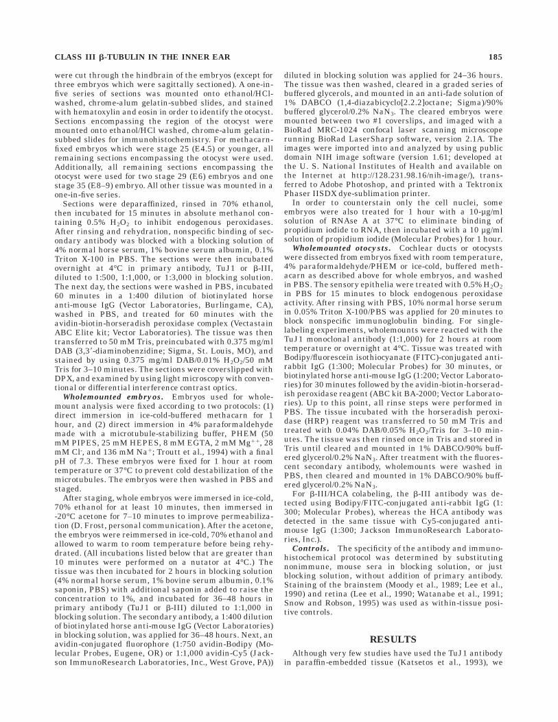
Fig. 7. Immature hair cells (arrows) emerging from the lumenal-streaky staining of the distal basilar papilla at stage 29 (embryonicday, E6). Note that cochlear ganglion fibers (open arrow) are beginningto penetrate the basilar papilla. Scale bar 5 20 µm.
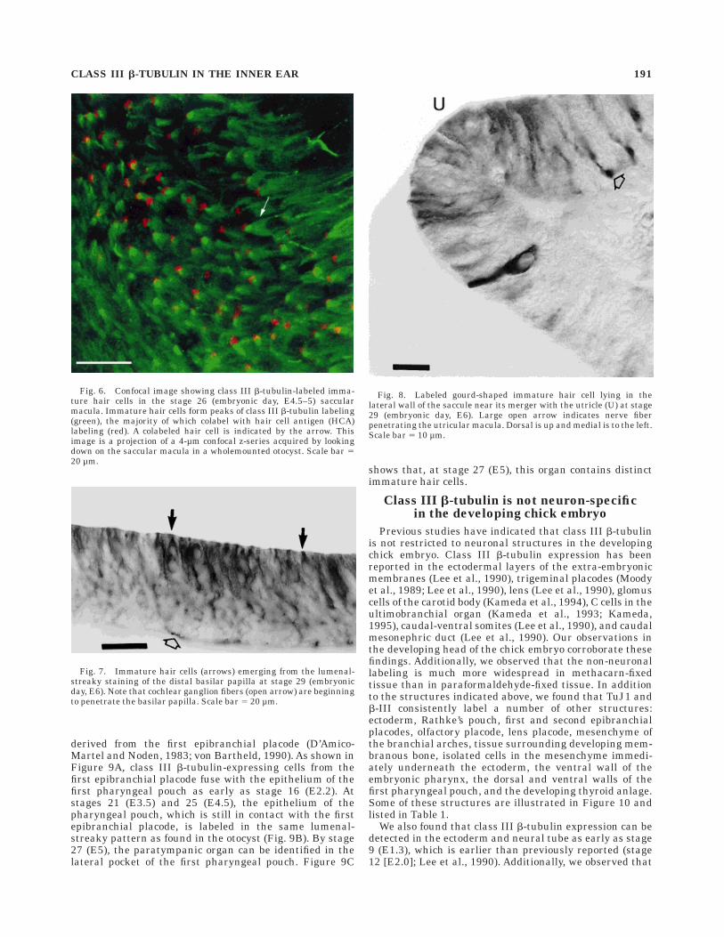
Fig. 8. Labeled gourd-shaped immature hair cell lying in thelateral wall of the saccule near its merger with the utricle (U) at stage29 (embryonic day, E6). Large open arrow indicates nerve fiberpenetrating the utricular macula. Dorsal is up and medial is to the left.Scale bar 5 10 µm.
CLASS III b-TUBULIN IN THE INNER EAR 191

TuJ1 and b-III typically produce the same lumenal-streaky pattern as we observed in the otic epithelium inother thickened epithelia expressing class III b-tubulin,including the proliferative zone of the neural tube, (seeFigs. 1, 2, and 10E). In the proliferative zone of the neuraltube, the staining is not limited to the trailing processes ofdensely stained cells migrating from the proliferativezone. Before stage 25 (E4.5), large regions of the prolifera-tive zone exhibit lighter, lumenal-streaky staining and thestaining appears to label all the cells in these regions.After stage 25 (E4.5), all the cells of the proliferative zoneappear to be labeled. Labeled mitotic spindles were alsoobserved in the proliferative zone of the neural tube as wellas the otic epithelium and ectoderm (data not shown).
DISCUSSION
A previous study showed that class III b-tubulin, awidely used neuron-specific marker, is expressed in ma-ture and regenerating hair cells but not the support cells ofthe avian inner ear (Stone et al., 1996a). In this study, weinvestigated the expression of this marker in the develop-ing avian inner ear. We found that class III b-tubulin is notneuron-specific in the developing avian embryo, but ap-pears to accumulate in neuronal cell types, including haircells, about the time of their differentiation. In the develop-ing inner ear, class III b-tubulin immunoreactivity wasfound in all regions of the otic epithelium from its forma-tion as the otic placode until about stage 21 (E3.5), whenthe prospective tegmentum vasculosum begins to lose itsstaining. By stage 35 (E8–9), most of the nonsensoryepithelia have lost their class III b-tubulin staining,leaving distinct regions of staining in nonsensory regionsbetween the morphological compartments of the inner ear,and in the outer circumference of the semicircular canals.Concurrent with the loss of class III b-tubulin stainingfrom nonsensory regions, the hair cells of the sensoryepithelia accumulate class III b-tubulin, whereas thesupporting cells decrease their staining for class III b-tubu-lin. We also observed a similar pattern of development in ahair cell organ, the paratympanic organ, that is notderived from the otocyst.
In the following sections, we will first discuss theneuronal specificity of class III b-tubulin, its accumulationin neuronal cell types, including hair cells. We will thendiscuss our observations with respect to several aspects ofinner ear development, in particular: the determination ofhair cells and support cells, the commonality of develop-mental programs in hair cell structures of different embry-onic origins, the determination and differentiation of non-sensory areas of the membranous labyrinth, and hair celldependence on innervation throughout development.
Neuronal specificity of class III b-tubulinand neurogenesis
Class III b-tubulin is widely used as a marker to detectneurons shortly after the onset of differentiation (e g.,Moody et al., 1989,1996; Lee et al., 1990; Watanabe et al.,1991; Easter et al., 1993; Katsetos et al., 1993; Gonzalezand Silver, 1994; Menezes and Luskin, 1994; Gates et al.,1995; Memberg and Hall, 1995; Snow and Robson, 1995).Earlier studies indicated that embryonic expression ofclass III b-tubulin is not solely restricted to neuronal celltypes (see Table 1). Our results also indicate that class IIIb-tubulin is expressed more widely in non-neuronal tis-
Fig. 9. Class III b-tubulin labeling in the developing paratympanicorgan (PT). A: Fusion (arrow) between the first epibranchial placode(EP) and the first pharyngeal pouch (PP) at stage 16 (embryonic day,E2.2). B: Lumenal-streaky class III b-tubulin labeling in the epithe-lium of the presumptive paratympanic organ at stage 21 (E3.5) in thefirst pharyngeal pouch. Note the heavily labeled facial nerve ganglioncells (VII) next to the paratympanic organ. C: Stage 27 (E5) paratym-panic organ with class III b-tubulin-positive immature hair cells(arrowheads) emerging from the lumenal-streaky staining of theepithelium. Dorsal is up in A–C. Medial is to the left in A and B, and tothe right in C. Scale bars 5 50 µm.
192 D. MOLEA ET AL.
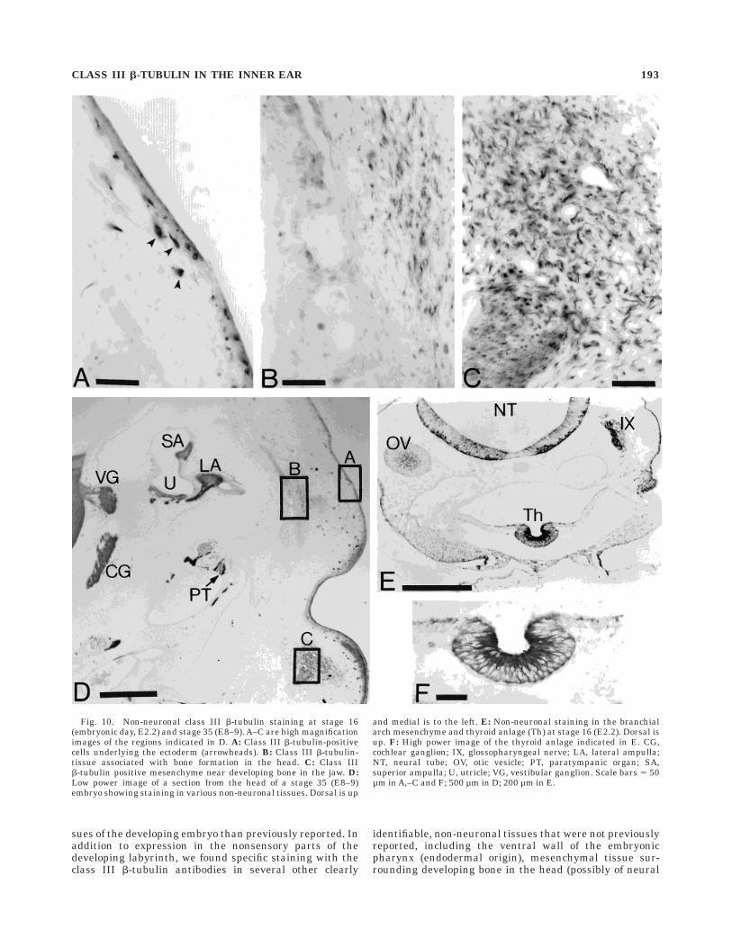
sues of the developing embryo than previously reported. Inaddition to expression in the nonsensory parts of thedeveloping labyrinth, we found specific staining with theclass III b-tubulin antibodies in several other clearly
identifiable, non-neuronal tissues that were not previouslyreported, including the ventral wall of the embryonicpharynx (endodermal origin), mesenchymal tissue sur-rounding developing bone in the head (possibly of neural
Fig. 10. Non-neuronal class III b-tubulin staining at stage 16(embryonic day, E2.2) and stage 35 (E8–9). A–C are high magnificationimages of the regions indicated in D. A: Class III b-tubulin-positivecells underlying the ectoderm (arrowheads). B: Class III b-tubulin-tissue associated with bone formation in the head. C: Class IIIb-tubulin positive mesenchyme near developing bone in the jaw. D:Low power image of a section from the head of a stage 35 (E8–9)embryo showing staining in various non-neuronal tissues. Dorsal is up
and medial is to the left. E: Non-neuronal staining in the branchialarch mesenchyme and thyroid anlage (Th) at stage 16 (E2.2). Dorsal isup. F: High power image of the thyroid anlage indicated in E. CG,cochlear ganglion; IX, glossopharyngeal nerve; LA, lateral ampulla;NT, neural tube; OV, otic vesicle; PT, paratympanic organ; SA,superior ampulla; U, utricle; VG, vestibular ganglion. Scale bars 5 50µm in A,–C and F; 500 µm in D; 200 µm in E.
CLASS III b-TUBULIN IN THE INNER EAR 193

crest origin), the thyroid anlage (endodermal origin), andthe ectoderm (including regions giving rise to epidermis).The non-neuronal staining in the tissue around formingbone is easily distinguishable from nerve fibers runningthrough the mesenchyme because the nerve fibers are veryheavily stained and the labeled mesenchymal cell bodiesare small, fusiform, and too numerous to be neuronalprecursors in these regions which normally do not havelarge populations of neurons. Previous studies also corre-lated the onset of class III b-tubulin expression withterminal mitosis and the onset of neuronal differentiation,because class III b-tubulin immunoreactivity was firstdetected in cells migrating from the proliferative zone ofthe neural tube, cells with neuronal morphologies, andneuronal populations undergoing terminal mitosis (Moodyet al., 1989; Lee et al., 1990). This correlation relied on theabsence of class III b-tubulin immunoreactivity in mitoticfigures and proliferating neurogenic tissues, particularlythe proliferative zone of the neural tube and neurogenicplacodes. Our observations, however, show that class IIIb-tubulin immunoreactivity can be detected in the prolif-erative zone of the neural tube, neurogenic placodes, andotic epithelium. Immunoreactive mitotic spindles werealso observed in these same structures. Hence, the expres-sion of class III b-tubulin is not, by itself, a reliableindicator of neuronal differentiation during embryonicdevelopment.
The widespread immunoreactivity observed with both ofthese antibodies is clearly due to binding of the primaryantibody and not a result of endogenous peroxidases, oradsorption of the secondary antibody or ABC reagents.Our use of methacarn, which fixes tissue by denaturationand coagulation, resulted in the observation of staining inmore tissues than previously reported. Because the TuJ1antibody has been shown to be specific for class IIIb-tubulin and not to cross-react with other isotypes ofb-tubulin or other proteins in chick embryo extracts underdenaturing conditions (Lee et al., 1990), we believe thatthe observed staining in methacarn-fixed tissue is the
result of specific binding of the antibodies to class IIIb-tubulin. Additionally, the carboxyl terminus of chickenclass III b-tubulin ends in a lysine residue (Sullivan andCleveland, 1986). Because lysine is one of the targets ofcross-linking fixatives like paraformaldehyde (Bancroftand Stevens, 1990), it is likely that less of the epitopewould be preserved with cross-linking fixatives than withcoagulative fixation. Earlier reports of class III b-tubulinas a neuronal marker used paraformaldehyde fixation,with fixation times ranging from 1 to 24 hours (Moody etal., 1989; Lee et al., 1990). As a result, low levels of class IIIb-tubulin expression may have been missed in previousstudies due to the destruction of the epitope duringfixation. In situ hybridization with specific probes to classIII b-tubulin mRNA may be needed in the future to confirmthat cells staining moderately with antibodies to class IIIb-tubulin actually express the molecule.
Our experience with the fixation sensitivity of theseantibodies suggests that, as fixation time increases, onlythose cells with the largest amount of the class III b-tubu-lin epitope are stained. Because neuronal cell types werethe only cells identified with TuJ1 in earlier studies,neuronal cells probably have significantly more class IIIb-tubulin than other cell types. Lee et al. (1990) were thefirst to suggest the accumulation of class III b-tubulin inneurons. They noted that labeled cells in the marginalzone of the neural tube were more intensely stained thancells migrating out of the proliferative zone, and observedweakly labeled cells in the dorsal root ganglion as neuronsbegan to differentiate. Additionally, Lee et al. (1990) ranimmunoblots of protein from whole embryonic chick headsat stages 12 (E2.0) through 23 (E3.5–4), and showed thatthe relative abundance of class III b-tubulin increasesduring this period when the number of differentiatingneurons is increasing. These observations led these au-thors to suggest that class III b-tubulin accumulates inneuronal cell types as they begin to differentiate.
Our observations support this hypothesis, but in adifferent context than suggested by Lee et al. (1990).Fixation with methacarn, as compared to paraformalde-hyde, appears to be much less detrimental to the detectionof class III b-tubulin by TuJ1 and b-III, thereby allowingus to detect lower levels of class III b-tubulin expression.As a result, we see staining in more cell types andstructures. Nevertheless, we always see dense staining inneuronal cells in the neural tube, cranial ganglia, neuro-genic placodes, and otic epithelium. Instead of seeing thesecells appear on a ‘‘background’’ of no class III b-tubulinexpression, we see these cells emerge from cell populationsexpressing lower levels of class III b-tubulin, and are ableto recognize them because of their accumulation of class IIIb-tubulin and distinctive morphology. Hair cells providean excellent example of this phenomenon. The otic epithe-lium clearly expresses class III b-tubulin prior to andduring the period of hair cell differentiation. Beginning atstage 29 (E6.0), we are able to distinguish hair cells fromthe other labeled epithelial cells because of their morphol-ogy, lumenal location, and because they stain more heavilythan the surrounding epithelium, i.e., both the surround-ing epithelium and the hair cells express class III b-tubu-lin, but hair cells express much more and stain moredensely. These observations suggest that, early in develop-ment, class III b-tubulin is expressed at low levels in mostneurogenic epithelia, such as the otic epithelia and neuraltube. As neuronal cells begin to differentiate, their expres-
TABLE 1. Tissues Expressing Class III b-Tubulin in the DevelopingChicken Embryo
Previously reported:C-cells of the ultimobranchial organ Kameda et al. (1993);
Kameda (1995)Glomus cells in the carotid body Kameda et al. (1994)Extra-embryonic membranes (ectodermal layers)LensCaudal mesonephric duct Lee et al. (1990)Caudal-ventral somiteTrigeminal placodesNeural tube–intermediate zoneNeural tube–marginal zone Lee et al. (1990);Neural crest–derived neurons Moody et al. (1989)Placodally-derived neurons
Reported in this paper:EctodermEmbryonic pharynx–
ventral wallfirst pharyngeal pouchthyroid anlage
Epibranchial placodes (first and second)Lens placodeNeural tube–proliferative zoneMesenchyme–
in the branchial archesaround developing boneisolated cells underlying the ectoderm
Olfactory placodeOtic placode, otic pit, and otocystParatympanic organRathke’s pouch
66
194 D. MOLEA ET AL.

sion of class III b-tubulin is significantly upregulated. Theexpression of class III b-tubulin in other cell phenotypes,however, is eventually downregulated.
At this time one can only speculate on the molecularevents underlying the bidirectional regulation of class IIIb-tubulin expression in neurogenic versus non-neurogeniccell types during differentiation. Anderson and colleagueshave described a 21-base pair sequence of genomic DNAdesignated as the neuron-restrictive silencer element(NRSE; also called RE-1; Mori et al., 1990; Wuenschell etal., 1990; Mori et al., 1992; Kraner et al., 1992) and anassociated protein binding factor, the neuron-restrictivesilencer factor (NRSF; also called REST or XBR; Chong etal., 1995; Schoenherr and Anderson, 1995a,b; Palm et al.,1998). This complex has been shown to negatively regulatethe expression of a large number of neuronal genes inneuronal precursors and non-neuronal cells (Mori et al.,1990; Wuenschell et al., 1990; Kraner et al., 1992; Mori etal., 1992; Chong et al., 1995; Schoenherr and Anderson,1995a; Schoenherr et al., 1996; Bessis et al., 1997; Edel-man and Jones, 1998; Kallunki et al., 1998). The chickenclass III b-tubulin gene has at least one NRSE and hasbeen shown to be regulated by NRSF (Schoenherr et al.,1996). Therefore, one is tempted to speculate that aconstitutive level of class III b-tubulin is expressed in allinner ear precursor cell types and that changes in class IIIb-tubulin levels are mediated by the NRSE/NRSF systemas neuronal and non-neuronal cells undergo differentia-tion.
Class III b-tubulin expression during haircell and sensory epithelia differentiation
We can first identify immature hair cells by class IIIb-tubulin immunoreactivity in wholemount preparationsof the saccular macula at about stage 26 (E4.5–5). By stage29 (E6.0), immature hair cells are detected in all thesensory epithelia. The appearance of hair cells with classIII b-tubulin in the saccular macula at stage 26 (E4.5–5)correlates with the appearance of hair cells as indicated byHCA labeling (this paper; Goodyear et al., 1995), and theelimination of gap junctions from hair cells reported byGinsberg and Gilula (1979). Similarly, in the basilarpapilla, class III b-tubulin-positive hair cells can be identi-fied at the same time that hair cells can be identified byHCA expression (Bartolami et al., 1991; Goodyear et al.,1995; Goodyear and Richardson, 1997) or stereocilia usingscanning electronic microscopy (SEM; Cotanche and Sulik,1984). Furthermore, class III b-tubulin staining followsthe same distal-to-proximal gradient of hair cell matura-tion (Cotanche and Sulik, 1984; Bartolami et al., 1991;Goodyear et al., 1995; Goodyear and Richardson, 1997).This evidence shows that accumulation of class III b-tubu-lin occurs early during hair cell differentiation, and iscontemporaneous with other available indicators of haircell differentiation.
Lateral inhibition has been suggested as a possiblemechanism for the developmental determination of haircells versus support cells, and the subsequent organizationof hair cells and support cells into a mosaic where the haircells are isolated from each other by the supporting cells(Goodyear et al., 1995; Goodyear and Richardson, 1997).This model proposes that, at some point in hair cell andsupport cell differentiation, a hair cell begins to inhibit itsneighbors from also differentiating into hair cells throughcontact-mediated signals. Thus, all the cells contacting a
hair cell become supporting cells. Variants of this ideahave been suggested as mechanisms that could producethe hair cell-support cell mosaic seen in the epithelia of theinner ear (Goodyear et al., 1995). All of these modelssuggest that hair cell-hair cell contacts should not beobserved. One advantage of class III b-tubulin over otherhair cell markers is that it allows us to see the morphologyof the cell body of the developing hair cell at about the timethat it begins to form stereocilia. Early hair cells have agourd-shaped appearance with a narrow neck extending tothe lumen. The lumenal area of these hair cells is muchsmaller than the amount of space occupied by the cell bodylocated below the lumen. Although we recognize that haircell-hair cell contact cannot be resolved using the tech-niques in this study, this gourd-shaped morphology, whichis present as the hair cells and supporting cells aredifferentiating, and the close packing of immature haircells, suggests that there is ample opportunity for haircell-to-hair cell contacts below the lumen of the epithe-lium, but does not preclude the possibility of thin support-ing cell processes separating the cells. The morphology ofearly differentiating hair cells suggests that there could besignificant contact occurring between hair cells below thelumen where the mosaic is observed. Additionally, sublu-menal contact between hair cells has been noted as late ashatching in chicken (Fermin and Cohen, 1984) and inadult quail (Ryals et al., 1992). These data suggest that, iflateral inhibition is the mechanism by which hair cells andsupport cells acquire their distinctive phenotypes, thetiming and degree of contact between cells may be moreimportant than just whether contact is occurring.
The paratympanic organ shows a similar sequence ofdevelopment as the otic epithelium. It is derived from anectodermal placode, the first epibranchial placode, whichappears to invade the distal first pharyngeal pouch assuggested by class III b-tubulin staining. Class III b-tubu-lin is expressed in a lumenal-streaky pattern throughoutthe early development of the paratympanic organ, andaccumulates in immature hair cells emerging from thelumenal-streaky epithelium as they begin to differentiate.BMP4, a marker used to identify developing sensoryepithelia in the inner ear, also localizes to the distal end ofthe first pharyngeal pouch where the paratympanic organdevelops, as early as stage 27 (E5), a stage when immaturehair cells are already present, and possibly as early asstage 16 (E2.2; Figs. 1 and 2 in Wu and Oh, 1996). Thesedata suggest that similar processes of development areoccurring in these two hair cell-producing structures, eventhough they are derived from different anlagen, and that acommon program of development may be occurring duringthe early differentiation of these sensory organs.
Class III b-tubulin expression in thenonsensory regions of the inner ear
Beginning at about stage 21 (E3.5), the expression ofclass III b-tubulin in all regions of the otic epitheliumbegins to give way to a distinct region of reduced class IIIb-tubulin expression in the lateral wall of the cochlearduct. This region is where the tegmentum vasculosum willdevelop (Knowlton, 1969; Cotanche and Sulik, 1982), andis indicated by an abrupt change in the density of stainingat the distal tip of the cochleosaccular duct evagination.The staining becomes increasingly more dense as oneapproaches the utricle. This abrupt change in staining iscontemporaneous with the specification of the sensory
CLASS III b-TUBULIN IN THE INNER EAR 195
epithelia in the cochlear duct as indicated by BMP4,Msx-1, and p75NGFR (Wu and Oh, 1996), suggesting thatthe determination of the tegmentum vasculosum may beoccurring as a distinct functional unit at the same time asthe sensory epithelia. This result indicates that the sen-sory and nonsensory regions of the inner ear are beingdetermined during the same period of development.
As development proceeds, more nonsensory regions be-gin to lose their class III b-tubulin staining, in particularthe endolymphatic sac, the perimacular region lateral butadjacent to the utricular macula, and regions involved inthe formation of the semicircular canals. By stage 35(E8–9), most of the nonsensory regions of the inner earhave lost their class III b-tubulin staining, leaving distinctregions of persistent staining between the medial walls ofthe cochlear duct and saccule, the confluence of the sac-cule, utricle and endolymphatic duct, and in the outercircumference of the semicircular canals. The first two ofthese regions occurs at the morphological transitionsbetween regions of the inner ear, and the staining in thesemicircular canals is located at the farthest extremes ofthe epithelia. One can imagine that these regions lieremote from possible signals directing morphogenesis andhave not yet been directed to differentiate.
With respect to the formation of semicircular canals, theidea that the persistent staining in the semicircular canallies remote from morphogenetic signals suggests thatthere is a signal directing the differentiation of the fusionplates of the semicircular canals which decreases withdistance from the center of the fusion plate. This constructwould place the signaling and differentiation, i.e., themorphogenetic action, of cells forming the semicircularcanals in the fusion plates, which subsequently disappear,and not in the remaining semicircular canals whose epithe-lia may have a predominately passive role. In Xenopus,Haddon and Lewis (1991) demonstrated that the epitheliaof the fusion plates are a source of glucosaminoglycans,which drive the formation of the semicircular canals, thusshowing that these cells are differentiating from the othercells in the epithelium of the semicircular canal. Addition-ally, Fekete et al. (1997) showed that, in chicken, cell deathfirst occurs at the center of the fusion plates and appears toexpand outward. Recently, Kiernan et al. (1997) proposeda model of semicircular canal determination based on theboundary model developed by Fekete (1996) and stainingpattern exhibited by two homeobox genes, SOHo-1 andGH6. These two genes display patterns of expressionremarkably similar to those we see with class III b-tubulinin the thin-walled nonsensory parts of the otocyst once thesemicircular canals begin to form. The model proposed,however, focuses on the specification of structures seen inthe mature labyrinth to explain the parceling of thelabyrinth into different structures during developmentand does not account for the complex morphogeneticmovements required to form the semicircular canals. Themodel suggested above would imply that the cells undergo-ing specification and specialization in the formation of thesemicircular canals lie in the fusion plates and subse-quently are not represented in the mature labyrinth.
Isolated hair cells and hair cell dependenceon innervation
Around stage 29 (E6.0), we observed gourd-shaped, classIII b-tubulin-positive cells, and HCA-positive/class III
b-tubulin-positive cells in putative nonsensory regions ofthe utricular and saccular spaces. Although some of thesecells may be part of a distinct population of nonsensorycells, because some of these cells also express HCA, wesuspect that some are isolated hair cells. These cells lie inregions devoid of innervation, and away from the identifi-able utricular and saccular maculae. Hair cells withoutinnervation are not observed in mature sensory epithelia.Several studies suggest that the presence of the AVG orcontact between hair cells and nerve fibers is not requiredfor hair cell differentiation (Fell, 1928; Orr, 1968; van deWater, 1976; Hirokawa, 1977, 1978; Corwin and Cotanche,1989; Swanson et al., 1990; Sokolowski et al., 1993). Thereis evidence, however, that hair cells and other receptorcells derived from the octavolateralis system do requiresome interaction with innervating fibers for their long-term maintenance (. 2 weeks). Whereas short termdenervation of these sensory epithelia does not produceany substantial degeneration (Fell, 1928; Orr, 1968; van deWater, 1976; Corwin and Cotanche, 1989; Swanson et al.,1990; Sokolowski et al., 1993), long-term denervationresults in a loss or dedifferentiation of hair cells withinthese sensory epithelia (Favre and Sans, 1991; Weislederet al., 1994, 1996). The extended length of time requiredfor hair cells to disappear suggests that the process may besecondary to other changes in the epithelium, or gradualchanges of a yet to be determined regulatory pathwaynormally set by neuronal interactions. We conclude thatthe initiation of hair cell or sensory epithelia differentia-tion occurs independent of neuronal influences, but that atsome point in development, hair cells or sensory epitheliabecome dependent on some neuronal influence for long-term maintenance and hence, these isolated hair cellsdeteriorate. The best test of this hypothesis will be studiesof the developing AVG and inner ear sensory epithelia inknockout mutants, such as the neurogenin1 knockouts(Ma et al., 1998), that do not seem to develop an AVG.
ACKNOWLEDGMENTS
The authors thank Dr. A. Frankfurter for kindly supply-ing the TuJ1 and b-III antibodies, and Dr. G. Richardsonfor supplying us with the antibody to HCA. We also thankC. Federhart for technical assistance, D. Cunningham, D.Frost, and G. MacDonald for numerous discussions aboutimmunohistochemistry and imaging, P. Schwartz andJanet Schukar for photographic assistance, E. Oesterleand L. Westrum for reading and commenting on themanuscript, and S. Cochran for stimulating discussion andhelp preparing the figures and manuscript.
LITERATURE CITED
Bancroft JD, Stevens A. 1990. Theory and practice of histological tech-niques. New York: Churchill Livingstone.
Bartolami S, Goodyear R, Richardson G. 1991. Appearance and distributionof the 275 kD hair-cell antigen during development of the avian innerear. J Comp Neurol 314:777–788.
Bessis A, Champtiaux N, Chatelin L, Changeux J-P. 1997. The neuron-restrictive silencer element: A dual enhancer/silencer crucial for pat-terned expression of a nicotinic receptor gene in the brain. Proc NatlAcad Sci USA 94:5906–5911.
Chong JA, Tapia-Ramırez J, Kim S, Toledo-Aral JJ, Zheng Y, Boutros MC,Altshuller YM, Frohman MA, Kraner SD, Mandel G. 1995. REST: Amammalian silencer protein that restricts sodium channel gene expres-sion to neurons. Cell 80:949–957.
196 D. MOLEA ET AL.

Corwin JT, Cotanche DA. 1989. Development of location-specific hair cellstereocilia in denervated embryonic ears. J Comp Neurol 288:529–537.
Cotanche DA, Sulik KK. 1982. Scanning electron microscopy of thedeveloping chick tegmentum vasculosum. Scan Electron Microsc Part3:1283–1294.
Cotanche DA, Sulik KK. 1984. The development of stereociliary bundles inthe cochlear duct of chick embryos. Brain Res Dev Brain Res 16:181–193.
D’Amico-Martel A. 1982. Temporal patterns of neurogenesis in aviancranial sensory and autonomic ganglia. Am J Anat 163:351–372.
D’Amico-Martel A, Noden DM. 1983. Contributions of placodal and neuralcrest cells to avian cranial peripheral ganglia. Am J Anat 166:445–468.
Easter SS Jr, Ross LS, Frankfurter A.1993. Initial tract formation in themouse brain. J Neurosci 13:285–299.
Edelman GM, Jones FS. 1998. Gene regulation of cell adhesion: A key stepin neural morphogenesis. Brain Res Brain Res Rev 26:337–352.
Favre D, Sans A.1991. Dedifferentiation phenomena after denervation ofmammalian adult vestibular receptors. Neuroreport 2:501–504.
Fekete DM. 1996. Cell fate specification in the inner ear. Curr OpinNeurobiol 6:533–541.
Fekete DM, Homburger SA, Waring MT, Riedl AE, Garcia LF.1997.Involvement of programmed cell death in morphogenesis of the verte-brate inner ear. Development 124:2451–2461.
Fell HB. 1928. The development in vitro of the isolated otocyst of theembryonic fowl. Archiv fur Experimentelle Zellforschung 7:69–81.
Fermin CD, Cohen GM. 1984. Developmental gradients in the embryonicchick’s basilar papilla. Acta Otolaryngol Stockh 97:39–51.
Fritzsch B, Barald KF, Lomax MI. 1998. Early embryology of the vertebrateear. In: Rubel EW, Popper AN, Fay RR, editors. Development of theauditory system. Vol. 9. Springer Handbook of Auditory Research. NewYork: Springer. p 80–145.
Gates MA, Thomas LB, Howard EM, Laywell ED, Sajin B, Faissner A, GotzB, Silver J, Steindler DA. 1995. Cell and molecular analysis of thedeveloping and adult mouse subventricular zone of the cerebral hemi-spheres. J Comp Neurol 361:249–266.
Ginzberg RD, Gilula NB. 1979. Modulation of cell junctions during differen-tiation of the chicken otocyst sensory epithelium. Dev Biol 68:110–129.
Gonzalez MdL, Silver J. 1994. Axon-glia interactions regulate ECM pattern-ing in the postnatal rat olfactory bulb. J Neurosci 14:6121–6131.
Goodyear R, Richardson G. 1997. Pattern formation in the basilar papilla:Evidence for cell rearrangement. J Neurosci 17:6289–6301.
Goodyear R, Holley M, Richardson G. 1995. Hair and supporting-celldifferentiation during the development of the avian inner ear. J CompNeurol 351:81–93.
Haddon CM, Lewis JH. 1991. Hyaluronan as a propellant for epithelialmovement: The development of semicircular canals in the inner ear ofXenopus. Development 112:541–550.
Hamburger V, Hamilton HL. 1951. A series of normal stages in thedevelopment of the chick embryo. J Morphol 88:49–92.
Hirokawa N. 1977. Disappearance of afferent and efferent nerve terminalsin the inner ear of the chick embryo after chronic treatment withb-bungarotoxin. J Cell Biol 73:27–46.
Hirokawa N. 1978. Synaptogenesis in the basilar papilla of the chick. JNeurocytol 7:283–300.
Kallunki P, Edelman GM, Jones FS. 1998. The neural restrictive silencerelement can act as both a repressor and enhancer of L1 cell adhesionmolecule gene expression during postnatal development. Proc NatlAcad Sci USA 95:3233–3238.
Kameda Y. 1995. Evidence to support the distal vagal ganglion as the originof C cells of the ultimobranchial gland in the chick. J Comp Neurol359:1–14.
Kameda Y, Kameya T, Frankfurter A. 1993. Immunohistochemical localiza-tion of a neuron-specific b-tubulin isotype in the developing chickenultimobranchial glands. Brain Res 628:121–127.
Kameda Y, Yamatsu Y, Kameya T,. Frankfurter A 1994. Glomus celldifferentiation in the carotid body region of chick embryos studied byneuron-specific class III b-tubulin isotype and leu-7 monoclonal antibod-ies. J Comp Neurol 348:531–543.
Katayama A, Corwin JT. 1989. Cell production in the chicken cochlea. JComp Neurol 281: 129–135.
Katsetos CD, Frankfurter A, Christakos S, Mancall EL, Vlachos IN, UrichH. 1993. Differential localization of class III b-tubulin isotype andcalbindin-D28k defines distinct neuronal types in the developing humancerebellar cortex. J Neuropathol Exp Neurol 52:655–666.
Kiernan AE, Nunes F, Wu DK, Fekete DM. 1997. The expression domain oftwo related homeobox genes defines a compartment in the chicken innerear that may be involved in semicircular canal formation. Dev Biol191:215–229.
Knowlton VY. 1967. Correlation of the development of membranous andbony labyrinths, acoustic ganglia, nerves, and brain centers of the chickembryo. J Morphol 121:179–208.
Kraner SD, Chong JA, Tsay H-J, Mandel G. 1992. Silencing the type IIsodium channel gene: A model for neural-specific gene regulation.Neuron 9:37–44.
Lee MK, Tuttle JB, Rebhun LI, Cleveland DW, Frankfurter A. 1990. Theexpression and posttranslational modification of a neuron-specificb-tubulin isotype during chick embryogenesis. Cell Motil Cytoskel 17:118–132.
Lurie DI, Rubel EW 1994. Astrocyte proliferation in the chick auditorybrainstem following cochlea removal. J Comp Neurol 346:276–288.
Ma, Q, Chen Z, Barrantes IdB, de la Pompa JL, Anderson DJ. 1998.neurogenin1 is essential for the determination of neuronal precursorsfor proximal cranial sensory ganglia. Neuron 20:469–482.
Memberg SP, Hall AK. 1995. Dividing neuron precursors express neuron-specific tubulin. J Neurobiol 27:26–43.
Menezes JRL, Luskin MB. 1994. Expression of neuron-specific tubulindefines a novel population in the proliferative layers of the developingtelencephalon. J Neurosci 14:5399–5416.
Moody SA, Quigg MS, Frankfurter A. 1989. Development of the peripheraltrigeminal system in the chick revealed by an isotype-specific anti-beta-tubulin monoclonal antibody. J Comp Neurol 279:567–580.
Moody SA, Miller V, Spanos A, Frankfurter A. 1996. Developmentalexpression of a neuron-specific b-tubulin in frog (Xenopus laevis): Amarker for growing axons during the embryonic period. J Comp Neurol364:219–230.
Mori N, Stein R, Sigmund O, Anderson DJ. 1990. A cell type-preferredsilencer element that controls the neural-specific expression of theSCG10 gene. Neuron 4:583–594.
Mori N, Schoenherr CJ, Vandenbergh DJ, Anderson DJ. 1992. A commonsilencer element in the SCG10 and type II Na1 channel genes binds afactor present in nonneuronal cells but not in neuronal cells. Neuron9:45–54.
Oh S-H, Johnson R, Wu DK. 1996. Differential expression of bone morpho-genetic proteins in the developing vestibular and auditory sensoryorgans. J Neurosci 16:6463–6475.
Orr MF. 1968. Histogenesis of sensory epithelium in reaggregates ofdissociated embryonic chick otocysts. Dev Biol 17:39–54.
Palm K, Belluardo N, Netsis M, Timmusk T. 1998. Neuronal expression ofzinc finger transcription factor REST/NRSF/XBR gene. J Neuorsci18:1280–1296.
Rubel EW. 1978. Ontogeny of structure and function in the vertebrateauditory system. In: Jacobson M, editor. Handbook of sensory physiol-ogy. Vol. IX. New York: Springer-Verlag. p 135–237.
Ryals BM, Westbrook EW, Stoots S, Spencer RF. 1992. Changes in theacoustic nerve after hair cell regeneration. Exp Neurol 115:18–22.
Schoenherr CJ, Anderson DJ. 1995a. The neuron-restrictive silencer factor(NRSF): A coordinated repressor of multiple neuron-specific genes.Science 267:1360–1363.
Schoenherr CJ, Anderson DJ. 1995b. Silencing is golden: Negative regula-tion in the control of neuronal gene transcription. Curr Opin Neurobiol5:566–571.
Schoenherr CJ, Paquette AJ, Anderson DJ. 1996. Identification of potentialtarget genes for the neuron-restrictive silencer factor. Proc Natl AcadSci USA 93:9881–9886.
Snow RL, Robson JA. 1995. Migration and differentiation of neurons in theretina and optic tectum of the chick. Exp Neurol 134:13–24.
Sokolowski BHA, Stahl LM, Fuchs PA. 1993. Morphological and physiologi-cal development of vestibular hair cells in the organ-cultured otocyst ofthe chick. Dev Biol 155:134–146.
Stone JS, Leano SG, Baker LP, Rubel EW. 1996a. Hair cell differentiation inchick cochlear epithelium after aminoglycoside toxicity: In vivo and invitro observations. J Neurosci 16:6157–6174.
Stone JS, Molea D, Oesterle EC, Rubel EW. 1996b. Class III beta tubulin inmature, Developing, and regenerating hair cells of the avian andmammalian inner ear. Mol Biol Cell 7:650a.
Sullivan KF, Cleveland DW. 1986. Identification of conserved isotype-defining variable region sequences for four vertebrate b tubulin polypep-tide classes. Proc Natl Acad Sci USA 83:4327–4331.
CLASS III b-TUBULIN IN THE INNER EAR 197

Swanson GJ, Howard M, Lewis J. 1990. Epithelial autonomy in thedevelopment of the inner ear of a bird embryo. Dev Biol 137:243–257.
Troutt LL, van Heumen WRA, Pickles JO. 1994. The changing microtubulearrangements in developing hair cells of the chick cochlea. Hear Res81:100–108.
van de Water TR. 1976. Effects of removal of the statoacoustic ganglioncomplex upon the growing otocyst. Ann Otol Rhinol Laryngol 85:2–32.
von Bartheld CS. 1990. Development and innervation of the paratympanicorgan (vitali organ) in chick embryos. Brain Behav Evol 35:1–15.
Watanabe M, Rutishauser U, Silver J. 1991. Formation of the retinalganglion cell and optic fiber layers. J Neurobiol 22:85–96.
Weisleder P, Lu Y, Zakon HH. 1994. Effects of denervation upon receptorcell survival and basal cell proliferation in tuberous electroreceptororgans of a weakly electric fish. J Comp Neurol 347:545–552.
Weisleder P, Lu Y, Zakon HH. 1996. Tuberous electroreceptor organs formin denervated regenerating skin of a weakly electric fish. J CompNeurol 367:563–574.
Whitehead MC, Morest DK. 1985a. The development of innervationpatterns in the avian cochlea. Neuroscience 14:255–276.
Whitehead MC, Morest DK. 1985b. The growth of cochlear fibers and theformation of their synaptic endings in the avian inner ear: A study withthe electron microscope. Neuroscience 14: 277–300.
Whitfield T, Haddon C, Lewis J. 1997. Intercellular signals and cell-fatechoices in the developing inner ear: Origins of global and of fine-grainedpattern. Semin Cell Dev Biol 8:239–247.
Wu DK, Oh S-H. 1996. Sensory organ generation in the chick inner ear. JNeurosci 16:6454–6462.
Wu DK, Nunes FD, Choo D. 1998. Axial specification for sensory organsversus non-sensory structures of the chicken inner ear. Development125:11–20.
Wuenschell CW, Mori N, Anderson DJ. 1990. Analysis of SCG10 geneexpression in transgenic mice reveals that neural specificity is achievedthrough selective derepression. Neuron 4:595–602.
198 D. MOLEA ET AL.