CISPLATIN-INDUCED TUMOR CELL DEATH SIGNALING AND POTENTIATION
58
DEPARTMENT OF ONCOLOGY-PATHOLOGY CANCER CENTER KAROLINSKA Karolinska Institutet, Stockholm, Sweden CISPLATIN-INDUCED TUMOR CELL DEATH SIGNALING AND POTENTIATION BY ENERGY METABOLISM INHIBITORS Linda Strandberg Ihrlund Stockholm 2007
Transcript of CISPLATIN-INDUCED TUMOR CELL DEATH SIGNALING AND POTENTIATION

DEPARTMENT OF ONCOLOGY-PATHOLOGY CANCER CENTER KAROLINSKA
Karolinska Institutet, Stockholm, Sweden
CISPLATIN-INDUCED TUMOR CELL DEATH SIGNALING
AND POTENTIATION BY ENERGY METABOLISM INHIBITORS
Linda Strandberg Ihrlund
Stockholm 2007

All previously published papers were reproduced with permission from the publisher.
Published and printed by ReproPrint AB, Solna, Sweden
© Linda Strandberg Ihrlund, 2006ISBN 91-7357-068-0
Gårdsvägen 4, 169 70 Solna
Published and printed byAll previously published papers were reproduced with permission from the publisher.
Published and printed by ReproPrint AB, Solna, Sweden
© Linda Strandberg Ihrlund, 2006ISBN 91-7357-068-0

ABSTRACT
Treatment of cancer is associated with challenges such as the development of resistance by the tumors. By elucidating intracellular signal transduction pathways elicited by therapy it is possible to identify structures and processes crucial for induction of cell death. The aim is to exploit this knowledge to further improve treatment strategies.
In this thesis, evidence is presented that cisplatin treatment of tumor cell lines induces separate signal transduction events. Upon cisplatin treatment, calpain, a cytosolic protease, is shown to become activated. Activated calpain is demonstrated to cleave the BH3 only protein Bid between residues Gly70 and Arg71, generating a fragment with the ability to induce release of cytochrome c from the mitochondria. Cisplatin induces signals via MEKK1, however, the activation of calpain is shown to be independent of overexpression of the dominante negative form of MEKK1 (dnMEKK1). The dominant positive form of MEKK1 (Dp MEKK1) induces a conformational change in the pro-apoptotic protein Bak. This conformational activation was revealed not to directly lead to apoptosis. However, cisplatin induces both conformational change of Bak as well as Bak oligomerization. Bak complexes of sizes between 80 kDa and 170 kDa are shown to be associated with apoptosis formation, indicating that in addition to activation of MEKK1 another signal is needed. This signal is constituted by JNK, shown as a JNK inhibitor blocked cisplatin-induced apoptosis and the formation of Bak complexes in the critical size range. These results are further confirmed using cells deficient for JNK.
In addition to the development of tumor resistance to treatment, another problem in tumor therapy is the toxicity to normal tissues unaffected by disease. An approach to overcome these processes is the use of therapy sensitization, i.e. using low dose of a standard treatment in combination with a drug that specifically targets crucial processes in the tumor cell to increase the effects of the other drug. In this thesis, the effects of sensitizing tumor cells to cisplatin by inhibition of their energy production are investigated. Both an inhibitor of glycolysis, 2-deoxyglucose (DG) as well as an inhibitor of fatty acid -oxidation, etomoxir, were shown to potentiate the effects of cisplatin-induced cell death in several tumor cell lines. These results are further confirmed in treatment of xenografted mouse tumors.

LIST OF PUBLICATIONS
This thesis is based on the following papers, which will be referred to in the text by their Roman numeral:
I. Mandic A, Viktorsson K, Strandberg L, Heiden T, Hansson J, Linder S, and Shoshan C.M. Calpain-Mediated Bid Cleavage and Calpain-Independent Bak Modulation: Two Separate Pathways in Cisplatin-Induced Apoptosis. Mol Cell Biol., 2002, 22: 3003-3013
II. Ihrlund L.S, Hernlund E, Viktorsson K, Panaretakis T, Barna G, Sabapathy K, Linder S, and Shoshan M.C. Two distinct steps of Bak regulation during apoptotic stress signaling: Different roles of MEKK1 and JNK1. Exp Cell Res., 2006, 312: 1581-1589
III. Ihrlund LS, Khan O, Hernlund E, Schwarz S, Castro J, Linder S, Panaretakis T and Shoshan MC. Cell death induction by glycolysis inhibitors per se and in combination with chemotherapeutic agents. Submitted to Cancer Chemother and Pharmacol., 2006
IV. Ihrlund LS, Khan O, Linder S and Shoshan MC Induction and potentiation of tumor cell death using the energy metabolism inhibitor etomoxir alone and in combinations with cisplatin and 2-deoxyglucose. Manuscript

CONTENTS1 INTRODUCTION ..............................................................................................................................1
1.1 CELL DEATH.................................................................................................................................11.1.1 Apoptosis ................................................................................................................................11.1.2 Necrosis ..................................................................................................................................21.1.3 Senescence..............................................................................................................................21.1.4 Autophagy...............................................................................................................................2
1.2 APOPTOTIC SIGNALING ................................................................................................................31.2.1 Caspases.................................................................................................................................31.2.2 BCL2 family proteins .............................................................................................................31.2.3 The role of mitochondria in apoptotic signaling...................................................................51.2.4 Calpain ...................................................................................................................................61.2.5 Mitogen activated protein kinases in cellular stress responses............................................71.2.6 Methods to study cell death..................................................................................................11
1.3 ENERGY METABOLISM ...............................................................................................................121.3.1 ATP production by oxidative and non-oxidative processes ................................................121.3.2 Energy metabolism in tumors ..............................................................................................131.3.3 Fatty acid metabolism..........................................................................................................15
1.4 TREATMENT OF CANCER............................................................................................................161.4.1 Chemotherapy ......................................................................................................................161.4.2 Tumor resistance to treatment .............................................................................................171.4.3 The concept of sensitization.................................................................................................18
2 RESULTS AND DISCUSSION.......................................................................................................20
2.1 PAPERS I AND II .........................................................................................................................202.1.1 Cisplatin induces calpain activation ...................................................................................202.1.2 Calpain cleaves Bid .............................................................................................................202.1.3 Calpain regulation ...............................................................................................................212.1.4 Bak conformational activation.............................................................................................222.1.5 Bak complex formation ........................................................................................................222.1.6 Requirement for JNK in Bak complex formation ................................................................23
2.2 PAPERS III AND IV.....................................................................................................................252.2.1 Different modes of action for DG and BP...........................................................................252.2.2 DG potentiates DNA-damaging agents ...............................................................................262.2.3 Etmoxir as a potentiator of DG and of cisplatin .................................................................272.2.4 Future perspectives for DG and etomoxir...........................................................................29
3 CONCLUDING REMARKS...........................................................................................................31
4 ACKNOWLEDGEMENTS.............................................................................................................33
5 REFERENCES..................................................................................................................................37

LIST OF ABBREVIATIONS
6-AN 6-aminonicotinamide ABC ATP-binding cassette AIF Apoptosis inducing factor AMACR -methylacyl CoA racemase AMP Adenosine monophosphate AMPK AMP-activated kinase ANT Adenine nucleotide translocator
AP-1 Activator protein 1 Apaf-1 Apoptosis protease activator factor 1 ASK Apoptosis stimulating kinase ATF-2 Activating transcription factor 2 ATM Ataxia telangiectasia mutated ATP Adenosine three phosphate Bad Bcl-2–associated death promoter Bak Bcl-2 homologous antagonist killer BAPTA-AM Cell permeable calcium chelator Bax Bcl-2 associated X protein Bcl-2 B-cell lymphoma 2 BH Bcl-2 homology Bid BH3-interacting domain death agonist Bim Bcl-2-interacting mediator of cell death BMH 1,6-bis- maleimidohexane BP Bromopyruvate Chk1 Cell Cycle Checkpoint Kinase 1 CK18 Cytokeratin 18 COX-2 Cyclooxygenase 2 CPT-1 Carnitine-palmitoyl-transferase I Ctr1 Copper transporter 1 DNA Deoxyribonucleic acid DG 2-deoxyglucose DR Death receptor ER Endoplasmic reticulum ERK Extracellular signal regulated kinase FASn Fatty acid synthase FDG Fluoro-deoxyglucose GLUT Glucose transporter

GSH Glutathione Her2 Human epidermal growth factor receptor 2 HXK Hexokinase JIP JNK-interacting proteins JNK c-Jun NH2-terminal protein kinase LDH Lactate dehydrogenase MAPK Mitogen-activated protein kinase MAPKK Mitogen-activated protein kinase kinase MAPKKK Mitogen-activated protein kinase kinase kinase Mcl Myeloid cell differentiation 1 MDM-2 Mouse double minute 2 MDR Multi drug resistance MEf Mouse embryo fibroblast MEK MAPK/ERK kinase MEKK MEK kinase MKK MAPK kinase MLK Mixed-lineage kinase MPT Mitochondrial permeability transition mRNA Messenger ribonucleic acid MS Mass spectrometry mTOR Mammalian target of rapamycin MTT 3-(4,5-dimethyl. thiazolyl-2)-2,5-diphenyltetrazolium bromide NADPH Nicotinamide-adenine dinucleotide phosphate NFkB Nuclear factor kappa beta NGF Nerve growth factor NMR Nucleic magnetic resonance PARP Poly (ADP-Ribose) polymerase PET Positron emission tomography P-gp P-glycoprotein PH Pleckstrin homology PI Propidium iodide PI3K Phosphatidyl inositol-3 kinase PUMA p53 up-regulated modulator of apoptosis PS Phosphatidyl serine ROS Reactive oxygen species SCO2 Synthesis of cytochrome c oxidase 2 SH3 Src-homology 3 TSC2 Tuberous sclerosis complex 2 TGF Transforming growth factor

TIGAR p53-induced glycolysis and apoptosis regulator TMRE Tetramethylrhodamine ethyl ester TNF Tumor necrosis factor TRAIL TNF-related apoptosis inducing ligand UCP Uncoupling protein UPR Unfolded protein response UV Ultraviolet VDAC Voltage dependent anion channel xIAP x-linked inhibitor of apoptosis protien
Mitochondrial membrane potential

1
1 INTRODUCTION
About one in three persons in the developed countries will contract a cancer related disease during their lifetime, making cancer the second most common cause of death after cardiovascular disease. Tumor treatment is faced with several challenges; cancer is a heterogeneous disease and countless genetic and epigenetic changes can arise in different tumors making each individual tumor case unique. In addition, anti-cancer drugs are ultimately designed to kill cells, conveying difficulties in avoiding toxicity to other tissues. However, by elucidating mechanisms behind cancer development and resistance to therapy, possibilities of development of preventative measures against tumor formation, of further improvement of existing treatment modalities and of better treatment options, arise.
Development of tumors have been attributed to six essential alterations in cell physiology (Hanahan and Weinberg 2000); self-sufficiency in growth signals, insensitivity to growth-inhibitory signals, evasion of programmed cell death, limitless replicative potential, sustained angiogenesis and tissue evasion and metastasis. These traits are considered as common to all human tumors and hence also provide possibilities to fight tumor development and means to target sensitive tumor targets for treatment. This thesis presents studies on programmed cell death (apoptosis) induced by chemotherapy in tumor cells, and crucial signal transduction pathways involved in this process. An important theme here is also the concept of interfering with the energy supply of tumor cells, in order to render them more susceptible to conventional cancer therapy. The overall aim is to gain better understanding of the mechanisms involved in cell death and how these can be exploited to render tumors more susceptible to therapeutic measures.
1.1 CELL DEATH
Cell death is an essential and highly regulated process that is well conserved between species. It plays a major role in tissue sculpting during embryo development and is necessary for tissue homeostasis, wound healing and elimination of pathogens in mature organisms. Defects in the cell death process are associated with numerous pathological disorders including diabetes, neurodegenerative disorders and cancer. Intense research on cell death has shown that it is not a uniform process but includes many different processes distinguished by differences in morphology and cellular signaling.
1.1.1 Apoptosis
The term apoptosis was first invented by Kerr and colleagues (Kerr, Wyllie et al. 1972) based on morphological criteria such as cellular shrinkage, condensation of nuclear chromatin, and DNA fragmentation. Studies performed in the nematode C. eleganshave provided important information on the tight genetic control of the apoptosis process. Several genes specifically required for induction and execution of apoptosis were identified and homologs to many of these genes have been found in mammals and

2
include both oncogenes and tumor suppressor genes (Liu and Hengartner 1999). The apoptotic cell eventually breaks up into membrane-enclosed particles called apoptotic bodies. These are quickly recognized and engulfed by immune cells or surrounding tissue. In this way, apoptotic cells do not inflict any tissue scarring or inflammation in neighboring tissues, making it suitable for normal tissue turnover in the body (Fadeel and Orrenius 2005).
1.1.2 Necrosis
Necrosis is classically described as an accidental form of cell death, characterized by irreversible swelling of the cytoplasm and mitochondria, loss of membrane integrity leading to cell rupture and leakage of the cellular compartments into the surrounding tissue, causing an inflammatory response (Fadeel and Orrenius 2005). However, recent studies indicate that necrosis may be a regulated process that accompanies apoptosis in some tissues during development. Also, there seems to be an overlap between apoptosis and necrosis in response to certain death inducing stimuli, i.e., many agents induce apoptosis at lower doses and necrosis at higher. Features of both apoptosis and necrosis may even coexist in the same cell and it has been proposed that necrosis can be seen as a different form of execution of programmed cell death (Proskuryakov, Konoplyannikov et al. 2003; Zong and Thompson 2006). The inflammatory response of necrosis may be associated with systemic toxicity and the activity of immune cells at the site may even promote malignancy (Zeh and Lotze 2005). On the other hand, the stimulation of the immune response could potentiate the killing of tumor cells (Jin and El-Deiry 2005).
1.1.3 Senescence
The term replicative senescence was first coined by Hayflick and Moorhead and described growth arrested human diploid cells that had gone through a certain number of doublings and thereafter were exhausted in their capacity to divide (Hayflick and Moorhead 1961). Later, it was discovered that during each cell cycle, 50-100 base pairs are lost from the ends of the chromosomes called the telomeres. This causes the chromosomes to lose their ability to protect the ends of chromosomal DNA, yielding end-to-end chromosomal fusions, and eventually leading to cell death (Counter, Avilion et al. 1992). Premature senescence is an inducible form of cellular senescence that is morphologically and biochemically related to replicative senescence. It is thought to be associated with DNA damage and signaling pathways involving ataxia telangiectasia mutated (ATM) and p53 activated by telomere shortening (d'Adda di Fagagna, Reaper et al. 2003).
1.1.4 Autophagy
Autophagy is a protective response to cellular stresses, e.g., starvation, changes in cell volume, oxidative stress, accumulation of misfolded proteins, hormonal signaling and irradiation. It is a process of sequestering organelles in vesicles inside the cell, which are subsequently delivered to the lysosome for degradation. Under conditions of prolonged cell stress the cell compartmentalizes and digests itself. The genetics behind autophagy have been characterized in yeast and regulation has been shown to involve the TOR and PI3 kinase pathways in response to starvation, suggesting that the same

3
signaling pathways are involved in mammals. Autophagy seems to play a complex role in ensuring cell survival during cell starvation and also in removing organelles or inducing death in damaged cells (Nelson and White 2004; Jin and El-Deiry 2005).
1.2 APOPTOTIC SIGNALING
Two main apoptotic pathways have been identified in mammals; the intrinsic, mitochondrial pathway and the extrinsic, death receptor pathway. The latter is triggered by ligands that bind to the death receptor members of the TNF receptor family, inducing intracellular signaling pathways leading to the execution of apoptosis. This is of fundamental importance in the maintenance of tissue homeostasis in the immune system. The mitochondrial pathway is triggered by a vast array of stimuli including DNA damage caused by chemotherapeutic drugs, by UV- or gamma-radiation, or by high levels of reactive oxygen species (ROS), activation of oncogenes or inadequate cell-matrix interactions (Fadeel and Orrenius 2005).
1.2.1 Caspases
Both the extrinsic and intrinsic pathways induce activation of caspases that constitute the executioners of the apotosis response. Caspases are cystein proteases found in the cytoplasm in their inactive pro-caspase form and are activated by cleavage at certain aspartate residues. Caspases can sequentially process and activate other caspases and are classified as initiator (casp-1, -2, -4, -5, -8, -9, -10 and -12) or effector caspases (such as casp-3, -6, -7, -11 and -13) (Thornberry and Lazebnik 1998). The signaling of caspases starts by activation of one of the initiator caspases, caspase-8 or -10 for the extrinsic pathway and caspase-9 for the intrinsic. These caspases can then activate a cascade reaction, eventually leading to activation of the main effector caspases -3, -6 or -7. The substrates of the effector caspases include nuclear and cytoskeletal components that are proteolyzed in order for the cell to develop the phenotypic characteristics of apoptosis (Thornberry and Lazebnik 1998; Chowdhury, Tharakan et al. 2006).
1.2.2 BCL2 family proteins
The initiation of the apoptotic response is tightly regulated and the B-cell/lymphoma-2 (Bcl-2) family of proteins play a critical role in this regulation, acting as gatekeepers between death signals and caspase activation. Expression of Bcl-2 has been detected frequently in human cancers and was first identified in human follicular B-cell lymphoma with the translocation t(14;18) (Tsujimoto, Cossman et al. 1985). Since then about 20 other members of the family have been characterized and divided into a pro- and anti-apoptotic subgroup respectively (Danial and Korsmeyer 2004). The Bcl-2 family of proteins share several conserved domains called the Bcl-2 homology (BH) regions, corresponding to alpha-helical segments. In general, the anti-apoptotic members including Bcl-2, Bcl-xL, Bcl-w and Mcl-1, show sequence conservation in all four domains, while the pro-apoptotic multi-BH domain members including Bax, Bak and Bok, lack the first alpha-helical segment, BH4. The pro-apoptotic members also include a subgroup called the BH3-only proteins, including Bid, Bad and Bim. Based

4
on deletion and mutagenesis studies, the BH3 domain is believed to be the critical death domain in all the pro-apoptotic Bcl-2 family members (Borner 2003; Burlacu 2003; Chan and Yu 2004).
The anti-apoptotic Bcl-2 family members exert their effects in numerous ways, e.g., as regulators of calcium homeostasis, as antioxidants and by inhibiting release of cytochrome c from the mitochondria. Following synthesis, the anti-apoptotic Bcl-2 family members are targeted to either mitochondrial or endoplasmic reticulum (ER) membranes where they integrate by way of a hydrophobic C-terminal domain. Furthermore, the BH domains of Bcl-2/Bcl-xL can form hydrophobic pockets with ability to hold the BH3 domain of the pro-apoptotic Bcl-2 family members. By this mechanism they are believed to sequester the BH3-only proteins, Bak/Bax proteins and other pro-apoptotic effectors, thereby inhibiting apoptosis induction. (Borner 2003; Burlacu 2003; Chan and Yu 2004; Jin and El-Deiry 2005).
Both the Bak/Bax and the BH3 only pro-apoptotic members are required for apoptosis induction. The BH3-only members are considered to be damage sensors positioned upstream of Bak/Bax, since they cannot induce apoptosis in the absence of Bak or Bax (Wei, Zong et al. 2001). In healthy cells the BH3-only proteins are kept in check by transcriptional and post-translational mechanisms. For example, Bim is maintained in its inactive form by binding to the microtubule-associated dynein motor complex. Upon cytokine deprivation or treatment with chemotherapeutic drugs, Bim is released from the microtubules and translocate to mitochondria where it binds Bcl-2/Bcl-xL and neutralizes their anti-apoptotic activity (Puthalakath, Huang et al. 1999). However, other Bim isoforms are not bound to the microtubules but are instead believed to be controlled at the transcriptional level since they are not found in healthy tissue (Chan and Yu 2004). Bid is another BH3-only protein known to be posttranslationally activated. Bid is normally kept in its inactive form in the cytosol via intramolecular interactions between it C-terminus and N-terminal BH3 domain. Bid is activated by cleavage, which exposes a glycine that can be N-myristolated, allowing Bid to target mitochondria and induce cell death (Zha, Weiler et al. 2000). Bid can act both by inhibition of Bcl-2/Bcl-xL but also by activation of Bak/Bax (Wei, Lindsten et al. 2000). Cleavage of Bid has been shown to be executed by caspase-8 (Li, Zhu et al. 1998), granzyme B (Heibein, Goping et al. 2000), lysozyme (Stoka, Turk et al. 2001) and calpain (Chen, He et al. 2001; Mandic, Viktorsson et al. 2002). Bad is another target for posttranslational modification, and is in normal cells phosphorylated at several serine residues, conveying preferential binding to 14-3-3 scaffold proteins. The phosphorylation has been attributed to AKT (Datta, Brunet et al. 1999), Raf (Wang, Rapp et al. 1996) and protein kinase A (Harada, Becknell et al. 1999), which all function as transducers of survival signaling. Death stimuli, e.g., growth factor withdrawal lead to dephosphorylation of Bad by calcineurin (Wang, Pathan et al. 1999) or protein phosphatase 2A (Chiang, Harris et al. 2001). Dephosphorylated Bad can then translocate to mitochondria and interact with Bcl-xL.
In healthy cells, the multi-domain pro-apoptotic protein Bax is mainly localized to the cytosol or loosely attached to membranes in its inactive form. After apoptotic stimulation, Bax undergoes conformational changes and translocates to the mitochondria where it inserts into the outer membrane (Wolter, Hsu et al. 1997). The

5
conformational change is associated with exposure of its N-terminal, an activation that can be monitored by an N-terminal specific antibody, and which has been shown to be induced by Bid (Desagher, Osen-Sand et al. 1999). Bax has been shown to be able to oligomerize in the outer mitochondrial membrane, a step that is thought to be critical for its pro-apoptotic function, leading to permeabilization of the membrane, possibly through formation of pores (Kuwana, Mackey et al. 2002). Unlike Bax, Bak is localized in the outer mitochondrial membrane in healthy cells. Analogous to Bax, Bak goes through a conformational change upon activation, exposing its N-terminal epitope (Griffiths, Dubrez et al. 1999), suggesting that the conformational change seen in Bax may not be causing its translocation to the mitochondria. Like Bax, Bak has been shown to oligomerize and induce the release of cytochrome c from the mitochondria (Wei, Lindsten et al. 2000).
1.2.3 The role of mitochondria in apoptotic signaling
Mitochondria play a crucial role in the intrinsic pathway as a target structure for the Bcl-2 family proteins, which regulate the release of a number of proapoptotic proteins from the mitochondrial intermembrane space, the most prominent being cytochrome c (Danial and Korsmeyer 2004). Upon its release, cytochrome c binds the adaptor molecule Apaf-1, which oligomerizes and recruits procaspase-9 in the presence of ATP, a complex termed the apoptosome. Procaspase-9 will undergo self-processing and activation and subsequently activates caspase-3 and the downstream apoptosis process. Other proteins released from the mitochondria can also induce apoptosis, albeit independently of caspases. Apoptosis-inducing factor (AIF) and the endonuclease EndoG can both translocate to the nucleus and induce fragmentation of DNA and thereby cell death (Fadeel and Orrenius 2005; Jin and El-Deiry 2005).
There are several theories on how the outer mitochondrial membrane becomes permeabilized to release cytochrome c; one is the mitochondrial permeability transition (MPT). MPT was long regarded as the major mechanism for release of cytochrome c and was originally defined as an sudden transition in the permeability of the inner mitochondrial membrane that occurred when mitochondria were treated with calcium and reagents that increased oxidative stress (Zoratti and Szabo 1995). MPT activation requires interaction between cyclophilin D, the adenine nucleotide translocator (ANT) and the voltage dependent anion channel (VDAC) at specialized contact sites between the inner and outer mitochondrial membranes to form a structure spanning both membranes. Opening of the MPT pore is followed by osmotic swelling of the mitochondrial matrix due to redistribution of water and ions between the cytosol and matrix, leading to rupture of the mitochondria. This will induce a subsequent release of mitochondrial proteins, including cytochrome c, providing a link between MPT and apoptosis (Armstrong 2006; Orrenius, Gogvadze et al. 2006). However, the importance of MPT for cytochrome c release during apoptosis has been challenged. These experiments were done on isolated mitochondria and it has been questioned whether mitochondrial swelling and cytochrome c release are associated in the context of a whole cell. For example mitochondrial swelling in HeLa cells has been shown not to induce cytochrome c release (Gao, Pu et al. 2001) and mitochondria from nerve growth factor (NGF) deprived neurons released cytochrome c into the cytosol but were

6
structurally intact and recovered their cytochrome c content when NGF was added back (Martinou, Desagher et al. 1999). Also, cyclophilin D deficient cells undergo apoptosis induced by various apoptotic stimuli whereas they exhibit resistance to necrotic cell death induced by excessive ROS and calcium (Nakagawa, Shimizu et al. 2005). In agreement with this; overexpression of cyclophilin D was shown to promote NO-induced necrosis whereas NO- or staurosporine-induced apoptosis was inhibited (Li, Johnson et al. 2004). Together these results suggest a role for MPT in necrosis but not apoptosis and that cytochrome c can be released from mitochondria by at least two distinct mechanisms.
1.2.4 Calpain
The calpains are a family of cytosolic calcium-dependent thiol proteases highly conserved between species. The calpains are widely expressed in the body with both ubiquitous and tissue-specific isoforms. The two most well-characterized calpains are expressed ubiquitously; the - and m-calpains, both containing a specific 80-kDa subunit and a common regulatory 28-kDa subunit. Calpains have been implicated in several important cellular functions including apoptosis, cell cycle regulation and cytoskeletal reorganization (Perrin and Huttenlocher 2002; Harwood, Yaqoob et al. 2005). The importance of calpains in tissue development is illustrated by the embryonic death of transgenic mice lacking calpain activity.
Regulators of calpain activity include cytosolic calcium concentrations, the endogenous inhibitor calpastatin, autoproteolytic cleavage, phosphorylation and intracellular distribution (Perrin and Huttenlocher 2002). It is clear that both m- and -calpains are absolutely dependent on calcium for their proteolytic function whereas they differ in dose required for activation in vitro. M-calpain requires millimolar calcium concentrations whereas -calpain settles for micromolar concentrations. Calcium binding to calpain induces a conformational change that is essential to assemble the active site. It has been proposed that calpains undergo autoproteolytic cleavage upon activation in order to free its active site. However, this is not completely clarified since mutated calpain without ability to undergo autoproteolysis also can cleave its substrates (Molinari and Carafoli 1997). Phosphorylation of calpains have been shown, mediated by, e.g., ERK signaling, activating calpain to participate in induction of cell migration, an important feature of tumor invasion (Shao, Chou et al. 2006). Calpains have been shown to translocate to the cellular membranes where they are activated, indicating the existence of membrane-bound activators of calpain (Molinari, Anagli et al. 1994). Unlike caspases, the calpains do not seem to recognize a specific cleavage site; instead the three-dimensional structure surrounding the cleavage site appears to mediate specificity. Calpain cleavage sites are often located at the boundaries between subunits of the substrate, thereby serving to modulate the function of these substrates rather than simply digesting them (Carafoli and Molinari 1998; Suzuki and Sorimachi 1998).
Calpain has been implicated in numerous pathologies including Parkinson´s disease, Alzheimer´s disease, multiple sclerosis, myocardial infarction and ischaemia. High levels of intracellular calcium and cell rupture and are both features of necrosis, a process that until recently was perceived as an non-regulated process. The fact that calpain is associated with both these features has contributed to the fact that, until

7
recently, calpain has not been extensively studied as a mediator of regulated cell death (Harwood, Yaqoob et al. 2005). Now, however, the role of calpain in cell death and the cross-talk with caspase signal transduction pathways, is attracting considerable interest. Calpain crosstalk with caspases includes its ability to cleave and activate caspase-3 and -12 (Nakagawa and Yuan 2000; Blomgren, Zhu et al. 2001). In addition, caspase-3 has been shown to cleave and inactivate calpastatin, leading to the activation of calpain and the proteolysis of target proteins (Porn-Ares, Samali et al. 1998; Wang, Posmantur et al. 1998). Calpain is also reported to interact with the Bcl-2 family of proteins, e.g. by inactivating Bcl-2 calpain is believed to trigger the intrinsic apoptotic pathway in lung carcinoma cells treated with the calcium ionophore ionomycin (Gil-Parrado, Fernandez-Montalvan et al. 2002). Other members of the Bcl-2 family that are reported to interact with calpain are: Bax has been shown to be cleaved during in the early stages of etoposide and staurosporine treatment rendering it inaccessible for Bcl-2 and thus enhancing its proapoptotic effect (Gao and Dou 2000); and Bid the cleavage of which increases its ability to induce cytochrome c release from mitochondria (Chen, He et al. 2001; Gil-Parrado, Fernandez-Montalvan et al. 2002; Mandic, Viktorsson et al. 2002).
1.2.5 Mitogen activated protein kinases in cellular stress responses
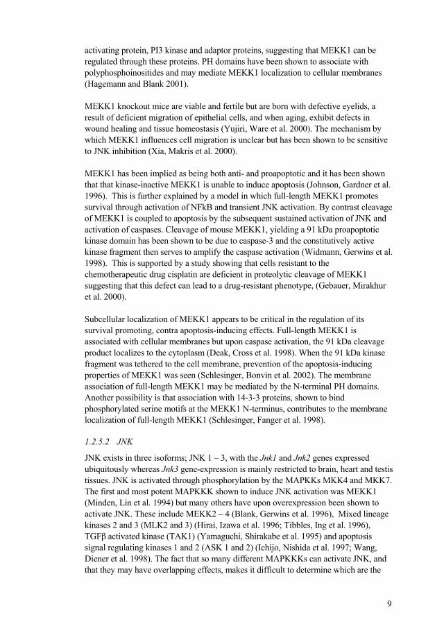
Regulatory mechanisms controlling proliferation, differentiation and apoptosis of cells are intertwined in a complex network of signaling pathways. The mitogen-activated protein kinase (MAPK) family constitutes a major network of pathways (Fig 1.), which potentially involves all of these crucial cellular events. The MAPK family kinases relay signals from the cell surface to the nucleus via sequential phosphorylations of its members. MAPKs are evolutionarily conserved in structure and function; in fact, all eukaryotic cells possess multiple MAPK pathways. Each MAPK cascade consists of three cytoplasmic kinases: a MAPkinase kinase kinase (MAPKKK), a MAP kinase kinase (MAPKK) and the MAP kinase itself.
At least 20 MAPKKK proteins have been identified in mammalian cells, and the possibility for these to be activated by different upstream stimuli allows the cell to respond coordinately, leading to a variety of possible responses. Stimuli shown to activate MAPKKKs are numerous and include hormones and growth factors that act through receptor tyrosine kinases, vasoactive peptides acting through G-protein coupled transmembrane receptors, transforming growth factor (TGF)- related polypeptides acting through serine-threonine kinase receptors and inflammatory cytokines of the tumor necrosis factor (TNF) family. Also, environmental stresses such as osmotic shock or genotoxic agents, ionizing radiation, ischemic injury, phosphorylation by other kinases, oligomerization and subsequent autophosphorylation are known to activate the MAPKKKs (Kyriakis and Avruch 2001; Uhlik, Abell et al. 2004).
Seven MAPKKs have been characterized (MEK1 and 2, MKK3–7) and are activated by the MAPKKKs through phosphorylation in conserved Ser/Thr motifs in the kinase domain (Kyriakis and Avruch 2001; Uhlik, Abell et al. 2004).
The MAPKs are activated by the MAPKKs through phosphorylation within a conserved Thr-X-Tyr motif in the activation loop of the kinase domain. There are three different MAPK classes; the extracellular signal-regulated kinase (ERK), c-Jun NH2-

8
terminal protein kinase (JNK) and p38-MAPK, all functioning as Ser/Thr kinases that can both phosphorylate other cytoplasmic targets but also have the ability to translocate to the nucleus and control gene expression via regulation of transcription factors. Mitogenic signaling induces activation and nuclear translocation of ERK, which results in proliferation, cell survival or differentiation whereas stress such as DNA-damaging chemotheraputic agents or irradiation activate JNK and p38 (Kyriakis and Avruch 2001; Uhlik, Abell et al. 2004; Viktorsson, Lewensohn et al. 2005).
Figure 1. MAPK signaling cascades
1.2.5.1 MEKK1
MEKK1 is a 196 kDa MAPKKK with a C-terminal catalytic domain and an N-terminal regulatory domain. MEKK1 was first named for its ability to phosphorylate the MAPKK MEK1, earlier known to be activated by Raf only. However, phosphorylation of MEK by MEKK1 induces only modest ERK activation whereas Raf-induced MEK activation effectively activates ERK, this despite the fact that the kinases phosphorylate the same amino acid residues. Furthermore, MEKK1 but not Raf, leads to activation of the JNK and p38 MAPKs, something that Raf is unable to accomplish.
MEKK1 contains both proline-rich sequences and pleckstrin homology (PH) domains at the N-terminus. Proline-rich sequences are involved in binding of proteins containing Src-homology 3 (SH3) domains such as tyrosine kinases, phospholipases, Ras GTPase
9
activating protein, PI3 kinase and adaptor proteins, suggesting that MEKK1 can be regulated through these proteins. PH domains have been shown to associate with polyphosphoinositides and may mediate MEKK1 localization to cellular membranes (Hagemann and Blank 2001).
MEKK1 knockout mice are viable and fertile but are born with defective eyelids, a result of deficient migration of epithelial cells, and when aging, exhibit defects in wound healing and tissue homeostasis (Yujiri, Ware et al. 2000). The mechanism by which MEKK1 influences cell migration is unclear but has been shown to be sensitive to JNK inhibition (Xia, Makris et al. 2000).
MEKK1 has been implied as being both anti- and proapoptotic and it has been shown that that kinase-inactive MEKK1 is unable to induce apoptosis (Johnson, Gardner et al. 1996). This is further explained by a model in which full-length MEKK1 promotes survival through activation of NFkB and transient JNK activation. By contrast cleavage of MEKK1 is coupled to apoptosis by the subsequent sustained activation of JNK and activation of caspases. Cleavage of mouse MEKK1, yielding a 91 kDa proapoptotic kinase domain has been shown to be due to caspase-3 and the constitutively active kinase fragment then serves to amplify the caspase activation (Widmann, Gerwins et al. 1998). This is supported by a study showing that cells resistant to the chemotherapeutic drug cisplatin are deficient in proteolytic cleavage of MEKK1 suggesting that this defect can lead to a drug-resistant phenotype, (Gebauer, Mirakhur et al. 2000).
Subcellular localization of MEKK1 appears to be critical in the regulation of its survival promoting, contra apoptosis-inducing effects. Full-length MEKK1 is associated with cellular membranes but upon caspase activation, the 91 kDa cleavage product localizes to the cytoplasm (Deak, Cross et al. 1998). When the 91 kDa kinase fragment was tethered to the cell membrane, prevention of the apoptosis-inducing properties of MEKK1 was seen (Schlesinger, Bonvin et al. 2002). The membrane association of full-length MEKK1 may be mediated by the N-terminal PH domains. Another possibility is that association with 14-3-3 proteins, shown to bind phosphorylated serine motifs at the MEKK1 N-terminus, contributes to the membrane localization of full-length MEKK1 (Schlesinger, Fanger et al. 1998).
1.2.5.2 JNK
JNK exists in three isoforms; JNK 1 – 3, with the Jnk1 and Jnk2 genes expressed ubiquitously whereas Jnk3 gene-expression is mainly restricted to brain, heart and testis tissues. JNK is activated through phosphorylation by the MAPKKs MKK4 and MKK7. The first and most potent MAPKKK shown to induce JNK activation was MEKK1 (Minden, Lin et al. 1994) but many others have upon overexpression been shown to activate JNK. These include MEKK2 – 4 (Blank, Gerwins et al. 1996), Mixed lineage kinases 2 and 3 (MLK2 and 3) (Hirai, Izawa et al. 1996; Tibbles, Ing et al. 1996), TGF activated kinase (TAK1) (Yamaguchi, Shirakabe et al. 1995) and apoptosis signal regulating kinases 1 and 2 (ASK 1 and 2) (Ichijo, Nishida et al. 1997; Wang, Diener et al. 1998). The fact that so many different MAPKKKs can activate JNK, and that they may have overlapping effects, makes it difficult to determine which are the

10
most important for JNK activation by certain stimuli, such as UV radiation or a certain genotoxin.
Protein-protein interactions are critical for signaling via the JNK pathway and signaling specificity may be mediated through the formation of protein complexes. Modulation of these complexes can be executed by scaffold proteins, which in response to specific stimuli appear to organize these complexes and thereby direct the signal. Thus, the JNK interacting protein (JIP) group of putative scaffolds has been identified. The assembly of a JNK complex by a scaffold protein in response to a certain stimulus may lead to efficient activation of JNK within a restricted region of the cell, notably the cytosol. The scaffold thus regulates the subcellular localization of JNK. In addition, the scaffold-bound signaling molecules may interact more efficiently because of their proximity and increased local concentration (Whitmarsh, Cavanagh et al. 1998; Davis 2000).
Regulatory inactivation of JNK may be executed by a specialized group of phosphatases called MAPK kinase phosphatases (MKPs), with phosphatase specificity to phosphotyrosine and phosphothreonine residues in the MAPK activation loop. MKPs are regulated through a highly reactive cystein, a subject for oxidation-mediated inhibition, which thus provides a means for activation of MAPKs in response to reactive oxygen species (ROS) or thiol-reactive compounds (Karin and Gallagher 2005).
JNK has been implicated in the apoptotic response to cellular stress, as exemplified by mouse embryo fibroblasts deficient for JNK, which exhibit resistance to UV-radiation treatment. The resistance mechanism is associated with failure to release cytchrome c from the mitochondria (Tournier, Hess et al. 2000). Also, chemotherapeutic drugs have been shown to require JNK for its apoptotic effects (Zanke, Boudreau et al. 1996; Sanchez-Perez and Perona 1999). However, the role of JNK in apoptosis is controversial in that JNK activation upon cellular stress may act either as a mediator of the stress response or alternatively provide a protective response. Possibly, this depends on cell type, the death stimulus, the duration of its activation and activity of other signaling pathways (Davis 2000; Jin and El-Deiry 2005). Most forms of environmental stress do not cause apoptosis under conditions sufficient for JNK activation. This may partly be because of survival signaling through pathways including NFkB, Akt and ERK, implying that JNK functions in a context of other signaling pathways (Davis 2000). Also, the time course of JNK activation appears to be of importance for JNK function (Chen and Tan 2000). This is exemplified by cytokines such as TNF that cause a transient activation of JNK that is not associated with apoptosis (Liu, Hsu et al. 1996). However when NFkB-deficient cells are stimulated with TNF, large amounts of ROS accumulate due to deficient expression of anti-oxidant enzymes, causing inhibition of the MKPs, and resulting in a prolonged JNK activation and subsequent apoptosis induction (Kamata, Honda et al. 2005).
JNK was first shown to exert its effects through phosphorylation of the transcription factor c-Jun, thereby increasing its transcriptional activity (Pulverer, Kyriakis et al. 1991), but also other proteins of the AP-1 transcription factor family such as JunB, JunD and ATF-2 can be activated. One proapoptotic protein that can be upregulated by

11
AP-1 is the Bcl-2 family member Bim (Ley, Ewings et al. 2005), providing an interesting link between JNK activation and proapoptotic signaling through the Bcl-2 family. The proapoptotic effects induced by AP-1 activation are further demonstrated as mutations of JNK phosphorylation sites on c-Jun, lead to partial protection against UV-induced cell death (Behrens, Sibilia et al. 1999).
However, UV-induced cell death has been shown not to require gene expression (Tournier, Hess et al. 2000) suggesting that JNK has other functions besides its transactivating effects. Indeed, JNK has been shown to also have post-translational effects on Bim, including phosphorylation of the Bim motif involved in binding to dynein motor complexes of microtubules. JNK-mediated phosphorylation would thus release Bim into the cytosol to exert its proapoptotic effects (Lei and Davis 2003). JNK may also phosphorylate Bcl-2 and Bcl-xL, thereby inhibiting their anti-apoptotic effects, e.g. sequestering Bim, PUMA, and Hrk (Basu and Haldar 2003; Brichese, Cazettes et al. 2004). It has also been reported that JNK can promote Bax proapoptotic activities by phosphorylation of 14-3-3 proteins, thereby releasing the 14-3-3 anchorage of Bax, which then can translocate to mitochondria and induce cytochrome c release (Tsuruta, Sunayama et al. 2004). Together, this confirms the complex role of JNK as a mediator of both pro- and antiapoptotic effects, and both through nuclear transactivating activities and cytosolic direct phosphorylations.
1.2.6 Methods to study cell death
There are many methods available for assessment of cell death, particularly apoptosis. The criteria for apoptosis proposed by Kerr and Wyllie are still valid for investigation of apoptosis but as the genetic background of apoptosis was clarified, additional possible approaches to study cell death were elaborated. These include more or less quantitative assays of, e.g., cytochrome c release, caspase activation and DNA cleavage. However, as the understanding of cell death processes evolves, problems with interpreting several assays have appeared. For example, caspase-independent cell death is put forward as a possible outcome, and there are in addition to cytochrome c other death-inducing factors that can be released from mitochondria (Hail, Carter et al. 2006). Also, the cell death response may not be exclusively apoptotic or necrotic, and mixed responses to a death stimulus are often the case. In addition one agent may induce different forms of cell death at different concentrations used, and at different time points.
Here, we have used caspase-specific cleavage of the epithelial cytoskeletal protein cytokeratin-18 (CK18) as method for apoptosis assessment. Caspase-mediated cleavage of CK18 leads to exposure of a neo-epitope in the resulting fragment and this stable fragment thus represents a specific marker of caspase-dependent apoptosis in epithelial cells (Hagg, Biven et al. 2002). Also, an antibody detecting total soluble CK18 has been used to measure both cleaved and uncleaved soluble CK18 released from dying cells, thus recognizing cell death due to both apoptosis and necrosis. Comparison of the levels of caspase-cleaved and total soluble CK18 will reflect the proportion of total cell death that ca be attributed to apoptosis (Kramer, Erdal et al. 2004). To assess and distinguish between apoptosis and necrosis, the annexin V protein, which has strong

12
specific affinity for phosphatidylserine (PS) was also used. Upon apoptosis induction, PS is translocated from the inner cytoplasmic leaflet of the plasma membrane to the cell surface, where it is recognized by the annexin V protein conjugated with e.g. FITC, which allows quantitation using flow cytometry. Annexin V can also be used in combination with fluorescent propidium iodide (PI), which intercalates with DNA. Because PI can only enter cells with compromised membrane integrity, i.e., necrotic or post-apoptotic cells, this method allows identification and quantitation of apoptotic and necrotic cells respectively. In time-course and dose-response experiments, it is thus possible to follow how chemotherapy-induced apoptosis may give way to necrosis over time and with increased dosage.
The MTT assay is generally used to measure cell viability. It is based on the reduction of tetrazolium salts from colorless to brightly colored insoluble derivatives called formazans. However, it has been shown that the MTT assay reflects levels of reducing equivalents NAD(P)H, produced mainly in the cytosol via glycolytic activity but also in the mitochondrial citric acid cycle. Thus, the MTT assay does not necessarily reflect the number of viable cells or their growth in a culture but instead reflects an integrated assembly of enzyme activities related to cell metabolism (Berridge, Herst et al. 2005). Here, we have used the MTT assay to study early effects on glycolysis/citric acid cycle activities after drug treatment.
1.3 ENERGY METABOLISM
1.3.1 ATP production by oxidative and non-oxidative processes
Glucose makes up a considerable part of the daily diet and is actively transported into mammalian cells, where it is converted by glycolysis into pyruvate, which in turn can be further metabolized by the citric acid cycle in the mitochondria. Glycolysis occurs both in the presence or absence of oxygen. Numerous prokaryotes and lower eukaryotes rely on anaerobic glycolysis to survive under non-oxygenated conditions, as shown already in the 19th century by Pasteur. However, higher organisms have found ways to more efficiently utilize glucose: by metabolizing the glycolysis product pyruvate in the citric acid cycle, followed by oxidative phosphorylation, 14 –fold more energy (ATP) can be obtained. However, this is at the price of oxygen dependence. Consequently, increased glycolysis confers the advantage of oxygen independence, whereas the concomitant major disadvantage is the low amount of energy produced (Ristow 2006).
13
1.3.2 Energy metabolism in tumors 1.3.2.1 Glucose metabolism
Otto Warburg discovered in the 1920s that tumor tissues exhibit a low respiratory rate compared to healthy non-malignant tissues, i.e., the malignant tissues mainly utilize glycolysis instead of mitochondrial oxidative phosphorylation, also in the presence of oxygen. This phenomenon was later termed the Warburg effect. The Warburg effect can be illustrated by the clinical use of [18F] fluoro-2-deoxyglucose (FDG) positron emission tomography (PET) in visualization of tumors. FDG, a glucose analog, is taken up by glucose transporters into the cell, where it is phosphorylated by hexokinase, the first enzyme of glycolysis. The FDG-6-phosphate formed cannot be further metabolized by glycolysis and can be eliminated by the renal system. However, because of the high glycolytic rate and the overexpression of glucose transporters in tumor cells, FDG accumulates in the tumor and can thus be imaged by PET (Gambhir 2002; Shaw 2006).
In accordance with their lower respiratory rate, it has been shown that high-proliferative tumors have fewer mitochondria compared to normal tissues and that these mitochondria are in general smaller (Pedersen 1978). Also, mitochondrial enzymes involved in oxidative phosphorylation such as the -F1 subunit of ATPsynthase, have been shown to be downregulated in human tumors of different origins (Cuezva, Krajewska et al. 2002; Isidoro, Martinez et al. 2004; Isidoro, Casado et al. 2005). In addition, uncoupling proteins, e.g., uncoupling protein 2 (UCP2), have been shown to be upregulated in tumor cells. UCP2 is a mitochondrial inner membrane anion carrier, which decreases the mitochondrial membrane potential and is considered a negative regulator of ROS production. Upregulation of UCP2 conveys cytoprotection by limiting oxidative injury and may be involved in mediating the switch from oxidative phosphorylation to glycosis as primary energy source in tumor cells (Harper, Antoniou et al. 2002; Horimoto, Resnick et al. 2004).
Several oncogenes have been implicated in the Warburg effect. The AKT oncogene is associated with enhanced glucose uptake and aerobic glycolysis. Akt is believed to increase the glucose transporters at the cell surface and to further stimulate hexokinase (Elstrom, Bauer et al. 2004). Also, the RAS, SRC and MYC oncogenes have been shown to be involved in induction of elevated levels of glucose transporters in the plasma membrane and to activate a wide array of glycolytic enzyme genes, such as those coding for phospho-fructokinase, lactate dehydrogenase and enolase (Flier, Mueckler et al. 1987; Osthus, Shim et al. 2000).
The tumor suppressor gene p53 has been shown to promote oxidative phosphorylation by transactivating the SCO2 gene required for assembly of the cytochrome c oxidase complex, a key component of the respiratory chain. Loss of SCO2 by loss of p53, results in defective oxidative phosphorylation, resulting in a switch to glycolysis for ATP production (Matoba, Kang et al. 2006). Another gene controlled by p53, which has recently been identified, is p53-induced glycolysis and apoptosis regulator (TIGAR). TIGAR expression lowers the levels of glycolysis and induces upregulation of the pentose phosphate pathway. This is involved in the synthesis of glutathione

14
(GSH), which provides protection against increased ROS levels. Consequently, p53-induced expression of TIGAR lowers glycolysis and protects cells from ROS-mediated apoptosis. In tumor cells with mutated p53, TIGAR-mediated inhibition of glycolysis does not occur, allowing for the higher glycolytic rate of tumor tissues (Bensaad, Tsuruta et al. 2006).
In support of the fact that oncogenes and tumor supressors play a role in tumor cell energy metabolism are observations that cells with activated Akt, Ras or Her2, or cells lacking tumor suppressor genes such as LKB1 and p53, undergo apoptosis under low-glucose circumstances. Interestingly, many of these cells are resistant to other forms of apoptotic stimuli, such as irradiation and chemotherapeutics (Shaw 2006).
1.3.2.1.1 AMP-activated protein kinase
An important enzyme in the control of the energy metabolism in the cell is the AMP-activated protein kinase (AMPK). It consists of an alpha catalytic subunit, containing a Ser/Thr protein kinase catalytic domain, and regulatory beta and gamma subunits, all highly conserved between species. The alpha subunit also contains a number of phosphorylatable residues, notably Thr-172, the phosphorylation of which is essential for AMPK activity (Carling 2005).
AMPK is activated allosterically by increased levels of 5´-AMP and is inhibited by ATP and glycogen. It is therefore regarded as an energy sensor. When a drop in the ATP:AMP ratio occurs, AMPK activates compensatory catabolic pathways such as glycolysis and fatty acid oxidation and induces increased glucose uptake through upregulation of the glucose transporters GLUT1 and -4. Energy-requiring processes such as fatty acid synthesis, protein synthesis and cell growth are switched off through effects on the mammalian target of rapamycin (mTOR) via the tumor suppressor tuberous sclerosis complex 2 (TSC2). Also, AMPK integrates signals from adipokines such as leptin and adiponectin to upregulate food intake, body weight and glucose-lipid homeostasis (Ashrafian 2006).
AMPK has been shown to be phosphorylated by LKB1 (Shaw, Kosmatka et al. 2004), a protein encoded by a tumor suppressor gene inactivated in cancer patients with Peutz-Jeghers syndrome (Hemminki, Markie et al. 1998; Jenne, Reimann et al. 1998). The role of AMPK in cancer is complex. Since mutated LKB1 is associated with cancer, the effects of AMPK would be associated with antitumor effects. This is in agreement with reports that AMPK phosphorylates and consequently activates p53, leading to replicative senescence (Jones, Plas et al. 2005) and as mentioned above, that AMPK can be involved in decreased proliferation by its effect on mTOR. On the other hand, the effects of AMPK to facilitate glucose and fatty acid metabolism would be associated with pro-survival effects, favoring tumor proliferation. An attempt to explain this paradox is proposed by Ashrafian (Ashrafian 2006), who suggests that AMPK has negative effects on tumor formation in the early stages but favor advanced tumors. Premalignant lesions with sufficient blood supply allow the tumor cells to produce ATP by oxidative phosphorylation rather than glycolysis. In such a setting, there is no advantage in importing excess glucose, and any AMPK activation would slow down cellular proliferation in these cells that may retain intact p53, TSC2 and mTOR. By

15
contrast, advanced tumors often have mutations in the tumor suppressors downstream of AMPK, allowing it to upregulate glycolysis and fatty acid metabolism without inducing any growth limiting effects.
1.3.3 Fatty acid metabolism
It has long been known that fatty acid synthesis occurs at very high rates in tumor tissues. Thus, fatty acid synthase (FASn), a lipogenic enzyme catalyzing the terminal steps in the de novo biogenesis of long-chain fatty acids, has been shown to be overexpressed and hyperactive in tumors (Kuhajda 2006). FASn exists in the cytoplasm of cells and consists of two identical polypeptides of 260 kDa that each contains several catalytical domains. Using acetyl-CoA and malonyl-CoA as two-carbon donors and NADPH as reducing equivalent, FASn can synthesize long-chain fatty acids. The principal product of FASn is the fatty acid palmitate, a 16-carbon molecule, but other shorter chains can also be formed. Fatty acids are important constituents of all biological membrane lipids but FASn activity is mostly used for energy storage since in well-nourished individuals, mainly circulating lipids from dietary intake are used for synthesis of biomembranes, even in tissues with high cellular turnover. FASn is therefore expressed at low or undetectable levels in most normal human tissues (Baron, Migita et al. 2004; Menendez, Vellon et al. 2005).
In contrast, in a wide array of human cancers FASn is overexpressed, also under circumstances of high levels of fatty acids in the circulation. FASn overexpression is mainly seen in epithelial carcinomas such as prostate, breast, colorectum, bladder, ovary, eosophagus, stomach, lung, thyroid and head and neck cancers. FASn overexpression can be coupled to poor prognosis and thus can serve as a prognostic indicator (Kuhajda 2000; Menendez and Lupu 2004). The fact that FAS is overexpressed in so many tumor types suggests that de novo synthesis of fatty acids is an important process required for growth and survival of transformed cells. It is possible that this is due to the greater need for energy of tumor cells.
It has indeed been shown that increased FAS activity provides substrates for energy extraction via -oxidation of fatty acids (Kuhajda 2000; Thupari, Landree et al. 2002). Studies of cancer patients, indeed show an increase in fatty acid turnover, oxidation and clearance (Legaspi, Jeevanandam et al. 1987; Hyltander, Drott et al. 1991). Another indication of enhanced -oxidation in tumors is that overexpression of -methylacyl CoA racemase (AMACR), an enzyme that converts fatty acids to isomers prior to -oxidation is expressed at high levels in most prostate cancers but not in normal prostate epithelium, indicative of an enhanced -oxidation in these tumors (Adley and Yang 2006). Pharmacological FASn inhibitors such as C75 and cerulenin as well as RNA interference of FASn lead to apoptosis of cancer cells. How this happens is not clear but one explanation may be that accumulation of malonyl-CoA is cytotoxic (Kuhajda 2000; Pizer, Thupari et al. 2000; De Schrijver, Brusselmans et al. 2003), possibly due malonyl-CoA inhibition of carnitine palmitoyltransferase-1 (CPT-1), the enzyme responsible for the uptake of fatty acids into the mitochondria where -oxidation takes place (Bandyopadhyay, Zhan et al. 2006). Thus, -oxidation of fatty acids may play an important role in fatty metabolism.
16
1.4 TREATMENT OF CANCER
The major treatment approaches for cancer consist of surgery, radiation, systemic chemotherapy, hormonal therapy and immunotherapy. Surgery and radiation are still the most successful means of treating cancer localized to the primary site, but neither is usually considered curative once the tumor has metastasized beyond regional lymph nodes. Instead, the systemic therapies can kill tumor cells that have metastasized to distant sites and these treatment modalities have a greater chance of curing patients with minimal tumor burden. Consequently, surgery and radiation may be useful in decreasing the tumor burden and thereby maximizing the impact of subsequent systemic approaches. Chemotherapeutic drugs must be given when the number of tumor cells is low enough to permit their destruction at doses that can be tolerated by the patient. Thus, the opportunity for cure is most likely during the early stage of disease or immediately after surgery when the tumor burden is minimized.
1.4.1 Chemotherapy
There are different classes of chemotherapeutic drugs, divided according to their mechanisms of action: alkylating and platinum agents – highly reactive agents that can form strong chemical bonds with thiol sulfurs and amino nitrogens in proteins and nucleic acids, e.g., cyclophosphamide and cisplatin; antimetabolites – agents that by mimicking the structure of a cellular reagent can interfere with its normal biochemical function such as DNA or folate synthesis, e.g., 5-fluorouracil and metohrexate; topoisomerase inhibitors – agents that inhibit the supercoiling of DNA by toposiomerases, thereby interfering with transcription and replication of DNA, e.g., camptothecin and etoposide; microtubular-targeting agents – which prevent cell division by inhibiting microtubule function, e.g. vincristine; and finally others – e.g., new target-specific agents such as tyrosine kinase inhibitors.
1.4.1.1 Cisplatin
Cisplatin is a platinum agent with a central role in cancer chemotherapy, and is one of the few chemotherapeutic drugs with ability to completely cure a tumor form, namely testicular cancer, for which the overall cure rate exceeds 90%. Other cancer forms treated with cisplatin include ovarian, cervical, head and neck and non-small cell lung cancer. The treatment is limited by side effects such as nephro- and neurotoxicity, of which neurotoxicity is dose-limiting and can result in peripheral neuropathy, tinnitus and hearing loss (Wang and Lippard 2005).The first platinum antitumor compounds were discovered as a result of studying effects of electrical currents on bacterial growth. Growth inhibition was found to occur but was caused by a platinum complex of ammonia and chloride produced in the medium at the platinum electrode. Several compounds were found to have antitumor effects against murine tumors and the most active was cisplatin (Rosenberg, Vancamp et al. 1965).
17
When cisplain has entered the cell it undergoes aquation where two chloride ligands are replaced by water molecules, facilitated by the low cellular concentration of chloride ions. The aquated molecule is positively charged and highly reactive towards DNA, RNA and proteins. Classically, DNA has been considered the main target structure of cisplatin, where it forms covalent bonds with purine bases to induce mainly intrastrand, but also interstrand, crosslinks (Wang and Lippard 2005). However, only about 1 % of the cisplatin that has entered the cell forms DNA adducts (Eastman 1991). It has been shown that cisplatin induces apoptosis also in the absence of nucleus (Mandic, Hansson et al. 2003), indicating that, in addition to its DNA-damaging effects, cisplatin also affects other cellular targets
1.4.2 Tumor resistance to treatment
Treatment of tumors is often faced with development of multidrug resistance (MDR). This is a phenomenon where tumor cells that have been exposed to one cytotoxic agent develop cross-resistance to a range of structurally and functionally unrelated compounds. In addition, MDR can also occur in a tumor without earlier exposure to any chemotherapeutic agent. There are three major mechanisms for drug resistance of tumor cells: 1) decreased uptake of drugs that require transporters to enter cells, 2)
changes that affect the capability of the drug to kill the tumor cell such as alterations of the cell cycle, increased repair of DNA damage, reduced apoptosis and alterations in the metabolism of drugs, and 3) increased energy dependent efflux of drugs (Longley and Johnston 2005).
There are numerous transporters in the cell membrane that have been proposed to be responsible for the uptake of different chemotherapeutic drugs. Cisplatin, for example, was long considered to enter the cell by passive diffusion but has now been reported to be actively transported by Ctr1, a high-affinity copper transporter (Ishida, Lee et al. 2002). Mutation or deletion of this gene results in increased cisplatin resistance and reduction of platinum levels inside the tumor cell (Wang and Lippard 2005).
Inactivating mutations or loss of the p53 tumor suppressor are the most common genetic alterations in human cancer, resulting in resistance to both radiation and cytotoxic drugs. Overexpression of Bcl-2 leading to downregulation of the intrinsic pathway is seen in many tumors, particularly B-cell lymphomas. Bax has been reported to be inactivated by mutations in colorectal cancers, and defective caspase activation has been observed in, e.g., ovarian cancer, leukemia and melanoma. Akt is another important resistance factor and has been shown to suppress apoptosis, e.g., by phosphorylating and inhibiting Bad and caspase-9 (Hersey, Zhuang et al. 2006; Meiler and Schuler 2006). A classic resistance mechanism is the increased efflux of cytotoxic drugs from the cell, mediated by the ATP-binding cassette (ABC) family of transporters. This family has many members but only about ten are involved in drug resistance, the most well-known being P-glycoprotein (P-gp), also known as MDR1. Expression of P-gp is usually highest in tumors derived from tissues that normally express some P-gp such as epithelial cells of the colon, kidney, adrenal, pancreas and liver, resulting in potential resistance to chemotherapeutic drugs even before therapy is initiated. In other tumors P-gp expression may be low at the start of treatment but

18
increase after exposure to chemotherapy. (Fardel, Lecureur et al. 1996; Gottesman, Fojo et al. 2002; Szakacs, Paterson et al. 2006).
1.4.3 The concept of sensitization
The fact that tumors develop resistance to cancer treatments has stimulated the pursuit of agents that can sensitize tumor cells to the treatment, e.g., drugs which target resistance mechanisms. The goal of treatment sensitization is to increase the efficacy of the therapy, in addition to decreasing the drug doses needed and thereby reducing toxic side effects of the treatment.
Inhibition of P-gp as a means to reverse MDR has been extensively studied and many agents that modulate the P-gp transporter, including verapamil, cyclosporine A and several calmodulin antagonists were identified already in the 1980s. Many of these are themselves substrates for P-gp and thus act by competing with the cytotoxic drug for efflux by the P-gp pump. High concentrations of the sensitizers were therefore required, resulting in high toxicity. However, further development has now led to drugs that specifically bind the P-gp pump, some of which are currently undergoing clinical trials (Thomas and Coley 2003).
There are many examples of attempts to potentiate apoptotic responses in tumor cells during treatment. The basic idea is to activate additional apoptotic signaling pathways, or to inhibit anti-apoptotic pathways. One approach is to induce the extrinsic pathway in order to add to the effects of the intrinsic pathway activated by chemotherapeutic drugs or radiation. The TNF family of proteins binds cell surface receptors whose activation will induce the extrinsic apoptosis pathway. One such member, the TNF-related apoptosis inducing ligand (TRAIL), has been shown to induce apotosis in a wide range of tumor cells. TRAIL binds the receptors DR4 and DR5, thereby recruiting adaptor proteins which lead to activation of caspase-8 and trigger apoptosis. Synergistic effects have been seen between TRAIL and ionizing radiation in breast cancer cells, leukaemias, colon carcinoma and gliomas. These effects seem to be p53 dependent and may involve radiation-induced upregulation of DR5. Also, DR5 deficient mice have exhibited resistance to ionizing radiation treatment indicating that endogenous TRAIL is a mediator of radiation-induced tissue damage. TRAIL in combination with several chemotherapeutic drugs has also resulted in synergistic apoptosis induction. This may be due to upregulation of DR4 and -5 in addition to downregulation of Bcl-2/Bcl-xL or upregulation of Bak or caspases (Cretney, Shanker et al. 2006).
COX-2 is a key enzyme required for conversion of arachidonic acid to prostaglandins and mainly mediates a pro-inflammatory role. COX-2 is often overexpressed in malignant tissue. Inhibition of COX-2 has been shown to increase the effects of irradiation in a number of cell systems. The mechanism behind this is not clear but increased apoptosis has been seen, perhaps elicited by inhibition of DNA damage repair. Another mechanism of the radiosensitizing effects of COX-2 is demonstrated by experimental rodent models where COX-2 inhibition is associated with decreased angiogenesis (Sminia, Kuipers et al. 2005).

19
Many cancer treatments induce cell death via DNA-damage, wherefore the DNA damage response is an interesting target for enhancing sensitivity. One approach to this is the development of small-molecule inhibitors of poly(ADP-ribose) polymerase (PARP) and the checkpoint kinase (Chk)1, both involved in the DNA damage response. PARP recognizes single-strand breaks caused by oxidative stress, radiation, topoisomerase inhibitors and alkyating agents. Inhibition of PARP activation during the DNA damage response may delay DNA repair and thus enhance the cytotoxicity. Mice deficient for PARP, exhibit hypersensitivity to alkylating agents and radiation, and display increased genomic instability after DNA damage (de Murcia, Niedergang et al. 1997; Trucco, Oliver et al. 1998; Masutani, Nozaki et al. 2000). Chk1 inactivates important cell cycle proteins and arrests the cell in G2/M transition, making time for DNA repair. Tumors are often defective in the G1 DNA damage checkpoint because of mutations in, e.g., p53 and pRb, rendering these cells more dependent on the G2
checkpoint. This implies abrogation of the G2 checkpoint as means to enhance DNA damaging therapies. Chk1 is thus a potential target for inhibition of G2 arrest and a number of drugs directed against Chk1 have shown promising effects as potentiators of a variety of tumor therapies including radiation, cisplatin, 5-fluorouracil, doxorubicin and gemcitabine (Luo and Leverson 2005). Normal cells are expected to be able to cope with a temporary loss of G2 checkpoint when subjected to chemotherapy whereas the tumor cells undergo cell death.
Another method for sensitization is the use of antisense oligonucleotides directed against gene products overexpressed in tumor cells, in addition to sublethal doses of chemotherapy. The antisense molecules are short stretches of 16-18 chemically modified nucleotides, designed to hybridize with specific complementary mRNA to block production of proteins encoded by these mRNA transcripts. This approach provides possibilities to downregulate anti-apoptotic proteins such as Bcl-2 and Bcl-xL (Yang, Feng et al. 2004; Yamanaka, Rocchi et al. 2005), MDM-2 (Bianco, Ciardiello et al. 2005) Raf-1 (Mewani, Tang et al. 2004) and xIAP (Lima, Martins et al. 2004).
Photodynamic therapy involves the administration of a photosensitizer, followed by local illumination of the tumor with light of the appropriate wavelength to activate the specific drug. Upon light absorption the drug is activated by excitation and can in the presence of oxygen react directly with oxygen or with a substrate leading to formation of reactive radicals. The effects of photodynamic therapy include induction of apoptosis and necrosis as well as damaging the vasculature of the tumor resulting in hypoxia and starvation of the tumor tissue. Photodynamic therapy has shown promising results as a chemosensitizer in experimental settings in combination with for example methotrexate (Sinha, Anand et al. 2006) and a COX-2 inhibitor (Ferrario, Fisher et al. 2005).
20
2 RESULTS AND DISCUSSION
2.1 PAPERS I AND II
A major goal of studying cellular signaling in tumor cells induced by chemotherapy treatment is to elucidate various basal and drug-induced signaling circuits and their interactions. Through a greater understanding of these pathways, efforts to increase sensitivity of tumor cells to treatment and to decrease resistance mechanisms can be made. Here, we have studied cisplatin in order to further elucidate the complex pathways it induces in tumor cells.
2.1.1 Cisplatin induces calpain activation
Earlier studies by the group on cisplatin-induced apoptosis has shown that it involves signaling via the pro-apoptotic Bcl-2 family member Bak. Activation of Bak is accompanied by a conformational change and this modulation was shown to be almost completely inhibited by a dominant negative form of MEKK1, dnMEKK1. However, this inhibition did not completely block apoptosis, suggesting that other pathways besides Bak activation are present after cisplatin treatment (Mandic, Viktorsson et al. 2001). This prompted the first study of this thesis, regarding the protease calpain and its role in cisplatin-induced apoptosis.
Calpain had at the time been implicated in radiation-induced apoptosis (Waterhouse, Finucane et al. 1998) and Paper I shows that two different calpain inhibitors with different modes of action, calpeptin and PD150606, both inhibited cisplatin-induced apoptosis by approximately half. Addition of calpeptin at 8 h after cisplatin did not inhibit apoptosis, showing that calpain activation leading to apoptosis is an early event.
Using fluorogenic calpain substrates, calpain was shown to be activated already after 3 h of cisplatin treatment in the melanoma cell line 224. Activation of calpain was seen also after treatment with camptothecin or etoposide. Furthermore, cisplatin-induced calpain activation could also be seen in lung- and breast cancer cell lines, suggesting that calpain activation is a process that can be induced in different tumor types in response to different death stimuli.
Calpain requires calcium for its activation and by using a cell-permeable calcium indicator, intracellular calcium was shown to be increased by 50% after 1 h of cisplatin treatment. As expected, calcium accumulation could be inhibited by treatment with the calcium chelator BAPTA-AM, which also inhibited cisplatin-induced apoptosis.
2.1.2 Calpain cleaves Bid
Calpain was shown to cleave the BH3-only protein Bid into its active form. This cleavage was blocked in cell culture by calpain inhibitors but not caspase- or cathepsin inhibitors. Classically, Bid has been implicated in the extrinsic, death receptor mediated pathway, where it is cleaved and activated by caspase-8 (Suzuki, Takahashi-Niki et al.
21
2004). We report that in cisplatin-induced apoptosis, cleavage of Bid is induced by calpain, but of course the possibility of a role for caspase-8 in this process could not be ruled out. Thus, a caspase-8 inhibitor was tested, and was found to inhibit neither cisplatin-induced Bid cleavage nor apoptosis induction. Similar results were seen with inhibitors of caspases -3 and -7. These results, in addition to Bid cleavage in caspase-3 deficient MCF-7 breast cancer cells, support a calpain-specific activation of Bid without involvement of caspases.
Importantly, calpain was shown to cleave recombinant Bid in vitro at a site between Gly70 and Arg71, generating a 14 kDa fragment with ability to induce release of cytochrome c from mitochondria. This truncated form of Bid is similar in size to caspase cleaved Bid, making them difficult to separate on a Western blot. This, in turn, implies that calpain-cleaved Bid may have been overlooked in earlier studies.
2.1.3 Calpain regulation
Calcium plays important roles for essential processes of the cell, e.g., proliferation, differentiation and apoptosis. Release of calcium from intracellular pools, mainly the endoplasmic reticulum (ER) is considered to trigger and modulate apoptotic signal transduction pathways in response to cellular stress. ER stress can be the result of both calcium depletion and overload, leading to disturbances in ER protein folding and induction of the unfolded protein response (UPR) pathway. The UPR serves to remove incorrectly folded proteins but may also, upon severe stress, contribute to apoptosis induction (Kaufman 2002; Orrenius, Zhivotovsky et al. 2003). Prolonged ER stress has been shown to activate caspase-12 by the action of calpain. Caspase-12 can then further activate downstream caspases, constituting a link between the calpain and caspase protease families (Nakagawa and Yuan 2000). Cytotoxic drugs has long been known to cause calcium influx into cells (Trump and Berezesky 1995) and it is possible that calpain can be differentially regulated by different mechanisms of calcium accumulation. Thus, some drugs may induce prolonged UPR with a late and indirect calcium release, whereas other drugs, e.g. cisplatin, induce an early and direct calcium influx into the cytoplasm. Hägg et al have further studied drug-induced calcium signaling in apoptosis and found correlations between late (7 h after treatment) calcium requirement, calpain activation and sustained JNK signaling. In contrast to what Paper I reports for cisplatin-induced apoptosis, late calcium release and calpain activation by several other drugs did not induce calpain cleavage of Bid or Bax (Hägg, Li et al. manuscript in preparation). This is in agreement with the possibility that there are different modes of activation of calpain, which in turn can activate different signaling processes. This is further illustrated by the fact that MEFs deficient for Bid did not exhibit resistance to cisplatin, i.e., indicating that calpain activation and calcium release in these cells induce other signaling events leading to apoptosis.
Another conclusion of the paper is that calpain and MEKK1 do not act via the same pathway. This is demonstrated as dnMEKK1 did not affect cisplatin-induced Bid cleavage but did inhibit Bak conformational activation, as earlier shown (Mandic, Viktorsson et al. 2001). Conversely, the calpain inhibitor calpeptin did not affect Bak modulation but inhibited cisplatin-induced calpain activation and apoptosis by approximately half. Finally, dnMEKK1 and calpeptin had additive effects on inhibition
22
of apoptosis, suggesting that cisplatin treatment induces at least two separate signal transduction pathways involving Bcl-2 family members.
2.1.4 Bak conformational activation
The importance of Bak for cisplatin-induced apoptosis, as exemplified by antisense Bak that rendered melanoma cells partially resistant to cisplatin (Mandic, Viktorsson et al. 2001), motivated further studies of the regulation of Bak in Paper II of this thesis. Bak activation can be demonstrated using an antibody directed against an N-terminal epitope that is exposed as Bak undergoes a conformational change associated with its apoptosis-inducing qualities (Griffiths, Dubrez et al. 1999). Here, Bak conformational activation was induced within 18 h by adenovirally mediated overexpression of a dominant positive mutant of MEKK1 (dpMEKK1). However, this activation of Bak is not necessarily coupled to apoptosis, since dpMEKK1 did not induce apoptosis until after 40 h, whereas cisplatin induced Bak conformational activation within16 h and massive apoptosis within 24 h.
Bak activation was also studied using the crosslinker 1,6-bis- maleimidohexane (BMH). BMH is a 16 Å, membrane permeable, homobifunctional maleimide that covalently cross-links sulfhydryl groups, i.e. that can intramolecularily crosslink the two cysteines of Bak. In SDS-PAGE, native Bak migrates as a 21 kDa species and loss of this form is associated with Bak activation (Wei, Lindsten et al. 2000). In kinetics experiments exposure of the N-terminal epitope in Bak was detected by flow cytometry already at 9h after cisplatin treatment, whereas loss of the 21 kDa species occurred after 13 h. It is problematical to compare time course experiments performed with different methods, but with these disparate time points of activation we can at least reflect on the possibility that Bak conformational activation takes place in two separate steps; one in the N-terminal exposing the N-terminal epitope, followed by a further deconvolution of the protein making its cysteines inaccessible for the BMH cross linker.
2.1.5 Bak complex formation
The mechanism(s) for cytochrome c release remain partially unclear (see section 1.2.3). However, the Bcl-2 family members Bak and Bax provide an attractive alternative of pore formation since they by numerous studies have been confirmed as regulators of cytochrome c release from mitochondria. For example, Bax has been shown to form pores in lipid membranes (Antonsson, Conti et al. 1997) and cleaved Bid has been shown to induce cytochrome c release from mitochondria by inducing Bak oligomerization (Wei, Lindsten et al. 2000).
Using gel filtration under native conditions, we studied cisplatin-induced Bak oligomers, and found that in non-apoptotic cells Bak occurs as mono- and dimers whereas apoptotic cells display Bak complexes in the size range of 80 - 170 kDa. We speculate that these complexes are Bak homo-oligomers and the size range would then correspond to trimers and up to hexamers, which would be able to form pore structures able to release cytochrome c. The timepoint of appearance of complexes a few hours before the execution of apoptosis is in agreement with pore formation leading to cytochrome c release. The possibility of other proteins being involved in the Bak complexes was of course considered, but no co-elution with any other Bcl-2 family

23
member was observed. We have particularly looked into the possibility of Bax-Bak complexes since such have been previously reported (Sundararajan, Cuconati et al. 2001). However, in our cell systems, translocation of Bax to mitochondria and Bax activation, as seen by a widely used antibody recognizing only the active Bax conformation, occurs much later than Bak activation and complex formation. Thus, Bax activation is seen when cells already have fragmented nuclei, i.e. after cytochrome c release and caspase activation. This makes Bax an unlikely candidate to be part of Bak complexes associated with apoptosis in our system. The results might indicate a possible drug-specificity in the mechanisms of cytochrome c release from mitochondria regarding choice of pore-forming protein. Bak and Bax have been shown to have overlapping activities, i.e., apoptosis can take place if one of these proteins is knocked out, but cells become unable to release cytochrome c from mitochondria if both are missing (Lindsten, Ross et al. 2000; Wei, Zong et al. 2001) Here, MEFs deficient for Bak were found to be twice as resistant to cisplatin as Bax deficient cells, suggesting that Bak is the preferred pore forming protein in cisplatin-induced apoptotic signaling. This is also supported by the absence of Bax in the Bak complexes. However, it is possible that in the complete absence of Bak, this role can be taken over by Bax.
Both Bak and Bax have been shown to release cytochrome c in interaction with VDAC independently of MPT (Shimizu, Narita et al. 1999), whereas other have not found any interactions (Wei, Lindsten et al. 2000). We did not observe co-elution of VDAC and Bak complexes. Interestingly, VDAC-2 has been reported to be associated with Bak in non-apoptotic cells, where it keeps Bak in its inactive form. After apoptotic stimuli, BH3-only proteins would displace VDAC-2 and induce Bak oligomerization (Cheng, Sheiko et al. 2003). Possibly, VDAC-2 might explain the presence of high molecular weight complexes, more than 170 kDa, in the non-apoptotic cells in our system.
2.1.6 Requirement for JNK in Bak complex formation
The role of JNK in apoptosis has long been unclear and its regulation appears to be quite complex. Paper II demonstrates that JNK activation is a prerequisite for the formation of the Bak complexes associated with apoptosis. Overexpression of dominant-positive MEKK1 induces Bak conformational activation but not the formation of Bak complexes and thus no apoptosis. The role of JNK was first shown using the JNK inhibitor SP600125, which blocked cisplatin induced complex formation and was confirmed by the finding that no Bak complexes appeared in MEFs deficient for JNK1. These cells were accordingly also resistant to cisplatin. Generally, JNK is believed to lie downstream of MEKK1 in the signal cascade. Paper II shows that dpMEKK1 was unable to induce Bak complexes, and apoptosis was induced only when an extraneously induced JNK signal was added. This is in agreement with studies showing that MEKK-induced apoptosis is independent of its role as regulator of JNK, and that the potentiating effects on irradiation-induced apoptosis by MEKK1 are not dependent on JNK activation (Muhlenbeck, Haas et al. 1998; Bild, Mendoza et al. 2002).
There have been no reports on a direct phosphorylation of Bak, which suggests that there must be a mediator of the JNK signal to Bak. Many studies report associations between JNK and other Bcl-2 family members. JNK has been shown to directly

24
phosphorylate Bcl-xL and Bcl-2 and to thereby block their inhibitory effect on apoptosis (Yamamoto, Ichijo et al. 1999; Basu and Haldar 2003). The BH3-only proteins Bid and Bim that have been reported to regulate the actions Bak and Bax (Letai, Bassik et al. 2002) are of course interesting as possible targets of JNK. Bid has been reported to induce oligomerization of Bak (Wei, Lindsten et al. 2000) but no phosphorylation of Bid by JNK has been shown. Importantly, we found that MEFs deficient for Bid were not resistant to cisplatin. Bim, on the other hand has been reported to be both phosphorylated by JNK and to induce activation of Bak and Bax leading to cytochrome c release (Lei and Davis 2003). In agreement with this, we report that Bim deficient MEFs were resistant to cisplatin induced apoptosis. Furthermore, unpublished results indicated that Bim was phosphorylated after 14 h of cisplatin treatment and that this effect was sensitive to the JNK inhibitor SP600125. These results indicate a role for Bim as mediator of JNK effects on Bak oligomerization, warranting further studies.
Together, Papers I and II demonstrate the complicated signaling circuits induced by cisplatin, a feature which this drug likely shares with many other highly reactive molecules that show high cytotoxicity. In summary, cisplatin induces three separate signals to MEKK1, calpain and JNK, respectively (Fig. 2). The relative importance of these signaling pathways may differ between tumor types and treatment modalities. Further studies may enable targeting of specific pathways crucial for a specific tumor type or treatment regimen.
Figure 2. Cisplatin-induced signaling patways

25
2.2 PAPERS III AND IV
Here, we have investigated the possibility of sensitizing tumors to chemotherapy by way of exploiting the fact that tumor cells have altered energy metabolism, notably increased dependence on upregulated glucose transporters, aerobic glycolysis and on fatty acid -oxidation.
2.2.1 Different modes of action for DG and BP
Paper III focuses on 2-deoxyglucose (DG) and 3-bromopyruvate (BP), both reported to inhibit glycolysis by targeting its first enzyme; hexokinase II (HXK). These drugs were used at doses that per se induced low level apoptosis (DG) and some necrosis (BP). In HCT 116 colon carcinoma cells, both DG and BP reduced cellular ATP by approximately half, showing that these drugs indeed influence energy production of the cells. Next, the MTT assay, which reflects cellular levels of NAD(P)H, derived from glycolysis and the citric acid cycle (Berridge, Herst et al. 2005) was used. A 3h incubation with DG or BP led to a DG-mediated decrease in MTT signal of approximately 40%, whereas BP had no effect. BP and DG have both been presented as direct inhibitors of HXK (Ko, Pedersen et al. 2001). The above results, however, indicate different modes of action of BP and DG. Since disturbance of oxidative phosphorylation/mitochondrial respiration would be expected to influence the mitochondrial membrane potential ( m), the effects of DG and BP on m were examined by assessing exclusion of the fluorescent probe TMRE from mitochondria. DG treatment at a dose that decreased both ATP and NADH levels did not have any effect on m, in agreement with its role as a glycolysis inhibitor. By contrast, BP, which had no effects in the MTT assay, inhibited m considerably at a dose that decreased ATP levels.
BP is an alkylating agent and can thus be expected to be highly reactive, have multiple modes of action and possibly interfere with many structures of the cell. The effects on
m shown here are in agreement with an earlier published observation that BP can inhibit succinate dehydrogenase (Sanborn, Felberg et al. 1971), a member of the electron transport chain. Loss of m has been associated with necrosis (Li, Johnson et al. 2004; Nakagawa, Shimizu et al. 2005). When BP was here combined with low doses of cisplatin or oxaliplatin, massive cell death was induced, as seen by an assay reflecting the release of lactate dehydrogenase (LDH) from cells. However, no potentiation of chemotherapy-induced apoptosis could be seen, indicating that the cell death is mainly due to induction of necrosis. Also, BP has recently been shown to induce inactivation of H+-vacuolar ATPase (Dell'Antone 2006), an enzyme responsible for acidification of many internal compartments in the cell, which is reported to participate in many important physiological processes as well as in pathological conditions e.g., diabetes, Alzheimer´s disease, cardiovascular diseases, osteoporosis and cancer (Bowman and Bowman 2005). Importantly, by affecting the H+-vacuolar ATPase, BP was shown to elicit severe deterioration of intracellular pH gradients, both in normal and tumor cells, implying that systemic use of BP may be associated with

26
severe toxicity (Dell'Antone 2006). Altogether, these observations, in combination with the fact that apoptosis may possibly be a more advantageous outcome of cancer therapy than necrosis (Zeh and Lotze 2005) led us to continue working with DG only. This since DG appears to be a specific glycolysis inhibitor and earlier has been shown to induce apoptosis in tumor cells (Aft, Zhang et al. 2002). In Paper III the combination of DG with a low dose of cisplatin did indeed lead to a significant potentiation of apoptosis. This potentiation could be inhibited by addition of pyruvate, also in support of DG effects via specific inhibition of glycolysis.
2.2.2 DG potentiates DNA-damaging agents
In Paper III, twenty agents selected to represent different modes of action were assessed at low doses for apoptosis induction in combination with DG. DG increased 24 h and 48 h apoptosis levels induced by some drugs, mainly DNA-damaging agents such as cisplatin, ellipticine and camptothecin. To study DG sensitization in a more physiological setting, MDA-MB-231 breast cancer tumor cells, xenografted to nude mice, were cut into 200 µM slices and treated for up to 72 h. Here, potentiation of apoptosis was seen with all three drugs tested. This is interesting since the MDA-MB-231 cells are highly resistant to chemotherapy treatment, due to P-gp expression and mutant Ras and p53. In addition, they were resistant to these drug combinations when assayed in standard tissue culture. This demonstrates that monolayer cultures differ greatly from the complex environmental conditions and differential gene expression (Erlichman and Vidgen 1984; Kobayashi, Man et al. 1993). That DNA-damaging drugs may be potentiatied by DG is in agreement with reports on DG as a potent radiosensitizer (Jain, Kalia et al. 1985; Varshney, Dwarakanath et al. 2005). Repair of DNA breaks is an ATP requiring process (Durante, Grossi et al. 1991). Thus, the mechanism for DG-induced sensitization of DNA damaging agents may involve attenuated DNA repair. Indeed, the radiosensitizing effects of DG have been attributed to inhibition of energy dependent processes involved in the repair of irradiation-induced DNA damage, leading to fixation of these DNA lesions (Jain, Kalia et al. 1985).
In a study of p53 and its effects on mitochondrial respiration, it was shown that p53 induces expression of the transporter synthesis of cytochrome c oxidase 2 (SCO2), a protein necessary for the assembly of cytochrome c oxidase in mitochondria (Matoba, Kang et al. 2006). Cytochrome c oxidase is involved in the fourth complex of the electron transport chain and is thus necessary for oxidative phosphorylation. Disruption of the p53 pathway will therefore, via reduced levels of SCO2, lead to attenuated oxidative phosphorylation which in turn causes cellular ATP production to switch to increased aerobic glycolysis. Indeed, cisplatin-induced apoptosis was potentiated also in p53 deficient cells.
In HCT 116 cells, cisplatin at 5 M induces premature senescence/cell cycle arrest rather than apoptosis (Berndtsson, Hagg et al. 2007). In the presence of DG, this dose of cisplatin induced significant levels of apoptosis. The second-generation platinum drug oxaliplatin was also used at 5 M but somewhat unexpectedly the apoptotic response was no potentiated by DG. Cisplatin induces an early (within 1 h) production of ROS, and superoxide scavengers can inhibit cisplatin-induced apoptosis

27
(Berndtsson, Hagg et al. 2007). Oxaliplatin does not require superoxide for execution of apoptosis (Hernlund, Panaretakis et al. manuscript submitted), suggesting that superoxide generation is a factor in DG sensitization. Production of superoxide and other ROS has been shown to influence and compromise important cellular processes such as DNA repair, cell proliferation and differentiation, thereby leading to induction of cell death (Orrenius, Gogvadze et al. 2006). The idea that ROS may play a part in DG sensitized apoptosis is supported by a study where DG-mediated potention of radiation-induced apoptosis was further potentiated by co-treatment with 6-aminonicotinamide (6-AN). 6-AN inhibits the pentose phosphate pathway, thereby downregulating cellular production of NADPH required for the maintenance of glutathione levels. Glutathione is a reducing agent fundamental for cellular redox regulation, i.e., reduced glutathione levels allows for increased cellular levels of ROS (Varshney, Dwarakanath et al. 2005). Also DG has in combination with radiation been shown to decrease glutathione levels in some cells (Varshney, Dwarakanath et al. 2005), i.e., this effect of DG may result in cells becoming more susceptible to radiation-induced ROS. ROS may induce apoptosis in cells that have undergone premature senescence in a p53-dependent manner (Macip, Igarashi et al. 2003). In agreement with this, DG did not potentiatie the effect of 5 µM cisplatin on HCT 116 cells deficient in p53.
2.2.3 Etmoxir as a potentiator of DG and of cisplatin
Etomoxir is not a DNA damaging agent, but was nevertheless potentiated by DG. Etomoxir obstructs mitochondrial fatty acid -oxidation by inhibiting carnitine-palmitoyl-transferase I (CPT-1), thereby impeding the uptake of long-chain fatty acids into mitochondria. The effects of etomoxir treatment, in combination with both DG and cisplatin, were further examined in Paper IV.
Assessment of intracellular ATP levels showed that compared to etomoxir, DG was more efficient in inhibiting ATP production. This can be explained by the fact that fatty acid oxidation provides substrates for the citric acid cycle, which in turn delivers electron transporters required for oxidative phosphorylation to generate ATP. This means that etomoxir only has indirect effects on ATP production. DG, on the other hand, inhibits ATP production both from the glycolysis itself in addition to limiting the supply of substrates for the citric acid cycle and subsequent oxidative phosphorylation (Fig 3).
In spite of this limited effect on ATP, etomoxir strongly potentiated cell death induction by cisplatin, indicating that etomoxir has additional effects. One clue to these effects may lie in the fact that etomoxir-mediated potentiation of cisplatin was inhibited by the superoxide scavenger Tiron®. Indeed, etomoxir has been reported to induce ROS (Cabrero, Alegret et al. 2002). Here, cisplatin was used at 12 M, a dose which induces both low-level apoptosis and premature senescence (Berndtsson, Hagg et al. 2007). Both effects may be sensitized by ROS (Macip, Igarashi et al. 2003; Sharma, Sen et al. 2005; Sato, Suzuki et al. 2006). However, paper IV shows that also p53-deficient cells are sensitized by etomoxir. One explanation may be that inhibition of -oxidation leads to accumulation of fatty acids and accumulation of saturated fatty acids has been associated with apoptosis (Paumen, Ishida et al. 1997). Saturated fatty acids are also

28
precursors of ceramide, a sphingolipid known to induce apoptosis (Lin, Chen et al. 2006). To explain why the combination of etomoxir and DG was not as efficient in inducing apoptosis as cisplatin in combination with either of these drugs, we propose that DG treatment results in reduced levels of acetyl-CoA and hence less substrate for fatty acid synthesis, thereby decreasing -oxidation. Thus, etomoxir will not have any pronounced impact via this pathway. The additive effect obtained with etomoxir and DG combination is therefore suggested to be due to fatty acid accumulation.
Figure 3. Overview of the cellular ATP-producing processes and the points of inhibition by DG and etomoxir.
The increased levels of fatty acid synthesis as well as increased -oxidation that are characteristic for many tumor cells (Kuhajda 2000; Luo, Zha et al. 2002; Rubin, Zhou et al. 2002; Menendez and Lupu 2004) may appear rather inefficient in terms of energy production and conservation. However, in tumor cells with up-regulated glycolysis, an excess of pyruvate and lactate is expected, thus providing high levels of acetyl-CoA. This means that increased synthesis of fatty acids would confer some protection against lactic acidosis as well as provide fatty acids for energy extraction.
29
2.2.4 Future perspectives for DG and etomoxir
Resistant tumor cells often exhibit lower mitochondrial membrane potential and a higher rate of aerobic glycolysis in combination with higher usage of fat for fuel in the mitochondria (Harper, Antoniou et al. 2002). This provides the appealing hypothesis that DG and etomoxir treatment may be tumor specific in that they should affect mainly cells with with this profile of energy production.
Interestingly, when DG was used to sensitize mouse tumors to irradiation treatment, DG was simultaneously shown to protect against radiation induced cytogenetic damage in the bone marrow (Singh, Singh et al. 1990). In normal tissues where oxidative phosphorylation is the main source of energy, DG-induced attenuation of glucose transport and glycolysis only has a small impact on energy supply. Nevertheless, this is proposed to be enough to inhibit cell progression and thus allows time for repair of DNA damage caused by irradiation. By contrast, in the tumor cells, ATP is drastically reduced, completely inhibiting DNA repair leading to increased radiation damage (Singh, Singh et al. 1990). This mechanism would apply also for chemotherapy-induced cell death and may be illustrated in our study by the fact that camptothecin-induced cell death in normal liver tissue was shown to be protected by both etomoxir and DG. A few studies have been undertaken where DG has been administered orally to patients in order to potentiate radiation treatment responses (Mohanti, Rath et al. 1996; Singh, Banerji et al. 2005). These studies showed promising results of DG as radiosensitizer and no severe toxic effects were reported. As can be expected from a drug that inhibits the cellular uptake of glucose, hypoglycemia was reported but not to any great extent, implying DG as a promising sensitizer of anti-tumoral therapy
The liver is the predominant site of fatty acid oxidation in the body and inhibition of this process can thus be expected to induce liver toxicity. This is indeed shown both in rat models and in humans (Fromenty and Pessayre 1995; Pessayre, Mansouri et al. 1999). However, in a clinical trial where patients received 80 mg of etomoxir daily for three months, no severe toxicity could be seen (Schmidt-Schweda and Holubarsch 2000), suggesting that also etomoxir is a good candidate for sensitization treatment.
Future perspectives for development of the proposed strategy for sensitization include an approach loosely termed metabolic profiling. This is a method for elucidating differences in metabolic signaling between tumors, and is in principle based on simultaneously assessing substrate flux within and among major metabolic pathways under various physiological conditions, growth phases and substrate environments. Data are generated using mass spectrometry (MS) or nuclear magnetic resonance spectroscopy (NMR) in combination with glucose molecules labeled with stable isotope (13C). The distribution of labeled carbons in different intermediates provides a measure of carbon flow towards the pentose phosphate cycle, glycolysis, direct glucose oxidation, the citric acid cycle and fatty acid synthesis. The results can be presented as flow charts in different settings, e.g, in different stages of the cell cycle, comparison of different cell lines, or comparison of processes activated before and after drug treatment (Boros, Cascante et al. 2002; Miccheli, Tomassini et al. 2006). Metabolic profiling can be used as a complement to genetic profiling in the drug discovery process by revealing metabolic adaptive changes that occur as a consequence of, e.g., gene expression and

30
changes in the microenvironment. Results obtained before and after drug treatment can thus be used to identify crucial targets for an anti-tumoral drug and to characterize its efficacy. It might also turn out to be useful for characterizing or identifying tumors susceptible to drugs such as DG and etomoxir that interfere with these and similar metabolic processes in the cell.

31
3 CONCLUDING REMARKS
The following conclusions are based on the work presented in this thesis: ∗ Cisplatin treatment of tumor cells can induce at least three separate signals to MEKK1, calpain and JNK respectively. ∗ Calpain cleaves Bid at Gly70/Arg71, generating a truncated Bid that can induce cytochrome c release from mitochondria, and which is close in size to caspase-8 cleaved and –activated Bid. ∗ Dominant-positive MEKK1 induces conformational activation of Bak, which is not sufficient to induce apoptosis. ∗ Formation of Bak complexes of 80-170 kDa, but not of greater molecular size, is associated with apoptosis. ∗ JNK is required for cisplatin-induced Bak complex formation from activated Bak molecules and for subsequent apoptosis, but is not required for Bak conformational activation. ∗ 2-deoxyglucose and 3-bromopyruvate, two drugs earlier reported to be glycolysis inhibitors, have different modes of action ∗ Single-dose treatment with 2-deoxyglucose can sensitize tumor cells, including cell lines wih mutant p53, to apoptosis induction by several chemotherapeutic agents, notably DNA-damaging drugs. ∗The carnitine-palmitoyl-transferase I inhibitor etomoxir sensitizes tumor cells to cisplatin-induced cell death, likely via at least two mechanisms.
Together, these studies have shed some light on the complexity of the signal transduction pathways elicited by cisplatin treatment. There are at least a hundred clinically used chemotherapeutic drugs, and it is quite plausible that many of these also induce differential signaling events. Thus, further studies of drug-induced signal transduction are undoubtedly needed. This would bring new insights into processes crucial for tumor survival and thus contribute to development of new treatment strategies, both by combinations of existing drugs, as well as by development of new ones.
Cisplatin is widely used clinically as therapy for a number of different tumor types. Although it is regarded as a DNA-damaging drug, it induces, as exemplified in this thesis, several different cellular signaling pathways. Some of them may in addition to executing cell death in tumors, also be involved in the side effects of cisplatin (nephro- and neuro/ototoxicity. To avoid the side effects of chemotherapy, much effort is being put into designing target-specific drugs. However, many of these new drugs have not been as efficient as standard chemotherapy, possibly reflecting the need to induce multiple signaling pathways in the tumor cell for induction of efficient execution. Combination therapies represent the current solution to decreasing side effects, i.e., combining lower doses of several different drugs. Combination of low-dose chemotherapy and pharmacological sensitizers is another possible option. Several different strategies for this are currently under investigation. Both DG etomoxir were here shown by different methods to potentiate cisplatin-induced cell death. These drugs have been tested clinically with no severe toxicity reported. We therefore believe that

32
the approach examined here, i.e., interfering with the altered mechanisms of energy production in the tumor cell, may be fairly easy to bring to the clinic

33
4 ACKNOWLEDGEMENTS
There are many people that in one way or the other have been involved in the making of this thesis. I would like to take this moment to offer my sincere gratitude to all of you, and especially mention:
First of all Mimmi Shoshan, my supervisor, for guiding me through this work and for being so very committed to science. You are driven and enthusiastic, always present ready to answer questions and discuss strange results. I am especially amazed by the way you can see interesting possibilities in the worst of western blots, it probably takes a true scientist! I am deeply impressed by you and your skills, I do not think there are many that have been so gifted in so many areas; natural science, art, languages, music…….. and the list continues.
Stig Linder, my co-supervisor, and head of our group. You possess an enormous amount of knowledge, during these years I have never ever heard you fail to answer a scientific question. Thanks for always happily taking time to help out with anything although your schedule is full. Also, thank you for nice times outside the lab, especially the ever so thrilling table-hockey tournaments
Maria Hägg Olofsson, I am so happy that we started that same day in the lab, few people are lucky to have such a generous and caring friend! I sincerely value our conversations and your advice that I ask for all the time. I admire you a lot for your frankness, you are one of the few I know that never pretend to be someone you are not. And of course, I am so glad for all the fun you seem to constantly lure me away on and I look forward to many more excursions of shopping and fika.
Maria Berndtsson, when I first met you I mainly saw your humble and sensible sides. I still believe you have those sought after qualities but now I know that you also have a great sense of humor. Your funny, laid back and mildly sarcastic insights on life make time spent with you always fun! You are such a very helpful, kind an unselfish person that I am happy to call my friend.
Aleksandra Mandic Havelka, it has been a great privilege to work with someone who is so competent and still relaxed. Even from the beginning I have never hesitated to ask you all the really stupid questions, which is a great credit to you and the way you make people around you feel comfortable. You are a truly gifted person and I am very glad that we have become friends.
Maria, Maria and Aleksandra, a lot of important things have happened to us all during these years: travels, weddings, houses have been bought and babies have been born. I am really grateful that I have got to share these things with you and I sincerely hope that we will continue drinking crazy amounts of tea while analyzing all that is important.

34
Emma Hernlund, you are such a cool person, completely doing your own thing. It was so much fun to come back from parental leave when you had showed up in the lab, making it a happier and definitively more creative place.
Takayuki Ueno, for being able to provide a protocol for any experiment imaginable and finding the reagents to do it. We have been lucky to have you in the lab, always helpful and always smiling.
Other past and present members in the Linder lab: Kristina, for lots of help in the beginning, introducing me to all the special peculiarities of lab work, Aris, for teaching me how to work the FACS, for always knowing the gossip and being such an enthusiastic party-goer, Kenneth, Marie, Naira, Gunilla, Maria S, Rong, Mia, Maria L, Elisabeth, Gabor, Canan, Stephan and Omar.
Jacob Ehnfors, for lighting up the writing room with your positive outlook on life and for never turning down a lunch at Parken.
Anna De Geer for being a truly great person, I always feel a little brighter after meeting you somewhere in the corridors. I am really glad you have joined in on the tennis lessons, making them even more fun!
Linn Hjortsberg, Lena Thyrell and Mira Ernkvist, I have very much enjoyed your company and the nice chats we have had.
Hanna-Stina Ahlzén for all the nice Friday-fika, I sincerely hope you are enjoying your American life.
All of you that make CCK work: former and present chairmen of the Department of Oncology and Pathology, Stefan Einhorn and Tina Dalianis for creating a productive and friendly research environment, Sören Lindén for being so nice and always helping with everything although you are heaped with work, Evi Gustafsson-Kadaka for always taking time and fixing any problem in no time, Joe Lawrence for keeping me posted on the latest news on Playstation games and of course for all help, Anders Eklöffor being able to solve all the impossible computer problems, Eva-Lena Toikka and Mari Becker for doing that little extra to make CCK such a nice place to work, Ann-Gitt Mathsson for making the administration run smoothly, Juan Castro for organizing the FACS unit so well and running beautiful cell cycle analyses, Elle, Elisabeth and Emily for making a visit to the shop time well spent.
Other members of CCK, past and present, especially: Karin, Tanya, Anders, Anna, Niina, Lars, Micke, Katja, Marianne, Edward, Lotte, Danne, Katarina, Malihe, Jamileh, Andrew, Janna, Christina, Åsa, Daniel and Arne.

35
To my great friends outside of work, I hope you all know how much you mean to me! Lise-Lott and Stefan you are the most down-to-earth people I know and I like that so much about you, thanks for all the nice times, Stefans great fish soup and Lotta, I think we should celebrate our 20th anniversary soon! Malin and Jonas, I enjoy your company immensely, you mean a lot to me, and seriously, no one can throw a party like you! Maria for being so kind, clever and caring and Frans for being such a special and fun person, you two have such a nice home where I always feel welcome. Linda and Magnus, for being truly good friends, Linda I very much enjoy conversations with you, you make sure they are never shallow and often lead somewhere unexpected. Jessicaand Ivan, although the distance between us has grown you never feel far, I really enjoy spending time with you, Jessica I hope we will always be close. To all of your beautiful babies and children: Emelie, Gustaf, Boel, Ida, Johanna and Albin, I look forward to many years of getting to know you!
Nina, my biomedicine-friend, I admire you for always being so driven and focused, every time we meet I hope to catch some of that! I enjoy our after-work wines a lot and hope we can make our schedules match better and meet more in the future.
Peter and Caroline, Bea and Björn, Christina and Peter, thanks for all the nice times and may there be more in the future!
My new family, Sigrid and Mats, thanks for making me feel welcome from the very beginning. For all the nice times we have shared, for having such a great son and for always helping us out, especially with babysitting when the hours of the day just do not add up. Anette, I am so happy that my husband came with a great friend too. You are such a beautiful person, always with a smile, I wish you all the best in life! My new relatives Ihrlundarna and Nordinarna, for having invited me into your circle, I always look forward to meet you. Especially to you Heidi, for being able to make anyone feel special and interesting, a truly great gift!
To all of my lovely relatives, thanks for putting my life into a greater context. We meet too seldom but I always enjoy it when we do, and our Midsummer celebrations are the best!
Mormor, för att du är världens bästa!
Petra, I am so lucky to have a sister like you, I have admired you my whole life and I think I always will. It is such a privilege to have a friend like you sharing so many memories. Mattias for being a great, caring person and for making my sister happy, and Edvin, you are a little sunshine, I hope you are prepared to spend a lot of time in the future with moster.
To mamma and pappa, my true role models in life! For your love, for always being there, always supporting me, making me feel safe and having allowed me to choose my own path. I owe everything to you. For loving Hedvig and always wanting to spend time with her. My goal in life is to be to her what you are to me.

36
To beautiful Hedvig, for showing up and letting me know what life is all about. You surprise me every day with your humor and cleverness and together with you everything falls into perspective. You make me see the truly important things in life and a day spent with you is always the best. I love you unconditionally.
This is for you Joacim, my true love. I am so happy that I picked you up on that Midsummer’s Eve . Thanks for letting me do this without ever complaining, I am aware of everything you have had to put up with. Thank you for believing in me and always helping when it is needed. You are the best dad Hedvig could ever have, just as I knew you would be. We share all that matters. Always yours.
37
5 REFERENCES
Adley, B. P. and X. J. Yang (2006). "Application of alpha-methylacyl coenzyme A racemase immunohistochemistry in the diagnosis of prostate cancer: a review." Anal Quant Cytol Histol 28(1): 1-13.
Aft, R. L., F. W. Zhang, et al. (2002). "Evaluation of 2-deoxy-D-glucose as a chemotherapeutic agent: mechanism of cell death." Br J Cancer 87(7): 805-12.
Antonsson, B., F. Conti, et al. (1997). "Inhibition of Bax channel-forming activity by Bcl-2." Science 277(5324): 370-2.
Armstrong, J. S. (2006). "Mitochondrial membrane permeabilization: the sine qua non for cell death." Bioessays 28(3): 253-60.
Ashrafian, H. (2006). "Cancer's sweet tooth: the Janus effect of glucose metabolism in tumorigenesis." Lancet 367(9510): 618-21.
Bandyopadhyay, S., R. Zhan, et al. (2006). "Mechanism of apoptosis induced by the inhibition of fatty acid synthase in breast cancer cells." Cancer Res 66(11): 5934-40.
Baron, A., T. Migita, et al. (2004). "Fatty acid synthase: a metabolic oncogene in prostate cancer?" J Cell Biochem 91(1): 47-53.
Basu, A. and S. Haldar (2003). "Identification of a novel Bcl-xL phosphorylation site regulating the sensitivity of taxol- or 2-methoxyestradiol-induced apoptosis." FEBS Lett 538(1-3): 41-7.
Behrens, A., M. Sibilia, et al. (1999). "Amino-terminal phosphorylation of c-Jun regulates stress-induced apoptosis and cellular proliferation." Nat Genet 21(3): 326-9.
Bensaad, K., A. Tsuruta, et al. (2006). "TIGAR, a p53-inducible regulator of glycolysis and apoptosis." Cell 126(1): 107-20.
Berndtsson, M., M. Hagg, et al. (2007). "Acute apoptosis by cisplatin requires induction of reactive oxygen species but is not associated with damage to nuclear DNA." Int J Cancer 120(1): 175-80.
Berridge, M. V., P. M. Herst, et al. (2005). "Tetrazolium dyes as tools in cell biology: new insights into their cellular reduction." Biotechnol Annu Rev 11: 127-52.
Bianco, R., F. Ciardiello, et al. (2005). "Chemosensitization by antisense oligonucleotides targeting MDM2." Curr Cancer Drug Targets 5(1): 51-6.
Bild, A. H., F. J. Mendoza, et al. (2002). "MEKK1-induced apoptosis requires TRAIL death receptor activation and is inhibited by AKT/PKB through inhibition of MEKK1 cleavage." Oncogene 21(43): 6649-56.
Blank, J. L., P. Gerwins, et al. (1996). "Molecular cloning of mitogen-activated protein/ERK kinase kinases (MEKK) 2 and 3. Regulation of sequential
38
phosphorylation pathways involving mitogen-activated protein kinase and c-Jun kinase." J Biol Chem 271(10): 5361-8.
Blomgren, K., C. Zhu, et al. (2001). "Synergistic activation of caspase-3 by m-calpain after neonatal hypoxia-ischemia: a mechanism of "pathological apoptosis"?" J Biol Chem 276(13): 10191-8.
Borner, C. (2003). "The Bcl-2 protein family: sensors and checkpoints for life-or-death decisions." Mol Immunol 39(11): 615-47.
Boros, L. G., M. Cascante, et al. (2002). "Metabolic profiling of cell growth and death in cancer: applications in drug discovery." Drug Discov Today 7(6): 364-72.
Bowman, E. J. and B. J. Bowman (2005). "V-ATPases as drug targets." J Bioenerg Biomembr 37(6): 431-5.
Brichese, L., G. Cazettes, et al. (2004). "JNK is associated with Bcl-2 and PP1 in mitochondria: paclitaxel induces its activation and its association with the phosphorylated form of Bcl-2." Cell Cycle 3(10): 1312-9.
Burlacu, A. (2003). "Regulation of apoptosis by Bcl-2 family proteins." J Cell Mol Med 7(3): 249-57.
Cabrero, A., M. Alegret, et al. (2002). "Increased reactive oxygen species production down-regulates peroxisome proliferator-activated alpha pathway in C2C12 skeletal muscle cells." J Biol Chem 277(12): 10100-7.
Carafoli, E. and M. Molinari (1998). "Calpain: a protease in search of a function?" Biochem Biophys Res Commun 247(2): 193-203.
Carling, D. (2005). "AMP-activated protein kinase: balancing the scales." Biochimie87(1): 87-91.
Chan, S. L. and V. C. Yu (2004). "Proteins of the bcl-2 family in apoptosis signalling: from mechanistic insights to therapeutic opportunities." Clin Exp Pharmacol Physiol 31(3): 119-28.
Chen, M., H. He, et al. (2001). "Bid is cleaved by calpain to an active fragment in vitro and during myocardial ischemia/reperfusion." J Biol Chem 276(33): 30724-8.
Chen, Y. R. and T. H. Tan (2000). "The c-Jun N-terminal kinase pathway and apoptotic signaling (review)." Int J Oncol 16(4): 651-62.
Cheng, E. H., T. V. Sheiko, et al. (2003). "VDAC2 inhibits BAK activation and mitochondrial apoptosis." Science 301(5632): 513-7.
Chiang, C. W., G. Harris, et al. (2001). "Protein phosphatase 2A activates the proapoptotic function of BAD in interleukin- 3-dependent lymphoid cells by a mechanism requiring 14-3-3 dissociation." Blood 97(5): 1289-97.
Chowdhury, I., B. Tharakan, et al. (2006). "Current concepts in apoptosis: The physiological suicide program revisited." Cell Mol Biol Lett 11(4): 506-25.
Counter, C. M., A. A. Avilion, et al. (1992). "Telomere shortening associated with chromosome instability is arrested in immortal cells which express telomerase activity." Embo J 11(5): 1921-9.

39
Cretney, E., A. Shanker, et al. (2006). "TNF-related apoptosis-inducing ligand as a therapeutic agent in autoimmunity and cancer." Immunol Cell Biol 84(1): 87-98.
Cuezva, J. M., M. Krajewska, et al. (2002). "The bioenergetic signature of cancer: a marker of tumor progression." Cancer Res 62(22): 6674-81.
d'Adda di Fagagna, F., P. M. Reaper, et al. (2003). "A DNA damage checkpoint response in telomere-initiated senescence." Nature 426(6963): 194-8.
Danial, N. N. and S. J. Korsmeyer (2004). "Cell death: critical control points." Cell116(2): 205-19.
Datta, S. R., A. Brunet, et al. (1999). "Cellular survival: a play in three Akts." Genes Dev 13(22): 2905-27.
Davis, R. J. (2000). "Signal transduction by the JNK group of MAP kinases." Cell103(2): 239-52.
de Murcia, J. M., C. Niedergang, et al. (1997). "Requirement of poly(ADP-ribose) polymerase in recovery from DNA damage in mice and in cells." Proc Natl Acad Sci U S A 94(14): 7303-7.
De Schrijver, E., K. Brusselmans, et al. (2003). "RNA interference-mediated silencing of the fatty acid synthase gene attenuates growth and induces morphological changes and apoptosis of LNCaP prostate cancer cells." Cancer Res 63(13): 3799-804.
Deak, J. C., J. V. Cross, et al. (1998). "Fas-induced proteolytic activation and intracellular redistribution of the stress-signaling kinase MEKK1." Proc Natl Acad Sci U S A 95(10): 5595-600.
Dell'Antone, P. (2006). "Inactivation of H+-vacuolar ATPase by the energy blocker 3-bromopyruvate, a new antitumour agent." Life Sci 79(21): 2049-55.
Desagher, S., A. Osen-Sand, et al. (1999). "Bid-induced conformational change of Bax is responsible for mitochondrial cytochrome c release during apoptosis." J Cell Biol 144(5): 891-901.
Durante, M., G. F. Grossi, et al. (1991). "Repair of potentially lethal damage by introduction of T4 DNA ligase in eucaryotic cells." Int J Radiat Biol 59(4): 963-71.
Eastman, A. (1991). "Mechanisms of resistance to cisplatin." Cancer Treat Res 57: 233-49.
Elstrom, R. L., D. E. Bauer, et al. (2004). "Akt stimulates aerobic glycolysis in cancer cells." Cancer Res 64(11): 3892-9.
Erlichman, C. and D. Vidgen (1984). "Cytotoxicity of adriamycin in MGH-U1 cells grown as monolayer cultures, spheroids, and xenografts in immune-deprived mice." Cancer Res 44(11): 5369-75.
Fadeel, B. and S. Orrenius (2005). "Apoptosis: a basic biological phenomenon with wide-ranging implications in human disease." J Intern Med 258(6): 479-517.
40
Fardel, O., V. Lecureur, et al. (1996). "The P-glycoprotein multidrug transporter." Gen Pharmacol 27(8): 1283-91.
Ferrario, A., A. M. Fisher, et al. (2005). "Celecoxib and NS-398 enhance photodynamic therapy by increasing in vitro apoptosis and decreasing in vivo inflammatory and angiogenic factors." Cancer Res 65(20): 9473-8.
Flier, J. S., M. M. Mueckler, et al. (1987). "Elevated levels of glucose transport and transporter messenger RNA are induced by ras or src oncogenes." Science235(4795): 1492-5.
Fromenty, B. and D. Pessayre (1995). "Inhibition of mitochondrial beta-oxidation as a mechanism of hepatotoxicity." Pharmacol Ther 67(1): 101-54.
Gambhir, S. S. (2002). "Molecular imaging of cancer with positron emission tomography." Nat Rev Cancer 2(9): 683-93.
Gao, G. and Q. P. Dou (2000). "N-terminal cleavage of bax by calpain generates a potent proapoptotic 18-kDa fragment that promotes bcl-2-independent cytochrome C release and apoptotic cell death." J Cell Biochem 80(1): 53-72.
Gao, W., Y. Pu, et al. (2001). "Temporal relationship between cytochrome c release and mitochondrial swelling during UV-induced apoptosis in living HeLa cells." J Cell Sci 114(Pt 15): 2855-62.
Gebauer, G., B. Mirakhur, et al. (2000). "Cisplatin-resistance involves the defective processing of MEKK1 in human ovarian adenocarcinoma 2008/C13 cells." Int J Oncol 16(2): 321-5.
Gil-Parrado, S., A. Fernandez-Montalvan, et al. (2002). "Ionomycin-activated calpain triggers apoptosis. A probable role for Bcl-2 family members." J Biol Chem277(30): 27217-26.
Gottesman, M. M., T. Fojo, et al. (2002). "Multidrug resistance in cancer: role of ATP-dependent transporters." Nat Rev Cancer 2(1): 48-58.
Griffiths, G. J., L. Dubrez, et al. (1999). "Cell damage-induced conformational changes of the pro-apoptotic protein Bak in vivo precede the onset of apoptosis." J Cell Biol 144(5): 903-14.
Hagemann, C. and J. L. Blank (2001). "The ups and downs of MEK kinase interactions." Cell Signal 13(12): 863-75.
Hagg, M., K. Biven, et al. (2002). "A novel high-through-put assay for screening of pro-apoptotic drugs." Invest New Drugs 20(3): 253-9.
Hail, N., Jr., B. Z. Carter, et al. (2006). "Apoptosis effector mechanisms: a requiem performed in different keys." Apoptosis 11(6): 889-904.
Hanahan, D. and R. A. Weinberg (2000). "The hallmarks of cancer." Cell 100(1): 57-70.
Harada, H., B. Becknell, et al. (1999). "Phosphorylation and inactivation of BAD by mitochondria-anchored protein kinase A." Mol Cell 3(4): 413-22.

41
Harper, M. E., A. Antoniou, et al. (2002). "Characterization of a novel metabolic strategy used by drug-resistant tumor cells." Faseb J 16(12): 1550-7.
Harwood, S. M., M. M. Yaqoob, et al. (2005). "Caspase and calpain function in cell death: bridging the gap between apoptosis and necrosis." Ann Clin Biochem42(Pt 6): 415-31.
Hayflick, L. and P. S. Moorhead (1961). "The serial cultivation of human diploid cell strains." Exp Cell Res 25: 585-621.
Heibein, J. A., I. S. Goping, et al. (2000). "Granzyme B-mediated cytochrome c release is regulated by the Bcl-2 family members bid and Bax." J Exp Med 192(10): 1391-402.
Hemminki, A., D. Markie, et al. (1998). "A serine/threonine kinase gene defective in Peutz-Jeghers syndrome." Nature 391(6663): 184-7.
Hersey, P., L. Zhuang, et al. (2006). "Current strategies in overcoming resistance of cancer cells to apoptosis melanoma as a model." Int Rev Cytol 251: 131-58.
Hirai, S., M. Izawa, et al. (1996). "Activation of the JNK pathway by distantly related protein kinases, MEKK and MUK." Oncogene 12(3): 641-50.
Horimoto, M., M. B. Resnick, et al. (2004). "Expression of uncoupling protein-2 in human colon cancer." Clin Cancer Res 10(18 Pt 1): 6203-7.
Hyltander, A., C. Drott, et al. (1991). "The effect on body composition and exercise performance of home parenteral nutrition when given as adjunct to chemotherapy of testicular carcinoma." Eur J Clin Invest 21(4): 413-20.
Ichijo, H., E. Nishida, et al. (1997). "Induction of apoptosis by ASK1, a mammalian MAPKKK that activates SAPK/JNK and p38 signaling pathways." Science275(5296): 90-4.
Ishida, S., J. Lee, et al. (2002). "Uptake of the anticancer drug cisplatin mediated by the copper transporter Ctr1 in yeast and mammals." Proc Natl Acad Sci U S A99(22): 14298-302.
Isidoro, A., E. Casado, et al. (2005). "Breast carcinomas fulfill the Warburg hypothesis and provide metabolic markers of cancer prognosis." Carcinogenesis 26(12): 2095-104.
Isidoro, A., M. Martinez, et al. (2004). "Alteration of the bioenergetic phenotype of mitochondria is a hallmark of breast, gastric, lung and oesophageal cancer." Biochem J 378(Pt 1): 17-20.
Jain, V. K., V. K. Kalia, et al. (1985). "Effects of 2-deoxy-D-glucose on glycolysis, proliferation kinetics and radiation response of human cancer cells." Int J Radiat Oncol Biol Phys 11(5): 943-50.
Jenne, D. E., H. Reimann, et al. (1998). "Peutz-Jeghers syndrome is caused by mutations in a novel serine threonine kinase." Nat Genet 18(1): 38-43.
Jin, Z. and W. S. El-Deiry (2005). "Overview of cell death signaling pathways." Cancer Biol Ther 4(2): 139-63.

42
Johnson, N. L., A. M. Gardner, et al. (1996). "Signal transduction pathways regulated by mitogen-activated/extracellular response kinase kinase kinase induce cell death." J Biol Chem 271(6): 3229-37.
Jones, R. G., D. R. Plas, et al. (2005). "AMP-activated protein kinase induces a p53-dependent metabolic checkpoint." Mol Cell 18(3): 283-93.
Kamata, H., S. Honda, et al. (2005). "Reactive oxygen species promote TNFalpha-induced death and sustained JNK activation by inhibiting MAP kinase phosphatases." Cell 120(5): 649-61.
Karin, M. and E. Gallagher (2005). "From JNK to pay dirt: jun kinases, their biochemistry, physiology and clinical importance." IUBMB Life 57(4-5): 283-95.
Kaufman, R. J. (2002). "Orchestrating the unfolded protein response in health and disease." J Clin Invest 110(10): 1389-98.
Kerr, J. F., A. H. Wyllie, et al. (1972). "Apoptosis: a basic biological phenomenon with wide-ranging implications in tissue kinetics." Br J Cancer 26(4): 239-57.
Ko, Y. H., P. L. Pedersen, et al. (2001). "Glucose catabolism in the rabbit VX2 tumor model for liver cancer: characterization and targeting hexokinase." Cancer Lett173(1): 83-91.
Kobayashi, H., S. Man, et al. (1993). "Acquired multicellular-mediated resistance to alkylating agents in cancer." Proc Natl Acad Sci U S A 90(8): 3294-8.
Kramer, G., H. Erdal, et al. (2004). "Differentiation between cell death modes using measurements of different soluble forms of extracellular cytokeratin 18." Cancer Res 64(5): 1751-6.
Kuhajda, F. P. (2000). "Fatty-acid synthase and human cancer: new perspectives on its role in tumor biology." Nutrition 16(3): 202-8.
Kuhajda, F. P. (2006). "Fatty acid synthase and cancer: new application of an old pathway." Cancer Res 66(12): 5977-80.
Kuwana, T., M. R. Mackey, et al. (2002). "Bid, Bax, and lipids cooperate to form supramolecular openings in the outer mitochondrial membrane." Cell 111(3): 331-42.
Kyriakis, J. M. and J. Avruch (2001). "Mammalian mitogen-activated protein kinase signal transduction pathways activated by stress and inflammation." Physiol Rev 81(2): 807-69.
Legaspi, A., M. Jeevanandam, et al. (1987). "Whole body lipid and energy metabolism in the cancer patient." Metabolism 36(10): 958-63.
Lei, K. and R. J. Davis (2003). "JNK phosphorylation of Bim-related members of the Bcl2 family induces Bax-dependent apoptosis." Proc Natl Acad Sci U S A100(5): 2432-7.

43
Letai, A., M. C. Bassik, et al. (2002). "Distinct BH3 domains either sensitize or activate mitochondrial apoptosis, serving as prototype cancer therapeutics." Cancer Cell2(3): 183-92.
Ley, R., K. E. Ewings, et al. (2005). "Regulatory phosphorylation of Bim: sorting out the ERK from the JNK." Cell Death Differ 12(8): 1008-14.
Li, H., H. Zhu, et al. (1998). "Cleavage of BID by caspase 8 mediates the mitochondrial damage in the Fas pathway of apoptosis." Cell 94(4): 491-501.
Li, Y., N. Johnson, et al. (2004). "Cyclophilin-D promotes the mitochondrial permeability transition but has opposite effects on apoptosis and necrosis." Biochem J 383(Pt 1): 101-9.
Lima, R. T., L. M. Martins, et al. (2004). "Specific downregulation of bcl-2 and xIAP by RNAi enhances the effects of chemotherapeutic agents in MCF-7 human breast cancer cells." Cancer Gene Ther 11(5): 309-16.
Lin, C. F., C. L. Chen, et al. (2006). "Ceramide in apoptotic signaling and anticancer therapy." Curr Med Chem 13(14): 1609-16.
Lindsten, T., A. J. Ross, et al. (2000). "The combined functions of proapoptotic Bcl-2 family members bak and bax are essential for normal development of multiple tissues." Mol Cell 6(6): 1389-99.
Liu, Q. A. and M. O. Hengartner (1999). "The molecular mechanism of programmed cell death in C. elegans." Ann N Y Acad Sci 887: 92-104.
Liu, Z. G., H. Hsu, et al. (1996). "Dissection of TNF receptor 1 effector functions: JNK activation is not linked to apoptosis while NF-kappaB activation prevents cell death." Cell 87(3): 565-76.
Longley, D. B. and P. G. Johnston (2005). "Molecular mechanisms of drug resistance." J Pathol 205(2): 275-92.
Luo, J., S. Zha, et al. (2002). "Alpha-methylacyl-CoA racemase: a new molecular marker for prostate cancer." Cancer Res 62(8): 2220-6.
Luo, Y. and J. D. Leverson (2005). "New opportunities in chemosensitization and radiosensitization: modulating the DNA-damage response." Expert Rev Anticancer Ther 5(2): 333-42.
Macip, S., M. Igarashi, et al. (2003). "Influence of induced reactive oxygen species in p53-mediated cell fate decisions." Mol Cell Biol 23(23): 8576-85.
Mandic, A., J. Hansson, et al. (2003). "Cisplatin induces endoplasmic reticulum stress and nucleus-independent apoptotic signaling." J Biol Chem 278(11): 9100-6.
Mandic, A., K. Viktorsson, et al. (2001). "Cisplatin induces the proapoptotic conformation of Bak in a deltaMEKK1-dependent manner." Mol Cell Biol21(11): 3684-91.
Mandic, A., K. Viktorsson, et al. (2002). "Calpain-mediated Bid cleavage and calpain-independent Bak modulation: two separate pathways in cisplatin-induced apoptosis." Mol Cell Biol 22(9): 3003-13.

44
Martinou, I., S. Desagher, et al. (1999). "The release of cytochrome c from mitochondria during apoptosis of NGF-deprived sympathetic neurons is a reversible event." J Cell Biol 144(5): 883-9.
Masutani, M., T. Nozaki, et al. (2000). "The response of Parp knockout mice against DNA damaging agents." Mutat Res 462(2-3): 159-66.
Matoba, S., J. G. Kang, et al. (2006). "p53 regulates mitochondrial respiration." Science312(5780): 1650-3.
Meiler, J. and M. Schuler (2006). "Therapeutic targeting of apoptotic pathways in cancer." Curr Drug Targets 7(10): 1361-9.
Menendez, J. A. and R. Lupu (2004). "Fatty acid synthase-catalyzed de novo fatty acid biosynthesis: from anabolic-energy-storage pathway in normal tissues to jack-of-all-trades in cancer cells." Arch Immunol Ther Exp (Warsz) 52(6): 414-26.
Menendez, J. A., L. Vellon, et al. (2005). "Does endogenous fatty acid metabolism allow cancer cells to sense hypoxia and mediate hypoxic vasodilatation? Characterization of a novel molecular connection between fatty acid synthase (FAS) and hypoxia-inducible factor-1alpha (HIF-1alpha)-related expression of vascular endothelial growth factor (VEGF) in cancer cells overexpressing her-2/neu oncogene." J Cell Biochem 94(5): 857-63.
Mewani, R. R., W. Tang, et al. (2004). "Enhanced therapeutic effects of doxorubicin and paclitaxel in combination with liposome-entrapped ends-modified raf antisense oligonucleotide against human prostate, lung and breast tumor models." Int J Oncol 24(5): 1181-8.
Miccheli, A., A. Tomassini, et al. (2006). "Metabolic profiling by 13C-NMR spectroscopy: [1,2-13C2]glucose reveals a heterogeneous metabolism in human leukemia T cells." Biochimie 88(5): 437-48.
Minden, A., A. Lin, et al. (1994). "Differential activation of ERK and JNK mitogen-activated protein kinases by Raf-1 and MEKK." Science 266(5191): 1719-23.
Mohanti, B. K., G. K. Rath, et al. (1996). "Improving cancer radiotherapy with 2-deoxy-D-glucose: phase I/II clinical trials on human cerebral gliomas." Int J Radiat Oncol Biol Phys 35(1): 103-11.
Molinari, M., J. Anagli, et al. (1994). "Ca(2+)-activated neutral protease is active in the erythrocyte membrane in its nonautolyzed 80-kDa form." J Biol Chem 269(45): 27992-5.
Molinari, M. and E. Carafoli (1997). "Calpain: a cytosolic proteinase active at the membranes." J Membr Biol 156(1): 1-8.
Muhlenbeck, F., E. Haas, et al. (1998). "TRAIL/Apo2L activates c-Jun NH2-terminal kinase (JNK) via caspase-dependent and caspase-independent pathways." J Biol Chem 273(49): 33091-8.

45
Nakagawa, T., S. Shimizu, et al. (2005). "Cyclophilin D-dependent mitochondrial permeability transition regulates some necrotic but not apoptotic cell death." Nature 434(7033): 652-8.
Nakagawa, T. and J. Yuan (2000). "Cross-talk between two cysteine protease families. Activation of caspase-12 by calpain in apoptosis." J Cell Biol 150(4): 887-94.
Nelson, D. A. and E. White (2004). "Exploiting different ways to die." Genes Dev18(11): 1223-6.
Orrenius, S., V. Gogvadze, et al. (2006). "Mitochondrial Oxidative Stress: Implications for Cell Death." Annu Rev Pharmacol Toxicol.
Orrenius, S., B. Zhivotovsky, et al. (2003). "Regulation of cell death: the calcium-apoptosis link." Nat Rev Mol Cell Biol 4(7): 552-65.
Osthus, R. C., H. Shim, et al. (2000). "Deregulation of glucose transporter 1 and glycolytic gene expression by c-Myc." J Biol Chem 275(29): 21797-800.
Paumen, M. B., Y. Ishida, et al. (1997). "Inhibition of carnitine palmitoyltransferase I augments sphingolipid synthesis and palmitate-induced apoptosis." J Biol Chem272(6): 3324-9.
Pedersen, P. L. (1978). "Tumor mitochondria and the bioenergetics of cancer cells." Prog Exp Tumor Res 22: 190-274.
Perrin, B. J. and A. Huttenlocher (2002). "Calpain." Int J Biochem Cell Biol 34(7): 722-5.
Pessayre, D., A. Mansouri, et al. (1999). "Hepatotoxicity due to mitochondrial dysfunction." Cell Biol Toxicol 15(6): 367-73.
Pizer, E. S., J. Thupari, et al. (2000). "Malonyl-coenzyme-A is a potential mediator of cytotoxicity induced by fatty-acid synthase inhibition in human breast cancer cells and xenografts." Cancer Res 60(2): 213-8.
Porn-Ares, M. I., A. Samali, et al. (1998). "Cleavage of the calpain inhibitor, calpastatin, during apoptosis." Cell Death Differ 5(12): 1028-33.
Proskuryakov, S. Y., A. G. Konoplyannikov, et al. (2003). "Necrosis: a specific form of programmed cell death?" Exp Cell Res 283(1): 1-16.
Pulverer, B. J., J. M. Kyriakis, et al. (1991). "Phosphorylation of c-jun mediated by MAP kinases." Nature 353: 670-674.
Puthalakath, H., D. C. Huang, et al. (1999). "The proapoptotic activity of the Bcl-2 family member Bim is regulated by interaction with the dynein motor complex." Mol Cell 3(3): 287-96.
Ristow, M. (2006). "Oxidative metabolism in cancer growth." Curr Opin Clin Nutr Metab Care 9(4): 339-45.
Rosenberg, B., L. Vancamp, et al. (1965). "Inhibition of Cell Division in Escherichia Coli by Electrolysis Products from a Platinum Electrode." Nature 205: 698-9.
Rubin, M. A., M. Zhou, et al. (2002). "alpha-Methylacyl coenzyme A racemase as a tissue biomarker for prostate cancer." Jama 287(13): 1662-70.
46
Sanborn, B. M., N. T. Felberg, et al. (1971). "The inactivation of succinate dehydrogenase by bromopyruvate." Biochim Biophys Acta 227(2): 219-31.
Sanchez-Perez, I. and R. Perona (1999). "Lack of c-Jun activity increases survival to cisplatin." FEBS Lett 453(1-2): 151-8.
Sato, T., M. Suzuki, et al. (2006). "Sequence-dependent interaction between cisplatin and histone deacetylase inhibitors in human oral squamous cell carcinoma cells." Int J Oncol 28(5): 1233-41.
Schlesinger, T. K., C. Bonvin, et al. (2002). "Apoptosis stimulated by the 91-kDa caspase cleavage MEKK1 fragment requires translocation to soluble cellular compartments." J Biol Chem 277(12): 10283-91.
Schlesinger, T. K., G. R. Fanger, et al. (1998). "The TAO of MEKK." Front Biosci 3:D1181-6.
Schmidt-Schweda, S. and C. Holubarsch (2000). "First clinical trial with etomoxir in patients with chronic congestive heart failure." Clin Sci (Lond) 99(1): 27-35.
Shao, H., J. Chou, et al. (2006). "Spatial localization of m-calpain to the plasma membrane by phosphoinositide biphosphate binding during epidermal growth factor receptor-mediated activation." Mol Cell Biol 26(14): 5481-96.
Sharma, H., S. Sen, et al. (2005). "Molecular pathways in the chemosensitization of cisplatin by quercetin in human head and neck cancer." Cancer Biol Ther 4(9): 949-55.
Shaw, R. J. (2006). "Glucose metabolism and cancer." Curr Opin Cell Biol.Shaw, R. J., M. Kosmatka, et al. (2004). "The tumor suppressor LKB1 kinase directly
activates AMP-activated kinase and regulates apoptosis in response to energy stress." Proc Natl Acad Sci U S A 101(10): 3329-35.
Shimizu, S., M. Narita, et al. (1999). "Bcl-2 family proteins regulate the release of apoptogenic cytochrome c by the mitochondrial channel VDAC." Nature399(6735): 483-7.
Singh, D., A. K. Banerji, et al. (2005). "Optimizing cancer radiotherapy with 2-deoxy-d-glucose dose escalation studies in patients with glioblastoma multiforme." Strahlenther Onkol 181(8): 507-14.
Singh, S. P., S. Singh, et al. (1990). "Effects of 5-bromo-2-deoxyuridine and 2-deoxy-D-glucose on radiation-induced micronuclei in mouse bone marrow." Int J Radiat Biol 58(5): 791-7.
Sinha, A. K., S. Anand, et al. (2006). "Methotrexate used in combination with aminolaevulinic acid for photodynamic killing of prostate cancer cells." Br J Cancer 95(4): 485-95.
Sminia, P., G. Kuipers, et al. (2005). "COX-2 inhibitors act as radiosensitizer in tumor treatment." Biomed Pharmacother 59 Suppl 2: S272-5.
Stoka, V., B. Turk, et al. (2001). "Lysosomal protease pathways to apoptosis. Cleavage of bid, not pro-caspases, is the most likely route." J Biol Chem 276(5): 3149-57.

47
Sundararajan, R., A. Cuconati, et al. (2001). "Tumor necrosis factor-alpha induces Bax-Bak interaction and apoptosis, which is inhibited by adenovirus E1B 19K." J Biol Chem 276(48): 45120-7.
Suzuki, K. and H. Sorimachi (1998). "A novel aspect of calpain activation." FEBS Lett433(1-2): 1-4.
Suzuki, Y., K. Takahashi-Niki, et al. (2004). "Mitochondrial protease Omi/HtrA2 enhances caspase activation through multiple pathways." Cell Death Differ11(2): 208-16.
Szakacs, G., J. K. Paterson, et al. (2006). "Targeting multidrug resistance in cancer." Nat Rev Drug Discov 5(3): 219-34.
Thomas, H. and H. M. Coley (2003). "Overcoming multidrug resistance in cancer: an update on the clinical strategy of inhibiting p-glycoprotein." Cancer Control10(2): 159-65.
Thornberry, N. A. and Y. Lazebnik (1998). "Caspases: enemies within." Science281(5381): 1312-6.
Thupari, J. N., L. E. Landree, et al. (2002). "C75 increases peripheral energy utilization and fatty acid oxidation in diet-induced obesity." Proc Natl Acad Sci U S A99(14): 9498-502.
Tibbles, L. A., Y. L. Ing, et al. (1996). "MLK-3 activates the SAPK/JNK and p38/RK pathways via SEK1 and MKK3/6." Embo J 15(24): 7026-35.
Tournier, C., P. Hess, et al. (2000). "Requirement of JNK for stress-induced activation of the cytochrome c-mediated death pathway." Science 288(5467): 870-4.
Trucco, C., F. J. Oliver, et al. (1998). "DNA repair defect in poly(ADP-ribose) polymerase-deficient cell lines." Nucleic Acids Res 26(11): 2644-9.
Trump, B. F. and I. K. Berezesky (1995). "Calcium-mediated cell injury and cell death." Faseb J 9(2): 219-28.
Tsujimoto, Y., J. Cossman, et al. (1985). "Involvement of the bcl-2 gene in human follicular lymphoma." Science 228(4706): 1440-3.
Tsuruta, F., J. Sunayama, et al. (2004). "JNK promotes Bax translocation to mitochondria through phosphorylation of 14-3-3 proteins." Embo J 23(8): 1889-99.
Uhlik, M. T., A. N. Abell, et al. (2004). "Wiring diagrams of MAPK regulation by MEKK1, 2, and 3." Biochem Cell Biol 82(6): 658-63.
Varshney, R., B. Dwarakanath, et al. (2005). "Radiosensitization by 6-aminonicotinamide and 2-deoxy-D-glucose in human cancer cells." Int J Radiat Biol 81(5): 397-408.
Viktorsson, K., R. Lewensohn, et al. (2005). "Apoptotic pathways and therapy resistance in human malignancies." Adv Cancer Res 94: 143-96.
Wang, D. and S. J. Lippard (2005). "Cellular processing of platinum anticancer drugs." Nat Rev Drug Discov 4(4): 307-20.
48
Wang, H. G., N. Pathan, et al. (1999). "Ca2+-induced apoptosis through calcineurin dephosphorylation of BAD." Science 284(5412): 339-43.
Wang, H. G., U. R. Rapp, et al. (1996). "Bcl-2 targets the protein kinase Raf-1 to mitochondria." Cell 87(4): 629-38.
Wang, K. K., R. Posmantur, et al. (1998). "Caspase-mediated fragmentation of calpain inhibitor protein calpastatin during apoptosis." Arch Biochem Biophys 356(2): 187-96.
Wang, X. S., K. Diener, et al. (1998). "MAPKKK6, a novel mitogen-activated protein kinase kinase kinase, that associates with MAPKKK5." Biochem Biophys Res Commun 253(1): 33-7.
Waterhouse, N. J., D. M. Finucane, et al. (1998). "Calpain activation is upstream of caspases in radiation-induced apoptosis." Cell Death Differ 5(12): 1051-61.
Wei, M. C., T. Lindsten, et al. (2000). "tBID, a membrane-targeted death ligand, oligomerizes BAK to release cytochrome c." Genes Dev 14(16): 2060-71.
Wei, M. C., W. X. Zong, et al. (2001). "Proapoptotic BAX and BAK: a requisite gateway to mitochondrial dysfunction and death." Science 292(5517): 727-30.
Whitmarsh, A. J., J. Cavanagh, et al. (1998). "A mammalian scaffold complex that selectively mediates MAP kinase activation." Science 281(5383): 1671-4.
Widmann, C., P. Gerwins, et al. (1998). "MEK kinase 1, a substrate for DEVD-directed caspases, is involved in genotoxin-induced apoptosis." Mol Cell Biol 18(4): 2416-29.
Wolter, K. G., Y. T. Hsu, et al. (1997). "Movement of Bax from the cytosol to mitochondria during apoptosis." J Cell Biol 139(5): 1281-92.
Xia, Y., C. Makris, et al. (2000). "MEK kinase 1 is critically required for c-Jun N-terminal kinase activation by proinflammatory stimuli and growth factor-induced cell migration." Proc Natl Acad Sci U S A 97(10): 5243-8.
Yamaguchi, K., K. Shirakabe, et al. (1995). "Identification of a member of the MAPKKK family as a potential mediator of TGF-beta signal transduction." Science 270(5244): 2008-11.
Yamamoto, K., H. Ichijo, et al. (1999). "BCL-2 is phosphorylated and inactivated by an ASK1/Jun N-terminal protein kinase pathway normally activated at G(2)/M." Mol Cell Biol 19(12): 8469-78.
Yamanaka, K., P. Rocchi, et al. (2005). "A novel antisense oligonucleotide inhibiting several antiapoptotic Bcl-2 family members induces apoptosis and enhances chemosensitivity in androgen-independent human prostate cancer PC3 cells." Mol Cancer Ther 4(11): 1689-98.
Yang, J. H., F. Feng, et al. (2004). "Chemosensitization of breast carcinoma cells with the use of bcl-2 antisense oligodeoxynucleotide." Breast 13(3): 227-31.

49
Yujiri, T., M. Ware, et al. (2000). "MEK kinase 1 gene disruption alters cell migration and c-Jun NH2-terminal kinase regulation but does not cause a measurable defect in NF-kappa B activation." Proc Natl Acad Sci U S A 97(13): 7272-7.
Zanke, B. W., K. Boudreau, et al. (1996). "The stress-activated protein kinase pathway mediates cell death following injury induced by cis-platinum, UV irradiation or heat." Curr Biol 6(5): 606-13.
Zeh, H. J., 3rd and M. T. Lotze (2005). "Addicted to death: invasive cancer and the immune response to unscheduled cell death." J Immunother 28(1): 1-9.
Zha, J., S. Weiler, et al. (2000). "Posttranslational N-myristoylation of BID as a molecular switch for targeting mitochondria and apoptosis." Science 290(5497): 1761-5.
Zong, W. X. and C. B. Thompson (2006). "Necrotic death as a cell fate." Genes Dev20(1): 1-15.
Zoratti, M. and I. Szabo (1995). "The mitochondrial permeability transition." Biochim Biophys Acta 1241(2): 139-76.