cis-Unsaturated Fatty Acids Induce the Fusion of ...
10
cis-Unsaturated Fatty Acids Induce the Fusion of Chromaffin Granules Aggregated by Synexin CARL E . CREUTZ Cell Biology and Biochemistry Section, Clinical Hematology Branch, National Institute of Arthritis, Metabolism and Digestive Diseases, National Institutes of Health, Bethesda, Maryland 20205. Dr. Creutz's present address is The Department of Pharmacology, University of Virginia, Charlottesville, Virginia 22908 . ABSTRACT When isolated chromaffin granules were aggregated by synexin (a Ca"-binding protein present in chromaffin and other secretory tissues) and then exposed to cis-unsaturated fatty acids at 37°C, they fused together to form large vesicles . The fusion was monitored by phase and electron microscopy and by turbidity measurements on the granule suspension . Arachidonic acid was the most effective fusogen, whereas trans-unsaturated fatty acids, saturated fatty acids, detergents or lysolecithin were inactive . During fusion some of the epinephrine of the granules was released but the soluble core proteins remained trapped in the resulting vesicles . These vesicles swelled to enclose the maximum volume . Although this swelling could be inhibited by increasing the osmotic strength of the medium, it did not appear to depend on the chemiosmotic properties of the granule membranes as it was not influenced by ATP, a proton ionophore, or an anion transport inhibitor . The regulators of this in vitro fusion-Ca 2+, synexin, and free, cis-unsaturated fatty acids- may be present in the cytoplasm of the chromaffin cell when it is stimulated to release epinephrine and granule proteins by exocytosis . Therefore, this fusion event may be the same that occurs between chromaffin granules undergoing compound exocytosis . Exocytosis is one of the most frequent events occurring in living cells that requires the fusion of two membranes. It is the basis for the release of many secretory products such as hor- mones, neurotransmitters, and digestive enzymes, which are stored in membrane-bounded vesicles before release . Histori- cally, a valuable system for the study of this process has been the secretion of catecholamines from the adrenal chromaffin cell. The nature of exocytosis was defined morphologically (13) and biochemically (17) in this system as the discharge of storage vesicle contents but not vesicle membranes. The chromaffin cell was also one of the first secretory systems in which the requirement for Ca" to activate exocytosis was recognized (15) . Recently, a model for the molecular basis of exocytosis has been developed based on data obtained from the chromaf- fin system (29) . This model explains the Ca" requirement in terms of synexin, a Ca"-binding protein found in many secre- tory tissues (7-9, 12, 22) . When activated by Ca", this protein causes the attachment of isolated secretory vesicles to one another, in a fashion that may be analogous to the interaction between vesicles that occurs during compound exocytosis . The model also defines the breakage of the secretory vesicle mem- brane and release of its contents during exocytosis as an osmotic THE JOURNAL OF CELL BIOLOGY " VOLUME 91 OCTOBER 1981 247-256 © The Rockefeller University Press - 0021-9525/81/10/0247/10$1 .00 lysis resulting from the ATP-dependent transport of protons and permeant anions into the vesicle interior (5, 10, 24, 31) . However, the relationship between those two events has re- mained poorly defined. In particular, it has not been clear that the regions of contact formed between secretory vesicles by synexin would be the most likely place for the membranes to break, as must occur during compound exocytosis . This communication describes observations suggesting that, in the presence of small amounts of unesterified, cis-unsatu- rated fatty acids, the regions of contact formed by synexin between chromaffin granules (the secretory vesicles of the adrenal medulla) do indeed break, leading to the fusion' of the granules and the formation of larger vesicles retaining some of the granule contents. ' In previous communications describing synexin (7, 8) the term "fu- sion" was used to refer to the formation of close contacts ("pentalam- inar complexes") between chromaffin granule membranes . In the present context it is more consistent with current terminology to reserve the term "fusion" to refer to the breakage of the membrane barriers (i.e ., "fission" in Palade's nomenclature 1231) and the formation of the larger vesicles . 247 Downloaded from http://rupress.org/jcb/article-pdf/91/1/247/1075256/247.pdf by guest on 03 January 2022
Transcript of cis-Unsaturated Fatty Acids Induce the Fusion of ...

cis-Unsaturated Fatty Acids Induce the Fusion
of Chromaffin Granules Aggregated by Synexin
CARL E. CREUTZCell Biology and Biochemistry Section, Clinical Hematology Branch, National Institute of Arthritis,
Metabolism and Digestive Diseases, National Institutes of Health, Bethesda, Maryland 20205.
Dr. Creutz's present address is The Department of Pharmacology, University of Virginia, Charlottesville,
Virginia 22908 .
ABSTRACT When isolated chromaffin granules were aggregated by synexin (a Ca"-bindingprotein present in chromaffin and other secretory tissues) and then exposed to cis-unsaturatedfatty acids at 37°C, they fused together to form large vesicles . The fusion was monitored byphase and electron microscopy and by turbidity measurements on the granule suspension .Arachidonic acid was the most effective fusogen, whereas trans-unsaturated fatty acids,saturated fatty acids, detergents or lysolecithin were inactive . During fusion some of theepinephrine of the granules was released but the soluble core proteins remained trapped inthe resulting vesicles . These vesicles swelled to enclose the maximum volume . Although thisswelling could be inhibited by increasing the osmotic strength of the medium, it did not appearto depend on the chemiosmotic properties of the granule membranes as it was not influencedby ATP, a proton ionophore, or an anion transport inhibitor.The regulators of this in vitro fusion-Ca2+, synexin, and free, cis-unsaturated fatty acids-
may be present in the cytoplasm of the chromaffin cell when it is stimulated to releaseepinephrine and granule proteins by exocytosis . Therefore, this fusion event may be the samethat occurs between chromaffin granules undergoing compound exocytosis .
Exocytosis is one of the most frequent events occurring inliving cells that requires the fusion of two membranes. It is thebasis for the release of many secretory products such as hor-mones, neurotransmitters, and digestive enzymes, which arestored in membrane-bounded vesicles before release . Histori-cally, a valuable system for the study of this process has beenthe secretion of catecholamines from the adrenal chromaffincell. The nature ofexocytosis was defined morphologically (13)and biochemically (17) in this system as the discharge of storagevesicle contents but not vesicle membranes. The chromaffincell was also one of the first secretory systems in which therequirement for Ca" to activate exocytosis was recognized(15) . Recently, a model for the molecular basis of exocytosishas been developed based on data obtained from the chromaf-fin system (29) . This model explains the Ca" requirement interms of synexin, a Ca"-binding protein found in many secre-tory tissues (7-9, 12, 22). When activated by Ca", this proteincauses the attachment of isolated secretory vesicles to oneanother, in a fashion that may be analogous to the interactionbetween vesicles that occurs during compound exocytosis . Themodel also defines the breakage of the secretory vesicle mem-brane and release of its contents during exocytosis as an osmotic
THE JOURNAL OF CELL BIOLOGY " VOLUME 91 OCTOBER 1981 247-256© The Rockefeller University Press - 0021-9525/81/10/0247/10$1 .00
lysis resulting from the ATP-dependent transport of protonsand permeant anions into the vesicle interior (5, 10, 24, 31).However, the relationship between those two events has re-mained poorly defined. In particular, it has not been clear thatthe regions of contact formed between secretory vesicles bysynexin would be the most likely place for the membranes tobreak, as must occur during compound exocytosis .
This communication describes observations suggesting that,in the presence of small amounts of unesterified, cis-unsatu-rated fatty acids, the regions of contact formed by synexinbetween chromaffin granules (the secretory vesicles of theadrenal medulla) do indeed break, leading to the fusion' of thegranules and the formation of larger vesicles retaining some ofthe granule contents.
' In previous communications describing synexin (7, 8) the term "fu-sion" was used to refer to the formation of close contacts ("pentalam-inar complexes") between chromaffin granule membranes. In thepresent context it is more consistent with current terminology to reservethe term "fusion" to refer to the breakage of the membrane barriers(i.e ., "fission" in Palade's nomenclature 1231) and the formation ofthelarger vesicles .
247
Dow
nloaded from http://rupress.org/jcb/article-pdf/91/1/247/1075256/247.pdf by guest on 03 January 2022
MATERIALS AND METHODS
MaterialsChromaffin granules were prepared from bovine adrenal medullary tissue by
differential centrifugation in 0.3 M sucrose as previously described (24), or bypurification on an isotonic step-gradient of sucrose and Metrizamide (30), col-lecting the granules at theinterface of densities 1.10 and 1.12 g/ml. The behaviorof the granules prepared by either method was indistinguishable in the assaysdescribed in this report.
Synexin was prepared from bovine liver using essentially the proceduresdescribed for the preparation of adrenal medullary synexin: precipitation in 20%ammonium sulfate and gel filtration on Ultragel ACA 34 (LKB Instruments,Inc., Rockville, Md .) (7, 8) . However, 2(N-morpholino)ethane sulfonic acid(MES)-NaOH, pH 6.0, was substituted for the histidine buffer previously used,and the initial extract was prepared from 250 g of tissue. The synexin obtainedfrom liver hasbeen shown to have thesame molecular parameters and interactionwith chrornaffin granules as had previously been described for adrenal medullarysynexin (9). The liver synexin was stored frozen at -20'C at a concentration of-100 jig/ml in the gel filtration buffer (0 .3 M sucrose, 40 mM MES-NaOH IpH6.01).
Fatty acids tested for fusogenic activity were obtained from Sigma ChemicalCo., St . Louis, Mo.
Aggregation and Fusion of Chromaffin GranulesSynexin-induced granule aggregation was carried out under the conditions of
the assay for synexin activity (7): incubation at 37'C of 1-ml samples containing10 to 20 l+g of synexin, 240 mM sucrose, 30 mM KCI, 32 mM MES-NaOH (pH6.0), 2.5 mM EGTA, and CaCl2 of appropriate concentration to give the desiredfree Ca!' concentration . The granule suspension had an initial absorbance at 540nm (A5ao) of 0.3, corresponding to 70-90 lLg/ml of granule protein . After incu-bation for a minimumof 15 min, the fusogenwas introduced as follows: Fusogenswere stored as 5 mg/ml solutions in ethanol at -20'C and diluted in sucrose-MES buffer to 100 Rg/ml before each series of experiments ; 40-100 Al of theresulting emulsion was added to the granule suspension. Stearic and myristicacids did not form emulsions at 1001ag/ml, so these compounds were introducedin ethanol, taking care that the final ethanol concentration in the granulesuspension was <1%a.
The turbidity (A .) of granule suspensions was monitored on a Gilford 250recording spectrophotometer (Gilford Instrument Laboratories, Oberlin, Ohio)equipped with an automatic cuvette positioner, which permitted the intermittentmonitoring offour simultaneous reactions .
For phase microscopy, a40-Al sample of the suspension was examined betweena glass slide (Kimble #75023 ; Kimble Div., Owens-Illinois, Inc., Toledo, Ohio)and a 24 x 30 mm cover slip (Kimble #75100). The slide and cover slip werescrubbed lightly and rinsed in deionized water before each series of observationsin order to remove contaminating fusogens .
For electron microscopy, 5-ml suspensions of granules were fixed by adding50% glutaraldehyde to a final concentration of 2% and placing the samples onice. After 15 min thegranules were sedimented at 20,000g for 15 min. The pelletswere collected in 1 ml of 2% glutaraldehyde and resedimented by centrifugationin a Beckman Microfuge (Beckman Instruments, Inc., Palo Alto, Calif.) . Thepellets were postfixed in 1% osmium tetroxide, before dehydration and embed-ding. Sections were stained with aqueous lead citrate and uranyl acetate andexamined by conventional transmission electron microscopy .
Release of Protein and Epinephrine from FusingGranules
5 minaftergranule fusion wasinduced, suspensions were centrifuged at 20,000g for 15 min at 4'C and the supernatant fractions assayed for protein andepinephrine content . Protein was assayed by the method of Lowry et al . (19)using bovine serum albumin as standard, after precipitation of protein in cold10% TCA. In addition, some protein determinations were performed using theBradford method (4) . Synexin showed poor color development in the Bradfordassay, relative to chromaffm granule proteins, and it wasfound that when bovineserum albumin was used as a standard for chrornaffin granule proteins (26) andbovine gamma globulin or ovalbumin was used as a standard for the protein
248 THE JOURNAL OF CELL BIOLOGY " VOLUME 91, 1981
contentof the synexin preparations (7), results consistent with the Lowry method(with serum albumin standard) were obtained . Epinephrine was assayed by thetluorometric trihydroxyindole method at pH 2 (2).
As an alternative to the centrifugation procedure, granule suspensions werechromatographed on PD-10 columns (Pharmacia Fine Chemicals, Piscataway,N. J .) containing Sephadex G-25, to separate free epinephrine from the granulesand membranes .
RESULTS
Initial Observation of Chromaffin GranuleFusionThe fusion of chrornaffin granules was first observed in the
phase microscope during a careful examination of large clumpsofgranules aggregated by synexin (similar to those in Fig. 1 A) .This approach was inspired by an electron micrograph of anisolated chrornaffin cell that had been exhaustively stimulatedto secrete epinephrine by the secretagogue veratradine (thismicrograph is published in reference 28) . The cytoplasm ofthiscell contained vacuoles that appeared to have been formed bythe fusion of large numbers ofcytoplasmic chromafffn granulesin the process of compound exocytosis . Subsequent examina-tion of thick sections of cells similarly treated revealed thatthese vacuoles were of sufficient size to be readily seen in thelight microscope . This observation suggested that, if such aninteraction between granules could be induced to occur in acell-free system, the formation of similar structures might bemonitored by phase microscopy . Accordingly, a search for invitro conditions that would induce fusion between large num-bers ofchromaffm granules was initiated using synexin to bringthe granules into close contact .
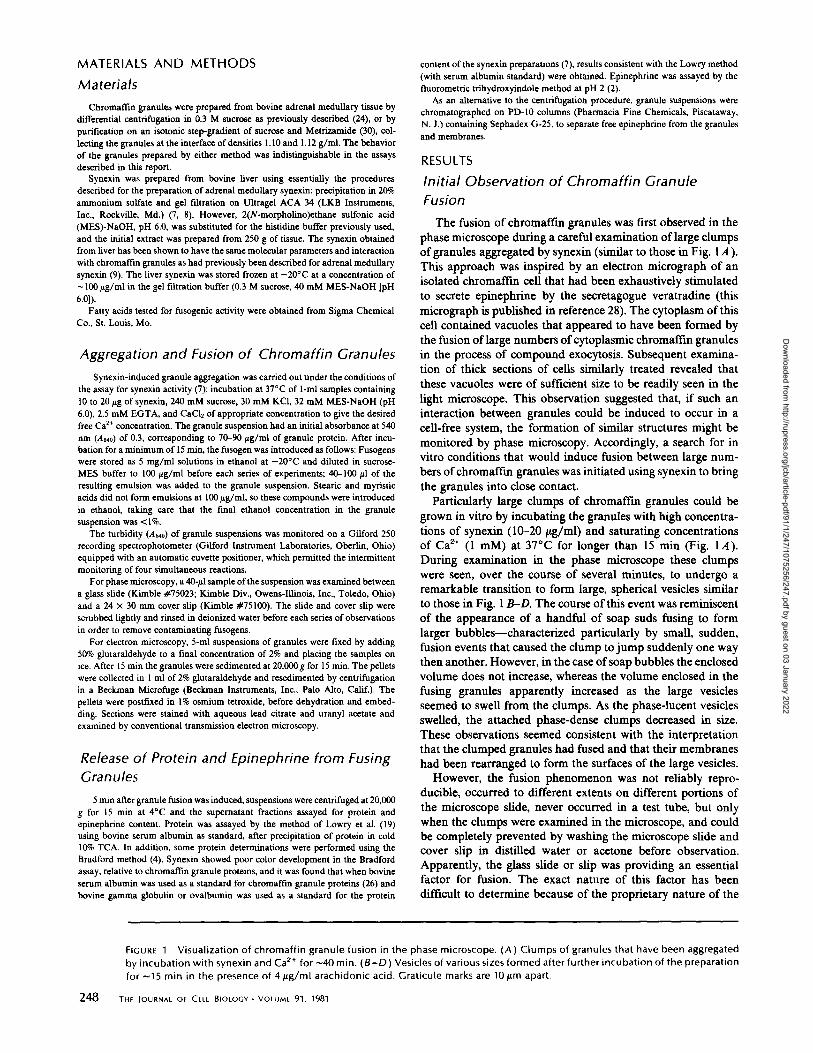
Particularly large clumps of chrornaffin granules could begrown in vitro by incubating the granules with high concentra-tions of synexin (10-20 ttg/ml) and saturating concentrationsof Ca" (1 mM) at 37°C for longer than 15 min (Fig . IA) .During examination in the phase microscope these clumpswere seen, over the course of several minutes, to undergo aremarkable transition to form large, spherical vesicles similarto those in Fig. 1 B-D. The course of this event was reminiscentof the appearance of a handful of soap suds fusing to formlarger bubbles-characterized particularly by small, sudden,fusion events that caused the clump to jump suddenly one waythen another. However, in the case ofsoap bubbles the enclosedvolume does not increase, whereas the volume enclosed in thefusing granules apparently increased as the large vesiclesseemed to swell from the clumps . As the phase-lucent vesiclesswelled, the attached phase-dense clumps decreased in size .These observations seemed consistent with the interpretationthat the clumped granules had fused and that their membraneshad been rearranged to form the surfaces of the large vesicles .However, the fusion phenomenon was not reliably repro-
ducible, occurred to different extents on different portions ofthe microscope slide, never occurred in a test tube, but onlywhen the clumps were examined in the microscope, and couldbe completely prevented by washing the microscope slide andcover slip in distilled water or acetone before observation .Apparently, the glass slide or slip was providing an essentialfactor for fusion. The exact nature of this factor has beendifficult to determine because of the proprietary nature of the
FIGURE 1
Visualization of chrornaffin granule fusion in the phase microscope . (A) Clumps of granules that have been aggregatedby incubation with synexin and Cat' for --40 min . (B-D) Vesicles of various sizes formed after further incubation of the preparationfor -15 min in the presence of 4 ftg/ml arachidonic acid . Graticule marks are 10 pen apart .
Dow
nloaded from http://rupress.org/jcb/article-pdf/91/1/247/1075256/247.pdf by guest on 03 January 2022
CREUTZ
Synexin and the Fusion of Chromaffin Granules
249
Dow
nloaded from http://rupress.org/jcb/article-pdf/91/1/247/1075256/247.pdf by guest on 03 January 2022
processes involved in the manufacture ofmicroscope slides andcover slips . However, this fortuitous observation led to thesurvey of potentially fusogenic compounds reported below.
Survey of Compounds for Fusogenic Activity
Because the fusion of chromaffin granules that was initiallyobserved apparently resulted from an exogenous factor intro-duced by the microscope slide or cover slip, a survey wasconducted to see if a similar effect could be induced bycompounds recognized as having fusogenic potential in cellularsystems. A particularly useful guide in selecting compoundswas the work of Ahkong et al. on the fusion of erythrocytes(1). The compounds that were tested are listed in Table I . Eachcompound was analyzed in two tests : the effect of the com-pound on the turbidity ofthe granule suspension and the effecton the appearance of granule aggregates in the phase micro-scope .
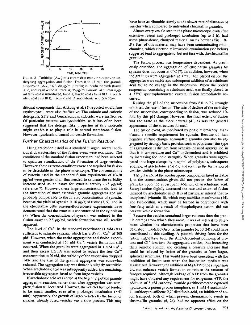
Fig. 2 illustrates the application of the turbidity assay tofusion in the presence of arachidonic or oleic acid, which werefound to be effective fusogens (see below) . The reaction wasstarted by adding the granules to the synexin and Cat+ . Theaggregation of granules was permitted to proceed for 15 minand this aggregation was manifested as an increase in turbidity(Fig. 2, traces a, b, and c) . When a cis-unsaturated fatty acidwas added, the turbidity of the suspension immediately fellbecause of the formation of the transparent vesicles from thedense clumps of granules and, also, probably to a lesser extent,because of some release of material from the granules (thislatter effect was also seen in the absence of synexin, when thegranules were not aggregated ; Fig. 2, trace d) . The decline inturbidity was largely complete in 5 min, and the extent of thedecline appeared to correlate with the extent of formation oflarge vesicles observable in the microscope (detergentlike mol-ecules, e.g., SDS or lysolecithin, were an exception in that theycaused granule lysis and a decline in turbidity but never formedobservable vesicles) . The decline in turbidity from the aggre-gated state (in which the turbidity was usually -160% of theinitial turbidity of the unaggregated suspension) to the state 5min after adding the fusogen is given in Table I as a quanti-tative characteristic of the effect of the compound on thesuspension . Although this parameter was useful for screeningcompounds for fusogenic activity, it must be emphasized thatit is not a direct measure of actual granule fusion because lysisor disaggregation can also contribute to the turbidity decline.Of the compounds tested, the only effective fusogens found
were the naturally occurring, cis-unsaturated fatty acids . Ar-achidonic acid was the most effective, causing microscopicallyobservable fusion at a minimum concentration of 2 tag/ml (6.6I,M) in the presence of 80 itg/ml granule protein (50 tLg/ml ofgranule lipid[ 16]) . Doubling the granule concentration doubledthe threshold concentration of arachidonic acid needed forfusion . The series of 18 carbon cis-unsaturated fatty acids-linolenic, linoleic, and oleic-had an effectiveness that de-creased as their degree of saturation increased . Higher levels ofthe less effective acids (e .g ., 10 ttg/ml of oleic acid) were ableto cause the same degree of fusion as lower levels of the mosteffective (4 FLg/ml of arachidonic acid) .A comparison of the fusogenic activity of several structural
and conformational isomers of oleic acid strikingly suggestedthat a particular stereochemistry was critical to induce fusion .Oleic acid has a double bond between the 9th and 10th carbonsin the cis configuration. Ifthis bond is in the trans configuration(elaidic acid), no fusion occurs . If the cis bond is moved closer
250
THE JOURNAL Or CELL BIOLOGY " VOLUME 91, 1981
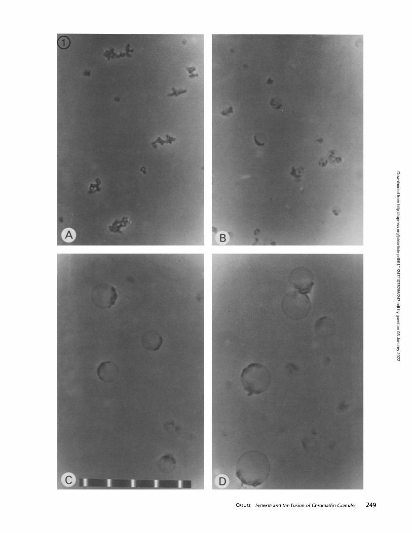
TABLE I
Compounds Tested for Fusogenic Activity
* Compounds were added to ^801íg/ml of granule protein after aggregationby synexin for 15 min.$ Change in turbidity of the granule suspension induced by the compoundafter 5 min. Similar to the analysis in Fig . 2, the change is given as a percentageof the Ay,o of the suspension before aggregation . The values given werereproducible to within five percentage units for a single preparation ofgranules or synexin . The relative order of effectiveness for different com-pounds was the same for all preparations of granules and synexin .§ Degree of vesicle formation seen in the phase microscope: ++++, extensivefusion, one or more large vesicles have developed from every clump; ++,moderate fusion, most clumps have developed vesicles but these are smaller,leaving a large part of the clump unfused; +, limited fusion, vesicles aredifficult to find, occurring in <5% of the clumps ; 0, no fusion, not a singlevesicle can be seen .
to the head group, to carbons 6 and 7 (petroselenic acid), nofusion occurs . If the cis bond is moved away from the headgroup to the 11, 12 position (vaccenic acid), the extent offusionis extremely reduced. In addition, if the head groups of arach-idonic acid or oleic acid are methylated, the compounds do notcause fusion. The saturated fatty acids tested, C12 through C,',(lauric, myristic, pahnitic, stearic), were all completely ineffec-tive. Stearyl amine, a-tocopherol, and trans-retinol-three ad-
Compound
Con-cen-tra-tion
ATur-bídity$
in 5min
Vesicleforma-tion§
pg/ml %
1 . Arachidonic acid 1 22 0[all cis 5,8,11,14 eicosatetraenoic 2 31 +acid (20:4)] 4 81 ++++
2. Linolenic acid 4 49 ++[all cis 9,12,15 octadecatrienoicacid (18:3)]
3. Linoleic acid 4 47 ++[all cis 9,12 octadecadienoic acid(18:2)]
4. Oleic acid 4 44 ++[cis 9 octadecenoic acid (18:1)] 10 78 ++++
5. Palmitoleic acid 4 41 .3 ++[cis 9 hexadecenoic acid (16:1)]
6. cis-Vaccenic acid 4 34 +[cis 11 octadecenoic acid (18:1)]
7. Erucic acid 4 25 .5 +[cis 13 docosenoic acid (22:1)]
8. Petroselenic Acid 4 0 0[cis 6 octadecenoic acid (18:1)] 10 4 0
9. Elaidic Acid 4 0 0[trans 9 octadecenoic acid (18:1)] 10 0 0
10 . Methyl arachidonate 4 0 011 . Methyl oleate 4 0 012 . Glycerol mono-oleate 4 0 013 . Lauric acid 4 0 014 . Myristic acid 5 0 015 . Palmitic acid 4 0 0
[hexadecanoic acid (16:0)]16. Stearic acid 5 0 0
[octadecanoic acid (18:0)]17. Stearyl amine 4 0 018 . Retinol 4 0 019. a-Tocopherol 4 0 020 . Prostaglandin Ez 4 0 021.Lysolecithin 4 0 0
(from egg yolk) 10 51 022 . SDS 4 12 023 . Benzalkonium chloride 4 0 0
8 0 0
Dow
nloaded from http://rupress.org/jcb/article-pdf/91/1/247/1075256/247.pdf by guest on 03 January 2022
a
10 15 20 25TIME, MINUTES
FIGURE 2
Turbidity (As4o) of a chromaffin granule suspension un-dergoing aggregation and fusion . From 0 to 15 min the granulesuspension (As4o, -0 .3 ; 80 jug/ml protein) is incubated with (tracesa, b, and c) or without (trace d) 15 ttg/ml synexin . At 15 min 4lig/ml fatty acid is introduced ; trace a, elaidic acid ( trans 18 :1) ; trace b,oleic acid (cis 18:1) ; traces c and d, arachidonic acid (cis 20 :4) .
ditional compounds that Ahkong et al . (1) reported would fuseerythrocytes-were also ineffective . The anionic and cationicdetergents, SDS and benzalkonium chloride, were ineffective .Of particular interest was lysolecithin, as it has often beensuggested that the detergentlike properties of this moleculemight enable it to play a role in natural membrane fusion .However, lysolecithin caused no vesicle formation.
Further Characteristics of the Fusion ReactionUsing arachidonic acid as a standard fusogen, several addi-
tional characteristics of the fusion event were examined . Theconditions ofthe standard fusion experiment had been selectedto optimize visualization of the formation of large vesicles .However, these extreme conditions were not required for fusionto be detectable in the phase microscope . The concentrationsof synexin used in the standard fusion experiments of 10-20ILg/ml were greater than that needed to saturate the turbidityincrease used as an assay for synexin activity (-5 tttg/ml;reference 7) . However, these large concentrations did lead tothe formation of more extensive granule aggregates and areprobably comparable to the in vivo concentration of synexin,because the yield of synexin is 10 ttg/g of tissue (7, 9), and inthe chromaffin cells immunofluorescence experiments havedemonstrated that the synexin is concentrated in the cytoplasm(9) . When the concentration of synexin was reduced in thefusion assay to 2.5 pg/ml, vesicle formation was still readilyapparent .The level of Ca" in the standard experiment (1 mM) was
sufficient to saturate synexin, which has a Kd for Ca2+ of 200g,M . However, when the entire aggregation and fusion experi-ment was conducted at 160 pM Ca2+, vesicle formation stilloccurred. When the granules were aggregated in 1 mM Ca",and then excess EGTA was added to reduce the free Ca2+
concentration to 20 g,M, the turbidity ofthe suspension dropped14%, and the size of the granule aggregates was somewhatreduced . The aggregation step was thus only slightly reversible.When arachidonic acid was subsequently added, the remaining,irreversible aggregates fused to form large vesicles .
If arachidonic acid was added at the beginning of the granuleaggregation reaction, rather than after aggregation was com-plete, fusion still occurred . However, the vesicles formed tendedto be much smaller, even after prolonged incubation (-45min) . Apparently, the growth oflarger vesicles by the fusion ofsmaller, already fused vesicles was a slow process. This may
have been attributable simply to the slower rate ofdiffusion ofvesicles when compared to individual chromaffin granules .
Almost every vesicle seen in the phase microscope, even afterextensive fusion and prolonged incubation (up to 2 h), hadsome phase-dense, clumped material on its border (Fig . 1 B-D) . Part of this material may have been contaminating mito-chondria, which electron microscopic examination (see below)suggested tend to aggregate to, butnot fuse with, the chromaffingranules.
The fusion process was temperature dependent . As previ-ously described, the aggregation of chromaffin granules bysynexin does not occur at 0°C (7) . In addition, however, whenthe granules were aggregated at 37°C, then placed on ice, theaggregates were stable and subsequent addition of arachidonicacid led to no change in the suspension. When the cooledsuspension, containing arachidonic acid, was finally placed ina 37°C spectrophotometer cuvette, fusion immediately oc-curred .
Raising the pH of the suspension from 6.0 to 7.2 stronglyinhibited the rate offusion . The rate of decline of the turbidityof the suspension, corresponding to fusion, was reduced 10-fold by this pH change. However, the final extent of fusionwas the same at the more neutral pH, as was the generalappearance of the structures formed .The fusion event, as monitored by phase microscopy, man-
ifested a specific requirement for synexin . Because of theirnegative surface change, chromaffin granules can also be ag-gregated by strongly basic proteins such as polylysine (this typeof aggregation is distinct from synexin-induced aggregation inthat it is temperature and Ca" independent and is inhibitedby increasing the ionic strength) . When granules were aggre-gated into large clumps by 4 ltg/ml of polylysine, subsequentaddition of arachidonic acid did not result in the formation ofvesicles visible in the phase microscope .The presence of the nonfusogenic compounds listed in Table
I, at the concentrations given, did not prevent the fusion ofgranules upon the subsequent addition of arachidonic acid .Stearyl amine slightly decreased the rate and extent of fusioninduced by arachidonic acid. It is of particular interest that a-tocopherol (vitamin E), which may stabilize membranes (14),and lysolecithin, which may be formed in conjunction withfree fatty acids as a result of phospholipase action, did notprevent vesicle formation .
Because the vesicles contained larger volumes than the gran-ule clumps from which they arose, it was of interest to deter-mine whether the chemiosmotic properties that have beendescribed in isolated chromaffin granules (6, 10, 24) could havecontributed to this swelling . A possible driving force for thefusion might have been the ATP-dependent pumping of pro-tons and C1- ions into the aggregated vesicles, thus increasingtheir osmotic content and creating a pressure increase thatcould be relieved by fusion of the granules to form larger,spherical structures . This would have been consistent with theinhibition of fusion seen when the incubation medium wasalkalinized . However, the addition ofMgATP to the suspensiondid not enhance vesicle formation or reduce the amount offusogen required. Although leakage of ATP from the granulesmight have obviated any requirement for exogenous ATP, theaddition of 5 I,M carbonyl cyanide p-trifluoromethoxyphenylhydrazone, a potent proton ionophore, or 1 mM 4-acetamido-4'-isothiocyanostilbene-2,2'-disulfonic acid, an inhibitor ofan-ion transport, both of which prevent chemiosmotic events inchromaffin granules (6, 24), had no apparent effect on the
CREUTZ
Synexin and the Fusion of Chromaffin Granules
251
Dow
nloaded from http://rupress.org/jcb/article-pdf/91/1/247/1075256/247.pdf by guest on 03 January 2022

fusion reaction . In contrast, however, when the aggregatedchromaffm granules were exposed to a hypertonic shock byadding sucrose to increase the osmolarity of the suspension to800 mosM, subsequent addition of arachidonic acid did notlead to the formation of the usual large vesicles . Whethermembrane fusion, per se, had been prevented was not deter-mined.
It was also of interest to determine whether metabolism ofthe fatty acids, particularly arachidonic acid, was important forfusion . However, the addition of 100 pM indomethacin toblock any cyclo-oxygenase activity that might have been pres-ent did not inhibit the process. In addition, as shown in TableI, prostaglandin E2, one of the prostaglandins that is formedfrom arachidonic acid and released from platelets during exo-cytosis (21), did not induce fusion .Using arachidonic acid uniformly labeled with 1°C, it was
found that, during fusion by 4 NAg/ml of the fatty acid, 93% ofthe label was apparently bound to the granules, as it could beremoved from the medium by centrifugation of the granules.Subsequent resolution ofthe membrane phospholipids by thin-layer chromatography (3) revealed no incorporation of thefatty acid into phospholipids. Therefore, the effective fusogenappears to have been the unesterified fatty acid incorporatednoncovalently into the granule membrane .
Retention of Granule Contents during FusionChemical analyses were performed to determine whether the
contents of the chromaffm granules were retained within thenew structures formed when fusion occurred. The aggregatedand fused granules were sedimented in the centrifuge and thesupernatant fractions were assayed for protein and epinephrinecontent relative to the total epinephrine and soluble protein ofundisturbed granules . An example of this type of analysis ispresented in Fig. 3. It is necessary to note as controls theamount of release that occurs when the granules are incubatedalone, with synexin only, or with arachidonic acid only. 100%of the epinephrine and 80% of the protein in the chromaffmgranule are in the core and are potentially soluble. Thus, weexpect leakage ofprotein and epinephrine, as percents oftotals,to be in the ratio of 4:5 . However, in the presence or absenceof arachidonic acid during the 20-min incubation there is apreferential leak of epinephrine . When fusion occurs, there isan additional release of epinephrine above the release occurringin the control samples. In this respect the fusion was apparentlyleaky. However, in the case ofsoluble core proteins, the analysisin Fig. 3 suggests that the fusion event may be completelyconservative, because no soluble protein escapes beyond con-trol levels .
Because the centrifugation procedure may have been harshenough to contribute to the leakage of epinephrine, the sepa-ration of granules and vesicles from epinephrine was alsoperformed by molecular exclusion chromatography on Sepha-dex G-25 . Using this procedure, however, we also found thatfusion was associated with additional epinephrine leakage.Thus, it appeared that the fusion event allowed the selectiverelease of some of the small components of the granule cores,while retaining the larger molecules.
Electron Microscopy of Fused ChromaffinGranules
Chromaffin granules that had been aggregated by synexinfor 15 min and then further incubated for 5 min in the presence
252
THE JOURNAL OF CELL BIOLOGY " VOLUME 91, 1981
FIGURE 3 Leakage of protein and epinephrine from chromaffingranules undergoing aggregation and fusion . Suspensions contain-ing 80 ug/ml granule protein were incubated as follows : a, 20 minwithout synexin or arachidonic acid ; b, 20 min without synexin, butwith 4 FLg/ml arachidonic acid added at 15 min; c, 20 min with 15
tLg/ml synexin alone; d, 20 min with synexin, with arachidonic acidadded at 15 min . Fusion occurs only in case d. Bar e indicates thedegree of leakage that should occur if fusion does not induce anyleakage above control levels (e = a + [ b - a ] + [c - a ]) . Leakageof protein in c, d, and e may be overestimated by a maximum of 2%of total because of unbound protein introduced in the synexinpreparation (7) . The error bars represent the standard deviations ofduplicate experiments .
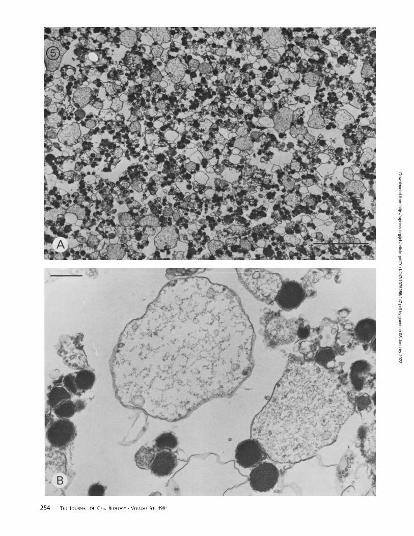
or absence of added arachidonic acid were examined by thin-section electron microscopy. In the absence ofarachidonic acidthe cores of the aggregated granules remained dense andseparated from one another by the closely apposed granulemembranes (Fig . 4A). However, in the presence ofarachidonicacid most of the granules fused to form larger structures boundby a single membrane and containing flocculent core mate-rials apparently greatly diluted from their original concentra-tion in the dense granules (Figs. 4B and 5) . These structureswere generally smaller than those illustrated in Fig. 1, becauseof the shorter incubation time before fusion (15 min vs . 30-45min) . They also appeared "relaxed" or "floppy" instead ofspherical, suggesting that they had not yet fully expanded, orthat the tensions maintaining the spherical shape may havebeen relaxed during the fixation and processing of the samples.In micrographs of control samples exposed to arachidonic acidin the absence of synexin, no evidence of granule fusion wasseen . Contaminating mitochondria were occasionally seen inthe electron micrographs, and they often appeared to havebeen attached to chromaffm granules by synexin action. How-ever, the mitochondria appeared very resistant to fusion be-cause they usually remained intact, even when seen attachedto the membranes of very large vesicles formed by the fusionof chromaffin granules.When granules were aggregated by polylysine and exposed
to arachidonic acid, many empty membrane envelopes wereseen (Fig . 6 B) . These envelopes were often larger than singlegranule membranes but smaller than the vesicles formed bysynexin. This suggested that a limited amount of membranefusion had occurred but that the resulting structures had brokenand released their contents. The inability of granules aggre-gated by polylysine to form large vesicles without the loss ofcore materials was consistent with the failure to observe fusionof such aggregates in the phase microscope .
Dow
nloaded from http://rupress.org/jcb/article-pdf/91/1/247/1075256/247.pdf by guest on 03 January 2022
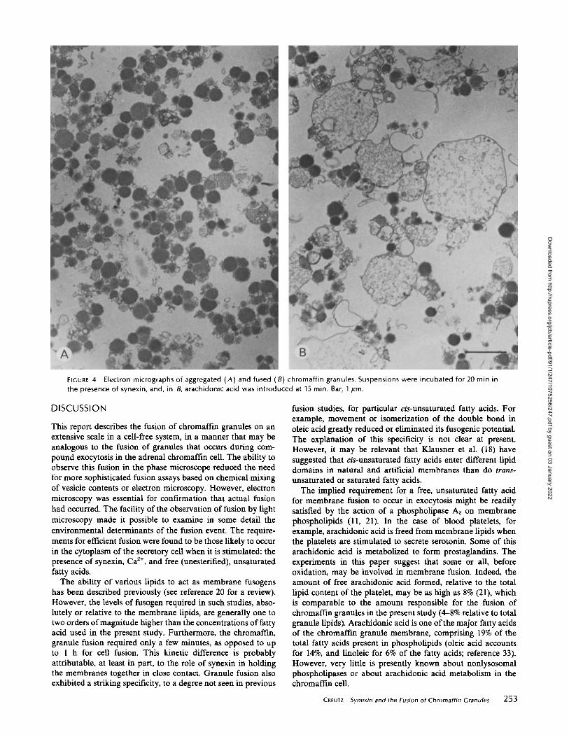
FIGURE 4
Electron micrographs of aggregated (A) and fused (8) chromaffin granules . Suspensions were incubated for 20 min inthe presence of synexin, and, in B, arachidonic acid was introduced at 15 min . Bar, 1 ttm .
DISCUSSION
This report describes the fusion of chromaffin granules on anextensive scale in a cell-free system, in a manner that may beanalogous to the fusion of granules that occurs during com-pound exocytosis in the adrenal chromaffin cell . The ability toobserve this fusion in the phase microscope reduced the needfor more sophisticated fusion assays based on chemical mixingof vesicle contents or electron microscopy. However, electronmicroscopy was essential for confirmation that actual fusionhad occurred . The facility of the observation of fusion by lightmicroscopy made it possible to examine in some detail theenvironmental determinants of the fusion event . The require-ments for efficient fusion were found to be those likely to occurin the cytoplasm of the secretory cell when it is stimulated : thepresence of synexin, Ca", and free (unesterified), unsaturatedfatty acids.The ability of various lipids to act as membrane fusogens
has been described previously (see reference 20 for a review) .However, the levels of fusogen required in such studies, abso-lutely or relative to the membrane lipids, are generally one totwo orders ofmagnitude higher than the concentrations offattyacid used in the present study . Furthermore, the chromaffin,granule fusion required only a few minutes, as opposed to upto 1 h for cell fusion . This kinetic difference is probablyattributable, at least in part, to the role of synexin in holdingthe membranes together in close contact . Granule fusion alsoexhibited a striking specificity, to a degree not seen in previous
fusion studies, for particular cis-unsaturated fatty acids . Forexample, movement or isomerization of the double bond inoleic acid greatly reduced or eliminated its fusogenic potential .The explanation of this specificity is not clear at present .However, it may be relevant that Klausner et al. (18) havesuggested that cis-unsaturated fatty acids enter different lipiddomains in natural and artificial membranes than do trans-unsaturated or saturated fatty acids .The implied requirement for a free, unsaturated fatty acid
for membrane fusion to occur in exocytosis might be readilysatisfied by the action of a phospholipase A2 on membranephospholipids (11, 21) . In the case of blood platelets, forexample, arachidonic acid is freed from membrane lipids whenthe platelets are stimulated to secrete serotonin. Some of thisarachidonic acid is metabolized to form prostaglandins . Theexperiments in this paper suggest that some or all, beforeoxidation, may be involved in membrane fusion . Indeed, theamount of free arachidonic acid formed, relative to the totallipid content of the platelet, may be as high as 8% (21), whichis comparable to the amount responsible for the fusion ofchromaffin granules in the present study (4-8% relative to totalgranule lipids). Arachidonic acid is one ofthe major fatty acidsof the chromaffin granule membrane, comprising 19% of thetotal fatty acids present in phospholipids (oleic acid accountsfor 14%, and linoleic for 6% of the fatty acids ; reference 33).However, very little is presently known about nonlysosomalphospholipases or about arachidonic acid metabolism in thechromaffin cell .
CREUrz
Synexin and the Fusion of Chromaffin Granules
253
Dow
nloaded from http://rupress.org/jcb/article-pdf/91/1/247/1075256/247.pdf by guest on 03 January 2022
254
THE JOURNAL OF CELL BIOLOGY " VOLUME 91, 1981
Dow
nloaded from http://rupress.org/jcb/article-pdf/91/1/247/1075256/247.pdf by guest on 03 January 2022
FIGURE 6
Chromaffin granules aggregated by polylysine and exposed to arachidonic acid . Suspensions were incubated for 20 minin the presence of polylysine and, in B, arachidonic acid was introduced at 15 min . Limited fusion and extensive membranebreakage is apparent in B. Bar, 1 Am .
In addition to specificity for particular fatty acids, the overallaggregation and fusion reaction also demonstrated a specificityfor Ca", for synexin, and for granule membranes. The Ca"specificity is accounted for by the specific requirement ofsynexin for this ion in order to bring the membranes together(7) . Aggregation by basic proteins such as polylysine did notlead to the extensive and semiconservative (i .e ., not leaky tolarger molecules) fusion induced by synexin . Although mito-chondria were attached to chromaftn granules by synexin,they very seldom fused with the granules in the presence ofarachidonic acid .
It is useful now to review the hypothesis that has been putforward for the roles that synexin and the chemiosmotic prop-erties of chromafiin granules may play in the process of exo-cytosis (29) and to examine explicitly what modifications tothis hypothesis the current observations suggest. When thechromaffm cell is stimulated to secrete epinephrine, the cyto-plasmic Cal l concentration increases . Synexin, which is pre-sumably free in the cytoplasm as a 47,000-dalton monomer atlow Ca", binds Ca2' and polymerizes to form small 50 x 150-A rods (8) . These rods may self-associate, and they may bindto chromaflin granule (7) and plasma membranes (25, 32) . Byso doing, they bring the membranes into close contact . The lowaffinity of synexin for Ca2+ (Kd - 200 jm) may convenientlyrestrict synexin action to the vicinity ofthe plasma membrane,where Ca2+ is at the highest concentration, assuming that itenters from the extracellular medium, At this point in the
process it has been suggested that granules attached to theplasma membrane (or to the membranes ofpreviously rupturedgranules) are exposed to the high, extracellular concentrationsof C1 - (27, 29) . This anion is then driven into the granuleinterior as a counter ion to protons being pumped into thegranule by the granule membrane MgATPase (10, 24) . Con-sequently, the osmotic strength in the granule increases, andeventually the granule ruptures, releasing its contents . Forexocytosis to occur the granule must rupture at the point ofcontact with the plasma membrane (or with the membrane ofa granule that has already released its contents). However, ithas been by no means clear that the "pentalaminar structure"formed by the synexin-induced apposition of membranes (7)would be preferentially weak and thus inclined to break .The implication of the present report is that the point of
contact formed between membranes by synexin can indeedbecome weak and will break if an unesterified, cis-unsaturatedfatty acid is present. If such an agent is made available by theaction of a phospholipase (perhaps Ca2' activated), exocytosiswill occur .
It is attractive to visualize that the rupture of granule mem-branes brought into contact by synexin and destabilized by anunsaturated fatty acid is driven by an increase in osmoticstrength within the granule. This could explain the swelling oflarge vacuolelike vesicles, seen in the present experiments, andthe inhibition of the formation of these structures by increasedosmotic strength, as well as the osmotic suppression of exocy-
FIGURE 5
Low and high magnification electron micrographs of fused chromaffin granules . Incubation as in Fig . 4 B. Bars : A, 5 Bm ;8, 0 .3 Bm .
CREUrz
Synexin and the Fusion of Chromaffin Granules
255
Dow
nloaded from http://rupress.org/jcb/article-pdf/91/1/247/1075256/247.pdf by guest on 03 January 2022
tosis, which has been described in several cell types (5, 27, 31).However, in the present experiments, the inability of an un-coupling agent (proton ionophore) or of an inhibitor of aniontransport to inhibit the fusion of granules, and the failure ofMgATP to accelerate the process raise doubts about the im-portance of the chemiosmotic swelling of the granules in thefusion event. Nonetheless, it seems clear that some type ofswelling (enclosed volume increase) must occur for vesicles toform which are large enough to be seen in the light microscope .Geometric considerations dictate that, when the surfaces of nsmall spheres are rearranged to form a single large sphere, thevolume enclosed in the large sphere is /n- times larger than thecollective volume of the n small spheres. The largest vesiclesformed from fused chromaffm granules were 10 ,,m in diameterand thus may have been constructed from the membranes of1,600 granules of 2,500-A diameter, assuming conservation ofmembrane area . Therefore, the contents of these vesicles werediluted 40-fold. This seems too great a volume increase to beaccounted for by the chemiosmotic properties ofthe membranewithin the time-frame of the fusion event or by the osmoticconsequence ofsolubilizing the small molecules trapped in thegranule cores. Perhaps the displacement of counterions fromnegatively charged matrix proteins (chromogranins) has led totheir expansion, thus driving the swelling of the vesicles.
In interpreting the present experiments, it should be empha-sized that the experimental conditions used were optimized forthe visualization of fusion in the phase microscope . Exocytosisin vivo may occur as a result of a far more limited degree offusion. A detailed investigation of granule fusion induced bysynexin and cis-unsaturated fatty acids under more restrictedconditions may now be valuable . Synexin is a widely distrib-uted protein, and cis-unsaturated fatty acids are common com-ponents of membrane phospholipids. Thus, the detailed studyof this event may help to elucidate the mechanism of Ca"-dependent membrane fusion in other systems as well.
I am indebted to James Jordan and N. Raphael Shulman for discus-sions of arachidonic acid metabolism, to Leonard Hjelmeland fordiscussions of detergent and lipid chemistry, to Mark Levine forsuggesting the vitamin E experiments and for supplying the compound,to Pat Fleming for a discussion of chromafffn granule lipids, and toHarvey Pollard, Chris Pazoles, Janet Scott, and Velia Fowler forcritical discussions of the data . I am also indebted to Howard Bladenfor advice in electron microscopy and the use of his facilities, to GeorgePappas for assistance with the ultrastructural analysis, to SamuelStopak for assistance with the epinephrine assay, and to Elise Urciolofor typing the manuscript .Some of the observations described here were reported in prelimi-
nary form in a discussion at the Laurentian Hormone Conference heldat the Mount Tremblant Lodge, Québec, Canada, August 24-29, 1980(28) .
Receivedfor publication 17 November 1980, and in revisedform 4 March1981.
REFERENCESl . Ahkong, Q . F., D . Fisher, W . Tampion, and J. A. Lucy. 1973 . The fusion of erythrocytes
by fatty acids, esters, retinal and a-tocopherol. Biochem . J. 136:147-155 .
256
THE JOURNAL OF CELL BIOLOGY - VOLUME 91, 1981
2. Anton, A . H ., andD . F . Sayer. 1962. A study of the factors affecting the aluminum oxide-trihydroxyindole procedure for the analysis of catecholi mines, J. Pharmakol. Exp. Ther .138:360-375 .
3 . Billah, M . M � E. G . Lapetina, and P . Cuatrecasas . 1980 . Phospholipase A, and phospho-lipase C activities of platelets. J. Biol. Chem. 255 :10227-10231 .
4. Bradford, M . M. 1976 . A rapid and sensitive method for the guantitation of microgramquantities of protein utilizing the principle of protein-dye binding. Anal Biochem. 72 :248-254 .
5. Brown, E. M ., C . J . Pazoles, C . E . Creutz, G. D . Aurbach, and H . B . Pollard . 1978 .Regulation of parathyroid hormone release from dispersed bovine parathyroid cells bypermeant anions. Proc. Nail. Acad. Sci. U. S. A . 75:876-880 .
6 . Casey, R . P., O. Njus, G . K . Radda, and P . A. Sehr . 1976. ATP-evoked catecholammerelease in chromaffm granules: osmotic lysis as a consequence of proton translocation .Biochem. J. 158 :583-588.
7. Creutz, C . E ., C. J. Pazoles, and H . B. Pollard. 1978. Identificatio n and purification of anadrenal medullary protein (synexin) that causes calcium dependent aggregation of isolatedchromaffin granules. J. Bial. Chem. 253 :2858-2866 .
8, Creutz, C. E., C . l. Pazoles, and H . B . Pollard . 1979 . Self-association of synexin in thepresence of calcium. J. Biot Chem. 254 :553-558 .
9, Creutz, C . E., C. J . Pazoles, and H. B . Pollard. 1980. Immunohistochemica l and biochem-ical studies on synexins and diverse tissues. In Calcium-binding Proteins and CalciumFunction in Health and Disease. F . L . Siegel et al editors. North Holland PublishingCompany, New York.
10 . Creutz, C. E., and H. B . Pollard. 1980 . A biophysical model of the chromaffm granule :accurate description of the kinetics of ATP and CI - -dependent granule lysis . Biophys. J.31 :255-270.
11 . Crews, F . T., Y. Morita, F . Hirata, J . Axelrod, and R . P. Siraganian . 1980. Phosphohpidmethylation affects immunoglobulin E-mediated histamine and arachidonic acid releasein rat leukemic basophils. Biochem. Biophys. Res. Commun. 93 :42-49.
12. Dabrow, M., S. Zaremba, and R. A . Hague-Angeletti . 1980 . Specificity of synexin-inducedchromaffin granule aggregation . Biochem. Biophys. Res. Commun. 96:1164-1171 .
13. De Robertis, E., and A . Vaz Ferreira, 1957. Electron microscopic study of the excretion ofcatechot-containing droplets in the adrenal medulla. Exp . Cell Res. 12 :568-574 .
14 . Diplock, A . T., and J. A. Lucy. 1973 . The biochemical modes of action of vitamin E andselenium: a hypothesis. FEBS (Fed. Eur. Biochem. Soc .) Lett. 29:205 .
15 . Douglas, W . W., and R. P. Rubin . 1961 . Th e role of calcium in the secretary response ofthe adrenal medulla to acetylcholine. J. Physiol. (Load.) . 159 :40-57.
16. Hillarp, N . A . 1959 . Further observations on the state of the catecholamines stored in theadrenal medullary granules. Acia Physiol Scand. 47 :271-279 .
17 . Kirshner, N ., H . J . Sage, W . J . Smith, and A . G . Kirshner. 1966. Release of catecholaminesand specific protein from adrenal glands . Science (Wash. D. C.) . 154 :529-531 .
18 . Klausner, R . D., A . M . Kleinfeld, R. L . Hoover, and M . J. Karnovsky . 1980. Lipi ddomains in membranes : evidence derived from structural perturbations induced by freefatty acids and lifetime heterogeneity analysis. J. Biol. Chem. 255:1286-1295 .
19 . Lowry, O . H., N . J. Rosebrough, A . L . Farr, and R. J . Randall, 1951 . Protein measurementwith the Folin phenol reagent . J Biol. Chem. 193:265-275 .
20 . Lucy, J . A. 1978 Mechanisms of chemically-induced cell fusion . In Membrane Fusion .Cell Surface Reviews . G. Poste and G . L . Nicolson, editors, North Holland PublishingCompany, New York. 5 :268-305 .
21 . Marcus, A. J . 1978. The role of lipids in platelet function: with particular reference to thearachidonic acid pathway . J. Lipid Res. 19 :793-826.
22. Morris, S . J ., and J . M. X. Hughes . 1979 . Synexin protein is nonselective in its ability toincrease Ca"-dependent aggregation of biological and artificial membranes. Biochem .Biaphys. Res. Comm. 91 :345-350 .
23. Palade, G . 1975 . Intracellular aspects of the process of protein synthesis. Science (Wash.D. C.) 189 :347-358 .
24. Pazoles, C. J ., and H. B. Pollard . 1978 . Evidence for stimulation of anion transport inATP-evoked transmitter release from isolated secretary vesicles. J. Biol. Chem. 253 :3962-3969 .
25. Pollard, H . B ., C . E. Creutz~ C. J. Pazoles, and J . H . Scott . 1980 . Fusion and fissionprocesses in exocytosis: possible roles for synexin and osmotic lysis in the two events. InProceedings of the Electron Microscope Society of America 38th Annual Meeting . C . W .Bailey, editor. Claitor's Publishing Division, Baton Rouge . 594597,
26 . Pollard, H . B ., R. Menard, H . A . Brandt, C. J. Pazoles, C . E. Creutz, and A . Ramu . 1978 .Application of Bradford's protein method to adrenal gland subcellular fractions . Anal.Biochem . 86:761-763 .
27. Pollard, H . B ., C . J . Pazoles, andC . E . Creutz . 1979. Evidenc e in support of a chemiosmoticmechanism for exocytosis from platelets, parathyroid and chromaffin cells . In Catechol-amines: Basic and Clinical Frontiers. 1 . Kopin and E. Usdin, editors . Pergamon Press,Oxford . 328-330.
28. Pollard, H . B ., C . J. Pazoles, and C . E . Creutz. 1981, Mechanism of calcium action andrelease of vesicle-bound hormones during exocytosis. Receni Prog. Horm . Res. 37 : 299-332.
29 . Pollard, H. B ., C . J. Pazoles, C . E. Creutz, and O. Zinder. 1979 . The chromaffin granuleand possible mechanisms of exocytosis . Int. Rev. Cytol. 58:160-198 .
30 . Pollard, H. B ., H. Shindo, C. E. Creutz, C . 1 . Pazoles, and J . S . Cohen. 1979 . Interna l pHand state ofATP in adrenergic chromaffm granules determined by "P nuclear magneticresonance spectroscopy . J. Biol. Chem. 254 :1170-1177.
31 . Pollard, H . B ., K. M . Tackman-Goldman, C. J . Pazoles, C. E. Creutz, and N. R . Shulman.1977 . Evidence for control of serotonin secretion from human platelets by hydroxyl iontransport and osmotic lysis . Proc. Nall Acad. Sci. U. S. A . 74 :5295-5299.
32 . Scott, J . H., C . E . Creutz, and H . B . Pollard. 1980 . Synexi n binding to chromaffin cellplasma membrane. Eur. J. Cell Biot 22 :186 (Abstr) .
33 . Winkler, H ., and A. D . Smith. 1968. Lipids of adrenal chromaffm granules: fatty acidcomposition of phospholipids, in particular lysolecithin. Naunyn-Schmiedebergs Arch.Pharmakol Exp. Pathol 261 :378-388 .
Dow
nloaded from http://rupress.org/jcb/article-pdf/91/1/247/1075256/247.pdf by guest on 03 January 2022
![Magnetoliposomes Loaded with Poly-Unsaturated Fatty Acids ... · reported using preparations of omega-3 fatty acids [29-35], a kind of long chain polyunsaturated fatty acids (PUFAs)](https://static.fdocuments.us/doc/165x107/5f8b025f23ab9a27c5624e1e/magnetoliposomes-loaded-with-poly-unsaturated-fatty-acids-reported-using-preparations.jpg)