Circulatory System Cardiovascular system is made up of the blood, heart, and blood vessels. *It...
31
Circulatory System • Cardiovascular system is made up of the blood, heart, and blood vessels. *It transports oxygen, nutrients, and hormones • **Lymphatic system is made up of the lymph, lymph nodes and lymph vessels, (**lymph nodes, spleen, and tonsils) • Together they form the Circulatory system
-
Upload
ralf-parker -
Category
Documents
-
view
215 -
download
0
Transcript of Circulatory System Cardiovascular system is made up of the blood, heart, and blood vessels. *It...

Circulatory System• Cardiovascular system is made up of the blood, heart, and blood vessels. *It transports oxygen, nutrients, and hormones• **Lymphatic system is made up of the lymph, lymph nodes and lymph vessels, (**lymph nodes, spleen, and tonsils)• Together they form the Circulatory system

Heart
• About the size of a fist, the heart will beat about 2.5 billion times in an average life span
• The heart lies in the thoracic (chest) cavity between the two lungs
• Pericardium is a tough sac like membrane that surround and protects the heart, and secretes a fluid that reduces friction as the heart beats

• Septum is a ‘wall’ that divided the heart vertically into 2 sides
• Right side: - Pumps blood to the lungs
• Left side- Pumps blood to other parts of the body

• Each side is divided into an upper and lower chambers
• The upper chamber is called an atrium• The Lower chamber is called the ventricle• Valves– **Found in veins Not arteries– Are flaps of tissue that open in 1 direction only
• **Mitral valve– Prevents backflow of blood into **left atrium
• **Semilunar valve– Prevents backflow of blood into ventricles

Circulation in the heart
• Superior vena cava sends deoxygenated blood from upper body to right atrium
(AKA it drains blood from the head and neck)• The right atrium sends deoxygenated blood to
the right ventricle• Muscles in the right ventricle contract and
force blood into the pulmonary arteries• Pulmonary artery sends blood to the lungs
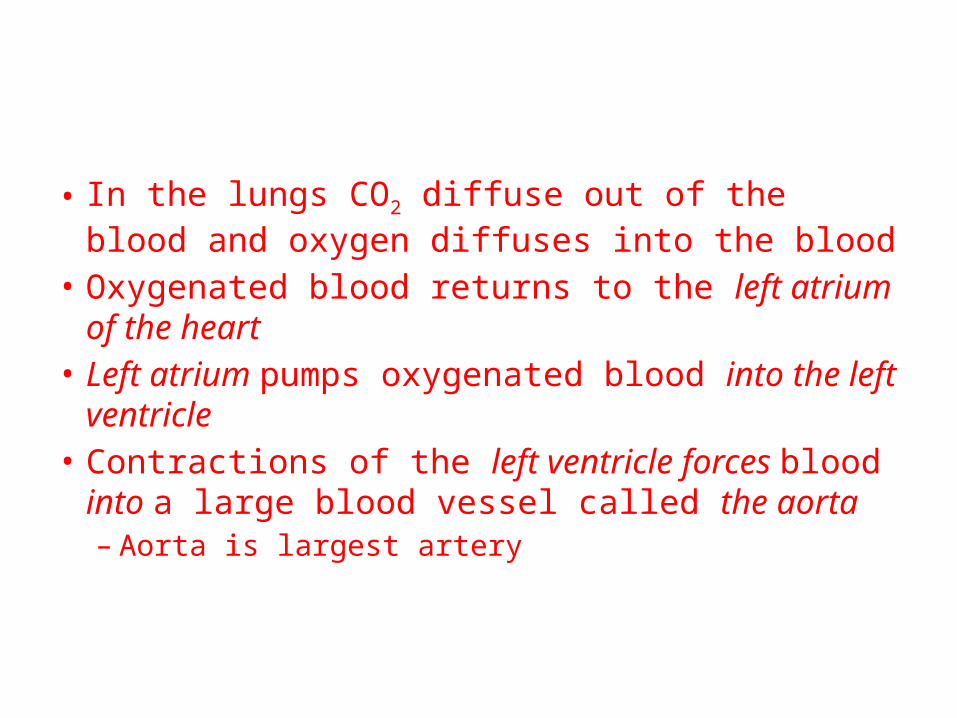
• In the lungs CO2 diffuse out of the blood and oxygen diffuses into the blood
• Oxygenated blood returns to the left atrium of the heart
• Left atrium pumps oxygenated blood into the left ventricle
• Contractions of the left ventricle forces blood into a large blood vessel called the aorta– Aorta is largest artery

• **Left ventricle pumps blood into the systemic circulation

Control of the heartbeat
• Sinoatrial (SA) node is a group of specialized heart-muscle cells found in the right atrium
• They initiate their own electrical impulse and contract
• *The SA node is often called the pace maker b/c it regulates the rate of contraction for the entire heart

• **Tricuspid valve called the AV node is found in the septum between the atria on the right side of the heart
• The **atrioventricular (Av) node relays the electrical impulse to the muscle cell that make up the ventricles
• Heartbeat has 2 phases1. Systole
occurs when ventricles contract
2. Diastole occurs when ventricles relax
These to phases result in the lub dub you hear, and we call a heartbeat

Blood Vessels• Blood vessels in the body include the arteries,
veins, and capillaries• *If a blood vessel has valves, it is probably a
vein• Arteries– Large vessels that carry blood away from heart
• Capillaries– Tiny vessels– They are so small that red blood cells can only
pass through single file

• **Arteriole conducts blood into the capillaries

• Veins– Blood flows from capillaries that merge to form
larger vessels called venules– Venules merge to form veins– Carries blood to heart– Veins returning deoxygenated blood from lower
parts of body merge to form the inferior vena cava– Veins returning deoxygenated blood from upper
parts of body merge to form the superior vena cava

• Pulmonary circulation– Deoxygenated blood from all body parts (except
lungs) into the right atrium– Pulmonary flow to and from the lungs– *Pulmonary circulation loop carries blood to the
lungs• Lymphatic System• *Excess fluids and proteins in the body are
returned to the bloodstream by the lymphatic system
• **connects with systemic circulation

• **Lymph tissue include;– Lymph nodes, spleen, tonsils

Homework
• Pg 939 #1-3, 5

Blood
• Made up of – red blood cells (RBC)– White blood cells (WBC)– Platelets
• You have 4-5liters of blood in your body• 55% is liquid, 45% formed elements• Blood is a liquid connective tissue

• Plasma– *Portion of blood containing metabolites, waste,
salts, proteins, and water– Liquid medium, sticky, and straw colored, 90% water
• **RBC (**erythrocytes)– **Transport oxygen to cells in all parts of body– Formed in the red marrow of bones– Immature RBC’s systhesize large amounts of an iron
containing protein called hemoglobin– ***Hemoglobin actually transports the oxygen– Lack nuclei (do they cannot divide)– Last about 120-130 days– There are more than 30 trillion RBC circulating in the body at
one time

• WBC– Help defend the body against disease– Formed in the red marrow of bones– Must travel to lymph nodes, tonsils, thymus or
spleen to mature–*Largest blood cell, aka leukocytes– Several types of WBC, only one type of RBC– Antibodies is one that helps destroy substances
like bacteria and viruses– IF you have an infection, WBC count can double– Less WBC than RBC

• **Phagocyte– Engulfs microorganisms
• ***Platlets– Cell fragments needed to form blood clots (fibrin)
• *Fibrin– Sticky protein threads that function in blood
clotting• **Lymph nodes – Filter components of lymph

Blood Types
• Determined by the type of antigen present on the surface of the RBC
• Antigen is a substance that stimulated an immune response
• If foreign antigen enter the body, cells respond by producing antibodies
• Antigen is an abbreviation for antibody-generating substance.

Blood typesBlood type Antigen in RBC Antibodies in
plasmaReceive from Give to
A A Anti-B O, A A, AB
*B B Anti-A *O, B B, AB
AB A and B None A, B, AB, O AB
O None Anti-A, Anti-B O A, B, AB, O

Rh factor
• An antigen that is sometimes present on the surface on the RBC
• 85% of the US population is Rh positive (Rh+)– Meaning that Rh antigens are present
• If an Rh- person receives blood from an Rh+ , antibodies may react with the antigen and agglutination may occur

Homework
• Pg 944 #1-2, 4-5

Respiratory System
• External respiration• Exchange of gases between atmosphere and blood
• Internal respiration• exchange of gases between blood and cells of body
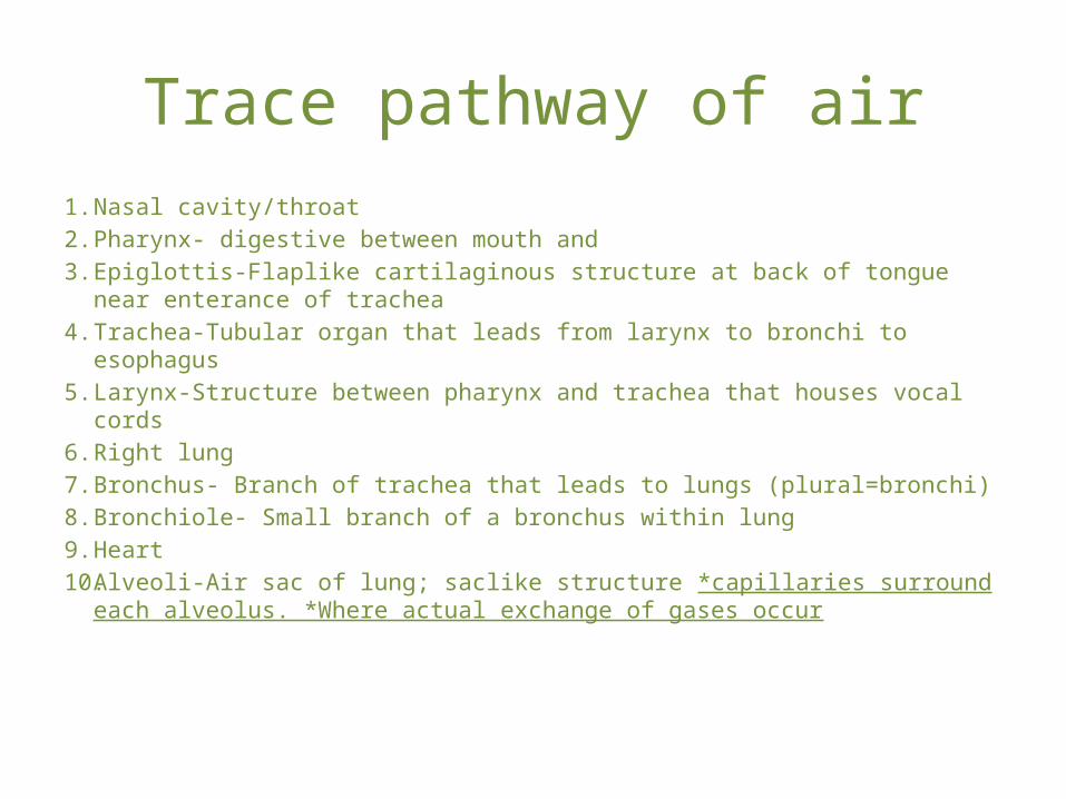
Trace pathway of air1. Nasal cavity/throat2. Pharynx- digestive between mouth and3. Epiglottis-Flaplike cartilaginous structure at back of tongue near enterance of
trachea4. Trachea-Tubular organ that leads from larynx to bronchi to esophagus5. Larynx-Structure between pharynx and trachea that houses vocal cords6. Right lung7. Bronchus- Branch of trachea that leads to lungs (plural=bronchi)8. Bronchiole- Small branch of a bronchus within lung9. Heart10. Alveoli-Air sac of lung; saclike structure *capillaries surround each alveolus.
*Where actual exchange of gases occur

• **Inspired air passes through the bronchioles just before entering the alveoli

Part Description Function
Nose Part of face, centered above mouth, below and between eyes
Provide entrance to nasal cavityHair filter incoming air
Nasal Cavity Hollow space behind nose Conducts air to pharynx, warms and moistens incoming air
Pharynx Chamber behind oral cavity and between nasal cavity
Passageway for air moving from nasal cavity to larynx and for food moving from oral cavity to esophagus
Larynx Enlargement at top of trachea prevents foreign objects, houses vocal cords
Trachea Flexible tube that connects larynx with bronchial tree
continues to filter incoming air
Bronchial tree Branched tubes that lead from trachea to alveoli
Continues to filter incoming air
Lungs Soft, cone shaped organs occupy a large portion of thoracic cavity
Contain alveoli, blood vessels, connective tissue, lymphatic vessels, and nerves of the lower respiratory tract

• The respiratory control center in the brain is most sensitive to concentrations of carbon dioxide (CO2) in the blood
• **The concentration gradients determine if CO2 is absorbed or released by blood
• **Increased levels of CO2 in the blood cause breathing rate to increase

• Pharynx
• **Larynx– Produces sounds for communication
• **Trachea– Is an air passageway
• *Epiglottis- Prevents food and liquid from entering the trachea
• Lungs– Site of gas exchange between atmosphere and
blood– The right lung has 3 lobes and is heavier than the
left lung which has 2 lobes– **Breathing is regulated mainly by the response
to the level of CO2 detected in blood

Inhalation
• *diaphragm contracts, moves downward, and rib cage moves upward and out