Circulatory System Blood vessels Arteries Capillaries Veins Heart Chambers Surfaces Coronary...
44
Circulatory System Blood vessels Arteries Capillaries Veins Heart Chambers Surfaces Coronary circulation Internal features Conducting system Fetal Circulation
-
Upload
martin-gilbert -
Category
Documents
-
view
279 -
download
1
Transcript of Circulatory System Blood vessels Arteries Capillaries Veins Heart Chambers Surfaces Coronary...
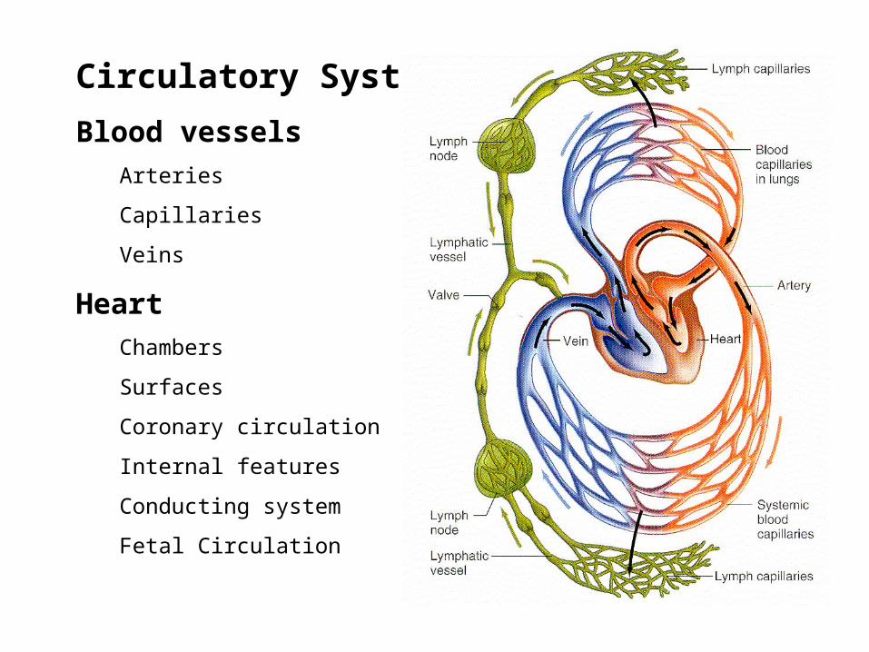
Circulatory System
Blood vessels
Arteries
Capillaries
Veins
Heart
Chambers
Surfaces
Coronary circulation
Internal features
Conducting system
Fetal Circulation
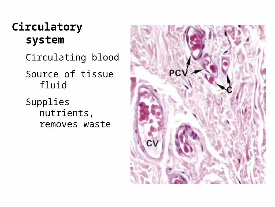
Circulatory system
Circulating blood
Source of tissue fluid
Supplies nutrients, removes waste
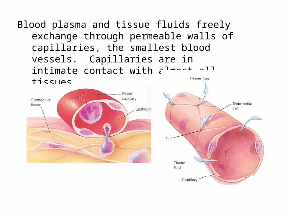
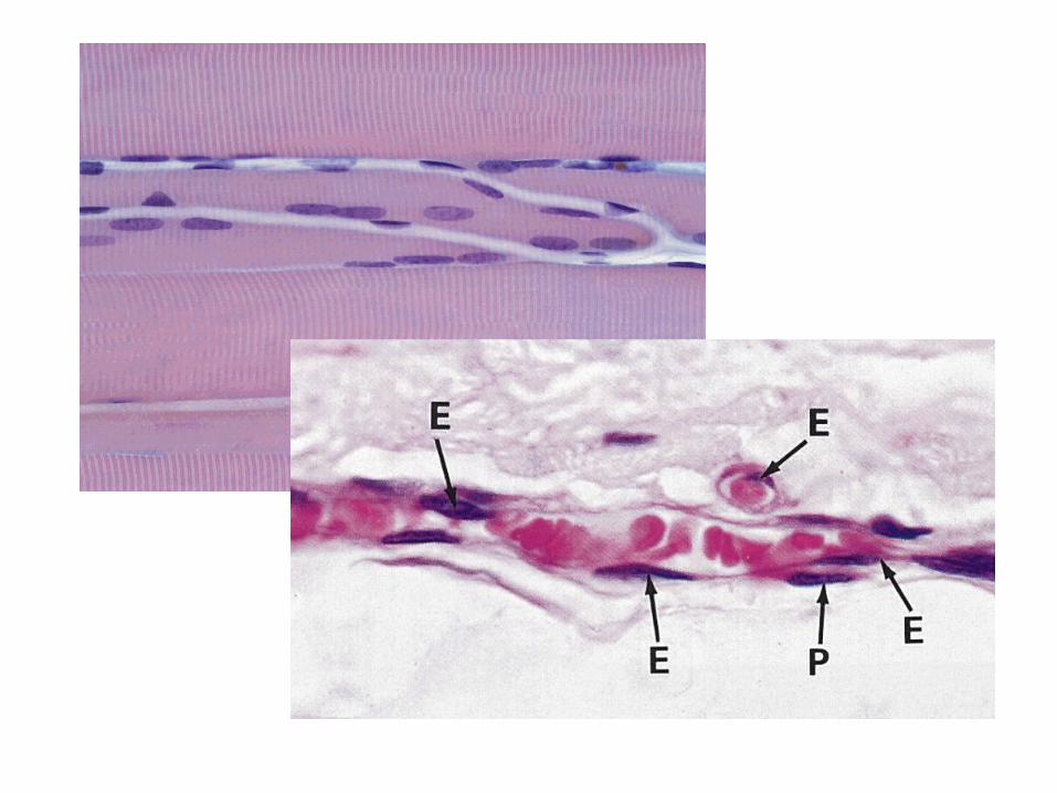
Blood plasma and tissue fluids freely exchange through permeable walls of capillaries, the smallest blood vessels. Capillaries are in intimate contact with almost all tissues.
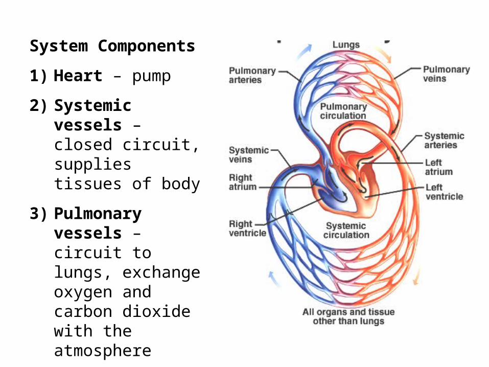
System Components
1) Heart – pump
2) Systemic vessels – closed circuit, supplies tissues of body
3) Pulmonary vessels – circuit to lungs, exchange oxygen and carbon dioxide with the atmosphere
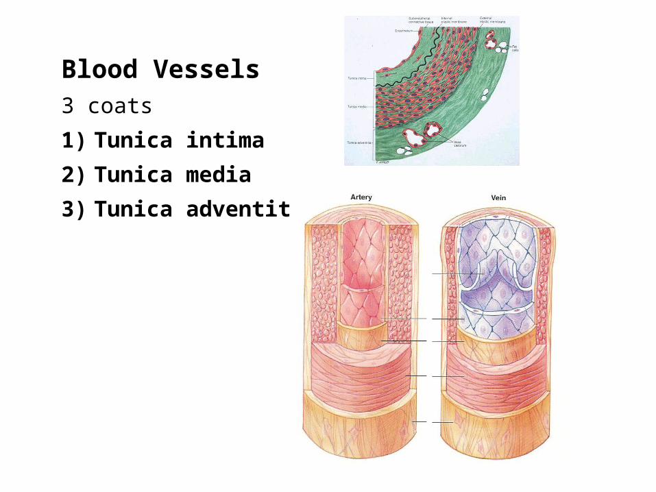
Blood Vessels3 coats
1) Tunica intima
2) Tunica media
3) Tunica adventitia

Blood Vessels1) Tunica intima
Endothelium – simple squamous epithelium
Loose CT
Internal elastic membrane (lamina) – sheet of protein elastin (like elastic fibers)
2) Tunica media
Mix: CT, smooth muscle, elastic fibers
3) Tunica adventitia
External elastic lamina
CT, vasa vasorum = small blood vessels that supply wall of very large arteries
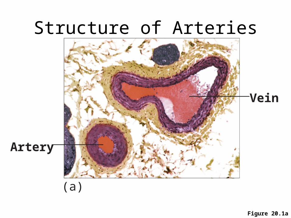
Structure of Arteries & Veins Capillaries
Figure 20.1a
Artery
Vein
(a)
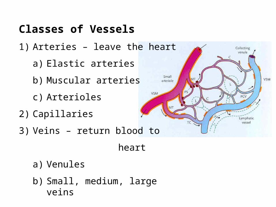
Classes of Vessels
1) Arteries – leave the heart
a) Elastic arteries
b) Muscular arteries
c) Arterioles
2) Capillaries
3) Veins – return blood to
heart
a) Venules
b) Small, medium, large veins
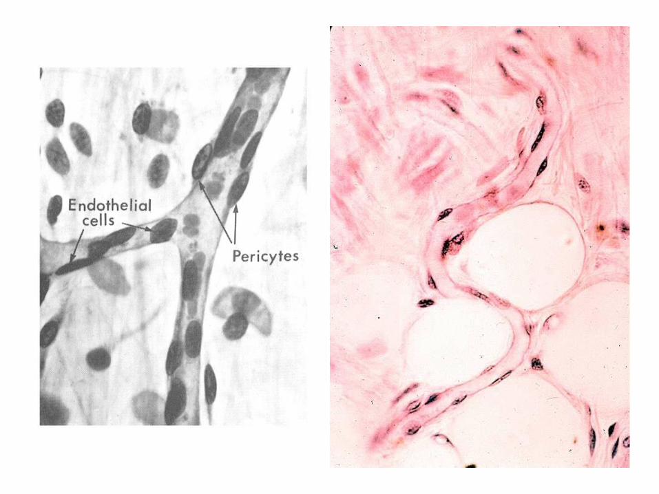
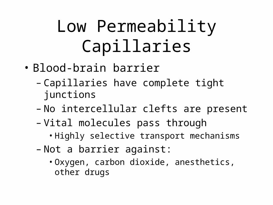
Low Permeability Capillaries
• Blood-brain barrier – Capillaries have complete tight junctions– No intercellular clefts are present– Vital molecules pass through
• Highly selective transport mechanisms
– Not a barrier against: • Oxygen, carbon dioxide, anesthetics, other drugs
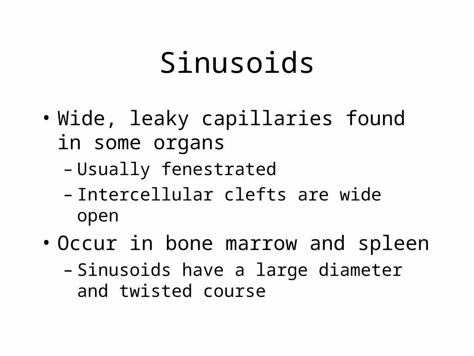
Sinusoids
• Wide, leaky capillaries found in some organs– Usually fenestrated– Intercellular clefts are wide open
• Occur in bone marrow and spleen– Sinusoids have a large diameter and twisted
course
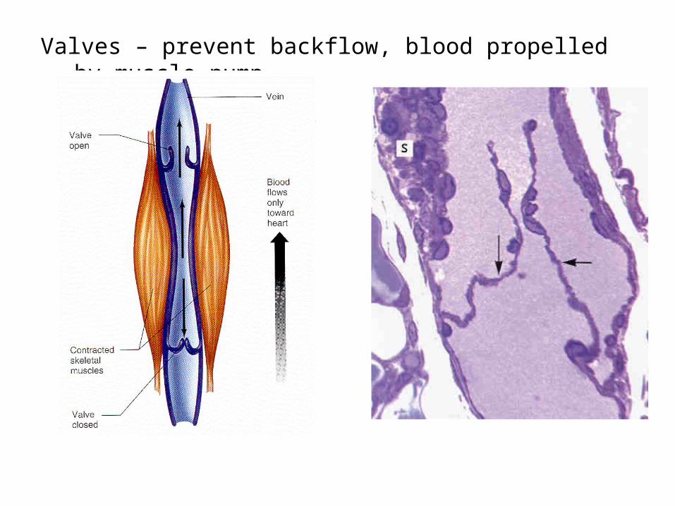
Valves – prevent backflow, blood propelled by muscle pump
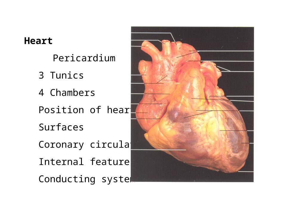
Heart
Pericardium
3 Tunics
4 Chambers
Position of heart
Surfaces
Coronary circulation
Internal features
Conducting system

Pericardium
=double walled serous sac
simple squamous epithelium (mesothelium) and some loose C.T.
1) Parietal layer
2) Visceral layer
Separated by serous fluid which decreases friction
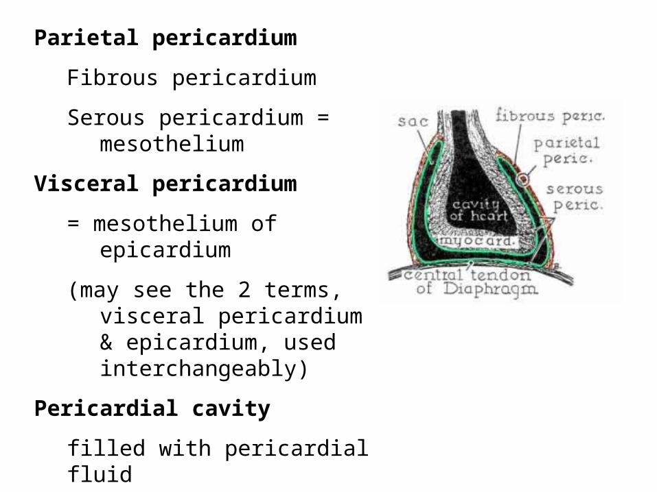
Parietal pericardium
Fibrous pericardium
Serous pericardium = mesothelium
Visceral pericardium
= mesothelium of epicardium
(may see the 2 terms, visceral pericardium & epicardium, used interchangeably)
Pericardial cavity
filled with pericardial fluid
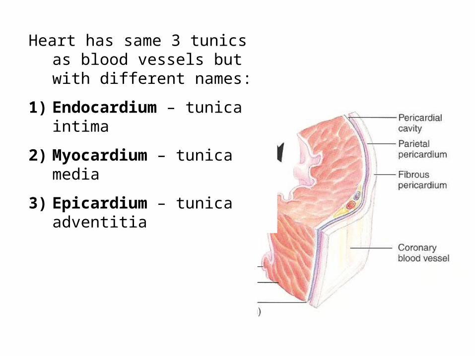
Heart has same 3 tunics as blood vessels but with different names:
1) Endocardium – tunica intima
2) Myocardium – tunica media
3) Epicardium – tunica adventitia
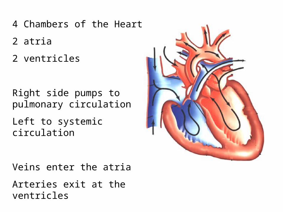
4 Chambers of the Heart
2 atria
2 ventricles
Right side pumps to pulmonary circulation
Left to systemic circulation
Veins enter the atria
Arteries exit at the ventricles
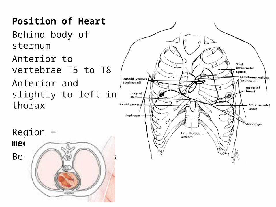
Position of Heart
Behind body of sternum
Anterior to vertebrae T5 to T8
Anterior and slightly to left in thorax
Region = mediastinum
Between the 2 lungs
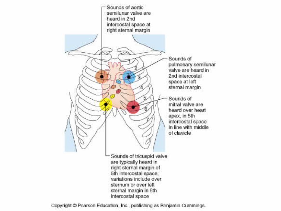
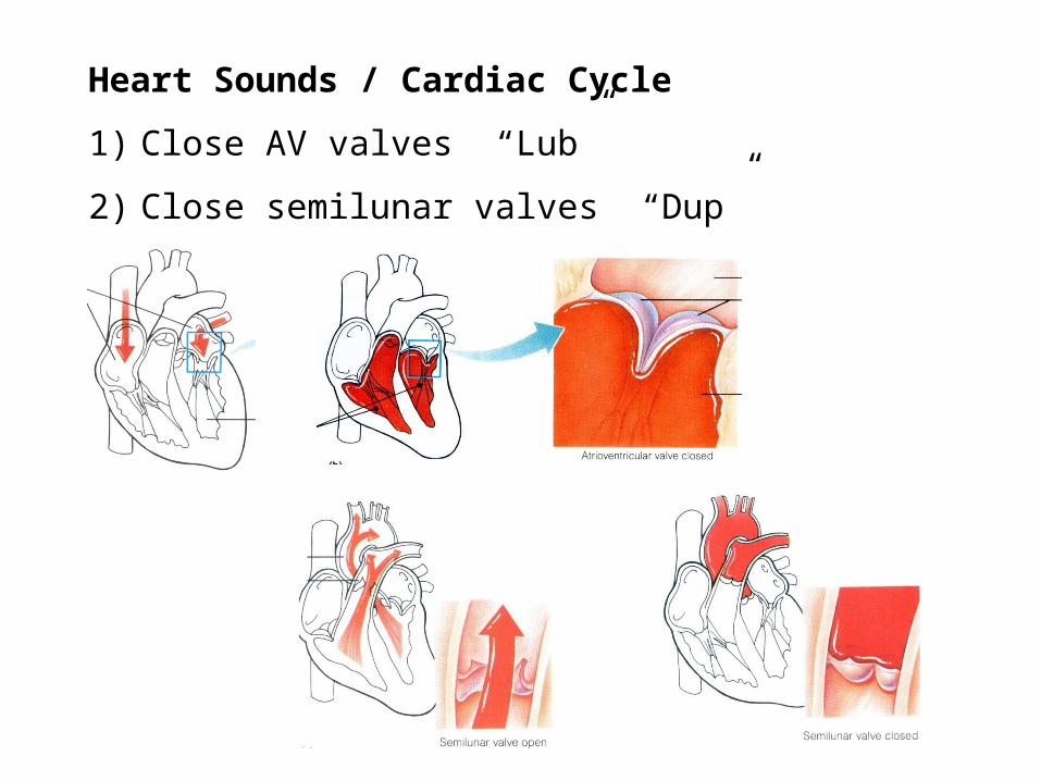
Heart Sounds / Cardiac Cycle
1) Close AV valves “Lub”
2) Close semilunar valves “Dup”
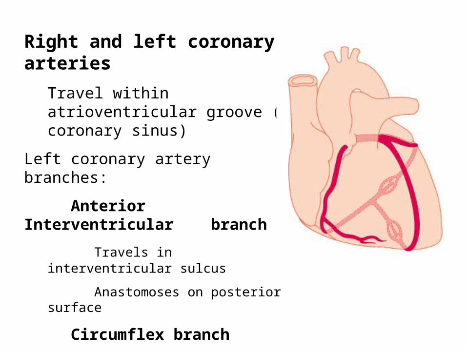
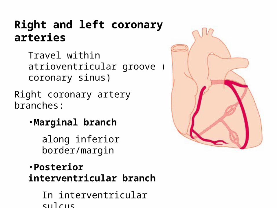
Right and left coronary arteries
Travel within atrioventricular groove (= coronary sinus)
Left coronary artery branches:
Anterior Interventricular branch
Travels in interventricular sulcus
Anastomoses on posterior surface
Circumflex branch
Anastomoses on posterior surface with right coronary
Right and left coronary arteries
Travel within atrioventricular groove (= coronary sinus)
Right coronary artery branches:
•Marginal branch
along inferior border/margin
•Posterior interventricular branch
In interventricular sulcus
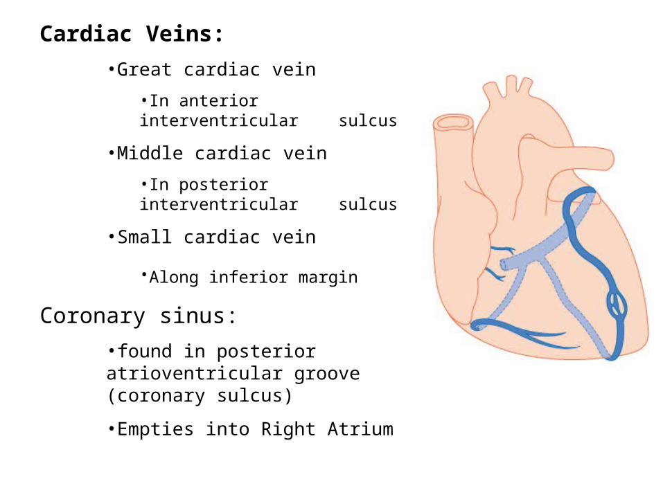
Cardiac Veins:
•Great cardiac vein
•In anterior interventricular sulcus
•Middle cardiac vein
•In posterior interventricular sulcus
•Small cardiac vein
•Along inferior margin
Coronary sinus:
•found in posterior atrioventricular groove (coronary sulcus)
•Empties into Right Atrium
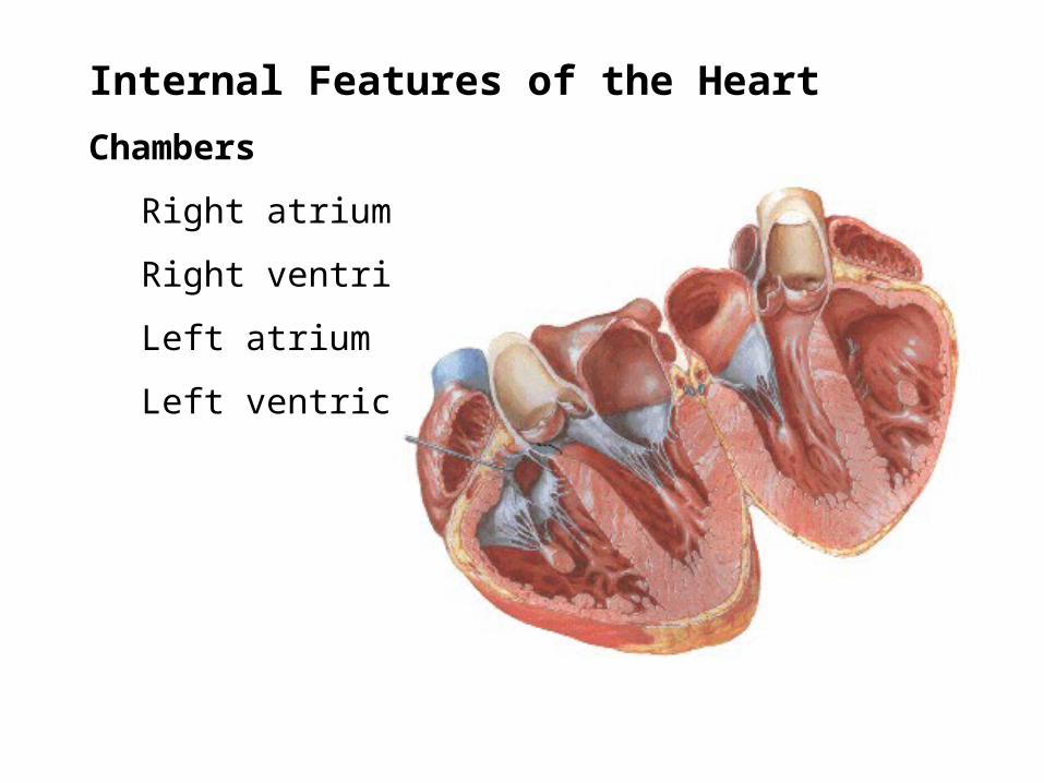
Internal Features of the Heart
Chambers
Right atrium
Right ventricle
Left atrium
Left ventricle
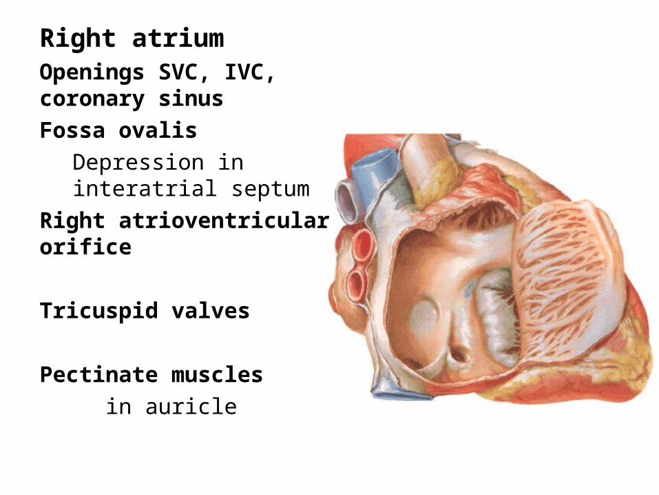
Right atriumOpenings SVC, IVC, coronary sinus
Fossa ovalis
Depression in interatrial septum
Right atrioventricular orifice
Tricuspid valves
Pectinate muscles
in auricle
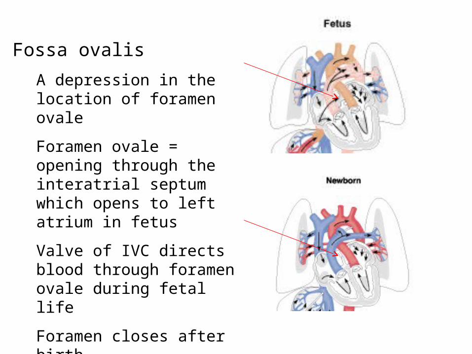
Fossa ovalis
A depression in the location of foramen ovale
Foramen ovale = opening through the interatrial septum which opens to left atrium in fetus
Valve of IVC directs blood through foramen ovale during fetal life
Foramen closes after birth
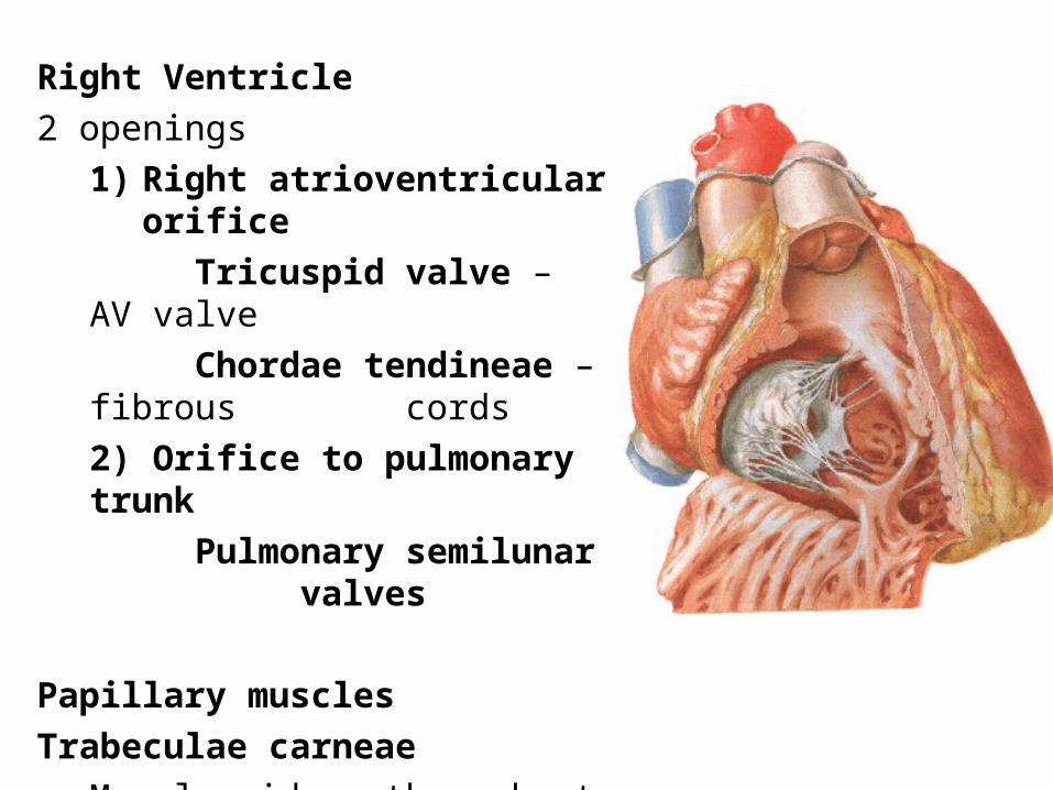
Right Ventricle
2 openings
1) Right atrioventricular orifice
Tricuspid valve – AV valve
Chordae tendineae – fibrous cords
2) Orifice to pulmonary trunk
Pulmonary semilunar valves
Papillary muscles
Trabeculae carneae
Muscle ridges throughout wall
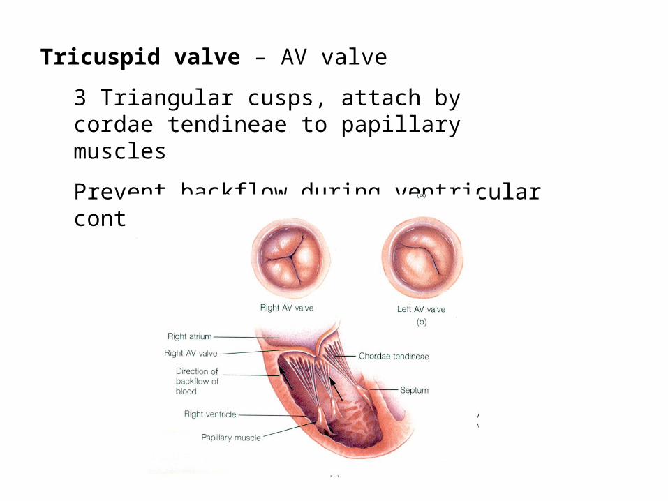
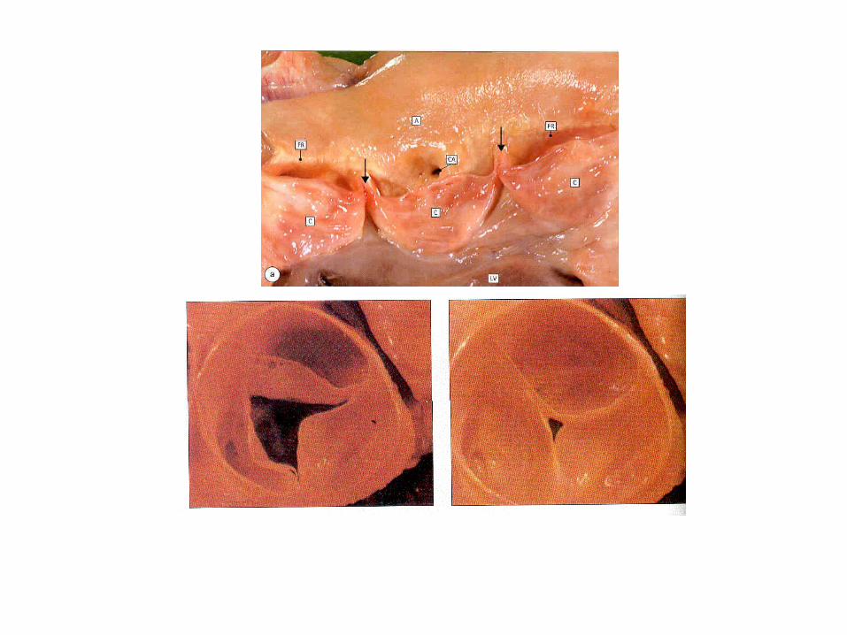
Tricuspid valve – AV valve
3 Triangular cusps, attach by cordae tendineae to papillary muscles
Prevent backflow during ventricular contraction
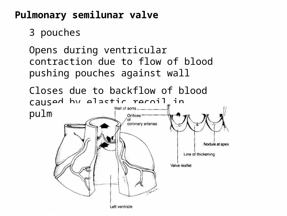
Pulmonary semilunar valve
3 pouches
Opens during ventricular contraction due to flow of blood pushing pouches against wall
Closes due to backflow of blood caused by elastic recoil in pulmonary artery, filling pouches
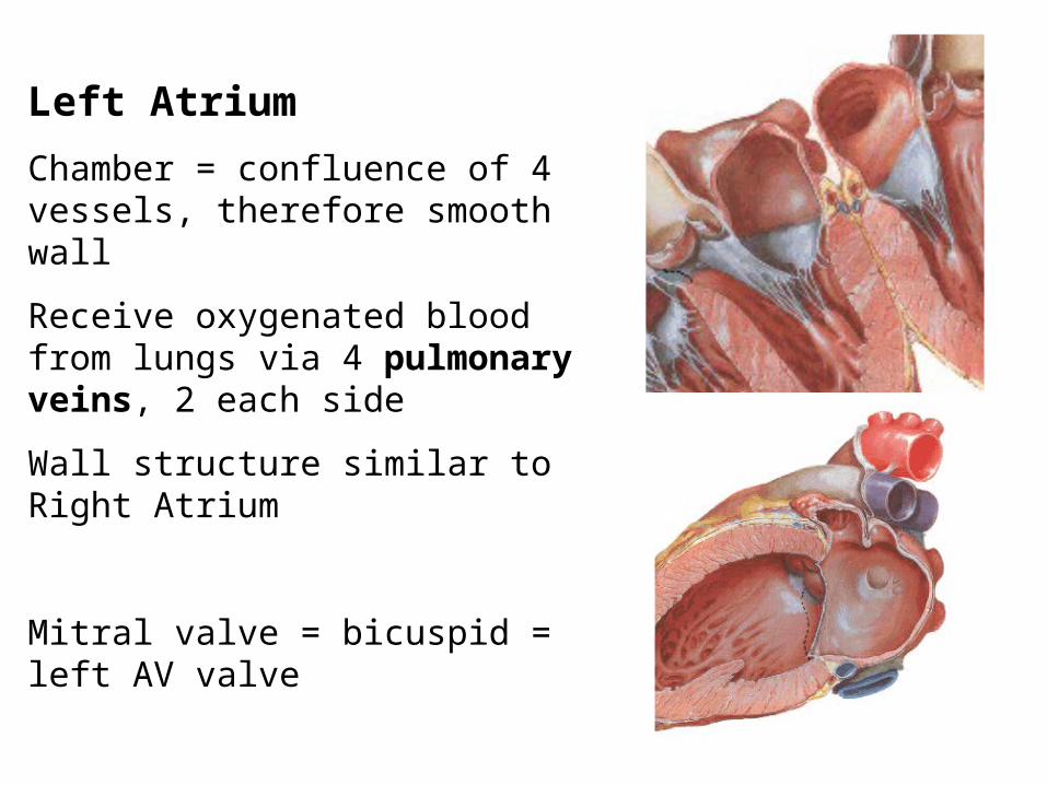
Left Atrium
Chamber = confluence of 4 vessels, therefore smooth wall
Receive oxygenated blood from lungs via 4 pulmonary veins, 2 each side
Wall structure similar to Right Atrium
Mitral valve = bicuspid = left AV valve
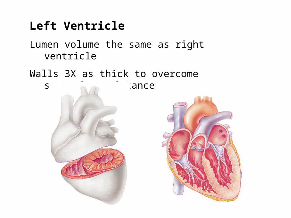
Left Ventricle
Lumen volume the same as right ventricle
Walls 3X as thick to overcome systemic resistance
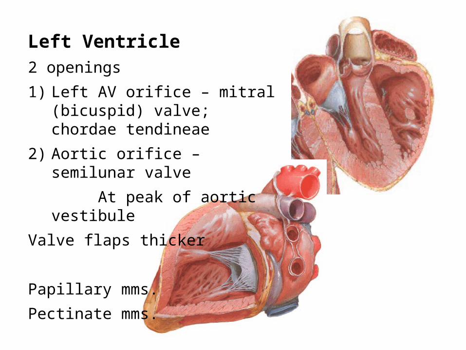
Left Ventricle2 openings
1) Left AV orifice – mitral (bicuspid) valve; chordae tendineae
2) Aortic orifice – semilunar valve
At peak of aortic vestibule
Valve flaps thicker
Papillary mms.
Pectinate mms.
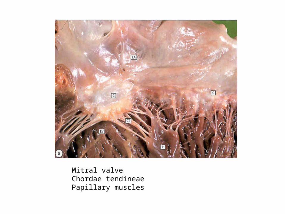
Mitral valveChordae tendineaePapillary muscles
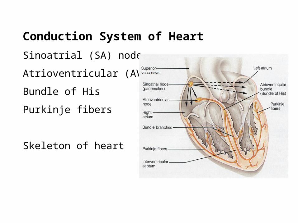
Conduction System of Heart
Sinoatrial (SA) node
Atrioventricular (AV) node
Bundle of His
Purkinje fibers
Skeleton of heart
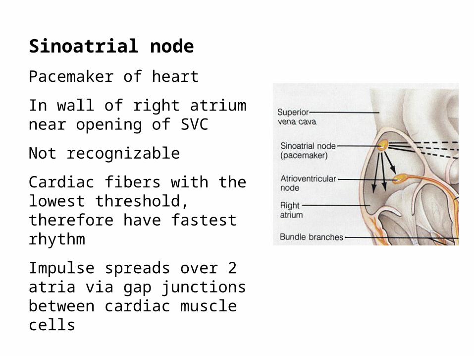
Sinoatrial node
Pacemaker of heart
In wall of right atrium near opening of SVC
Not recognizable
Cardiac fibers with the lowest threshold, therefore have fastest rhythm
Impulse spreads over 2 atria via gap junctions between cardiac muscle cells
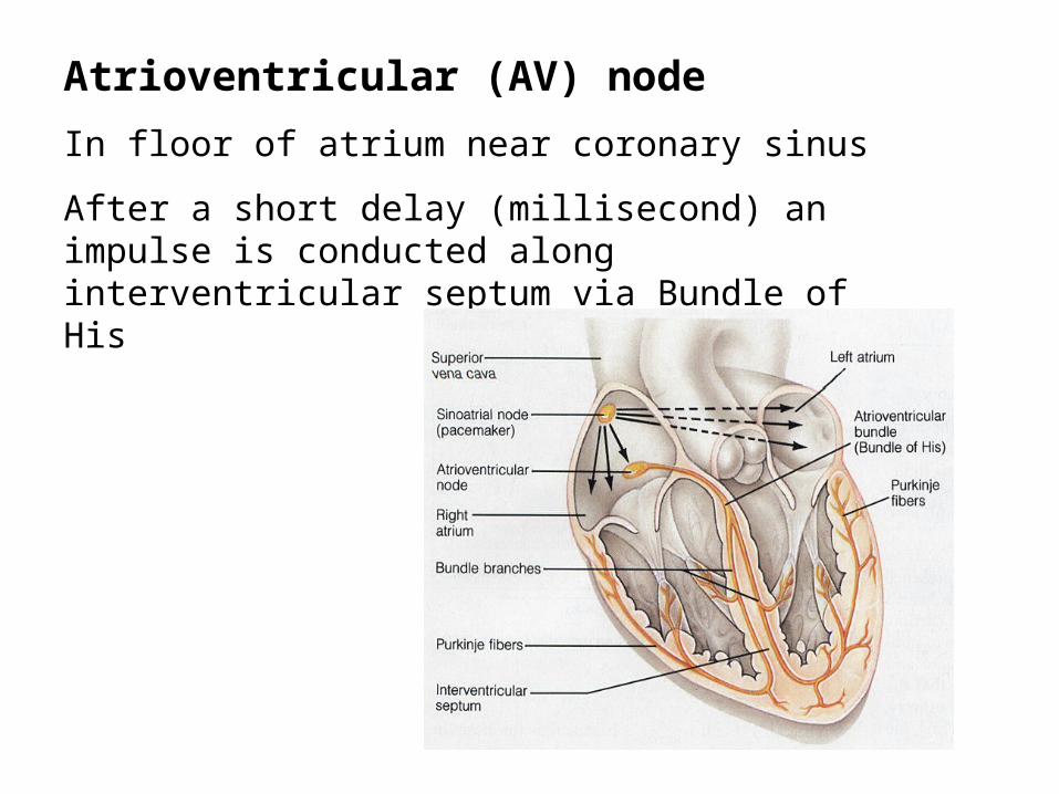
Atrioventricular (AV) node
In floor of atrium near coronary sinus
After a short delay (millisecond) an impulse is conducted along interventricular septum via Bundle of His
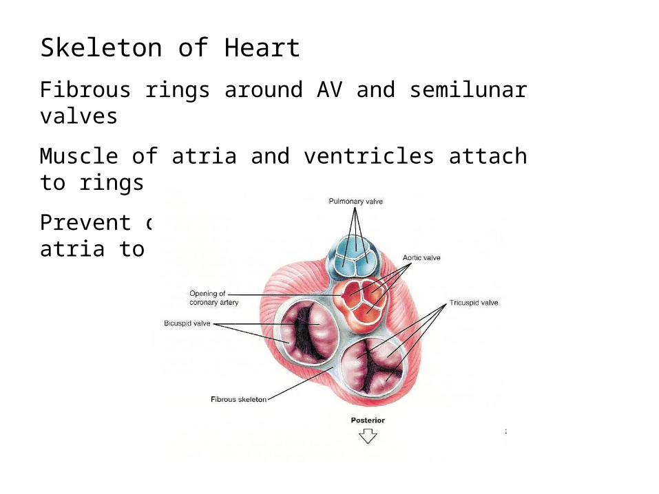
Skeleton of Heart
Fibrous rings around AV and semilunar valves
Muscle of atria and ventricles attach to rings
Prevent conduction of impulse from atria to ventricles
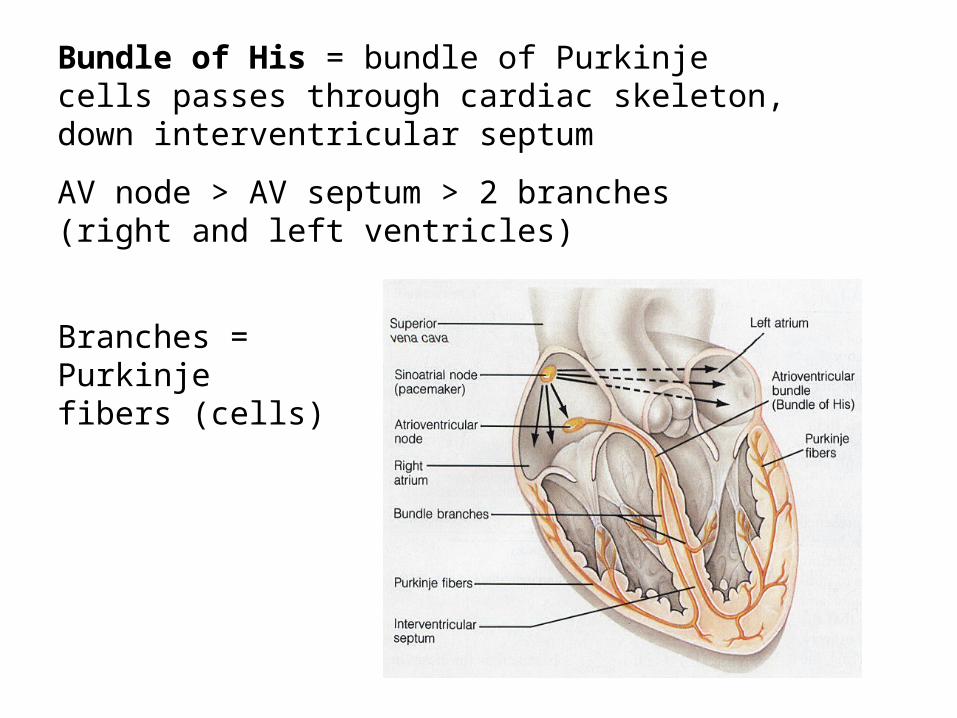
Bundle of His = bundle of Purkinje cells passes through cardiac skeleton, down interventricular septum
AV node > AV septum > 2 branches (right and left ventricles)
Branches = Purkinje fibers (cells)
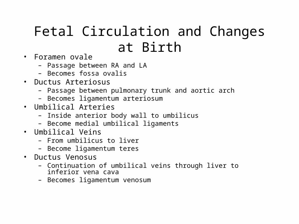
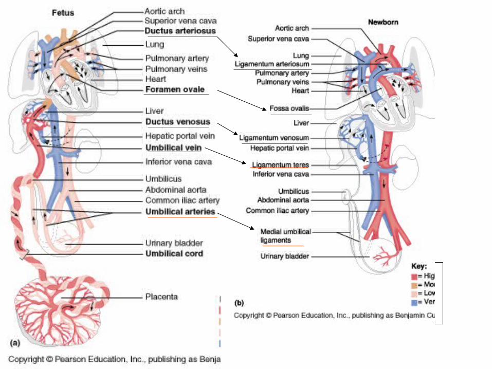
Fetal Circulation and Changes at Birth• Foramen ovale
– Passage between RA and LA– Becomes fossa ovalis
• Ductus Arteriosus– Passage between pulmonary trunk and aortic arch– Becomes ligamentum arteriosum
• Umbilical Arteries– Inside anterior body wall to umbilicus– Become medial umbilical ligaments
• Umbilical Veins – From umbilicus to liver– Become ligamentum teres
• Ductus Venosus– Continuation of umbilical veins through liver to inferior vena cava– Becomes ligamentum venosum