Circulating Nucleic Acids in Plasma and Serum · Circulating Nucleic Acids in Plasma and Serum ......
28
Circulating Nucleic Acids in Plasma and Serum
Transcript of Circulating Nucleic Acids in Plasma and Serum · Circulating Nucleic Acids in Plasma and Serum ......

Circulating Nucleic Acids in Plasma and Serum

Peter B. GahanEditor
Circulating Nucleic Acidsin Plasma and Serum
Proceedings of the 6th InternationalConference on Circulating Nucleic Acidsin Plasma and Serum Held on 9–11November 2009 in Hong Kong
123

EditorProf. Peter B. GahanKing’s College LondonAnatomy & Human SciencesLondon BridgeSE1 1UL LondonUnited [email protected]
ISBN 978-90-481-9381-3 e-ISBN 978-90-481-9382-0DOI 10.1007/978-90-481-9382-0Springer Dordrecht Heidelberg London New York
Library of Congress Control Number: 2010933647
© Springer Science+Business Media B.V. 2011No part of this work may be reproduced, stored in a retrieval system, or transmitted in any form or byany means, electronic, mechanical, photocopying, microfilming, recording or otherwise, without writtenpermission from the Publisher, with the exception of any material supplied specifically for the purposeof being entered and executed on a computer system, for exclusive use by the purchaser of the work.
Printed on acid-free paper
Springer is part of Springer Science+Business Media (www.springer.com)
Contents
Part I Current Developments
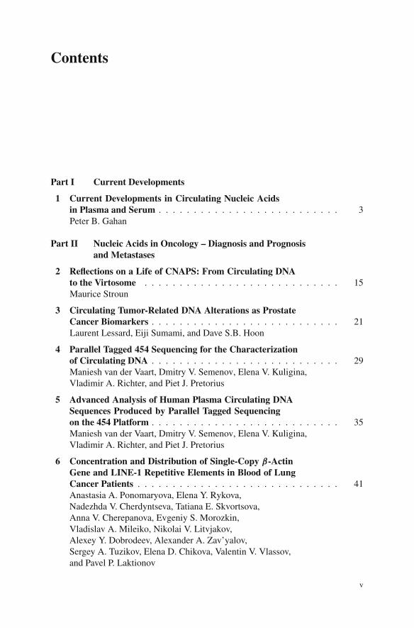
1 Current Developments in Circulating Nucleic Acidsin Plasma and Serum . . . . . . . . . . . . . . . . . . . . . . . . . . 3Peter B. Gahan
Part II Nucleic Acids in Oncology – Diagnosis and Prognosisand Metastases
2 Reflections on a Life of CNAPS: From Circulating DNAto the Virtosome . . . . . . . . . . . . . . . . . . . . . . . . . . . . 15Maurice Stroun
3 Circulating Tumor-Related DNA Alterations as ProstateCancer Biomarkers . . . . . . . . . . . . . . . . . . . . . . . . . . . 21Laurent Lessard, Eiji Sumami, and Dave S.B. Hoon
4 Parallel Tagged 454 Sequencing for the Characterizationof Circulating DNA . . . . . . . . . . . . . . . . . . . . . . . . . . . 29Maniesh van der Vaart, Dmitry V. Semenov, Elena V. Kuligina,Vladimir A. Richter, and Piet J. Pretorius
5 Advanced Analysis of Human Plasma Circulating DNASequences Produced by Parallel Tagged Sequencingon the 454 Platform . . . . . . . . . . . . . . . . . . . . . . . . . . . 35Maniesh van der Vaart, Dmitry V. Semenov, Elena V. Kuligina,Vladimir A. Richter, and Piet J. Pretorius
6 Concentration and Distribution of Single-Copy β-ActinGene and LINE-1 Repetitive Elements in Blood of LungCancer Patients . . . . . . . . . . . . . . . . . . . . . . . . . . . . . 41Anastasia A. Ponomaryova, Elena Y. Rykova,Nadezhda V. Cherdyntseva, Tatiana E. Skvortsova,Anna V. Cherepanova, Evgeniy S. Morozkin,Vladislav A. Mileiko, Nikolai V. Litvjakov,Alexey Y. Dobrodeev, Alexander A. Zav’yalov,Sergey A. Tuzikov, Elena D. Chikova, Valentin V. Vlassov,and Pavel P. Laktionov
v
vi Contents
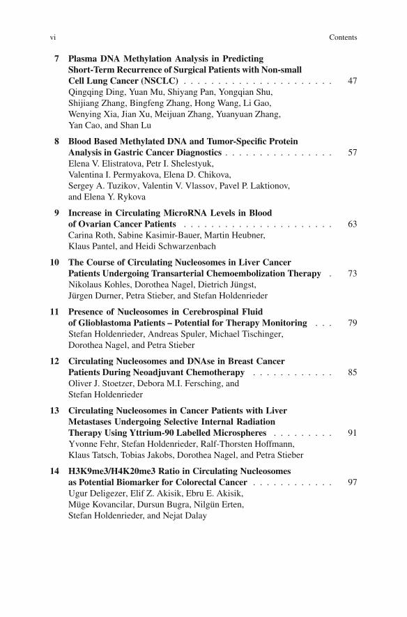
7 Plasma DNA Methylation Analysis in PredictingShort-Term Recurrence of Surgical Patients with Non-smallCell Lung Cancer (NSCLC) . . . . . . . . . . . . . . . . . . . . . . 47Qingqing Ding, Yuan Mu, Shiyang Pan, Yongqian Shu,Shijiang Zhang, Bingfeng Zhang, Hong Wang, Li Gao,Wenying Xia, Jian Xu, Meijuan Zhang, Yuanyuan Zhang,Yan Cao, and Shan Lu
8 Blood Based Methylated DNA and Tumor-Specific ProteinAnalysis in Gastric Cancer Diagnostics . . . . . . . . . . . . . . . . 57Elena V. Elistratova, Petr I. Shelestyuk,Valentina I. Permyakova, Elena D. Chikova,Sergey A. Tuzikov, Valentin V. Vlassov, Pavel P. Laktionov,and Elena Y. Rykova
9 Increase in Circulating MicroRNA Levels in Bloodof Ovarian Cancer Patients . . . . . . . . . . . . . . . . . . . . . . 63Carina Roth, Sabine Kasimir-Bauer, Martin Heubner,Klaus Pantel, and Heidi Schwarzenbach
10 The Course of Circulating Nucleosomes in Liver CancerPatients Undergoing Transarterial Chemoembolization Therapy . 73Nikolaus Kohles, Dorothea Nagel, Dietrich Jüngst,Jürgen Durner, Petra Stieber, and Stefan Holdenrieder
11 Presence of Nucleosomes in Cerebrospinal Fluidof Glioblastoma Patients – Potential for Therapy Monitoring . . . 79Stefan Holdenrieder, Andreas Spuler, Michael Tischinger,Dorothea Nagel, and Petra Stieber
12 Circulating Nucleosomes and DNAse in Breast CancerPatients During Neoadjuvant Chemotherapy . . . . . . . . . . . . 85Oliver J. Stoetzer, Debora M.I. Fersching, andStefan Holdenrieder
13 Circulating Nucleosomes in Cancer Patients with LiverMetastases Undergoing Selective Internal RadiationTherapy Using Yttrium-90 Labelled Microspheres . . . . . . . . . 91Yvonne Fehr, Stefan Holdenrieder, Ralf-Thorsten Hoffmann,Klaus Tatsch, Tobias Jakobs, Dorothea Nagel, and Petra Stieber
14 H3K9me3/H4K20me3 Ratio in Circulating Nucleosomesas Potential Biomarker for Colorectal Cancer . . . . . . . . . . . . 97Ugur Deligezer, Elif Z. Akisik, Ebru E. Akisik,Müge Kovancilar, Dursun Bugra, Nilgün Erten,Stefan Holdenrieder, and Nejat Dalay
Contents vii
15 Functionality of CNAPS in Cancer: The Theory of Genometastasis 105Dolores C. García-Olmo, Hector Guadalajara,Carolina Dominguez-Berzosa, María G. Picazo,Mariano García-Arranz, and Damián García-Olmo
Part III Nucleic Acids in Foetal Medicine
16 Circulating Fetal DNA/RNA in Maternal Plasmafor Aneuploidy Detection . . . . . . . . . . . . . . . . . . . . . . . 111Y.K. Tong, R.W.K. Chiu, and Y.M.D. Lo
17 A “Fluid-Agnostic” Approach to Analysis of Fetaland Neonatal Developmental Gene Expression . . . . . . . . . . . . 125Jill L. Maron and Diana W. Bianchi
18 Non-invasive Prenatal Diagnosis: An Epigenetic Approachto the Detection of Common Fetal Chromosome Disordersby Analysis of Maternal Blood Samples . . . . . . . . . . . . . . . 133Maj A. Hultén, Elisavet A. Papageorgiou, Floriana DellaRagione, Maurizio D’Esposito, Nigel Carter, andPhilippos C. Patsalis
19 Comparative Study of Extracellular DNA by FISH . . . . . . . . . 143Evgeniy S. Morozkin, Ekaterina M. Loseva,Vladislav A. Mileiko, Kira S. Zadesenets, Nikolay B. Rubtsov,Valentin V. Vlassov, and Pavel P. Laktionov
20 An Additional Pre-amplification Step for the EarlyDetermination of Fetal RHD from Maternal Plasma . . . . . . . . 147Tadeja Dovc-Drnovšek, Nataša Toplak, Irena Bricl,Tanja Blejec, Minka Kovac, and Primož Rožman
21 The Correlation of Circulating Cell-Free DNA, Cell-FreeFetal DNA and MicroRNA 325 Levels to ClinicalCharacteristics and Laboratory Parameters in Pre-eclampsia . . . 153Levente Lázár, Bálint Nagy, Attila Morvarec, and János Rigó
Part IV Other Clinical Exploitation of CNAPS
22 Comparison of Plasma Cell-Free DNA Levels with GeneExpression Profiles of Peripheral Blood Cells DuringHaemodialysis . . . . . . . . . . . . . . . . . . . . . . . . . . . . . . 159Ales Horinek, Ales Panczak, Magdalena Mokrejsova,Katarina Rocinova, Marie Korabecna, Dalibor Cerny, andVladimir Tesar
23 Low-Molecular-Weight DNA of Blood Plasmaas an Indicator of Pathological Processes . . . . . . . . . . . . . . . 165Irina N. Vasilyeva, Tatyana V. Ivtchik, and Igor A. Voznyuk

viii Contents
24 The Clinical Significance of Plasma DNA Quantificationfor Quake Trauma Patients . . . . . . . . . . . . . . . . . . . . . . 171Dan Chen, Shiyang Pan, Shijiang Zhang, Peijun Huang,Wenying Xia, Erfu Xie, Bing Gu, Fang Wang, Jian Xu,Ting Xu, Yachun Lu, Di Yang, and Shan Lu
Part V The Biology of CNAPS
25 Methylated Cell-Free DNA In Vitro and In Vivo . . . . . . . . . . . 185Tatyana E. Skvortsova, Olga E. Bryzgunova,Alena O. Lebedeva, Viktoria V. Mak, Valentin V. Vlassov, andPavel P. Laktionov
26 Circadian Rhythmicity and Clearance of Cell-Free DNAin Human Plasma . . . . . . . . . . . . . . . . . . . . . . . . . . . . 195Marie Korabecna, Ales Horinek, Nikola Bila, andSylvie Opatrna
27 Fragments of Cell-Free DNA (cfDNA) EnhanceTranscription Activity in Human Mesenchymal Stem Cells(hMSCs) and Inhibit Their In Vitro Differentiation . . . . . . . . . 199Elena M. Malinovskaya, Svetlana V. Kostyuk,Aleksey V. Ermakov, Marina S. Konkova,Tatjana D. Smirnova, Larisa V. Kameneva,Liudmila V. Efremova, Anna Yu. Alekseeva,Liudmila N. Lyubchenko, and Natalya N. Veiko
28 Cell-Surface-Bound DNA Inhibits Poly(I:C)-Activated IL-6and IL-8 Production in Human Primary Endothelial Cellsand Fibroblasts . . . . . . . . . . . . . . . . . . . . . . . . . . . . . 207Anna V. Cherepanova, Alexander V. Bushuev,Valentin V. Vlassov, and Pavel P. Laktionov
29 Accumulating Fragments of Extracellular DNA (ecDNA)Influence Rat Primary Cerebellum Granule Cell Culture . . . . . . 213Liudmila V. Efremova, Svetlana V. Kostyuk,Leonid G. Khaspekov, and Natalya N. Veiko
30 Cell Free DNA (cfDNA) Influences Nitric Oxide and rosLevels in Human Endothelial Cells . . . . . . . . . . . . . . . . . . 219Anna Yu. Alekseeva, Natalia V. Bulycheva,Svetlana V. Kostyuk, Tatjana D. Smirnova, andNatalya N. Veiko
31 Development of the Adaptive Response and BystanderEffect Induced by Low-Dose Ionising Radiation in HumanMesenchymal Stem Cells . . . . . . . . . . . . . . . . . . . . . . . . 225Aleksey V. Ermakov, Marina S. Konkova, Svetlana V. Kostyuk,Tatjana D. Smirnova, Liudmila V. Efremova,Liudmila N. Lyubchenko, and Natalya N. Veiko
Contents ix
32 Extracellular RNA as Regulators of Cellular Processes . . . . . . . 233Dmitry V. Semenov, Grigory A. Stepanov,Dmitry N. Baryakin, Olga A. Koval, Elena V. Kuligina, andVladimir A. Richter
33 Microvesicles Circulating in Plasma of Rats Contain DNA:Are These Small Vesicles a Main Source of Cell-Free DNAin Plasma? . . . . . . . . . . . . . . . . . . . . . . . . . . . . . . . . 239Gemma Serrano-Heras, Damián García-Olmo, andDolores C. García-Olmo
Part VI New Technologies for CNAPS
34 Rapid Isolation and Detection of Cell Free CirculatingDNA and Other Disease Biomarkers Directly from Whole Blood . 247Rajaram Krishnan and Michael J. Heller
35 Yields of Viral and Circulating Cell-Free Nucleic AcidsUsing the QIAamp R© Circulating Nucleic Acid Kit . . . . . . . . . 259Martin Horlitz, Tanja Hartinger, Simone Graf,Annabelle Lucas, Annette Nocon, andMarkus Sprenger-Haussels
36 Comparison of Nucleosomes and Quantitative PCR UsingDiverse DNA Isolation Methods . . . . . . . . . . . . . . . . . . . . 269Michael Fleischhacker, Bernd Schmidt, Sabine Weickmann,Debora M.I. Fersching, Gloria S. Leszinski, Barbara Siegele,Oliver J. Stoetzer, and Stefan Holdenrieder
37 MicroRNA Analysis in the Spinal Fluid of AlzheimerPatients: A Methodological Feasibility Study . . . . . . . . . . . . 275Argonde van Harten, Joyce Mulders, Cagla Çevik,Maartje Kester, Philip Scheltens, Wiesje van der Flier, andCees Oudejans
Index . . . . . . . . . . . . . . . . . . . . . . . . . . . . . . . . . . . . . 283
Contributors
Elif Z. Akisik Department of Basic Oncology, Istanbul University OncologyInstitute, Istanbul, Turkey, [email protected]
Ebru E. Akisik Department of Basic Oncology, Istanbul University OncologyInstitute, Istanbul, Turkey, [email protected]
Anna Yu. Alekseeva Research Centre for Medical Genetics, Russian Academyof Medical Sciences, Moscow, Russia, [email protected]
Dmitry N. Baryakin Institute of Chemical Biology and Fundamental MedicineSB RAS, Novosibirsk, Russia, [email protected]
Diana W. Bianchi Division of Genetics, Department of Pediatrics, FloatingHospital for Children at Tufts Medical Center, Boston, MA, USA; Divisionof Genetics, Department of Pediatrics, Floating Hospital for Children at TuftsMedical Center, Boston, MA, USA, [email protected]
Nikola Bila Faculty of Medicine in Pilsen, Charles University in Prague, Pilsen,Czech Republic
Tanja Blejec Department of Perinatology, University Medical Centre, Ljubljana,Slovenia
Irena Bricl Blood Transfusion Centre of Slovenia, Ljubljana, Slovenia
Olga E. Bryzgunova Siberian Division of the Russian Academy of Sciences,Institute of Chemical Biology and Fundamental Medicine, Novosibirsk, Russia,[email protected]
Dursun Bugra Deparment of Surgery, Istanbul Medical Faculty, IstanbulUniversity, Istanbul, Turkey, [email protected]
Natalia V. Bulycheva Research Centre for Medical Genetics, Russian Academyof Medical Sciences, Moscow, Russia, [email protected]
Alexander V. Bushuev Siberian Division of the Russian Academy of Sciences,Institute of Chemical Biology and Fundamental Medicine, Novosibirsk, Russia
xi
xii Contributors
Yan Cao Department of Laboratory Medicine, The First Affiliated Hospital ofNanjing Medical University, Nanjing, China, [email protected]
Nigel Carter Wellcome Trust Sanger Institute, Cambridge, UK
Dalibor Cerny 1st School of Medicine, Charles University, Prague, CzechRepublic, [email protected]
Cagla Çevik Department Clinical Chemistry, VU University Medical Center,De Amsterdam, The Netherlands
Dan Chen Department of Laboratory Medicine, The First Affiliated Hospital ofNanjing Medical University, Nanjing, China, [email protected]
Nadezhda V. Cherdyntseva Siberian Division of the Russian Academy of MedicalSciences, Cancer Research Institute, Tomsk, Russia, [email protected]
Anna V. Cherepanova Siberian Division of the Russian Academy of Sciences,Institute of Chemical Biology and Fundamental Medicine, Novosibirsk, Russia,[email protected]
Elena D. Chikova Siberian Division of the Russian Academy of Sciences, Instituteof Chemical Biology and Fundamental Medicine, Novosibirsk, Russia,[email protected]
R.W.K. Chiu Centre for Research into Circulating Fetal Nucleic Acids, Li KaShing Institute of Health Sciences, and Department of Chemical Pathology, Princeof Wales Hospital, The Chinese University of Hong Kong, Shatin, Hong KongSAR, China, [email protected]
Maurizio D’Esposito Institute of Genetics and Biophysics ‘A. Buzzati Traverso’,CNR, Naples, Italy, [email protected]
Nejat Dalay Department of Basic Oncology, Istanbul University OncologyInstitute, Istanbul, Turkey, [email protected]
Ugur Deligezer Department of Basic Oncology, Istanbul University OncologyInstitute, Istanbul, Turkey, [email protected]
Qingqing Ding Department of Laboratory Medicine, The First Affiliated Hospitalof Nanjing Medical University, Nanjing, China; Department of Oncology, TheFirst Affiliated Hospital of Nanjing Medical University, Nanjing, China,[email protected]
Alexey Y. Dobrodeev Siberian Division of the Russian Academy of MedicalSciences, Cancer Research Institute, Tomsk, Russia, [email protected]
Carolina Dominguez-Berzosa IdiPAZ, “La Paz” University Hospital, UniversidadAutónoma de Madrid, Madrid, Spain, [email protected]
Tadeja Dovc-Drnovšek Blood Transfusion Centre of Slovenia, Ljubljana,Slovenia, [email protected]

Contributors xiii
Jürgen Durner Institute of Clinical Chemistry, University-HospitalMunich-Grosshadern, Munich, Germany, [email protected]
Liudmila V. Efremova Research Centre for Medical Genetics, Russian Academyof Medical Sciences, Moscow, Russia, [email protected]
Elena V. Elistratova Siberian Division of the Russian Academy of Sciences,Institute of Chemical Biology and Fundamental Medicine, Novosibirsk, Russia,[email protected]
Aleksey V. Ermakov Research Centre for Medical Genetics, Russian Academy ofMedical Sciences, Moscow, Russia, [email protected]
Nilgün Erten Department of Internal Medicine, Istanbul Medical Faculty, IstanbulUniversity, Istanbul, Turkey, [email protected]
Yvonne Fehr Institute of Clinical Chemistry, University-HospitalMunich-Grosshadern, Munich, Germany, [email protected]
Debora M.I. Fersching Institute of Clinical Chemistry, University HospitalMunich, Munich, Germany, [email protected]
Michael Fleischhacker Medical Clinic – Oncology and Haematology, UniversityMedicine Charité Berlin, Berlin, Germany, [email protected]
Peter B. Gahan Anatomy and Human Sciences, King’s College London, London,UK, [email protected]
Li Gao Department of Laboratory Medicine, The First Affiliated Hospital ofNanjing Medical University, Nanjing, China, [email protected]
Mariano García-Arranz IdiPAZ, “La Paz” University Hospital, UniversidadAutónoma de Madrid, Madrid, Spain, [email protected]
Damián García-Olmo IdiPAZ, “La Paz” University Hospital, Department ofSurgery, Universidad Autónoma de Madrid, Madrid, Spain, [email protected]
Dolores C. García-Olmo Experimental Research Unit, General UniversityHospital of Albacete, Albacete, Spain, [email protected]
Simone Graf R&D Department, QIAGEN GmbH, Hilden, Germany,[email protected]
Bing Gu Department of Laboratory Medicine, The First Affiliated Hospital ofNanjing Medical University, Nanjing, China, [email protected]
Hector Guadalajara IdiPAZ, “La Paz” University Hospital, UniversidadAutónoma de Madrid, Madrid, Spain, [email protected]
Tanja Hartinger R&D Department, QIAGEN GmbH, Hilden, Germany,[email protected]
xiv Contributors
Michael J. Heller Department of Bioengineering, Department ofNanoengineering, UCSD Moores Cancer Center, University of CaliforniaSan Diego, La Jolla, CA, USA, [email protected]
Martin Heubner Department of Gynaecology and Obstetrics, Universityof Duisburg-Essen, Essen, Germany, [email protected]
Ralf-Thorsten Hoffmann Institute of Clinical Radiology, University-HospitalMunich-Grosshadern, Munich, Germany, [email protected]
Stefan Holdenrieder Institute of Clinical Chemistry, University-HospitalMunich-Grosshadern, Munich, Germany,[email protected]
Dave S.B. Hoon Department of Molecular Oncology, John Wayne CancerInstitute, Santa Monica, CA, USA, [email protected]
Ales Horinek 1st School of Medicine, Charles University, Prague, CzechRepublic; General Teaching Hospital, Prague, Czech Republic, [email protected]
Martin Horlitz R&D Department, QIAGEN GmbH, Hilden, Germany,[email protected]
Peijun Huang Department of Laboratory Medicine, The First Affiliated Hospitalof Nanjing Medical University, Nanjing, China, [email protected]
Maj A. Hultén Warwick Medical School, University of Warwick, Coventry, UK,[email protected]
Tatyana V. Ivtchik St-Petersburg State Medical University named after I. P.Pavlov, St-Petersburg, Russia, [email protected]
Tobias Jakobs Institute of Clinical Radiology, University-HospitalMunich-Grosshadern, Munich, Germany, [email protected]
Dietrich Jüngst Medical Clinic II, University-Hospital Munich-Grosshadern,Munich, Germany
Larisa V. Kameneva Research Centre for Medical Genetics, Russian Academy ofMedical Sciences, Moscow, Russia
Sabine Kasimir-Bauer Department of Gynaecology and Obstetrics, University ofDuisburg-Essen, Essen, Germany, [email protected]
Maartje Kester Departments of Neurology, VU University Medical Center,Amsterdam, The Netherlands: Departments of Epidemiology/Biostatistics, VUUniversity Medical Center, Amsterdam, The Netherlands
Leonid G. Khaspekov Research Centre for Medical Genetics, Russian Academyof Medical Sciences, Moscow, Russia, [email protected],[email protected]

Contributors xv
Nikolaus Kohles Institute of Clinical Chemistry, University-HospitalMunich-Grosshadern, Munich, Germany, [email protected]
Marina S. Konkova Research Centre for Medical Genetics, Russian Academy ofMedical Sciences, Moscow, Russia, [email protected]
Marie Korabecna Department of Biology, Faculty of Medicine in Pilsen, CharlesUniversity in Prague, Pilsen, Czech Republic, [email protected]
Svetlana V. Kostyuk Research Centre for Medical Genetics, Russian Academy ofMedical Sciences, Moscow, Russia, [email protected]
Minka Kovac Omega d.o.o., Ljubljana, Slovenia
Olga A. Koval Institute of Chemical Biology and Fundamental Medicine SB RAS,Novosibirsk, Russia, [email protected]
Müge Kovancilar Department of Basic Oncology, Istanbul University OncologyInstitute, Istanbul, Turkey, [email protected]
Rajaram Krishnan Department of Bioengineering, Department ofNanoengineering, UCSD Moores Cancer Center, University of California SanDiego, La Jolla, CA, USA
Elena V. Kuligina Institute of Chemical Biology and Fundamental Medicine SBRAS, Novosibirsk, Russia, [email protected]
Pavel P. Laktionov Siberian Division of the Russian Academy of Sciences,Institute of Chemical Biology and Fundamental Medicine, Novosibirsk, Russia,[email protected]
Levente Lázár Department of Obstetrics and Gynecology, SemmelweisUniversity, Budapest, Hungary, [email protected]
Alena O. Lebedeva Siberian Division of the Russian Academy of Sciences,Institute of Chemical Biology and Fundamental Medicine, Novosibirsk, Russia,[email protected]
Laurent Lessard Department of Molecular Oncology, John Wayne CancerInstitute, Santa Monica, CA, USA, [email protected]
Gloria S. Leszinski Institute of Clinical Chemistry, University Hospital Munich,Munich, Germany, [email protected]
Nikolai V. Litvjakov Siberian Division of the Russian Academy of MedicalSciences, Cancer Research Institute, Tomsk, Russia, [email protected]
Y.M.D. Lo Centre for Research into Circulating Fetal Nucleic Acids, Li Ka ShingInstitute of Health Sciences, and Department of Chemical Pathology, Prince ofWales Hospital, The Chinese University of Hong Kong, Shatin, Hong Kong SAR,China, [email protected]
xvi Contributors
Ekaterina M. Loseva Siberian Division of the Russian Academy of Sciences,Institute of Chemical Biology and Fundamental Medicine, Novosibirsk, Russia,[email protected]
Yachun Lu Department of Laboratory Medicine, The First Affiliated Hospitalof Nanjing Medical University, Nanjing, China, [email protected]
Shan Lu Department of Medicine, University of Massachusetts Medical School,Worcester, MA, USA, [email protected]
Annabelle Lucas R&D Department, QIAGEN GmbH, Hilden, Germany,[email protected]
Liudmila N. Lyubchenko Research Centre for Medical Genetics, RussianAcademy of Medical Sciences, Moscow, Russia, [email protected]
Viktoria V. Mak Siberian Division of the Russian Academy of Sciences, Instituteof Cytology and Genetics, Novosibirsk, Russia, [email protected]
Elena M. Malinovskaya Research Centre for Medical Genetics, RussianAcademy of Medical Sciences, Moscow, Russia, [email protected]
Jill L. Maron Division of Newborn Medicine, Department of Pediatrics, FloatingHospital for Children at Tufts Medical Center, Boston, MA, USA,[email protected]
Vladislav A. Mileiko Siberian Division of the Russian Academy of Sciences,Institute of Chemical Biology and Fundamental Medicine, Novosibirsk, Russia,[email protected]
Magdalena Mokrejsova General Teaching Hospital, Prague, Czech Republic;1st School of Medicine, Charles University, Prague, Czech Republic,[email protected]
Evgeniy S. Morozkin Siberian Division of the Russian Academy of Sciences,Institute of Chemical Biology and Fundamental Medicine, Novosibirsk, Russia,[email protected]
Attila Morvarec Department of Obstetrics and Gynecology, SemmelweisUniversity, Budapest, Hungary
Yuan Mu Department of Laboratory Medicine, The First Affiliated Hospitalof Nanjing Medical University, Nanjing, China, [email protected]
Joyce Mulders Department Clinical Chemistry, VU University Medical Center,De Amsterdam, The Netherlands, [email protected]
Dorothea Nagel Institute of Clinical Chemistry, University-HospitalMunich-Grosshadern, Munich, Germany, [email protected]
Bálint Nagy Department of Obstetrics and Gynecology, Semmelweis University,Budapest, Hungary
Contributors xvii
Annette Nocon R&D Department, QIAGEN GmbH, Hilden, Germany,[email protected]
Sylvie Opatrna Faculty of Medicine in Pilsen, Charles University in Prague,Pilsen, Czech Republic, [email protected]
Cees Oudejans Department Clinical Chemistry, VU University Medical Center,De Amsterdam, The Netherlands, [email protected]
Shiyang Pan Department of Laboratory Medicine, The First Affiliated Hospitalof Nanjing Medical University, Nanjing, China, [email protected]
Ales Panczak General Teaching Hospital, Prague, Czech Republic; 1st School ofMedicine, Charles University, Prague, Czech Republic, [email protected]
Klaus Pantel Institute of Tumour Biology, University Medical CentreHamburg-Eppendorf, Hamburg, Germany, [email protected]
Elisavet A. Papageorgiou Department of Cytogenetics and Genomics, TheCyprus Institute of Neurology and Genetics, Nicosia, Cyprus, [email protected]
Philippos C. Patsalis Department of Cytogenetics and Genomics, The CyprusInstitute of Neurology and Genetics, Nicosia, Cyprus, [email protected]
Valentina I. Permyakova Siberian Division of the Russian Academy of Sciences,Central Clinical Hospital, Novosibirsk, Russia, [email protected]
María G. Picazo General University Hospital of Albacete, Albacete, Spain
Anastasia A. Ponomaryova Siberian Division of the Russian Academy ofMedical Sciences, Cancer Research Institute, Tomsk, Russia,[email protected]
Piet J. Pretorius Biochemistry Division, School for Physical and ChemicalSciences, North-West University, Potchefstroom, South Africa,[email protected]
Floriana Della Ragione Institute of Genetics and Biophysics ‘A. BuzzatiTraverso’, CNR, Naples, Italy, [email protected]
Vladimir A. Richter Institute of Chemical Biology and Fundamental MedicineSB RAS, Novosibirsk, Russia, [email protected]
János Rigó Department of Obstetrics and Gynecology, Semmelweis University,Budapest, Hungary
Katarina Rocinova General Teaching Hospital, Prague, Czech Republic; 1stSchool of Medicine, Charles University, Prague, Czech Republic,[email protected]
Carina Roth Institute of Tumour Biology, University Medical CentreHamburg-Eppendorf, Hamburg, Germany, [email protected]

xviii Contributors
Primož Rožman Blood Transfusion Centre of Slovenia, Ljubljana, Slovenia
Nikolay B. Rubtsov Siberian Division of the Russian Academy of Sciences,Institute of cytology and genetics, Novosibirsk, Russia, [email protected]
Elena Y. Rykova Siberian Division of the Russian Academy of Sciences, Instituteof Chemical Biology and Fundamental Medicine, Novosibirsk, Russia,[email protected]
Philip Scheltens Departments of Neurology and Epidemiology/Biostatistics,VU University Medical Center, Amsterdam, The Netherlands
Bernd Schmidt Medical Clinic – Infectiology and Pulmonology, UniversityMedicine Charité Berlin, Berlin, Germany, [email protected]
Heidi Schwarzenbach Institute of Tumour Biology, University Medical CentreHamburg-Eppendorf, Hamburg, Germany, [email protected]
Dmitry V. Semenov Institute of Chemical Biology and Fundamental MedicineSB RAS, Novosibirsk, Russia, [email protected]
Gemma Serrano-Heras Experimental Research Unit, General University Hospitalof Albacete, Albacete, Spain, [email protected]
Petr I. Shelestyuk Novosibirsk Oncological Dispensary, Novosibirsk, Russia,[email protected]
Yongqian Shu Department of Oncology, The First Affiliated Hospital of NanjingMedical University, Nanjing, China, [email protected]
Barbara Siegele Institute of Clinical Chemistry, University Hospital Munich,Munich, Germany, [email protected]
Tatiana E. Skvortsova Siberian Division of the Russian Academy of Sciences,Institute of Chemical Biology and Fundamental Medicine, Novosibirsk, Russia,[email protected]
Tatjana D. Smirnova Research Centre for Medical Genetics, Russian Academy ofMedical Sciences, Moscow, Russia, [email protected]
Markus Sprenger-Haussels R&D Department, QIAGEN GmbH, Hilden,Germany, [email protected]
Andreas Spuler Department of Neurosurgery, Helios Klinikum Berlin-Buch,Berlin, Germany, [email protected]
Grigory A. Stepanov Institute of Chemical Biology and Fundamental MedicineSB RAS, Novosibirsk, Russia, [email protected]
Petra Stieber Institute of Clinical Chemistry, University-HospitalMunich-Grosshadern, Munich, Germany, [email protected]
Contributors xix
Oliver J. Stoetzer Hematology/Oncology Outpatient Specialty Center Munich,Munich, Germany, [email protected]
Maurice Stroun OncoXL, Geneva, Switzerland, [email protected]
Eiji Sumami Department of Molecular Oncology, John Wayne Cancer Institute,Santa Monica, CA, USA, [email protected]
Klaus Tatsch Department of Nuclear Medicine, University-HospitalMunich-Grosshadern, Munich, Germany; Department of Nuclear Medicine,Municipal Hospital Karlsruhe, Karlsruhe, Germany,[email protected]
Vladimir Tesar General Teaching Hospital, Prague, Czech Republic; 1st School ofMedicine, Charles University, Prague, Czech Republic,[email protected]
Michael Tischinger Department of Psychiatry, University of Munich, Munich,Germany, [email protected]
Y.K. Tong Centre for Research into Circulating Fetal Nucleic Acids, Li Ka ShingInstitute of Health Sciences, and Department of Chemical Pathology, Prince ofWales Hospital, The Chinese University of Hong Kong, Shatin, Hong Kong SAR,China, [email protected]
Nataša Toplak Omega d.o.o., Ljubljana, Slovenia
Sergey A. Tuzikov Siberian Division of the Russian Academy of MedicalSciences, Cancer Research Institute, Tomsk, Russia, [email protected]
Wiesje van der Flier Departments of Neurology, VU University Medical Center,Amsterdam, The Netherlands; Departments of Epidemiology/Biostatistics, VUUniversity Medical Center, Amsterdam, The Netherlands; Alzheimer Center, VUUniversity Medical Center, Amsterdam, The Netherlands, [email protected]
Maniesh van der Vaart Biochemistry Division, School for Physical and ChemicalSciences, North-West University, Potchefstroom, South Africa,[email protected]
Argonde van Harten Departments of Neurology, VU University Medical Center,Amsterdam, The Netherlands; Departments of Epidemiology/Biostatistics, VUUniversity Medical Center, Amsterdam, The Netherlands, [email protected]
Irina N. Vasilyeva St-Petersburg Scientific Research Institute ofPhthisiopulmonology, St-Petersburg, Russia, [email protected]
Natalya N. Veiko Research Centre for Medical Genetics, Russian Academy ofMedical Sciences, Moscow, Russia, [email protected]
Valentin V. Vlassov Siberian Division of the Russian Academy of Sciences,Institute of Chemical Biology and Fundamental Medicine, Novosibirsk, Russia,[email protected]
xx Contributors
Igor A. Voznyuk Military Medical Academy, St-Petersburg, Russia,[email protected]
Fang Wang Department of Laboratory Medicine, The First Affiliated Hospital ofNanjing Medical University, Nanjing, China, [email protected]
Hong Wang Department of Laboratory Medicine, The First Affiliated Hospital ofNanjing Medical University, Nanjing, China, [email protected]
Sabine Weickmann Medical Clinic – Infectiology and Pulmonology, UniversityMedicine Charité Berlin, Berlin, Germany, [email protected]
Wenying Xia Department of Laboratory Medicine, The First Affiliated Hospital ofNanjing Medical University, Nanjing, China, [email protected]
Erfu Xie Department of Laboratory Medicine, The First Affiliated Hospital ofNanjing Medical University, Nanjing, China, [email protected]
Jian Xu Department of Laboratory Medicine, The First Affiliated Hospital ofNanjing Medical University, Nanjing, China, [email protected]
Ting Xu Department of Laboratory Medicine, The First Affiliated Hospital ofNanjing Medical University, Nanjing, China, [email protected]
Jian Xu Department of Laboratory Medicine, The First Affiliated Hospital ofNanjing Medical University, Nanjing, China
Di Yang The Platform for Molecular Diagnosis and Biotherapy of GravenessDisease of Jiangsu Province, Nanjing, China, [email protected]
Kira S. Zadesenets Siberian Division of the Russian Academy of Sciences,Institute of cytology and genetics, Novosibirsk, Russia, [email protected]
Alexander A. Zav’yalov Siberian Division of the Russian Academy of MedicalSciences, Cancer Research Institute, Tomsk, Russia,[email protected]
Shijiang Zhang Department of Surgery, The First Affiliated Hospital of NanjingMedical University, Nanjing, China, [email protected]
Bingfeng Zhang Department of Surgery, The First Affiliated Hospital of NanjingMedical University, Nanjing, China, [email protected]
Meijuan Zhang Department of Laboratory Medicine, The First Affiliated Hospitalof Nanjing Medical University, Nanjing, China, [email protected]
Yuanyuan Zhang Department of Laboratory Medicine, The First AffiliatedHospital of Nanjing Medical University, Nanjing, China,[email protected]

Part ICurrent Developments

Chapter 1Current Developments in Circulating NucleicAcids in Plasma and Serum
Peter B. Gahan
Abstract DNA and RNA fractions have been isolated from the whole blood, serum,plasma, the surface of blood cells, urine, saliva and spinal fluid from both healthyindividuals and patients. The ability to isolate, quantify, and analyze these moleculeshas led to the identification of specific nucleic acid fragments related to a varietyof clinical disorders thereby permitting their early diagnosis and prognosis. Thischapter summarizes the work reported in this volume.
Keywords Circulating nucleic acids · Fetal medicine · Oncology · Newtechnology · Biology of CNAPS
Introduction
The current volume concerns the meeting of the sixth international conference oncirculating nucleic acids in plasma and serum (CNAPS) held in Hong Kong on 9-11 November 2009. The aim of the meeting was to bring together clinicians andscientists working in this field to present their latest findings on the basic biology,methodology and clinical applications of circulating nucleic acids in blood, urine,cerebro-spinal fluid and saliva.
Since the first publication by Mendel and Métais (1948) reporting the circula-tion of DNA in blood and its increase in amount in cancer patients, studies haveevolved from just considering the amounts of DNA circulating in CNAPS dur-ing cancer and other clinical situations (Leon et al. 1977; Koeffler et al. 1973;Tan et al. 1966; Stroun et al. 1989) to more recent developments in the use ofDNA, nucleosomes, mRNA and micro RNAs as both early markers and prognos-tic tools. Most work has concentrated upon the role of isolated DNA fractions and
P.B. Gahan (B)Anatomy and Human Sciences, King’s College London, London, UKe-mail: [email protected]
3P.B. Gahan (ed.), Circulating Nucleic Acids in Plasma and Serum,DOI 10.1007/978-90-481-9382-0_1, C© Springer Science+Business Media B.V. 2011
4 P.B. Gahan
nucleosomes and to a lesser degree, mRNA. However, it is clear that the nucleicacids present in blood are derived from a number of sources including the break-down of cells in the blood - both blood cells and circulating cancer cells, cell-surfacebound DNA, the presence of bacteria and viruses, tissue necrosis, cell and tis-sue apoptosis, release of a newly synthesized DNA/RNA lipo-protein complex(the virtosome), exosomes, transposons and retrotransposons (Gahan and Stroun2010). This medley of sources offers range of choice in the nucleic acid fractionto be assessed with respect to a particular disorder. In addition, the development ofapproaches involving the exploitation of epigenetic events such as methylation andhypermethylation, histone modifications in circulating nucleosomes, RNA-singlenucleotide polymorphism, epigenetic allelic ratios and epigenetic-genetic chromo-some dosage has offered more sensitive diagnostic methods that are applicable inthe clinical environment. The following comments will highlight advances pre-sented at the symposium and will raise questions as to the future developmentsneeded.
Nucleic Acids in Oncology – Diagnosis and Prognosisand Metastases
Diagnosis and Prognosis
Two aspects of CNAPS in oncology have provided areas of development, namely,the clinical application in early diagnosis and prognosis and the other, a betterunderstanding of the origins of metastases.
Nucleosomes have provided the basis for a number of studies in both early diag-nosis and prognosis in cancer. Thus, trans-arterial chemo-embollization, the newloco-regional anticancer treatment option for advanced hepatocellular carcinomapatients, has been assessed in terms of serum nucleosome levels by Kohles et al.Although an initial decline was found in nucleosome levels shortly after treatment,by 24 hours there was a marked increase possibly due to the release of nucleosomesfrom the increased number of necrotic cells. Hence, this may provide a means ofestimating the efficiency of the therapy. Likewise for the studies by Fehr et al. onnucleosome levels in patients after treatment by selective internal radiation therapy.This is a loco-regional anticancer treatment option for advanced cancer patients withliver metastases or liver cancer employing Yttrium-90 labelled microspheres.
A similar prognostic value might be available through the results of a prelimi-nary investigation of nucleosome levels in both blood and cerebro-spinal fluid frompatients with glioblastoma by Holdenreider et al. They showed that patients devel-oping oedema after operation had a substantial increase in the nucleosome levels inthe cerebo-spinal fluid. Additional studies may show this to be a useful marker ofthe development of post-operational complications.
As a variation on the nucleosome theme, Delizeger et al. examined a modifica-tion of the histone fraction of nucleosomes as a marker for colorectal cancer. Hence,

1 Current Developments in Circulating Nucleic Acids in Plasma and Serum 5
an analysis of the trimethylation of histone H3 lysine 9 (H3K9me3) and histoneH4 lysine 9 (H4K20me3), at the pericentric heterochromatin of the nucleosomescirculating in plasma, exploited the H3K9me3/H4K20me3 ratio for normalizingH3K9me3 concentrations. In this way, it was possible to distinguish patients withcolorectal cancer (median 0.8) from the healthy group (median 3) and those withmultiple myeloma (median 4.7).
An alternative approach in which allelic imbalance and DNA hypermethylationanalyses have been combined, has been exploited by Lessard et al. who demon-strated a significantly improved sensitivity for the method to detect prostate cancer.This approach, using loss of heterozygosity combined with hypermethylation, hasproved to be more sensitive than the currently used prostate specific antigen serumlevels.
Another approach for the detection of prostate cancer was emplyed by Van derVaart et al. using parallel tagged sequencing of circulating DNA on the GSFLXsequencer from 454 life sciences. A total of ~3600 unique sequences were ana-lyzed and were seen to be distributed over the human genome with a slightly highermutation rate being observed for DNA obtained from the cancer patients when com-pared to the control group. A further characterization of this array of sequenceswas performed by comparative analysis of chromosome distribution, repeat con-tent and epigenetic characteristics of plasma DNA. Satellite repeats attributed tochromosome 12 were elevated in plasma of prostate cancer patients.
Although the average concentration of circulating DNA, measured as LINE-1repetitive elements, in plasma was shown to be similar in healthy individuals andnon-small cell lung cancer patients, Ponomaryova et al. also found that the concen-tration of cell-surface-bound circulating DNA was significantly low. This correlatedwith a poor disease prognosis. The ratio of the β-actin gene to LINE-1 was found tobe elevated in the cell-surface-bound DNA of the non-small cell lung cancer patientscompared to healthy individuals,. Hence, these results indicate a possible role forβ-actin gene and LINE-1 fragments circulating in non-small cell lung cancerpatients in both tumour detection and prognosis. Ding et al. also found that thequantification of methylated RASSF1A after operation provided a useful prognosticbiomarker for predicting the recurrence in non-small cell lung cancer patients aftercurative-intent surgery.
Epigenetic effects in the shape of promotor methylation rates of three tumoursuppressor genes from both plasma DNA and cell-surface-bound DNA from gas-tric cancer patients were considered by Elistratova et al. Methylated forms of p15,MGMT and hMLH1 genes were detected with high rates at stages II, III and IVof gastric cancer. However, no significant correlation was found between epigeneticand protein markers so indicating their independent development in gastric tumorpathogenesis.
A different approach has been taken by Roth et al. in studies on ovariancancer patients. The concentrations of four circulating microRNAs (miRNA10b,miRNA34a, miRNA141 and miRNA155) were measured in the serum of 59patients with ovarian cancer and 29 healthy individuals. The levels of total RNA,miRNA10b, miRNA34a, miRNA141, and miRNA155 in ovarian cancer patients,
6 P.B. Gahan
were significantly higher than those from the healthy controls. A significantcorrelation was also recorded of increasing amounts of miRNA34a with lymph nodemetastases.
Metastases
Amongst the DNAs circulating in cancer patients will be DNAs released fromtumour cells either by necrosis or apoptosis or as newly synthesised virtosomes(Adams et al. 1997; Anker et al. 1994; Garcia-Olmo et al. 2010; Stroun et al.1989). CNAPS DNAs can readily enter cells and in some cases be expressedin a way that modifies the biology of the recipients cell (Adams et al. 1997;Anker and Stroun 1972; Anker et al. 1980, 1994; Bulicheva et al. 2008; Ermakovet al. 2008; Garcia-Olmo et al. 2010; Ottolenghi and Hotchkiss 1960; Skvortsovaet al. 2008). Thus, it is not only possible for cancer cells circulating in the bloodto result in metastases, but also for the DNA released from tumour cells to dolikewise.
One of the most common alterations of tumour related DNA found in CNAPSDNA from cancer patients is its hypermethylation. Thus, methylated fragmentsof the RAR2 gene from CNAPS have been shown to be taken up by HeLa andhuman umbilical vein endothelial cells twice as efficiently as unmethylated frag-ments. Since the methylated RAR 2 gene fragments are more prevalent than theunmethylated fragments in intracellular traffic, they would appear to pose a highertransformation potential (Skvortsova et al. 2008).
It has been shown that the SW 480 cell line, originating from a human coloncarcinoma and containing a point mutation of the K-ras gene on both alleles, canbe released in the form of the newly synthesised, virtosomal DNA/RNA-lipoproteincomplex containing the mutated K-ras gene. Culturing NIH/3T3 cells in the pres-ence of the non-purified SW 480 cell supernatant containing the virtosome complexresulted in the appearance of transformed foci. The presence of a mutated ras genein the transfected foci of the 3T3 cells was confirmed by hybridization after PCRand by sequencing the PCR product (Anker et al. 1994). In a similar fashion, thevirtosomes released from mouse tumour cell lines J774 cells (leukemia) and P497cells (glial tumour) entered non-stimulated lymphocytes and resulted in their stimu-lation to synthesize DNA for cell division (Adams et al. 1997). Therefore, it comesas no surprise that Garcia-Olmo et al. (2010) have proposed the Genometastasesconcept in which the DNA released from tumour cells into the blood moves to othercell sites – possibly stem cells – which are transformed into secondary tumours(Garcia-Olmo et al. 1999). Experimental evidence comes from studies by Garcia-Olmo et al. (2010) in which cultures of NIH-3T3 cells were supplemented withsamples of plasma from patients with either K-ras-mutated colorectal tumours orfrom healthy subjects. This was made by either direct addition of plasma to cul-tures in standard plates or avoiding plasma-cell contact by placing membranes with0.4 μm pores between the plasma and the cultured cells to act as a filter and so avoidthe involvement of any free host cancer cells. Human gene transfer occurred in most
1 Current Developments in Circulating Nucleic Acids in Plasma and Serum 7
cultures of NIH-3T3 cells, as shown by the presence of human K-ras sequences, p53sequences and ß-globin encoding sequences. Furthermore, the NIH-3T3 cells wereshown to be oncogenically transformed after being cultured with plasma from coloncancer patients by the development of carcinomas in NOD-SCID mice injectedwith the transformed NIH-3T3 cells. Cultures with an artificial membrane con-taining 0.4 μm diameter pores placed between the NIH-3T3 cells and the plasmagave similar results showing that the transforming factor was smaller than 0.4 μm(Garcia-Olmo et al. 2010). The presence of small vesicle-like structures was con-firmed by Serrano-Heras et al. through the demonstration of an increased release ofDNA-containing vesicles in the bloodstream of tumour bearing, compared to nor-mal, rats. The DNA was shown to contain K-ras sequences and, hence, may be thesource of the transforming DNA in the bloodstream. This is strong confirmation tothe idea that circulating DNA released from tumour cells can be the direct cause ofmetastases (Garcia-Olmo et al. 1999).
Nucleic Acids in Foetal Medicine
Pregnant women often opt for prenatal diagnosis to test for foetal chromosomal ane-uploidies, the most common aneuploidies including trisomy 21, trisomy 18, trisomy13 (Savva et al. 2010) and monosomy X in females (Ranke and Saenger 2001). Thisusually involves the invasive procedures of chorionic villus sampling and amnio-centesis in order to obtain foetal genetic material for analyses, such procedures, attimes, resulting in the loss of the foetus (Tabor et al. 1986).
The discovery that cell-free foetal DNA contributes a mean of 3–6% of the totalmaternal plasma DNA (Lo et al. 1998b). has permitted the development of somemethods that have already been translated into clinical use e.g. the non-invasivedetermination of fetal rhesus D status (Lo et al. 1998a; Daniels et al. 2009) and theexclusion of sex-linked disorders (Costa et al. 2002). The early approaches focussedon the detection of foetal-specific RNA/DNA molecules for chromosome dosagedetermination involving RNA-single nucleotide polymorphism (SNP), epigeneticallelic ratios and epigenetic-genetic chromosome dosage. Tong et al. presented ahighly sensitive polymorphism-independent approach using a very precise digi-tal polymerase chain reaction platform together with a single molecule countingtechnology and a parallel sequencing platform for the direct detection of foetalchromosomal aneuploidies from maternal plasma.
An alternative analyses has been considered by Hultén et al. in which methylatedDNA immunoprecipitation in combination with high resolution oligonucleotidemicroarray analysis has permitted the identification of chromosomal DNA methy-lation patterns using a high-throughput approach. The methylation patterns ofchromosomes 13, 18, 21 and the sex chromosomes in female peripheral blood, CVSand placental DNA will form the basis of non/minimally-invasive prenatal analysis.Morozkin et al. have employed fluorescent in situ hybridization to examine extra-cellular DNA versus genomic or apoptotic DNA from culture medium and boundto the cell surface of human primary endotheliocytes, human primary fibroblasts

8 P.B. Gahan
and HeLa cells. An over-representation was found for chromosome 9 fragments andthe regions of the short arms of chromosomes 13, 14, 15, 21, 22 in DNA isolatedfrom the culture medium of primary fibroblasts. These finding offer DNA targetsfor diagnostic purposes.
While the above approaches are important in identifying some chromosomalabnormalities, foetal sex and Rhesus factors in the first trimester, only a relativelysmall fraction of foetuses are affected with trisomy 21. There are many infants andchildren with a variety of developmental disorders that are not due to aneuploidyand who could benefit from a real-time genomic approach to better understandfoetal development and to identify key genes involved in the pathogenesis of dis-orders such as a means of targeting for therapy. Working with neonatal mRNArather than DNA, Maron and Bianchi in a “fluid agnostic” approach, have con-centrated on mRNA fractions from maternal and neonatal whole blood, amnioticfluid, and neonatal saliva as potential sources of genomic information that couldassist an understanding of foetal development, pathology, and diagnosis. Workingwith mRNA will give a better chance to study differentially regulated genes and soexpand the range of developmental and pathological targets.
Other Clinical Exploitation of CNAPS
The level of circulating DNA has been shown to increase in patients presentingwith injury, the concentration relating to the severity of the injury (Lan et al. 2003).DNA measurement on admission could be used to predict the outcome in terms oforgan failure, acute lung injury, acute respiratory syndrome and death. Similarly,ß-globulin DNA concentration was found to be higher in patients presenting withstroke and could be used as a predictor of death (Rainer and Lam 2006) as werenucleosomes (Geiger et al. 2007). A new duplex real-time PCR assay with internalcontrol developed by Chen et al. was used by them to study circulating plasma DNAlevels in trauma patients from the Wenchuan, China earthquake in 2009. During theearly stage of injury, the median plasma DNA level of patients was more than fivetimes that of the healthy controls and a statistically significant difference of plasmaDNA concentration between patients with and without organ injury was determined.
Cerebrovascular accidents are also characterized by the increase in low molecularweight DNA concentration in the course of 3 days after acuity with a maximum after3 hours in the case of hemorrhage and after 24 hours in the case of ischemia. Recentanalysis by Vasilyeva et al. of such low molecular weight DNA from the spinal fluidfrom patients with severe cerebral vascular circulatory problems showed a sharpincrease within 3 h from the start of the attack, similar to that seen with the DNAfraction from blood. However, since the spinal fluid contains no blood cells duringthe first 24 h after the attack, the DNA is likely to have the brain lesion as its source.
Horinek et al. have found that plasma DNA levels increase sharply in patientsundergoing dialysis and although the levels drop subsequently, they do not return tothe control values. The increased DNA levels could be due to apoptosis as shownby over-expression of the pro-apoptotic genes BAX and CASP8.
1 Current Developments in Circulating Nucleic Acids in Plasma and Serum 9
The Biology of CNAPS
As has already been mentioned, there are a variety of sources of CNAPS andthere are many questions relating to the biology of these nucleic acid fractions tobe answered. In some instances CNAPS have been demonstrated to be activelyreleased from cells, readily taken up by other cell populations and biologically active(reviewed Gahan and Stroun 2010). The mechanisms controlling the production andrelease of both the DNA and RNA fractions, the mechanisms of release and uptakeand the way in which they can modify the recipient cell’s biology are still to beclarified.
Methylated DNA enters cells more easily than non-methylated DNA as shownby the uptake of methylated fragments of RAR2 gene into HeLa and humanumbilical vein endothelial cells being twice as efficient as that of unmethylatedfragments. A common alteration of tumour related DNA found in CNAPS con-cerns the hypermethylation of DNA from cancer patients. Since the methylatedRAR 2 gene fragments are more prevalent than the unmethylated fragments inintracellular traffic, they would appear to pose a higher transformation potential(Skvortsova et al. 2008). Skortsova et al. have gone on to show that when humanCNAPS is injected into mice methylated DNA was degraded less quickly than theunmethylated form. In addition, a quantitative study of RARbeta2 gene methyla-tion in cell-free DNA and genomic DNA of primary and transformed cells showedan over-representation of methylated DNA sequences in the circulating DNA ofprimary cells.
From the results of Korabecna et al., it would appear that plasma DNAase IImakes only a minor contribution to the degradation of circulating DNA.
A number of studies have reinforced the concept that the circulating nucleic acidscan enter host cells and modify the biology of those cells. Thus, Malinovskayaet al. have shown that CpG-enriched rDNA accumulating in human cfDNA sig-nificantly stimulates gene transcription in mesenchymal stem cells by activatingTLR9 and MyD88-dependent signaling pathways and inhibiting differentiation ofmesenchymal stem cells into adipocytes. Inhibition of Poly(I:C)-activated IL-6 andIL-8 Production in Human Primary Endothelial Cells and Fibroblasts was alsodemonstrated by Cherepanova et al.
The accumulating CpG-rich ribosomal repeat was demonstrated by Efremovaet al. to influence brain cell function in pathology and injury being accompanied byintensive DNA liberation from cells as the result of apoptosis or necrosis. pBRTRRRsignificantly up-regulated iNOS gene expression, being more effective in low con-centrations as was the case for iNOS gene expression. Similarly, Alexseeva et al.also showed that cell free DNA could influence the elaboration of NO and ROSdepending upon the sample concentration and the content of CG-DNA markerwith cell free DNA isolated from blood from patients with cardiovascular diseasesinfluencing ROS synthesis more efficiently than did cell free DNA from healthydonors.
Ermakov et al. have previously shown that low-dose ionizing radiation induced inhuman G0-lymphocytes the development of an adaptive response that was accom-panied by transposition of homologous-chromosomes loci within the cell nucleus
10 P.B. Gahan
and activation of the chromosomal nucleolar-forming regions. Such reactionswere transmitted to unirradiated lymphocytes via the bystander-effect mecha-nism (Ermakov et al. 2008). Similar results have been obtained with monolayermesenchymal stem cell cultures after the development of the radiation inducedbystander effect.
Few studies have been made on the effects of circulating RNA to enter cellsthough RNA has previously been shown to be capable of transforming cells (Skoget al. 2008). Semenov et al have developed a number of analogues of both single-and double-stranded RNAs that have readily entered cells and produced a variety ofbiological effects in the recipient cells.
New Technology
Although the use of massively parallel sequencing (Rogers and Ventner 2005) hasfacilitated the development of the analyses of CNAPS such as for diagnosis of tri-somy 21 (Chiu et al. 2010), the inclusion of CNAPS as a major player in predictiveand preventive medicine will depend upon the reliability of the way that the with-drawn blood is handled prior to nucleic acid extraction and the mechanism employedfor the extraction of the nucleic acids as well as the development of rigorous andrepeatable techniques linking a particular nucleic acid fragment to a specific clinicaldisorder.
A way forward designed by Krishnan and Heller allows the nucleic acidsto be directly removed from whole blood, even immediately after withdrawal.Using a microarray dielectrophoretic system, high molecular weight DNAcan be both detected and rapidly isolated directly from whole blood. Levelsof < 260 ng per ml DNA are detectable. The method can also be applied for theisolation of nanoparticles at < 9.5 × 10⁹ particles per ml.
Given the variability between available commercial methods for the extractionof nucleic acids from plasma and serum, Fleischhacker et al. have made a compar-ison of three kits in an effort to establish which kit yields the most DNA isolatedversus the immunological quantification of circulating nucleosomes using the CellDeath Detection ELISA plus. The study was performed simultaneously in two sep-arate laboratories. Comparable results were obtained with large differences beingrecorded between the different procedures and with the MagNA-Pure isolationsystem giving the highest DNA yield.
The isolation of nucleic acids from plasma, serum and urine has been improvedby the development of a new QIAamp R© Circulating Nucleic Acid Kit by Horlitzet al. This large volume kit yields 7–9 times as much as that derived with the theQIAamp Blood Mini as well as offering improved recovery of short DNA fragments.It would appear that the QIAamp Circulating Nucleic Acid Kit can serve as a samplepreparation solution for processing up to 5 ml cell-free body fluid and can extractand concentrate circulating nucleic acids, including microRNA, and viral nucleicacids up to 250-fold.
1 Current Developments in Circulating Nucleic Acids in Plasma and Serum 11
One of the newer developments in CNAPS has been the introduction ofmicroRNAs as early markers for diagnosis and prognosis. One area in which thishas been applied concerns the study of Alzheimer disease by van Harten et al.Using the Megaplex protocol with Taqman Array MicroRNA cards on small RNAisolated with the MirVana Paris kit it was possible to isolate all 667 currently knownmicroRNAs from the spinal fluid of Alzheimer patients.
Conclusions
There is still a long way to go before CNAPS will become fully integrated intopredictive and preventive medicine. However, a strong beginning has been estab-lished in both the technology available and the identification of the relevant nucleicacid fragments linked to specific clinical disorders. The trialling of foetal diagnosticmethods in national health programmes in some countries is good evidence for this.Nevertheless, it is clear that quality assured methodologies will be needed for threeimportant areas, namely, whole blood handling prior to nucleic acid extraction, themechanisms employed for the nucleic acid extraction and the development of rigor-ous and repeatable techniques linking a particular nucleic acid fragment to a specificclinical disorder for either prediction or prognosis.
References
Adams DH, Diaz N, Gahan PB (1997) In vitro stimulation by tumour cell mediaof [3H]thymidineincorporation by mouse spleen lymphocytes. Cell Biochem Funct 15:1191–1126
Anker P, Stroun M (1972) Bacterial ribonucleic acid in the frog brain after a bacterial peritonealinfection. Science 178:621–623
Anker P, Jachertz D, Stroun M, Brogger R, Lederrey C, Henri J, Maurice P (1980) The role ofextracellular DNA in the transfer of information from T to B human lymphocytes in the courseof an immune response. J Immunogenet 6:475–481
Anker P, Lyautey J, Lefort F, Lederrey C, Stroun M (1994) Transformation of NIH/3T3 cells andSW 480 cells displaying K-ras mutation. CR Acad Sci III 10:869–874
Bulicheva N, Fidelina O, Krtumova MN, Neverova M, Bogush A, Bogush M, Roginko O, Veiko N(2008) Effect of cell-free DNA of patients with cardiomyopathy and rDNA on the frequencyof contraction of electrically paced neonatal rat ventricular myocytes in culture. Ann NY AcadSci 1137:273–277
Chiu RWK, Sun H, Akolekar R, et al (2010) Maternal plasma DNA analysis with massively parallelsequencing by ligation for noninvasive prenatal diagnosis of trisomy 21. Clin Chem 56:459–463
Costa JM, Benachi A, Gautier E (2002) New strategy for prenatal diagnosis of X-linked disorders.N Engl J Med 346:1502
Daniels G, Finning K, Martin P, et al (2009) Noninvasive prenatal diagnosis of fetal blood groupphenotypes: current practice and future prospects. Prenat Diagn 29:101–107
Ermakov AV, Kostyuk SV, Konkova MS, Egolina NA, Malinovskaya EM, Natalya N, Veiko NN(2008) Extracellular DNA fragments: factors of stress signalling between X-irradiated andunirradiated human lymphocytes. Proc NY Acad Sci 1137:41–46