Circular RNA-ZFR Inhibited Cell Proliferation and Promoted ...
22
│ http://www.e-crt.org │ 1396 Copyright ⓒ 2018 by the Korean Cancer Association This is an Open-Access article distributed under the terms of the Creative Commons Attribution Non-Commercial License (http://creativecommons.org/licenses/by-nc/4.0/) which permits unrestricted non-commercial use, distribution, and reproduction in any medium, provided the original work is properly cited. Cancer Res Treat. 2018;50(4):1396-1417 pISSN 1598-2998, eISSN 2005-9256 https://doi.org/10.4143/crt.2017.537 Open Access Circular RNA-ZFR Inhibited Cell Proliferation and Promoted Apoptosis in Gastric Cancer by Sponging miR-130a/miR-107 and Modulating PTEN Original Article Purpose This study aimed to probe into the associations among circular RNA ZFR (circ-ZFR), miR- 130a/miR-107, and PTEN, and to investigate the regulatory mechanism of circ-ZFRmiR- 130a/miR-107PTEN axis in gastric cancer (GC). Materials and Methods GSE89143 microarray data used in the study were acquired from publicly available Gene Expression Omnibus database to identify differentially expressed circular RNAs in GC tissues. The expressions of circ-ZFR, miR-130a, miR-107, and PTEN were examined by real-time reverse transcription polymerase chain reaction, while PTEN protein expression was meas- ured by western blot. The variation of GC cell proliferation and apoptosis was confirmed by cell counting kit-8 assay and flow cytometry analysis. The targeted relationships among circ- ZFR, miR-130a/miR-107, and PTEN were predicted via bioinformatics analysis and demon- strated by dual-luciferase reporter assay and RNA immunoprecipitation assay. The impact of ZFR on gastric tumor was further verified in xenograft mice model experiment. Results Circ-ZFR and PTEN were low-expressed whereas miR-107 and miR-130a were high- expressed in GC tissues and cells. There existed targeted relationships and interactions between miR-130a/miR-107 and ZFR/PTEN. Circ-ZFR inhibited GC cell propagation, cell cycle and promoted apoptosis by sponging miR-107/miR-130a, while miR-107/miR-130a promoted GC cell propagation and impeded apoptosis through targeting PTEN. Circ-ZFR inhibited cell proliferation and facilitated apoptosis in GC by sponging miR-130a/miR-107 and modulating PTEN. Circ-ZFR curbed GC tumor growth and affected p53 protein expres- sion in vivo. Conclusion Circ-ZFR restrained GC cell proliferation, induced cell cycle arrest and promoted apoptosis by sponging miR-130a/miR-107 and regulating PTEN. Key words Circ-ZFR, miR-130a, miR-107, PTEN, Stomach neoplasms Tonglei Liu, MM 1 Shuang Liu, MB 2 Yu Xu, MD 1 Ruo Shu, MB 1 Feng Wang, MB 1 Cheng Chen, MB 1 Yujian Zeng, MD 1 Huayou Luo, MD 1 + + + + + + + + + + + + + + + + + + + + + + + + + + + + + + + + + + + + + + + + + + + + + + + + + + + + + + + + + + + + + + + + + + + + + + + + + + + + + + + + + + + + + + + + + + + + + + + + + + + + + + + + + + + + + + + + + + + + + + + + + + + + + + + + + + + + + + + + + + + + + + + + + + + + + + + + + + + + + + + + + + + + + + + + + + + + + + + + + + + + + + + + + + + + + + + + + + + + + + + + + + + + + + + + + + + + + + + + + + + + + + + + + + + + + + + + + + + + + + + + + + + + + + + + + + + + + + + + + + + + + + + + + + + + + + + + + + + + + + + + + + + + + + + + + + + + + + + + + + + + + + + + + + + + + + + + + + + + + + + + + + + + + + + + + + + + + + + + + + + + + + + + + + + + + + + + + + + + + + + + + + + + + + + + + + + + + + + + + + + + + + + + + + + + + + + + + + + + + + + + + + + + + + + + + + + + + + + + + + + + + + + + + + + + + + + + + + + + + + + + + + + + + + + + + + + + + + + + + + + + + + + + + + + + + + + + + + + + + + + + + + + + + + + + + + + + + + + + + + + + + + + + + + + + + + + + + + + + + + + + + + + + + + + + + + + + + + + + + + + + Correspondence: Yujian Zeng, MD Department of Gastrointestinal Surgery, First Affiliated Hospital of Kunming Medical University, No. 295 Xichang Road, Kunming 650032, Yunnan, China Tel: 86-0871-65324888 Fax: 86-0871-65336015 E-mail: [email protected] Co-correspondence: Huayou Luo, MD Department of Gastrointestinal Surgery, First Affiliated Hospital of Kunming Medical University, No. 295 Xichang Road, Kunming 650032, Yunnan, China Tel: 86-0871-65324888 Fax: 86-0871-65336015 E-mail: [email protected] Received November 10, 2017 Accepted January 22, 2018 Published Online January 24, 2018 *Tonglei Liu, Shuang Liu, and Yu Xu contributed equally to this work. Departments of 1 Gastrointestinal Surgery and 2 Ultrasonics, First Aliated Hospital of Kunming Medical University, Kunming, China
Transcript of Circular RNA-ZFR Inhibited Cell Proliferation and Promoted ...

│ http://www.e-crt.org │1396 Copyright ⓒ 2018 by the Korean Cancer AssociationThis is an Open-Access article distributed under the terms of the Creative Commons Attribution Non-Commercial License (http://creativecommons.org/licenses/by-nc/4.0/)
which permits unrestricted non-commercial use, distribution, and reproduction in any medium, provided the original work is properly cited.
Cancer Res Treat. 2018;50(4):1396-1417
pISSN 1598-2998, eISSN 2005-9256
https://doi.org/10.4143/crt.2017.537
Open Access
Circular RNA-ZFR Inhibited Cell Proliferation and Promoted Apoptosis in Gastric Cancer by Sponging miR-130a/miR-107 and Modulating PTEN
Original Article
PurposeThis study aimed to probe into the associations among circular RNA ZFR (circ-ZFR), miR-130a/miR-107, and PTEN, and to investigate the regulatory mechanism of circ-ZFRmiR-130a/miR-107PTEN axis in gastric cancer (GC).
Materials and MethodsGSE89143 microarray data used in the study were acquired from publicly available GeneExpression Omnibus database to identify differentially expressed circular RNAs in GC tissues.The expressions of circ-ZFR, miR-130a, miR-107, and PTEN were examined by real-time reverse transcription polymerase chain reaction, while PTEN protein expression was meas-ured by western blot. The variation of GC cell proliferation and apoptosis was confirmed bycell counting kit-8 assay and flow cytometry analysis. The targeted relationships among circ-ZFR, miR-130a/miR-107, and PTEN were predicted via bioinformatics analysis and demon-strated by dual-luciferase reporter assay and RNA immunoprecipitation assay. The impactof ZFR on gastric tumor was further verified in xenograft mice model experiment.
ResultsCirc-ZFR and PTEN were low-expressed whereas miR-107 and miR-130a were high-expressed in GC tissues and cells. There existed targeted relationships and interactions between miR-130a/miR-107 and ZFR/PTEN. Circ-ZFR inhibited GC cell propagation, cellcycle and promoted apoptosis by sponging miR-107/miR-130a, while miR-107/miR-130apromoted GC cell propagation and impeded apoptosis through targeting PTEN. Circ-ZFR inhibited cell proliferation and facilitated apoptosis in GC by sponging miR-130a/miR-107and modulating PTEN. Circ-ZFR curbed GC tumor growth and affected p53 protein expres-sion in vivo.
ConclusionCirc-ZFR restrained GC cell proliferation, induced cell cycle arrest and promoted apoptosisby sponging miR-130a/miR-107 and regulating PTEN.
Key wordsCirc-ZFR, miR-130a, miR-107, PTEN, Stomach neoplasms
Tonglei Liu, MM1
Shuang Liu, MB2
Yu Xu, MD1
Ruo Shu, MB1
Feng Wang, MB1
Cheng Chen, MB1
Yujian Zeng, MD1
Huayou Luo, MD1
+ + + + + + + + + + + + + + + + + + + + + + + + + + + + + + + + + + + + + + + + + + + + + + + + + + + + + + + + + + + ++ + + + + + + + + + + + + + + + + + + + + + + + + + + + + + + + + + + + + + + + + + + + + + + + + + + + + + + + + + + ++ + + + + + + + + + + + + + + + + + + + + + + + + + + + + + + + + + + + + + + ++ + + + + + + + + + + + + + + + + + + ++ + + + + + + + + + + + + + + + + + + + + + + + + + + + + + + + + + + + + + + ++ + + + + + + + + + + + + + + + + + + ++ + + + + + + + + + + + + + + + + + + + + + + + + + + + + + + + + + + + + + + ++ + + + + + + + + + + + + + + + + + + ++ + + + + + + + + + + + + + + + + + + ++ + + + + + + + + + + + + + + + + + + ++ + + + + + + + + + + + + + + + + + + ++ + + + + + + + + + + + + + + + + + + ++ + + + + + + + + + + + + + + + + + + ++ + + + + + + + + + + + + + + + + + + ++ + + + + + + + + + + + + + + + + + + + + + + + + + + + + + + + + + + + + + + ++ + + + + + + + + + + + + + + + + + + ++ + + + + + + + + + + + + + + + + + + ++ + + + + + + + + + + + + + + + + + + ++ + + + + + + + + + + + + + + + + + + +
Correspondence: Yujian Zeng, MDDepartment of Gastrointestinal Surgery, First Affiliated Hospital of Kunming MedicalUniversity, No. 295 Xichang Road, Kunming 650032, Yunnan, ChinaTel: 86-0871-65324888 Fax: 86-0871-65336015E-mail: [email protected]
Co-correspondence: Huayou Luo, MDDepartment of Gastrointestinal Surgery, First Affiliated Hospital of Kunming MedicalUniversity, No. 295 Xichang Road, Kunming 650032, Yunnan, ChinaTel: 86-0871-65324888Fax: 86-0871-65336015E-mail: [email protected]
Received November 10, 2017Accepted January 22, 2018Published Online January 24, 2018
*Tonglei Liu, Shuang Liu, and Yu Xu contributedequally to this work.
Departments of 1Gastrointestinal Surgery and 2Ultrasonics, First Aliated Hospital of Kunming Medical University, Kunming, China

VOLUME 50 NUMBER 4 OCTOBER 2018 1397
Introduction
Gastric cancer (GC) is one of the most common lethal neo-plasms and also the second cause of cancer-incurred deatharound the world [1]. In the recent years, with rapid advan-ces of diagnostic apparatus and therapeutic approaches, themortality and morbidity rates of GC have been on a steadydownward trend [2]. However, the prognostic outcome ofadvanced stage disease still remains poor and 5-year overallsurvival rate is less than 30% due to frequent tumor recur-rence and metastasis [3]. Therefore, it is imperative to inves-tigate the in-depth molecular mechanism of GC for thedevelopment of effective targeted therapies.
Circular RNAs (circRNAs) are a large class of endoge-nously expressed non-coding RNAs characterized by cova-lently closed loop structures with neither 5 to 3 polarity norpolyadenylated tail [4]. Numerous researches have revealedthat circRNAs play critical roles in regulating multiple cel-lular activities and pathological processes [5]. It has been alsosubstantiated that circRNAs participate in the initiation andprogression of several types of cancers [6]. Huang et al. [7]reported that circular RNA 0000745 (hsa_circ_0000745) expression was downregulated that is closely correlated withtumor differentiation. Guo et al. [8] disclosed that overex-pression of circular RNA 0000069 (hsa_circ_0000069) pro-moted cell propagation and metastasis in colorectal cancer.Zhang et al. [9] verified that increased circ-UBAP2 could facilitate cell growth and inhibit apoptosis in osteosarcoma.Nonetheless, the regulatory function of circular RNA ZFR(circ-ZFR) on GC remains unknown, which led us to wonderwhether or not circ-ZFR could serve as a novel biomarker forGC diagnosis and treatment.
MicroRNAs (miRNAs) are small non-coding RNAs thatbind to the 3-untranslated regions (3-UTR) of targetmRNAs and involved in the several types of cancer as tumorfacilitator or inhibitor, thereby potentially modulating the biological processes [10,11]. Furthermore, circRNAs havebeen demonstrated to function as miRNA sponge to regulategene expression in multiple cancers [12]. For instance, circu-lar RNA MTO1 was found to suppress hepatocellular carci-noma progression by sponging miR-9 in the recent study ofHan et al. [13]. Li et al. [14] demonstrated that circular RNAFUT10 reduced proliferation and facilitated differentiationof myoblasts by sponging miR-133a. Nevertheless, fewerstudies focused on the effects of circ-ZFR on GC, and thusthe regulatory function of circ-ZFRmiRNAsmRNAs axisin GC needs to be further elucidated.
Herein, we were dedicated to investigating the associa-tions among circ-ZFR, miR-130a/miR-107, and PTEN as wellas the regulatory mechanism of circ-ZFRmiR-130a/miR-107–PTEN axis in GC.
Materials and Methods
1. Tissue samples
GC tissues and adjacent tissue samples were collected from48 GC patients in the Gastroenterology Center of First Affil-iated Hospital of Kunming Medical University from Febru-ary 2011 to February 2016. The final diagnosis of each patientwas confirmed by histopathology and evaluated in accor-dance with tumor nodes metastasis (TNM) staging systemand National Comprehensive Cancer Network OncologyClinical Practice Guidance (V.1.2012).
2. CircRNA microarray analysis
Total RNA expression was analyzed through AffymetrixHuman Genome U133 Plus 2.0 Array (Affymetrix, SantaClara, CA). Robust multiarray average normalization wasperformed using R language and environment (http://www.r-project.org/). GSE89143 microarray data used in the studywere acquired from publicly available Gene Expression Omnibus database to identify differentially expressed circR-NAs. The screening threshold of differentially expressed cir-cRNAs was set as log(fold change) > 2 and p < 0.05.
3. Cell culture
Human gastric epithelial cell line GES-1 and human GCcell lines AGS, AZ521, and HGC-27 were acquired fromBeNa Culture Collection (BNCC, Beijing, China). All celllines received short tandem repeat profiling authenticationand mycoplasma contamination tests. GC cell line AGS wascultured in 90% Ham’s F12 nutrient medium (Sigma, St.Louis, MO) supplemented with 10% fetal bovine serum(FBS); AZ521 cells were stored in Dulbecco’s modified Eaglemedium plus 10% FBS (Gibco, Grand Island, NY); the cultureof HGC-27 cell line was performed in 80% RPMI-1640(Gibco) containing 20% FBS. All the cell lines were main-tained in the medium at 37°C overnight.
4. Cell transfection
AGS cells in the logarithmic growth phase were harvestedand 2105 cells were inoculated onto 6-well plates. Subse-quently, AGS cells were respectively transfected with pcDNA-3.1circ-ZFR or pcDNA3.1-PTEN, miR-107 mimics or inhi-bitor, miR-130a mimics or inhibitor following the instructionsof Lipofectamine 3000 reagent (Life Technologies, Carlsbad,CA) for 24 hours. The experiment grouping was as following:(1) negative control (NC) group and pcDNA3.1-ZFR group;(2) NC group, pcDNA3.1-ZFR group, miR-107mimics group,
Tonglei Liu, The Regulatory Mechanism of Circ-ZFRmiR-130a/miR-107PTEN Axis in GC

miR-107inhibitor group, and miR-107–mimics+pcDNA3.1-ZFR group; (3) NC group, miR-130a–mimics group, miR-130ainhibitor group, miR-130a–mimics+pcDNA3.1-ZFRgroup; (4) NC group, miR-107–mimics group, pcDNA3.1-PTEN group, and pcDNA3.1-PTEN+miR-107–mimics group;(5) NC group, miR-130a–mimics group, pcDNA3.1-PTENgroup, and pcDNA3.1-PTEN+miR-130a–mimics group.
5. RNA preparation and real-time reverse transcriptionpolymerase chain reaction
The nuclear and cytoplasmic extracts were preparedthrough NE-PER nuclear and cytoplasmic extraction rea-gents (Pierce, Brockville, ON, Canada), followed by totalRNA isolation using TRIzol (Life Technologies). In terms ofRNase R treatment, total RNA (2 mg) with or without 3 U/mg of RNase R was incubated for 30 minutes at 37°C(Epicentre Technologies, Madison,WI). Subsequently, RNeasyMinElute cleaning Kit (Qiagen, Hilden, Germany) was uti-lized to purify the resulting RNA. TaqMan MicroRNA assays(Life Technologies) was employed to quantify the amount ofmature miRNA, and glyceraldehyde 3-phosphate dehydro-genase (GAPDH) as an internal standard. To quantify theamount of mRNA and circRNA, cDNA was synthesizedfrom 500 ng of RNA by via the PrimeScript RT Master Mix(Takara, Shiga, Japan), and the real-time polymerase chainreaction (PCR) results were analyzed through SYBR PremixEx Taq II (Takara Bio Inc., Otsu, Japan). To confirm the absolute quantity of RNA, the purified PCR product, whichis amplified from cDNA in line with the circ-ZFR sequence,was serially diluted to produce a standard curve. The primersequences were exhibited in Table 1.
6. Western blot
Total protein was extracted using RIPA buffer (Sigma). Theprotein concentration was verified by the BCA protein assay
kit (Pierce). The proteins were segregated by sodium dodecylsulfate polyacrylamide gel electrophoresis (Bio-Rad, Her-cules, CA) and transferred onto polyvinylidene difluoridemembrane. The primary antibodies used were anti-PTEN(1:1,000, ab170941, Abcam, Cambridge, MA), anti-p53(1:1,000, ab131442, Abcam), and anti-GAPDH (1:10,000,ab181602, Abcam). The membranes were washed and thenincubated in goat anti-rabbit horseradish peroxidaseconju-gated secondary antibody (1:2,000) for 2 hours at 37°C.GAPDH was deemed an internal control. An enhancedchemiluminescence kit (Life Technology) used to visualizethe immunoblot bands, of which the optical density was analyzed by ImageJ2X software.
7. Immunohistochemistry
Nude mice tumor tissues were fixed by paraformaldehydesolution, dewaxed in xylene, rinsed with phosphate bufferedsaline (PBS) and incubated with 3% hydrogen peroxide in50% methanol for 30 minutes at 37°C. After the eliminationof endogenous peroxidase activity, the sections were rinsedin PBS again and incubated in protein block solution (Bio-Genex, San Ramon, CA) in a humid chamber for 30 minutes.One hundred microliters PTEN was added to the sections,which were then incubated with primary antibody anti-ZFR(1:100, ab170941, Abcam) in a humid chamber overnight. Sec-tions exposed to diluents alone without primary antibodyserved as negative controls. The slides were then rinsed threetimes in PBS for 5 minutes each and incubated with goat anti-mouse secondary antibody for 30 minutes. The reaction wasdeveloped using the peroxidase substrate diaminobenzidine.The sections were counterstained with hematoxylin.
8. Cell counting kit-8 assay
Cell proliferation was assessed using cell counting kit-8(CCK-8) kit (Dojindo Laboratories, Kumamoto, Japan). AGS
Table 1. Polymerase chain reaction primer sequencesPrimer sequences (5-3)
Circ-ZFR forward AACCACCACAGATTCACTATCirc-ZFR reverse AACCACCACAGATTCACTATmiR-107 forward ATACCGCTCGAGTGCCATGTGTCCACTGAAT miR-107 reverse ATACCGCTCGAGTTCCATGCCTCAACTCCTmiR-130a forward TTCACATTGTGCTACTGTCTGCmiR-130a reverse GTGCAGGGTCCGAGGTPTEN forward TAGAGCGTGCAGATAATGACAAGGAPTEN reverse TGAACTGCTAGCCTCTGGATTTGAGAPDH forward ATAGCACAGCCTGGATAGCAACGTACGAPDH reverse CACCTTCTACAATGAGCTGCGTGTG
1398 CANCER RESEARCH AND TREATMENT
Cancer Res Treat. 2018;50(4):1396-1417

cells were seeded in the 96-well plates (2103 cells/well) con-taining 10 µL CCK-8 solutions (Beyotime, Jiangsu, China)and were incubated at 37°C for 1 hour. The absorbance wasmeasured by microplate reader (Pharmacia Biotech, Piscat-away, NJ) at 450 nm.
9. Flow cytometry assay
Cell cycle and apoptosis of transfected cells were deter-mined by flow cytometry assay. After transfection for 96hours, 1106 cells were digested using 0.25% trypsin withoutEDTA, washed by pre-cooled PBS for three times and col-lected by centrifugation at 2,000 rpm. Then cells werewashed twice using 5% bovine serum albumin (BSA), afterwhich the supernatant was discarded and then the cells wereadded with 300 µL 5% BSA and 700 µL of 70% pre-cooled alcohol and stored in a fridge at –20°C overnight. Subse-quently, the cells were centrifuged at 2,000 rpm for 5 min-utes, rinsed and resuspended in PBS. And then, AGS cellswere incubated with 1 µL 10 mg/µL RnaseA for 20 minutes.Three hundred microliters 10 µg/mL propidum iodide (PI;Sigma) was applied to cell staining for 15 minute at 4°C. Fur-thermore, cell apoptosis was performed by reference to Annexin V-FITC apoptosis detection kit instructions (Sigma),100 µL binding buffer and 5 µL Annexin-V-FITC (20 µg/mL)were added into each tube, followed by 15-minute incuba-tion in the dark. Next, 150 µL binding buffer and 10 µL PI dye(50 µg/mL) were added into tube. Cell cycle and apoptosisrate was measured by FACSCanto II flow cytometer (BD Bio-sciences, San Jose, CA).
10. RNA immunoprecipitation assay
RNA immunoprecipitation (RIP) assay was conducted viathe Magna RIP RNA-Binding Protein ImmunoprecipitationKit (Millipore, Bedford, MA) following the producer’s pro-tocol. Briefly, 1107 cells at 80% confluence were harvestedand were lysed in RIP lysis buffer. After that, 100 µL celllysate was incubated with RIP buffer containing magneticbeads conjugated with human anti-argonaute 2 antibody(Millipore) or NC normal mouse IgG (Millipore). The speci-mens were incubated with proteinase K to segregate immunoprecipitated RNA. The extracted RNAs were furtherused to confirm circRNA-MYLK through real-time reversetranscription polymerase chain reaction (qRT-PCR).
11. Dual-luciferase reporter gene assay
The relationship between circ-ZFR and miR-130a/miR-107was verified through starbase prediction software (http://starbase.sysu.edu.cn/) and the relationship between miR-130a/miR-107 and PTEN was detected by TargetScan (http:
//www.targetscan.org/mamm_31/). Cells were seeded into96-well plates (100 µL/well) and incubated at 37°C over-night. Plasmid vector pcDNA3.1 (0.2 µg) and FugeneHD (0.3 µL) were purchased from Promega (Beijing, China). Wildtype and mutated type ZFR3-UTR were respectively inserted into the XhoI/NotI sites of pcDNA3.1 to constructrecombinant. Afterwards, the cells were co-transfected withrecombinant pcDNA3.1-ZFR, miR-107 mimics, miR-107 inhibitor, miR-130a mimics, miR-130a inhibitor, or miR-NCusing Lipofectamine 2000. At 48 hours for transfection, therelative luciferase activity was confirmed following the Dual-Luciferase Reporter Assay kit instructions (Promega). Fireflyluciferase activity was normalized relative to Renilla luciferase.
12. Xenograft mice model experiment
Ten male BALB/c Nude mice (8-week-old) were obtainedfrom Centre of First Affiliated Hospital of Kunming MedicalUniversity and used for the construction of nude micemodel. All mice were kept under the pathogen-free circum-stance during the entire research. Generally, 1107 AGS cellstransfected with pcDNA 3.1-ZFR overexpression plasmidswere subcutaneously injected into the flank of nude mice. Allmice were sacrificed at the fifth week after treatment. Tumorvolume was measured following the formula of “tumor vol-ume=lengthwidthwidth/2.”
13. Statistical analysis
Statistical data were analyzed using GraphPad Prism 6.0(GraphPad Software, San Diego, CA) and documented asmeans±standard deviation. The Pearson’s correlation coeffi-cient analysis was employed to confirm the correlations.Comparisons among groups were assessed through Stu-dent's t test and one-way ANOVA. p < 0.05 was deemed sta-tistically significant.
14. Ethical statement
This animal experiment has been conducted following theauthenticated animal protocols of Ethical Committee of Animal Welfare of First Affiliated Hospital of KunmingMedical University.
This research had gotten ratification from the Human Research Ethics Committee of First Affiliated Hospital ofKunming Medical University and obtained informal writtenconsents from all subjects. All clinical specimens and datawere collected in a randomized and double-blind waythroughout the research.
VOLUME 50 NUMBER 4 OCTOBER 2018 1399
Tonglei Liu, The Regulatory Mechanism of Circ-ZFRmiR-130a/miR-107PTEN Axis in GC

Results
1. Circ-ZFR was significantly downregulated in GC tissuesand cells
To explore the role of circRNAs in the development of gas-tric cancer, we analyzed GC tumor tissues and its matchedadjacent tissues by using microarrays. The different expres-sion circRNAs were shown in Fig. 1A. We screened the top
eight high expressions and low expressions circRNAs in GCand graphing the Heatmap, and then circ-ZFR was decreasedin GC tissue and selected for further study (Fig. 1B). Mean-while, qRT-PCR also displayed that circ-ZFR was drasticallydownregulated in tumor tissues compared with adjacent tis-sues (p < 0.05) (Fig. 1C). Moreover, the expression of circ-ZFR was considerably lower in human GC cell lines (AGS,AZ521, and HGC-27) than in gastric epithelial cell line GES-1 (p < 0.05) (Fig. 1D). Since AGS cell line presented the mostsignificant difference on circ-ZFR expression in comparison
Fig. 1. Circular RNA ZFR (circ-ZFR) was significantly downregulated in gastric cancer (GC) tissues and cells. (A, B) Circ-ZFR was low-expressed in GC tumor tissues compared with adjacent tissues confirmed by microarray analysis and shownin Volcano plot and heat map. (C) Circ-ZFR was significantly downregulated in tumor tissues compared with adjacent tissues.(D) The expression of circ-ZFR was considerably lower in human GC cell lines (AGS, AZ521, and HGC-27) than in gastric epithelial cell line GES-1 determined by real-time reverse transcription polymerase chain reaction. Among three GC cell lines,AGS cell line presented the most significant difference of circ-ZFR expression in comparison with GES-1 cell line. (E) The pres-ence of circ-ZFR in AGS cells showed that there was no less expression after the RNase was processed. GAPDH, glyceraldehyde3-phosphate dehydrogenase. *p < 0.05, **p < 0.01, compared with adjacent tissues or GES-1 or normal control (NC).
Rela
tive
expr
essio
n of
circ
-ZFR 3
2
0
1
Adjacent tissues Tumor tissues
C
Rela
tive
expr
essio
n of
circ
-ZFR 1.5
1.5
1.0
0.5
0
–0.5
–1.0
–1.5
1.0
0
0.5
GES-1 AGS HGC-27AZ521
D
Rela
tive
RNA
expr
essio
n
1.5
1.0
0
0.5
GAPDH circZFR
ENC RNase R
Log1
0(cor
rete
d p-
valu
e)
Log2(FC)
15
10
0
5
–5 5
Adjacent tissues Tumor tissues
–10 0 10
A B
ClasscircZFRcircEIF3IcircMYO9BcircGSK3BcircPDSS1circUGP2circABHD2circTTC39CcircSTIM1circUFD1LcircDNA2circEPS15circALDH1A2circRRBP1circSFMBT2circVDAC3
1400 CANCER RESEARCH AND TREATMENT
Cancer Res Treat. 2018;50(4):1396-1417
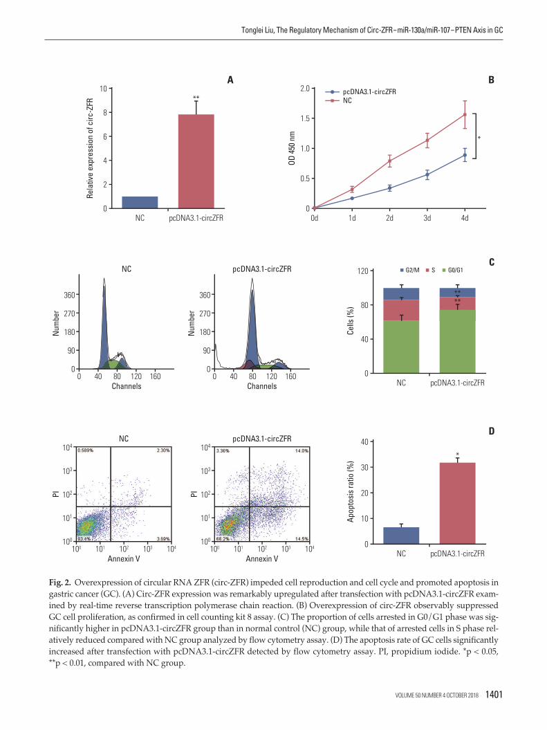
Fig. 2. Overexpression of circular RNA ZFR (circ-ZFR) impeded cell reproduction and cell cycle and promoted apoptosis ingastric cancer (GC). (A) Circ-ZFR expression was remarkably upregulated after transfection with pcDNA3.1-circZFR exam-ined by real-time reverse transcription polymerase chain reaction. (B) Overexpression of circ-ZFR observably suppressedGC cell proliferation, as confirmed in cell counting kit 8 assay. (C) The proportion of cells arrested in G0/G1 phase was sig-nificantly higher in pcDNA3.1-circZFR group than in normal control (NC) group, while that of arrested cells in S phase rel-atively reduced compared with NC group analyzed by flow cytometry assay. (D) The apoptosis rate of GC cells significantlyincreased after transfection with pcDNA3.1-circZFR detected by flow cytometry assay. PI, propidium iodide. *p < 0.05, **p < 0.01, compared with NC group.
Rela
tive
expr
essio
n of
circ
-ZFR
10
0
4
2
8
6
A
NC
OD 45
0 nm
0
1.0
0.5
2.0
1.5
0d 1d 2d 3d 4d
pcDNA3.1-circZFRNC
pcDNA3.1-circZFR
Cells
(%)
0
40
120
80
C
NC pcDNA3.1-circZFR
B
G2/M S G0/G1
Apop
tosis
ratio
(%)
0
20
10
40
30
D
NC pcDNA3.1-circZFRAnnexin V
PINu
mbe
r
101
100
104
103
102
100 101 102 103 104
Channels
90
0
360
270
180
0 40 80 120 160
Annexin V
pcDNA3.1-circZFRNC
PI
101
100
104
103
102
100 101 102 103 104
Num
ber
Channels
pcDNA3.1-circZFRNC
90
0
360
270
180
0 40 80 120 160
VOLUME 50 NUMBER 4 OCTOBER 2018 1401
Tonglei Liu, The Regulatory Mechanism of Circ-ZFRmiR-130a/miR-107PTEN Axis in GC
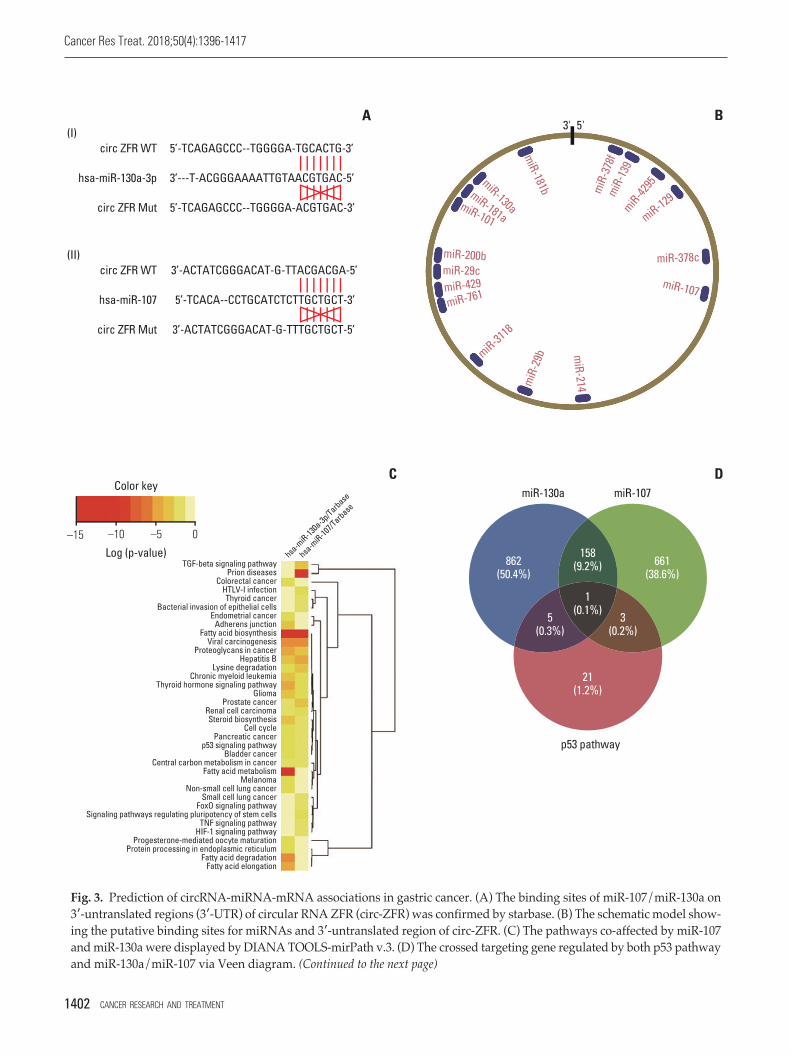
Fig. 3. Prediction of circRNA-miRNA-mRNA associations in gastric cancer. (A) The binding sites of miR-107/miR-130a on3-untranslated regions (3-UTR) of circular RNA ZFR (circ-ZFR) was confirmed by starbase. (B) The schematic model show-ing the putative binding sites for miRNAs and 3-untranslated region of circ-ZFR. (C) The pathways co-affected by miR-107and miR-130a were displayed by DIANA TOOLS-mirPath v.3. (D) The crossed targeting gene regulated by both p53 pathwayand miR-130a/miR-107 via Veen diagram. (Continued to the next page)
circ ZFR WT 5’-TCAGAGCCC--TGGGGA-TGCACTG-3’
3’---T-ACGGGAAAATTGTAACGTGAC-5’
5’-TCAGAGCCC--TGGGGA-ACGTGAC-3’
hsa-miR-130a-3p
circ ZFR Mut
(I)A B
C D
circ ZFR WT
miR-130a miR-107
p53 pathway
3’-ACTATCGGGACAT-G-TTACGACGA-5’
5’-TCACA--CCTGCATCTCTTGCTGCT-3’
3’-ACTATCGGGACAT-G-TTTGCTGCT-5’
hsa-miR-107
circ ZFR Mut
TGF-beta signaling pathwayPrion diseases
Colorectal cancerHTLV-I infectionThyroid cancer
Bacterial invasion of epithelial cellsEndometrial cancer
Adherens junctionFatty acid biosynthesis
Viral carcinogenesisProteoglycans in cancer
Hepatitis BLysine degradation
Chronic myeloid leukemiaThyroid hormone signaling pathway
GliomaProstate cancer
Renal cell carcinomaSteroid biosynthesis
Cell cyclePancreatic cancer
p53 signaling pathwayBladder cancer
Central carbon metabolism in cancerFatty acid metabolism
MelanomaNon-small cell lung cancer
Small cell lung cancerFoxO signaling pathway
Signaling pathways regulating pluripotency of stem cellsTNF signaling pathway
HIF-1 signaling pathwayProgesterone-mediated oocyte maturation
Protein processing in endoplasmic reticulumFatty acid degradation
Fatty acid elongation
hsa-miR-13
0a-3p/Tarbase
hsa-miR-10
7/Tarbase
Color key
Log (p-value)
(II)miR-181bmiR-181a
miR-3118
miR
-29b m
iR-214
miR-378c
miR-
378f
miR-
139
miR-4295
miR-129
miR-107
miR-200bmiR-29cmiR-429miR-761
miR-101
miR-130a
1(0.1%)5
(0.3%)3
(0.2%)
158(9.2%)862
(50.4%)661
(38.6%)
21(1.2%)
–15 0–5–10
5'3'
1402 CANCER RESEARCH AND TREATMENT
Cancer Res Treat. 2018;50(4):1396-1417

with GES-1 cell line (p < 0.01), it was selected for the later experiments. In addition, to further confirm the characteris-tics of circ-ZFR, we used a highly processive 3 to 5 exori-bonuclease (RNase R enzyme) that does not act on circRNAsbut linear RNAs. As expected, circ-ZFR was resistant toRNase treatment in contrast to GAPDH as shown in Fig. 1E.
2. Overexpression of circ-ZFR retarded cell propagationand cell cycle and promoted apoptosis in GC
qRT-PCR results presented that circ-ZFR expression wasremarkably increased after transfection with pcDNA3.1-ZFR(p < 0.01) (Fig. 2A). Furthermore, overexpression of circ-ZFRobservably suppressed GC cell proliferation, as confirmed inCCK-8 assay (p < 0.05) (Fig. 2B). In addition, flow cytometryanalysis disclosed the variance of cell cycle and apoptosiscondition after transfection with pcDNA3.1-ZFR. As exhib-ited in Fig. 2C and D, the percentage of cells arrested inG0/G1 phase was significantly higher in pcDNA3.1-ZFRgroup than in NC group, while that of arrested cells in Sphase relatively declined compared with NC group (both p< 0.01). Meanwhile, the apoptosis rate of GC cells signifi-cantly increased after transfection (p < 0.05). Overall, over-
expression of circ-ZFR arrested cell propagation and cellcycle and promoted apoptosis in GC.
3. Prediction of circRNA-miRNA-mRNA associations inGC
Through starbase, a bioinformatics prediction website, weentered circ-ZFR into starbase and search the potential tar-geting miRNAs, founding that the binding sites of miR-107/miR-130a on 3-UTR of ZFR were showed in Fig. 3A andB. Depending on DIANA TOOLS-mirPath v.3 website, wesought to find a pathway that is co-influenced by miR-130aand miR-107, p53 signaling pathway was selected for furtherstudy (Fig. 3C). And then, we crossed the targeting gene wasregulated by both p53 pathway and miR-130a/miR-107 viavenn diagram, and intersection was PTEN showed in Fig. 3D. Finally, we utilized Targetscan to find out the rela-tionship between miR-130a/miR-107 and PTEN (Fig. 3E).The network clarified the mechanism of circ-ZFR, miR-130a/miR-107, and PTEN in GC showed in Fig. 3F, which the experimental research idea was depended on.
Fig. 3. (Continued from the previous page) (E) The binding sites of miR-107/miR-130a on 3-UTR of PTEN was validated byTargetScan. (F) A biomathematically predicted target network between circ-ZFR, miR-130a/miR-107, and PTEN.
PTEN 3’ UTR WT 5’-UCACAUCCUACCCCUUUGCACUU-3’
3’-UACGGGAAAAUUGUAACGUGAC-5’
5’-UCACAUCCUACCCCUAACGUGAU-3’
hsa-miR-130a-3p
PTEN 3’ UTR Mut
(I)E F
PTEN 3’ UTR WT 5’-AUAAAAUAUUUUGUA--AUGCUGCA-3’
3’-ACUAUCGGGACAUGUUACGACGA-5’
5’-AUAAAAUAUUUUGUA--AUGCUGCA-3’
hsa-miR-107
PTEN 3’ UTR Mut
(II)
hsa-miR-130a
hsa-miR-107
circ-ZFR
CDK19
FNDC4
SLC2A4RG
MEOX2
COX8C
SPG20
TRERF1FAM175B
MIF4GD
PPP2R3C
AGO1
UNC80DUS1L
ZNRF2
TWF1
PRKCE
UBC
ST18
PTEN
VOLUME 50 NUMBER 4 OCTOBER 2018 1403
Tonglei Liu, The Regulatory Mechanism of Circ-ZFRmiR-130a/miR-107PTEN Axis in GC

Fig. 4. Confirmation of targeted relationships between miR-107/miR-130a and PTEN or circular RNA ZFR (circ-ZFR). (A)MiR-107 mimics and miR-130a mimics led to overexpression of miR-107 and miR-130a, respectively detected by real-timereverse transcription polymerase chain reaction. (B) The expression of circ-ZFR was no significant difference both in miR-107mimics and miR-130a–mimics group. (C, D) Overexpression of miR-107 and miR-130a both significantly repressed theluciferase activity of the luciferase reporter containing ZFR 3-untranslated regions (3-UTR)WT but not the reporter con-taining ZFR 3-UTRMut confirmed by dual luciferase reporter assay. (E, F) There existed some interactions between ZFRand miR-107/miR-130a verified by RNA immunoprecipitation assay. (Continued to the next page)
miR-107–
NC
miR-107–
mimics
miR-103a
–mimics
miR-130a
–NC
Rela
tive
expr
essio
n of
miR
-107
/miR
-130
a
10
0
4
2
6
8
A
circ-ZFR WT circ-ZFR Mut
Luci
fera
se ra
tio
0.4
0
0.1
0.2
0.3
C
miR-107–
NC
miR-107–
mimics
miR-103a
–mimics
miR-130a
–NC
Rela
tive
expr
essio
n of
circ
-ZFR
1.25
0
0.50
0.25
0.75
1.00
B
NC miR-107–mimics
circ-ZFR WT circ-ZFR Mut
Luci
fera
se ra
tio
0.4
0
0.1
0.2
0.3
DNC miR-130a–mimics
AGO2 lgG
Rela
tive
enric
hmen
t
20
0
5
10
15
ENC miR-107–mimics
AGO2 lgG
Rela
tive
enric
hmen
t
20
0
5
10
15
FNC miR-130a–mimics
1404 CANCER RESEARCH AND TREATMENT
Cancer Res Treat. 2018;50(4):1396-1417

4. Confirmation of targeted relationships and expressioncorrelations between miR-107/miR-130a and PTEN/circZFR
qRT-PCR displayed that miR-107 mimics and miR-130amimics contributed to miR-107 and miR-130a overexpressionrespectively (p < 0.05) (Fig. 4A). While, the expression of circ-ZFR was detected and the results revealed that miR-107mimics and miR-130a mimics have little influences on circ-ZFR level (Fig. 4B). That is to say, the relationship betweencirc-ZFR and miR-107/miR-130a was unidirectional. More-over, overexpression of miR-107 and miR-130a both signifi-cantly repressed the luciferase activity of the luciferasereporter containing circ-ZFR 3-UTRWT but not the reporter containing circ-ZFR 3-UTRMut, as shown in dualluci-ferase reporter assay (both p < 0.05) (Fig. 4C and D). RIPassay verified that there existed some interactions betweencirc-ZFR and miR-107/miR-130a both (p < 0.05) (Fig. 4E andF). Furthermore, we also verified that miR-107/miR-130acould bind to 3’ UTR of PTEN (both p < 0.05) (Fig. 4G andH). Additionally, in Fig. 5A and B, miR-107 and miR-130awere found to be highly expressed, whereas PTEN was lowlyexpressed in tumor tissues compared with adjacent tissues(p < 0.05). Furthermore, Pearson correlation analysis revea-led that the expressions of miR-107 and miR-130a were bothnegatively correlated with those of ZFR and PTEN (Fig. 5Cand F).
5. Circ-ZFR influenced GC cell propagation, cell cycle, andapoptosis by sponging miR-107/miR-130a
qRT-PCR displayed that miR-107/miR-130a mimics led tooverexpression of miR-107/miR-130a, while miR-107/miR-130a inhibitor downregulated miR-107/miR-130a expres-sions (p < 0.05) (Fig. 6A and C). CCK-8 assay (Fig. 6B and D)revealed that upregulation of miR-107 and miR-130a expres-
sion both promoted cell propagation, whereas downregula-tion of miR-107 and miR-130a expressions restrained propa-gation compared with NC group (p < 0.05). Moreover, nosignificant disctinction was found between miR-107/miR-130a+pcDNA3.1-ZFR group and NC group (p > 0.05). How-ever, the proliferation ability of the cells in miR-107/miR-130a+pcDNA3.1-ZFR group was significantly stronger than thatin pcDNA3.1-ZFR group, while weaker compared with miR-107/miR-130a–mimics group (p < 0.05).
For the analysis of cell cycle and apoptosis, flow cytometryassay (Fig. 7A and B) indicated that the proportion of thecells arrested in G1 phase in pcDNA3.1-ZFR group, miR-107inhibitor group and miR-130a inhibitor group was noticeablylarger than in NC group. Conversely, the arrested cell in G1phase in miR-107–mimics group and miR-130a–mimicsgroup was relatively fewer (p < 0.05). Nonetheless, the arrested cell in G1 phase in miR-107/miR-130a+pcDNA3.1-ZFR group was remarkably fewer than that in pcDNA3.1-ZFR group, while more than that in miR-107/miR-130a–mimics group (p < 0.05). Similarly, no conspicuous distinc-tion was found between miR-107/miR-130a+pcDNA3.1-ZFRgroup and NC group (p > 0.05). The apoptosis of cells trans-fected with pcDNA3.1-ZFR and miR-107/miR-130a inhibitorwas significantly enhanced, while that of the cells transfectedwith miR-107/miR-130a mimics was remarkably inhibited(p < 0.05) (Fig. 8A and B). No significant difference of apop-tosis rate was observed between miR-107/miR 130a+pcDNA-3.1-ZFR group and NC group (p > 0.05). However, the cellapoptosis rate in miR-107/miR-130a+pcDNA3.1-ZFR groupwas observably lower than that in pcDNA3.1-ZFR group,while significantly higher compared with miR-107/miR-130a–mimics group (p < 0.05).
Fig. 4. (Continued from the previous page) (G, H) miR-107/miR-130a could bind to 3-UTR of PTEN confirmed by dual luciferasereporter assay. *p < 0.05, compared with normal control (NC) group.
PTEN WT PTEN Mut
Luci
fera
se ra
tio
0.4
0
0.1
0.2
0.3
GNC miR-107–mimics
PTEN WT PTEN Mut
Luci
fera
se ra
tio
0.4
0
0.1
0.2
0.3
HNC miR-130a–mimics
VOLUME 50 NUMBER 4 OCTOBER 2018 1405
Tonglei Liu, The Regulatory Mechanism of Circ-ZFRmiR-130a/miR-107PTEN Axis in GC

Fig. 5. Correlations on expression between miR-107/miR-130a and PTEN/circ-ZFR. (A, B) MiR-107 and miR-130a werehighly expressed whereas PTEN was lowly expressed in tumor tissues compared with adjacent tissues examined by real-time reverse transcription polymerase chain reaction. (C, D) The expressions of miR-107 and miR-130a were both negativelycorrelated with ZFR expression determined by Pearson correlation analysis. (E, F) The expressions of miR-107 and miR-130awere both negatively correlated with PTEN expression determined by Pearson correlation analysis. *p < 0.05, comparedwith adjacent tissues. GC, gastric cancer.
miR-107 miR-130a
Rela
tive
miR
-107
/miR
-130
a ex
pres
sion 8
0
2
4
6
AAdjacent tissues Tumor tissues
Adjacent tissues Tumor tissues
Rela
tive
PTEN
exp
ress
ion
6
0
2
4
B
0.50 1.0 1.5
Rela
tive
expr
essio
n of
miR
-107
Relative expression of circZFR
8
0
2
4
6
CR2=0.577, p < 0.001
0.50 1.0 1.5
Rela
tive
expr
essio
n of
miR
-130
a
Relative expression of circZFR
8
0
2
4
6
DR2=0.518, p < 0.001
20 6 8
Rela
tive
expr
essio
n of
PTE
N
Relative expression of miR-107
2.0
0
0.5
1.0
1.5
4
FR2=0.580, p < 0.001
20 4 8
Rela
tive
expr
essio
n of
PTE
N
Relative expression of miR-130a
2.0
0
0.5
1.0
1.5
6
FR2=0.522, p < 0.001
1406 CANCER RESEARCH AND TREATMENT
Cancer Res Treat. 2018;50(4):1396-1417

Fig. 6. Circ-ZFR suppressed gastric cancer cell reproduction by sponging miR-107/miR-130a. (A, C) MiR-107/miR-130amimics significantly increased the expression of miR-107/miR-130a, while miR-107/miR-130a inhibitor remarkably decreased miR-107/miR-130a expressions examined by real-time reverse transcription polymerase chain reaction. (B, D)Overexpression of miR-107 and miR-130a both promoted cell proliferation whereas downregulation of miR-107 and miR-130a expressions restrained propagation. No significant difference of cell proliferation was found between miR-107/miR-130a+pcDNA3.1-ZFR group and normal control (NC) group. However, the proliferation ability of the cells in miR-107/miR-130a+pcDNA3.1-ZFR group was significantly stronger than that in pcDNA3.1-ZFR group, while weaker comparedwith miR-107/miR-130a–mimics group. *p < 0.05, **p < 0.01, compared with NC group; #p < 0.05, compared with pcDNA3.1-ZFR group; †p < 0.05, compared with miR-107/miR-130a–mimics group.
Rela
tive
expr
essio
n of
miR
-107
10
0
4
2
8
6
A
OD 45
0 nm
2.5
0
1.0
0.5
2.0
1.5
0 1 2 3 4
miR-107–mimics+pcDNA3.1-ZFRmiR-107–inhibitormiR-107–mimicspcDNA3.1-ZFRNC
#
†
B
NC
miR-107–mimics
miR-107–inhibitor
Rela
tive
expr
essio
n of
miR
-130
a
8
0
4
2
6
C
OD 45
0 nm
2.5
0
1.0
0.5
2.0
1.5
0 1 2 3 4
miR-130a–mimics+pcDNA3.1-ZFRmiR-130a–inhibitormiR-130a–mimicspcDNA3.1-ZFRNC
#
†
D
NC
miR-130a–mimics
miR-130a–inhibitor
Time (day)
Time (day)
VOLUME 50 NUMBER 4 OCTOBER 2018 1407
Tonglei Liu, The Regulatory Mechanism of Circ-ZFRmiR-130a/miR-107PTEN Axis in GC

6. miR-107/miR-130a promoted GC cell propagation andimpeded apoptosis through targeting PTEN
Western blot confirmed that the expression of PTEN wasremarkably repressed after transfection with miR-107–mim-ics/miR-130a–mimics (both p < 0.05), while it was overex-pressed followed transfection with pcDNA3.1-ZFR andmiR-107 inhibitor/miR-130a inhibitor (both p < 0.01). In addition, no significant difference was found between miR-107/miR-130a+pcDNA3.1-ZFR group and NC group (p >0.05) (Fig. 9A and B). qRT-PCR results displayed that aftertransfection with pcDNA3.1-PTEN, the expression of pcDNA-3.1-PTEN in GC cells was dramatically upregulated (p < 0.01)
(Fig. 9C). Furthermore, CCK-8 assay exhibited that overex-pression of PTEN significantly attenuated GC cell propaga-tion ability, while that of miR-107/miR-130a mimics pro-moted cell propagation. Meanwhile, the proliferation abilityof the cells in miR-107/miR-130amimics+pcDNA3.1-PTENgroup was significantly stronger than that in pcDNA3.1-PTEN group, while weaker compared with miR-107/miR-130amimics group (p < 0.05) (Fig. 9D and E). No obviousdistinction of propagation ability was found between miR-107/miR-130amimics+pcDNA3.1-PTEN group and NCgroup (p > 0.05).
Additionally, flow cytometry assay (Fig. 10A and B) indi-cated that the proportion of the cells arrested in G1 phase in
Fig. 7. Circular RNA ZFR (circ-ZFR) induced cell cycle arrest by sponging miR-107/miR-130a in gastric cancer. (A, B) Theproportion of the cells arrested in G1 phase in pcDNA3.1-ZFR group, miR-107 inhibitor group and miR-130a inhibitor groupwas considerably larger than in normal control (NC) group. Conversely, the percentage of arrested cell in G1 phase in miR-107–mimics group and miR-130a–mimics group was relatively smaller compared with NC group. Nonetheless, the percent-age of arrested cell in G1 phase in miR-107/miR-130a+pcDNA3.1-ZFR group was significantly smaller than that inpcDNA3.1-ZFR group, while larger than that in miR-107/miR-130a–mimics group. There was no conspicuous distinctionbetween miR-107/miR-130a+pcDNA3.1-ZFR group and NC group. *p < 0.05, compared with NC group; #p < 0.05, comparedwith pcDNA3.1-ZFR group; †p < 0.05, compared with miR-107/miR-130a–mimics group. (Continued to the next page)
Cells
(%)
120
0
40
80
A
G2/M S G0/G1
#†
Num
ber
Channels
200
100
0
500
400
300
0 40 80 120 160
NC
Num
ber
Channels
200
100
0
500
400
300
0 40 80 120 160
pcDNA3.1-ZFR
Num
ber
Channels
200
100
0
500
400
300
0 40 80 120 160
miR-107–mimics
Num
ber
Channels
200
100
0
500
400
300
0 40 80 120 160
miR-107–inhibitor
Num
ber
Channels
200
100
0
500
400
300
0 40 80 120 160
miR-107–mimics+pcDNA3.1-ZFR
miR-107–mimics+pcDNA3.1-ZFRNC
miR-107–mimics
miR-107–inhibitor
pcDNA3.1-ZFR
1408 CANCER RESEARCH AND TREATMENT
Cancer Res Treat. 2018;50(4):1396-1417

miR-107/miR-130a–mimics group dramatically reduced,while that in S phase increased. Conversely, the arrested cellsin G1 phase in pcDNA3.1-PTEN group conspicuously increased in comparison with NC group, whereas those in Sphase relatively decreased (p < 0.05). Nonetheless, the per-centage of arrested cell in G1 phase in miR-107/miR-130a+pcDNA3.1-PTEN group was significantly smaller than thatin pcDNA3.1-PTEN group, while larger than that in miR-107/miR-130a–mimics group (p < 0.05). Furthermore, theapoptotic rate of the cells transfected with miR-107/miR-130a mimics considerably fell, while cell apoptosis drasticallyrose in pcDNA3.1-PTEN group (p < 0.05) (Fig. 11A and B).No significant difference of apoptosis ratio was detected between miR-107/miR-130a+pcDNA3.1-PTEN group andNC group (p > 0.05). Nevertheless, the cell apoptosis rate inmiR-107/miR-130a+pcDNA3.1-PTEN group was consider-ably lower than that in pcDNA3.1-PTEN group, while sig-nificantly higher compared with miR-107/miR-130a–mimicsgroup (p < 0.05).
7. Circ-ZFR curbed GC tumor growth and affected p53 pro-tein expression in vivo
As displayed in Fig. 12A, the tumor volume of the mice inpcDNA3.1-ZFR group grew more slowly than that in NCgroup (p < 0.05). Moreover, the tumor weight of mice inpcDNA3.1-ZFR group was significantly lighter comparedwith NC group (p < 0.05) (Fig. 12B and C). The expressionlevel of miR-107 and miR-130a was decreased comparedwith NC group after transfected with pcDNA3.1-ZFR detected by qRT-PCR (p < 0.01) (Fig. 12D and E). Meanwhile,IHC assay revealed that after transfection with pcDNA3.1-ZFR, the expression level of p53 and PTEN protein dramati-cally increased in comparison with NC group (p < 0.01) (Fig. 12F and G).
Fig. 7. (Continued from the previous page)
Cells
(%)
120
0
40
80
B
G2/M S G0/G1
#†
Num
ber
Channels
200
100
0
500
400
300
0 40 80 120 160
NC
Num
ber
Channels
200
100
0
500
400
300
0 40 80 120 160
pcDNA3.1-ZFR
Num
ber
Channels
200
100
0
500
400
300
0 40 80 120 160
miR-130a–mimics
Num
ber
Channels
200
100
0
500
400
300
0 40 80 120 160
miR-130a–inhibitor
Num
ber
Channels
200
100
0
500
400
300
0 40 80 120 160
miR-130a–mimics+pcDNA3.1-ZFR
miR-130a–mimics+pcDNA3.1-ZFRNC
miR-130a–mimics
miR-130a–inhibitor
pcDNA3.1-ZFR
VOLUME 50 NUMBER 4 OCTOBER 2018 1409
Tonglei Liu, The Regulatory Mechanism of Circ-ZFRmiR-130a/miR-107PTEN Axis in GC

Discussion
Based on a series of experiment, we verified the regulatoryrole of circ-ZFR–miR-130a/miR-107–PTEN network in GC.We first identified that circ-ZFR was downregulated in GCand acted as a tumor inhibitor in cell propagation and apop-tosis of GC. Meanwhile, we validated the targeted relation-ships between miR-107/miR-130a and PTEN/circ-ZFR. Fur-thermore, we disclosed that circ-ZFR expression was nega-tively correlated with miR-130a and miR-107 expressions,which were also correlated inversely with their target PTENexpression. Our study finally demonstrated that circ-ZFR
inhibited GC cell proliferation and promoted apoptosis bysponging miR-130a/miR-107 and modulating PTEN.
CircRNAs, a novel type of endogenous non-coding RNAs,have been recently considered a crucial regulator of gene expression and pathological processes. Accumulating resear-ches have suggested that aberrant circRNA expression playsimportant roles in carcinogenesis and tumor progression[15]. Fu et al. [16] demonstrated that circular RNA 0004018(hsa_circ_0004018) was lowly expressed and played a role incarcinogenesis and metastasis of hepatocellular carcinoma.In the recent study of Li et al. [17] circular RNA 0000096(hsa_circ_0000096) was also found to be significantly down-regulated in GC tissues and knockdown of hsa_circ_0000096
Fig. 8. Circular RNA ZFR (circ-ZFR) facilitated gastric cancer cell apoptosis by sponging miR-107/miR-130a (A, B), Theapoptosis of cells was significantly enhanced after transfected with pcDNA3.1-ZFR and miR-107/miR-130a inhibitor, whilethat of the cells transfected with miR-107/miR-130a–mimics was remarkably inhibited. No significant difference of apoptosisrate was detected between miR-107/miR-130a+pcDNA3.1-ZFR group and normal control (NC) group. However, the cellapoptosis rate in miR-107/miR-130a+pcDNA3.1-ZFR group was considerably lower than that in pcDNA3.1-ZFR group,while significantly higher compared with miR-107/miR-130amimics group. *p < 0.05, ** p < 0.01, compared with NC group;#p < 0.05, compared with pcDNA3.1-ZFR group; †p < 0.05, compared with miR-107/miR-130a–mimics group. (Continued tothe next page)
#†
Annexin V
miR-107–mimics
PI
101
100
104
103
102
100 101 102 103 104
miR-107–mimics+pcDNA3.1-ZFR
Annexin V
pcDNA3.1-ZFR
PI
101
100
104
103
102
100 101 102 103 104
Annexin V
miR-107–mimics+pcDNA3.1-ZFR
PI
101
100
104
103
102
100 101 102 103 104
Annexin V
NC
PI
101
100
104
103
102
100 101 102 103 104
Annexin V
miR-107–inhibitor
PI
101
100
104
103
102
100 101 102 103 104
NC
miR-107–mimics
miR-107–inhibitor
pcDNA3.1-ZFR
Apop
tosis
ratio
(%)
30
0
10
20
A
1410 CANCER RESEARCH AND TREATMENT
Cancer Res Treat. 2018;50(4):1396-1417

significantly inhibited cell propagation and migration in vitroand in vivo. Herein, circ-ZFR was identified to be observablydownregulated in GC tissues based on microarray analysis,and overexpression of circ-ZFR could retard propagationand cell cycle and promoted apoptosis in GC confirmed byCCK-8 and apoptosis assay.
It is well recognized that miRNAs play a critical role in theregulation of gene expression and various biological pro-cesses, such as proliferation, metastasis, apoptosis and soforth. The circRNA-miRNA association and their interactioninfluence on various cancers have been widely studied. Cir-cRNAs has been demonstrated to serve as miRNA sponge,thereby regulating gene transcription and cellular activitiesin many researches [18]. Liang et al. [19] disclosed that circ-ABCB10 promoted breast cancer proliferation and progres-sion through sponging miR-1271. Tang et al. [20] reportedthat circ_0001982 facilitated breast cancer cell carcinogenesisthrough repressing miR-143. Zhong et al. [21] unraveled thatcirc-TCF25 accelerated cell propagation and mobility in blad-der cancer through sponging miR-103a-3p/miR-107. How-
ever, Zou et al. [22] found that miR-107 was a tumor pro-gression promoter in hepatocellular carcinoma, which sug-gested that miR-107 takes different effects in different can-cers. A recent study also revealed that miR-107 was upregu-lated in GC [23], which displayed the similar result as ourstudy. In the current study, we predicted the association between circ-ZFR and miR-130a-3p/miR-107, and substan-tiated the circ-ZFRmiR-130a-3p/miR-107 regulatory loopin GC. Circ-ZFR inhibited GC cell propagation, induced cellcycle arrest and promoted apoptosis by sponging miR-107/miR-130a.
PTEN, as a tumor suppressor, is mutated in a large numberof cancers at high frequency [24]. It negatively regulates intracellular levels of phosiphatidylinositol-3,4,5-trisphos-phate in cells and AKT/PKB signaling pathway, suppressingcellular functional activities [25]. Actually, emerging evi-dence has substantiated that PTEN could be targeted andregulated by miR-130a and miR-107 to influence cancerouscellular activities [26,27]. Nonetheless, there are fewer studieson the targeted relationships between miR-130a/miR-107
Fig. 8. (Continued from the previous page)
#†
Annexin V
miR-130a–mimics
PI
101
100
104
103
102
100 101 102 103 104
miR-130a–mimics+pcDNA3.1-ZFR
Annexin V
pcDNA3.1–ZFR
PI
101
100
104
103
102
100 101 102 103 104
Annexin V
miR-130a–mimics+pcDNA3.1-ZFR
PI
101
100
104
103
102
100 101 102 103 104
Annexin V
NC
PI
101
100
104
103
102
100 101 102 103 104
Annexin V
miR-130a–inhibitor
PI
101
100
104
103
102
100 101 102 103 104
NC
miR-130a–mimics
miR-130a–inhibitor
pcDNA3.1-ZFR
Apop
tosis
ratio
(%)
30
0
10
20
B
VOLUME 50 NUMBER 4 OCTOBER 2018 1411
Tonglei Liu, The Regulatory Mechanism of Circ-ZFRmiR-130a/miR-107PTEN Axis in GC

Fig. 9. miR-107/miR-130a promoted gastric cancer cell reproduction through targeting PTEN. (A, B) The expression of PTENwas remarkably repressed after transfection with miR-107mimics/miR-130amimics, while it was overexpressed followedtransfection with pcDNA3.1-ZFR and miR-107 inhibitor/miR-130a inhibitor detected with western blot. (C) The expressionof pcDNA3.1-PTEN in gastric cancer cells was dramatically upregulated after transfection with pcDNA3.1-PTEN examinedby real-time reverse transcription polymerase chain reaction. (D, E) The proliferation ability of the cells in miR-107/miR-130amimics+pcDNA3.1-PTEN group was significantly stronger than that in pcDNA3.1-PTEN group, while weaker com-pared with miR-107/miR-130amimics group. No significant difference of cell proliferation was observed betweenmiR-107/miR-130amimics+pcDNA3.1-PTEN group and normal control (NC) group. *p < 0.05, compared with NC group;#p < 0.05, compared with pcDNA3.1-PTEN group; †p < 0.05, compared with miR-107/miR-130amimics group.
NC
miR-107–mimics
miR-107–mimics
+pcDNA3.1-ZFR
miR-107–inhibito
r
pcDNA3.1-ZFR
PTEN p53
Rela
tive
prot
ein
expr
essio
n
3
0
1
2
ANCmiR-107–mimicsmiR-107–mimics+pcDNA3.1-ZFR
pcDNA3.1-ZFRmiR-107–inhibitor
PTEN
p53
GAPDH#† #
†
NC
miR-130a–mimics
miR-130a–mimics
+pcDNA3.1-ZFR
miR-130a–inhibito
r
pcDNA3.1-ZFR
PTEN p53
Rela
tive
prot
ein
expr
essio
n
3
0
1
2
BNCmiR-130a–mimicsmiR-130a–mimics+pcDNA3.1-ZFR
pcDNA3.1-ZFRmiR-130a–inhibitor
PTEN
p53
GAPDH#† #
†
OD 45
0 nm
2.5
0
1.0
0.5
2.0
1.5
0d 1d 2d 3d 4d
pcDNA3.1-PTEN+miR-130a–mimicspcDNA3.1-PTENmiR-130a–mimicsNC
#
†
E
OD 45
0 nm
2.5
0
1.0
0.5
2.0
1.5
0d 1d 2d 3d 4d
pcDNA3.1-PTEN+miR-107–mimicspcDNA3.1-PTENmiR-107–mimicsNC #
†
D
Rela
tive
expr
essio
n of
PTE
N 6
0
4
2
NC pcDNA3.1-PTEN
C
1412 CANCER RESEARCH AND TREATMENT
Cancer Res Treat. 2018;50(4):1396-1417

Fig. 10. miR-107/miR-130a influenced gastric cancer cell cycle through targeting PTEN. (A, B) Flow cytometry assay dis-played that the proportion of the cells arrested in G1 phase in miR-107/miR-130a–mimics group dramatically reduced com-pared with normal control (NC) group, while that in S phase increased and there was no significant difference in G2 phase.On the contrary, the percentage of arrested cell in G1 phase in pcDNA3.1-PTEN group conspicuously increased in comparisonwith NC group whereas that in S phase relatively decreased. Nonetheless, the percentage of arrested cell in G1 phase inmiR-107/miR-130a+pcDNA3.1-PTEN group was significantly smaller than that in pcDNA3.1-PTEN group, while largerthan that in miR-107/miR-130a–mimics group. There was no significant distinction between miR-107/miR-130a+pcDNA3.1-PTEN group and NC group. *p < 0.05, compared with NC group; #p < 0.05, compared with pcDNA3.1-PTEN group; †p < 0.05, compared with miR-107/miR-130a–mimics group.
pcDNA3.1-PTEN+miR-107–mimicsNC
miR-107–mimics
pcDNA3.1-PTEN
Cells
(%)
120
0
40
80
AG2/M S G0/G1
#†Nu
mbe
r
Channels
200
100
0
500
400
300
0 40 80 120 160
NC
Num
ber
Channels
200
100
0
500
400
300
0 40 80 120 160
miR-107–mimics
Num
ber
Channels
200
100
0
500
400
300
0 40 80 120 160
pcDNA3.1-PTEN
Num
ber
Channels
200
100
0
500
400
300
0 40 80 120 160
pcDNA3.1-PTEN+miR-107–mimics
pcDNA3.1-PTEN+miR-130a–mimicsNC
miR-130a–mimics
pcDNA3.1-PTEN
Cells
(%)
120
0
40
80
BG2/M S G0/G1
#†Nu
mbe
r
Channels
200
100
0
500
400
300
0 40 80 120 160
NC
Num
ber
Channels
200
100
0
500
400
300
0 40 80 120 160
miR-130a–mimics
Num
ber
Channels
200
100
0
500
400
300
0 40 80 120 160
pcDNA3.1-PTEN
Num
ber
Channels
200
100
0
500
400
300
0 40 80 120 160
pcDNA3.1-PTEN+miR-130a–mimics
VOLUME 50 NUMBER 4 OCTOBER 2018 1413
Tonglei Liu, The Regulatory Mechanism of Circ-ZFRmiR-130a/miR-107PTEN Axis in GC

Fig. 11. miR-107/miR-130a impeded cell apoptosis in gastric cancer through targeting PTEN. (A, B) The apoptotic rate ofthe cells transfected with miR-107/miR-130a–mimics considerably fell whereas that of the cells transfected with pcDNA3.1-PTEN drastically rose. No significant difference of apoptosis ratio was found between miR-107/miR-130a+pcDNA3.1-PTENgroup and normal control (NC) group. Nevertheless, the cell apoptosis rate in miR-107/miR-130a+pcDNA3.1-PTEN groupwas considerably lower than that in pcDNA3.1-PTEN group, while significantly higher compared with miR-107/miR-130a–mimics group. *p < 0.05, compared with NC group; #p < 0.05, compared with pcDNA3.1-PTEN group; †p < 0.05, comparedwith miR-107/miR-130a–mimics group.
#†
Annexin V
miR-107–mimics
PI
101
100
104
103
102
100 101 102 103 104Annexin V
NC
PI
101
100
104
103
102
100 101 102 103 104
Annexin V
pcDNA3.1-PTEN+miR-107–mimics
pcDNA3.1-PTEN+miR-107–mimics
pcDNA3.1-PTEN+miR-130a–mimics
PI
101
100
104
103
102
100 101 102 103 104Annexin V
pcDNA3.1-PTEN
PI
101
100
104
103
102
100 101 102 103 104
NC
miR-107–mimics
pcDNA3.1-PTEN
Apop
tosis
ratio
(%)
20
0
5
10
15
A
#†
Annexin V
miR-130a–mimics
PI
101
100
104
103
102
100 101 102 103 104Annexin V
NC
PI
101
100
104
103
102
100 101 102 103 104
Annexin V
pcDNA3.1-PTEN+miR-130a–mimics
PI
101
100
104
103
102
100 101 102 103 104Annexin V
pcDNA3.1-PTEN
PI
101
100
104
103
102
100 101 102 103 104
NC
miR-130a–mimics
pcDNA3.1-PTEN
Apop
tosis
ratio
(%)
20
0
5
10
15
B
Cancer Res Treat. 2018;50(4):1396-1417
1414 CANCER RESEARCH AND TREATMENT

Fig. 12. Circular RNA ZFR curbed gastric cancer (GC) tumor growth and affected p53 protein expression in vivo (A) Thetumor volume of the mice in pcDNA3.1-ZFR group was significantly grew more slowly than that in normal control (NC)group. (B, C) The tumor weight of mice in pcDNA3.1-ZFR group was significantly lighter compared with NC group. (D, E)The expression level of miR-107 (D) and miR-130a (E) was decreased after transfected with pcDNA3.1-ZFR. (F) The expressionlevel of p53 protein dramatically increased after transfected with pcDNA3.1-ZFR confirmed by immunohistochemistry (IHC)assay. (G) The expression level of PTEN increased after transfected with pcDNA3.1-ZFR confirmed by IHC assay. *p < 0.05,**p < 0.01, compared with NC group.
PTEN
IHC
stai
ning
scor
es o
f PTE
N 4
3
0
2
1
NC pcDNA3.1-ZFR
NC pcDNA3.1-ZFR
G
p53
IHC
stai
ning
scor
es o
f p53 4
3
0
2
1
NC pcDNA3.1-ZFR
NC pcDNA3.1-ZFR
NC
pcDNA3.1-ZFR
F
Rela
tive
expr
essio
n of
miR
-130
a
1.5
1.0
0
0.5
NC pcDNA3.1-ZFR
ERe
lativ
e ex
pres
sion
of m
iR-1
071.5
1.0
0
0.5
NC pcDNA3.1-ZFR
D
Rela
tive
tum
or w
eigh
t 1.5
1.0
0
0.5
NC pcDNA3.1-ZFR
C
Rela
tive
tum
or vo
lum
e
Time (wk)
150
100
0
50
1 2 3 4 5
A BpcDNA3.1-ZFRNC
Tonglei Liu, The Regulatory Mechanism of Circ-ZFRmiR-130a/miR-107PTEN Axis in GC
VOLUME 50 NUMBER 4 OCTOBER 2018 1415

and PTEN in GC. Herein, we verified that miR-130a/miR-107 could target and modulate PTEN expression throughbioinformatics analysis, dual-luciferase reporter gene assayand RIP assay.
In addition, circRNAs have been demonstrated to act asmiRNA sponges and play crucial roles in regulating gene expression through a circRNA-miRNA-gene pathway. Chenet al. [28] substantiated that circ-WDR77 affected vascularsmooth muscle cells proliferation and migration by spongingmiR-124 and modulating its target FGF-2. Peng et al. [29] revealed that circ-ZNF609 regulated AKT3 expression bysponging miR-150-5p in Hirschsprung disease. Xie et al. [30]found that circRNA-001569 exerted influence on the propa-gation and invasiveness of colorectal cancer via circRNA-001569miR-145-E2F5/BAG4/FMNL2 network. In our stu-dy, circ-ZFR was identified as a sponge of miR-130a/miR-107 and indirectly modulated their target PTEN expression,thereby repressing GC cell propagation and promotingapoptosis (Fig. 13). There were still some shortcomings in ourresearch. Only the effect of circ-ZFR was explored in vivo,while the effect of miR-130a and miR-107 in vivo still remained unclear. Further study ought to be put into prac-
tice. On the other hand, we only focus on the influences ofcirc-ZFR–miR-130a/miR-107–PTEN axis in gastric cancer. Inthe follow-up study, we will pay attention to other molecularmechanisms and signaling pathways on GC procession.
In conclusion, circ-ZFR inhibited cell proliferation and pro-moted apoptosis in GC by sponging miR-130a/miR-107 andmodulating PTEN. The current research revealed a novel molecular mechanism of circ-ZFR in regulating GC cell prop-agation, cell cycle and apoptosis, which provided a compre-hensive insight into the regulatory role of circ-ZFR–miR-130a/miR-107–PTEN axis in GC and facilitated the discoveryof novel therapeutic targets in GC treatment.
Conflicts of Interest
Conflict of interest relevant to this article was not reported.
Acknowledgments
This study was supported by Leader Training Program in Med-ical Subjects of Health and Family Planning commission of YunnanProvince (D-201657).
Fig. 13. The mechanism of circular RNA ZFR (circ-ZFR)–miR-107/miR-130a–PTEN axis. Circ-ZFR was identified as a spongeof miR-130a/miR-107 and indirectly modulated their target PTEN expression. GC, gastric cancer.
GC cells AGS
3’5’
miR-107
miR-130a
PTEN
PTENp53
circ ZFR
Cell cycleApoptosis Proliferation
p53
Apoptosis
Cell cycle
Proliferation
Cancer Res Treat. 2018;50(4):1396-1417
1416 CANCER RESEARCH AND TREATMENT

1. Xi HQ, Zhang KC, Li JY, Cui JX, Gao YH, Wei B, et al. RNAi-mediated inhibition of Lgr5 leads to decreased angiogenesisin gastric cancer. Oncotarget. 2017;8:31581-91.
2. Li X, Li H, Zhang R, Liu J, Liu J. MicroRNA-449a inhibits pro-liferation and induces apoptosis by directly repressing E2F3in gastric cancer. Cell Physiol Biochem. 2015;35:2033-42.
3. Allemani C, Weir HK, Carreira H, Harewood R, Spika D,Wang XS, et al. Global surveillance of cancer survival 1995-2009: analysis of individual data for 25,676,887 patients from279 population-based registries in 67 countries (CONCORD-2). Lancet. 2015;385:977-1010.
4. Liu J, Liu T, Wang X, He A. Circles reshaping the RNA world:from waste to treasure. Mol Cancer. 2017;16:58.
5. Chen LL, Yang L. Regulation of circRNA biogenesis. RNABiol. 2015;12:381-8.
6. Fu L, Wu S, Yao T, Chen Q, Xie Y, Ying S, et al. Decreased expression of hsa_circ_0003570 in hepatocellular carcinomaand its clinical significance. J Clin Lab Anal. 2017 May 11[Epub]. https://doi.org/10.1002/jcla.22239.
7. Huang M, He YR, Liang LC, Huang Q, Zhu ZQ. Circular RNAhsa_circ_0000745 may serve as a diagnostic marker for gastriccancer. World J Gastroenterol. 2017;23:6330-8.
8. Guo JN, Li J, Zhu CL, Feng WT, Shao JX, Wan L, et al. Com-prehensive profile of differentially expressed circular RNAsreveals that hsa_circ_0000069 is upregulated and promotes cellproliferation, migration, and invasion in colorectal cancer.Onco Targets Ther. 2016;9:7451-8.
9. Zhang H, Wang G, Ding C, Liu P, Wang R, Ding W, et al. Increased circular RNA UBAP2 acts as a sponge of miR-143 topromote osteosarcoma progression. Oncotarget. 2017;8:61687-97.
10. Izaurralde E. GENE REGULATION. Breakers and blockers-miRNAs at work. Science. 2015;349:380-2.
11. Gattolliat CH, Uguen A, Pesson M, Trillet K, Simon B, DoucetL, et al. MicroRNA and targeted mRNA expression profilinganalysis in human colorectal adenomas and adenocarcinomas.Eur J Cancer. 2015;51:409-20.
12. Hansen TB, Kjems J, Damgaard CK. Circular RNA and miR-7in cancer. Cancer Res. 2013;73:5609-12.
13. Han D, Li J, Wang H, Su X, Hou J, Gu Y, et al. Circular RNAcircMTO1 acts as the sponge of microRNA-9 to suppress hepatocellular carcinoma progression. Hepatology. 2017;66:1151-64.
14. Li H, Yang J, Wei X, Song C, Dong D, Huang Y, et al. Circ-FUT10 reduces proliferation and facilitates differentiation ofmyoblasts by sponging miR-133a. J Cell Physiol. 2018;233:4643-51.
15. Yao JT, Zhao SH, Liu QP, Lv MQ, Zhou DX, Liao ZJ, et al.Over-expression of CircRNA_100876 in non-small cell lungcancer and its prognostic value. Pathol Res Pract. 2017;213:453-6.
16. Fu L, Yao T, Chen Q, Mo X, Hu Y, Guo J. Screening differential
circular RNA expression profiles reveals hsa_circ_0004018 isassociated with hepatocellular carcinoma. Oncotarget. 2017;8:58405-16.
17. Li P, Chen H, Chen S, Mo X, Li T, Xiao B, et al. Circular RNA0000096 affects cell growth and migration in gastric cancer. BrJ Cancer. 2017;116:626-33.
18. Li J, Yang J, Zhou P, Le Y, Zhou C, Wang S, et al. CircularRNAs in cancer: novel insights into origins, properties, func-tions and implications. Am J Cancer Res. 2015;5:472-80.
19. Liang HF, Zhang XZ, Liu BG, Jia GT, Li WL. Circular RNAcirc-ABCB10 promotes breast cancer proliferation and pro-gression through sponging miR-1271. Am J Cancer Res.2017;7:1566-76.
20. Tang YY, Zhao P, Zou TN, Duan JJ, Zhi R, Yang SY, et al. Cir-cular RNA hsa_circ_0001982 promotes breast cancer cell car-cinogenesis through decreasing miR-143. DNA Cell Biol.2017;36:901-8.
21. Zhong Z, Lv M, Chen J. Screening differential circular RNAexpression profiles reveals the regulatory role of circTCF25-miR-103a-3p/miR-107-CDK6 pathway in bladder carcinoma.Sci Rep. 2016;6:30919.
22. Zou CD, Zhao WM, Wang XN, Li Q, Huang H, Cheng WP, etal. MicroRNA-107: a novel promoter of tumor progression thattargets the CPEB3/EGFR axis in human hepatocellular carci-noma. Oncotarget. 2016;7:266-78.
23. Wang S, Ma G, Zhu H, Lv C, Chu H, Tong N, et al. miR-107regulates tumor progression by targeting NF1 in gastric can-cer. Sci Rep. 2016;6:36531.
24. Milella M, Falcone I, Conciatori F, Cesta Incani U, Del CuratoloA, Inzerilli N, et al. PTEN: multiple functions in human malignant tumors. Front Oncol. 2015;5:24.
25. Yu G, Chen X, Chen S, Ye W, Hou K, Liang M. MiR-19a, miR-122 and miR-223 are differentially regulated by hepatitis Bvirus X protein and involve in cell proliferation in hepatomacells. J Transl Med. 2016;14:122.
26. Wei H, Cui R, Bahr J, Zanesi N, Luo Z, Meng W, et al. miR-130a deregulates PTEN and stimulates tumor growth. CancerRes. 2017;77:6168-78.
27. Wang S, Yuan L. Predictive biomarkers for targeted and cyto-toxic agents in gastric cancer for personalized medicine. BiosciTrends. 2016;10:171-80.
28. Chen J, Cui L, Yuan J, Zhang Y, Sang H. Circular RNA WDR77target FGF-2 to regulate vascular smooth muscle cells prolif-eration and migration by sponging miR-124. Biochem BiophysRes Commun. 2017;494:126-32.
29. Peng L, Chen G, Zhu Z, Shen Z, Du C, Zang R, et al. CircularRNA ZNF609 functions as a competitive endogenous RNA toregulate AKT3 expression by sponging miR-150-5p in Hirs-chsprung's disease. Oncotarget. 2017;8:808-18.
30. Xie H, Ren X, Xin S, Lan X, Lu G, Lin Y, et al. Emerging rolesof circRNA_001569 targeting miR-145 in the proliferation andinvasion of colorectal cancer. Oncotarget. 2016;7:26680-91.
References
Tonglei Liu, The Regulatory Mechanism of Circ-ZFRmiR-130a/miR-107PTEN Axis in GC
VOLUME 50 NUMBER 4 OCTOBER 2018 1417