
Circadian Rhythms: An Electric Jolt to the Clock
3
Circadian Rhythms: An Electric Jolt to the Clock The animal circadian pacemaker is composed of two transcriptional feedback loops, which regulate electrical activity in circadian neurons. Surprisingly, a new study reports that electrical activity can reprogram circadian transcription, and identifies CREB proteins as candidates for this reprograming. Patrick Emery In animals, the molecular circadian pacemaker consists of two interlocked transcriptional feedback loops [1]. These two loops drive antiphasic waves of output gene transcription, which impact cell metabolism, physiology and behavior. For the pacemaker neurons that control circadian behaviors such as the sleep/wake cycle, membrane physiology and neuronal activity are critical circadian outputs [2–4]. Do these rhythmic outputs feedback on the molecular pacemaker to contribute to its oscillations? And do they modulate downstream clock-controlled genes? In this issue of Current Biology, Mizrak et al. [5] examine these important questions by looking at the consequences to the Drosophila circadian transcriptome of manipulating membrane properties of circadian neurons. Ten years ago, Nitabach et al. surprisingly found that when they electrically silenced the Drosophila circadian pacemaker neurons (the small ventral lateral neurons; sLNvs), the molecular clock stopped functioning under constant conditions [6]. Hence, they proposed that proper electrical membrane physiology is needed for self-sustained molecular circadian rhythms, at least in circadian neurons. Interestingly, the resting membrane potential of the sLNvs shows a rhythm during the course of the day; it is hypopolarized near dawn and becomes hyperpolarized after dusk [2]. This raises the possibility that electrical activity rhythms could feed back on the transcriptional pacemaker to contribute to its oscillations and affect circadian output gene rhythms. So what happens to the circadian pacemaker, and to the expression of the genes it controls, if one forces membrane activity to be constantly low (evening-like), or constantly high (morning-like)? This is the question Mizrak et al. [5] elegantly addressed. They expressed the potassium channel Kir to electrically silence the sLNvs, or the bacterial sodium NachBac channel to increase their neural activity [7] (Figure 1). The authors then took the technically challenging approach of isolating mRNAs from larval sLNvs (only eight neurons per brain!) at different time points, and performed whole genome expression studies to examine the consequences of manipulating electrical activity on the sLNv transcriptome. Two very interesting and provocative conclusions were drawn. First, there is a strong enrichment of genes under circadian control in the pool of genes whose expression was altered by membrane excitability manipulation. Second, there is a strong correlation between the directionality of the change in response to electrical manipulation and the time at which a gene is normally expressed. Indeed, the pattern of transcription in the morning becomes more evening-like when an evening-like neural activity is induced by Kir expression, while circadian transcription in the evening becomes more morning-like when morning-like neural activity is rendered through NachBac expression (Figure 1). Even the mRNA levels of circadian pacemaker genes were affected, although frequently not with the evening/morning logic just described. Thus, changes in electrical activity can somehow bypass, or at least modulate, the transcriptional program driven by the circadian pacemaker, and the authors interpret their results as an indication that electrical activity encodes time information. These new data give support to previous studies promoting the idea that membrane physiology is an important part of the time-keeping mechanism in clock neurons [6,8]. However, a study published in Current Biology at the end of last year challenges the notion that electrical activity is required for circadian pacemaker function [9]. Depetris-Chauvin et al. [9] were able to turn Kir expression on and off in sLNvs, and showed that this results in temporary electrical silencing. As in the earlier study in which sLNvs were permanently silenced, flies became behaviorally arrhythmic, which is expected since the pacemaker neurons cannot fire action potentials. However, once Kir expression was blocked, circadian rhythms not only reemerged, but strikingly did so with the exact same phase they had before disappearing. Immunostaining revealed that oscillations of the key pacemaker protein PERIOD (PER) actually persisted during electrical silencing, as long as this silencing was not done for too long a period of time. Thus, electrical activity does not appear to be required for circadian pacemaker function in clock neurons. Nevertheless, it is entirely possible that during reversible electrical silencing, many of the genes shown by Mizrak et al. [5] to be under electrical activity control are actually misregulated. Moreover, per (and timeless) mRNA levels are much less sensitive to membrane physiology manipulations than those of circadian output genes [5]. This would explain why PER protein cycling is almost unaffected by inducible electrical silencing. Electrical activity would thus encode time-of-day information, but this information would be primarily used to control output gene rhythms. It is, however, important to keep in mind that the manipulations made to electrical activity by Mizrak et al. are quite extreme, with prolonged hypo- and hyperexcitability that are probably well beyond the daily range of fluctuation in wild-type flies. The strikingly different results obtained between inducible and constitutive Kir expression are a warning that such manipulations are not without potential caveats [6,9]. Thus, in the future, it will be important to find ways to measure gene expression levels when membrane properties are more mildly altered. Another issue that would be interesting to address is the extent to which altered electrical activity per se contributes to the transcriptional changes observed in sLNvs. Indeed, these neurons form a network with other circadian neurons, exchanging Current Biology Vol 22 No 20 R876
-
Upload
patrick-emery -
Category
Documents
-
view
214 -
download
0
Transcript of Circadian Rhythms: An Electric Jolt to the Clock
Current Biology Vol 22 No 20R876
Circadian Rhythms: An Electric Jolt tothe Clock
The animal circadian pacemaker is composed of two transcriptional feedbackloops, which regulate electrical activity in circadian neurons. Surprisingly,a new study reports that electrical activity can reprogram circadiantranscription, and identifies CREB proteins as candidates for thisreprograming.
Patrick Emery
In animals, the molecular circadianpacemaker consists of two interlockedtranscriptional feedback loops [1].These two loops drive antiphasicwaves of output gene transcription,which impact cell metabolism,physiology and behavior. For thepacemaker neurons that controlcircadian behaviors such as thesleep/wake cycle, membranephysiology and neuronal activity arecritical circadian outputs [2–4]. Dothese rhythmic outputs feedbackon the molecular pacemaker tocontribute to its oscillations? Anddo they modulate downstreamclock-controlled genes? In this issueof Current Biology, Mizrak et al. [5]examine these important questionsby looking at the consequences to theDrosophila circadian transcriptome ofmanipulating membrane properties ofcircadian neurons.
Ten years ago, Nitabach et al.surprisingly found that when theyelectrically silenced the Drosophilacircadian pacemaker neurons (thesmall ventral lateral neurons; sLNvs),the molecular clock stoppedfunctioning under constant conditions[6]. Hence, they proposed thatproper electrical membrane physiologyis needed for self-sustained molecularcircadian rhythms, at least incircadian neurons. Interestingly, theresting membrane potential of thesLNvs shows a rhythm during thecourse of the day; it is hypopolarizednear dawn and becomeshyperpolarized after dusk [2]. Thisraises the possibility that electricalactivity rhythms could feed back on thetranscriptional pacemaker tocontribute to its oscillations and affectcircadian output gene rhythms.
So what happens to the circadianpacemaker, and to the expression ofthe genes it controls, if one forcesmembrane activity to be constantly low(evening-like), or constantly high
(morning-like)? This is the questionMizrak et al. [5] elegantly addressed.They expressed the potassium channelKir to electrically silence the sLNvs, orthe bacterial sodium NachBac channelto increase their neural activity [7](Figure 1). The authors then took thetechnically challenging approach ofisolating mRNAs from larval sLNvs(only eight neurons per brain!) atdifferent time points, and performedwhole genome expression studies toexamine the consequences ofmanipulating electrical activity onthe sLNv transcriptome. Two veryinteresting and provocativeconclusions were drawn. First, there isa strong enrichment of genes undercircadian control in the pool of geneswhose expression was altered bymembrane excitability manipulation.Second, there is a strong correlationbetween the directionality of thechange in response to electricalmanipulation and the time at whicha gene is normally expressed. Indeed,the pattern of transcription in themorning becomes more evening-likewhen an evening-like neural activity isinduced by Kir expression, whilecircadian transcription in the eveningbecomes more morning-like whenmorning-like neural activity isrendered through NachBac expression(Figure 1). Even the mRNA levels ofcircadian pacemaker genes wereaffected, although frequently not withthe evening/morning logic justdescribed. Thus, changes in electricalactivity can somehow bypass, or atleast modulate, the transcriptionalprogram driven by the circadianpacemaker, and the authors interprettheir results as an indication thatelectrical activity encodes timeinformation.
These new data give support toprevious studies promoting the ideathat membrane physiology is animportant part of the time-keepingmechanism in clock neurons [6,8].However, a study published in Current
Biology at the end of last yearchallenges the notion that electricalactivity is required for circadianpacemaker function [9].Depetris-Chauvin et al. [9] were able toturn Kir expression on and off in sLNvs,and showed that this results intemporary electrical silencing. As in theearlier study in which sLNvs werepermanently silenced, flies becamebehaviorally arrhythmic, which isexpected since the pacemaker neuronscannot fire action potentials. However,once Kir expression was blocked,circadian rhythms not only reemerged,but strikingly did so with the exactsame phase they had beforedisappearing. Immunostainingrevealed that oscillations of the keypacemaker protein PERIOD (PER)actually persisted during electricalsilencing, as long as this silencing wasnot done for too long a period of time.Thus, electrical activity does not
appear to be required for circadianpacemaker function in clock neurons.Nevertheless, it is entirely possible thatduring reversible electrical silencing,many of the genes shown by Mizraket al. [5] to be under electrical activitycontrol are actually misregulated.Moreover, per (and timeless) mRNAlevels are much less sensitive tomembrane physiology manipulationsthan those of circadian output genes[5]. This would explain why PER proteincycling is almost unaffected byinducible electrical silencing. Electricalactivity would thus encode time-of-dayinformation, but this information wouldbe primarily used to control outputgene rhythms. It is, however, importantto keep in mind that the manipulationsmade to electrical activity by Mizraket al. are quite extreme, with prolongedhypo- and hyperexcitability that areprobably well beyond the daily range offluctuation in wild-type flies. Thestrikingly different results obtainedbetween inducible and constitutive Kirexpression are a warning that suchmanipulations are not without potentialcaveats [6,9]. Thus, in the future, it willbe important to find ways to measuregene expression levels whenmembrane properties are more mildlyaltered. Another issue that would beinteresting to address is the extent towhich altered electrical activity per secontributes to the transcriptionalchanges observed in sLNvs. Indeed,these neurons form a network withother circadian neurons, exchanging
NachBac
Evening
Morning
Evening
Morning
Evening
Morning
Kir
Current Biology
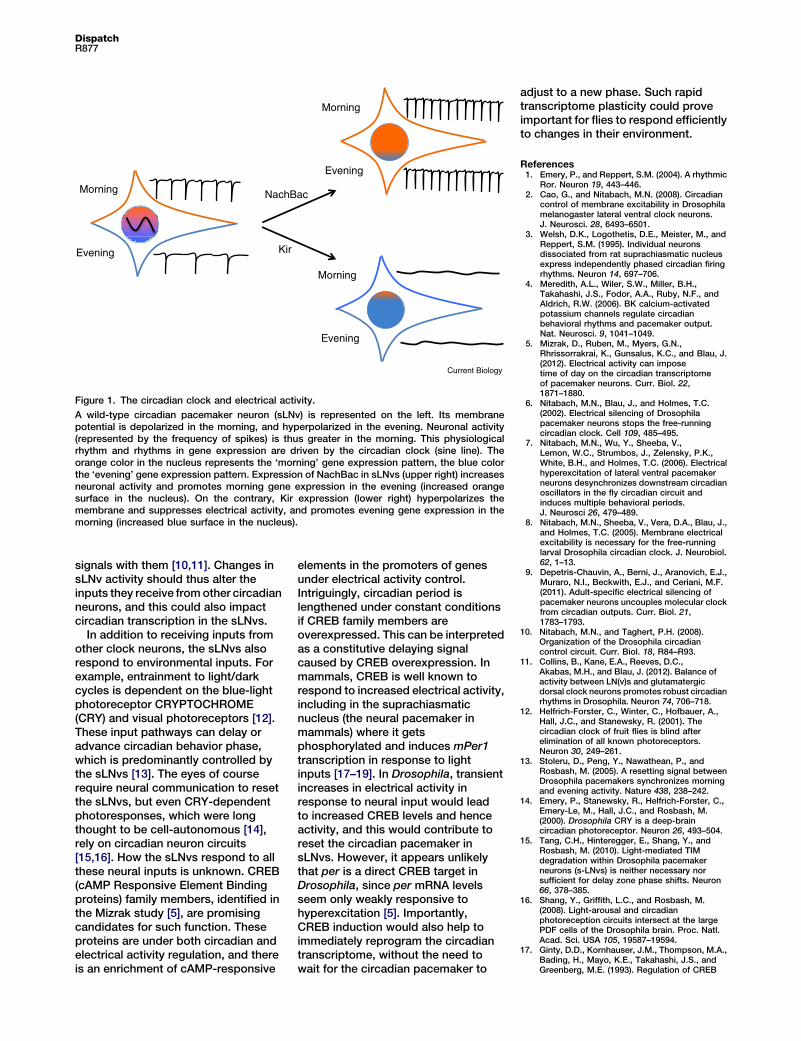
Figure 1. The circadian clock and electrical activity.
A wild-type circadian pacemaker neuron (sLNv) is represented on the left. Its membranepotential is depolarized in the morning, and hyperpolarized in the evening. Neuronal activity(represented by the frequency of spikes) is thus greater in the morning. This physiologicalrhythm and rhythms in gene expression are driven by the circadian clock (sine line). Theorange color in the nucleus represents the ‘morning’ gene expression pattern, the blue colorthe ‘evening’ gene expression pattern. Expression of NachBac in sLNvs (upper right) increasesneuronal activity and promotes morning gene expression in the evening (increased orangesurface in the nucleus). On the contrary, Kir expression (lower right) hyperpolarizes themembrane and suppresses electrical activity, and promotes evening gene expression in themorning (increased blue surface in the nucleus).
DispatchR877
signals with them [10,11]. Changes insLNv activity should thus alter theinputs they receive fromother circadianneurons, and this could also impactcircadian transcription in the sLNvs.
In addition to receiving inputs fromother clock neurons, the sLNvs alsorespond to environmental inputs. Forexample, entrainment to light/darkcycles is dependent on the blue-lightphotoreceptor CRYPTOCHROME(CRY) and visual photoreceptors [12].These input pathways can delay oradvance circadian behavior phase,which is predominantly controlled bythe sLNvs [13]. The eyes of courserequire neural communication to resetthe sLNvs, but even CRY-dependentphotoresponses, which were longthought to be cell-autonomous [14],rely on circadian neuron circuits[15,16]. How the sLNvs respond to allthese neural inputs is unknown. CREB(cAMP Responsive Element Bindingproteins) family members, identified inthe Mizrak study [5], are promisingcandidates for such function. Theseproteins are under both circadian andelectrical activity regulation, and thereis an enrichment of cAMP-responsive
elements in the promoters of genesunder electrical activity control.Intriguingly, circadian period islengthened under constant conditionsif CREB family members areoverexpressed. This can be interpretedas a constitutive delaying signalcaused by CREB overexpression. Inmammals, CREB is well known torespond to increased electrical activity,including in the suprachiasmaticnucleus (the neural pacemaker inmammals) where it getsphosphorylated and induces mPer1transcription in response to lightinputs [17–19]. In Drosophila, transientincreases in electrical activity inresponse to neural input would leadto increased CREB levels and henceactivity, and this would contribute toreset the circadian pacemaker insLNvs. However, it appears unlikelythat per is a direct CREB target inDrosophila, since per mRNA levelsseem only weakly responsive tohyperexcitation [5]. Importantly,CREB induction would also help toimmediately reprogram the circadiantranscriptome, without the need towait for the circadian pacemaker to
adjust to a new phase. Such rapidtranscriptome plasticity could proveimportant for flies to respond efficientlyto changes in their environment.
References1. Emery, P., and Reppert, S.M. (2004). A rhythmic
Ror. Neuron 19, 443–446.2. Cao, G., and Nitabach, M.N. (2008). Circadian
control of membrane excitability in Drosophilamelanogaster lateral ventral clock neurons.J. Neurosci. 28, 6493–6501.
3. Welsh, D.K., Logothetis, D.E., Meister, M., andReppert, S.M. (1995). Individual neuronsdissociated from rat suprachiasmatic nucleusexpress independently phased circadian firingrhythms. Neuron 14, 697–706.
4. Meredith, A.L., Wiler, S.W., Miller, B.H.,Takahashi, J.S., Fodor, A.A., Ruby, N.F., andAldrich, R.W. (2006). BK calcium-activatedpotassium channels regulate circadianbehavioral rhythms and pacemaker output.Nat. Neurosci. 9, 1041–1049.
5. Mizrak, D., Ruben, M., Myers, G.N.,Rhrissorrakrai, K., Gunsalus, K.C., and Blau, J.(2012). Electrical activity can imposetime of day on the circadian transcriptomeof pacemaker neurons. Curr. Biol. 22,1871–1880.
6. Nitabach, M.N., Blau, J., and Holmes, T.C.(2002). Electrical silencing of Drosophilapacemaker neurons stops the free-runningcircadian clock. Cell 109, 485–495.
7. Nitabach, M.N., Wu, Y., Sheeba, V.,Lemon, W.C., Strumbos, J., Zelensky, P.K.,White, B.H., and Holmes, T.C. (2006). Electricalhyperexcitation of lateral ventral pacemakerneurons desynchronizes downstream circadianoscillators in the fly circadian circuit andinduces multiple behavioral periods.J. Neurosci 26, 479–489.
8. Nitabach, M.N., Sheeba, V., Vera, D.A., Blau, J.,and Holmes, T.C. (2005). Membrane electricalexcitability is necessary for the free-runninglarval Drosophila circadian clock. J. Neurobiol.62, 1–13.
9. Depetris-Chauvin, A., Berni, J., Aranovich, E.J.,Muraro, N.I., Beckwith, E.J., and Ceriani, M.F.(2011). Adult-specific electrical silencing ofpacemaker neurons uncouples molecular clockfrom circadian outputs. Curr. Biol. 21,1783–1793.
10. Nitabach, M.N., and Taghert, P.H. (2008).Organization of the Drosophila circadiancontrol circuit. Curr. Biol. 18, R84–R93.
11. Collins, B., Kane, E.A., Reeves, D.C.,Akabas, M.H., and Blau, J. (2012). Balance ofactivity between LN(v)s and glutamatergicdorsal clock neurons promotes robust circadianrhythms in Drosophila. Neuron 74, 706–718.
12. Helfrich-Forster, C., Winter, C., Hofbauer, A.,Hall, J.C., and Stanewsky, R. (2001). Thecircadian clock of fruit flies is blind afterelimination of all known photoreceptors.Neuron 30, 249–261.
13. Stoleru, D., Peng, Y., Nawathean, P., andRosbash, M. (2005). A resetting signal betweenDrosophila pacemakers synchronizes morningand evening activity. Nature 438, 238–242.
14. Emery, P., Stanewsky, R., Helfrich-Forster, C.,Emery-Le, M., Hall, J.C., and Rosbash, M.(2000). Drosophila CRY is a deep-braincircadian photoreceptor. Neuron 26, 493–504.
15. Tang, C.H., Hinteregger, E., Shang, Y., andRosbash, M. (2010). Light-mediated TIMdegradation within Drosophila pacemakerneurons (s-LNvs) is neither necessary norsufficient for delay zone phase shifts. Neuron66, 378–385.
16. Shang, Y., Griffith, L.C., and Rosbash, M.(2008). Light-arousal and circadianphotoreception circuits intersect at the largePDF cells of the Drosophila brain. Proc. Natl.Acad. Sci. USA 105, 19587–19594.
17. Ginty, D.D., Kornhauser, J.M., Thompson, M.A.,Bading, H., Mayo, K.E., Takahashi, J.S., andGreenberg, M.E. (1993). Regulation of CREB

Current Biology Vol 22 No 20R878
phosphorylation in the suprachiasmaticnucleus by light and a circadian clock. Science260, 238–241.
18. Gau, D., Lemberger, T., von Gall, C., Kretz, O.,Le Minh, N., Gass, P., Schmid, W., Schibler, U.,Korf, H.W., and Schutz, G. (2002).Phosphorylation of CREB Ser142 regulates
light-induced phase shifts of the circadianclock. Neuron 34, 245–253.
19. Obrietan, K., Impey, S., Smith, D., Athos, J., andStorm, D.R. (1999). Circadian regulation ofcAMP response element-mediated geneexpression in the suprachiasmatic nuclei.J. Biol. Chem. 274, 17748–17756.
Department of Neurobiology, University ofMassachusetts Medical School, Worcester,MA 01605, USA.E-mail: [email protected]
http://dx.doi.org/10.1016/j.cub.2012.08.003
Affective Neuroscience: Food‘Wanting’ Hotspot in Dorsal Striatum
New research has uncovered a micro-domain within dorsal neostriatum whereenkephalin surges are triggered by the opportunity to consume tasty foods andwhere m-opioid microinjections generate intense motivational ‘wanting’ to eatwithout enhancing food ‘liking’.
Andrew D. Lawrence
The increased prevalence of obesityposes a global challenge to health.Over-consumption of aggressivelymarketed, abundant, cheap, tasty,energy-dense foods is one contributorto intake in excess of metabolicdemand [1], resulting in increasedattention to neural mediators of foodreward. A new study [2], reported inthis issue of Current Biology, hasuncovered a novel brain substratein dorsal neostriatum that mediatesexcessive consumption ofenergy-dense foods, where m-opioidsignalling generates intensemotivational ‘wanting’ to eat withoutelevating the hedonic impact (‘liking’)of feeding.
It is well established that signallingvia brain m-opioid receptors has potenteffects on feeding and that the ventralstriatum (nucleus accumbens) isa critical substrate where opioids exerttheir rewarding effects [3]. A strikingaspect of m-opioid-induced eating isits specificity for foods rich in sugar, fat,or both [3]. Kelley et al. [3] proposedthat, whilst normal feeding can occurwithout release of enkephalin (anendogenous m-opioid receptor ligand),enkephalin release, by enhancing thepleasure of eating, serves to stimulateintake of energy-dense foods beyondthat required to maintain energybalance. In an evolutionary context,a m-opioid-driven urge to overeat whenpresented with a calorific food sourcewould serve to increase fat stores,aiding survival in the event of futurefamine [3]. But in the current food-richenvironment, such a mechanismseems more of a hindrance than a help.
A detailed account of the role ofaccumbens opioid transmissionin food reward is emerging, basedon the work of Pecina et al. [4,5].The hedonic impact of tastes can bemeasured objectively by orofacial tastereactivity patterns (‘liking’ reactions),which are homologous across rodentand primate species [4] and whichfluctuate in similar ways to humansubjective pleasure during hunger/fullness states. For example, sweettastes elicit a positive hedonic patternof reactions including tongueprotrusions (licking of the lips) [4].
By selectively stimulating m-opioidreceptors in discrete rat brain regionsvia local microinjection of m-opioidagonists and studying the extent towhich such manipulations enhance‘liking’ reactions, Berridge et al. [4,5]have previously identified discretehedonic ‘hotspots’: micro-domainswhere m-opioid receptor stimulationpowerfully increases ‘liking’ reactionsto sweet tastes. One such hotspot(1 mm3 volume) resides in themedial-dorsal accumbens shell [4,5].Another (0.8 mm3 volume) resides ina caudal zone of the ventral pallidum,chief accumbens output target [4,5].These hedonic hotspots act in concertto increase ‘liking’ reactions to sweetsensations [4,5]. The hedonic hotspotsare also ‘wanting’ hotspots, in thatthe same microinjections of m-opioidagonist simultaneously increase both‘liking’ and motivational ‘wanting’ forfood, as reflected in vigorous eating[4,5]. The tight localization of opioidhedonic hotspots contrasts strikinglywith a looser distribution of substrates,encompassing almost the entire medialaccumbens shell (plus amygdaloid
regions), where m-opioid stimulationgenerates only ‘wanting’ to eat (largeincreases in food intake) withoutenhancing ‘liking’ reactions (pure‘wanting’ hotspots) [4,5]. Treatmentwith m-opioid agonists/antagonistsalso elevates/suppresses consumptionof sweet and fatty foods in the widerventral striatum [3–5].The new study [2] reveals that
m-opioids stimulate food ‘wanting’without enhancing food ‘liking’ not onlyin ventral, but also in the dorsalstriatum, a region seldom associatedwith reward. The dorsal striatum hasa mosaic organization comprisingisland-like striosomes/patchesembedded in a more extensive matrix[6]. Striosomes are distinguishedfrom matrix by a dense concentrationof m-opioid receptors [6]. Orbitofrontal,cingulate and insular corticespreferentially innervate striosomes,which form part of a ‘limbic’ circuitembedded in sensorimotorand associative striatum [6].DiFeliceantonio et al. [2] focused onthe medial dorsal striatum, which,like the accumbens, contains neuronsresponding to food [7], is enriched inm-opioid receptors [8], receivesamygdaloid projections [9], andreceives body-state signals from lateralhypothalamus via midline thalamicprojections [3].In vivo measurements are critical for
revealing normal functioning of opioidtransmission within striatal networks.Microdialysis is one approach forin vivo studies; however, until recently,measurement of opioid-peptiderelease in behaving animals has beenstymied by problems with recoveryand detection sensitivity [10].DiFeliceantonio et al. [2] adopteda novel analysis method — capillaryliquid chromatography coupled off-lineto multistage mass spectrometry,to determine endogenous opioidsin microdialysis samples collectedin vivo. This technique has beenvalidated for measurement of opioidpeptides, showing high sensitivityand specificity [10]. Using this


![Journal of Circadian Rhythms BioMed · 2017. 8. 28. · circadian rhythms that repeat approximately every 24 hours [1,2]. Examples of circadian rhythms include oscil-lations in core](https://static.fdocuments.us/doc/165x107/60c1699fd6e56d72e306568a/journal-of-circadian-rhythms-biomed-2017-8-28-circadian-rhythms-that-repeat.jpg)


