CIBA-TM Series- 2015- 2 Aquatic Animal Health... · Training Manual On Aquatic Animal Health...
146
CIBA-TM Series- 2015- 2
Transcript of CIBA-TM Series- 2015- 2 Aquatic Animal Health... · Training Manual On Aquatic Animal Health...

CIBA-TM Series- 2015- 2

Training Manual
On
Aquatic Animal Health Management in
Brackishwater Aquaculture
Contributed by:
K.K.Vijayan, S.V.Alavandi, K.V.Rajendran, K.P. Jithendran,
C.P.Balasubramanian, M.Jayanthi, M. Muralidhar, A.
Panigrahi, K.Ambasankar, J. SyamaDayal, T.K.Ghosal, Debasis
De, M.Poornima, R. Saraswathy, P.K.Patil, S.K.Otta, Sanjoy
Das, K.P.Kumaraguru Vasagam, P.Ezhil Praveena,
T.Bhuvaneswari, GourangaBiswas ,
Prem Kumar, N. Lalitha, R. Kumara Raja, L. Christina, K.P.
Sandeep, Satheesha Avunje, T. Sathish Kumar, and R. Vidya.
Aquatic Animal Health and Environment Division
Central Institute of Brackishwater Aquacutlrue,
75, Santhome High Road, R.A. Puram,
Chennai 600028
CIBA-TM Series-2015-2

DISCLAIMER
All rights reserved. No part of this publication may be reproduced, stored in a retrieval
system, or transmitted in any form or by any means, electronic, mechanical, recording or
otherwise, without theprior written permission of Director, CIBA.
The author and publisher are providing this book and its contents on an “as is” basis and
make no representations or warranties of any kind with respect to this book or its contents. In
addition, the author and publisher do not represent or warrant that the information accessible
via this book is accurate, complete or current.
Except as specifically stated in this book, neither the author or publisher, nor any authors,
contributors, or other representatives will be liable for damages arising out of or inconnection
with the use of this book.

CONTENT
Part I
Sl. No Title Page No
1. Overview of BrackishwaterAquaculture 1
2. Better management practices (BMPs) 9
3. Biosecurity and Quarantine Measures for Aquaculture
Health Management
25
4. Diseases surveillance in brackishwater aquaculture:
recent developments
31
5. Nutrition and feed management in brackishwater
aquaculture with special reference to aquatic animal health
36
6. Live feeds and health management in the
brackishwater larviculture
40
7. Application of probiotics and immunostimulants in
aquaculture for pond health and disease management
44
8. Pond preparation and management practices for
prevention of diseases in brackish water aquaculture
48
9. Water and soil quality requirements and their management
in brackish water aquaculture
55
10. Introduction to Nucleic Acids 62
11. Viruses, structure, biology – viral replication, infection,
variations / methods
67
12. Viral diseases and their management with special
reference to white spot disease and Indian shrimp farming
74
13. Update on Acute hepatopancreatic necrosis disease
(AHPND)
78
14. Diseases in shrimp aquaculture-Current scenario in India:
white faeces/gut syndrome (WFS), growth variation /
retardation, white muscle syndrome, running mortality
syndrome (RMS) and and Hepatopancreatic
microsporidiosis
81
15. Shrimp Diseases : An overview 86
Part II
Sl.No Title Page No
1. Requirements for Aquatic Animal Disease Diagnostic
Laboratory
93
2. Investigating disease in brackishwater aquaculture:
Overview
95
3. Shrimp anatomy, dissection, tissue preservation; sampling,
preservation / fixation and transport of samples for disease
investigation
100
4. Bacteriological methods for disease investigation:
Sampling, isolation and identification of bacteria
108
5. Application of molecular diagnostics in disease
management
114
6. Extraction and quantification of nucleic acids 118
7. Principles and practice of polymerase chain reaction 123
8. PCR diagnosis of some important OIE listed viruses and
bacterial pathogens of shrimp
127
9. Histopathological techniques in shrimp disease diagnosis 134
10. Collection, preservation and despatch of aquatic animal
sample to a diagnostic laboratory / referral laboratory for
disease diagnosis
136

Part I
Concepts in Aquatic Animal Health
Management

1
Overview of Brackishwater Aquaculture in India
Ever increasing human population
coupled with decreasing availability of
space for land-based food production
system (such as agriculture, animal
husbandry) has
resultedshortageandinconsistentsupplyofq
ualityfoodforlargenumberofpeople across
the globe. A realistic solution of the
problem could be sustainable utilization of
water resources as most of the global
water resources are lying unutilized or
underutilized. Amongst a variety of food
items present in the aquatic system, fishes
are considered as the most important
group of the organisms suitable for human
consumption. While capture fisheries is
showing the signs of almost stagnation for
more than a decade, aquaculture offers a
vast scope of expansion.
India is the second most populous
country in the world with a total
population of 1.21 billion as in 2011
representing 17.5% of the world’s
population and occupying only 2.4% of
the world’s landmass. In line with the
global trend, greatest challenge the
country faces is to ensure food security of
the largely undernourished protein
starved population in rural as well as
urban areas, especially in the context of
declining land resources available for
agriculture and animal husbandry. Hence
fisheries, mainly aquaculture sector would
have to emerge as the saviour to meet
increased food demand. With its limited
resources of man and materials various
central and state govt. agencies are
making all out efforts at augmenting
production by way of exploiting
sustainably all potential sources of aquatic
organisms in various kinds of
waterresources.
Indian aquaculture has demonstrated
a six and half fold growth over the last
two decades. Carp in freshwater and
shrimps in brackishwater form the major
areas of activity. Aquaculture in India is
generally practiced with the utilization of
lowtomoderatelevelsofinputs,especiallyor
ganic-basedfertilizersandfeed.About 40%
of the available 2.36 million hectares of
freshwater resources and 13% of a total
potential brackishwater resource of 1.24
million hectares is under use at present.
This offers vast scope for both horizontal
and vertical expansion of these sectors. As
aquaculture plays vital role in socio-
economic development in terms of income
and employment, environment friendly
aquaculture has been accepted as a tool for
rural development. It also has huge
potential for foreign exchangeearnings.
Initiatives have also been taken up to
use the unutilized and underutilized
resources in several regions of the country.
Issues like investment in fish and shrimp
hatcheries, establishment of aquaculture
estates, feed mills, R&D support and
ancillary industries have been given
special emphasis to strengthen the pace of
growth of thesector.
Origin and development
Brackishwater farming in India is an
age-old traditional system confined
mainly to
the'bheries'(manmadeimpoundmentsincoas
talwetlands) of WestBengal, ‚gheris‛ in
Odisha,‚pokkali' (salt
resistantdeepwaterpaddy)fieldsinKerala,‚k
harlands‛ in Karnatakaand‚khazans‛ in Goa
coasts.Thesesystemshave been sustaining
production of 500–750 kg/ha/year with
shrimp contributing 20–25% with no

2
additional input, except that of trapping
the naturally bred juvenile fish andshrimp
seed during tidal influx. In this traditional
method, low lying areas near the banks of
saline water rivers and creeks are
encircled by peripheral dyke and tidal
water is allowed to enter the
impoundment along with natural seeds of
various species of shrimps, crabs and fish.
Water is retained with periodical
exchanges during lunar cycles and the
animals are allowed togrow.
Realizing the importance of shrimp
farming in Indian economy, Central
Inland Fisheries Research Institute (CIFRI)
under the Indian Council of Agricultural
Research (ICAR) established first
Experimental Brackish water Fish Farm at
Kakdwip, West Bengal in 1973. This was
followed by inception of All-India
Coordinated Research Project on Brackish
water Fish Farming in 1975 by the ICAR
with centres in West Bengal, Odisha,
Andhra Pradesh, Tamilnadu, Kerala and
Goa. At the same time, shrimp seed
production studies were initiated by the
Central Marine Fisheries Research
Institute (CMFRI) of ICAR. Commercial
scale shrimp farming started gaining roots
only after 1988–1989 and the semi-
intensive farming technology
demonstrated production levels reaching
4–6 tons/ha. Area under shrimp farming
increased substantially during 1990–1994
with remarkable growth rate till 1995 as
the boom period of commercial-scale
shrimp culture and the bust came in 1995-
96, with the outbreak of viral disease. The
fact that most of the farmers were new to
commercial scale and high intensive
shrimp farming, the general ignorance of
good aquaculture practices and the lack of
suitable extension services, led to a host of
problems. Later with the advent of bio-
secured closed culture technology using
better management practices, semi-
intensiveand intensive shrimp farming
again started to regain its lost glory during
early years of the present century. Farmed
shrimp production increased from 102940
tons in 2001-02 to record production of
144346 tons in 2006-2007 and operating at
around 100000 tons over the years.
Brackishwater aquaculture development
in India was mostly oriented till 2009 to
tiger shrimp, Penaeus monodon culture
only. Other shrimp species like P. indicus,
P. merguiensis, P. penicillatus, P. japonicus
and P. semisulcatus are not yet cultured on
large commercial level. As tiger shrimp
farming became regressed by viral
diseases since 1995 and profitability was
decreasing due to abnormal hike in input
cost and decreasing unit sale value, Indian
farmers were looking an alternative. In
2009, the Coastal Aquaculture Authority
of India (CAA) permittedthe
entrepreneurs to introduce a new species,
P. vannamei (Pacific white leg shrimp) in
India with prescribed guidelines. Before
introduction, risk analysis was carried out
by Central Institute of Brackishwater
Aquaculture (CIBA) and National Bureau
for Fish Genetic Resources (NBFGR) after
pilot scale initiation 2003.At the same time
CAA is very keen in the bio security and
approval for culture of P. vannamei. Since
its introduction, vannamei farming
showed rapid growth.
The state wise area and production of
shrimpis asfollows:
Among Indian states, Andhra Pradesh
is leading in P. vannamei farming
contributingnearly80%ofIndianproduction
.Table3depictsstatewiseareaunder culture
and production of P. vannamei (Pacific
white leg shrimp) inIndia.

3
Table 1: Areas (ha) under shrimp cultivation by state
State 1990 1994 1999 2014
WestBengal 33815 34400 42525 48410
Orissa 7075 8500 11332 6302
AndhraPradesh 6000 34500 84269 36123
TamilNadu 250 2000 2670 7804
Kerala 13000 14100 14595 12917
Karnataka 2500 3500 3540 394
Goa 525 600 650 31
Maharashtra 1800 2400 970 1486
Gujarat 125 700 997 2359
Total 65090 100700 161548 115826
Table 2: Area (ha) under farming, production and percent of total production during2013-2014
State Area under
farming
Production(mt) Percent of total
production
WestBengal 48410 52581 19.4
Orissa 6302 14532 5.4
AndhraPradesh 36123 159083 58.7
TamilNadu 7804 25815 9.5
Kerala 12917 5175 1.9
Karnataka 394 664 0.2
Goa 31 63 0.02
Maharashtra 1486 3513 1.3
Gujarat 2359 9393 3.5
Total 115826 270189 -
Source: MPEDA,2014
There is huge potential for mud crab
farming in the country. Still there is no
organized aquaculture of mud crab for
supporting the export trade. Major reason
is the non-availability or inconsistent
availability of crab seeds for farming.
Technology for seed production, culture
and fattening of green mud crab Scyllla
serreta has been developed by CIBA. Some
farmers are practicing crab fattening in the
coastal regions with considerable success.
After the inception of crab hatchery by the
Rajiv Gandhi Centre for Aquaculture
(RGCA) in Nagapattinam district in Tamil
Nadu, hatchery produced crab seeds are
now available. Some progressive farmers
have started crab grow-out farming with
better performance of hatchery
producedseedscomparedtowildones.

4
Table 3: State wise area under culture (ha) and production (tons) of P. vannamei inIndia
States 2009-10 2010-11 2011-12 1012-13 1013-14 2014-15
WestBengal Area 0 0 0 0 130 326
Production 0 0 0 0 479 395
Orissa Area 0 0 25 46 485 2340
Production 0 100 436 2907 11866
Andhra
Pradesh
Area 264 2739 7128 20198 49764 37560
Production 1655 16913 75385 133135 210639 276077
Tamil Nadu &
Pondicherry
Area 0 34 397 1511 5087 5037.1
Production 0 109 2863 8595 26281 32687.8
Kerala Area 0 0 0 0 0 5.8
Production 0 0 0 0 0 11.75
Karnataka Area 0 0 72 154 157 124.76
Production 0 0 232 484 517 623.2
Goa Area 0 0 0 1 29 27.2
Production 0 0 0 15 67 88.2
Maharashtra Area 10 94 127 439 908 1274.51
Production 30 508 941 1503 3291 4901.04
Gujarat Area 9 64 88 366.71 707 3545.4
Production 46 717 1195 3348.19 6326 26763
Total Area 283 2931 7837 22715.71 57267 50240.77
Production 1731 18247 80717 147516.2 250507 353413.1
Source: MPEDA(Kochi)
High value carnivorous fishes like
Asian seabass (Lates calcarifer), snappers
(Lutjanus sp.) and
herbivorous/omnivorous fishes like
Striped grey mullet (Mugil cephalus),
Tade mullet (Liza tade), Parsia (Liza
parsia), Milk fish (Chanos chanos) and
Pearl spot (Etroplus suratensis) are
available for farming in the Indian
coastal ecosystem. In addition to this,
fishes like Mystus gulio, are also being
cultured. Successful technology has been
developed for the seed production of
Asian seabass under controlled
conditions and farming by CIBA since
1997. Some enterprising farmers in
Tamilnadu and West Bengal have taken
up seabass culture. Monoculture of
Asian seabass is in practice in those areas
where cheap trash fish is available in
plenty. Polyculture of Asian seabass
following ‛predator-prey culture‟
system using tilapia as prey material has
also been tried with considerable
success. In addition to this an avenue has
come by successful breeding and seed
production of cobia (Rachycentron
canadam) using land based pond reared
brooders by CIBA. Cobia farming in
India is gaining momentum. Controlled
breeding of grouper (Epinephelus
tauvina), striped grey mullet and pearl
spot has also been successful. Mullets,
milkfish and pearl spot have shown
promises for commercial aquaculture in
inland saline soil / water areas.
Production potential ranging from 0.5 to
3 tons/ hectare/ year has been
demonstrated from such waters.
Farmingsystems
There are five different shrimp
aquaculture practices mentioned in the
literature, ranging from traditional to
ultra-intensive techniques, but the most
common techniques followed in India are
traditional, extensive, semi-intensive, and
intensive. These three categories are
divided, according to their stocking
densities (shrimp/ m2), and the extent of

5
management over grow-out parameters,
i.e. level of inputs (Table4).
Traditional culture practices
dependent completely on the natural
tidal entry for seed, food and water
exchange. Furthermore, traditional
systems are often characterized by
polyculture with fish or by rotation with
rice, e.g. in the bheris of West Bengal and
pokkalis of Kerala in India. In this method
of aquaculture low lying areas near the
banks of saline water rivers and creeks
are encircled by peripheral dyke and tidal
water is allowed to enter in the
impoundment along with natural seeds
of various species of shrimps, crabs and
fishes. Water is retained with periodical
exchanges during lunar cycles and the
animals are allowed to grow. After 3–4
months harvesting is done partially
during lunar cycles. Productivity in this
system ranged between 500–750 kg/ha, of
which about 30 percent is contributed by
prawns/ shrimps and 70 per cent
bymullets
Extensive shrimp aquaculture is
primarily used in areas with limited
infrastructure. Producers rely on the tides
to provide most of the food for the
shrimp and as a means of water
exchange. Feed for shrimp is naturally
occurring, in some cases fertilizers or
manure is added to promote algal
growth. Low stocking densities result in
modest yields. Land and labour are the
principal inputs, which keeps operational
cost at a minimum. The extensive
farming is commonly known as
improved traditional farming. This
system involves construction of
peripheral canals/ ponds of size ranging
from 1–5 ha. Shrimp seed at the rate of
15000 – 20000/ha are stocked. Water
management is done by tidal effect. The
average yield is 1500 – 1700 kg/ha,
including fin fishes. In most of the cases,
the stock is left atthe mercy of nature and
the predators. Supplementary feeding is
not generally practiced as the entire
production system is dependent on
utilization of natural productivities.
However some farmers use oil cakes and
rice bran as supplementary feed.
Semi-intensive cultivation involves
stocking densities beyond those that the
natural environment can sustain without
additional inputs. Consequently these
systems depend on a reliable shrimp post
larvae (PL) supply, and a greater
management intervention in the pond‟s
operation compared to extensive ponds.
Semi-intensive shrimp aquaculture relies
on water pumps to exchange up to 25%
of pond volume daily; however, mostly
closed culture is practiced at present.
With stocking rates of 6-20 shrimp PL per
m2, fertilizers are applied to augment
natural
foodintheponds.Supplementaryfeedingis
doneusingformulatedfeedspreferably in
pelletized form. Maximum annual yields
range from 2 to 6 tons per hectare. The
risk of crop failure increases with
increasing farming intensity, which is
mainly due to the impact on water
quality exerted by the high stocking
densities and supplementary feeding. All
of the costs associated with semi-
intensive production are much higher
relative to those for extensive production,
including a more complex system of
ponds, installation of a pump system to
regulate waterexchange, skilled
management, labour, purchased feed and
seed stock,and increased energy usage
for aeration and other purposes. The
higher the culture intensity, the higher
the capital required and the higher the
risks involved.
Thus,theincreasedcapitalinputsrequiredf
orsemi-intensivecultureoftenprecludeits
adoption by small-scale producers. Tiger

6
shrimp farming and low density pacific
white leg shrimp farming in India falls
under thiscategory.
Intensive grow-out systems utilize
ample supplies of clean sea / estuarine
water, adequate infrastructure, and well-
developed hatchery and feed industries.
Intensive shrimp farming introduces
small enclosures (down to 0.1 ha), high
stocking densities (20-50 hatchery-
produced shrimps/m2), around-the-clock
management, very high inputs of
formulated feeds, and aeration. Aeration,
the addition of oxygen to the water
permits much higher stocking and
feeding levels. Yields range from 7 to 15
tons per hectare per year. The risk of
disease can be serious in intensive
culture, especially if water discharge
from one pond or farm is taken into
another to be reused. Most of the P.
vannamei farming in India is conducted in
this method using specific pathogen free
(SPF) seeds under strict
biosecurityprotocol.
Table 4: Farming practices based on level of management, stocking density and
production followed in India:
Traditional Extensive Semi-
intensive
Intensive
Pond size(ha) 0.1-50 1-10 0.2-2 0.1-1
Stocking Natural Natural +artificial Artificial Artificial
Stocking density
(seed/m2)
Unregulated 2-6 6-20 20-50
Seedsource Wild Wild + Hatchery Hatchery +
wild
Hatchery
Annual
Production
(ton/ha/yr)
<0.6 0.6-1.5 2-6 7-15
Feedsource Natural Natural Natural +
Formulated
Formulated
Fertilisers No Yes Yes Yes
Waterexchange Tidal Tidal +pumping Pumping Pumping
Aeration No No Yes Yes
Diversity of
crops
Polyculture Monoculture,
polyculturerarely
Monoculture Monoculture
Disease problems Rare Rare Moderate to
frequent
Frequent
Employment
(persons/ha)
1-2 2-3 3-4 4-5
Human resource development
through brackishwater
aquaculture
Employment opportunities in
coastal areas have increased greatly with
the development of shrimp farming.
According to an estimate, the average
labour requirement in shrimp farming
has been estimated to be about 600 labour
days/crop/ha. In contrast, labour
days/crop/ha in paddy cultivation is 180,
which is much lower than shrimp
farming. Case studies carried out at a sea-
7
based farm in the Nellore District of
Andhra Pradesh showed an increase of
2–15 percent employment and 6–22
percent income for farm labourers
following the establishment of shrimp
farms. Hatcheries and feed mills in the
brackish water sector are also providing
substantial employment opportunities.
Jobs generated in the main and
supporting sectors of the shrimp
aquaculture sector was estimated to be
over three lakhs inIndia.
Laws andregulations
There are many laws and
regulations, which are relevant to
aquaculture adopted at state level,
several key laws and regulations relevant
to aquaculture at the central level. Those
include the century-old Indian Fisheries
Act (1897), which penalizes the killing of
fish by poisoning water and by using
explosives; the Environment Protection
Act (1986) with provisions for all
environment related issues; Water
(Prevention and Control of Pollution) Act
(1974) and the Wild Life Protection Act
(1972). The Supreme Court prohibited the
construction / set up of shrimp culture
ponds except traditional and improved
traditional types of ponds within the
Coastal Regulation Zone (CRZ) on 11th
December, 1996. It also ruled that an
authority should be constituted to protect
the ecologically fragile coastal areas, sea
shore, water front and other coastal areas
and specially to deal with the situation
created by the shrimp culture industry in
the coastal states / union territories. To
perform the functions indicated by the
Supreme Court, Coastal Aquaculture
Authority was formed in accordance with
the Environment (Protection) Act. The
Authority, to which specific
responsibilities for aquaculture have been
allocated, falls under the administrative
control of the Ministry of Agriculture.
Institutionalsetup
Division of Fisheries under the
Department of Animal Husbandry,
Dairying, and Fisheries of the Ministry of
Agriculture, Govt. of India is the nodal
agency for planning, monitoring and the
funding of several centrally sponsored
developmental schemes related to
fisheries and aquaculture in all of the
Indian States. Most of the states possess a
separate Ministry for Fisheries and also
have well-organized fisheries
departments, with fisheries executive
officers at district level and fisheries
extension officers at block level, who are
involved in the overall development of
the sector. Centrally sponsored schemes
are implemented through 422 (Fish
Farmer‟s Development Agency) FFDAs
in freshwater sector and 39
(Brackishwater Fish Farmer‟s
Development Agency) BFDAs in the
maritime districts substantially
contributes to brackishwater aquaculture
development. The ICAR under the
Department of Agricultural Research and
Education, which in turn is within the
Indian Ministry of Agriculture, has a
Division of Fisheries, which undertakes
the R&D on aquaculture and fisheries
through a number of research institutes.
There are about 400 Krishi Vigyan
Kendras (KVKs) operated through State
Agricultural Universities, ICAR Research
Institutes and NGOs, most of which also
undertake aquaculture development
activities. The MPEDA functioning under
the Ministry of Commerce, besides its
role in the export of aquatic products also
contributes towards the promotion of
coastal aquaculture. Many other
organizations like Department of Science
and Technology, Departments of
Biotechnology, University Grants

8
Commission also support or conduct
R&D in the subject. Various NGOs and
private organizations contribute
substantially in thiscontext.
Research andDevelopment
Eight fisheries research institutes
are there under ICAR, the nodal agency
for aquaculture research in India, of
which CIBA, in Chennai is responsible
for research on brackishwater
aquaculture. These institutes have their
regional centres located in different agro-
ecological regions to undertake research
on problems of regional importance.
Research programmes are set depending
on national priority and regional
necessity, farmers' feedback is also given
due emphasis. Fisheries colleges under
different State Agriculture Universities,
as well as other universities and
organizations also undertake aquaculture
research. The institutes transfer the
developed techniques and technology
through research publications and on-
farm demonstrations. To disseminate the
emerging
technologies,electronicmediaalsoplayvital
role.
Wayforward
Exports will remain the mainstay of
the sector for years to come. Institutional
agencies focused towards this end must
seek to examine the scope for
diversification of markets, communicate
to exporters and processors the niche
markets that exist for exclusive markets
for value added fish and shrimp. Non-
tariff barriers to trade will continue to
assume different forms and dimensions.
It is the versatility and capability of the
seafood industry to adapt that will enable
them to survive such onslaughts on their
territory. Domestic markets for fish and
fish products not only provide an entirely
new opportunity for growth but also can
act as a buffer in case of gluts in the
international markets. Institutional
agencies such as National Fisheries
Development Board (NFDB) have an
onerous task on hand for enabling the
domestic markets for fish to establish
itself. Mud crab farming is one of the
avocations started in the brackishwater
sector recently to enhance the production
of mud crabs as well as to uplift the
socio-economic condition of coastal rural
population. In the brackish water sector
there were issues of waste generation,
conversion of agricultural land,
salinization, and degradation of soil and
the environment due to the extensive use
of drugs and chemicals and destruction
of mangroves. Efforts towards adoption
of improved farming technologies like
recirculatory aquaculture system (RAS),
improved polyculture, integrated
multitrophic aquaculture (IMTA) may
make brackishwater aquaculture more
environmentallyacceptable.

9
Better management practices (BMPs)
Crop related risk factors in an
aquaculture system either increases or
decreases the probability of occurrence of an
adverse event during a specified time
period. Presence of the necessary cause
alone like white spot disease (WSD) will not
lead to an outbreak in a pond. In a farm
situation, a number of ‚component causes‛
(risk factors) along with the ‚necessary
cause‛ might become ‚sufficient cause‛ to
produce WSD outbreaks. These risk factors
occur throughout the shrimp cropping cycle
and can be categorized as shown below
during the different stages of the crop cycle:
Stocking season
Pond preparation
Pond water preparation
Seed quality and screening
Water management
Pond bottom management
Feed management
Disease treatments
Effective management of these factors can
be essentially the core aspect in controlling
the environmental factors and thereby
reduce the risks of disease occurrence in the
pond.
Risk management
The risk management is to develop
practical measures for
containing/preventing shrimp disease
outbreaks that should specifically cover
identification of disease risk factors,
diagnosis and management strategies to
control disease in farms. Eventually two key
areas are identified:
Better management practices (BMPs) that
are practical farm-level interventions to
address the key ‚risk factors‛.
Farmer organization/self-help
groups/clusters to address social and
financial risks associated with farming and
allow effective dissemination of the BMPs
among group members.
In order to improve the sustainability
of shrimp farming by increasing the
consistency of production many
organizations focus on the implementation
of Best Management Practices (BMPs).
Recently, number of stakeholders insists on
Good Aquaculture Practices (GAPs) during
certification programmes and these two
practices can be differentiated as:
BMPs tend to be farm management practices
prepared to minimize the potential for farm-
raised fishery products to be contaminated
with pathogens, chemicals, or unapproved
or misused animal drugs.
GAP can be defined as those practices
necessary to address food safety concerns.
BMPs are based on the International
Principles for Responsible Shrimp Farming
which covers farm selection, farm design,
water use, broodstock and post-larvae, feed
management, health management, food
safety and social responsibility.
The BMPs have to be simple and
practical but science based their adoption
requires an understanding of the farmers
and their culture systems. Once
implemented BMPs are analyzed both to
understand how they were developed, how
they work, and what further needed for
adoption by other producers. Based on the
adoption analysis of BMP for shrimp
culture, responsible shrimp farming in
countries like Indonesia, Vietnam, Thailand
and India is being rigorously practiced. The
goal is to constantly seek out better
practices, not just because they reduce
impacts, but also because they are more

10
efficient and more profitable. The
experiences in different countries have
shown that well designed and implemented
BMPs can support producers to:
Increase efficiency and productivity by
reducing the risk of shrimp health
problems.
Reduce or mitigate the impacts of
farming on the environment.
Improve food safety and quality of
shrimp farm product; and
Improve the social benefits from shrimp
farming and its social acceptability and
sustainability.
BMPs can be country specific and are
developed for a particular location, taking
account of local farming systems, social and
economic issues, markets and environments.
In India experiences of NACA have shown
that although principles are widely
applicable there is considerable local
variation in BMPs. Thus, implementation
programmes of BMPs in various countries
give boost to farmer societies and through
these societies the traceability and
certification programmes and access to
premium international/domestic markets
can be achieved. So, BMPs are often
voluntary practices for local regulations but
can form an important tool for certification
programmes.
Pre-stocking preparations
Generally in unprepared ponds that are
not well dried, the organic matter from
previous crops accumulated in pond bottom
cause crop losses. Even if the bottom is
partially dried, the rich blackish organic
muddy soils are removed and just put inside
of the dike which again is the cause of poor
pond preparation. Thus, for shrimp culture
to be successful much depends on good
bottom soil condition and its pre-
preparation before shrimp stocking.
However, there is a diversity of soil
which plays a vital role in shrimp culture
systems. Among different soils, some soils
may have undesirable properties like
potential acid sulphate acidity, high organic
matter content or excessive porosity. On the
other hand, even if the site is good, the
problems may still crop up due to the large
quantity of inputs used during culture like
feed and fertilizers, which lead to excessive
phytoplankton production, low dissolved
oxygen, high ammonia, poor bottom soil
condition and other problems. Most of these
problems can be avoided by adopting
proper best management practices during
pond preparation and culture period.
1. POND PREPARATION
The main objectives of pond preparation
are to provide the shrimp with a clean pond
base and appropriate stable water quality.
Pond preparation has to be done for both
virgin and newly constructed and in already
existing ponds.
1.1 Newly constructed ponds
In newly dug out ponds, the
characteristics of the soil has to be analysed
first before adopting the various measures to
prepare the pond. Soil deficiencies should be
identified and treated in new ponds before
stocking. If the soil of a new pond is acidic, it
should be limed before initiation of
aquaculture.
1.2 Post harvest pond preparation
Before initiating a second crop, the pond
has to be prepared by drying, removing the
earlier organic matter etc. However, after
every cropping, the soil conditions change
tremendously and are never of the same
characteristics as the virgin ponds or before
the first crop initiation. The various pond
preparation strategies include:

11
i) Cleaning
The considerable quantity of waste
accumulated in the ponds depending upon
the culture practices must be removed to
ensure sustained production. Removal of
waste by draining and drying of the pond
bottom after the end of the production cycle
is followed for keeping pond environment
clean. Two systems are commonly used to
clean the pond after a production cycle. One
is to allow the pond to dry out and then
remove the waste or the other is to wash
away the waste before it dries off.
ii) Drying
After the final drain harvest, the pond
bottom is allowed to dry and crack,
primarily to oxidize the accumulated
organic components. The pond bottom
should be dried for 7-10 days or the soil
should crack to a depth of 25 - 50 mm. The
waste can either be removed manually or
with machines at least 5cm of the top soil.
Drying and cracking of pond bottom
enhances aeration and favours microbial
decomposition of soil organic matter.
Drying also enhances the mineralization of
organic phosphorous which is dependent to
the availability in the water column and
pond soil. One of the main BMP components
is the disposal of solid waste away from the
pond site instead on the pond bunds which
is cost driven and needs site for dumping.
iii) Wet method
Alternatively in ponds where complete
drying is not possible organic,
biodegradable, piscicides such as Mahua oil
cake (100-150 ppm) and tea seed cake (15-20
ppm) can be used for eliminating unwanted
organisms. Formalin, potassium
permanganate, benzyl chromium chloride,
provodone iodine etc can be used to kill
bacteria and external parasites and these
compounds are degraded within culture
systems and usually do not causes water
pollution. In this method, after the final
drain harvest, the accumulated black
material on the pond bottom is flushed in
the form of thin slurry using a pump. In
certain areas sodium metabisulfate is used
as a post harvest treatment of shrimp and
this substance is acidic and it reacts to
remove dissolved oxygen from water.
However, the disposal of used sodium
metabisulfate solutions has to be treated
before draining in natural waters as it can
cause killing of localized fish. Use of other
products to a lesser extent in aquaculture
include zeolite, aluminum sulfate, ferrous
chloride, sodium bicarbonate, MS-222,
rotenone, chlorine compounds, herbicides,
insecticides, bacterial cultures, enzyme
preparations and possibly others which
essentially improve the soil quality and
convert the waste matter. Thus, removal of
the slurry is a quick and more efficient
process than the dry method, reducing the
period between production cycles. The
advantage of this method is that waste is
removed in suspension. This method needs
a settling pond where waste is removed
from the water and treated repeatedly to
avoid polluting the local environment.
iv) Pond maintenance
During pond preparation the weak dikes
are strengthened with soil and the inner
slope of the dike is consolidated with
outside soil other than from the pond
bottom. Tunnels and holes caused by
burrowing organisms are plugged.
Reconditioning of the bottom trench,
levelling of pond bottom using tractors and
repairs of sluice structures and sluice screens
are also done.
v) Liming
Liming of aquaculture ponds is done to
neutralize soil acidity and increase total
alkalinity and total hardness concentrations
in water. This enhances pond productivity
of food organisms for higher aquatic animal
production. Based on either the total

12
alkalinity or soil pH, agricultural limestone
dose is estimated. If both are available and
values are not in agreement, use the variable
that gives the greatest agricultural limestone
dose. Brackishwater ponds with total
alkalinity below 60 mg l-1 and any pond with
soil pH below 7 usually will benefit from
liming.
The amount of usage of lime materials to
raise the pH to 7 varies in different lime
materials. Agricultural limestone is a safe
product. However, burnt lime and hydrated
lime can result in dangerously high pH if
used in excessive amounts. So, agricultural
limestone should be spread uniformly over
the bottom of empty ponds up to the top of
the dike and left for 10 - 15 days, or
alternatively, it may be spread uniformly
over water surfaces. A large proportion of
the lime should be spread on the feeding
areas and any part of the pond that has
remained wet. Agricultural limestone will
not react with dry soil, so when applying
over the bottoms of empty ponds, it should
be applied while soils are still visibly moist.
Tilling after liming can improve the reaction
of agricultural limestone with soil.
Generally, for low pH 4.0 to 4.5 of about 11.5
to17.0 tons/ha is required of quick lime or
agricultural lime or dolomite with lowest for
quick lime. Similarly for different soil pH,
the quantities have been assessed and
applied accordingly.
vi) Tilling
Tilling bottom soils can enhance drying
to increase aeration and accelerate organic
matter decomposition and oxidation for
reduction of compounds. Soil amendments
such as agricultural limestone or burnt lime
can be mixed into soil by tilling.
Accumulations of organic matter of other
substances in the surface layer of soil also
can be mixed with deeper soils to reduce
concentrations of the substances in the
surface layer. Pond bottoms are not be tilled
when they are too wet as the tillage
machinery may not function to its capacity.
Ruts caused by machinery will fill with soft
sediment and be likely sites for anaerobic
conditions. Ruts also interfere with draining
and increase the difficulty of drying pond
bottoms. Depth of tillage usually should be 5
to 10 cm, so mould board plows, often called
turning plows, can be used to turn soil over.
Tilling can be counterproductive in ponds
where heavy mechanical aeration is used.
Tilling will loosen the soil particles and
aerator-induced water currents will cause
severe erosion of the pond bottom. Thus, if
bottoms of heavily aerated ponds are tilled,
they should be compacted with a heavy
roller before refilling.
vii) Fertilization
Decomposition in organic soils is slow
because pH usually is low and the amount
of carbon to nitrogen (C: N ratio) is high.
Nevertheless, because of high organic matter
content, such soil often becomes anaerobic
during shrimp culture. Application of
agricultural limestone and inorganic
nitrogen fertilizers increases the pH and
supply of nitrogen for faster soil organic
matter degradation during fallow periods
between crops. Urea can be spread over
pond bottoms at 200 to 400 kg ha-1 at the
beginning of the fallow period to accelerate
decomposition of organic soil. Agricultural
limestone should not be applied until a few
days after urea is applied to prevent a high
pH. Sodium nitrate can be applied @ 20 to 40
g m-2 to wet soil to encourage organic matter
decomposition in wet areas. However,
nitrate fertilizers are more expensive and are
not recommended where soils can be
adequately dried.
The rate of application of inorganic
fertilizers ranges from 25 - 100 kg/ha as a
basal dose during pond preparation with
minimum water depth of 10 - 15 cm. When
the shrimp culture progresses, depending

13
upon the phytoplankton density as
exemplified by turbidity of the pond water,
required quantity of the fertilizers may be
applied in split doses at short intervals for
sustained plankton production. The main
nutrient limiting phytoplankton production
in brackishwater ponds is phosphorus.
Hence both phosphorus and nitrogen
should be applied in the ratio of 1:1.
Excessive application of urea and
ammonium fertilizers may cause ammonia
toxicity to shrimp and also may lead to algal
blooms reducing dissolved oxygen. During
over blooming of phytoplankton copper
sulfate can be used to ‚thin‛ phytoplankton
blooms by applying the product at a safe
concentration of one-one hundredth of the
total alkalinity as it quickly precipitates to
the pond bottom and does not remain in the
water. However, accidental overdoses or
spills can cause shrimp mortality in ponds
which has to be monitored.
Shrimp being bottom dwellers, benthic
organisms constitute their main food items.
Hence fertilization of soil instead of water is
more effective. Productivity of benthic
organisms may be low in ponds with
concentrations of organic carbon below 0.5
to 1.0%. Organic fertilizer can be applied to
such soils to enhance organic matter
concentration. Chicken and other animal
manures have been applied at 1,000 to 2,000
kg ha-1 to pond bottoms during the fallow
period. In brackishwater conditions
decomposition of cattle dung is slow and
hence application of chicken manure, if
available, is advisable. The rate of chicken
manure is one-third of cattle dung.
However, application of a higher quality
organic matter such as plant meals-e.g., rice
bran, soybean meal, and crushed corn-or
low-protein-content animal feed at 500 to
1,000 kg ha-1 are more efficient. When
organic fertilization of pond bottoms is
practiced, ponds should be filled with 10 to
20 cm of water and allowed to develop a
dense plankton bloom. In shrimp farming,
both organic manures and inorganic
fertilizers are supplementary to each other
and one cannot be exchanged for the other.
It is always better to apply both organic and
inorganic fertilizers together as a basal dose
during pond preparation for optimum
result.
viii) Nutrient exchange between soil and
water
The two most important nutrients in
pond aquaculture are nitrogen and
phosphorus because these two nutrients
often are present in short supply and limit
phytoplankton growth. These two nutrients
are added to ponds in fertilizers, manures,
and feeds. Fertilizer nitrogen usually is in
the form of urea or ammonium, and urea
quickly hydrolyzes to ammonium in pond
water. Ammonium may be absorbed by
phytoplankton, converted into organic
nitrogen, and eventually transformed into
nitrogen of shrimp protein via the food web.
Ammonium may be oxidized to nitrate by
nitrifying bacteria, and nitrate may be used
by phytoplankton or denitrified by
anaerobic microorganisms in the sediment.
Nitrogen gas formed by denitrification
diffuses from sediment to pond water to the
atmosphere. Ammonium is in equilibrium
with ammonia, and ammonia also can
diffuse from pond waters to the atmosphere.
A small amount of ammonium may be
adsorbed on cation exchange sites in pond
bottom soils. Uses of salt at concentrations
up to 100 mg/L chloride are safe which can
counteract nitrite toxicity. Organic nitrogen
in plankton and in aquatic animal feces may
settle to the bottom to become soil organic
nitrogen. Nitrogen in soil organic matter
may be mineralized to ammonia and
recycled to the pond water, but the rate is
slow.
Phosphorus usually is present in fertilizer
as calcium or ammonium phosphate.

14
Phytoplankton can rapidly remove
phosphate from water, and phosphorus in
phytoplankton may enter the food web
culminating in shrimp. Pond soil strongly
adsorbs phosphorus, and the capacity of
pond soil to adsorb phosphorus increases as
a function of increasing clay content. Most of
soil phosphorus was tightly bound, and only
a small amount was water soluble. Pond
soils are not a major source of phosphorus to
water because soil-adsorbed phosphorus is
highly insoluble. Phosphorus released by
decomposition of organic matter in pond
bottoms is rapidly adsorbed by soil and little
of it enters the water. Soils that are near
neutral in pH have less capacity to adsorb
phosphorus and a greater tendency to
release phosphorus than do acidic or
alkaline soils. Nevertheless, even neutral
soils remove phosphorus from the water
and are a sink rather than a source of
phosphorus. Once dissolved in the water,
nitrogen and phosphorus originating from
manures and feed also will enter the same
pathways as nitrogen and phosphorus
applied in chemical fertilizers.
ix) Application of microbial products for
improvement of soil quality
A number of products are promoted to
enhance beneficial chemical and biological
processes and to improve soil quality. These
products include cultures of living bacteria,
enzyme preparations, composted or
fermented residues, plant extracts, and other
concoctions. There is no evidence from
research that any of these products will
improve soil quality. Nevertheless, they are
not harmful to the culture species,
surrounding environment, workers, or
quality of aquaculture products.
x) Raising of water level
The pond is filled with brackish or
seawater by pumping or by opening the
sluice with proper screens to prevent entry
of unwanted organisms into the pond. The
water level is maintained to 30 - 40 cm and
allowed to remain for 10 - 15 days. By this
time, the colour of water may turn dark
green with algal bloom and a layer of
benthic algae along with associated food
organisms will form at the bottom.
Subsequently small doses of organic and
inorganic fertilizers are applied based on the
Secchi disc observations (transparency with
25-30 cm -optimal) of algal production. The
water level is then raised to 100-125 cm.
Now the pond is ready for stocking post
larvae of shrimps.
xi) Monitoring of soil parameters during
pond preparation
Monitoring of soil quality condition can
be valuable in shrimp culture pond
management. Major concerns in pond
bottom soil management are low soil pH,
high soil organic matter, loss of the oxidized
layer, and accumulation of soft sediment. In
older ponds with impaired soil quality,
problems should be corrected and prevented
from recurring. These materials have
combined effect on the environment of the
pond bottom. To understand the condition
of the pond bottom, the following
parameters are monitored :
a) Soil pH
This is one of the most important soil
quality parameters since it affects the pond
condition. Generally, soil pH ranging
between 6.5 and 7.5 is the best suited where
availability of nitrogen, phosphorus,
potassium, calcium and magnesium is
maximum. The micronutrient whose
requirements are very small is also available
in this pH range. The low pH of bottom
sediment indicates unhygienic condition.
b) Organic matter - Redox-potential
The changes observed during culture
period in the soil bottom in terms of
increasing organic load forms a indicator of
the soil quality conditions. Anaerobic

15
condition developes in pond, when input of
organic matter exceeds and the supply of
oxygen needed for decomposition of organic
matter depletes. This reducing condition can
be measured as the redox potential (Eh).
Redox potential indicates whether the water
or soil is in reduced condition or oxidized
(Eh with ‘+’ ve value) condition. Reduced or
anaerobic sediments may occur at the pond
bottom in heavily stocked pond with heavy
organic load and poor water circulation.
Under anaerobic condition in the pond
bottom, reduced substances such as H2S,
NH3, CH4 etc. is formed which are toxic to
benthic organisms. In shrimp ponds,
development of highly reducing conditions
at the inter phase layer of water and pond
mud is highly undesirable. Water circulation
by water exchange, wind or aeration helps
to move water across mud surface and
prevents the development of reduced
condition. Draining at the centre of pond, as
is being practiced by some farmers, is an
ideal remedy for the prevention of formation
of highly reducing condition especially
during the last phase of culture period.
Bottoms should be smoothened and sloped
to facilitate draining of organic waste and
toxic substances. The redox potential (Eh) of
mud when exceed -200 mV, the pond
condition is critical. So, during pond
preparation, drying of the pond is essential
as it changes anaerobic condition to aerobic
condition of bottom soil.
Thus adoption of best management
practices during pond preparation paves
way for higher production, lesser incidence
of disease outbreaks and crop losses
provided further BMP procedures are
strictly followed during stocking and culture
phase also. One of the BMPs principles that
governs a successful crop in an area is strict,
uniformed and disciplined adoption of all
principles by one and all in the shrimp
culturists within the culture activity zone.
Shrimp seed selection
Best management practices in shrimp
seed production can be defined as the
practices or measures or methods adopted to
secure a disease free environment in all
production phases in the hatchery for
improved seed quality. BMP is the ability to
prevent losses to disease through effective
elimination of pathogens and their carriers.
The shrimp aquaculture industry has been
experiencing severe setbacks due to the
devastating viral diseases. These diseases
are believed to be transferred between
regions through the importation of hatchery
broodstock, postlarvae and shrimp
products. Once new pathogens are imported
to an area, infection of wild stock appears to
be inevitable, eliminating future possibilities
of using uncontaminated wild stock to
culture.
BMP encompasses policy, regulatory and
programme frameworks in response to
managing risks associated with diseases.
The basic elements of a BMP programme in
a shrimp hatchery include the physical,
chemical and biological methods necessary
to protect the hatchery from all diseases of
high risk. Responsible hatchery operation
must also consider the potential risk of
disease introduction into the natural
environment and its effects on neighbouring
aquaculture operations and the natural
fauna.
The BMP issues in shrimp hatcheries may
be either internal concerning the
introduction and transfer of pathogens
within the facility or external concerning the
introduction and transfer of pathogens from
outside sources to the facility or vice versa.
In case of disease outbreak within the
aquaculture facilities the options available
are either treatment- by application of
methods that reduce the effects of the
diseases, containment- by restriction of the
disease from spreading to other

16
tanks/facilities, or elimination- of the
diseases from the vicinity. Implementation
of BMP programme for a shrimp
hatcheryshould include the following
elements:
Specific pathogen free (SPF) or high
health (HH) shrimp stocks should be
used.
All the incoming stocks should be
quarantined in the designated area.
All incoming stock should be analyzed
for diseases.
All incoming water sources should be
treated to eliminate pathogens.
Equipment and materials should be
sterilized and maintained clean.
Personal hygiene measures like shower
bath including washing of hands and
feet and clothing.
Knowledge of the potential pathogenic
diseases and the sources of risk and
methods and techniques for their
control and /or eradication
Specific pathogen resistant (SPR) stocks
to be used.
Maintenance of optimum
environmental conditions.
Immune enhancers and probiotics to be
used in place of antibiotics.
Infrastructure requirements
Shrimp hatcheries should be designed (or
modified, in the case of existing hatcheries)
to ensure good biosecurity, efficiency, cost-
effectiveness and the implementation of the
hatchery standard operating procedures. A
well-designed shrimp hatchery will consist
of separate facilities for quarantine,
acclimatization, maturation, spawning and
hatching, larval and nursery rearing, indoor
and outdoor algal culture, and for the
hatching of Artemia. Additionally, there will
be supporting infrastructure for handling
water (facilities for abstraction, storage,
filtration, aeration, heating and distribution),
and feed (laboratories for analysis and
preparation and storage facilities), as well as
maintenance areas, packing areas for nauplii
and PL, offices, storerooms and staff living
quarters. The physical separation or
isolation of the different production facilities
is a feature of good hatchery design and
should be incorporated into the construction
of new hatcheries. In existing hatcheries
with no physical separation, effective
isolation may also be achieved through the
construction of barriers and implementation
of process and product flow controls. The
hatchery facility should have a wall or fence
around the periphery of the property, with
enough height to stop the entrance of
animals and unauthorized persons. This will
help to reduce the risk of pathogen
introduction by this route, as well as
increase overall security.
Water quality and treatment
Water for the hatchery should be filtered
and treated to prevent entry of vectors and
any pathogens that may be present in the
source water. This may be achieved by
initial filtering through sub-sand well
points, sand filters (gravity or pressure), or
mesh bag filters into the first reservoir or
settling tank. Following primary disinfection
by chlorination, and after settlement, the
water should be filtered again with a finer
filter and then disinfected using ultraviolet
light (UV) and/or ozone. The use of
activated carbon filters, the addition of
ethylene diamine tetra acetic acid (EDTA)
and temperature and salinity regulation may
also be features of the water supply system.
Each functional unit of the hatchery
should have independent water treatment
facility and it should be isolated from other
water supply systems for other areas.
Separate recirculation systems may be used
for part or the entire hatchery to reduce
17
water usage and further enhance
biosecurity, especially in high-risk areas. The
discharge water from the hatchery,
particularly that is known or suspected to be
contaminated (for example, water
originating from the quarantine areas)
should be held temporarily and treated with
hypochlorite solution (>20 ppm active
chlorine for not less than 60 minutes) or
another effective disinfectant prior to
discharge. This is particularly crucial where
the water is to be discharged to the same
location as the abstraction point.
Broodstock selection and quarantine
Some viral diseases are believed to be
transmitted vertically from parent to
offspring and this may be eliminated by the
use of SPF domesticated shrimp. If SPF
shrimp are not available, broodstock should
be tested for infection by an appropriate
diagnostic test and any infected individuals
should be destroyed. Shrimps testing
negative for the disease or pathogen should
still be considered a risk and placed in a
quarantine facility until their health status is
fully known.
Hatcheries normally prefer wild
broodstocks since they produce more
healthy nauplii. In recent years, however,
the high risk of introducing viral pathogens
with wild broodstock has changed this
preference. Domesticated stocks either
genetically improved or suspected to be
resistant or tolerant to specific pathogens
may be used as broodstock. SPF stocks are
generally maintained in highly biosecure
facilities and their offspring (designated
‚high health‛ rather than SPF) are supplied
to the industry. SPR shrimp are those that
are not susceptible to infection by one or
several specific pathogens, and Specific
Pathogen Tolerant (SPT) shrimp are those
that are intentionally bred to develop
resistance to the disease caused by one or
several specific pathogens.
The quarantine facilities are essentially a
closed holding area where shrimp are kept
in individual tanks until the results of
screening for viruses (and for bacteria,
where applicable) are known. The
broodstock quarantine unit should be
physically isolated from the rest of the
hatchery facilities. If this is not possible, the
hatchery design should be altered so that
there is no possibility of contamination from
the quarantine or holding area into the other
production areas. Particular care should be
taken with waste disposal and effluent
treatment. Staff working in this area should
not be permitted to enter other production
sections and should follow sanitary
protocols at all times. The quarantine unit
should have the following characteristics:
Adequate isolation from all of the
rearing and production areas to avoid
any possible cross contamination.
Should be in an enclosed and covered
building with no direct access to the
outside.
There should be means provided for
disinfection of feet (footbaths containing
hypochlorite solution at >50 ppm active
ingredient) and hands (bottles
containing iodine-PVP (20 ppm and/ or
70% alcohol) to be used upon entering
and exiting the unit.
Entrance to the quarantine area should
be restricted to the personnel assigned
to work exclusively in this area.
Quarantine unit staff should enter
through a dressing room, where they
remove their street clothes and take a
shower before going to another dressing
room to put on working clothes and
boots. At the end of the working shift,
the sequence should be reversed.
An adequate number of plastic buckets
should be available in the quarantine
room to facilitate effective daily routine
18
movement of shrimp in and out of the
facility.
The quarantine facility should have an
independent supply of water and air
with separate treatment and disinfection
systems and a system for the treatment
of effluents to prevent the potential
escape of pathogens into the
environment.
The seawater to be used in the facility
must enter a storage tank where it will
be treated with hypochlorite solution
(20 ppm active ingredient for not less
than 30 minutes) before inactivating
with sodium thiosulfate (1 ppm for
every ppm of residual chlorine) and
strong aeration.
All wastewater must be collected into
another tank for chlorination (20 ppm
for not less than 60 minutes) and
dechlorination before release to the
environment.
All mortalities or infected animals must
be incinerated or disposed of in another
approved manner.
Used plastic containers and hoses must
be washed and disinfected with
hypochlorite solution (20 ppm) before
reuse.
All the implements used in the
quarantine unit must be clearly marked
and should remain in the quarantine
area. Facilities for disinfection of all
equipment at the end of each day
should be available.
Broodstock health screening
When broodstock are large in numbers,
the tests may be carried out on pools of 10
individuals from different broodstock
groups. Although PCR testing should be
conducted on broodstock upon arrival
during their quarantine, it is worthwhile to
conduct additional PCR testing (at least for
WSSV) after spawning. This is because there
is evidence that broodstock that tested PCR-
negative for WSSV during quarantine may
test positive if analyzed following exposure
to a stress such as spawning. Infected
animals should be disposed of by
incineration or some other method (e.g.
autoclaving and deep burial) that will
prevent the potential spread of virus.
Animals should be kept under observation
in the quarantine facility until all tests are
completed prior to transferring them to the
acclimatization area. The equipment used
for the transfer should be kept separate from
that used in the quarantine room and
disinfected before and after transport. All
equipment used in the quarantine area
should remain in the quarantine area and be
disinfected at the end of each day in tanks
specially designated for that purpose.
Broodstock nutrition
Fresh feeds such as squid, polychaetes,
Artemia, mussels, oysters, clams etc., must be
screened for contamination before use and it
is always better to use fresh feeds. The feeds
may be sterilized or pasteurized to
inactivate any virus as long as this does not
affect the acceptability or nutritional quality
of the feed. Different types of frozen feeds
should be stored in separate freezers to
avoid cross contamination.
Spawning and hatching
Spawning should be done in a separate
room from the maturation area in order to
keep the spawning area clean and to be able
to carry out daily washing and disinfection
of tanks without disturbing the broodstock.
Water-purification steps should be taken
including UV light treatment and passage
through activated carbon and cartridge
filtration to <1 µm. The eggs should be
collected and washed with adequately
treated seawater (filtered and sterilized) and
then disinfected using iodine-PVP (50–100

19
ppm/10–60 sec) before rinsing again with
abundant clean seawater.
Hatching facility should also be a
separate one. The nauplii should be
collected and washed with adequately
treated seawater (filtered and sterilized) and
then disinfected using iodine-PVP (50–100
ppm/10–60 sec) before rinsing again with
abundant clean seawater. While
transporting nauplii the transport vehicle
should first be disinfected before entering
the hatchery facilities. After unpacking the
nauplii, the packing material must be
incinerated. The spawning and hatching
tanks are to be washed daily with calcium
(or sodium) hypochlorite solution (30 ppm
active ingredient), and rinsed with abundant
treated water before being refilled. This
disinfection will help to reduce the risk of
disease transmission.
Larval rearing and maintenance
Entrance to the larval rearing areas
should be restricted only to the personnel
who work in these areas. Sanitary mats or
footbaths containing a disinfectant solution
must be placed at the entrance of each room
of the hatchery. The disinfectant solution
must be replaced as necessary. At each
entrance to the larval rearing room,
containers with iodine-PVP (20 ppm) and/or
70% alcohol should be available and all
personnel must wash their hands in the
disinfection solution on entry to, and exit
from, the rooms.
Each room should have a complete
complement of materials such as filters,
meshes, buckets etc. for routine operation. A
tank (500–600 litres) containing disinfectant
(hypochlorite solution, 20 ppm active
ingredient) should be provided to disinfect
hoses, buckets, etc. Common-use equipment
can be placed in this disinfecting tank at the
end of every day and rinsed before re-use
the following day. The disinfectant in this
tank should be replaced daily or as required.
Additionally, beakers, nets etc. used for each
tank should be maintained in a bucket filled
with sodium hypochlorite solution (20 ppm
active ingredient) and dedicated to that one
tank to prevent cross-contamination
between tanks within the same unit.
Samples of larvae and postlarvae for routine
checking should be taken in disposable
plastic containers that are disposed of once
used. After the daily check is complete, the
larvae or postlarvae should be discarded
into a plastic container with sodium
hypochlorite (20 ppm active ingredient) or
another suitable disinfectant. Larvae and
postlarvae used in the daily checks must
never be returned to the larval rearing
rooms or larval tanks.
Larval nutrition and feed management
All sources of live, fresh or frozen food
should be considered from the point of view
of pathogen risk. Staff from these areas
should not be able to enter other production
areas.
Algae
Appropriate sanitary and microbiological
procedures should be used to ensure the
quality of the culture. Contamination with
protozoans that feed on algae, other species
of algae, and bacteria (in particular harmful
Vibrio spp.) should be avoided.
Alternatively, pure starter cultures can be
purchased from reputable algal culture
laboratories and be on-grown in the
hatchery’s massive tanks using sanitary
procedures. The procedure of buying one lot
of pure algal culture and continuously sub-
culturing it throughout each larval culture
cycle is not recommended, as it can easily
lead to contamination of the algae and
eventually, of the larvae themselves.
Following disinfection of the algal culture
tanks with calcium (sodium) hypochlorite
solution (10 ppm active ingredient), they
should be rinsed with clean, treated water

20
and washed with a 10% muriatic acid before
being left to dry.
Artemia
Certification may be requested for
freedom from TSV, WSSV and YHV viruses
by PCR analysis for all Artemia cysts
purchased. After harvest, the tanks used to
hatch Artemia must be washed with
detergent and water, and then disinfected
using a sponge dipped in sodium
hypochlorite solution (20 ppm active
ingredient), rinsed with abundant treated
(filtered and sterilized) water and washed
again with a 10% solution of muriatic acid.
Frozen Artemia nauplii or adults should be
stored in a separate, exclusive freezer.
Use of Probiotics
Probiotic based hatchery managements
are more and more accepted where good
strain of probiotic bacteria or bioremediator
are used to keep the pathogenic strain at
bay.
Mode of application
Probiotics as a feed can be supplied from
protozoea stage onwards along with
microalgal feed. There are three widely used
routes of administration of probiotics -
either as a food supplement or as an
additive to the water. But in hatcheries
preferred mode of application is immersion
method or bioencapsulation of probiotic
components through live food organisms.
Usually in hatchery systems, its application
can start in the early stage of water
treatment to prevent proliferation of
pathogenic bacteria and as water
conditioner agent via immersion method.
Protozoea being filter feeding organisms
can ingest beneficial bacteria in the rearing
systems. During mysis stage probiotic is fed
through either bioencapsulation of live food
organisms like Artemia and rotifer with
probiotic bacteria or directly incorporated in
to microencapsulated diets. In the later stage
of larval life cycle mainly post larvae cell
wall component of probiotic bacteria like
peptidoglycan, LPS and yeast beta glucan
are incorporated in to formulated diets
Larval condition and health assessment for
quality seed
Assessment of the larval health and
general condition are carried out on regular
basis. The most simple and widely used
criterion is the visual observation of fry.
Active fry with dark colour is considered to
be best for stocking. The PL with clean
carapace should be selected and it indicates
the animal is growing fast and moulting
frequently. Slow growth is indicated by the
presence of pathogens and necrosis.
Vibriosis is the most commonly occurring
bacterial disease with variety of clinical
signs such as necrosis of appendages;
exuvial entrapment; reddening of the
pleopods, pereiopods and gills; cessation of
feeding; white intestine; excessive fouling;
luminescence in the water and larval bodies.
The diseases mostly encountered in hatchery
includes Monodon baculo virus (MBV),
White Spot Syndrome Virus (WSSV),
Baculoviral midgut gland necrosis virus
(BMNV) among the viral diseases, Vibriosis
(bacterial) and larval mycosis (fungal) and
other protozoan diseases.
Health assessment in the hatchery at
three levels helps producing high
health larvae.
Level I: Examination of health
condition, deformity, feeding
behaviors, activity and stress
tolerance;
Level II: Bacterial load of the system
and animal, microscopic observation;
Level III: Screening through PCR and
other high tech tools
Data collection and record keeping
regarding day to day operations, larval
health, treatments/ chemicals used, water

21
quality and other relevant information are to
be performed and monitored.
Muscle gut ratio
It was reported that the wild fry has a tail
muscle generally exceeds their hind gut
diameter by a ratio of at least 4:1. Based on
this, muscle gut ratio is used widely to
assess the PL quality in many hatcheries.
The measurement is taken half way between
the telson and last abdominal segment. The
muscle should completely fill the shell form
the gut down to the ventral side. Poor
quality fry will often have muscle gut ratio
less than 4:1. This method is proved to be
very successful. However, it should be noted
that this procedure is limited to stages
before PL20, as in older fry (>22 days) it is
hard to measure due to the prominent
pigmentation.
Postlarva size
Increased growth and reduced variability
in size during post larval stage is proved to
be related to further growth to juvenile stage
(Castle et al 1993). Uniformity of length of
hatchery reared PL is widely used as an
early indicator of PL quality. Studying the
production characteristics of
Litopenaeusvannamei Clifford (1999)
proposed a scale. Length uniformity was
evaluated using the coefficient ofvariation
(C. V.), which is calculated as the standard
deviation divided by the mean. If the C. V is
lesser than 10% the population is considered
to be excellent for stocking, and if C. V. is
greater than 15% the population may have
been infected.
Screening of postlarvae
Larval screening for pathogens has
become a pivotal part of the larval quality
evaluation. Testing for virus using
molecular diagnostics such as Polymerase
Chain Reaction (PCR) has become
increasingly common. The Principle of the
PCR is of amplifying DNA of the target
organisms (in this case white spot syndrome
virus and detected by electrophoresis. If
band in the gel correspond to a specific
disease primer (eg. WSSV), the virus is
present.
Stocking period
Each separate unit of larval rearing tanks
within a hatchery or, preferably, the whole
hatchery should be stocked with nauplii in
as short a time period as possible, usually
limited to three to four days. Prolonging this
stocking period often results in increased
incidence of disease for the later-stocked
larvae, presumably through bacterial
contamination from the older to the younger
tanks. This phenomenon is often associated
with the so-called ‚zoea-2 syndrome‛,
where late zoea 1 and early zoea 2 stage
larvae refuse to eat and suffer high mortality
with associated bacterial problems. This
problem may be controlled through
restricting the time of stocking to less than
four days, using probiotics and maintaining
good cleanliness in all areas of the hatchery
at all times.
Shipping and transfer of postlarvae
All shipping containers and equipments
(nets, air stones, air lines etc.) should be
disinfected before and after use. If plastic
bags are used, they should be incinerated
after use, they should not be re-used for
shipping postlarvae or broodstock shrimp.
The vehicles that deliver the postlarvae are a
potential source of contamination, as they
may visit several farms and hatcheries in the
course of making deliveries. If possible,
postlarva packing should take place at a
point isolated from the production facilities,
and the transport trucks (at least the wheels
and tires) should be disinfected before entry
to the hatchery.
Facility maintenance
After each cycle, sanitary dry out (for
larval rearing) for at least every three to four

22
months (for maturation facilities), with a
minimum dry period following cleaning of
seven days should be practiced. This will
help prevent the transmission of disease
agents from one cycle to the next. Concrete
tanks painted with marine epoxy or plastic-
lined tanks are easier to clean and maintain
than bare cement tanks. Tanks used for
broodstock spawning, egg hatching, and
holding of nauplii and postlarvae should be
thoroughly cleaned after each use. The
procedures used for cleaning and
disinfection are basically the same for all
tanks and equipment. They include
scrubbing with clean water and detergent to
loosen all dirt and debris, disinfecting with
hypochlorite solution (20–30 ppm active
ingredient) and/or a 10% solution of
muriatic acid (pH 2–3), rinsing with
abundant clean water to remove all traces of
chlorine and/or acid, and then drying. The
walls of tanks may also be wiped down with
muriatic acid; outdoor tanks and small tanks
can be sterilized by sun drying. All
equipment and other material used in the
room (filters, hoses, beakers, water and air
lines etc.) can be placed in one of the tanks
containing hypochlorite solution after first
cleaning with a 10% muriatic acid solution.
All hatchery buildings (floors and walls)
should be periodically disinfected. Before
stocking tanks for a new cycle, they should
once again be washed with detergent, rinsed
with clean water, wiped down with 10%
muriatic acid and once more rinsed with
treated water before filling. Disinfection
procedures may require adjustment
according to the special needs of the facility.
Appropriate safety measures must be taken
when handling the chemicals used for
disinfection.
Stocking
Selection of good quality seed for
stocking into a pond is the first important
step of the shrimp grow-out management.
The farmer must ensure that he or she gets
healthy seed by purchasing them from
reliable hatchery or hatcheries. It may not
always be possible to obtain the desired
shrimp seed due to limitations in availability
and quantity. The following parameters
should be taken into consideration in
purchasing shrimp seed for stocking which
forms one of the BMP.
(i) Size
Seeds of PL 15-20, indicated by the
appearance of 4-6 spines on the rostrum, are
recommended for stocking in a pond. The
healthy PL should have the muscle-to-gut
ratio in the sixth abdominal segment of
about 4:1 or the thickness of the gut should
be about the thickness of the muscle.
Practically, seed from the first and second
spawning of a broodstock with uniform size
can be used.
(ii) Morphology
The postlarvae should have normal
appearance of trunk, appendages and
rostrum. The abdominal muscle must be
clear, no discoloration or erosion on any
parts of the body, the gut should be full of
food, and the muscle should fill the
carapace.
(iii) Colour
Post larvae with the presence of pigment
cells in the uropods should be used since
this indicates the stage of development. PL
that will have high survival and growth
rates will be light gray, brown to dark
brown and black in colour. Signs of red or
pink coloration are normally related to
stress.
(iv) Behaviour
Healthy seed swim straight, respond
rapidly to external stimuli such as a tap on
the side of the basin, actively swim against
the current when the water is stirred, and
cling to the sides rather than aggregate or be
23
swept down into the center of the container
when the current has subsided.
(v) External Fouling
Seeds should be free from external
parasites, bacteria and other fouling
organisms. The presence of these organisms
indicates unhealthy conditions, which will
affect growth and survival of the PL. It is
recommended that before purchasing, the
farmer should visit the hatchery to check the
seed once or twice either in the early
morning or late afternoon, especially one
day prior to stocking. However, healthy
seed with some fouling may be used when
the animals are in good condition after
treatment.
(vi) Pathogen Free
Seed should be checked for the presence
of viral occlusion bodies. Seed with large
numbers of occlusions indicate stress
conditions and will not so vigorous in the
pond.
Stocking density
Stocking the pond with hatchery reared
disease free and high quality shrimp seed
will ensure a successful culture. When a
farm is ready for operation, the optimum
stocking density of PL in a pond should be
determined in accordance with the
production capacity of the farm and the
culture system, which include the soil and
water quality, food availability, seasonal
variations, target production, and farmer’s
experience. It is recommended that farmers
should start a new crop with a low stocking
density to access the production capacity of
the pond. If production is successful, then
the stocking density could be increased for
subsequent crops. Overstocking should be
avoided since it may result in management
problems and loss of entire production.
The stocking density between 6- 10
no./m2 is for improved traditional, 10-20
PL/m2 is usually practiced in a semi-
intensive culture. In an intensive culture, a
well-managed pond with consistent good
water quality can stock up to 25-30 PL/m2 at
1.2 m water depth and up to 40-50 PL/m2 at
1.5 m water depth or deeper. However, it
must be emphasized that intensive cultures
involve high densities and can only be
sustained in well-managed farms under an
experienced farmer.
Technique of stocking
Proper stocking techniques will prevent
unnecessary mortality of seed. The
following methods have shown excellent
results.
(i) Transportation
Seed are normally transported in plastic
bags. The bags are usually filled up to 1/3
with water, oxygenated and then placed
inside styrofoam boxes. If the transportation
is longer than 6 hours, small bags of ice
should be added into the boxes to reduce the
water temperature and maintain it at 20-
22ºC. The densities of PL in a bag are 1,000-
2,000 seed/l for PL 15 and 500-1,000 PL/l for
PL 20. The ideal time for transportation is in
the early morning or evening to avoid
excessively high temperatures during the
day, unless a covered vehicle is used.
(ii) Acclimation
To eliminate stress, the seed should be
maintained in water of constant salinity for
at least 1-week prior to transfer. The
adjustment of salinity by about 3 ppt daily is
advisable. Acclimation of seed to the water
pH and temperature of the pond must be
rendered upon arrival. Two common
techniques are used for gradual acclimation
of seed to the water conditions in the pond.
The first method is accomplished by placing
the seed and water from the transported bag
into a tank at the side of a pond containing
an equal volume of well-aerated pond
water. The seed will be kept for 0.5-1 hr
before being siphoned into the pond. The

24
second method, the most favourable one, is
to float the plastic bag in the pond until it
has reached equilibrium. The bags are
opened one by one and pond water is added
gradually to an equal volume. After a
further 30 min of acclimatization, the seed
are released directly into the pond by
distributing them throughout the area of the
pond or into a nursing system. The actual
numbers of seed at stocking can be
estimated by counting the PL individually in
3-5 bags with a spoon or small net to attain
the average number in each bag and
multiplied by the total number of bags.
(iii) Nursing of shrimp postlarvae
To ensure high survival and adequate
feeding of seed during the first 2-3 weeks,
some farms may stock the PL in a separate
nursing pond or a small impoundment,
usually 5-10 % of the total pond area, within
the culture pond. The nursing system will
help in concentrating the seed in a limited
area until they reach PL 30-40 and in more
accurate monitoring for survival and feeding
of the PL. However, it appears that the
separate nursing pond system may lead to
some unfavorable results in that the size of
the PL varies widely, (‘broken sizes’), and
the seed difficult to harvest and would
experience stress during harvest and
transport to the culture pond. As a result, a
farmer prefers to nurse the seed in an
impoundment installed inside the pond,
rather than in a separate pond. Recently,
some farmers employ a system in which
high densities of seed (100-200 PL/m2) are
stocked into a pond for 1-2 months, and then
approximately half of the juveniles are
transferred to another pond by large lift
nets. The same acclimation process should
be performed during seed and juvenile
stocking.
In a very intensive pond (stocking
density greater than 30 PL/m2) where the
nursing impoundment is not available, a
survival pen may be installed to estimate the
survival of the seed during the first 2 weeks
after stocking to allow accurate feeding
management. The survival pen may be a
small net pen or hapa of approximately 1 m2
containing 100 seed or a large net pen of
usually 10 m2 at 100 PL/m2 stocking density.
In the small pen, the seed can be counted
accurately while the seed in the large pen
may be counted by using a 1 m2 lift net
placed with 10% of the feed. In this method,
seed should appear in the lift net at 3-4 days
after stocking and the number of shrimp in
the net should be counted at 2 hours after
feeding once daily. The survival number of
shrimp can then be estimated.
If the survival rate during the nursing
period is less than 50%, the problems that
cause this initial mortality must be identified
and rectified and the addition of more seed
should be considered. Seed can be added up
to 30 days post-stocking without causing a
variation in size at harvest. If the survival is
less than 30%, the pond should be drained
and prepared for a new crop. Some farmers
release seed directly into the pond. In this
direct stocking method, the survival number
of seed during the first 2 weeks post
stocking may not be accurately estimated,
since the shrimp will not approach the
feeding trays during this period.

25
Biosecurity and Quarantine Measures for Aquaculture Health
Management
Aquaculture has rapidly grown
during the last couple of decades,
contributing significantly to the National
economic development through export of
fish and fishery products and providing
food security to the country. Indian
aquaculture sector has achieved
remarkable growth during the past five
years especially, with respect to the
shrimp production through aquaculture
after the introduction of Pacific white
shrimp. During 2014-15, seafood exports
crossed 10,51,243 tons valued at `
33,441.61 crore (USD 5511.12 million).
However, disease related losses in
aquaculture have been a challenge to
achieve higher
productivity.Suchsetbackshavebeenexper
iencedcountriesworldover.
Aquatic animals are widely
translocated across countries for
enhancing aquaculture productions and
species diversification. Such trans-
boundary movement of live aquatic
animals has the risk of introduction of
new diseases. Responsible fisheries
emphasizes the need to minimize the risk
of disease transfer and adverse effects on
wild and cultured stocks associated with
the introduction of non-native species
and transport of eggs, larvae, brood stock
and other live organisms. Presently two
amphibian, ten fish, seven molluscan and
eightcrustacean diseases have been listed
by the World Organization for Animal
Health (OIE). In molluscs, parasitic
diseases are important, while in fish and
crustaceans viral diseases are cause of
concern. Whether a listed disease is due
to a virus, fungus, bacterium or a
parasite, the occurrence of the disease
may adversely affect international trade
among trading partners that have, or do
not have, the listed disease. The purpose
of this article is to emphasize the
importance of quarantine measures and
biosecurity of aquaculture in minimizing
risks of disease and maximizing
aquacultureproductions.
Introduction of exotic species,
import risk analysis and quarantine
Introduction of exotic species has both
benefits and adverse impact. The exotics
may be introduced with a view to
enhancing growth, genetic up-gradation
of local stocks, as biological control
agents, disease resistance and for
ornamental purpose.
The introduced species may escape
into natural waters and establish in the
wild.
Theadverseeffectassociatedwithexoticsca
nbedividedintothreemaincategories
i.e. ecological, genetic and health.
Ecological impacts may be due to habitat
alteration, competition and predation.
The genetic impact may result in the loss
of native germplasm and genetic
diversity, which has evolved through
centuries to adapt to our conditions. The
exotic may introduce pathogens of
concern in the open water and destroy
the native species. For the economic,
social and trade development, movement
of live aquatic organisms across national
boundaries is necessary even though
such activities may lead to the
introduction of new pathogens and pose
risks to the importing country. However,
movement of live aquatic animals into
our country should be considered only

26
after critically examining the options for
utilizing the natural biodiversity of India.
The import of live aquatic organisms has
to be considered taking into
consideration the
ConventiononBiodiversity(CBD)aswellas
theBiodiversityActofIndia.
Asthemovementofliveaquaticanimalsi
nvolvescertaindegreeofrisktothe
importing country, an import risk
analysis (IRA) to assess the possible risk
associated with the import is imperative.
The main components of IRA are hazard
identification, risk assessment, risk
management and risk communication.
Though various countries use different
methods, they should be science-based
and transparent with detailed
documentation. The importing country
has an obligation to ensure that IRA is
scientifically sound, adequately
documented and critically evaluated as
per international obligations. The claims
about its own aquatic animal health
status should be accurate and based on
scientific data and rigorous surveillance
as demanded by the exporting country.
The exporting country should also ensure
that the information provided on its
health status is accurate and based on
international standards. It also has an
obligation to report any significant
change in the health status to all trading
partners and international conventions.
In India, Penaeus vannamei, a non-native
species of shrimp, has been introduced to
brackishwater aquaculture system as an
alternative to P. monodon, culture,
recently. The introduction was mainly
driven by the negative impact of white
spot
disease(WSD)causedbywhitespotvirus(W
SV)onthesustainabilityofP.monodon
culture. The introduction of this exotic
species was facilitated by the Coastal
Aquaculture Authority and ICAR-CIBA
and NBFGR carried out import risk
analysis prior to the introduction of the
species in the country and recommended
for establishment and maintenance of
quarantine facility byGovernment.
The strategy of the aquatic quarantine
and health management system,
primarily involves the protection of a
country’s aquatic biodiversity from exotic
organisms, pathogens and containment
of the diseases. The guiding principles in
establishing a quarantine policy for
responsible movement of aquatic
organism and products have been
formulated by consensus among various
countries. According to FAO (2006),
“Quarantine means maintaining a group of
aquatic animals inisolation with no direct or
indirect contact with other aquatic animals,
in order to undergo observation for a specified
length of time and, if appropriate, testing and
treatment, including proper treatment of the
effluent waters.‛ Detailed aquatic animal
quarantine guidelines are provided in
Asia Regional Technical Guidelines on
Health Management for the Responsible
Movement of Live Aquatic Animals and
the Beijing Consensus and
Implementation Strategy (FAO/NACA,
2000). For the success in formulating and
implementing aquatic quarantine, health
management and certification system, it
is necessary to identify pathogens of
concern. Viruses such as white spot virus
and bacteria such as vibrios are of great
concern. International Aquatic Animal
Health code requires that all the member
countries make available through OIE
whatever information is necessary to
minimize the spread of important
pathogens and their etiological agents to
assist in achieving better worldwide
control of these diseases. The OIE has
two lists of Diseases of Aquatic animals
i.e. diseases notifiable to OIE‟ and „other
significant diseases‟. These diseases,

27
especially the notifiable ones, are of
significance in international trade. At
present, the OIE notifiable and other
significant diseases are only considered
for quarantine purposes. But there are
other diseases, which are infectious and
capable of causing economic, and
biodiversity loss. To comply with the OIE
guidelines to deal with the diseases, it is
essential that a country has to develop
disease information and reporting system
built on a disease surveillance
programme. In this direction, Aquatic
Animal Disease Surveillance programme
has been implemented in India since
2013, and diseases of concern are being
listed. India also established its Aquatic
Quarantine Facility (AQF) in the year
2009 at Chennai at the behest of Ministry
of Agriculture for quarantining the P.
vannamei broodstock imported to India.
Establishment of this facility has boosted
vannamei farming inIndia.
Aquaculturebiosecurity
Biosecurity is a broad concept and the
application of biosecurity concepts to
shrimp aquaculture will contribute
significantly to reduce losses due to
diseases and make this sector more
sustainable and environmentally
responsible. Biosecurity means „life
protecting‟, but its use appears to be
restricted to issues related to preventing
the introduction, establishment or spread
of unwanted biological organisms or
agents. The principles of biosecurity
should be considered to keep the
pathogen not only out of the culture
environment but also out of the country
and the region. Implementation of
biosecurity practices is an increasingly
pressing issue for fisheries and
aquaculture managers, considering the
importance of this sector in terms of
economic development of the people.
Resource protection, food security, trade,
production and development issues are
driving this change
(Beers,etal,2005).Developingbiosecuritypr
ogrammeincludesidentificationofthe
species at-risk that is required to be
protected by the programme,
threats,pathways of hazards; assessment
of the level of harm that would result;
measures that could be used to mitigate
the risk; documentation of the
programme, its performance and
auditing of the programme, preparation
of contingency plans, and finally
provision of adequate resources to
implement the programme. The sanitary
and phyto-sanitary measures (SPS
Agreement) provide an internationally
enforceable set of rights and obligations
on the use of biosecurity measures by
governments.
A biosecurity programme is
developed after scientific analysis of
information with the aim of adopting
procedures to manage risks to an
acceptably low level. Theuse of sound
epidemiological principles and a logical,
structured approach will help in
achieving the biosecurity. At farm level,
implementing biosecurity plan requires
modifying existing farms and
management routines. In this instance,
white spot disease (WSD) is taken as a
case considering its devastating nature
and biosecurity measures required for
minimizing risks associated with this
disease in aquaculture are outlined. Basic
husbandry practices have to be expanded
to include elements of disease prevention
and control. Biosecurity measures
implemented appropriately can be a cost-
effective way of managing disease risks.
Among the viruses causing diseases in
shrimp, the white spot syndrome virus
(WSSV) is an extremely virulent
pathogen. It is one of the most prevalent

28
and widespread viruses in shrimp
aquaculture systems around the globe.
Transmission of viral diseases in
aquaculture occurs through two
pathways, viz., horizontally (transmitted
by direct contact, or indirectly, by
ingestion of infected organisms, and
water), and vertically (virus is passed
from an infected female parent to her
progeny). A number of vectors such as
rotifers, marine molluscs, polychaete
worms and non-decapod crustaceans
including Artemia salina and the
copepods, as wellas non-crustacean
aquatic arthropods such as sea Slaters
(Isopoda) and Euphydradae insect larvae
can serve as host to this virus and
transmit the disease in cultured stock. To
date, more than 93 species of arthropods
have been reported as hosts or carriers of
WSSV either from culture facilities, the
wild or experimental infection. Birds also
can serve as potential sources of disease
transmission. At present there is no
treatment available to interfere with the
unrestrained occurrence and spread of
the disease, while biosecurity and better
management practices involving rigorous
sanitation practices on shrimp farms have
been helpful in prevention and control of
thedisease.
Farm level of biosecurity measures
have to be implemented by the farmers.
Achieving biosecurity in hatcheries and
farms requires preventing the entry of
WSV into hatcheries and farms,
monitoring the health status of the
shrimp population, adoption of better
management practices including
recommended protocols from using
pathogen-free stock, pond preparation
and management measures.
Preventing the entry of WSSV in
hatcheries and grow-out phase of
aquaculture can be achieved primarily
using specific pathogen-free (SPF) or
specific pathogen- resistant (SPR) and
genetically improved (selective breeding
method) stock. Specific pathogen-free
history comes only from a long-term
captive breeding and disease surveillance
programme at a facility that has a fully
functional and effective biosecurity plan.
Pathogen carriers such as vectors,
intermediate hosts, reservoir hosts, non-
host biological carriers such as birds,
insects, other predators, human beings
and, fomites such as water, vehicles,
buckets, shoes, nets, clothing also pose
serious threat and management measures
need to be incorporated to prevent entry
of pathogens from these sources.
Adopting quarantine measures for
broodstock prior to their use, adopting
better management practices (BMPs) and
standard operating protocols (SOPs) by
implementing good sanitary practices,
treating water before use, optimizing
stocking density of larvae and
maintaining good water quality, treating
hatchery effluent during seed production
in hatchery will help in achieving
biosecurity in hatcheries.
Main preventive measures at pond /
farm level include proper pond
preparation to eliminate pathogens and
their carriers, treatment of water in
reservoirstoinactivatefreevirusesandkillvi
ruscarriers,waterfiltration usingfine
filters to keep carriers out, closed systems
to avoid contamination from source
water, reduced water exchange to
minimize the entry from source water
and even changing the water source.
Transmission of virus can be prevented
by providing crab fencing, fencing, foot
baths, wheel baths, and disinfection
protocols. Improved husbandry practices
have been successfully employed for the
control of diseases. Shrimp ponds with a
history of disease outbreaks have a

29
greater likelihood of future disease outbreaks, and hence, special attention is
required during pond preparation in
suchfarms.
Pond preparation is essential to reduce
the risks of shrimpdisease outbreaks.
Removal of bottom sludge, especially in
ponds with high stocking densities,
ploughing of soil when wet, use of lime
during pond preparation will help in
minimizing disease risks. Farms with
poor bottom soil quality such as
presenceof a black soil layer, will suffer
crop failures. Hence, the sludge must be
removed and disposed away from the
pond site. Extra precaution should be
taken while disposing sludge from farms
affected by disease outbreak during the
last crop. Sludge removal should pay
attention to areas of the pond where there
is a high accumulation of organic matter
from pervious crop, such as feeding
areas. Ponds must be ploughed to expose
the black soil layer underneath bottom
soil to sunlight and atmospheric oxygen.
By this process, the organic waste
(sludge) will be oxidized. Ploughing on
wet soil is particularly recommended for
ponds if the sludge
cannotberemovedproperlybymanualorm
echanicalmethods.Afterploughing,
Ponds must be dried for 2-3 weeks and
even more when pond had a history of
WSD outbreak since WSSV can be viable
for three weeks despite sun-drying. In
case a heavy tractor is used for
ploughing, then plough the dry soil and
then fill the pond with water to wet the
soil and then again dry. Ploughed pond
bottom leads toturbid water conditions
during culture period. Therefore,
compaction of the bottom using heavy
rollers after the whole process of pond
preparation, i.e., before water intake,
helps avoid the turbid water condition.
Liming during pond preparation
optimizes pH and alkalinity conditions of
soil and water. The type and amount of
lime to be added depends mainly on the
soil pH and also on pond waterpH.
Some important biosecurity
measures to be adopted in the
shrimpfarm:
Stocking should be done with disease-
free SPFpost-larvae.
The post-larvae should only be procured
from registeredhatchery.
The stocking density should not be
very high.
The crab fencing and bird scare lines
should be inplace.
Movement of people should be
restricted inside thefarm.
There should be proper education
and awareness among the farmers on
biosecurity.
Environmental cleanliness should
bemaintained.
Proper pond preparation measures
should be followed.
Foot bath and hand disinfection should
be there at the entrance.
If possible, the separate feeding
trays, boats, refractometer and
Potassium permanganate footbath

30
sampling materials should be used
for eachpond.
Summary
Aquaculture has been contributing
significantly to the National economic
development in India. However,
diseases have become the major limiting
factor determining aquaculture
production. Further, as translocation of
aquatic animals for diversification has
emerged as a potential threat and
source of new diseases, quarantine and
health management have become
integral components of aquaculture.
The strategy of the aquatic quarantine
and health management system,
primarily involves the protection of our
country‟s aquatic biodiversity from
exotic organisms, pathogens and
containment of the diseases. In the
process of introducing P. vannamei in
India, ICAR-CIBA and NBFGR carried
out import risk analysis and
recommended introduction of the
species and this introduction has
resulted in substantial increase in
shrimp production. This is also
facilitated by the establishment of
Aquatic Quarantine Facility (AQF) in
the year 2009 at Chennai for P. vannamei
broodstock imported to India. In this
situation, application of biosecurity
concepts will contribute significantly to
reduce losses due to diseases and make
shrimp aquaculture sustainable and
environmentallyresponsible.

31
Diseases surveillance in brackishwater aquaculture: recent
developments
Introduction
Global wild fisheries have been
enduring under pressure of continuous
exploitation, and during the last many years
the catches are showing a flattened curve,
the trend in Indian capture fisheries also
shows similar stagnation. The promotion
and progress of aquaculture sector is
emerging as the only viable alternative, in
meeting the ever increasing valuable fish
protein. The Food and Agriculture
Organization (FAO, 2012) estimates show
that the catch production will remain
relatively the same at about 90 million
tonnes until the year 2030, while the
production of aquaculture is expected to
increase approximately to about 93 million
tonnes by 2030 from the 63.6 million tonnes
in 2011. In India barcksihwater aquaculture,
especially the shrimp farming is the engine
of industrial phase of aquaculture,
contributing a major share in the export
earnings, which crossed 30,000 crores
during 2013-14. The Indian shrimp farming
sector follows a bubble and bust cycle,
where early nineties showed a remarkable
growth with tiger shrimp farming, but later
collapsed with the outbreak of white spot
disease (WSSV) pandemic, even forcing a
crop holiday for shrimp farming during
1995. However from 2009, with the
introduction of specific pathogen free (SPF)
Pacific white shrimp, India’s brackishwater
aquaculture saw a new dawn with
productions crossing three lakh metric
tonnes in 2013. Fisheries sector in India
contributes about 0.83% of the total GDP
and about 4.75% of agricultural GDP (Anon,
2014) valued at about `30213.26 crores
during 2013-14.This scenario could be
sustained provided aquaculture
intensification progresses on a responsible
aquaculture mode. Long-term
sustainability of brackishwater
aquaculture sector can be achieved with
meticulous planning and effective
management of aquaculture inputs and
rearing systems, with proper use of novel
biotechnologies in seed, feed and health
aspects of the host and farming systems.
As in the case of other rearing systems
such as agriculture and animal
husbandry, disease issues and related
crop failures is the most significant
challenge in the growth, sustainability and
profitability of brackishwater aquaculture
sector.
Diseases problems and related crop loss
are the major constraints in aquaculture
production
Across the world, the aquaculture
production is showing an impressive
growth, and the expansion is also
significant, where the introduction of new
species, including exotic species is playing
a major role. In the process of
introduction and movements of live
aquatic animals and animal products and
their life stages (broodstock, seed, and
feed), has become a normal activity in
India and other aquaculture countries.
This intensified aquaculture, along with
the introduction process, is opening up
new opportunities for opportunistic and
exotic pathogens, ultimately resulting in
the spread and incursion of diseases in
shellfish and finfish rearing systems.
Disease outbreaks in aquatic environment
are complex and the etiology is often
difficult to determine since a series of
events which are involved (environmental
factors, stock health condition, infectious

32
agent, poor husbandry and management
practices), in the process of disease
development and outbreaks. Aquatic
animal disease caused by viruses, bacteria,
fungi, parasites and other undiagnosed and
emerging pathogens continue to have
significant impacts, as the industry expands
to meet the challenges of increased
production, and the unavoidable
intensification.
Over the last couple of decades,
diseases such as white spot syndrome
caused by white spot virus (WSSV),
yellowhead disease caused by yellow head
virus (YHV) and Taura syndrome caused by
taura virus (TSV), heavily impacted shrimp
aquaculture in the Asiatic region and in
Americas and caused the collapse of the
Penaeus monodon industry. The combined
loss from shrimp diseases, at the global
level from 11 shrimp farming countries for
the period 1987–1994, was estimated at 3019
million USD (Israngkura and Sae-Hae,
2002).WSD alone is estimated to cause
losses of over US$6 billion since its
emergence in 1992. Since 2009, the newly
emerging diseases such as early mortality
syndrome (EMS), specifically known as
Acute Hepatopancreatic Necrosis Disease
(AHPND), has caused large losses among
shrimp farmers in China, Vietnam,
Malaysia and Thailand. Losses due to
AHPND are reported to be well in excess of
US$ 1 billion per year. Recently in 2013,
AHPND was also detected in Mexico with
more than 80% mortality in about 50% of its
shrimp farms in operation and the loss was
estimated to be $ 116.2 million. These case
histories clearly reflect the capacity of the
lethal disease causing pathogens and the
damage caused by the disease outbreaks to
the aquaculture sector. Considering the
limited therapeutic options available for the
control of viral diseases, only timely disease
detection using novel diagnostic tools for
disease surveillance followed by active
response to adopt and practise proper
health management approaches would
ameliorate the magnitude of the problem.
Aquatic animal health monitoring and
aquatic animal disease surveillance
The term aquatic animal health
monitoring is nothing but comprehensive
collection, analysis and dissemination of
information about diseases that are known
to occur in the population which is being
monitored. It is used to evaluate the
frequency and trends of diseases, the risk
factors associated with it and its economic
impacts. Monitoring programs are used in
conjunction with disease control and
eradication programs frequently. The term
surveillance implies an active process in
which data are collected, analysed,
evaluated and reported to those involved
with a goal of providing better control of a
disease or condition. It aids in detecting
an exotic or new disease within a given
population. Disease surveillance programs
are designed to argue freedom from
specific diseases and should always elicit
action in the event of an exotic disease
introduction. Important questions often
asked as part of the surveillance programs
include: (i) is the frequency of the disease
remaining constant, increasing or
decreasing?; (ii) are there differences in
the geographical pattern of the condition?;
(iii) does the disease have any impact on
productivity and / or profitability?; (iv) is
the disease absent from a particular
species / strain, region, or nation?; and (v)
is a control or eradication program cost-
effective?
Due to the wide variety of species
cultured, the pathogens and management
systems, is necessary to establish
surveillance systems; these should be
designed to demonstrate freedom of
aquatic animals from infectious diseases,
taking into account the definition of the

33
population, including any sub-populations
that should be targeted to improve the
probability of detecting disease, clustering
of disease, documentation of the
methodology used, survey design and data
analysis procedures, the test or test system
being used, the design prevalence or
minimum expected prevalence in the
presence of disease, sampling approaches,
and quality assurance systems. Surveillance
has been recognized as one of the key
elements of any animal health policy, giving
priority to preventive approach, early
detection and rapid response.
Approaches to Aquatic Animal Disease
Surveillance
The various approaches in
performing surveillance programme are
General or Passive surveillance and
Targeted or active surveillance.
General or Passive Surveillance
relies on the use of secondary that is
collected from disease records, sales of
medicines and production records in the
farms. Stakeholders play a critical role in
passive surveillance and hence they should
be involved in the surveillance programs.
Continuous training programs and
periodical feedback with results will help in
motivating them in their commitment
towards such initiatives.
Active surveillance is based on
primary data collection encompassing steps
through sample collection through data
analysis. Active disease surveillance
programmes (based on survey techniques
which provide representative samples of
the susceptible population of interest) are
aimed at (i) detecting occurrence of any
significant diseases; (ii) may target specific
diseases; or (iii) may monitor the progress
of specific disease control or eradication
efforts. This kind of surveillance provides
the data required to prove that the specified
populations are free of a specific disease.
A third type of approach to
surveillance is the “Risk based
surveillance (RBS)”, in which the
exposure and risk assessment methods
have been applied together with
traditional design approaches in order to
ensure appropriate and cost effective data
collection‛ (Stärket al. 2006). Risk-based
surveillance is useful to support both
strategic and operational decision making.
The aim of risk-based surveillance is to
identify surveillance needs, set priorities
and allocate resources effectively and
efficiently. Targeted surveillance is a part
of RBS, and involves making best use of
resources.
Aquatic Animal Disease Surveillance -
International scenario
The diseases which are listed by
OIE are a major concern at the
international level. Codex Alimentarius
Commission (CAC) has set standards for
various diseases and aquaculture end
products. The primary purposes of
surveillance is to gather information on
the occurrence, course and treatment of
aquatic animal diseases and to provide
guidelines and set standards for health
regulations, including diagnostic
techniques which are also applicable to
international trade in aquatic animals. The
Code describes internationally agreed
upon scientific and technical information
regarding important aquatic animal
diseases, risk analysis procedures and
health certification protocols. All the
member countries are required to send
report on the occurrence of notifiable and
other diseases of which are concern to
OIE. Through Disease Information System
it is disseminated to member countries by
OIE. India is a member country of the OIE
and its participation in the aquatic disease
reporting system contributes to overall
aquatic disease surveillance. Many
countries have surveillance system in

34
aquatic animals. Countries like Australia
and New Zealand has its own well
developed aquatic disease surveillance
programme. Moreover Nordic countries
have surveillance programme for various
fish diseases.
Implications of disease surveillance
programme
Diseases surveillance and reporting
is considered to be a fundamental
component of any national or regional
strategy on aquatic animal health
programme. Disease surveillance has its
own advantages. They form the source of
ringing the alarm bell in any emerging
disease outbreak in aquatic animal health.
Planning and executing aquatic disease
control programmes can be done efficiently
if an efficient aquatic disease surveillance
system is in place. The surveillance
programme helps set guidelines / standards
in assessing and managing risks of diseases
associated with trade. Besides helping to
identify the etiology of the disease, the
proactive surveillance will also provide the
data required for rapid response. The
aquatic animal health surveillance directly
benefits farmers and stakeholders;
certification of exports; international
reporting and verification of freedom from
diseases of concern. The information
obtained from surveillance programme can
be used to support contingency planning
and monitoring of diseases control
measures. Disease surveillance also helps in
zoning, which is the process of delineating
infected and uninfected populations within
a country or group of countries, with regard
to specific diseases. Further, the zones
defined by appropriate surveillance
mechanisms as being free of diseases may
be used to facilitate trade and to protect
against the introduction of pathogens.
Zones defined as having the presence of a
specific pathogen may also have
unrestricted transfers to zones positive for
the same pathogen. Thus, a zone which is
positive for a disease is not, necessarily
subject to cessation of trade (Subasinghe et
al., 2004).
India’s initiative in aquatic animal
disease surveillance
As the disease management is
difficult proposition in the aquaculture,
the best option available is to have a two
pronged approach of prevention and
protection against the existing and
emerging pathogens through surveillance
and monitoring. Better management
practices (BMPs) and regulatory
framework in the movement of aquatic
animals across the farming areas, in par
with international standards is required.
Timely response and targeted actions are
important in the assessment of threat and
follow up contingency plans. A good
surveillance programme, using novel
diagnostics in place can identify the
pathogen and prevent the emergence and
spread of the diseases.
New national Surveillance
programme initiated through a network of
fisheries research institutions active in
aquatic animal health, with National
Bureau of Fish Genetic Resources
(NBFGR) and National Fisheries
Development Board (NFDB), is a way
forward in the aquaculture disease
monitoring in India. The project was
conceived with the following objectives:
(i) Investigate and detect new and exotic
infectious disease outbreak in aquatic
animals; (ii) Provide evidence of freedom
from diseases of concern within a defined
geographical area; (iii) Collect information
on the distribution and occurrence of
diseases of concern; (iv) Assess progress in
control or eradication of selected disease
pathogens; with a total budget outlay of
about `32 crores hopefully would achieve
desired results. About 22 leading national
35
institutions covering fourteen states with
passive and active surveillance in more than
100 districts would cover disease problems
in shrimp, carp, catfish, tilapia,
ornamentals, cold water species, freshwater
prawn and molluscs. A Technical Advisory
Committee (TAC) has been constituted to
oversee the implementation of the project
with NACA, Bangkok as a special invitee.
Initiatives byCentral Institute of
Brackishwater Aquaculture in disease
surveillance
Disease monitoring in brackishwater
aquaculture has been a major activity of this
institute since its inception. During initial
phase of brackishwater aquaculture
development during late 80's, soft shell
syndrome of shrimp and EUS of finish were
plaguing our brackish water sector, where
CIBA initiated disease investigation and
diagnosis. Scientific shrimp farming took
off in India with the DBT-Govt of India pilot
project at Nellore, AP. The project with the
Andhra Pradesh Shrimp seed production,
supply and research centre (TASPARC)
with DBT funds injected great impetus and
interest on semi intensive shrimp farming.
Initially sporadic occurrence of bacterial
diseases such as the bacterial septicaemia in
grow-out farms caused limited losses, and
from 1994 the new white spot syndrome or
white spot disease (WSD), caused by one of
the lethal and largest DNA viruses showed
and it presence causing large scale and
massive mortalities, devastating the entire
shrimp farming sector. Many brackishwater
shrimp farmers shifted to scampi farming in
brackishwater ponds considering its ability
to carry higher viral load, which also
suffered setbacks due to another new viral
disease, the white tail disease, caused by an
RNA virus, the macrobrachium nodavirus
(MrNV). Since 2006, the black tiger shrimp
farming continued to face challenges with
WSSV, but also with other disease
conditions such as loose shell syndrome
(LSS), followed by the monodon slow
growth syndrome. Since the introduction
of the Pacific white shrimp in India in
2009, issues of zoea syndrome in
hatcheries and mortality problems during
early days of stocking and continuous low
level mortalities during the crop have also
been reported.
Concluding remarks
Considering the complexities of
the aquatic ecosystem, success of aquatic
animal disease surveillance programme
requires a sustained effort. Surveillance
programme is really a challenge in the
multifaceted aquatic animal health
system. More sophisticated models of the
processes underlying disease emergence
have to be taken into account, inter-alia,
the role of multi-host pathogens and
reservoir populations and a greater
understanding of the way pathogens are
transmitted in aquatic systems are needed
to support measures to minimize the
number and impact of new diseases. The
quantum of both legal and illegal trade in
aquatic animals far outstrips that of land-
dwelling animals, and it is one of the
biggest challenges in preventing the
spread of emerging aquatic diseases. In
addition, coming few epochs, aquaculture
is likely to expand and grow further, such
legal/illegal trade is set to increase, and
pressures of limited water resources and
climate change effects are likely to
combine to increase the rate at which
diseases emerge and spread. In the face of
these combined challenges, the demand
for epidemiological approaches to curtail
measures to protect aquatic animal health
will undoubtedly increase. Sustained
efforts by all stake holders in India, the
institutional, corporate, farmers level and
allied sectors, active in aquaculture need
to come together to achieve enhanced
aquaculture production, to meet the
increasing demand of quality food.

36
Nutrition and feed management in brackish water
aquaculture with special reference to aquatic animal health
Feedmanagementmeansoptimumutilizat
ionoffeedwithminimumwastage by
achieving best feed conversion ratio and
maximum growth. A very good quality
feed can produce poor result if the feed
management is poor whereas, a moderate
feed can produce very good results under
good feed management. The foremost
critical factor in feed management is
selection of appropriate feeds and planning
of optimal feeding regimens. Suitable feed
should fulfill the nutritional
requirementsof species under culture.
Proteins, lipids, carbohydrates, vitamins,
minerals and water are the six major
classes of nutrients, which are used for
building, maintenance of tissues and
supply of energy. The requirement for
these nutrients varies depending on the
species according to their feeding habit,
habitat in which they live in and the stage
in their life cycle. Our aim should therefore
be to produce nutritionally balanced feed
with optimum protein energy ratio. It
should also ensure that nutrients are not
lost in water during the feeding process.
Therefore, aquaculture feeds of different
formulations are processed using the
special technologies to ensure the diet
remains intact in water before ingestion,
and nutrients are prevented from
dissolving. These general categories of
feeds used in aquaculture are wet feeds
with moisture contents of 50-70 percent,
semi moist formulated feed with moisture
contents of 20-40 percent and dry pelleted
feeds with moisture contents of less than 10
percents. Since problems are associated
with the distribution, handling, utilization,
storage and quality of wet feeds and moist
feeds, more and more dry feedsare
manufactured either by steam pelleting or
by extrusionpelleting.
Feeding management in shrimp
culturesystem
Proper feed management is essential for
successful and profitable shrimp farming.
As feed alone costs 50-55% of total culture
expenditure, strict supervision on feeding
is required. Following points should be
strictly followed while feeding the shrimp
for maintaining good pond hygiene and to
reduce wastage of feed and to avoid
accumulation in pondbottom.
Proper feeding guidelines should be
followed to fix ration size for shrimp
culture pond
Check tray should be monitored daily
Time and frequency of feeding should
be proper.
Shrimp appetite will vary due to the
environmental conditions i.e., water
quality, water temperature, sunny/overcast
days and physiological conditions such as
disease and moulting. Feed should never
be given in excess as uneaten feed
pollutesthewater.Asshrimpsarethenocturna
lfeeder,largerdosesmaybeoffered in the
evening and during night. Regular
observations and experience helps in
mastering the management of feeding in a
culture farm. Generally during new moon
and full moon moulting of shrimp takes
place and they become sluggish and reduce
the feed intake. Quantity of feed offered
should be reduced at the extent of 30-50 %
during thatperiod.

37
Rationsize
Generally the method of calculating the
daily ration is based on the body weight of
shrimp (Table 1 and 2). Daily ration should
be divided and given 2 to 5 times a day
(Table 1, 2 and 3). The feeding activity and
quantity of feed consumed may be checked
by keeping feed in check trays (size: 80 cm
x 80 cm) @ 6 nos./ha in different places in
pond. After one month of stocking,
consumption of feed shouldbe checked by
using check trays. Besides the ration size,
the optimal feed particle size also affects
the feed intake and growth of shrimp. Feed
particle size should
varyasperbodyweightofshrimp(Table4).Fee
dshouldbebroadcastedevenlyina periphery
of about 2 meters from dyke in all sides of
thepond.
Table1:Feedingscheduleforfirstfiftydaysofshrimpfarming
Age
(Days)
Feed increment per
day(g)
No. of meals per
day
Feed per day per lakh
PL-20(Kg)
1 - 2 2.0
2-10 400 2 2.4-5.6
11-30 600 3 6.2-17.6
31-50 500 4 18.1-27.6
Table 2: Feeding schedule after 50 days of culture based on check tray performance
Days of
culture
Expected
ABW(g)
% of ABW to be
used asfeed
Feed % in
Check tray
No. of meals
perday
51-55 6-7 5.0-4.8 2.0 4
56-60 7-8 4.8-4.6 2.2 4
61-65 8-9 4.6-4.4 2.2 4
66-70 9-10 4.4-4.2 2.4 4
71-77 10-12 4.2-4.0 2.6 4
78-83 12-14 4.0-3.7 2.7 4
84-90 14-16 3.7-3.5 2.8 4
91-97 16-18 3.5-3.2 2.9 4
98-104 18-21 3.2-2.9 3.0 4
105-110 21-24 2.9-2.7 3.2 4
111-117 24-27 2.7-2.5 3.3 5
118-124 27-30 2.5-2.2 3.5 5
125-131 30-33 2.2-2.0 3.6 5
131-133 33-36 2.0-1.8 3.7 5
Table 3: Feeding Schedule forshrimp
Feed
type
Shrimp weight
(g)
Time offeeding
6.00
AM
11.00
AM
6.00
PM
10.00
PM
2.00AM
Starter Up to4.0 30% - 35% 35% -
Grower 4 –25 25% 15% 30% 30% -
Finisher >25 25% 15% 20% 25% 15%

38
Table 4: Recommended shrimp pellet size
Feedtype Size of shrimp(g) Pelletsize
Starter 0-4.0 0.5-1.0 mmcrumble
Grower 4.0-25.0 2 - 2.3 mm x 4 - 5 mm
Finisher >25 2-2.5 mm x 6 – 8mm
Check traymonitoring:
Quantity of feed to be kept in check tray
depends upon pond size and average body
weight of shrimp and can be determined
using the following formula:
The check trays should be observed after
2 hrs of feeding. Depending on the quantity
of feed consumed in the check tray, the next
dose should be increased or decreased.
Special care should be taken during
moulting, low dissolved oxygen and
stressed condition due to heavy rain, high
temperature, unfavourable pond bottom
and waterquality.
Success of feed management depends on
the farmer‟s experience and observation on
the feeding behaviour and feed intake of
shrimp. Following a strict feed management,
survivability up to 80 % and average weight
of 30 g can be achieved in culture duration
of 120 days.
Waterquality
The interrelationships between feeding
and water quality in aquaculture is complex.
By providing optimal species-specific
requirements such as temperature, dissolved
oxygen, pH and salinity, adequate feeding
to satiation, improved growth and high
survival can be ensured. When the water
quality parameters fall below optimal levels,
the species under culture will be stressed
and feeding and growth will be impaired.
Accumulation of left over feed together with
excretory products is associated with high
BOD, NH3, H2S, CH4 and harmful effects of
eutrophication. This is a critical issue in
management since effluent quality can be
linked directly to feeds and feeding practices
and is regulated under water pollution
control laws in many countries. Thus,
feeding regimes should be designed to
minimize the nutrient loss and faecal output
and to maximize the nutrient retention and
health status of the cultured fishes/shrimps.
Handling and storage of feeds
Optimizing handling and storage
procedures on farms is an essential
component of good management practice.
High quality feed can readily spoil and
denature if
storedunderinadequateconditionsorfortoolo
ngaperiod.Incorrectlystoredfeeds may not
only be unappetizing to fish or lacking in
essential nutrients but also may contain
toxic and antinutritional factors. This can
lead to abnormal behavior, poor feeding
response and growth. Hence different feed
types such as wet feeds, moist feeds and dry
feeds must be handled and stored under
appropriateconditions.
Disease preventivemeasures
Always use dry pellet feed, in which
the bacteria cannot grow due to low
water activity andmoisture.
Supply nutritionally balanced diet for
respective species to avoid nutrition
deficiency.
Avoid dumping more feed in culture
pond without monitoring the feed
intake as uneaten fed will accumulate

39
and deteriorate the soil and water
quality of pond.
Feed should be offered in split doses
instead of a single dose for better
utilization by fish/shrimp and less
accumulation of uneaten feed in pond
bottom.
Feed should be stored properly to
avoid pest and disease infestation in
feed. Freshly prepared good quality
feed proven with best potential FCR,
could reduce feed waste and disease
infish/shrimp.
Feed with poor water stability, which
have lost their nutritional potency and
are poorly accepted by the fish or
shrimp should berejected.
Appropriate particle size of the feed
should be designed for a particular
stage of lifecycle.
The ration size and feeding Schedules
should be regulated with reference to
feeding guides, response of fish and
environmental conditions.
At poor feed management, bloom may
be formed in pond which causes pH
and D.O. fluctuation and cause stress
to the fish/shrimp. Water exchange,
liming and aeration may be required
to reduce the stress offish/shrimp.
Judicious feed management is
important factor in achieving good
feed efficiency and reducing
feedwastage.
40
Live feeds and health management in the brackishwater
larviculture
Introduction
Aquaculture is one of the fastest
growing food producing sectors in the
world, accounting for almost half of the
total food fish supply. The aquaculture
industry prefers the hatchery produced
larvae of finfish and shellfish as seed for
the farming rather than the wild collected
ones. In larviculture of any aquatic
species, providing ideal starter feed for
the larvae is the challenging task. Many of
these larvae are with small mouth gape
and therefore the larval feed should be
sufficiently small enough to be consumed
by the tiny larvae. Suitability of larval
feeds are not only valued based on their
particle size, but also on their nutritional
composition and their ability to preserve
the quality of rearing water. Till today, no
replacement for tiny plant and animal
water creatures commonly called as live
feeds has been found to match their range
of particle sizes and nutritional quality.
Several species of micro algae and
plankton crustaceans have been used as
classical larval feeds in hatchery
production finfishes and
shellfisheslarvae.
The development of mariculture and
brackishwater aquaculture around the
world can be attributed to the
development of standard techniques for
mass production of live feed. The
hatchery production of penaeid shrimp
post-larvae depends on the use of live
diatoms (Chaetoceros spp., Skeletonema
spp. and Thalassiosira spp.) for the early
stages and Artemia for later stages.
Globally the hatchery production of
juveniles of marine and brackishwater
finfish is achieved by
theuseofrotifersandArtemia(Fig.1).Microal
gaearealsoroutinelyusedinthe,
greenwatertechnique‟employedinlarvalreari
ngespeciallyformarinefinfishand
crablarviculture.
Importance of live feed is due to
several factors such as presence of
essential nutrients, broad spectrum
composition of food, better intake due to
the movement, auto-digestion
characteristics, better nutrient
assimilation in larvae, stimulation of
feeding behavior due to soft texture and
attractability and ample scope for
enrichment. Live feed organisms are able
to swim / disperse in the water column
and thus they are constantly available to
the larvae. The movement of live feed in
water stimulates larval feeding responses.
Live feed organisms with a thin
exoskeletonandhighwatercontentmaybem
orepalatabletothelarvaewhen compared
to the hard formulated diets. This is why
live feed organisms are commonly called
living capsules ofnutrition.
Apart from providing essential
nutrients to the larvae, many of the live
feed organisms has the potential to
prevent many of the diseases in the
marine and brackishwater larviculture.
Currently many studies are going on all
over the world
ontheabilityoflivefeeds(mostlymicroalgae
)inhealthmanagementinlarviculture and
on producing more number of healthy
larvae in eachcycle.

41
Satisfyingthenutritionalrequireme
nt
The live feeds provide most of the
essential nutrients to the finfish and
shellfish larvae, which are required for
growth and development. Among the
nutrients,lipids play a crucial role in the
larval development, especially the
essential fatty acids. There are some
microalgae consist of high amount of
essential fatty acids, lipids, proteins,
vitamins andminerals.
The nutritional value of algal species
mainly depends on its cell size,
digestibility, production of toxic
compounds and biochemical
composition. The presence of highly
unsaturated fatty acids (HUFA), in
particular eicosapentaenoic acid (20:5n-3,
EPA), arachidonic acid (20:4n-6, ARA),
and docosahexaenoic acid (22:6n-3,
DHA), is of major importance in the
evaluation of the nutritional composition
of an algal species to be used as food for
marine and brackishwater larval
nutrition. Microalgae are primary
producers of n-3 PUFA, and some
Chlorophyta species such as
Nannochloropsis and Tetraselmis are
excellent sources of EPA, while some
Haptophyta species Isochrysis galbana and
Pavlova lutheri are outstanding sources of
DHA. Besides being excellent n-3 PUFA
producers, microalgae can be mass
cultured and their EPA and DHA content
can bemodulated. Nutritionally rich
microalgae can be used to enrich other
live feeds
(Rotifers,ArtemiaandCopepods)forbetterg
rowthandsurvivaloflarvae.
Creating a healthy environment for
the larvae
The presence of microalgae in the
larval rearing tank will improve and
stabilize the water quality in static
systems (eg: Remove metabolic by-
products and produce oxygen). They can
be a direct food source through active
uptake by the larvae and the
polysaccharides present in the algal cell
walls may possibly stimulate the non-
specific immune system in the larvae.
Apart from that, they can be an indirect
source of nutrients for fish larvae (i.e. by
enhancing the nutritional value of the live
prey organisms in the larval rearing tank).
Interestingly the green water system
(larval rearing tanks with microalgae-
unialgal or mixed algal) may control
microbial load in tank water and/or larval
gut (FAO fisheries technical paper:361).
There are certain factors, which
determine the larval health and survival
such as proper environment, suitable feed
at right time, absence of pathogens and
overallbiosecurity.Allthesefactorsareconn
ectedeachother.Agreenwater system or
the presence of certain microalgae can
make all the essential
requirementsforbetterlarvalhealth.
Maintaining water quality parameters
in the optimal range is an important task
in all hatchery operations. Among various
water quality measures, ammonia and
CO2 will make more problems to the
larvae when their concentrations are
above certain limits. Microalgae culture is
an effective method for mitigation of
ammonia and CO2 from the larval rearing
systems. The experiments showed that a
microalgae Scenedesmus dimorphus can
assimilate up to 98.6% of ammonia from
the system, with an ammonia removal
rate of 51mg/L/day (Kang and Wen,
2015). The photosynthetic microalgae will
use the CO2 and produceoxygen.
Different algal species used in green
water systems in larval rearing has
proved to be beneficial for larval

42
performance among marine finfish
species like turbot, Atlantic halibut, sole,
sea breams, sea bass, striped mullet,
damsels, flounders and crabs (e.g. mud
crab- Scylla serrata). The effect of
microalgae in fish larval rearing system is
not completely understood. The
hypotheses listed in scientific studies
include improvement of water quality,
direct nutrition through active ingestion,
indirect nutrition by enriching the live
prey in the rearing tank, micronutrient
stimulus for feeding behavior or
physiological processes, and regulation of
opportunistic bacteria by antibacterial or
probiotics action (Muller- Feuga et al.,
2003). The selection of suitable algal
species for larval rearing is also
important.
Antimicrobial property of
microalgae
Widespread antibiotic resistance
among pathogenic bacteria and the low
specificity of these drugs made an urgent
need for the development of novel
antibacterial agents. Addition of
microalgae (green water system) often
has a positive effect on the survival rates
of finfish and shellfish larvae. Fishes are
susceptible to a wide variety of bacterial
pathogens which can cause variety of
diseases. These bacteria only become
pathogenic when fishes are
physiologically unbalanced, nutritionally
deficient, or under various stress such as
poor water quality, overstocking etc.
(Anderson, 1995). Antimicrobial activity
of microalgae has been studied by many
researchers (Austin et al., 1992).
Microalgae produce compounds at
intracellular and extracellular levels,
however, as a large proportion of these
compounds is not excreted but remains
within the cells (Guedes et al., 2011).
Microalgae have been explored for their
bioactive compounds with promising
applications consisting antibacterial,
antiviral and antifungal activities.
Health management by other live
feeds
Other live feeds like rotifers, artemia
and copepods are also widely used in
larviculture in different larval stages of
different species. The rotifers and artemia
can be enriched with suitable microalgae
or commercial enrichment product to get
betterlarvalsurvivalandhealth.Thecopepo
dsarenutritionallysuperiortoartemia and
rotifers. The broad range of size (30 µm to
1 mm) of copepods is also suitable for
marine larviculture. Unfortunately the
usage of copepods in hatchery operations
are limited as the single species mass
culture of copepods are not yet
standardized in many countries and it is
verydifficult.
Live feeds asimmune-stimulants
The production of fish larvae is often
hampered by high mortality rates, and it
is believed that most of this economic loss
due to infectious diseases. The
immunomodulation of fish larvae has
been proposed as a potential method for
improving larval survival by increasing
the innate responses of the developing
animals. Conversely, there is a school of
thought that raises the concern of
immunomodulating a larvae before its
immune system is fully formed as this
may
adverselyaffectthedevelopmentofanormal
immuneresponse.
Microalgae play a crucial role in
present shrimp culture management
strategies. Algal compounds and their
metabolites have been shown to improve
the immune system of the shrimp and
increase its resistance against pathogens.
Someof these compounds have been

43
known to exhibit either antiviral or
antibacterial activities.
Application of
transgenicmicroalgae
There are researches going on in the
production of transgenic microalgae and
its application in aquatic health
management. Researchers developed a
safer, more effective and less expensive
biological bactericide for aquaculture use
(Li and Tsai., 2009). A stable transgenic
line of Nannochloropsis oculata, which has
bactericidal activity, was developed. By
feeding this transgenic N. oculata to a
small model fish medaka, significant
increase in the survival rate was observed
24 hrs after bacterial
pathogenicinfectionwithVibrioparahaemoly
ticusinthefish‟sdigestivetract.
Conclusion
According to the scientific literature,
live microalgae with high nutritive value
and appropriate physical properties can
provide a healthy rearing environment to
the aquaculture system. Finally, a better
understanding of the mechanism of green
water systems both in intensive and
extensive culture will aid in optimizing
the usage of microalgae in larval culture.
A broader range of microalgae species,
especially mixtures and including species
rich in DHA, should be assessed in green
water systems. Further research has to be
done on the exact mechanisms on the
improvement of growth, survival and
health of finfish and shellfish larvae by
differentmicroalgae.
Table 1: Commercial algal culture and its applications:
Source:Hemaiswaryaetal.,2011
Species Use in aquaculture
Nannochloropsis Growingrotifersandinfinfishhatcheries;veryhighEPAlevel
Pavlova UsedtoincreasetheDHA/EPAlevelsinoysters, clams,mussels andscallops
Isochrysis Enrichment of zooplankton such as artemia, used in shellfish hatcheries and used
in some shrimp hatcheries, good size for feeding brine shrimp and copepods,
oysters, clams, mussels, andscallops
Tetraselmis Excellent feed for shrimp larvae and contains natural amino acids that stimulate
feeding in marine animals, used in conjunctionwith
Nannochloropsis for producing rotifers, excellent feed for
increasinggrowthratesandfightingzoeasyndrome
Thalassiosira
weissflogii
Used in the shrimp and shellfish larviculture, considered by several
hatcheries to be the single best alga for larval shrimps, also goodfor
feedingcopepodsandbrineshrimps
Dunaliella Used as source of vitamins and pigments for shrimp larval forms.
Chaetoceros Used as source of vitamins for shrimp larvalforms

44
Application of probiotics and immunostimulants in
aquaculture for pond health and disease management
Export of marine products has become
one of the major foreign exchange earners
in recent years. India exported 10.51 lakh
tons of marine products worth ` 33,441.61
crores (USD 5.51 billion) during the year
2014-15. Shrimp constitutes major
portions with 3.57 tons worth ` 22,468.12
crores (USD 3.71 billion), while fresh
finfish constitutes 3.09 lakh tons worth `
3,778.50 crores (USD 0.61 billion)
(www.mpeda.com). For almost one
decade the wild catch has been stagnant
and contribution from the aquaculture is
growing. Ever increasing population,
awareness about the health benefits and
the higher demand for aqua food
products in international markets has
made aquaculture as one of the profitable
businesses in developing countries
especially India. During the last two
decades aquaculture in India has grown
many folds, however health management
of is one of the major area of concern for
the economical sustainability of the
industry. Diseases like,white spot
syndrome, vibriosis and emerging
diseases like Early mortality syndrome,
white gut, white muscle, white faeces has
necessitated the development of strategies
to combat the issues related to disease
which are safe for host and environment.
Recent trend of intensification in
aquaculture after introduction of Penaeus
vannamei has posed several challenges
both for safety of host and maintaining
the quality of environment. Similar trend
is observed in almost all the major shrimp
producing countries in the world. Recent
intensification in aquaculture in India has
resulted in deterioration in pond
environment exposing the animals to
stressful conditions. Such animals are
more susceptible to opportunistic
pathogensespecially those leading to
vibriosis, resulting in severe economic
loss. Extensive use of antibiotics for
prevention of bacterial infections leads to
the development of antibiotic resistance
and exportrestrictions.
In an effort to achieve higher
production targets various chemicals and
biological products are used in hatchery
and grow-out culture without clear
understanding of the scientific basis.
Probiotics and immunostimulants are one
such group of products, which are used
and abused extensively in Indian
aquaculture. Probiotics are single or
mixed culture of live microorganisms,
which has the beneficial effect on host
and the environment. On the other hand,
immunostimulants are microbial, animal,
plant, chemical or synthetic substances
effectively stimulate the immune system
to enable the host to fight stress and
infections. Though probiotics and
immunostimulants are effectively used in
human and veterinary medicine, their
efficacy and mode of application needs
intensive study inaquaculture.
In a recent estimate aqua farmers
spend about `15-20 worth probiotics and
immunostimulants per kg of shrimp
produced which is about 10% of
theproduction cost. Clear scientific
understanding at the level of scientific
community and proper advise to the
farming sector will help in effective
utilize the beneficial effect of these
products in Indian finfish and shellfish
culture operations. There is an urgent
need to set standards for quality of the

45
probiotic and immune stimulating agents
so that genuine products are made
available in themarket.
Probiotics in shrimp aquaculture
Generally probiotics for aquaculture
applications can be classified as
gutprobiotics, water probiotics and soil
probiotics. However, some bacteria have
demonstrated beneficial effects in both
gut and soil. Several mechanisms have
been attributed to the beneficial effects of
probiotics which include
competitiveexclusion of pathogenic
bacteria, helps digestion and provide
nutrients, utilize organicmatter, enhance
immunity and improved water quality.
While immune stimulating agents of
microbial or plant origin are most
commonly used in aquaculturepractice.
Gutprobiotics
Several bacterial species have been
reported to have beneficial effect in gut of
aquatic animals. However, only few
species are extensively used in
commercial probiotic products, like,
Bacillus pumilus, B. megaterum, B. subtilis,
B. polymyxa, B. licheniformis, Cellolomonas
spp., etc. Gut probiotics generally
colonize the gut and competitively inhibit
the pathogenic bacteria in addition to
possible release of some bactericidal
molecules. These probiotics are applied
regularly throughout culture period as
feed topdressing.
Waterprobiotics
It has been estimated that almost 70%
of the feed in shrimp pond is left
unconsumed and accumulates in pond
bottom. Build-up of uneaten feed and
practice of zero water exchange
deteriorates the pond environment
causing stress to animals. It is important
to note that in line with human medicine,
most of the emerging diseases in recent
times are due to stress due to pollution.
Over a period of culture cycle toxins
stockpile in the environment causing
stress to animalswhich leads to reduce
immune system making animals
susceptible foropportunistic pathogens.
Stressed animals also face metabolic
malfunction and show reduced growth
and mortality. Nitrosomonas spp. and
Nitrobacter spp. are the autotrophic
bacteria extensively used as water
probiotics in aquaculture. These bacteria
are associated with nitrogen recycling
and help in mitigating the toxic effects of
ammonia, nitrite and nitrate in
pondwater.
Soilprobiotics
Accumulation of uneaten feed
deteriorates the pond bottom leading to
black soil due to hydrogen sulphide and
other sulphur metabolites. This condition
needs immediate attention otherwise
leading to releases of toxic gases into
water column. Bacteria commonly used in
soil probiotics are Paracoccus spp.,
Rhodococcus spp., Rhodobacter spp. and
Thiobacillus denitrificans. These are mostly
sulphur recycling bacteria establish in the
pond bottom and help in converting toxic
hydrogen sulphide.
Immunostimulantsinshrimpaquac
ulture
The stress imposed due to intensive
aquaculture practices by the high-density,
supress the immune system of animals.
Immune compromised animals are
susceptible to pathogens ultimately
leading to retarded growth, mortality and
economic loss. Environmental hazards of
using chemicals in aquaculture have
prompted the scientists to search for
alternative strategies that improve the
immune competence of aquatic animals.
For development of effective immune
stimulatory compounds, it is essential to

46
understand the basic mechanisms of host
physiology and the defencesystem.
Depending on the origin,
immunostimulants used in aquaculture
can be classified as bacterial, algae-
derived, animal-derived, nutritional
factors as immunostimulants, and
hormones/cytokines. Immunostimulating
agents are defined as „naturally occurring
compound that modulates the immune
system by increasing the host‟s resistance
against diseases that in most
circumstances are caused by pathogens‟.
Unlike vertebrates, shrimp do not have
adaptive immune system and depend
mostly on innate immune mechanism.
Recent interest in immunostimulants as
an alternative to the drugs, chemicals and
antibiotics for health management in
aquaculture is mainly due to the ability of
immunostimulating agents to enhance the
innate (or non-specific) immune response.
The major advantages of
immunostimulants are that it can be
administered by bathing or orally as feed
top-dressing to shrimp. Live and killed
bacteria, bacterial cell wall,
lipopolysaccharides, peptidoglycan,
glucans and chitin/chitosan are some of
the extensively studied immune
stimulating agents for aquatic species.
However, synthetic compounds,
polysaccharides, vitamins and animal and
plant extracts are also reported to
enhance the non-specific immune
response in finfish andshellfish.
Logic in method of probiotics and
immunostimulantapplication
For probiotics bacteria to colonize the
environment and exhibiting the beneficial
functions, application of these products in
optimum dosage and schedule is very
important. Suitable conditions required
for multiplication of the probiotic bacteria
depends on the concentration and
schedule of application, combination of
probiotics bacteria and mode of
application. It is essential to understand
that bacteria in the probiotic products are
very sensitive to water parameters like
pH, temperature, salinity, DO, nitrite,
ammonia etc. Hence it is not necessary
that a probiotic product works similarly
in all seasons, regions and stage of grow-
out culture. Similarly immune stimulating
agents reach specific organs in the host
and enhance the secretary and
differentiating functions for effective
mitigation of stress and to encounter the
pathogens effectively. Application of
these products in appropriate dose and
schedule play very important role in
expression of beneficial effects on health
and growth ofanimals.
Labelling ofproducts
Medicinal products including
probiotics and immunostimulants used in
human veterinary and aquaculture
practice need to be labelled with contents,
concentration, dose and indications.
However, most of the products do not
follow such regulations in Indian
aquaculture. Thus the market is full of
products without proper labelling and
some manufacturers claim effectiveness
of the product in removing ammonia,
cleaning the pond bottom and improve
the growth andsurvival of shrimp. So,
farmers should be aware of such products
and avoid their applications without
scientificbacking.
Conclusion
Though the mechanism behind the
beneficial effects of probiotic and
immunostimulant application in
aquaculture is not unequivocally
established, it is definitely an important
activity in Better Management Practices
(BMPs), both in hatchery and grow-out
cultures. It is essential to understand the

47
composition of products and mode of
application, suitable for the use in
different types of culture systems. Since
these products are prophylactic in nature
and not therapeutic agents, hence need
regular application. It is essential to
understand that, beneficial effects of these
products could be realized only when
quality products are applied in prescribed
schedule and dose. Farmers need to
understand the scientific basis behind the
application of these products to obtain
their intended prophylactic effects and
reduce the cost of production.

48
Pond preparation and management practices for prevention
of diseases in brackish water aquaculture
The water and soil quality variables
affecting shrimp/fish survival and
growth are determining factors for
disease outbreaks. Disease is an
expression of a complex interaction
between host (shrimp/fish), pathogen
(bacteria/virus) and environment (pond
soil and water quality). Severe
alterations in the culture environment
deviated from the optimum pose stress
on the system leading to reduced
immune status of the animal to fight
infections. Generally disease does not
occur when the culture environment
(water and soil parameters) is
maintained at optimum and balanced
condition. Adverse water quality
conditions compromise management
and increase stress level thus, making
them more susceptible to diseases. Even
if the site is good with optimum soil and
water characteristics, problems may still
crop up by the large quantity of inputs
like feed and fertilizers, which lead to
excessive phytoplankton production,
low dissolved oxygen, high ammonia,
poor bottom soil condition and other
problems. Most of these problems can
be avoided by proper management
practices during pond preparation and
cultureperiod.
Pondpreparation
The main objectives of pond
preparation are to provide the animal
with a clean pond base and appropriate
stable water quality. Pond preparation is
generally dealt in two categories viz.,
newly constructed ponds and existing
culture ponds. In newly dug out ponds,
the characteristics of the soil have to be
understood first, and soil deficiencies
should be identified and treated in new
ponds instead of waiting until poor
bottom soil quality develops later. For
example, if soil in a new pond is acidic,
it should be limed before initiation of
aquaculture. The pond preparation after
harvest before initiating next crop is
entirely different from that of a newly
dug-out pond and comprises of removal
of waste accumulated during the
previous crop by draining and drying of
the pondbottom.
Drying
The pond bottom should be dried for
at least 7-10 days for mineralization of
organic matter and release of nutrients.
Exposure of the pond bottom to sunlight
until it dries and cracks, enhances
aeration and favours microbial
decomposition of soil organic matter.
The optimum moisture content for
drying is 20%, but it might vary among
soils from different ponds. Pond drying
certainly enhances the mineralization of
organic nitrogen andphosphorus.
Tilling
Tilling bottom soils can enhance
drying to increase aeration and
accelerate organic matter decomposition
and oxidation of reduced compounds.
Soil amendments such as agricultural
limestone or burnt lime can be mixed
into soil by tilling. Accumulation of
organic matter and other substances in
the surface layer of soil also can be
mixed with deeper soils to reduce
concentrations of the substances in the
surface layer. Pond bottom should not
be tilled when they are too wet to
support tillage machinery. Ruts caused

49
by machinery will fill with soft
sedimentand be likely sites for anaerobic
conditions. Depth of tillage usually
should be 5 to 10 cm, mould board plow
often called turning plow, can be used to
turn soilover.
Liming
The reason for liming aquaculture
ponds is to neutralize soil acidity and
increase total alkalinity and total
hardness concentrations in water. This
can enhance availability of nutrients in
the pond water and improves the
conditions for productivity of food
organisms and increase aquatic animal
production. Either total alkalinity or soil
pH may be used to estimate the liming
dose. If both are available but values are
not in agreement, use the variable that
gives the greatest limingdose.
Brackishwater ponds with total
alkalinity below 60 mg l-1 and any pond
with soil pH below 7 usually will benefit
from liming. Agricultural limestone will
not react with dry soil, so when
applying over the bottom of empty
ponds, it should be applied while soils
are still visibly moist but dry enough to
walk on. In ponds with highly acidic soil
(pH < 6), liming can increase
phosphorus availability byincreasing
the soil pH. The amount of different
lime materials required to raise the pH
to7 is given in Table1.
Table1:Amountoflime(tons/ha)toraisethesoilpHto7.0
SoilpH
Quantity of lime material (tons/ha)
Dolomite Agriculturallime Quicklime
6 to6.5 5.7 to2.8 5.5 to2.8 4.6 to2.3
5.5 to6.0 8.5 to5.7 8.3 to5.5 6.9 to4.6
5.0 to5.5 11.3 to8.5 11.1 to8.3 9.2 to6.9
4.5 to5.0 14.2 to11.3 13.9 to11.1 11.5 to9.2
4.0 to4.5 17.0 to14.2 16.6 to13.9 13.8 to11.5
Agricultural limestone will not react
with dry soil, hence when applying over
the bottom of empty ponds, it should be
applied while soils are still visibly
moistbut dry enough to walk on.
Generally lime is applied after slight
turning over of bottomsoil.
Fertilization and nutrientstransformation
The two most important nutrients in
pond aquaculture are nitrogen and
phosphorus, because these two nutrients
often are present in short supply and limit
phytoplankton growth. These two
nutrients are added to ponds in fertilizers,
manures, and feeds. Fertilizer nitrogen
usually is in the form of urea or
ammonium, and urea quickly hydrolyses
to ammonium in pond water.
Ammonium may be absorbed by
phytoplankton, converted to organic
nitrogen, and eventually transformed into
nitrogen of fish protein via the food web.
Ammonium may be oxidized to nitrate by
nitrifying bacteria, and nitrate may be
used by phytoplankton or denitrified by
anaerobic microorganisms in the
sediment. Nitrogen gas formed by
denitrification diffuses from sediment to
pond water to the atmosphere.
Ammonium is in equilibrium with
ammonia, and ammonia also can diffuse
from pond waters to the atmosphere. A
small amount of ammonium may be
adsorbed on cation exchange sites in
pond bottom soils. Organic nitrogen in
plankton and in aquatic animal faeces
may settle to the bottom to become soil

50
organic nitrogen. Nitrogen in soil organic
matter may be mineralized to ammonia
and recycled to the pond water, but the
rate is slow.
Soils that are near neutral in pH have
less capacity to adsorb phosphorusand a
greater tendency to release phosphorus
than do acidic or alkaline soils. Phosphate
is released from iron and aluminium
combination when reducing conditions
develop from oxygen depletion. A
dynamic equilibrium exists between
sediment and overlaying water so that a
small amount of phosphorus is
maintained in solution. Phosphorus
exchange between soil and water can
conceivably be influenced by pond
management procedures which influence
dissolved oxygen concentrations in
bottom water, disturb the bottom soil
surface, suspend soil particles into the
water, mix interstitial water into pond
water, influence pH, or alter
concentrations of iron, aluminium, and
calcium.
Management of pond bottom soil during
culture period
Allaquaculturepondssoilbottombecom
ecoveredwithsedimentandthissediment
can be considered as aquaculture pond
soil. In describing various
physical,chemical and biological
processes occurring in the pond bottom, it
is convenient to refer to bottom deposit as
sediment. The sediment – water interface
is an intricate system where complex
chemical and microbial changes occur
and plays important role in brackish
water aquaculture. In the optimum
conditions, organic matter
presentinthesoilwillbemineralizedbyusin
gdifferentmicroorganismssuchas
autotrophic, heterotrophic
microorganisms and it will release the
nutrients in the availableform.
Monitoring of soil parameters during
culture period
Monitoring of soil quality can be
valuable in fish culture pond
management. During culture the
carbonaceous matter, suspended solids,
faecal matter and dead plankton etc. also
settle at the pond bottom. Major concerns
in pond bottom soil management are low
soil pH, highsoil organic matter, loss of
the oxidized layer and accumulation of
soft sediment. Pond managers should still
strive to prevent severe soil quality
problems from developing. In older
ponds with impaired soil quality,
problems should be corrected and
prevented from recurring. These
materials have combined effect on the
environment of the pond bottom. In order
to characterize the soils based on soil
type, a pond core sampler fabricated by
the Environment Section of CIBA can be
used for the depth-wise collection of cores
The low pH of bottom sediment
indicates unhygienic condition needs
regular check-up. The change in the
bottom in terms of increasing organic
load should be recorded regularly for the
management of the pond bottom.
Anaerobic condition can be developed in
pond, when input of organic matter
exceeds the supply of oxygen needed for
decomposition of organic matter. This
reducing condition can be measured as
the redox potential (Eh). Eh indicates
whether the water or soil is in
reduced(Ehwith`-vevalue) or
oxidized(Ehwith`+'vevalue)condition.In
anaerobic sediment, some
microorganisms decompose organic
matter by fermentation reactions that
produce alcohols, ketones, aldehydes,and
other organic compounds as metabolites.
Other anaerobic microorganisms are able
to use oxygen from nitrate, nitrite, iron
and manganese oxides, sulfate, and

51
carbon dioxide to decompose organic
matter, but they release nitrogen gas,
ammonia, ferrous iron, manganous
manganese, hydrogen sulphide, and
methane as metabolites.
Some of these metabolites ydrogen
sulfide, ammonia and nitrite can enter the
water and be potentially
toxictofishes.Theredox potential
(Eh) of mud should not exceed -200 mV.
The oxidized layer at the sediment
surface is highly beneficial and should be
maintained throughout the culture
period. Ponds should be managed to
prevent large accumulations of fresh
organic matter at the soil surface, or in the
upper few mm of soil. Hence, it is
extremely important to maintain the
oxidized layer at the sediment surface in
cultureponds.
Watermanagement
Maintenance of good water quality is
essential for both survival and optimum
growth of fish. Water treatment is an
important step during pond preparation
for the maintenance of good water quality
at later stage.
Culture of brackishwater species
under varying source waters
Shrimp species P. monodon and P.
vannamei and, finfishes Seabass, Mullet
and Milk fish are being cultured by
farmers in sea, brackish and fresh waters.
Though high salinity and clear water with
less plankton always causes shrimp stunt,
butthis high salinity water affects shrimp
only at juvenile stage when they mainly
consume zooplankton. Bacterial infection
and pond bottom deterioration generally
caused by over blooming of
phytoplankton as in brackishwater ponds
are not observed in seawater based
culture ponds. Culture in freshwater
requires closed system to avoid viral
diseases as virus carriers grow very fast
in fresh water. Groundwater may differ
significantly in terms of its relative ionic
composition compared to seawater. Most
saline groundwater is deficient in
potassium although other key ions such
as sodium, chloride, calcium and
magnesium can also vary considerably
depending on the aquifer. Low salinity
water can also react with bottom soils,
significantly affecting the ionic
composition of water held in open ponds.
Major ion deficiencies can have serious
physiological consequences ranging from
stunted or poor growth through to
asphyxiation, oedema anddeath.
Potassium has an essential role in
regulating sodium and therefore fluid
balance within the haemolymph. Hence
there is a need to supplement potassium
as and whenrequired.
Intake watertreatment
Polluted or self-polluted source water
through aquaculture causes slow growth,
disease outbreak and accelerated
mortalities of cultured animals. Direct use
of creek or sea water carries the risk of
introducing the virus through aquatic
crustaceans. There is a need to eliminate
these from water before use in culture
ponds. Reservoir has to be integral
component and should be attached to
grow-out ponds for sedimentation to
settle organic loads and silt and
chlorination treatment. Adding treated
water from reservoir (approximately 30%)
throughout the crop is essential to
prevent excess salinity which may
gradually increase through evaporation.
Following points should be considered
for management of water quality.
Water from the source is filtered
through filter nets of 60 micron
mesh/cm2 to prevent the entry of
52
parasites and crustaceans that are
carriers of diseases.
Inorganic turbidity (suspended solids)
should be removed by providing
sedimentation/ reservoir pond before
water to be taken into production
ponds.
Chlorination as a means to sterilize
the water is practiced by many
farmers. To achieve this enough
chlorine should be applied so as to
overcome the chlorine demand of
organic matter and other substances in
the water. Water should be taken in
reservoir ponds and treated with
calcium hypochlorite @ 30 ppm.
Permissible level of chlorine residuals
in treated water for use in grow-out
ponds should be less than 0.001ppm.
Monitoring of water stress parameters
during culture period
Salinity
Optimal salinity range of 10 to 35 ppt
is considered optimum for growth and
proper metabolic processes of tiger
shrimp. Researchers indicated that a
population of P. vannamei juveniles
infected by IHHNV (Infectious
Hypodermal and Hematopoietic Necrosis
Virus) grew at a slower rate when reared
in a high salinity (49 ppt) than in lower
salinities (5-15 or 25 ppt).
Temperature
Temperature is one factor controlling
the speed of biochemical reactions and
regulating the activities of cultured
animals. The temperature below and
above the optimum range (28 to 32oC) is
known to weaken the immune status of
the shrimp making it more susceptible to
diseases due to Vibrio. If shrimp are
infected, eitheras PL or older shrimp, they
can survive reasonably well as long as the
temperature remains above 300 C.
However, if the temperature drops below
around 270C, mortality rates increases.
Studies show that that the rate of
mortality in shrimp infected with some
virus diseases such as WSSV and TSV is
affected by water temperature and had
total crop failures unlike those who
stocked later when the temperature was
high andstable.
pH
The pH stress could trigger the disease
outbreaks by reducing the immune
defense mechanisms of the host. pH in
pond waters should be maintained in the
range of 7.5-8.5. The influence of pH is
harmful to the shrimp are usually caused
by the mechanism of increasing the
concentration of toxic or poisonous
substances, such
asanincreaseinanionicammonia(NH3)atp
Habove7.Whereasinwaterswith low pH
will cause an increase in the fraction of
anionic sulphide (H2S) and the toxicity of
nitrite, as well as physiological disorders
inshrimp.
DO (Dissolvedoxygen)
In intensive aquaculture practices,
dissolved oxygen (DO) is a major limiting
factor especially in the bottom layers of
shrimp culture ponds. Decomposition of
accumulated feed and the animal faeces
lead to hypoxic and sometime anoxic
conditions particularly at night time. DO
levels were above 4 mg/L during day time
with aeration, whereas the levels may go
less than 2 to 3 mg/L during night
time/early morning. DO less than 2.8 mg/l
is considered hypoxic condition and it is
known to influence growth, survival,
feeding, moulting, behavior,
osmoregulatory capacity and immune
response of Penaeidshrimps.
Transparency and watercolour

53
It reflects the type and density of
plankton. The more intense colour of
water signifies the more number of
existing plankton. Too high plankton
density may affect fluctuations in
dissolved oxygen and pH in the pond. On
a sunny day, the amount of dissolved
oxygen will be very high and the pH
tends to lower, while the evening will be
very high pH and DO can decrease to less
than2ppm. Transparency must be
maintained at a level of 30-40cm.
Metabolites
Unfortunately a single metabolite may
not be responsible for retarded growth or
mortality of shrimp in ponds. It is
essential to study at what level of toxicity
shrimp can tolerate under combinations
of two or more metabolites (ammonia,
nitrite, sulphide).
TAN (Total AmmoniaNitrogen)
The concentration of total ammonia
nitrogen (TAN) in intensive grow-out
ponds increases as culture progress and
levels of more than 1.0 ppm are toxic. The
percentage of the toxic form increases as
pH and temperature rise during the day
and can reach critical levels. In addition
to immune response, elevated
concentration of TAN affects the growth,
moulting, oxygen consumption and
ammonia excretion. Increased
concentration of TAN decreases the
activity of superoxide dismutase
responsible for the scavenging of reactive
oxygen species (ROS) leading to increase
in superoxide anion. Reduced phagocytic
activity and
clearanceefficiencyleadtoincreasedsuscept
ibilitytobacterialinfections.
Nitrite
Among the metabolic toxicants nitrite
is considered most dangerous as it can
accumulate in haemolymph up to 10 fold
higher than in water via active chloride
uptake mechanism and passive entry. The
higher concentration of the nitrite is
known to decrease the levels of total
haemocyte counts to the reduced
prophenoloxidase and phagocytic
activities. Nitrite is more toxic in low
saline conditions compared to brackish
and seawater based culture ponds.
Hydrogensulphide
Sulphide is an ionization product of
hydrogen sulphide and pH regulates the
distribution of total sulphide among its
forms (H2S, HS- and S2-). Un-ionized
hydrogen sulphide is toxic to aquatic
organisms. Concentration of 0.01 to 0.05
mg/Litre of H2S may be lethal to aquatic
organisms and any detectable
concentration is undesirable. Presence of
sulphide affects the immune parameters
like total haemocyte count, hyaline cells,
phenol oxidase activity,
phagocyticactivity and clearance
efficiency thereby making the shrimp
more susceptible to pathogenic infections
like,vibriosis.
Waterexchange
Traditionally the management of water
quality is through water exchange to
reduce organic and to flush excess
nutrients and plankton (cyanobacteria)
out of the pond. Periodic partial removal
of cyanobacteria and algal blooms by
flushing or scooping out the scum
facilitates optimum density and prevents
sudden die-off of the bloom.
Minimization of water exchange will
prevent viruses and carriers / bacterial
pathogens from entering the ponds and
reduce the possibility of disease
transmission into culture ponds. But the
reduction of water exchange requires
closer control of water quality parameter
such as pH and ammonia, effective
54
sediment management, careful control of
feeding and reduction of stocking
density. However, improperly managed
closed system increases the risk of
stressful rearing conditions, bad water
quality and diseases in ponds. Hence, the
best water management option available
to farmers is limited water exchange from
treated
reservoir,whichenablesgoodwaterqualityc
onditionsinponds,whilereducingthe
potential of disease introduction to the
farms through intakewater.
Aeration
In a typical black tiger shrimp pond,
low rpm (revolution per minute) aerators
may suffice but those with high rpm are
required for P. vannamei culture. Paddle
wheel aerators are commonly used and
the newer ones such as the long arm
aerators and spiral aerators can circulate
oxygen to the pond bottom and apply
more efficient aeration. In general,
aeration to achieve more than 4 ppm of
DO is related to production targets,
stocking density, feed usage and salinity.
Manage the concentration of DO in pond
waters are very closely related to the
amount and type of phytoplankton, the
number and condition of the existing
aerator, shrimp biomass, total organic
matter content in the pond, and bacterial
activity. Generally, one horsepower is
suggested for 500 kg production and 50
PL/m. The placement of
aeratorsisimportanttopreventlocalizeddep
ositionofsludge.
Feed management and water quality
The practice of providing food for the
shrimp is trade-off between food source
and water quality in the pond. It has been
estimated that as much as 0.4 ppm
ammonia can be added to the system for
each 100 kg of feed used. Overfeeding,
even in one feed can lead to sudden
increases in ammonia, sometimes called
ammonia spikes. These spikes can often
be missed during daily or weekly
sampling of water for ammonia levels.
Thus, it is a prudent management
strategy to reduce ammonia in ponds,
even at lower pH. Feeding quantity
should be strictly controlled, according to
the weather, water quality, containing
shrimp density and the actual flexibility
to adjust food intake and other factors, so
that smaller meals and scientificfeeding.
Conclusion
Sustainability of aquaculture depends
on the maintenance of a good
environment. The two-pronged approach
of combining pond management and
health monitoring is the key for
successful shrimp/finfish production. It is
important to know how much biomass
can be supported by the pond
environment (carrying capacity of pond).
Although the ideal carrying capacity can
be low, higher production volumes can be
achieved by partial harvesting more than
once. The well-designed management
practices pertaining to soil and water
quality should increase the efficiency and
productivity by reducing the risk of
shrimp / fish health problems, and reduce
or mitigate the impacts of farming on the
environment. Regular monitoring of
environmental parameters and timely
mitigation using appropriate biological
agents is the key to protect potential
losses due to stress and opportunistic
bacterial infections. The understanding on
ecological process occurring in culture
ponds through regular monitoring will
help to solve some of the disease issues
faced by thefarmers.

55
Water and soil quality requirements and their management
in brackish water aquaculture
The water and soil quality variables
affecting shrimp survival and growth are
determining factors for disease outbreaks.
Disease is an expression of a complex
interaction of host, pathogen and
environment. Darkish hepatopancreas in
shrimp is disastrous as consumers will not
accept and it is more significant in White
legged pacific shrimp, Litopenaeus
vannamei where the contrast between the
dark areas and the pink body of the
shrimp is more noticeable. This problem
occurs when shrimp are farmed under
poor pond conditions and stressful
harvest procedures. Generally disease will
not occur when the culture environment
(water and soil parameters) is maintained
at optimum and balanced condition.
Bacterial and fungal diseases can be
usually controlled by good management.
Adverse water quality conditions
compromise management and increase
shrimp stress level, thus, making them
more susceptible to diseases.
1. Sea, brackish and freshwater based
L.vannamei culture
Vannamei can be cultured in sea,
brackish and fresh waters. Culture with
seawater has to be developed in farms
along the coastlines where wastewater
and disease pathogens can be completely
drained out to open sea. Though high
salinity and clear water with less plankton
always causes shrimp stunt, but this high
salinity water affects shrimp only at
juvenile stage when they mainly consume
zooplankton. Bacterial infection and pond
bottom deterioration generally caused by
over blooming of phytoplankton as in
brackishwater ponds are not observed in
seawater based culture ponds. Culture in
freshwater (inland areas) requires closed
system to avoid viral diseases as virus
carriers grow very fast in freshwater.
Groundwater may differ significantly in
terms of its relative ionic composition
compared to seawater. Most saline
groundwater is deficient in potassium
although other key ions such as sodium,
chloride, calcium and magnesium can also
vary considerably depending on the
aquifer. Low salinity water can also react
with bottom soils, significantly affecting
the ionic composition of water held in
open ponds. Major ion deficiencies can
have serious physiological consequences
ranging from stunted or poor growth
through to asphyxiation, oedema and
death. Potassium has an essential role in
regulating sodium and therefore fluid
balance within the hemolymph. Hence
there is a need to supplement potassium
as and when required.
2. Water and soil stress parameters
The maintenance of good water quality
in ponds is essential in providing a low
stress rearing environment for L.vannamei.
It is impossible to determine all the
variables to evaluate the water quality in
ponds and is very important to prevent
the shrimp experience stress that can
accelerate the shrimp to various diseases.
The important water and soil stress
parameters that requires management are
detailed below.
A. Transparency and water colour
It reflects the type and density of
plankton. The more intense the color of
water signifies the more number of
existing plankton. Too high plankton
density may affect fluctuations in

56
dissolved oxygen and pH in the pond. On
a sunny day, the amount of dissolved
oxygen will be very high and the pH
tends to lower, while the evening will be
very high pH and DO can decrease to less
than 2 ppm.
Transparency must be maintained at a
level of 30-40 cm. Flocculation and turning
water milkfish colour with little or no
primary productivity and excessive
amount of foam are the causes for slow
shrimp growth.
B. pH (Potential hydrogen)
The shrimp should not experience
stress in adjusting pH of the body to its
environment. pH in pond waters should
be maintained in the range of 7.5-8.5. The
influence of pH is harmful to the shrimp
are usually caused by the mechanism of
increasing the concentration of toxic or
poisonous substances, such as an increase
in anionic ammonia (NH3) at pH above 7.
Whereas in waters with low pH will cause
an increase in the fraction of anionic
sulfide (H2S) and the toxicity of nitrite, as
well as physiological disorders in shrimp.
In the long term, low pH conditions
would result in the release of sodium into
the water body.
C. DO (Dissolved Oxygen)
DO is a key factor for the success of
shrimp culture. DO content in the
morning should be above 4 ppm and
above 6 ppm during the day. DO
concentrations below 4 ppm make shrimp
difficulty capturing oxygen, and the
shrimp will rise to the surface of the water
to get oxygen. If this goes on for a long
time, the shrimp will suffocate.
Concentration of DO in the pond waters
affects the physiology of the shrimp.
Shrimp growth will be slow because the
rate of feed consumption decreases with
decrease in DO concentration.
D. Salinity
Optimal salinity is required for shrimp
to establish the metabolic processes
properly. If the salinity in the shrimp body
fluids is higher than the environment, the
water in the environment will enter into
the shrimp body so that the cell will swell.
On the contrary, if the environmental
salinity is higher than the salinity of
shrimp body fluids, the water in the
shrimp body will come out so that the
shrimp become thin. Optimal salinity for
growth of shrimp is 15-30 ppt. Researchers
indicated that a population of L. vannamei
juveniles infected by IHHNV (Infectious
Hypodermal and Hematopoietic Necrosis
Virus) grew at a slower rate when reared
in a high salinity (49 ppt) than in lower
salinities (5-15 or 25 ppt).
E. Temperature
Temperature is one factor controlling
the speed of biochemical reactions and
regulating the activities of cultured
animals. The optimum temperature for L.
vannamei is 28 to 320 C. In brackishwater
shallow ponds, where regular exchange
between the tidal water and the pond
water is not maintained during the hot
dry months, the temperature of pond
water may shoot up beyond the tolerance
limit causing mortality of reared shrimps.
The high rate of evaporation will also
occur increasing the water salinity beyond
the tolerance level. Similarly, during the
winter season, the low temperature will
have a chilling effect reducing metabolic
and growth rates of cultured shrimps.
Although it is common knowledge that
stocking is best done when the
temperature is higher and stable, many
farmers continue to take risks by stocking
early in the year to get better prices with a
higher risk of failure if the temperature
drops. Studies show that that the rate of
mortality in shrimp infected with some

57
viral diseases such as WSSV and TSV is
affected by water temperature and had
total crop failures unlike those who
stocked later when the temperature was
high and stable. If shrimp are infected,
either as PL or older shrimp, they can
survive reasonably well as long as the
temperature remains above 300 C.
However, if the temperature drops below
around 270C, mortality rates increase.
F. Alkalinity
Alkalinity is the amount of carbonate,
bicarbonate and hydroxide contained in
the water. Alkalinity is important because
of its ability to sustain the pH, because the
addition of acid without lowering the pH
value. Alkalinity should be equal to or
greater than 80 ppm.
G. Metabolites
Unfortunately a single metabolite may
not be responsible for retarded growth or
mortality of shrimp in ponds. It is
essential to study at what level of toxicity
shrimp can tolerate under combinations of
two or more metabolites (ammonia,
nitrite, sulphide).
i) TAN (Total Ammonia Nitrogen)
Ammonia is present in water in two
forms, a toxic un-ionized ammonia (NH4+)
form and a non-toxic ionized ammonia
(NH3) form, The relative amounts of these
are dependent on the pH of water and to a
lesser extent on water temperature. The
percentage of the toxic form increases as
pH and temperature rise during the day
and can reach critical levels. Shrimp
growth and survival can be reduced with
long-term exposure to un-ionised
ammonia at 0.1ppm and short term
exposure to as low as 0.4 ppm. At pH
levels below 8, un-ionised ammonia
should be less than 10% of the total
ammonia measured. It is necessary to
know the pH of the pond water and use
conversion tables to estimate the level of
un-ionised ammonia in the pond. Due to
high ammonia levels, the gills of shrimp
will be turned yellow. A standard level of
ammonia in the pond is not more than
0.01 ppm.
ii) Hydrogen sulphide
Under anaerobic condition, certain
heterotrophic bacteria can use sulphate
and other oxidized sulphur compounds as
terminal electron acceptors in metabolism
and excrete sulphide. Sulphide is an
ionization product of hydrogen sulphide
and pH regulates the distribution of total
sulphide among its forms (H2S, HS- and
S2-). Un-ionized hydrogen sulphide is
toxic to aquatic organisms. Concentration
of 0.01 to 0.05 mg/l of H2S may be lethal to
aquatic organisms and any detectable
concentration is undesirable.
iii) Nitrite
Nitrite (NO2) the intermediate product
of bacteria mediated conversion of
ammonia to nitrates more toxic in
freshwater compared to brackish and
seawater based culture ponds.
H. Soil pH
This is one of the most important soil
quality parameters since it affects the
pond condition. Generally, soil pH
ranging between 6.5 and 7.5 is the best
suited where availability of nitrogen,
phosphorus, potassium, calcium and
magnesium is maximum. The
micronutrient whose requirements are
very small is also available in this pH
range. The low pH of bottom sediment
indicates unhygienic condition and needs
regular check-up.
I. Organic matter
Unutilized feed, carbonaceous matter,
dissolved solids, faecal matter, dead
plankton etc. settle at the pond bottom

58
and results in the accumulation of organic
loads. The change in the bottom in terms
of increasing organic matter load should
be recorded regularly for the management
of the pond bottom.
J. Redox potential
Oxygen is required for the
decomposition of organic waste settling at
the pond bottom during culture
operations. The quantity of organic load
increases with the progress of the culture.
When an input of organic waste exceeds
the supply of oxygen, anaerobic condition
develops. This reducing condition can be
measured by redox meter. Redox-
potential is represented as Eh, which
indicates whether the bottom soil is in
reduced or oxidized condition. Reduced
or anaerobic sediments may occur at the
pond bottom of heavily stocked pond
with heavy organic load and poor water
circulation. Under anaerobic condition of
the pond bottom, reduced substances such
as H2S, NH3 which are toxic to benthic
organisms are liberated and diffused into
water phase.
3. Management practices to maintain the
stress parameters
Good pond management is critical as
the water quality can deteriorate quickly
due to the accumulation of organic matter
from uneaten feed, faeces, dead shrimp
and algal bloom crashes. Shrimp pond
water quality is influenced by both
environmental and management factors.
Better control of water quality within the
ponds became vital when farms reported
incidences of shrimp coming up to the
surface and problems of shrimp mortality.
Water quality management is basically the
management of water quality parameters
daily to keep it in optimal conditions for
growth of shrimp. Water management for
the production of L. vannamei is to focus
attention on measures to maintain color
changes (plankton density) and increase
DO concentration, use of chemical and
biological technologies to improve water
quality and sediment, and fed high-
quality food to reduce water pollution.
A. Intake water treatment
Polluted or self-polluted source water
through aquaculture causes slow growth,
disease outbreak and accelerated
mortalities in shrimp. Reservoir has to be
integral component and should be
attached to grow-out ponds for
sedimentation to settle organic loads and
silt and chlorination treatment. Adding
treated water from reservoir
(approximately 30%) throughout the crop
is essential to prevent excess salinity
which may gradually increase through
evaporation.
B. Water exchange
Traditionally the management of water
quality is through water exchange to
reduce organic and to flush excess
nutrients and plankton (cyanobacteria)
out of the pond. Periodic partial removal
of cyanobacterial and algal blooms by
flushing or scooping out the scum
facilitates optimum density and prevents
sudden die-off of the bloom. However,
due to increasing farm density,
deteriorating intake water quality and rise
in viral diseases, the use of water
exchange as a method of pond water
quality management is questionable. This
practice increases the operating costs due
to high water and energy consumption,
and the lower retention time of nutrients
within the culture systems, which would
otherwise be available for biogeochemical
recycling by bacteria and phytoplankton,
thereby increasing the availability of
natural food. Minimisation of water
exchange will prevent viruses and
carriers/bacterial pathogens from entering
the ponds and reduce the possibility of

59
disease transmission into shrimp ponds.
This also led to the reduction of
wastewater discharges and only the
wastewater during harvest needs to be
treated. But the reduction of water
exchange requires closer control of water
quality parameters such as pH and
ammonia, effective sediment
management, careful control of feeding
and reduction of stocking density.
However, improperly managed closed
system increases the risk of stressful
rearing conditions, bad water quality and
diseases in ponds. Hence, the best water
management option available to farmers is
limited water exchange from treated
reservoir, which enables good water
quality conditions in ponds, while
reducing the potential of disease
introduction to the farms through intake
water. The potential of zero water
exchange system will be greater if the
nutrients generated within the system and
further accumulated in the sediment could
be removed.
C. Aeration
In a typical black tiger shrimp pond,
low rpm (revolution per minute) aerators
may suffice but those with high rpm are
required for L. vannamei culture. Paddle
wheel aerators are commonly used and
the newer ones such as the long arm
aerators and spiral aerators can circulate
oxygen to the pond bottom and apply
more efficient aeration. In general,
aeration to achieve more than 4 ppm of
DO is related to production targets,
stocking density, feed usage and salinity.
Manage the concentration of DO in pond
waters are very closely related to the
amount and type of phytoplankton, the
number and condition of the existing
aerator, shrimp biomass, total organic
matter content in the pond, and bacterial
activity. Generally, one horsepower is
suggested for 500 kg production and 50
PL/m. The placement of aerators is
important to prevent localized deposition
of sludge. Maintaining sufficient level of
DO facilitates oxidation of ammonia to
harmless nitrate by nitrifying bacteria.
D. Feed management
The practice of providing food for the
shrimp is trade-off between food source
and water quality in the pond. It has been
estimated that as much as 0.4 ppm
ammonia can be added to the system for
each 100 kg of feed used. Overfeeding,
even in one feed can lead to sudden
increases in ammonia, sometimes called
ammonia spikes, a few hours later. These
spikes can often be missed during daily or
weekly sampling of water for ammonia
levels. Thus, it is a prudent management
strategy to reduce ammonia in ponds,
even at lower pH. Feeding quantity
should be strictly controlled, according to
the weather, water quality, containing
shrimp density and the actual flexibility to
adjust food intake and other factors, so
that smaller meals and scientific feeding.
E. Pond bottom management
Pond bottom management is very
important because most of the shrimp
activities performed in the pond bottom.
Pond bottom is a feeding area which is
also where the accumulation of dirt as a
result of the culture process. Keeping the
pond bottom clean will indirectly protect
water quality and shrimp health. Ponds
with soft sludge give poorer yields.
However, earthen pond bottoms can be
improved with oxygenation by the tilling
of the pond bottom and followed by
sufficient drying and oxidation at least
once a year. The accumulated materials on
the pond bottom have combined effect on
the pond environment. Water circulation
by water exchange, wind or aeration helps
to move water across mud surface and
prevent the development of reduced

60
condition. Bottom should be smoothened
and sloped to facilitate draining of organic
waste and toxic substances. Central
drainage canal in the pond may also help
in the removal of organic waste
periodically. Negative (-) redox value
shows reducing condition, whereas
positive (+) value shows aerobic condition
of the pond bottom mud. Eh of pond mud
should not exceed -200 mV.
F. Use of chemicals, disinfectants and
probiotics
Various chemicals have been
recommended for reducing the load of
harmful bacteria in the pond. There is
very little evidence for the efficiency of
these compounds. Most of the
recommended substances are broad-
spectrum disinfectants including
quaternary ammonium compounds
(Benzalkonium chloride), buffered
iodophores and calcium hypochlorite.
External fouling is usually associated with
deterioration in the pond bottom or the
water quality. Chemical treatment should
be resorted only if the environment has
been improved but the shrimp have not
moulted.
If the pH in pond waters are under the
range of standardized, it must be
enhanced by the provision of lime.
Zeolites, although widely used, have been
shown in several studies to be ineffective
in reducing ammonia at salinities above 1
ppt due to competition with other ions in
salt water such as sodium, potassium,
magnesium and calcium. Formalin (37 to
40% formaldehyde) @ 25 to 30 ppm is
recommended to treat external fouling in
shrimp ponds. The aerators should be
allowed to run to help disperse formalin
and maintain good amount of dissolved
oxygen levels. Formalin is a reducing
agent, which removes DO from the water,
therefore if it is applied at night, DO levels
must be very carefully monitored.
Application of gas adsorbents or
probiotics to adsorb or reduce ammonia
and H2S are being practiced. However,
application of probiotics can give
inconsistent results due to wide
differences between bacteria counts and
strains, differences in the environmental
conditions in which they are used, and the
slow growth of many probiotic bacteria
strains in ponds.
4. Wastewater management
Coastal Aquaculture Authority has
made wastewater (effluent) treatment
system as mandatory for L.vannamei
farming irrespective of the size of the
farm. Shrimp farm wastewater after
harvesting has to be treated and
disinfected by chlorine before discharge to
open water sources. The wastewater from
the pond may be allowed into a settlement
pond before letting it into the
environment so that suspended solids
may settle at the bottom and the sludge
has to be removed periodically. Shrimp
farm wastewater is rich in nutrients such
as nitrogen and phosphorus and can be
utilised by integration with other
aquaculture production systems. Culture
of finfish, molluscs and seaweeds in the
wastewater from shrimp ponds can
remove nutrients and particulate organic
matter. To reuse the water, reservoir is
required to ensure that water treated
along the treatment system is within the
standards acceptable for culture.
5. Conclusion
The two-pronged approach of combining
pond management and health monitoring
is the key for successful shrimp
production. Sustainability of aquaculture
depends on the maintenance of a good
environment. The understanding of the
ecological processes occurring in source
water bodies and in L.vannamei shrimp

61
culture ponds through regular monitoring
will help us understand and solve someof
the disease issues faced by shrimp
farmers. It is important to know how
much shrimp can be supported by the
pond environment (carrying capacity of
pond). Although the ideal carrying
capacity can be low, higher production
volumes can be achieved by partial
harvesting more than once. The
promotion of growth of natural planktonic
or benthic microbial and microalgal
communities (bioflocs and periphyton,
respectively) present in the pond
environment helps in the utilization of
nutrients through autotrophic and
heterotrophic processes accelerating the
removal of organic and inorganic wastes,
thus improving water quality in addition
their biomass can be used as a source of
food by the cultivated organisms

62
IntroductiontoNucleicAcids
Introduction
The primary aim of this lecture isto givea
brief introduction to the basic structure
and physical and chemical propertiesof
nucleic acids so asto easily understand
the in vitro amplificationofDNAthrough
Polymerase Chain Reaction (PCR). The
knowledge on these basic principleswill
helpto manipulate the reaction to suit
ones need and
effectivelyusethePCRtechnology in
shrimp disease
diagnosis.Thiswillalsohelpin
understanding and solving the various
problems one may face during the
application of this technology. Therefore,
I have avoidedthe cumbersome chemistry
of the nucleic acids, but the most relevant
principles that are required for the
understanding the PCR are discussed and
explained.
DNA was isolated way back in 1869
DNAwasfirstisolatedfrompuscellsandfro
msalmonspermbyFriedrichMeiescherin
1869.Sinceitwasisolatedfromnucleiitwasca
llednuclein.DNAfromdifferentcells and
viruses vary in their nucleotide sequence,
nucleotide ratioandmolecularweight.In
fact nucleicacids are the major component
ofthe cell. The genomiccontent of the cell
varies from 0.01 pg in prokaryotes to 0.3
to 10 pg in higher plants and animals.
However, the vast majority of the
nucleicacid in cells is present as
complexes with proteins. Prokaryotic
DNA forms complexes with polyamines
and proteins while eukaryotic DNA is
associated with histones and variousnon-
histone proteins. The amount of DNA in
any given species of cell or organism
isconstant and can not be altered either by
environmental or nutritional or metabolic
conditions. The germ cells (sperm/egg) of
higher animals possess only one half of the
amount of DNA found in
somaticcellsofthesamespecies.
Nucleotidesarethebuilding blocks of
Nucleic acids
Justastheaminoacidsarebuildingblocksofpe
ptides(proteins),thenucleotidesare the
building blocks for nucleic acids. The
monomericunitsofDNAarecalled
deoxyribonucleotides.Eachofnucleotidecont
ainsthreecharacteristiccomponents.(a) a
heterocyclic nitrogenous base, derivative of
either a pyrimidine or purine. (b) a pentose
sugar molecule and (c) a moleculeof
phosphate. There are four different
deoxyribonucleotideswhichserve as the
major building blocks ofDNA
macromolecule. They are all similar except
for the nitrogenous base. Each nucleotide is
named after the
base.Thepurinederivativesareadenine(A)an
dguanine(G)whilethepyrimidine
derivatives are cytosine (C) and thymine
(T). Similarly, fourdifferent ribonucleotides
are the building blocks for the RNA. They
are the purine bases adenine and guanine.
The pyrimidine bases are cytosine and
uracil (U). The pentose sugar isdifferent in
DNA and RNA,DNAcontains2-
deoxyribosesugar, while RNA contains
ribose sugar.
Nucleic Acids exist in different forms
Nucleicacidsexistintwomajortypes,namely
DNAandRNA.ThoughDNAexistsin one
type they exist in different forms, as linear,
circular, singlestranded and double
stranded forms. On the contrary mostly
RNAexists assingle stranded form. There
are three types of RNA present in a living
cell- messenger RNA (mRNA), Ribosomal

63
RNA
(rRNA)andtransferRNA(tRNA).Eventhou
gh RNAsaresinglestranded,theyform
extensivesecondarystructuresasinthecase
oftRNAsandrRNAs.MessengerRNAs of
eukaryotic cells are uniquethat they
contain long stretches of poly (A)
sequences at the 3’ ends (the carbon
atomsarenumberedbyadding prime to the
number, for eg. 2nd
carbonatominthesugariswrittenas2’,3rdas3
’etc). IngeneralDNAcontainsthe
geneticinformation;however,incertainviru
sesRNAcontainsthegeneticinformation.
DNA/RNAareformed bycovalent links
of Deoxy/Oxyribonucleotides
Anucleicacidispolynucleotide-
thatisapolymer
consistingofnucleotides.The pentose
sugar is a cyclic five carbon ribose sugar
in caseofRNAand2’deoxyribose sugar in
the case of DNA. A purine or pyrimidine
base is attached to the 1’ carbon atom of
the pentose sugar by an N-glycosidic
bond. A phosphate is attached to the 5’
carbon of the sugar by phosphoester
bond. It is this phosphate which gives the
strong negative charge for the nucleotides
and the nucleic acids. This property is
used in agarose gel
electrophoresisofnucleicacids.Thenucleoti
desinnucleicacidsarecovalentlylinked by
a second phosphoester bond that joins
the5’ phosphate of onenucleotide and the
3’ OH group of adjacent nucleotide. This
phosphateplusitsbondstothe3’and5’carbo
n atomsiscalledaphosphodiesterbonds.
DNA exhibits base equivalence
Edwin Chargaff and his colleagues using
quantitative chromatographicseparation
methods analyzed the base composition
of nucleic acids and proposed the
Chargaff’s rule for DNA. (1) The base
composition of
DNAvariesfromspeciestospecies.(2)DNA
specimensisolated different tissues ofthe
same species have the same base
composition. (3) The base
compositionofDNAinagivenspeciesdoesnot
changewith age, nutritional state or
changesin environment. (4) The number of
adenine residues is always equal to the
number ofthymine residues, A=T.
SimilarlyGuanine residues is
alwaysequaltocytosineresidues,G=C.(5)DN
A extracted from closelyrelated species
have similar base composition whereas
those of widely different species have
widely differentbasecomposition.
Watson and Crick model of DNA
In the year 1953 Watson and Crick
proposeda structure for the DNA based on
the crystallographic structure. It was an
epoch making proposal, which
revolutionized the world, for which they
got the Nobel Prize. The structure proposed
by Watson and Crick explained the long
known Chargaff’s rule. The DNA exists in a
double helix state. The two strands run in
opposite direction(antiparallel).
ThepolarityoftheDNAstrandis5’ to 3’. These
two strands are held togetherby H-bonding
between complementary N-
bases.AbondswithTandGbondswithC.Phos
phateandsugarformbackboneson the
outside and hydrophobicN-
basesareinside,stacking on top of one
another. The
doublehelixis20Åindiameter.Thehelixmake
sacompleteturnwithin10nucleotides
atadistanceof34Å.Theraisepernucleotide
is3.4Å.Itisimportant tonotethatnucleic acids
have polarity. This means that their ends
(termini) are not the same. One end of the
polynucleotide chain bears a 5’ phosphate
group whereas the other end bears a 3’
hydroxyl group. By convention the
sequence of the nucleic acidis written in the
5’-3’
direction,the5’terminusalwaysbeingtothelef
t.

64
DNAcan existindifferentforms
The geometry of the DNA double helix
was deduced from X-ray diffraction
studies using DNA fibers. Analysis of
such patterns revealed the existence of
three different conformations of the DNA.
Theyare calledas A-DNA, B-DNA andC-
DNA. The Watson and Crick model is
nothing butthe B-DNA. A-DNA differs
from the B structure in the following
respects. The base pairs, althoughparallel
to one another and spaced by the
sameamount(0.30nm)asintheBform,are
inclinedbyabout20degreestotheplanes
perpendicular to the helix axis. The
number of bases per turn is 12 and hence
the
structureisfatter,butlongitudinallymoreco
mpact. The two grooves that run around
the outside helix are approximately the
same sizeinA-DNA.Butthegrooves vary
in their size in B-DNA, called major and
minor grooves. The base pairs overlap in
B-DNA such
thatthetopviewofthehelixappearsto
befullofbasepairsbutA-
DNAhasaholedown the middle. C-DNA
is a distorted B structure with anon-
integral number of bases per full turn and
the base pairs are somewhat more
inclined to planesparallel to the axis.
There is also another form of (synthetic)
DNA the Z-DNA. It is left handed. The
base pairs are inclined to 7degrees and
there are 12 base pairs per full turn. The
biological significance ofZ-
DNAisstilluncertain.
Secondary structure of DNA is
drasticallydifferentfromtheprimarystruc
ture
Linear nature of a DNA sequenceis
regarded as theprimary structure. In the
case of DNA, secondary structure consists
of two independent, covalentlylinked
chains coiled arounda
commonaxisandformsdoublehelix.In
thecaseofRNAmoleculewhichis primarily
single stranded, a number of intra-strand
H-bonding can be found. This intra strand
bending of the helix axisin different
directions iscalled the secondary structure.
They are termed differently as hairpin
loops, hairpin bends, stem loops cruciform
etc. Secondary structures of RNA are
believed to play important biological roles
such as
recognitionregionsforcertainenzymes.
DNAisstabilizedbyvariousforces
WhatholdstheDNAdoublehelixtogether?
One of the factors isthehydrogenbonding.
The Watson-Crick Model
isthermodynamically correct. A:T base
pairing hastwo hydrogen bonding, while
G:C pairing has three hydrogen bonding
which is more stable
thantheformer.Hydrogenbondingis notthe
only stabilizing factor. Hydrophobic
interactionsin base stacking interactions
between aromatic rings
insidethehelixarethe major stabilizing
forces against repulsionby negatively
charged phosphates. The
presenceofcounterionssuchasMg2+
andK+alsoplayaroleinstabilizationofDNA
doublehelix.
DNA is very stable compared to RNA
Compared to DNA, RNA is less stable. This
is primarily dueto the single stranded
nature. The nature ofthe
pentosesugaralsoplaysaroleinthestability.R
NAcan be
easilyhydrolyzedbydilutealkali.Thesugarm
oietycontains2’hydroxyl group.Dilute
sodium hydroxide produces a mixture of
nucleosides, 2’ and 3’ phosphates. Cyclic 2’,
3’ monophosphates are the first products of
the action of alkali on RNA. Theyare
further
hydrolyzedbyalkali,whichattackseither
oneofthetwoP-O-Clinkagestoyielda

65
mixtureof2’and3’nucleosidephosphates.Si
nceDNAhasno2’OHgroupitcannot
behydrolyzedbyalkali.
DNAcan be denatured
byheatandacidsbutnotbyalkali
Gentle acid hydrolysis of DNA at pH 3.0
causes selective hydrolytic removal of all
its purine bases without affecting the
pyrimidine deoxyribose bonds or the
phosphodiester bonds of the backbone.
The resulting DNA derivative, which is
devoid of purine bases is called
apurinicacid. Selectiveremoval of the
pyrimidine baes, accomplished by
somewhatdifferentchemicalconditionspro
ducesapyrimidinicacid.Asmentionedabov
e alkali cannot hydrolyze theDNA.
Nucleases also hydrolyze the nucleic
acids. Nucleic acids can be denatured by
heating.This process is called melting.
When nucleic acid is denatured the
stacking is lostandhencetheUV-
absorbance increases. This increase is
called hyperchromic shift. For total
denaturation of the double stranded
DNA, the hyper
chromiceffectisoftheorderof30%.Thetemp
eratureatwhichthesolutioncontains
50%denaturedand50%doublestrandedDN
Aiscalledthemeltingtemperature(Tm).
The value of Tm is the function of the
nature of the DNA, ions in the solutions
and the ionic strength. Renaturation is not
simply the reversal of denaturation. If the
denatured DNAsolutionsaremaintained5-
10ºCbelowthemeltingtemperature, the
complementary strands will slowly re-
associate andthe double helical structures
will reform. The whole process
isconcentration dependent. Thermal
denaturation is one of
thepropertiesexploitedinpolymerasechain
reaction(PCR).
DNAreplicates bysemi-
conservativemechanismThe most
striking feature ofthe Watson-Crick model
of DNA, from the genetic point of
viewis,thatthetwostrandsofdoublehelicalD
NAarecomplementary.Thereplication of
each to form new complementary strands
results in formation of two daughter duplex
DNA molecules, each of which contains
one strand from the parental DNA. This
process is called semi conservative
replication.
Thismodelwasconclusivelyproved by
MeselsonandStahlin1957byingeniousexperi
mentusingbacteria.Thesameistrue in other
dividing cells.
DNA polymerase is the keyenzyme in
DNA replication
Theenzymaticmechanismbywhichthe
DNAisreplicatedwaselucidated by
A.Kornberg and his colleagues in 1956. The
enzyme involved in this process is DNA
polymerase I. Later it was found out that,
other enzymes, (Pol II and Pol III) were also
involvedinreplication.Now it is shown that
Pol III is the major enzyme concerned in the
replicationprocess,althoughPolI
participates.Thisalso
functionsinrepairofDNA.It also has the 3’-5’
and 5’-3’ exonuclease activity. The most
striking and the characteristic property of
DNA polymerase is that it requires the
presence of some pre-existing DNA called
primer, in the absence of which thepurified
enzyme will not be able to make any DNA
at all.
Pre-existingDNAprimerandtemplate is
essential for DNA replication
Okazaki and his colleagues discovered that
nascent DNA occurs in short pieces, called
Okazaki fragments. These
fragmentsarefoundinviral,bacterialandeuka
ryoticcells during DNA replication.
Replication of DNA in short steps is a
device that permits
replicationofbothstrandsofDNAbyDNApol
ymerase that replicates only in 5’-3’

66
direction. These short pieces are quickly
joined bycovalent bonds. It was also
shown that the purified DNA polymerase
cannot utilize the native DNA strand as a
primer. Therefore, DNA replication is
preceded bythe formation of a short
strand of RNA complementary to a
section of double
strandDNA.ThisprimingRNAisgenerated
bya DNA directed RNA polymerase.
Once the
primingRNAstrandhasbeenmade,DNA
polymerase begins to add nucleotides to
formDNA from 5’-3’ direction. This is the
principle that is involved in the necessity
of primers for the PCR.
RNA too can act as a template for DNA
synthesis.
There are many viruses whose genetic
material is made of RNA. These viruses
replicate their genome via synthesis of
DNA.Thisismediatedby an enzyme called
reverse transcriptase (RT). This enzyme
was first isolated by Temin and
Baltimore. It is primarily an RNA
dependent DNApolymerase. Such
enzymes are purified from RNA tumor
viruses. This enzyme is used in
molecularbiologytosynthesizecompliment
ary
DNA(cDNA)frommRNAs.Theyarealsous
ed to amplify RNA viral genes through
RT- PCR. This technology is used for
detectingthepresenceofRNAviruses.
Polymerase Chain Reaction (PCR) is the
resultofsuccessfulexploitationofthe
propertiesofDNAanditsreplication.
DNApolymeraseusessinglestrandedDNA
asatemplate forthesynthesisofa
complementarynewstrand. These single
stranded DNA
templatescanbeproducedby
simplyheatingdoublestrandedDNAtotem
peraturesnearboiling.DNApolymerase
also requires a small section
ofdoublestranded DNA to initiate (prime)
synthesis. Therefore the starting point for
DNA synthesis can be specified by
supplying an oligonucleotide primer (a
small piece of DNA with 15-40 nucleotides)
that anneals to the template-DNA at that
point. This is the first important feature
ofthe PCR- that DNA polymerase can be
directed to synthesize a specific region of
DNA. Both DNA strands
canserveastemplatesforsynthesis,provided
an oligonucleotide primer is supplied for
each strand. For a PCR, the primers are
chosen to flank the region of DNA that is to
be amplified so that the newlysynthesized
strands of DNA, starting at each primer,
extend beyond the position ofthe primer on
the opposite strand. Therefore, new primer
binding sites are generated on each newly
synthesized DNA strand. The reaction
mixture is again heated to separate the
original and newly synthesized strands,
which are then available for further cycles
of primer hybridization, DNA synthesis
and strand separation.The net result ofa
PCR is the amplification of the DNA-
molecules in geometric proportion. This is
all possible due the DNA polymerase from
a heat stable bacteriaThermus aquaticus
(Taq). Using this technology it is now
possible to
synthesizeDNAinthepurestform,whichisthe
basic requirement for any genetic
manipulations.PCRtechnologynowformsthe
most preferred, rapid, sensitive and
specificdiagnostictoolbothinhumansandani
mals.
Conclusion
The basic knowledge on the structure and
function of nucleic acid is the basis of many
revolutionary developments in
biology.PCR is one such technologythat has
revolutionized molecular biology. The
deeperunderstanding of the chemistry of
nucleic acids will help not only to
understand the

67
noveltechnologiesbutalsotodevelopnew
technologiesin future.
68
Viruses, structure, biology – viral replication, infection,
variations / methods
Biology of virus
A virus is a submicroscopic small infectious
agent that replicates only inside the living
cells of other organisms. Viruses can infect
all types of life forms, from animals and
plants to microorganisms, including bacteria
and archaea. The true infectious part of any
virus is its nucleic acid, either DNA or
RNA but never both. The nucleic
acid encodes the genetic information unique
for each virus. The infective, extracellular
(outside the cell) form of a virus is called
the virion. It contains at least one unique
protein synthesized by specific genes in the
nucleic acid of that virus. In virtually all
viruses, at least one of these proteins forms a
shell or capsid around the nucleic acid.
Certain viruses also have other proteins
internal to the capsid; some of these proteins
act as enzymes, often during the synthesis of
viral nucleic acids. Viruses are depend on
the host cell for almost all of their life-
sustaining functions. The virion capsid has
three functions: (1) to protect the viral
nucleic acid from digestion by certain
enzymes (nucleases), (2) to furnish sites on
its surface that recognize and attach (adsorb)
the virion to receptors on the surface of the
host cell, and, in some viruses, (3) to provide
proteins that form part of a specialized
component that enables the virion to
penetrate through the cell surface membrane
or, in special cases, to inject the infectious
nucleic acid into the interior of the host cell.
Viroids(meaning ‚viruslike‛) are disease-
causing organisms that contain only nucleic
acid and have no structural proteins.
The amount and arrangement of the
proteins and nucleic acid of viruses
determine their size and shape. The nucleic
acid and proteins of each class of viruses
assemble themselves into a structure called a
nucleocapsid. Some viruses have more than
one layer of protein surrounding the nucleic
acid; still others have lipoprotein membrane
(called an envelope), derived from the
membrane of the host cell, that surrounds
the nucleocapsid core. Penetrating the
membrane are additional proteins that
determine the specificity of the virus to host
cells. Most viruses vary in diameter from 20
nanometres to 250–400 nm. Animal viruses
exhibit extreme variation in size and shape.
They may be icosahedrons , helical ,
polygonal and elongated cylinders either
naked or enveloped. The nucleic acids of
virions are arranged into genomes which
may be segmented or non segmented. They
may be single or double stranded and some
have circular genomes.
Viral classification
The International Committee on Taxonomy
of Viruses established the viral classification
scheme in 1966. From the broadest to the
narrowest level of classification, the viral
scheme is: Order, Family, Subfamily, Genus,
Species and Strain/type. For the purposes of
classification, however, three criteria are
paramount. These criteria are the host
organism or organisms that the virus
utilizes, the shape of the virus particle, and
the type and arrangement of the viral
nucleic acid.
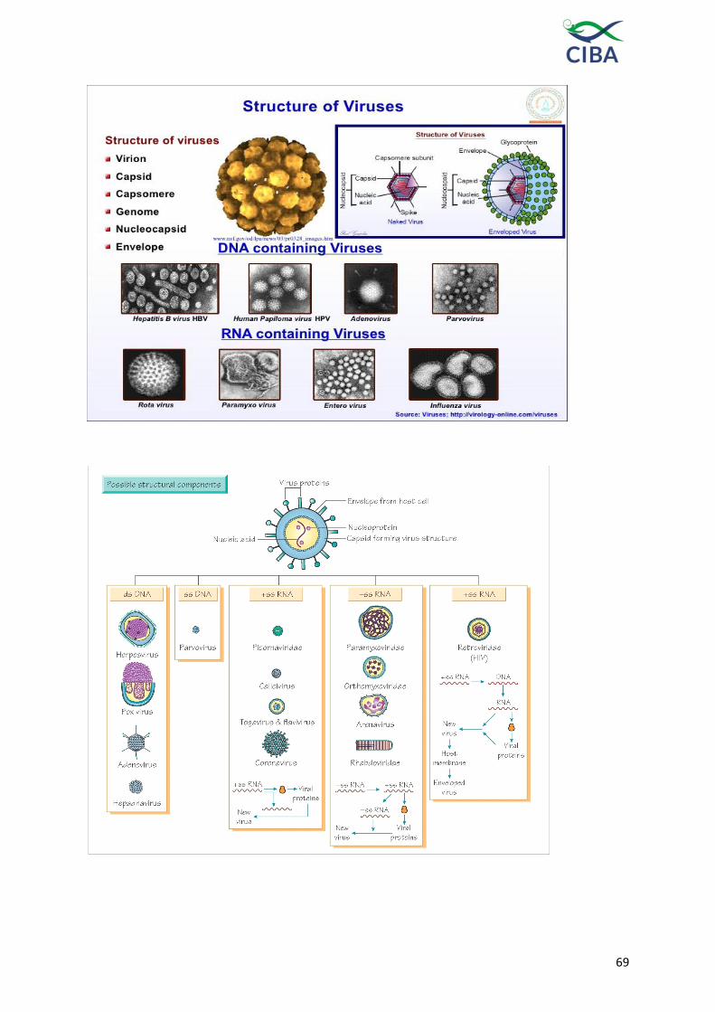
69

70
VIRUS REPLICATION
The following steps take place during viral
replication
• Adsorption - virus binds to the host cell.
• Penetration - virus injects its genome
into host cell.
• Viral Genome Replication - viral genome
replicates using the host's cellular
machinery.
• Assembly - viral components and
enzymes are produced and begin to
assemble.
• Maturation - viral components assemble
and viruses fully develop.
• Release - newly produced viruses are
expelled from the host cell
1. Adsorption
The virus becomes attached to the cells,
and at this stage, it can be recovered in the
infectious form without cell lysis by
procedures that either destroy the
receptors or weaken their bonds to the
virions. Viruses typically attach to cells via
specific cell surface receptors. Cells lacking
specific receptors are resistant. Many
different molecules are used as receptors
by different viruses, including cell surface
glycoproteins, components of the
extracellular matrix and receptors involved
in cell signalling and activation. A number
of viruses also use coreceptors to gain
entry to cells
2. Penetration
Penetration rapidly follows adsorption,
and the virus can no longer be recovered
from the intact cell. Following attachment,
the virus must traverse the lipid bilayer
surrounding the cell, without killing the
cell. Once inside the cell, the virus must the
disassemble itself in such a way that (1) its
genetic information and any associated
enzymes remain intact and (2) the viral
nucleic acid and associated enzymes are
directed to the appropriate cellular
compartment . The process of virus entry
can proceed via several pathways. Some
virus able to fuse directly with the host
cell membrane at neutral pH, via the
action of the viral fusion protein (F). This
results in uncoating of the viral genome
at the cell membrane.
Receptor-mediated endocytosis. Other
viruses do not fuse directly with the host
cell membrane, but in stead undergo
uptake into endocytotic vesicles.
Endosome fusion. In some cases, the
virus takes advantage of the natural
process of endosome acidification. the
HA (hemagglutininneuraminidase)
protein of influenza virus, which
undergoes a low-pH induced shape
change, such that the protein enters a
fusion-competent state. This allows the
virus to fuse with the host cell membrane,
bringing the virus particle into the cell.
Endosome lysis. Non-enveloped viruses
lack the lipid membranes found in
enveloped viruses. As a result, these
viruses cannot enter cells via a simple
process of membrane fusion between the
virus envelope and the host cell
membrane. One strategy employed by
non-enveloped viruses is virus attaches
immunoglobulin-like molecule, and it
then binds to cellular integrins. Following
binding, the integrin and virus are
internalized, and the virus then begins to
disassemble in the mildly acidic
environment of the early endosome (~pH
6). As the endosome acidifies further, is
thought to trigger endosome lysis,
thereby mediating the escape of the
partially disassembled core particle into
the cytosol. Pore formation. Some non-
enveloped viruses, such as
picornaviruses, form pores in the cell
membrane.

71
3. Uncoating
A key step in uncoating is the acidification
of the content of the endosome to a pH of
about 5. The low pH causes rearrangement
of coat components, which then expose
normally hidden hydrophobic sites. They
bind to the lipid bilayer of the membrane,
causing the extrusion of the viral core into
the cytosol.
4. Viral Nucleic Acid Replication
Virulent viruses, either DNA and RNA,
shut off cellular protein synthesis and
disaggregate cellular polyribosomes,
favouring a shift to viral synthesis.
DNA viruses replicate their genetic
material by one of three modes:
• Bidirectional replication from a circular
substrate. This process may proceed via a
‚theta-form‛ intermediate (eg,
papillomaviruses), or in some cases via a
‚rolling circle‛ mechanism that results in
the generation of concatemeric (head-
totail) viral genomes.
• Replication from a linear substrate. In this
case, synthesis of new DNA strands is not
simultaneous. Rather, it occurs
sequentially (ie, first one strand is made in
its entirety & then the next strand is
made). Examples include adenoviruses.
• Replication via an RNA intermediate.
Hepadnaviruses (hepatitis B virus) are
unique since they contain a partially
dsDNA genome that must be converted
into an RNA form by the virion enzyme
reverse transcriptase during the virus life
cycle
Replication of RNA Viruses. RNA viruses
have a variety of modes of replication.
Three important points about their
replication are as follows:
1. The viral RNA genome can act as its own
message (positive strand viruses) OR the
complementary strand can be the mRNA
(negative strand viruses).
2. All RNA viruses except retroviruses
encode an RNA-dependent RNA
polymerase. In the negative strand RNA
viruses this polymerase is part of the
virion, and it must enter the cytosol along
with the viral genome. This is necessary
in order for the virus to generate mRNAs
from its genome.
3. All RNA viruses replicate in the
cytoplasm except orthomyxoviruses
(influenza A & B), borna disease virus,
hepatitis delta virus and retrovirses.
Additional feature of orthomyxoviruses:
these viruses have segmented genomes
(influenza A has 8 separate strands of
genomic RNA)
4. Retroviruses are unique. These viruses
have a positive sense RNA genome
which must be converted into a dsDNA
form by the virion enzyme reverse
transcriptase (an RNA-dependent DNA
polymerase). This double-stranded DNA
is then integrated (at random sites) into
the host cell chromosome by the viral
integrase enzyme. Upon integration into
the host chromosome, the viral DNA can
then be transcribed by cellular RNA
polymerase II, to produce new genomic
RNA molecules.
Viral variation
Genetic variation is an essential feature of
all living organisms. It provides the
resource for natural selection and for the
progressive adaptation of the population
to a changing environment. Viruses face
continuous environmental change as they
pass from host to host. Evasion of the
host defences is a central feature of the
survival strategy of all viruses. However,
allelic variations in host genes present a
changing environmental landscape that
can determine susceptibility to infection
or efficiency of replication.

72
Viral variation can be generated by a
number of mechanisms. Major
rearrangements in genome structure and
genetic recombination, gene duplications,
gene exchanges and gene adoptions.
However, the most common form of
variation is mutation by nucleotide
substitution. This occurs as a consequence
of polymerase error in reading the
template during replication. As viruses
replicate rapidly and prodigiously, viral
variation has significant implications for
diagnosis and epidemiology.
Genetic variation in RNA and DNA
viruses
From a genetic perspective, viruses can be
classified as genome comprises RNA or
DNA. RNA viruses are inherently
hypervariable as RNA polymerases, which
replicate the viral genome, lack proof
reading and error editing functions
resulting rate of nucleotide
misincorporation in RNA viruses . Some
mutations are lethal. However, many
mutations result in viable genomes that
continue to replicate and contribute to the
virus population. In this way, RNA viruses
continually refine their genetic structure to
accommodate the changing environment.
Some RNA viruses may also undergo
genetic rearrangements that allow
exchange of corresponding genes or gene
segments during mixed infections . These
recombination and reassortment events
allow the most efficient and
environmentally adapted combinations of
genes to emerge from the available genetic
pool, increasing the potential for viral
survival.RNA viruses known to infect
farmed shrimp include Taura syndrome
virus (TSV), yellow head virus (YHV), gill-
associated virus (GAV), lymphoid organ
virus (LOV) and rhabdovirus of penaeid
shrimp (RPS). Each of these viruses is
likely to replicate with a high mutation
frequency. Some may also have a
capacity for genetic recombination.
In DNA viruses, the mutation rate is
usually far lower than in RNA viruses .
DNA polymerases, both cellular and
viral, do employ proof reading and repair
functions to reduce the intrinsic error
rate. However, some small DNA viruses
(eg. parvoviruses) appear to produce
factors that suppress the repair function,
generating an error rate similar to that of
RNA viruses. DNA viruses have also
been reported to produce sequence
duplications and insert host DNA
sequences into the viral genome.DNA
viruses known to infect farmed shrimp
include white spot syndrome virus
(WSSV), monodon baculovirus (MBV),
baculoviral midgut necrosis virus
(BMNV), infectious hypodermal and
haematopoeitic necrosis virus (IHHNV),
spawner mortality virus (SMV) and
hepatopancreatic parvovirus (HPV). A
recent study of WSSV DNA from sources
in different geographic locations has
suggested little sequence variation
between isolates.
Genetic variation in viral detection and
disease diagnosis
Observations of genetic variability in
viruses proclaim the need for care in the
use of molecular methods for disease
diagnosis. Mutations in the nucleotide
sequence can prevent binding of PCR
primers to target sequences, cause
primers to bind non-specifically to non-
target sequences, or prevent PCR
extension. Sequence insertions or
duplications can generate size variations
in the PCR product. In each case, the
result may appear falsely negative. At the
protein level, mutations and other
variations in sequence can affect the
binding of diagnostic reagents. Variations
can also cause closely related strains to

73
have significantly different biological
properties such as pathogenicity, tissue
tropism or host range. An understanding
of these factors is important for accurate
interpretation of data obtained for disease
diagnosis, epidemiolgical investigation or
screening for disease-free certification.
Virological Methods
The most commonly used laboratory
methods for the detection of viruses and
virus components in biological samples can
be– visualization of virus particles ,
measurement of virus infectivity, viral
serology and molecular methods.
Direct visualization of virus particles:
Visualization of virus particles requires the
use of the electron microscope (EM). The
EM accelerates electrons to high energy,
and focuses them using a magnet. The high
energy of the electrons gives them a short
wavelength, which is much smaller than
that of viruses. it is necessary to coat or
stain samples for EM analysis. This process
is called ‚shadowing‛ and it involves the
deposition of a thin layer of a heavy metal
such as osmium on the surface of the
sample to be analyzed. One particularly
useful aspect of EM analysis is that it
allows one to directly count the number of
virus particles within a sample – regardless
of whether or not those particles are
actually infectious. One problem of the
shadowing process is that it tends to result
in sample desiccation, and in some loss of
structural information – particularly for
enveloped viruses. As a result, the
procedure of cryoEM analysis was
developed. In this case, the virus sample is
rapidly frozen onto an EM grid in such a
way that it becomes embedded in a thin
sheet of vitreous ice (ice that does not
contain any large crystals). This process
retains some moisture in the virus, and it
results in enhanced preservation of viral
structures. The virus particles are then
visualized in the microscope at very low
temperature, to prevent thawing.
Measurement of Virus Infectious Units
To study and quantitate virus infectivity
in the laboratory, animal viruses must be
propagated in cells and cell lines. One
very widely used assay is the plaque
assay. This method is very similar to the
procedure used to titer bacteriophages,
and it relies upon (1) the use of confluent
monolayers of cells which are susceptible
to the virus, (2) the induction of a visible
cytopathic effect by the virus and (3) the
use of a semisolid overlay of agar which
prevents virus diffusion from one
infected cell to other nearby cells. As a
result, small round plaques (clear areas)
form in the cell monolayer as the virus
replicates. These plaques can be
visualized by staining the cells with a
vital dye like crystal violet or neutral red.
Endpoint dilution assay can be used to
determine titers of many viruses. In this
case, add serially diluted aliquots of virus
to cultured cells, which are typically
plated into multiwell plates. Allow the
cells to grow for fixed period of time,
after which the wells are scored for the
presence of viral replication. This is most
often done by analysis of virally induced
cytopathic effect. Endpoint dilution
assays provide a widely used basis for
the calculation of virus titers. Typically,
the 50% infectious dose (TCID50 or tissue
culture 50% infectious dose) is calculated
using a mathematical analysis of the data.
Popular methods include that of Reed
and Munch.
Reed-Münch formula I = (% of w ells infected at dilution above 50% - 50%)
(% of wells infected at dilution above 50% - % of wells infected at dilution below)
50% endpoint titer = 10 log total dilution above 50% - (I x log h)
I = Interpolated value of the 50% endpoint h = dilution factor

74
Table 1: Classification of known shrimp virus
Name Approximate
Virion size
Nucleic
Acid
Probable
Classification Reference
White spot syndrome virus (WSSV) 80-330 nm dsDNA Whispovirus Wongteerasupayaet al,
1995
Infectious hypdermal and hematopoietic
necrosis virus (IHHNV)
22 nm ssDNA Parvovirus Lightneret al, 1983
Monodon baculovirus (MBV) 75X300 nm dsDNA Baculovirus Lightneret al, 1983
Hepatopancreatic parvovirus (HPV) 22-24 nm ssDNA Parvovorus Lightner and Redman,
1985
BaculovirusPenaei (BP) 50-75X300 nm dsDNA Baculovirus Couch, 1974
Baculoviralmidgut gland necrosis (BMN) 75X300 nm dsDNA Baculovirus Sano et al, 1981
Yellow head virus (YHV) 44X173nm ssRNA Rhabdovirus Flegelet al, 1995
Taura syndrome virus (TSV) 30-32nm ssRNA Picornavirus Lightneret al, 1995
Infectious myonecrosis virus (IMNV) 40 nm dsRNA Totivirus Poulos et al., 2006
Laem-Singh virus (LSNV) 30nm ssRNA Luteovirus-like
(unclassified)
Srisala et al., 2010
covert mortality nodavirus (CMNV), 32 nm ssRNA Noda virus Zhang et al., 2014
Reo like virus 70nm dsRNA Reo-like virus Tsing and Bonami, 1987
Lymphoid organ vacuolization virus
(LOVV)
30XSSnm ssRNA Toga-like virus Bonamiet al, 1992
Rhabdovirus of penaeid shrimp (RPS) 75X125nm ssRNA Rhabdovirus Nadalaet al, 1992
75
Viral diseases and their management with special reference
to white spot disease and Indian shrimp farming
Introduction
Aquaculture is a rapidly expanding
industry, which augments cheap protein
to a large portion of world population. In
terms of value, shrimp is considered as
the single largest seafood commodity
because of its high export value. More
than 75% of shrimp production is in the
form of aquaculture and several
modifications are being constantly carried
out to increase the production. However,
adoption of high stocking density,
increase use of inputs and lack of proper
biosecurity has resulted in the outbreak of
diseases. All most all kinds of
microorganisms have been foundto be
capable of producing some forms of
disease to shrimp. However, the
emergence of viral diseases since 1981 has
made the situation more complicated and
the industry is constantly facing huge loss
due to mortality and crop failure. So far
more than 20 viruses have been reported
from shrimp and new viruses are
frequently being added up to the existing
list. Amongst all the viruses, white sot
syndrome virus (WSSV) many times acts
as a single most causative agent
threatening the sustainability of shrimp
aquacultureindustry.
White spot syndrome
virus(WSSV)
WSSV is a rapidly replicating and highly
virulent shrimp virus that has wide
spread presence throughout the world.
Originating from Taiwan in 1992, it
spread to Japan during 1993 and
subsequently very quickly to all other
Asian countries. By 1995, it had already
spread to North America and further by
1999 to South America. The quick spread
of this virus to different regions and
simultaneous investigation by different
scientists speculated the same agent to be
different ones and thereby called them in
different names such as Systemic
Ectodermal and Mesodermal Baculovirus
(SEMBV), rod shaped nuclear virus of
Penaeus Japonicus (RV-Pj), Hypodermal
and Haematopoietic Necrosis Baculovirus
(HHNBV), third Penaeus monodon non-
occluded virus (PmNOB III), penaeid rod
shaped DNA virus or white spot
baculovirus. On subsequent investigation
and data analysis when it was known that
all these names are for the same agent, it
was unanimously called as white spot
syndrome virus. Other than the penaeid
shrimps which serve as host for this virus,
a large number of other crustaceans serve
as carrier and therefore it has been
impossible to eradicate this virus from the
culture system. The typical clinical
symptoms of WSSV infection are the
formation of circular whites spotson the
carapace. However, there can be many
other reasons for this and therefore the
infectionshouldgetconfirmedbyvariousdi
agnosticprotocols.

76
Fig. 1: Typical (circular white spot on the carapace) clinical sign of WSSV (A); The
detection of the virus by PCR (B); Histopathology (C) and in situ hybridization
(D)
Prevention andcontrol
Virus exclusion and preventing the
entry
Geneticselection
Wherever possible, specific pathogen free
(SPF) and specific pathogen resistant (SPR)
animals should be stocked to avoid the
disease occurrence. After determining the
disease resistance markers, animals can be
selected for the production
oftotallydiseaseresistanceprogenies.Wherec
ompleteresistancestrainproductionis not
possible, care can be taken to produce at
least pathogen free stocks as has been tried
for Penaeus vannamei. As in this case the
seed will be free from major disease
causing agents such as WSSV and
subsequentbettermanagement practices can
be followed to get a successfulharvest.
Screening oflarvae
Stress test should be carried out to choose
the healthy larvae. Further, the larvae
should be screened by PCR for the
presence of virus. Early, accurate and
sensitive detection of pathogens are
important and at least a nested PCR
protocol should be carried out to rule out
the presence of WSSV inlarvae.
Better management practices (BMPs)
Once the larvae are shifted from the
hatchery to the pond, it is subjected to
enormous stress. Stress is the main factor to
initiate disease and this can be overcome to
a large extent through BMP. Soil and water
qualities are theimportant aspects for
health management. Ponds should be
prepared adequately to ensure it as
pathogen free. Sufficient gap should be
there between the culture cycles to
adequately prepare the ponds. WSSV can
remain viable and infectious for
considerabletimeperiodintheponds.Therefo
re,pondsshouldbedriedsufficiently
beforetherealpreparation.
Biosecurity
As has been already mentioned, WSSV is
a highly virulent virus and has a wide host
range. Utmost care should therefore be
taken to prevent the spread of the causative
agents. Source water, seed, equipment,
workers and invasive organisms are the
main source of virus spread. Step by step
protocols and precautions should be taken
from the beginning to avoid the entry of
the pathogens to culture system.If strict
biosecurity measures are adopted, disease
can be avoided to a large extent. Adoption
of proper sanitation protocols,
development of a reservoir pond, bird
fencing and crab fencing are some of the
common biosecurity measures to avoidthe
spread ofWSSV.
Development of recirculatory/zero
water exchange culture system
Water can be an important source of
infection as far as WSSV is considered.
Care should be taken to avoid the virus
from the intake water. Recirculatory/zero

77
water exchange will ensure the prevention
of pathogen entry into the system.
Culturing the virus free larvae and
adopting better management practice, it
will then possible to prevent the entry of
WSSV into the system.
Prevention of the spread
Once a disease outbreak occurs, it
becomes a rich source of pathogen to
contaminate the nearby brackishwater
fishery resources. Therefore, it is necessary
to confine the infected pond and do
immediate treatment to ensure complete
pathogen killing. Sufficient biosecurity and
emergency planning should be in hand to
prevent the spread through carrier aquatic
organisms orbirds.
Boostingtheimmunityandincreasing
diseaseresistancecapacity
Immunostimulants
Non-specific immune system of shrimp is
the primary defence against a wide range
of pathogens. This system can be
stimulated through various microbial and
plant based products to provide better
protection. Lipopolysaccharide (LPS) from
gram negative bacteria and
peptidoglycan/beta glucan from gram
positive bacteria / yeast are widely used
immunostimulants. Different plant based
products with proven medicinal properties
have been successfully used as
immunostimulants to provide partial or
full protection. In addition to providing
protection against
diseases,theseimmunostimulantshavealsob
eenfoundusefultoprovidebetter growth.
Because of the lack of memory in shrimp, it
is necessary to apply these
immunostimulants very frequently during
a culture period for successful protection.
Similarly, the shrimp larvae can also be
treated through immersion or
feedtohavebetterprotectionduringtheinitial
criticalperiodofculture.
Adoption of Biofloctechnology
This recently developed eco-friendly
technology where beneficial
microorganisms are multiplied in an
ecosystem through the manipulation of
carbon and nitrogen ratio has been found
to be useful in boosting the immune system
and thereby increasing the disease
resistance capacity. Similar to probiotics,
the microbes produced here maintain the
water quality and at the same time
stimulate the immune system of shrimp
when taken as feed. Through this
technology, the wastes generated are
efficiently utilized and uses of many
additional inputs are also avoided.
Therefore, this technology has several
added advantages to avoid disease
problems, particularly from that ofWSSV.
Use ofprobiotics
Probiotics are a group of ‟good bacteria‟,
which are proved to improve the host
immune system and thereby provide good
health when consumed. These bacteria go
and colonize in the gut. In this way they
occupy the space and do not allow the
pathogenic bacteria to settle down. They
also produce specific molecules which has
ability to stimulate the host immune
system. Based on its application, probiotics
in aquatic system can be of two types. Gut
probiotics does the usual function in
replacing the pathogenic bacteria and
stimulating the immune system. Whereas
the water probiotics helps in increasing the
diversity of good bacteria in water and
thereby do not allow the multiplication of
pathogenic bacteria. These bacteria are also
known to secret extracellular products that
has inhibitory effect against
harmfulbacteria.
A number of probiotics products,
consisting of several bacteria species such
as Lactobacillus spp., Bacillus spp.,
Pseudomonas spp., etc either as single

78
species or as consortium, are available for
the use both in fish and shellfish culture.
Experimental evidences regarding
precipitation of WSSV by secondary
bacterial infection has been generated.
Therefore, during this period, application
of probiotics will prevent the
multiplication of pathogenic bacteria and
thereby prevent the quick precipitation
ofdisease.
Controlmeasures
Preventingtheprecipitation
There are evidences where farms can
continue with WSSV and have successful
harvest.Thisismainlybypreventingthepreci
pitation.Theviruscanremainina
latent/dormant stage and cannot cause
disease. This is mainly through good water
quality maintenance, BMP and other
precautionarymeasures.
RNA interference(RNAi)
Experimentally, this technology has been
proved to be very useful for the control of
several pathogens including WSSV. It has
been particularly very effective for the
control of many viral diseases where
treatments through medicines are not
possible. Specific virulence genes of the
pathogens are targeted to develop short
RNA fragments (either single or double
stranded) and this is either injected or
supplied through oral route after
modification. This brings degradation of
the pathogen through post translational
modification. The RNAi system has been
found to be functional in shrimp. This
method is particularly look promising for
the treatment of viral diseases of shrimp
where neither any treatment methods nor
any vaccines are available. Several
experiments have been carried out
targeting some of the common virulent
genes of WSSV such as VP28, VP19, VP15,
rr1, rr2 etc. Unfortunately, many of these
protections have been
demonstratedthrough injection which is
not a practical method during culture
practice. However, the injection method to
produce viral fee larvae will be helpful for
some of the penaeid shrimps like Penaeus
monodon where development of SPF
stocks has not been possible.
Developmentofanti-WSSVmedicine
A lot of medicinal plants have shown to
have antiviral molecules and have been
effectively used against a wide range of
animal and human viral pathogens. Such
types of screening from plants for the
development of anti-WSSV molecules
should also be tried seriously. Similarly
many of the marine macro and
microorganisms are also a good source of
antiviral molecules and effort should be
there for their screening. The newly
developed concept, ‚synthetic biology‛
should also be applied
tosyntheticallydevelopdifferentmoleculesal
readyproventohaveanti-viraleffect.
Conclusion
WSSV is a highly virulent pathogen that
can bring mass mortality within a few days
of time period. It can be a single most
important factor in threatening the
sustainability of shrimp aquaculture
practice. Though, it has been more than
two decades after its emergence, no
solution to this problem has been possible
to develop. In this context, general
management practices to prevent the
entrythrough viral exclusion will be a
better mechanism. However, further
research
shouldbecarriedouttodevelopaneffectiveme
dicineforcontrolmeasure.

79
Update on Acute hepatopancreatic necrosis disease (AHPND)
An emerging disease known as early
mortality syndrome (EMS) or acute
hepatopancreatic necrosis syndrome
(AHPNS) has been recently causing
significant losses in shrimp farms in
China, Vietnam, Malaysia and Thailand
since 2009. The disease affects both black
tiger shrimp Penaeus monodon and Pacific
white shrimp, P. vannamei and is
characterised by mass mortalities during
the first 20-30 days of stocking.
Considering the severity of the disease, the
Network of Aquaculture Centres in Asia-
Pacific (NACA) jointly organized an
emergency regional consultation on
EMS/AHPNS with the Australian
Department of Agriculture, Fisheries and
Forestry (DAFF), Australia, in Bangkok,
on 9-10 August 2012, to share information
on this emerging disease, its occurrence,
pathology and diagnosis, and to develop a
coordinated regional response where
international shrimp health experts,
regional governments and industry
personnel participated.
History of EMS / AHPNS and its impact
The EMS / AHPNS has been so far
reported from Vietnam, China, Malaysia
and Thailand since 2009. In Vietnam, the
disease was observed since 2010, but the
most widespread devastation due to EMS
was reported since March 2011 in the
Mekong Delta (South Vietnam). EMS
affected the main shrimp production areas
of Tien Gang, Ben Tre, Kien Giang, Soc
Trang, Bac Lieu and Ca Mau provinces with
a total shrimp pond area of around 98,000
hectares. In June 2011, unprecedented losses
in P.monodon were reported in 11,000 ha of
shrimp farms in Bac Lieu, 6,200 ha in Tra
Vinh (causing a lossof over VND12 billion),
and 20,000 ha in Soc Trang (causing VND1.5
trillion in losses) (Mooney, 2012). In China,
the occurrence of EMS in 2009 was initially
ignored by most farmers. But in 2011,
outbreaks became more serious especially in
farms with culture history of more than five
years and those closer to the sea using high
saline water (Panakorn, 2012). Shrimp
farming in Hainan, Guangdong, Fujian and
Guangxi suffered during the first half of
2011 with almost 80% losses. In Malaysia,
EMS was first reported in mid-2010 in the
east coast of peninsular states of Pahang and
Johor. The outbreaks of EMS resulted in
significant drop in P. vannamei production,
from 70,000 mt in 2010 to 40,000 mt in 2011.
Production for 2012 (up to May) was only
30,000 mt and worse was expected to come
as unconfirmed reports on EMS outbreaks in
the states of Sabah and Sarawak came in
April 2012. In Thailand, so far 0.7% total
shrimp ponds were reported to be affected
by EMS, in Rayong, Chantaburi, Trat,
Chacheongsao provinces located along the
eastern Gulf.
Investigations on EMS/AHPNS and their
findings
Pathology of EMS / AHPNS has been
explicitly described by Dr. DV Lightner of
University of Arizona, in both P. monodon
and P. vannamei and he informed that the
pathology appeared to be limited to the
hepatopancreas (HP). He described EMS /
AHPNS as idiopathic since no specific
disease causing agent (infectious or toxic)
was so far found to be associated. The
clinical signs such as significant atrophy of
the HP, which may be often pale to white
due to pigment loss, sometimes with visible
black spots or streaks, which does not
squash easily between the thumb and
forefinger could be used for presumptive

80
diagnosis in cases of shrimp mortality
starting as early as 10 days post-stocking.
Progressive degeneration and dysfunction of
the HP tubule epithelial cells progressing
from proximal to distal ends of HP tubules
and its degenerative pathology suggested
of a toxic etiology. However, he also
mentioned that the anecdotal information
suggested that disease spread patterns
may be consistent with an infectious
agent. According to Dr. Tim Flegel of
CENTEX shrimp, in the EMS affected
shrimp, in addition to various well studied
pathogens such as WSSV, YHV and
vibrios that are commonly linked to EMS,
they also found certain groups of bacteria
using metagenomics tools. While the role
of certain crustaceacides such as
cypermethrin was ruled out, the disease
transmission trials are still inconclusive.
The potential pathogens are integral
components of all ecosystems and disease
emergence and subsequent spread often
resulted from some disturbance in the
ecology, which can upset the natural
balance resulting in a normally innocuous
organism emerging as a new disease
agent. The current aquaculture practices
are artificial and un-natural high density
culture activities and promote emergence
of pathogens (Dr. Peter Walker, CSIRO).
Considering the need for identifying the
etiology of EMS / AHPNS of shrimp, a
number of new molecular methods such
as sequence-assisted and sequence-
independent virus discovery could be
applied to discover viruses or other
pathogens associated with AHPNS (Dr Jeff
Cowley, CSIRO).
The epidemiology and risk factors
involved in EMS / AHPNS require
systematic studies. Until epidemiological
approaches are applied systematically to
include hatchery, transport, pond, farm
and location specific data, it will be very
difficult to pinpoint and prioritize risk
factors for AHPNS (Dr Flavio Corsin and
Dr Matthew Briggs). However, the
potential risk factors included most of the
generic factors such as high stocking
densities, older farms closer to the sea
using higher salinity water, farms not
employing reservoirs, farms overusing
chemicals, inadequate aeration, and
presence of toxic levels of H2S etc.
Way forward
While the role of cypermethrin was
ruled out, involvement of biotic and
abiotic factors and toxins was required to
be investigated. An in depth analysis is
required to identify the etiology that
should unravel cryptic pathogens using
molecular tools and pyro-sequencing.
Robust challenge studies to prove Koch’s
and Rivers’ postulates would have to be
repeated using fresh tissues using
appropriate challenge protocols by oral,
reverse gavage, cohabitation, etc.
With regard to surveillance, reporting,
disease prevention and control, quality
data / information on suspect parameters
such as pH, H2S, use of probiotics etc to
understand potential risk factors is
essential. Information has to be provided
to farmers on how the disease appears,
what samples to be collected, mode of
sample collection etc for lab investigations.
It is necessary to provide training to the
farmers on how to detect the disease and
field officers on sampling.
Biosecurity, emergency response and
disease management could be
immediately addressed by imposing
restrictions on movement of live affected
animals to areas free from EMS and that
buffer zones be created and monitored.
Areas that are free from EMS should take

81
precautions for stocking. It is also
important to take utmost care in
processing produce from affected EMS
areas. Affected ponds must be treated
before release of pond water into wild.
Information on EMS in broodstock and PL,
and wild organisms as carriers has to be
generated.
Summary
For the purpose of detection and
surveillance of EMS / AHPNS, the definition
proposed by Prof Don Lightner, which relies
mainly on histopathology of hepatopancreas
may be employed along with the clinical
signs. It is important that histological
examination be carried out to confirm that
suspected occurrences that fit the AHPNS.
Identifying the primary cause of EMS /
AHPNS is necessary and can be addressed
by robust scientific programme. Until this
information becomes available, it is essential
to increase disease awareness among the
shrimp farmers. Considering the great
economic loss that EMS is likely to cause in
the region’s shrimp aquaculture, ways of
preventing the spread and/or occurrence of
this disease should be formulated by
concerned experts, officials and other
regulatory bodies. Farmers, on the other
hand, should also cooperate with the
concerned agencies by promptly reporting
any suspected mortalities among cultured
shrimp that appear to be similar to the
clinical description of EMS / AHPNS.
Further, it is necessary to impose restrictions
on movement of live affected animals to
areas free from EMS either for culture or for
processing purposes, and movement of live
shrimp may be undertaken only after
conducting robust import risk analyses.

82
Diseases in shrimp aquaculture-Current scenario in India:
white faeces/gut syndrome (WFS), growth variation /
retardation, white muscle syndrome, running mortality
syndrome (RMS) and Hepatopancreatic microsporidiosis
Aquaculture continues to be the
fastest growing food production sector
with an annual average growth rate
exceeding 6 % (FAO, 2014) and having
enough potential to meet the growing
demands for aquatic food. Aquaculture
delivers not only economic income and
high quality food products, but also
employment to both skilled and
unskilled workers. Over the last three
decades, shrimp farming has been one
of the most rapidly growing aquaculture
sectors throughout the world. Shrimp
continues to be the largest single
seafood commodity by value,
accounting for 15% of all internationally
traded fishery products. Farm raised
shrimp is comprised of 55 % of global
shrimp production and this is entirely
dominated by two species – the black
tiger shrimp (Penaeus monodon) and the
white Pacific white shrimp (Penaeus
vannamei) (FAO 2014). In India, shrimp
aquaculture started as a traditional
practice in natural water bodies such as
bheries or pokkali fields and
subsequently transformed to
commercial industry during 90‟s.
Initially it was dominated by a single
species, Penaeus monodon and the
production of which reached a
maximum of 1.44 lakh tons in 2006-07
(www.mpeda.com). Due to disease
outbreak and other social issues, the
farmers felt it difficult to continue
further with this species and thereafter
an exotic species; the Pacific white
shrimp
P. vannamei was introduced into the
brackishwater aquaculture system of
India. Because of its SPF status, fast
growth rate and culture feasibility in
wide salinity range, this got readily
accepted by the farmers and
subsequently became the dominant
culturedspecies.
The increasing trend in
intensification and commercialization
has exacerbated the epidemics of
diseases and become a major constraint
for the sustainability of this industry.
Severe disease related mortality and
thereby economic loss due to different
viral agents such as monodon
baculovirus (MBV) in Taiwan, infectious
hypodermal and hematopoietic necrosis
virus (IHHNV) in the Americas, yellow
head virus (YHV) in Thailand and Taura
syndrome virus (TSV) in the Americas
during the different periods have been
reported. In addition to all these, the
major disease outbreak due to White
Spot Syndrome Virus (WSSV) has also
been reported from all parts of the
world. During the recent past, the havoc
createdbyEarlyMortalitySyndrome(EMS
)indifferentSouthEastAsiancountries has
also been found to have severe
economic impact on shrimp industry of
these regions. Further, a number of
other diseases by unidentified etiologies
are constantly being associated with
shrimp culture practice which has been
responsible either for direct mortality or
growth reduction and thereby bringing
loss to farmers. Shrimp hatcheries are

83
also prone to losses either due to disease
outbreak or other unknownfactors.
Some of the problems associated with
current shrimp culture practice in India
are discussed below. In addition to the
already existing WSSV problems, the
industry at present is going through a
very critical phase due to several
uncharacterizedfactors.
Currently prevailing diseases in
Grow-outsystems
White faeces syndrome(WFS)
White faeces syndrome reported
since last decade, has recently been
noted as
seriousproblemforP.vannameithroughou
ttheworld.However,thisdiseasehas been
reported from both cultured black tiger
shrimp and pacific white shrimp. White
faeces syndrome usually occurs after 60
days of culture (DOC) and it may be
accompanied by high shrimp mortality.
Ponds affected with white faeces
syndrome show white faecal strings
floating on the pond surface while the
shrimps show white/golden
brownintestine, reduced feed
consumption, growth retardation and
often associated with loose shell. The
disease can cause moderate to severe
economic loss by reducingthe shrimp
survival by 20–30 percent when
compared to normal ponds. While
investigating the aetiology of WFS this
disease has been associated with
presenceof vermiform like gregarine
bodies, vibriosis, Enterocytozoan
hepatopenaei, bluegreen algae and loose
shell syndrome. Sriurairatana et al
(2014) revealed that 96% of the
pondsexhibitingWFSpresentedvermifor
mbodiesresemblinggregarines.Whenthe
contents of the gut or faecal strings were
examined in squash mounts with the
light microscope, they consisted of
masses of vermiform bodies that
superficially resembled gregarines.
Bacteriological results showed that total
bacteria and Vibrio spp. found in
haemolymph and intestine were
significantly higher in diseased shrimp
than in healthy shrimp. Six species of
fungi(Aspergillusflavus,A. ochraceus, A.
japonicus, Penicillium spp., Fusarium spp.,
and Cladosporium cladosporioides) were
isolated from shrimp naturally infected
with white faeces syndrome.
Histopathological examination revealed
diffused haemocyte encapsulation and
dilated hepatopancreatic tubules
accompanied by necrosis.
Tangprasittipap et al., 2013 revealed that
the microsporidian newly found in P.
vannamei is nonspecific with previously
described E. hepatopenaei and it is not
causally associated with WFS.
Sriurairatana et al. (2014) concluded
with Transmission electron microscopy
(TEM) study that vermiform structures
superficially resembling gregarines and
commonly found now in the HP of
Asian cultivated shrimp are not
independent organisms but result from
the transformation, sloughing and
aggregation of microvilli from the HP
tubule epithelial cells themselves and
the denuded epithelial cells
subsequently undergo lysis, can lead to
the phenomenon called white faeces
syndrome (WFS) and transformed
microvilli (ATM) in very severe cases
they may retard shrimp growth and
may predispose shrimp to opportunistic
pathogens. Furthermore it has been
estimated that the Thai production
losses due to WFS in 2010 were 10–15%.
The cause of white faeces syndrome and
treatment is uncertain. However
reduced
stockingdensity,properwaterexchangeto
getherwithbettermanagementpractices

84
willbehelpfulinevadingWhiteFaecessyn
drome(WFS).
Running Mortality Syndrome(RMS)
Since 2011, a new syndrome has
brutally affected the shrimp industry
and causing substantial mortality. The
disease has been loosely termed as
Running Mortality Syndrome (RMS) by
the farming community. The affected
ponds show different mortality patterns
with unusual symptoms, no relation to
any known diseases anda slow mortality
rate (e.g. <1%/day), but the cumulative
loss over phase will be high. Some
farmers have lost up to four crops, with
mortality percentage reaching 70% in
most of the cases. Generally mortalities
start after a month or 40 days of culture
(DOC) but a portion of shrimp continue
to survive and can grow to fully
harvestable size. Investigations
(conducted in ICAR-CIBA) revealed no
associationof Running Mortality
Syndrome (RMS) with known shrimp
viral infection. Further, bacteriological
examination of haemolymph samples of
RMS affected shrimp indicated
predominance of Vibrio spp., such as
Vibrio parahaemolyticus and Vibrio
azureus. The population of anaerobic
bacteria in the gut of RMS affected
shrimp ranged from 72 - 252 x 1014 cfu
mL-1 and were identified based on16S
rRNA gene analysis as Enterococcus
faecium, E hirae, and Lactobacillus
plantarum. Bacterial diversity of RMS
affected shrimp gut examined by
Denaturing Gradient Gel
Electrophoresis (DGGE) revealed a
number of uncultured bacterial
sequences. Histopathological
examination of the hepatopancreas was
largely normal. However, some samples
showed karyomegaly and increased
inter hepatopancreatic tubular space
with haemolymph infiltration, muscle
necrosis, loosened LO tubule cells and
constricted lumen. Bioassay experiments
carried out by feeding RMS affected
shrimp tissue to healthy 13-14 g shrimp
did not elicit any disease in the
experimental shrimp. All the
experimental animals were healthy and
active even after 44 hrs of feeding RMS
affected shrimp tissue like that of
control animals. RMS affected shrimp
showed recovery and appeared healthy
and active after 155 hrs of transferring to
wet lab in water with optimal
parameters. Co-habitation experiment
with healthy shrimp and the infected
animals also failed to induce RMS. All
shrimp appeared healthy and active.
Relatively few studies done on Running
Mortality syndrome, and still the
causative agents or aetiology of RMS
areunknown.
Size variation/ Growthretardation
More recently shrimp farmers have
been reported several cases of size
variation / growth retardation in P.
vannamei grow-out cultures. It is
reported that viruses,viz., infectious
hypodermal and haematopoietic
necrosis virus (IHHNV), lymphoid
organ vacuolization virus (LOVV),
monodon baculovirus (MBV),
hepatopancreatic
parvovirus(HPV)andLaem-
Singhvirus(LSNV)areassociatedwithslo
wgrowthand size variation in shrimp. In
India, Madhavi et al. (2002) recorded

85
multiple viral infections in shrimp with
stunted growth. Rai et al. (2009)
observed IHHNV, MBV and HPV
associated with slow growth shrimp and
stated that IHHNV could be oneof the
causes of slow growth in cultured P.
monodon. In the event of white faeces
syndrome affected animals there is a
decrease in feed consumption and
growth rates were reduced as revealed
by average daily weight gain (ADG) for
the whole crop operation of less than 0.1
g/day compared to 0.2 g/day in normal
ponds. Feed conversion ratios (FCR)
ranged from 1.7 to 2.5 when compared
to 1.5 or less for normal ponds
(Sriurairatana et al, 2014). Recently
Enterocytozoan hepatopenaei found to be
associated with size variation/growth
retardation (Tangprasittipap et al.,2013).
White gutdisease
This disease of P. vannamei is mostly
caused by different species of Vibrio and
is very much prevalent in Andhra
Pradesh and Tamilnadu. Vibrios are
normally present in water bodies. But
sometimes stressed environmental
factors such as sudden change of
environment and salinity, low DO,
mechanical injury, higher stocking
density, etc. caused rapid multiplication
of this organism in the gut and
hepatopancreas. Vibriosis caused red
discolouration and melanization of
appendages (red disease), necrosis of
tail, broken antennae, etc. Six species of
Vibrio viz. V. harveyi, V. parahaemolyticus,
V. alginolyticus, V. anguillarum, V.
vulnificus and V. splendidus are generally
associated with the diseased shrimp.
The diagnosis of the disease can be done
by isolation of organism by plating
haemolymph on the TCBS agar followed
by identification of the species of Vibrio
by biochemical tests or 16S rRNA
genesequencing.
Hepatopancreatic microsporidiosis
Hepatopancreatic microsporidiosis
(HPM) is caused by Enterocytozoon
hepatopenaei (EHP). It was first
reported as an unnamed
microsporidian from growth
retarded giant or black tiger shrimp
Penaeus monodon from Thailand in
2004. It was subsequently
characterized in detail and named in
2009. It also has much smaller spores
(approximately 1 μm in length) and
is currently known to infect both P.
monodon and P. vannamei.It has been
found that EHP can be transmitted
directly from shrimp to shrimp by
cannibalism and cohabitation. E.
hepatopenaei (EHP) is confined to
tubule epithelial cells of the shrimp
HP and shows no gross signs of
disease except retarded growth. It is
likely that other penaeid shrimp
and/or other crustaceans or even
other marine or brackish water
species in the region may also be
susceptible to infection. For example,
some samples of polychaetes and
mollusks have tested positive by
PCR, but it is still not known
whether they are infected or passive
carriers. More recently, samples of
frozen Artemia mass has been
reported to test positive for EHP by
PCR , but again, it is not known
whether Artemia is susceptible to
EHP infection or just a mechanical
carrier. It is urgent that these
possibilities be explored in order to
improve control measures.Although
EHP does not appear to cause
mortality in P. monodon and P.
86
vannamei, information from shrimp
farmers indicates that it is associated
with severe growth retardation in P.
vannamei . Thus, farmers and
hatchery operators monitor P.
vannamei and P. mondon for EHP in
broodstock, PL and rearing ponds.
The best approach for maturation
and hatchery facilities to avoid EHP
is not to use wild, captured, live
animals (e.g., live polychaetes, clams,
oysters, etc.) as feeds for broodstock.
Better would be pasteurization
(heating at 70oC for 10 minutes) .
Another alternative would be to use
gamma irradiation with frozen feeds.
Alternatively, polychaetes could be
selected and tested for freedom from
shrimp pathogens and then reared as
broodstock feed in biosecure settings
designed to maintain their freedom
from shrimp pathogens (i.e. SPF
polychaetes).
Conclusion
Aquaculture is now integral to the
economies of many countries. Growing
demand for seafood and limitations on
production from capture fisheries will
inevitably lead to the increased
intensification in commercialization of
shrimp aquaculture. This in turn
increases the number of diseases and
leads to emergence of new diseases. The
emergence and spread of infectious
disease is usually the result of a series of
linked events involving the interactions
between the host (including the
physiological, reproductive and
developmental stage conditions), the
environment and the presence of
pathogens. Focusing efforts on
producing high quality seed, better
pond manage to reduce stress and risk
of infection, following routine farm
biosecurity, responsible trade practices,
response to disease outbreak, and
improved better management practices
shall aid in preventing the epidemics of
diseases. Further health management is
a shared responsibility, and each
stakeholder‟s contribution is essential to
the health managementprocess.

87
Shrimp Diseases: An overview
Recent past years advances in intensive
aquaculture have brought parallel acute
concern for problems associated with
diseases both by sudden catastrophic
epizootics and slow continuous attrition
which leads to great economic loss.
Generally infectious diseases of shrimp and
other aquatic animals are caused by virus,
bacteria, fungi and parasites, and other
diseases and abnormalities due to
environmental stresses, genetic factors and
nutritional deficiencies. Risk of diseases is
inherent with transboundary movement of
live aquaculture species and translocation of
alien species poses potential risk of spread of
pathogens that may lead to disease
outbreaks that might associate with
significant production and economic losses.
Indian government has recently permitted L.
vannamei for culture in India, with CAA
approval by allowing importation of Specific
Pathogen Free (SPF) broodstock from other
countries after undergoing required
quarantine of such consignments. SPF
shrimp are expected to be free from the OIE
listed pathogens that are of economic
importance in shrimp aquaculture.
However, SPF status cannot guarantee the
disease free status after importation. In this
context awareness on the diseases and
preparedness for health management are
being discussed.
WHITE SPOT DISEASE (WSD)
Described elsewhere in this manual
YELLOW HEAD DISEASE (YHD)
Yellow head disease was the first major
viral disease that caused extensive losses to
shrimp farms in Thailand during 1990-91.
YHD outbreaks have been reported in the
black tiger prawn and the white Pacific
shrimp. YHD has been reported in China,
Taipei, India, Indonesia, Malaysia, the
Philippines, Sri Lanka, Thailand and
Vietnam. Outbreaks of YHD with heavy
mortalities have been reported in farmed
black tiger shrimp and pacific white shrimp.
It is reported to be highly prevalent (>50%)
sampled farmed and wild populations in
Australia, Asia, East Africa and
Mexico.YHD is caused by yellow head virus
(YHV), and its close relatives such as gill-
associated virus (GAV). YHV is rod-shaped
enveloped viruses of 40-60 nm by 150-200
nm size, containing single stranded RNA.
YHV affects tissues of ectodermal and
mesodermal origin including lymphoid
organ, haemocytes, haematopoietic tissue,
gill lamellae and spongy connective tissue of
the sub-cutis, gut, antennal gland, gonads,
nerve tracts and ganglia. YHV principally
affects pond reared juvenile stages of 5-15 g.
Affected shrimp typically feed voraciously
for two to three days and then stop feeding
abruptly and are seen swimming near the
periphery of the pond. YHV infections can
cause swollen and light yellow colored
hepatopancreas in infected shrimp, and a
general pale appearance, before dying
within a few hours. YHD can cause up to
100% mortality in infected P. monodon ponds
within 3-5 days of the first appearance of
clinical signs. GAV has been reported to be
associated with mortalities of up to 80% in P.
monodon ponds in Australia.Yellow head
virus can be detected by RT-PCR or with a
probe designed for dot-blot and in situ
hybridization tests. It can also be diagnosed
histologically in moribund shrimp by the
presence of intensely basophilic inclusions,
most easily in H&E stained sections of
stomach or gill tissue, or simply by rapid
fixation and staining of gill tissue and
microscopic examination. The primary
mechanism of spread of YHV in pond

88
culture appears to be through water and
mechanical means. Infected broodstock can
pass on the virus to larvae in the
maturation/hatchery facilities if thorough
disinfection protocols are not strictly
adhered to. Methods of YHV eradication in
ponds are much the same as for other
viruses and involve BMPs that include pond
preparation by disinfection and elimination
of carriers and production of virus free
broodstock and PL for pond stocking.
TAURA SYNDROME (TS)
Taura Syndrome was first identified from
farms around the Taura River in Ecuador in
1992 and the disease spread rapidly to the
whole of Latin and North America within
three years. Subsequently, TS was also
reported from Asia including Mainland
China and Taiwan (from 1999), and in late
2003 in Thailand, probably through the
regional and international transfer of live PL
and broodstock of L.vannamei. Initial work
suggested that TS was caused by a toxic
pesticide. However, it is now knownthat a
single or perhaps several very closely
related strains (mutants) of the Taura
syndrome virus
(TSV) are responsible for the TS. TSV is a
single stranded RNA virus of 32 nm size,
non-enveloped icosahedrons and more
prone to mutations causing more concern.
TSV infections occur in juvenile shrimp
(0.1-1.5 g body weight) within two to four
weeks of stocking ponds and occur largely
within the period of a single moult cycle. In
the acute phase of the disease, during pre-
moult stage, the shrimp are weak, soft-
shelled, have empty gut and diffuse
expanded chromatophores that appear red,
particularly in the tail (hence the common
name - red tail disease). Such animals will
usually die during moulting (5-95%). Adult
shrimp are known to be more resistant than
juveniles. Shrimp that survive infection
show signs of recovery and enter the chronic
phase of the disease. Such shrimp show
multiple, randomly distributed, irregular,
pitted, melanised lesions of the cuticle.
These gross lesions will persist, but may be
lost during moulting, and the shrimp
thereafter appear normal. TS can be
diagnosed using standard histological and
molecular methods of detection. Specific
DNA probes applied to in situ hybridization
assays with paraffin sections provide the
confirmatory diagnosis. Reverse
transcriptase polymerase chain reaction (RT-
PCR) assay is commonly used for larger
sample sizes and non-lethal sampling for
broodstock. Histological demonstration of
enlarged lymphoid organs (LO) with
multiple LO spheroids and multifocal areas
of necrosis in the cuticular epithelium of the
general body surface, appendages, gills,
hindgut, and foregut (the oesophagus,
anterior and posterior chambers of the
stomach).The mechanism of transmission of
TSV can be through contaminated PL and
broodstock. Recently it has been shown that
mechanical transfer through insect. The
disease can be prevented by avoidance of
reintroduction of the virus from wild shrimp
and carriers and stocking with TSV-free PL
produced from TSV-free broodstock.
INFECTIOUS HYPODERMAL AND
HAEMATOPOIETIC NECROSIS (IHHN)
IHHN was first discovered in L. vannamei
and P. stylirostris in the Americas in the year
1981. However, it was thought to have been
introduced along with live P. monodon from
Asia. IHHNV has probably existed for some
time in Asia without detection due to its
insignificant effects on P. monodon, the major
cultured species in Asia. Recent studies have
revealed geographic variations in IHHNV
isolates, and suggested that the Philippines
was the source of the original infection in
Hawaii, and subsequently in most shrimp
farming areas of Latin America. Large-scale
epizootics were responsible for multi-
million dollar losses in L. vannamei culture in

89
the Americas during the 1990s. IHHNV is
caused by a small (20-22 nm) single-
stranded DNA-containing parvovirus. Gross
signs of disease are not specific to IHHN,
but may include reduced feeding, elevated
morbidity and mortality rates, fouling by
epicommensals and bluish body coloration.
Larvae, PL and broodstock rarely show
symptoms. In L. vannamei, IHHNV can cause
runt deformity syndrome (RDS), which
typically results in cuticular deformities
(particularly bent rostrums), slow growth,
poor FCR and a greater size variation at
harvest, contributing substantially to
reduction in profits. IHHNV typically causes
no problems for P. monodon since they have
developed a tolerance to it over a long
period of time, but they may suffer with
RDS. P. merguiensis and P. indicus appear
refractory to the IHHNV. However, these
species may be life-long carriers of the virus
and so could easily pass it onto L. vannamei,
which typically suffer from RDS when
exposed to IHHNV.IHHNV can be
diagnosed using methods such as DNA
probes in dot blot and in situ hybridization
and PCR techniques (including real-time
PCR) as well as histological analysis of
H&E-stained sections looking for
intracellular, Cowdry type A inclusion
bodies in ectodermal and mesodermal
tissues such as cuticular epithelium, gills,
foregut, hind gut, lymphoid organ and
connective tissues. Transmission of IHHN is
known to occur rapidly by cannibalism
shrimp. It can also be transmitted through
waterborne route and cohabitation. Vertical
transmission from broodstock to larvae is
common. Strict hatchery biosecurity
including checking of broodstock by PCR, or
the use of SPF broodstock, washing and
disinfecting of eggs and nauplii is essential
in combating this disease.
INFECTIOUS MYONECROSIS (IMN)
Infectious myonecrosis is an emerging L.
vannamei disease, first detected in Brazil
during 2004, and then in Indonesia in 2006.
To date, IMN has been detected in East Java,
Bali, and West Nusa Tenggara provinces.
The principal host species in which IMNV is
known to cause significant disease
outbreaks and mortalities is L. vannamei.
IMN is caused by a virus, a putative
totivirus. IMNV particles are icosahedral in
shape and 40 nm in diameter. Juveniles and
sub-adults of L. vannamei, farmed in marine
or low salinity brackish water, appear to be
the most severely affected by IMN disease.
The principal target tissues for IMNV
include the striated muscles (skeletal and
less often cardiac), connective tissues,
haemocytes, and the lymphoid organ
parenchymal cells. IMN disease causes
significant mortality in grow-out ponds and
is characterized by acute onset of gross signs
including focal to extensive whitish necrotic
areas in the striated muscle, especially of the
distal abdominal segments and the tail fan,
which may become necrotic and reddened
similar to the colour of cooked shrimp.
Severely affected shrimp become moribund
and mortalities can be instantaneously high
and continue for several days. Mortalities
from IMN range from 40 to 70% in
cultivated L. vannamei, and food conversion
ratios (FCR) of infected populations increase
from normal values of ~ 1.5 to 4.0 or higher.
IMN can be confirmed by histopathology,
using routine haematoxylin-eosin (H&E)
stained paraffin sections and demonstrating
characteristic coagulative necrosis of striated
skeletal muscle fibres, often with marked
oedema among affected muscle fibres. IMN
may be also rapidly diagnosed using a
nested reverse-transcriptase polymerase
chain reaction (RT-PCR) method which
provides a rapid, sensitive and specific test
to detect IMNV in penaeid shrimp.
Published methods are available for the
molecular detection of IMNV by in-situ
hybridisation (ISH), nested RT-PCR and
real-time RT-PCR. IMNV has been
demonstrated to be transmitted through

90
cannibalism. Transmission via water and
vertical transmission from broodstock
(transovarian or by contamination of the
spawn eggs) to progeny is also likely to
occur. IMNV may also be transmitted
among farms by faeces of seabirds or shrimp
carcasses. Outbreaks of IMN with sudden
high mortalities may follow stressful events
such as capture by cast-net, feeding, sudden
changes in salinity or temperature, etc., in
early juvenile, juvenile, or adult L. vannamei
in regions where IMNV is enzootic. Stocking
only pre-screened broodstock and/or their
spawned eggs/ nauplii and discarding those
that test positive for the IMN virus by
reverse-transcription polymerase chain
reaction (RT-PCR). The disease can be
prevented by stocking with virus free PL
produced from IMNV-free broodstock.
SPHERICAL BACULOVIROSIS (PENAEUS
MONODON-TYPE BACULOVIRUS,
MONODON BACULOVIRUS)
It is associated with high mortalities in
hatchery-reared larval, post-larval and early
juvenile stages of Penaeus monodon, caused
by dsDNA virus, PmSNPV (for singly
enveloped nuclear polyhedrosis virus from
P. monodon) belonging to the genus
Nucleopolyherdovirus which was first
reported in Taiwan during 1983 and in India
during 1995. The existence of different
strains of this virus is likely based on its
wide geographical and host species range. It
infects other penaeid species also but in L.
vannamei it causes no pathogenesis.
TETRAHEDRAL BACULOVIROSIS
(BACULOVIRUS PENAEI, PVSNPV-
SINGLY ENVELOPED
NUCLEOPOLYHEDROVIRUS FROM
PENAEUS VANNAMEI)
The International Committee on Virus
Taxonomy lists the related virus MBV
(spherical baculovirosis) as a tentative
species in the genus Nucleopolyherdovirus.
Therefore, this should also be considered as
a tentative species in this genus. At least
three geographical strains have been
demonstrated so far. Baculovirus penaei
(BP) infections have been reported in one or
more species of penaeid genera or
subgenera. All penaeid species may be
potential hosts in all life stages, except eggs
and nauplii. BP is strictly enteric infecting
mucosal epithelial cells of the
hepatopancreas tubules and the anterior
midgut. Persistent infection occurs
commonly in penaeid hosts of BP. Wild
adult P. vannamei females that are heavily
infected with BP have been shown to excrete
BP-contaminated faeces when spawning,
thereby contaminating the eggs and passing
the virus to the next generation. None are
known in natural infections, but the rotifer
Brachionus plicatilis and Artemia salina nauplii
were used as passive carriers of BP to
deliver the virus to larval stages of P.
vannamei in experimental infections.
Transmission of BP is horizontal by
ingestion of infected tissue (cannibalism),
faeces, occlusion bodies, or contaminated
detritus or water. Highest mortality occurs
in protozoa, mysis and post larval stages.
High mortality rates are unusual as a
consequence of BP infection in the juvenile
or adult stages, but infection may cause poor
growth and reduced survival in nursery or
grow-out ponds.
PENAEUS VANNAMEI NODAVIRUS
(PVNV) INFECTION
The virus is also reported to cause muscle
necrosis. This was first reported in 2004
from L. vannamei cultured in Belize. Infection
with this virus resulted up to 50% reduction
in production in some ponds of the affected
farms. Because of molecular &
ultrastructural characteristics, PvNV is
placed in the Nodaviridae family. PvNV is
most similar to MrNV, agent of White Tail
Disease in Macrobrachiumrosenbergii. It is not
highly virulent in lab challenges. Not
associated with major on-farm mortality.

91
Thegross and histological signs were
whitened abdominal muscles, coagulative
muscle necrosis with haemocytic
aggregation and basophilic inclusions
similar to the signs of IMNV. Effect (s) on
farmed shrimp has not been fully evaluated
and is not clear at present. RT-PCR and In-
situ Hybridization (ISH) methods are
needed to detect PvNV & differentiate it
from IMNV.
COVERT MORALITY DISEASE (CMD)
The disease first reported from China. In
China since 2009, continuous mortality of
shrimp especially peaked at 60-80 days with
cumulative losses reaching up to 80%.T he
new disease was named as Covert mortality
disease (CMD) because affected shrimp died
at the bottom of the pond and farmers
would initially be unaware of the mortality.
The causative agent was identified and
named as covert mortality nodavirus
(CMNV). The virus cause gross signs of
muscle whitening, similar to that caused by
infectious myonecrosis virus (IMNV) and
Penaeus vannamei nodavirus (PvNV).
Histopathologically enlarged nuclei in the
shrimp hepatopancreas together with
coagulative muscle necrosis observed . An
RT-PCR detection method was developed
and virus was detected in affected shrimp
such as Penaeus (Fenneropenaeus) chinensis
and Penaeus (Marsupenaeus) japonicas.
Experimental studies conducted at Thailand
showed injection of extracts derived from
tissue homogenates of RT-PCR positive
shrimp into naïve shrimp at 32oC resulted in
conversion of the injected shrimp to CMNV-
positive status, but was not associated with
any gross signs of disease or mortality.
NECROTIZING HEPATOPANCREATITIS
(NHP)
This disease is also known as Texas
necrotizing hepatopancreatitis (TNHP),
Texas pond mortality syndrome (TPMS),
and Peru necrotizing hepatopancreatitis
(PNHP). NHP has been reported as an
important disease since its first diagnosis in
1985. It has been reported to cause mass
mortalities to the tune of 20-90 percent of L.
vannamei in highly saline commercial grow-
out ponds nearly every year since then. By
1993, NHP spread to Ecuador, Guatemala,
Honduras, Mexico and Peru and by 1995,
coincided with warm waters with high
salinity associated with El Nino, and caused
severe mortalities (60-80 percent mortality)
of L. vannamei and L. stylirostris throughout
Ecuador. NHP has not yet been reported in
Asia, but could cause significant damage
were it to be transferred here with untested
shrimp introduction. Necrotizing
hepatopancreatitis is caused by obligate
intracellular Rickettsia-like bacterium, a
member of the order a-Proteobacteria
(Gram-negative, pleomorphic, rod-shaped
or helical-shaped bacterium). Affected
shrimp are lethargic, anorexic with empty
gut and show epibiotic fouling. Exoskeleton
becomes soft and show abdominal muscle
atrophy. Affected ponds have increased FCR
and growth of affected shrimp is retarded.
The hepatopancreas becomes watery with
white or black streaks. Mortality rates reach
up to 90% within 30 days of the appearance
of clinical signs. NHP can be diagnosed by
demonstration of lipid droplets and
melanisation of hepatopancreas by
microscopic examination of wet mount of
preparations. It may be confirmed by
histopathological examination showing
atrophy and the presence of granuloma in
the hepatopancreas, and haemocyte
aggregations around the hepatopancreatic
tubules. Intra-cytoplasmic Rickettsia-like
bacteria may be prominently seen in the
cytoplasm. Molecular diagnostic tools such
as in situ hybridization, dot blot
hybridisation, and PCR for specific a-
Proteobacterial DNA are also available.
NHP could be transmitted horizontally with
infected PLs. Maintaining optimal

92
environmental parameters using BMPs will
be useful in preventing NHP.
General strategy for disease prevention
and control
The disease prevention and control
strategy is the best practice for successful
hatchery and grow-out culture practices in
shrimp industry.
1. Ponds should be dried before starting
the culture.
2. Strict biosecurity measures to be
adopted.
3. Sieve should be used at water inlet
and the water should be bleached
before stocking to weed out wild
shrimp, fishes and intermediate hosts.
4. Good water quality should be
maintained through out the culture.
5. Zero water exchange or minimal water
exchange from reservoir ponds.
6. Disease-free stock should be used
from good genetic strain of
broodstock.
7. Development and use of disease
resistant stocks will help in prevention
of catastrophic disease out break and
loss.
8. Coastal Aquaculture Authority (CAA)
guidelines should be followed for
optimum shrimp stocking density in
grow out culture system.
9. Quarantine measures should strictly
be adopted to import broodstock to
avoid entry of existing or emerging
pathogen.
10. Adequate balanced good nutrition to
be made available to avoid problems
associated with cannibalism and
horizontal spread of diseases.
11. Proper destruction and disposal of
infected as well as dead animals to be
regularly monitored.
12. Animals should be handled with good
care to avoid unwanted stress.
13. Proper chemical prophylaxis and
vaccine development is needed for
immunological protection.
14. Regulations are required to prevent
transfer of pathogens from one host
population to another, nationally or
internationally.
15. Sanitation and disinfection of hatchery
and equipments are to be strictly
followed.

93
PartII
Laboratory Experience for Aquatic
Animal Health Management

94
Requirements for Aquatic Animal Disease Diagnostic
Laboratory
The requirements for the the
aquatic animal disease diagnostic
laboratory here is for Level III diagnosis
including Clinical examination and
epidemiological description (level-I),
simple laboratory tests such as microscopic
examination of blood or tissue samples
(level-II) and sophisticated laboratory tests
for confirmatory diagnosis (Level-III). The
major components of the aquatic animal
disease diagnosis include molecular
diagnostic work, histopathological work
and microbiological work, and hence the
aquatic animal disease diagnostic
laboratory must have facilities that include
(i) molecular biology laboratory for
isolating nucleic acids and performing
polymerase chain reaction (PCR) test, (ii)
histology laboratory, and (iii) microbiology
laboratory. Additionally wet lab facility for
pathogen challenge tests, bioassay or any
other test as per the requirement would
add value.
A. Molecular biology lab
Polymerase Chain Reaction has
been one of the sensitive methods for
detection of microorganisms (pathogenic
or other importance). There should be one
dedicated room for molecular biology
related work. Initially only PCR related
work can get started and it should have
scope for future expansion for any other
molecular biology works. One of the main
problems in PCR work is contamination
that can lead to false results. The entire
PCR work should be divided and
performed in three parts (preferably in
different locations).
Pre-PCR Area: Pre-PCR area will receive
samples and the samples will be prepared
for diagnosis. This step involves dissection
of animals and DNA / RNA extraction.
Animal dissection materials include
scissors, forceps, microfuge racks,
micropipettes of different capacities, etc.,
besides consumables such as chemicals for
buffer preparation and sample collection,
gloves, tissue paper, tips for the
micropipettes, microcentrifuge tubes (1.5
ml and 2.0 ml capacity), sample containers,
etc.
PCR Area: The PCR part will involve
setting up of the PCR reaction. This will
involve use of various molecular biology
chemicals and reagents. Extreme care
should be taken not to contaminate any of
these components.
Post-PCR area: Post PCR will involve
electrophoresis of amplified product and
detection of the PCR product using a UV
transilluminator, photographic facility for
imaging gels, and computer and software
for analysis of gel images.
As far as possible, it is advisable to have
three separate compartments in different
locations for each of the above activities
(Pre-PCR, PCR and Post-PCR).
The facilities required for the PCR
laboratory include major equipment such
as refrigerated centrifuge, water bath,
freezer (-20C and -80C), ice flaking
machine, PCR work station, thermocycler,
gel documentation system with a
computer, electrophoresis apparatus
(Power pack, gel casting tray, combs, etc.),
and minor equipment such as dedicated
micropipettes (10 ul to 1000 ul capacity),
pH meter, etc will be required. Besides,
consumables for PCR chemicals, Cryobox

95
to hold temperature sensitive chemicals,
PCR tubes / Strips / plates, filter tips, tissue
paper, gloves, etc will be also required.
B. Histology Laboratory
The histology laboratory will involve
various activities that include collection of
samples, dissection, fixation, processing,
sectioning, staining and observation. The
major equipments required would be (i)
Hot air oven to keep paraffin wax at a
particular temperature (for manual
processing), chemical hood to work with
acids / formaldehyde, tissue processing by
manual method, staining etc., (ii)
microtome for cutting sections, (iii)
histoembedder (optional, tissue
embedding can be done manually also),
(iv) automatic tissue processor (optional, if
the number of samples handled is very
large, this equipment is advisable,
Otherwise, the processing can be done in
coupling jars manually). And finally, a
good microscope is required for
observation of the slides. Minor
equipment and consumables for histology
laboratory include micropipettes, water
bath chemicals for tissue processing and
fixing, preparation of stains, sample
containers, gloves, tips, glass slides,
coverslips etc, coupling jars, slide holder,
slide rack etc.
C. Microbiology Laboratory
This lab will involve routine
isolation of microbes (bacteria and fungi)
and their identification. The major
equipments required would include
laminar flow for sample inoculation and
processing, autoclave for sterilization,
incubator for incubation of culture plates /
tubes at a particular temperature, hot air
oven for the sterilisation of glassware,
microscope for routine observation of
microorganisms. Besides, minor
equipment such as micropipettes, water
bath, test tube racks, dissection apparatus
etc, consumables such as different
microbiological media, chemicals, cotton,
gloves, tissue paper, test tubes, petri plates,
microcentrifuge tubes, tips, spreader,
inoculating loops, glass slides, conical
flasks, measuring cylinders, beakers etc.
will be required.
D. Wet Lab Facility
This lab will be required to rear
experimental animals and carry out
challenge tests or bioassay experiments to
determine virulence of microorganisms.
This facility can also be used for any other
purposes as per the requirement. The wet
laboratory would require FRP tanks of
different capacities to hold aquatic animals
as required, clean / filtered sea water,
aerators /blower, aerator tubes, stones,
etc as required.

96
Investigating disease in brackishwater aquaculture:
Overview
Introduction
In the recent years Indian shrimp
farming is experiencing a rapid growth
with the introduction of the exotic
species Litopenaeus vannamei as an
alternate candidate species for culture in
the coastal states of India. The
advantages of this species are the
availability of Specific Pathogen Free
(SPF) seed, fast growth in euryhaline
conditions, maximum yield and high
market demand in other countries. The
culture of L. vannamei has been
expanding very rapidly in India as most
of the farmers switched over to culture L.
vannamei and the prospects are very high
to substitute P. monodon culture
gradually. Despite the progress, there is a
threat that L. vannamei culture system in
India may be hit by viruses unless
precautionary measures are adopted. As
a consequence of the rapid growth and
the current development of the penaeid
aquaculture industry, many of the most
significant shrimp pathogens were
moved from the regions where they
initially appeared to new regions even
before the ‘‘new‛ pathogen had been
recognized, named, proven to cause the
‘‘new‛ disease, and before reliable
diagnostic methods were developed
(Lightner, 2011). L. vannamei is known to
be vulnerable to a wide range of viral
diseases, and the reports of mass
mortalities due to these infections and
failure of the culture system have also
been recorded. The social and economic
impact of the pandemics caused by these
diseases is profound in our country. This
paper deals with clinical approach and
current status of the diseases L. vannamei
in India.
Clinical Approach
Farm history
It starts with getting the detailed
history of the farm from the farmer
which includes stocking density, seed
source, feed, culture details, water
source, salinity, dissolved oxygen,
application of fetilizer if any, mortality
pattern, symptoms exhibited by the
morbid shrimp and previous disease
outbreak, if any.
Gross observations
Gross observations of clinical signs in
shrimp can be easily made at the farm or
pond side. Although, in most cases, such
observations are insufficient for a definite
diagnosis, such information is essential for
preliminary compilation of a strong ‚case
description‛ (or case history). Accurate and
detailed gross observations also help with
initiation of an action plan which can
effectively reduce losses or spread of the
disease, e.g., destruction or isolation of
affected stocks, treatments or alterations to
husbandry practices (i.e., feeding regimes,
stocking densities, pond fertilization, etc.).
These can all be started while waiting for
more conclusive diagnostic results.
Behaviour
Abnormal shrimp behaviour is often the
first sign of a stress or disease problem.
Farmers or farm workers, through daily
contact with their stocks, rapidly develop a
subconscious sense of when ‚something is
wrong‛. This may be noticeable changes in
feeding behaviour, swimming movement
or unusual aggregations. Even predator

97
activity can provide clues to more
‚hidden‛ changes such as when fish or
shrimp-eating birds congregate around
affected ponds. Record-keeping can
provide valuable additional evidence that
reinforces such observations and can
indicate earlier dates when problems
started to appear. It is important that
farmers and workers on the farm, as well
as field support staff, get to know the
‚normal‛ (healthy) behaviour of their
stocks. Since some species and growing
environments may demonstrate or evoke
subtle differences in behaviour, these
should be taken into account, especially if
changing or adding species, or when
information gathered from a different
growing environment is used. Where any
change from normal behaviour affects
more than small numbers of random
individuals, this should be considered
cause for concern and warrants
investigation. Some clues to look out for in
shrimp stocks include:
Unusual activity during the daytime –
shrimps tend to be more active at
night and stick to deeper water during
the day.
Swimming at or near pond surface or
edges - often associated with lethargy
(shrimp swimming near the surface
may attract predatory birds).
Increased feed consumption followed
by going off-feed.
Reduction or cessation of feeding.
Abnormal feed conversion ratios,
length/ weight ratios.
• General weakening - lethargy (note:
lethargy is also characteristic in
crustaceans when the water
temperature or dissolved oxygen
levels are low, so these possibilities
should be eliminated as potential
causes before disease investigations
are started).
Mortalities
Mortalities that reach levels of concern
to a farmer should be examined for any
patterns in losses, such as:
Relatively uniform mortalities
throughout a system should be
examined immediately and
environmental factors determined.
Apparently random or sporadic
mortalities may indicate a within-
system or stock problems. If the
following conditions exist - (a) no
history of stock-related mortalities,
(b) all stock originate from the same
source, and (c) there have been no
changes to the rearing system prior to
mortality problems - samples of affected
and unaffected shrimp should be
submitted for laboratory examination,
as appropriate, and supported by gross
observations and stock history.
Mortalities that spread suggest an
infectious cause and should be sampled
immediately. Affected shrimp should be
kept as far away as possible from
unaffected shrimp until the cause of the
mortalities can be established.
Feeding
Abnormalities in feeding behaviour and
lack of feed in the gut are good indicators
of potential problems. Daily gut content
checks can be made on shrimp caught in
feeding trays or bowls (where used) or,
less frequently, from samples taken to
determine growth. Ideally examination of
feeding behaviour should be made every 1-
2 weeks, even in extensive farming
systems. Feeding behaviour is most easily
checked by placing feed in a tray or bowl
and seeing how quickly the shrimp
respond, ideally after the shrimp has not

98
been fed for at least a few hours. It is
important that the feed used is attractive to
the shrimp as poorly formulated, old or
badly stored feeds may not be attractive to
the shrimp. Gut contents can be checked by
holding the shrimp against a light to show
the gut in the tail segments. If these are
empty, especially just after providing feed,
it may indicate either of the following
conditions: i) underfeeding, or ii) onset of
cessation of feeding (anorexia). Feed
records should be maintained to determine
normal feed consumption patterns (i.e.,
feeding activity by healthy shrimp), which
can be compared with ‚suspect‛ feeding
activity. In many cases of chronic loss,
daily feed consumption patterns may
remain stable or oscillate over periods of
several weeks. These can be detected by
making a graph of daily feed consumption
or by comparing daily feed consumption in
the record book over an extended period
(e.g. 3-4 weeks).
Colonisation and Erosion
Colonisation of the shell (cuticle) and
gills of a crustacean is an on-going process
that is usually controlled by grooming. The
presence of numerous surface organisms
(e.g. ‚parasites‛ - which damage their host;
or ‚commensals‛ - that do not adversely
impact their host) suggests sub-optimal
holding conditions or a possible disease
problem. Apparent wearing away (erosion)
of the cuticle or appendages (legs, tail,
antennae, rostrum), or loss of appendages,
with or without blackening (melanization)
are also highly indicative of a disease
problem. Breakage of the antennae is an
early warning sign. In healthy penaeid
shrimp, these should extend
approximately 1/3 past the length of the
body (when bent back along the body line).
Likewise, erosion or swelling of the tail
(uropods and telson), with or without
blackening, is an early sign of disease.
Cuticle Softening, Spots and Damage
Softening of the shell, other than during
a moult, may also indicate the presence of
infection. Damage or wounds to the shell
provide an opportunity for opportunistic
infections (mainly bacterial and fungal) to
invade the soft-tissues and proliferate,
which can seriously impact the health of
the shrimp. Certain diseases, such as White
Spot Disease, directly affect the appearance
of the shell; however, few changes are
specific to a particular infection. In the case
of white spots on the cuticle, for example,
recent work has shown that bacteria can
produce signs similar to those produced by
White Spot Disease and Bacterial White
Spot Syndrome.
Colour
Shrimp colour is another good indicator
of health problems. Many crustaceans
become more reddish in color when
infected by a wide range of organisms, or
when exposed to toxic conditions,
especially those that affect the
hepatopancreas. This is thought to be due
to the release of yellow-orange (carotenoid)
pigments that are normally stored in the
hepatopancreas. This red colour is not
specific for any single condition (or groups
of infections), however, so further
diagnosis is needed. Yellowish coloration
of the cephalothorax is associated with
yellowhead disease and overall reddening
can be associated with gill associated virus
infections, white spot disease or bacteria,
as described above, or bacterial septicemia.
In some cases, the colour changes are
restricted to extremities, such as the tail fan
or appendages and these should be
examined closely. It should be noted that
some shrimp brood stock, particularly
those from deeper waters, can be red in
colour (thought to be due to a carotenoid-
rich diet). This does not appear to be
related to health and its normality can be

99
established through familiarization with
the species being grown. Under certain
conditions, some crustaceans may turn a
distinct blue colour. This has been shown
to be due to low levels of a carotenoid
pigment in the hepatopancreas (and other
tissues), which may be induced by
environmental or toxic conditions. Normal
differences in colouration (light to dark)
within a species may be due to other
environmental variables. For example,
Penaeusmonodon grown in low salinities,
are often much paler than P. monodon
grown in brackish-water or marine
conditions. These variations do not appear
to be related to general health.
Soft-Tissue Surfaces
A readily observable change to soft
tissues is fouling of the gill area, sometimes
accompanied by brown discoloration. This
can be due to disease and should trigger
action since it reduces the shrimp’s ability
to take up oxygen and survive. Shrimp
with brown gills or soft shells (or a
representative sub-sample), should be
transferred to a well aerated aquarium
with clean sea water at the same salinity as
the pond from which they came. They
should be observed every 1-2 hrs over 1
day. If the shrimp return to normal activity
within a few hours, check environmental
parameters in the rearing pond(s).
Removal of the shell in the head region of
shrimp allows gross examination of this
region, particularly the hepatopancreas. In
some conditions, the hepatopancreas may
appear discoloured (i.e., yellowish, pale,
red), swollen or shrunken, compared with
healthy shrimp. If the hepatopancreas is
gently teased out of the shell, the mid-gut
will become visible and permit direct
examination of colour (dark - feeding;
light/white/yellow - mucoid, empty or not
feeding). This information is useful for
determining the health of the shrimp and if
infectious disease agents are present.
Sample collection
This is the most important and crucial
step in disease diagnosis wherein proper
sample collection is essential for acurrate
disease diagnosis. This includes
collection of tissues from the moribund
shrimps, feed from the farm for
toxicologic analysis. These all will helps
in arriving at a tentative diagnosis of the
shrimps.
Bacterial disease
For diagnosing bacterial diseases
haemolymph should be collected from
the moribund shrimp and it can be
plated on Zobell marine agar plates
(ZMA) / Thiosulphate Citrate Bile Salt
(TCBS) agar plates. Staining of the
haemolymph smear will also give details
about the infection.
Viral disease
The infected shrimps should be
collected in 90% alcohol, RNA later or in
ice for virus isolation and for molecular
tests. For histopathological examination
the tissues should be collected in
Davidson’s fixative or in 10% formal
saline. Small shrimps can be collected as
such in fixative but if it is of >12g size the
abdomen can be slit open and then
immersed into the fixative.
Miscellaneous diseases
For identifying parasitic and other
diseases shrimps should be collected in
physiological saline. On reaching the
laboratory the squash preparation can be
made from gills, hepatopancreas and
other tissues which can be examined
under microscope.
Environmental Parameters
Environmental conditions can have a
significant effect on crustacean health, both

100
directly (within the ranges of physiological
tolerances) and indirectly (enhancing
susceptibility to infections or their
expression). Examples include changes to
dissolved oxygen levels and/or pH which
may promote clinical expression of
previously latent yellowhead disease and
white spot disease or the effect of salinity
on the expression of necrotising
hepatopancreatitis. This is especially
important for species grown under
conditions that bear little resemblance to
the wild situation. Water temperature,
salinity, turbidity, fouling and plankton
blooms are all important factors. Rapid
changes in conditions, rather than gradual
changes, are particularly important as
potential triggers for disease. Therefore,
the farm manager and workers, should
attempt to keep pond rearing conditions
within the optimum range for the species
and as constant as possible within that
range. High stocking rates are common in
aquaculture but predispose individuals to
stress so that even minor changes in
environmental conditions may precipitate
disease. In addition, many small changes
do not affect shrimp health. However,
when several of these small changes occur
simultaneously, results can be far more
severe.

101
Shrimp anatomy, dissection, tissue preservation, sampling,
preservation/fixation and transport of samples for disease
investigation
Scientific classification:
Kingdom: Animalia
Phylum: Arthropoda
Subphylum: Crustacea
Class: Malacostraca
Order: Decapoda
Suborder: Dendrobranchiata
Superfamily: Penaeoidea
Family: Penaeidae
Morphology of shrimp
External Morphology
The entire shrimp can be divided into two
parts – Head and abdomen. The head
region containing the chest part is called
cephalothorax. The head is covered by a
hard cuticle called carapace. The carapace
again contains different spines and one
‚S‛ shaped organ in the front called
rostrum. Towards the upper end of head
and on both sides, two compound eyes are
present. Other important organs in the
frontal part includes antenna, antennule
and maxilliped. The down part of the
head contains 5 pairs of walking legs
called as periopods. The abdominal part is

102
divided into six segments. At the end of
the abdominal segment, a apine like
structure called telson and flnaking to this
two fan like structures called as uropods.
In the down part of first 5 abdominal
segment, a pair of leg present called as
pleopods or swimming leg
.
Anatomy of shrimp
Internal Anatomy
Major internal organs of shrimp include
hapatopancreas, heat, digestive system,
lymphoid organ, nerves and arteries. The
hepatopancreas is an important part which
contains different enzymes and functions
both as liver and pancreas. Heart of shrimp
contains hemolymph which becomes green
in colour because of a copper containing
pigment called haemocyanin. Digestive
system includes mouth, mandibles, cardiac
stomach, pyloric stomach, midgut, hindgut
and anus.
Dissection for sampling
Depending upon the situation, lethal or non-
lethal sampling can be done. For valuable
animals lie broodstocks or fully grown
cultured shrimps, non-lethal sampling is
preferred where only tip of the pleopod is
cut and taken for testing. At the same time,
if lethal sampling has to be done, the entire
pleopod can be cut and taken as sample.As
lethal sampling, many of the other organs
can be taken as sample after dissecting out
the animal. This depends on the type of
pathogen to be analysed. For example: for
WSSV and IHHNV, gill can be taken as a
sample. If IMNV has to be detected, telson is
preferred as a sample. For HPV, MBV,
AHPND and EHP, hepatopancreas,

103
stomach, midgut etc can be taken as
samples. Lymphoid organ is a preferred
sample for many of the shrimp viruses.
Depending upon the requirement,
samples can be collected either on
alcohol, RNAlater or Davidson’s fixative.
Propersamplingprocedureisverymuchi
mportantforappropriatediagnosis of fish
and shrimp diseases. Improper sampling
may result in wrong disease diagnosis
leading to incorrect interpretation.
Sampling should be carried out in such a
way as to provide the best possible
likelihood that the sample will be
representative for the population. For
proper sampling, the actual objective of
the surveillance programme should be
considered. The principal objective of
sampling from a population is to select
the subsets units from the population,
which is representative of that particular
population. The simple random selection
ofsample is called as probability sampling
and is one of the most common sampling
techniques. When this is not possible, the
sampling should provide the best
practical generating optimal inferences
about disease patterns in the target
population. Usually, the disease survey is
done with the objective of exhibiting
presence or absence of a certain factor
(e.g. Disease) or to estimate the
prevalence of anydisease.
Sampling methods for disease
diagnosis of shrimps
The target organs in case of crustaceans
depend upon the disease or pathogen to
be tested, age and size of the animals and
objectives of testing i.e. diagnosis of overt
disease, detection of sub-clinical
pathogens or carriers or sampling for
targeted surveillance to show absence of
specific diseases. For collection of shrimp
samples, the proper record of sample is
very much important and the sample
should be properly labeled preferably
with a sample code and proper record
should be noted against the sample code.
The record should include name and
address of the farm, contact information,
cultured species along with stocking

104
density, seed source, water exchange
information, days of culture (DOC), date
and timeof commencement of disease
symptom, details of samples (organs,
collection media, etc.), water source, feed
used, number of ponds, sampling pond
number, water salinity, time of collection
of samples and date of submission to the
laboratories. When any specific disease is
suspected based on symptoms and
history, special consideration should be
given targeting that particulardisease.
General precaution in microbiology
laboratory
Working in microbiology lab needs
some sorts of special precaution which
everyone working in microbiology lab
should understand. The general
precaution in microbiology lab should be:
Keep microbiology lab clean and
wear a lab coat during the
microbiological work.
Do not eat or drink anything in the
microbiology laboratory.
Report all accidents even though
you consider them as minor – such
as cuts, pricks, abrasion to the
scientist or incharge of the
laboratory.
Place all the solid wastes (Cotton,
discarded plate) in a container
provided for the purpose.
Smear desk top with mercuric
chloride (1: 1000), or alcohol (70%)
prior to and after plating of cultures
to reduce contamination.
Wash your hands with soap before
and after work.
Plan and organise your work in
advance, as microbiology work
needs a lot of preliminary
arrangement.
Sampling
The first step in disease diagnosis is
sampling. The sample for microbiology
work should be collected in aseptic
condition and better to be processed
immediately or otherwise should be kept
on ice or at 40C. The sample for molecular
biology work should be preserved in 95%
ethanol and for histopathological work in
10% neutral buffer formalin. Apart from
fish and shrimp, water and soil sample
should also be collected from a disease
affected pond.
1. Water sampling: Using a sterile
container, 100 ml – 500 ml of water
sample should becollected keeping at
least 10% of air space to allow adequate
space for mixing the sample prior to
analysis. Care must be taken to avoid
sample contamination during collection.
To make a representative sample within
pond water should be collected from at
least 3 places. For best results, the sample
should be inoculated within 6 hours after
collection and should be maintained at or
below 10 °C, but not frozen.
2. Soil sampling: Using a sterile
container, soil sample should be collected
from at least fiveplaces within a pond.
Then the sub-sample should be mixed to
make a composite sample. Sample should
be collected in a sterile sample container.
Before use the sample should be
homogenised.
3. Sampling of shrimp
For bacteriologicalanalysis
Live shrimp is the most suitable sample
for bacteriological analysis. The moribund
shrimp samples (preferably minimum
three in number) can be collected in
double polypack with oxygen. The bags
should be kept in styrofoam or thermocol
boxes to keep the temperature cool. While
sending to long distance, either ice pack

105
or gel pack should be kept inside the
boxes. The equal number of normal
shrimp may also be collected and sent to
the laboratory in similar condition for
comparison study. However, if live
shrimps are not available, the dead
shrimp samples for bacteriological
analysis can be dispatched onice.
Alternatively, the haemolymph from the
moribund shrimp may be drawn with the
help of syringe and 1-2 drops of
haemolymph may be spread on pre-dried
Thiosulfate citrate bile salt sucrose (TCBS)
agar or Zobell marine agar (ZMA). After
inoculation, the plates should be
incubated at 30 ºC for 18-24 hrs. When
thesample has to be collected from parts
with necrosis or blisters, the area should
be cleaned thoroughly with sterile normal
saline. Then the sample can be collected
from infected area with the help of a
sterileswab.
For collection of sample from internal
organs: After carefully dissecting the
shrimp aseptically, the sample can be
collected from hepatopancreas with an
inoculating loop or swab. Similarly, other
organs can also becollected, homogenized
in 0.1 % peptone water and inoculated
into culture broth or solid culture plate.
The particular species of bacteria can be
identified by Gram‟sstaining, different
biochemical tests or different molecular
methods such as PCR, Real time PCR,
hybridization with DNA probe, 16S
rRNA gene sequencing,etc.
For molecular diagnosis by PCR
Sampling can be either lethal or non-
lethal. Non-lethal sampling is usually
carried out with broodstocks or
cultured adult shrimps where a piece of
pleopod is cut and used for PCR
without sacrificing the animal. For
lethal sampling, entire animal (in case
of larvae) or any tissue material can be
collected based on the type of viral
pathogen needed for detection.
Different parts those are usually
collected for nucleic acid extraction
include hemolymph, gill, muscle,
pleopod, lymphoid organ,
hepatopancreas, eye stalk and faecal
matter. While moribund shrimps are
usually preferred to detect the actual
disease status, samples can also be
collected from healthy shrimps to find
out whether a particular virus is present
or not. The collected samples may be
transported in 95 % ethyl alcohol. If the
samples are intended for detection of
RNA viruses, then it can be preserved in
any RNA preservation media (e.g. RNA
later). For PCR analysis, the sample to be
transported in frozen or chilled condition
preferablyindryice.

106
Table 1. List of important shrimp viruses
Letters in bold indicates preferred tissues for PCR
Forhistopathologicalanalysis
For histopathological analysis, the
moribund shrimp should be selected.
Theshrimp should have preferable the
clinical symptom and is about to die.
Davidson’s fixative is the most suitable
media for preservation of shrimp for
histopathology.
Davidson’sfixative:
Formalin: 220ml
Ethanol (95 %): 330ml
Distilled water: 335ml
Glacial acetic acid: 115ml
The shrimp should be injected with
Davidson‟s fixative at the rate of 10 % of
the
bodyweight.Iftheweightoftheshrimpis20g,t
hen2mlofDavidson’sfixativeis required. At
first, the fixative should be directly
injected into hepatopancreas to ensure
rapid fixation. Then the remaining
portion of the fixative should be injected
at different parts of body of theshrimp.
Then the cuticle of the shrimp should be
silted along the midline to ensure
penetration of fixative. Then the sample
should be placed in a container with 10
volumes of fixative. If the size of shrimp
is 20 g, then requirement of fixative will
be 200 ml. Now, the sample in this
condition is ready to be transported to
laboratory.
When diagnosis has to be done for
specific disease, the target sample will
vary depending upon the disease
suspected. The appropriate sample to be
collected for specific disease has been
summarized in Table1.

107
Table 1: Samples to be collected for different diseases
Disease Causativeagent Preferredsample
Shrimpdiseases
Early mortality
syndrome
Specific strain of Vibrio
parahaemolyticus
For bacteriology: Live/
moribund shrimp
For histopathology:
Shrimp fixed in Davidson’s
fixative.
Vibriosis inshrimp Different species of Vibrio Haemolymph aspirated
directly from heart on
TCBS or ZMA media for
isolation and identification
of Vibrio colony
Gut and hepatopancreas
for isolation ofbacteria
Finding of luminescence in
case of luminescent
Vibrio(e.g.
V.harveyi)underdark

108
White spotdisease White spot syndrome
virus(Whispovirus)
Gills and cuticular
epithelium for microscopy
and PCRanalysis
Haemolymph: For
demonstration of
aggregates of WSSV
virions
Infectious hypodermal
and haematopoietic
necrosis (IHHN) disease
Viral infection caused by
Brevidensovirus
(Parvoviridaefamily)
Forhistopathology: Ectodermal
and mesodermal tissues
including gill, cuticular
epithelium, connective tissue,
haematopoietictissue, lymphoid
organs, antennal gland,etc.
Black gilldisease Bacteria
(Flavobacteriumspp.,
Cytophaga spp.) and
parasite(e.g.
Zoothamniumspp.)
Gills for observing parasites
directly under the microscope
and also for isolation of
specific bacteria.
Hepatopancreatic
parvovirus (HPV)
infection
Brevidensovirus
(Parvoviridaefamily)
Hepatopancreas of the affected
shrimp
Yellow HeadDisease Yellow head virus
(Okavirus under
Roniviridaefamily)
For histopathology:
Cephalothorax tissueof
moribund shrimp
forhaematoxylin-
eosinstaining.
Light microscopy of tissues of
ectodermal and mesodermal
origin for finding of
cytoplasmic inclusion bodies.
Taurasyndrome Taura syndrome virus
(Aparavirus under
familyDicistroviridae)
Cuticular epithelium,
appendages, gill, hindgut and
subcuticular connectivetissue
Loose shellsyndrome Unknownaetiology For histopathology:
Hepatopancreas, lymphoid
organs andmuscle.
Proper sampling is one of the pre-
requisites for disease diagnosis. The
aquatic health experts should take care at
each and every steps of processing of
samples for correct diagnosis of every
disease. Every aspects of the sampling such
as sample size, objective of the surveillance
programme, choice of proper fixatives (as
in case of histopathology), choice of proper
media (as in case of microbiological
analysis), proper incubation temperature
should be taken into consideration for
sampling procedure of both finfish
andshrimp.

109
Bacteriological methods for disease investigation:
Sampling, isolation and identification of bacteria
Sampling methods for bacterial diseases
Nauplii, larvae and postlarvae - Use the
whole animal after rinsing in sterile
seawater or 2.5% NaCl saline solution.
Pooled animal are homogenize, dilution
made and streak on agar plates.
Juveniles - Do surface disinfection (1%
calcium hypochlorite, 1-2% povidone
iodine and 70% ethyl alcohol) of the
shrimp samples. Rinse in sterile seawater
or 2.5% NaCl saline solution. Target tissue
excise using flame sterilized dissecting
tools and isolation made as follows:
o Systemic infections: excise a block of
abdominal muscle or the heart, touch
it to the surfaceof the agar plate, steak
and incubate
o Enteric infections: excise the
hepatopancreas, midgut and foregut
and touch the exposedinner surfaces
or contents of the excised organ to the
surface of the agar plate, streak and
incubate
Sub-adult and adult - Preferred sample is
the hemolymph. This can be removed either
by using a syringe or cutting the tail (if
animal is to be sacrificed). Place a drop of
the hemolymph onto the agar plate and
streak with sterile loop. If needed, dilutions
can be made.
Aseptic techniques
Maintenance of aseptic conditions
during all steps of microbiology is the
first step for successful microbiological
investigation. For ensuring this, all
glassware, plasticware, media,
solutions etc. are sterilized before use.
Personnel care should be taken to wash
the hands with 70% ethanol before start
of any culture work. Sterilization
methods employed for different kinds
of materials used in the laboratory are
given in Table 1.
Table 1. Common sterilization methods
Sl. Methods of Condition Materials
No. sterilisation
1
Dry
sterilization 1600C for two hour or All types of glassware like pipettes, tubes,
by hot air oven 1800C for one hour
flasks, petridish etc. and lipid like paraffin
oil
2 Autoclaving 1210C, 15 lb pressure for Bacteriological media, discarded media,
15 min plasticware, steel items, corks, rubber
materials, filter pads, distilled water,
buffers,
solutions and also glassware
3 Filtration 0.22 µm or 0.45 µm pore Tissue culture media, antibiotics, sera, heat
size membrane filter sensitive solutions like carbohydrate,
amino acids etc.
Autoclaving: This is used for the sterilisation of media. Usually, 15 minutes at 15 lb
110
steampressure which gives the
temperature of about 121°C is sufficient
for sterilisation of media.
Precaution
1. Media containing sugar should not
be sterilised for more than 15 minute
since prolonged heating hydrolyse
sugars.
2. Flask containing media should be
placed with enough spacing in the
autoclave. This will facilitate the
steam circulation uniformly.
3. Cotton plugged flask, tubes should
be covered with heavy paper or
aluminium foil before autoclaving.
This prevents cotton becoming
saturated with moisture.
4. Before raising pressure in the
autoclave, make sure that all the air
in the autoclave has been displaced
by steam. This can be done by
keeping the valve open for 5 minutes
after the autoclave starts steaming.
5. At the close of sterilisation do not
release the pressure suddenly. This
results in the plugs being blown out
or getting soaked by the boiling
media.
Preparation of culture media
Media refers to any substance that will
enable the microorganisms to grow and
multiply. It may be liquid media (broth)
or solid medium. Both, liquid or solid
media must be sterile before use.
Procedure
Take known volume of distilled
water (1 liter) in an Erlenmeyer flask.
Add the required amount of
dehydrated agar and stir with a glass
rod to prevent lumping. The quantity
required/ liter is always written over
the media bottle.
Set the flask on an electric hot plate to
completely dissolve the media. Then,
close it with a cotton plug.
Autoclave at 15 lb pressure for 15
minutes.
After taking out from autoclave,
allow the media to cool to about 50°C
(the agar should be warm and
melted, but not too hot to handle in
its flask). Then after quickly pour the
melted, sterile agar into a series of
petri dishes to about one-third
capacity. Replace each petri dish top
as the plate is poured. When the
plates are cool (agar solidified), invert
them to prevent condensing moisture
from accumulating on the agar
surfaces.
Place inverted agar plates and tubes
of sterilized nutrient broth at 37°C in
incubator for at least 24 hours to
ensure they are sterile.
Isolation of pure culture of bacteria
Isolation of pure culture of bacteria is
necessary to characterize it further by
cultural, biochemical, molecular methods.
A number of methods are used for this
purpose. These are:
1. Streak plate technique (streaking
onto solid media)
2. Pour plate technique (Incorporation
into molten semi-solid media)
3. Spread plate technique
Streak plate method
This is the most commonly employed
technique for obtaining pure culture of
specific bacterium from mixed bacterial
populations.
Procedure
1. Inoculate the infected larvae/ affected
tissues/ haemolymph/water sample
on the culture plates with the help of

111
sterile bacteriological loop and streak
the inoculum to get isolated colonies.
2. Incubate the inoculated agar plates at
optimal temperature (28°C - 30°C) for
24-48 h and observe for development
of bacterial colonies.
3. Examine cultural characteristics of the
bacterial colonies as given in the
subsequent sections and record.
4. Obtain pure culture of bacteria by
picking up morphologically distinct
colonies with the help of a sterile
bacteriological loop and subculture on
ZMA for further characterization.
Pour plate technique
The pour plate method requires only small
volume of samples like 1 ml, 0.1 ml, and
0.01 ml or 0.001 ml of sample.. These
dilutions are prepared by pipetting 1 ml of
undiluted sample into 9 ml of buffered
dilution water. The TSA medium is
preferable for counting total heterotrophic
bacterial population and 15-18 ml of the
medium is poured to each dilution and
mixed well and allowed to set with
inoculums. After solidification, the plates
are incubated upside-down at 35o C for 48
hours.
Spread Plate technique
The sterile TSA plates are inoculated and
spread uniformly with 100µl of 10-1 to 10-7
dilutions of samples (depending upon the
type of sample and amount of bacteria
present) in duplicate and the plates are
incubated upside-down at 35o C for 48
hours. Between spreading operations of
each plate, the glass rod used for spreading
is sterilized by dipping in alcohol and
flaming. After 48 hours of incubation, the
colonies developing in the each plate are
counted using Quebec colony counter. The
sample volumes and dilutions are selected
to get the total number of colonies on a
plate between 30 and 300. The number of
samples to be plated at any one time is
limited so that not more than 20 minutes
(preferable 10 minutes) elapse between the
dilution of the first sample and the pouring
of the last plate. The sample container is
mixed thoroughly before performing
dilutions (approximately 25 times) and
sterile pipette is used with sterile microtips
for each sample. TPC i.e. Colony-Forming
Units (CFU)/g of sample is calculated as
follows.
CFU/g sample = Average count x Dilution
factor.
In the case of colony counts from
spreaders, the average count has to be
doubled before calculation of TPC. The
spread plate technique is commonly used
for TVC with the same procedure except
using TCBS agar.
Identification of bacteria
Bacterial cells are very small in size,
usually in the range of 0.5µ to 20µ (1 µ =
10-3 mm = 10-6 m). Limit of vision of
human eye is just 100 µ, so they are not
visible to naked eye. Hence a microscope is
used for observing bacterial cell. But
bacterial cells are semi-transparent, so,
suitable dyes are required to stain them to
obtain better contrast. Staining and
microscopy represents the first stage in
microbial identification and provides the
crucial information with regards to shape,
size, capsule, spore etc.

112
Staining
Dyes used for staining purpose of living
organisms are called stain. There are many
stains available, and depending on our
requirement, suitable ones are selected.
Based upon staining principal, staining
techniques are broadly classified into two
categories. These are:
1. Simple staining
2. Differential staining
Simple staining: Staining of bacterial cells
by the use of a single staining solution is
calledsimple staining. Ex: Methylene blue
staining, Carbol fuchsin staining, Crystal
violet staining. Simple staining is done by
flooding the staining solution on fixed
bacterial smear over a slide. It reveals the
shape and size of bacteria and
distinguishes bacterial cells from non
living material.
Differential staining: It involves staining
of bacterial cells by more than one
stainingsolution which may be applied
separately or mixed and applied in single
step. It is used to differentiate between
bacterial cells or parts of bacterial cells. Ex:
Grams staining, Acid-fast staining, spore
staining, capsular staining.
A. Gram staining of bacteria
Principle
Gram staining is widely used for
classification of bacteria into gram-
positive and gram-negative bacteria. The
cell wall of bacteria contains
peptidoglycans, which is a thick layer in
the Gram-positive bacteria. The
pararozaniline dye such as crystal violet
treated with iodine mordant remains
trapped in the cell wall and hence, the
cells are not destained when treated with
alcohol. Gram negative bacteria are the
primary concern in shrimp culture,
mostly belonging to Vibrio spp.
Material required
1. Primary stain – Crystal violet
2. Mordant – Gram’s iodine
3. Decolorizer – 95% ethanol
4. Counter stain – Safranin
5. Glass slide with smear
6. Immersion oil
7. Blotting paper
8. Microscope
Protocol
Prepare smear of bacteria on clean glass
slide using sterile nichrome loop by
mixing with a drop of sterile normal
saline.
Fix the smear after air-drying by
gently passing the slide over flame.
Stain the smear with crystal violet
solution for 1 minute
Wash in tap water for few seconds.
Flood the smear with iodine solution
for 30 seconds.
Wash in tap water for 15 seconds.
De-colorize with 95% ethanol for 10
seconds.
Counters stain with safranin for 1
min.
Wash in tap water, blot dry and
observe upon oil immersion.
Violet coloured bacteria – gram
positive. Red/Pink coloured bacteria
– gram negative.
Record size, shape and arrangement
and other morphological
characteristics.

113
B. Observation of bacterial motility by
hanging drop technique
Principle
This test is performed to find out the
motility of the bacteria by examining the
living bacteria microscopically by the
hanging drop method.
Material required
1. Cavity glass slide
2. Cover slip
3. Vaseline
4. Microscope
Protocol
1. Sterilize the inoculating loop, take a
drop of culture and place it on the centre
of the clean cover slip.
2. Add a small drop of Vaseline at the
four corners of the clover glass.
3. Invert the cleaned cavity slide and
keep it on the cover glass carefully in
such a way that when the slide is
inverted, the hanging drop in suspended
in the depression on the slide.
4. Keep the hanging drop preparation
on the microscope stage and allow the
path of light through the object (hanging
drop) by raising the condenser. Focus
and observe the edge of the hanging
drop using low power objective.
5. Turn to high power and using fine
adjustment focus the bacteria and
observe motility.
6. A darting zig–zag movement indicate
polar flagellation and less vigorous and
more vibratory movement indicates
peritrichate flagellation.
C. Total Plate Count
Principle
In this method, serial dilution of the
inoculum is made before spreading on
the surface of an agar plate. At higher
dilutions the number of bacteria will be
less and small number of well isolated
colonies will be formed in the agar plates
inoculated with them. This is to
determine the number of bacteria/ml in a
specific solution. The hatchery and
aquaculture farm samples were analysed
for total bacterial loads.
Materials required
1. Inoculum
2. Sterile saline
3. Sterile test tubes
4. Zobell marine Agar medium in
petridish
5. Bunsen burner
6. Sterile L-shaped bent glass rods
Protocol
Prepare serial ten fold dilutions of the
inoculum (10-1 to 10-7)
Place 0.1 ml of the inoculum on the
agar surface and the evenly spread the
entire surface using a sterile bent glass
rod.
For each dilution one plate is
inoculated as described above.
All the inoculated plates are
incubated at 36°C for 24-48 hours.
After incubation the plates are
observed for bacterial growth and
colonies are counted. In the higher
dilutions discrete colonies can be
observed.

114
TPC (cfu/g or cfu/ml) = Average
Count X dilution factor
D. Total Vibrio Counts (TVC)
Protocol
0.5 ml each of the appropriate sample
dilution was pipetted into sterile pre-
dried petri plates containing TCBS agar,
in duplicate and distributed over the
surface with sterile bent glass rods. The
plates were incubated in inverted
position at 36°C for 18-24 hours. The total
of yellow and green colonies for each
dilution was recorded as presumptive
Vibrio count.
Total Vibrio count (cfu/g or cfu/ml) =
Average Count X dilution factor X 2
E. Standard Culture Method
Zobell’s Marine agar is the preferred
general agar medium to obtain the
greatest number and most variety of
marine organisms present in the sample.
Alternatively, the following general
purpose media containing 2 to 2.5%
NaCl can also be used:
a. Tryptic Soya Agar (TSA)
b. Beef Heart Infusion Agar (BHIA)
c. Nutrient Agar (NA)
These culture media will be used for
primary isolation and purification of the
bacteria. Likewise, Thiosulfate Citrate
Bile Salts sucrose (TCBS) agar selective
for Vibrio spp. can be used to make
tentative diagnosis to involvement of
potentially pathogenic Vibrio spp. to
bacterial infection.
Culture and General Tests
Check plates at 12 to 18 hours for
luminescent colonies as
luminescence tends to fade within 24
hours after incubation
Purification is made by streak
dilution technique to obtain pure
colonies. One well separated pure
colony from each plate was streaked
on TSA slants for further
confirmation, identification and
storage purpose, primary
identification can be employed in 24-
hour culture using the following
identification strategy:
Rapid Identification Test Kits –
Biolog, API NFT strips
Classic methods – Isolated bacteria
were subjected to a series of
phenotypic and biochemical tests
such as Grams stain, motility,
oxidase, salt tolerance (0%, 3%, 8%
and 11%), amino acid
decarboxylation (arginine, lysine and
ornithine), production of indole,
methyl red and acetyl methyl
carbinol, sugar fermentation test as
described in Bergey’s manual of
Systematic Bacteriology (2005). For
identification of Vibrios, the criteria
proposed by Alsina and Blanch
(1994, 1994a).
Antibiotic sensitivity of the bacteria.

115
Application of molecular diagnostics in disease management
Shrimp farming is one of the fastest
growing aquaculture sectors in many parts
of the world. The world marine shrimp
aquaculture industry has experienced rapid
changes over the last few years. Culture of
the introduced species, Pacific white leg
shrimp, Litopenaeus vannamei has become
very popular among shrimp farmers in
S.E.Asian countries including India, as an
alternative species to black tiger shrimp,
Penaeus monodon which was affected by
disease problems and syndromes leading to
near collapse of the industry. In intensive
farming, with the increase in stocking
density, diseases are a natural consequence
and damage due to them is very visible
because of their negative impact. It is an
established fact that viruses are the most
important causative agents of several
shrimp diseases. A quick response and
damage control is required to prevent the
spread of the disease. Early detection of
sick/dying shrimp, use of pond side
diagnostics and safe disposal of dying
shrimp will help reduce the impact and
spread of viral diseases.
Molecular methods
Due to the importance of shrimp
culture, the availability of easy and rapid
methods that allow early diagnosis is
essential for routine monitoring of the
animal health status and to restrain further
disease outbreaks. Rapid detection of
pathogens would be very essential for
effective health management in
aquaculture. While conventional
microbiological isolation methods are used
in case of bacterial pathogens,
histopathology is widely used to detect
viral infections. However, these methods
are time consuming and lack sensitivities
to detect latent pathogens. Efforts to
overcome these problems have led to the
development of immunoassay and DNA-
based diagnostic methods. These
molecular techniques facilitate the specific
detection with high sensitivity, thus
allowing rapid screening of viral
pathogens.
1. Nucleic acid based diagnostic methods
1.1. Polymerase Chain Reaction
Polymerase chain reaction (PCR) is a
highly sensitive and robust technique for
detection of shrimp pathogens. By
definition, PCR is a nucleic acid
amplification technique wherein a specific
portion of nucleic acid from a target
organism is amplified in vitro. Herein
amplification is achieved using
oligonucleotide primers that are specific for
the portion of the DNA to be amplified. By
designing oligonucleotide primers that are
specific for an organism, it is possible to
design PCR to amplify specifically DNA
from any desired organism. The
amplification requires the enzyme DNA
polymerase, and the building blocks of
DNA, the deoxyribonucleotides (dATP,
dTTP, dGTP, dCTP). The reaction is
performed in several cycles, each cycle
consisting of three steps (a) DNA
denaturation: this is the step in which the
target DNA strands are separated by heating
to about 95°C, (b) Primer annealing: this is
the step in which the primer binds
specifically to the target region. This step is
carried out at 55-65°C,
(c) Primer extension: this is the step in
which the new DNA strand is synthesized
by the DNA polymerase on the template
strand. Normally about 30 cycles of
reaction are performed. Since each cycle

116
involves denaturation of DNA at 95°C, the
DNA polymerase used in the reaction
should be thermostable. The discovery of
thermostable DNA polymerase from the
thermophilic bacterium Thermus aquaticus
led to rapid application of PCR in
diagnostics.
PCR has been widely applied to the
detection of shrimp viruses so that the risk
of disease can be controlled. This includes
screening of broodstock, larvae and post
larvae in the hatchery and before stocking.
PCR is also used for identifying carriers,
checking water and sediment for viral
contamination and monitoring health of
shrimp in grow out ponds.
1.2. Nested PCR
The sensitivity and specificity are the
most important parameters of a detection
method; nested PCR has been developed
for this purpose, in which two sets of PCR
primers are sequentially used. The first
primer set amplifies a target sequence,
which then serves as the template for a
second amplification. The second primer
set lies internal to the first amplicon. This
secondary amplification will not occur if
the primary amplification did not happen.
A major shortcoming of nested PCR is that
the reaction vessel needs to be opened
toad the second primer set which increases
the contamination probability from the
laboratory environments.
1.3. Multiplex PCR
Multiplex PCR has been successfully
applied in many areas of nucleic acid
diagnostics. The cost and limited volume
of test samples are the key points for the
pathogen detection. The process is termed
multiplex PCR, since multiple sets of
primers are included in a single reaction
tube. In this procedure, more than one
target sequence is amplified in a single
reaction system by including more than
one pair of primers. A key point in the
development of a multiplex PCR assay is
the design of the primers. All of the
primers must be designed with very close
annealing temperature, and the
amplification products need to be of
markedly different sizes so as to be easily
differentiated by agarose gel
electrophoresis. In addition, the multiplex
primers might cause interference in the
amplification process, which often makes
it difficult for optimization of the reaction,
especially when the number of primer
pairs in the reaction system increases.
1.4. Reverse-transcription PCR
In reverse-transcription PCR, the RNA
target as of a RNA virus is first converted
into a complementary DNA (cDNA) by
the reverse transcriptase enzyme. This
cDNA is used as template and amplified
by standard PCR methods. Reverse-
transcription PCR is used not only to
detect pathogens, but also to detect the
specific expression of certain genes during
the course of growth or infection since
they are amplified at a much higher
number of messenger or ribosomal RNA
than the number of DNA copies. In
contrast to the detection of DNA from
nonviable organisms using standard PCR,
the detection of cDNA from messenger
RNA encoded by a pathogen using
reverse-transcription PCR could be
evidence of active infection.
1.5. Real-time PCR
Real-time PCR which is used to amplify
and simultaneously quantify a targeted
DNA molecule enables detection and
quantification of the viral pathogen in the
tissues of infected shrimp. It offers
continuous monitoring of PCR product
formation throughout the reaction and

117
eliminates post-PCR analysis process. Thus,
it shortens detection time compared to
standard PCR, and reduces the risk of
amplicon contamination by frequent
handling during various steps of
conventional PCR. By using this technique
the viral load in infected shrimp can be
accurately determined which in turn helps
in risk assessment as well as disease
monitoring during culture. Four types of
indicators have been used most frequently
in real-time PCR methods for pathogen
detection: TaqMan probes, SYBR Green
dyes, molecular beacons, fluorescence
resonance energy transfer (FRET)
hybridization probes.
In TaqMan probe, a single stranded
oligonucleotide probe complementary to a
segment of 20 to 60 nucleotides with in
DNA template and located between the
two primers is used. In this assay a
fluorescent reporter and quencher are
covalently attached to the 5’ and 3’ ends of
the probe, respectively. The single
stranded probe does not show
fluorescence due to close proximity of
fluorochrome and quencher. During PCR
the 5’ to 3’ exonuclease activity of Taq
polymerase degrades the portion of the
probe that has annealed to the template,
releasing the fluorochrome from proximity
to the quencher. Thus fluorescence is
directly proportional to the fluorophore
released and amount of DNA template
present in the PCR product.
SYBR Green chemistry is an alternate
method used to perform real-time PCR
analysis. SYBR Green is a dye that binds to
the minor groove of double stranded
DNA. Here the intensity of the
fluorescence emission increases with the
amount of SYBR Green dye that binds to
double stranded DNA. As the synthesis of
double stranded amplicons continues in
an exponential manner, SYBR Green dye
signal increases.
In real time PCR assay, the exponential
increase in the fluorescence is used to
determine the cycle threshold (Ct), which
is the number of PCR cycles at which
significant exponential increase in
fluorescence is detected. Using a standard
curve for Ct values at different DNA
concentrations, quantitation of target DNA
in any sample can be made. Real time PCR
assays have been successively applied for
detection and quantification of IHHNV,
TSV, WSSV, YHV, HPV etc. The real time
multiplex PCR for the detection of more
than two viral pathogens has been
developed.
1.6. Loop-Mediated Isothermal
Amplification (LAMP)
Loop-mediated isothermal amplification
(LAMP) is a nucleic acid amplification
method using single temperature
incubation. It allows amplification of DNA
with high specificity, sensitivity and
rapidity.This technique can amplify target
nucleic acid to 109 copies at 60–65 æ%C
within 1 h. The amplification of nucleic
acid is based on the principle of strand
displacement DNA synthesis by the Bst
DNA polymerase large fragment. The
specificity, sensitivity and rapidity of
LAMP are due to the high strand
displacement activity of the Bst
polymerase and a set of two inner primers
and two outer primers. LAMP is highly
specific for the target sequence because of
the recognition of the target sequence by
six independent sequences in the initial
stage and by four independent sequences
in the later stages of the LAMP reaction.
The amount of amplicons generated can
be quantified in real-time either by
measuring the turbidity or by the signals

118
produced by fluorescent dyes that
intercalate the DNA. As the reaction is
conducted under isothermal conditions, it
can be carried out with a simple and
inexpensive water bath so that a thermal
cycler is not required. In addition to
being inexpensive, isothermal
amplification technique is further
simplified by the use of chromatographic,
lateral flow dipstick. Rapid detection of
viruses by LAMP of genomic material
with high specificity and sensitivity can be
applied for diagnosis, monitoring and
control of diseases in shrimp aquaculture.
LAMP has been developed for the
detection of major shrimp viruses
including TSV, YHV, WSSV, IMNV,
IHHNV, MBV, and HPV.
1.7. PCR–Enzyme Linked Immunosorbent
Assay (PCR–ELISA)
The PCR–ELISA is an alternative method
for the detection of nucleic acids which
mimic enzyme linked immunosorbant
assays. The technique mainly involves
amplification of viral DNA by PCR followed
by hybridization of the PCR product with a
specific probe and finally the detection of
the hybridized product by ELISA technique.
In this assay, the PCR products will be
hybridized to an immobilized capture probe
with sequences internal to the PCR product.
Thus, it is an alternative and less expensive
technique than real-time PCR. PCR–ELISA,
a promising diagnostic tool has been
developed for detection of major shrimp
viruses. This technique could detect up to
three viral particles. Hence, PCR–ELISA is
more sensitive than conventional PCR and
histological examination and can be used for
field level applications where large numbers
of samples can be analyzed simultaneously.
1.8. Probe Techniques
The development of non-radioactive
labeling of nucleic acid frgments has made
gene probe technology readily available in
shrimp diagnosis. This technology was
first developed for the diagnosis of
IHHNV and now it is being used for other
shrimp viruses. Non-radioactively labeled
digoxigenin (DIG) DNA probe has been
used in dot blot, in situ hybridization and
southern blot hybridization for detection
and analysis of major viral pathogens of L.
vannamei viz IHHNV, TSV, YHV, WSSV,
HPV, MBV etc.

119
Extraction and quantification of nucleic acids
Isolation of the genomic DNA and RNA
from shrimp tissues is prerequisite for
diagnosis of pathogenic virus and bacteria of
shrimp using PCR. Hence expertise on
isolation and quantification of the nucleic
acids from shrimp tissue is basic
requirement for PCR .
Equipments and Materals
Refrigerated centrifuge, Deep freezer, Ice
flaker, Shaking water bath,
Sphectrophotometer, Vortex mixer,
Autoclaved Milli Q water, Adjustable
mocropipettes, Micro-centrifuge
tubes,Homogenizer, Disposable gloves
Sample material: Specific tissue from
shrimp or bacterial colony for DNA
isolation.
DNA extraction
Principle
Good quality DNA is an essential
requirement for molecular biology work.
DNA extraction protocols comprise of the
basic steps of disruption of the cell and
nuclear membrane to release the DNA into
solution followed by precipitation of DNA
while ensuring removal of other
biomolecules such as the proteins,
polysaccharides, lipids, phenols and other
secondary metabolites. In addition to
conventional DNA extraction protocols,
many others modifications protocols to suit
a particular tissue type or downscaling
applications are available. In addition to
these basic protocols, DNA isolation kits
based on either anion exchange
chromatography or silica gel membranes are
available commercially. While using any kit,
the instruction given by the manufacturer
should be followed step wise to extract
DNA.
1. Extraction of nucleic acid
a. DNA
Method I : DNA extraction by lysis
method
Lysis buffer
10mM Tris Hcl pH 8.0 0.15g
25mM EDTA 0.93g
0.5% SDS 0.5g
100mM NaCl 0.58g
Make up to 100ml with dd water
Procedure
Lysis: Add 0.2ml The lysis buffer to
the tissue and homogenize. Make up
to 0.5ml with lysis buffer. Add 5 µl
Proteinase K to the homogenate. Boil
for 10 min in water bath.
Centrifuge at 12,000 g for 10 min at
40C
Take out 300 µl of supernatant in to
fresh tube.
Add 600 µl Phenol – Chloroform (1:2
)
Centrifuge at 12,000 g for 10 min at
40C.
Take out supernatant in to fresh
tube
Add equal volume of isopropanol
Centrifuge at 10,000 g for 10 min at
40C.
Wash the pellet with 70% ethanol at
8,000 g for 5 min at 40C.
Discard supernatant and air dry the
pellet
Method II : DNA extraction by CTAB
method
Chemicals required:
a) NaCl b) Hexadecyltrimethyl-Ammonium
Bromide (CTAB) c) Tris HCl
d) Na2- EDTA e) Proteinase K f) RNase A g)
β-mercaptoethanol h) Phenol i) Chloroform

120
j) Iso-amyl alcohol k) Iso-propyl alcohol l)
Sodium acetate
Preparation of Buffers:
CTAB buffer:100mM Tris-HCl, pH 8.01.4M
NaCl 20mM EDTA2% w/v hexadecyl-
trimethyl-ammonium bromide (CTAB) (1.4
M NaCl, 2% (w/v) CTAB, 0.1 M Tris-Base,
0.015 M EDTA)
This buffer stabilizes nucleic acids and aids
in the separation of organic molecules.
1x TE buffer: (10 mM Tris, pH 8.3, 1 mM
EDTA)
For 100 ml 1x TE buffer combine 1 ml 1 M
Tris (pH 8.3) and 200 μl 0.5 M EDTA (pH
8.0) and adjust the volume to 100 ml with
de-ionized water. Autoclave and store at
room temperature.
Phenol:Chloroform:Iso-amyl alcohol
(24:24:1):
For 100 ml phenol:chloroform:iso-amyl
alcohol (24:24:1) combine 49 ml phenol, 49
ml chloroform and 2ml iso-amyl alcohol.
Mix properly and store at room temperature
under the fume hood.
Method:
1. DNA was extracted from 20-50 mg of
sample material. 500 μl extraction buffer is
added to 1.5 ml micro-centrifuge tube
containing the sample DNA. To that add 2μl
of proteinase-K to dissolve the protein
constituent and 2 μl RNase-A to remove
RNA from the DNA extract and add 25 μl of
β-mercaptoethanol to the mixture. This Keep
it in the water bath for 1 hr at 60°C.
2. After 1 hr incubation add 500 μl of
phenol:chloroform:iso-amyl alcohol (24:24:1)
to the mixture in micro-centrifuge tubes to
precipitate the cell debris and protein
present in the crude extract. The micro-
centrifuge tubes should be mixed gently by
inverting for 5 min and kept for
centrifugation at 10,000 rpm for 10 min at
4°C temperature .
3. After centrifugation, pipette out 250 μl of
the upper phase and transfer to new micro-
centrifuge tube. To this, add25 μl of 3M
sodium acetate and 250 μl of iso-propyl
alcohol. The mixture gently swirl. In this
step, the DNA get precipitated and appears
like a white coil in the solution, and keep it
at -20°C for 1 hr.
4. Then centrifuge the tubes at 10,000 rpm
for 10 min at 4°C. Discard the supernatant.
A pure DNA appears as translucent pellet
on the wall of the tube. The tubes were kept
upside down on tissue paper to air dry
samples at RT overnight.
5. Warm TE buffer at 50°C and add 200 μl to
the DNA pellet and incubated for 10 min at
same temperature. Solubilize DNA by
tapping gently and stored at -20°C until
further use. DNA in TE should be suitably
diluted before use in PCR reactions. DNA
can also be resuspended in H2O
Quantification of DNA
The concentration of DNA and RNA should
be determined by measuring the absorbance
at 260 nm (A260) in a spectrophotometer. The
ratio of the readings at 260 nm and 280 nm
(A260/A280) provides an estimate of DNA
purity with respect to contaminants that
absorb UV light, such as protein. The
A260/A280 ratio is influenced considerably by
pH. For accuracy, pure DNA has an A260/A280
ratio of 1.8–2.0 alkaline buffer (e.g., 10 mM
Tris·Cl, pH 8.5)

121
For quantification of DNA, take 10µl of
DNA and dilute in 990µl of TE buffer (1:100
dilutions). Take the O.D at 260nm and the
concentration of DNA to be calculated as
follows.
1 O.D of double stranded DNA at 260nm =
50µg/ml.
Therefore DNA concentration (µg/µl) = OD
X Dilution factor X 50
1000
Storage of DNA
Purified DNA should be stored at –20°C or –
70°C under slightly basic conditions (e.g.,
TrisCl, pH 8.0 or TE buffer) because acidic
conditions can cause hydrolysis of DNA.
Avoid repeated freeze-thawing as this will
lead to precipitates. Diluted solutions of
nucleic acids (e.g., dilution series used as
standards) should be stored in aliquots and
thawed once only.
Isolation of RNA
Principle:
RNA (Ribonucleic acid) is a polymeric substance
present in living cells and many viruses. RNA is
used in all the steps of protein synthesis in all
living cells and carries the genetic information
for many viruses. The isolation of RNA with
high quality is a crucial step required to perform
various molecular biology experiment.
TRIzol Reagent is a ready-to-use reagent
used for RNA isolation from cells and
tissues. TRIzol works by maintaining RNA
integrity during tissue homogenization,
while at the same time disrupting and
breaking down cells and cell components.
Addition of chloroform, after the
centrifugation, separates the solution into
aqueous and organic phases. RNA remains
only in the aqueous phase. After transferring
the aqueous phase, RNA can be recovered
by precipitation with isopropyl alcohol.
Total RNA extracted by TRIzol Reagent is
free from the contamination of protein and
DNA. This RNA can be used in Northern
blot analysis, in vitro translation, poly (A)
selection, RNase protection assay, and
molecular cloning.
Stabilization of RNA in biological samples
Traditionally, samples harvested for
RNA analysis are immediately frozen
in liquid nitrogen and stored at –80°C
until processed. Stabilization
reagents, available from commercial
suppliers, can alternatively be used to
stabilize RNA in biological samples.
Preventing measures of RNase
contamination:
RNases can be introduced into the RNA
preparation during the isolation procedure.
To reduce this, Always wear disposable
gloves. Use sterile, disposable plastic ware
and automatic pipettes reserved for RNA
work to prevent cross-contamination.
Isolation of RNA using TRIZOL method
Reagents
TRIZOL® Reagent
Chloroform
Isopropyl alcohol
75% Ethanol (in DEPC-treated
water)
RNase-free water or 0.5% SDS
solution
DEPC inactivates the RNases by the covalent
modifications of the histidine residues. To
prepare RNase-free water, draw water into

122
RNase-free glass bottles. Add
diethylpyrocarbonate (DEPC) to 0.01% (v/v).
Let stand overnight and autoclave.
Procedure
1. Tissues Homogenization
Homogenize per 50-100 mg of tissue
samples in 1 ml of TRIZOL® Reagent tissue
using a homogenizer. Following
homogenization, remove insoluble material
from the homogenate by centrifugation at
12,000 × g for 10 minutes at 4°C. Transfer the
clear homogenate solution to a fresh tube
and proceed with chloroform addition and
phase separation.
2. Phase Separation
Incubate the homogenized samples for 5
minutes at room temperature (15 to 30°C) to
permit the complete dissociation of
nucleoprotein complexes.
Add 0.2 ml of chloroform per 1 ml of
TRIZOL® Reagent. Shake tubes vigorously
by hand for 15 seconds and incubate them at
for 2 to 3 minutes.
Centrifuge the samples at 12,000 × g for 15
minutes at 4°C. Following centrifugation,
the mixture separates into a lower red,
phenol-chloroform phase, an interphase,
and a colorless upper aqueous phase. RNA
remains exclusively in the aqueous phase.
3. RNA Precipitation
Transfer the aqueous phase to a fresh tube.
Precipitate the RNA from the aqueous phase
by mixing with 0.5 ml of isopropyl alcohol.
Incubate samples at 15 to 30°C for 10
minutes. Centrifuge at 12,000 × g for 10
minutes at 4°C. The RNA precipitate, often
invisible before centrifugation, forms pellet
on the side and bottom of the tube.
4. RNA Wash
Remove the supernatant. Wash the RNA
pellet once with 1 ml of 75% ethanol. Mix
the sample by vortexing and centrifuge at
7,500 × g for 5 minutes at 4°C.
5. Re-dissolving the RNA
Air dry the RNA pellet. It is important not to
let the RNA pellet dry completely as this
will greatly decrease its solubility. Dissolve
RNA in RNase-free water and incubating for
10 minutes at 55 to 60°C. RNA can also be
redissolved in 100% formamide (deionized)
and stored at -70°C. The RNA precipitate
can be stored in 75% ethanol at 2 to 8°C for
at least one week, or at least one year at –5 to
-20°C.
Determining RNA quality
The ratio of the readings at 260 nm and
280 nm (A260/A280) provides an estimate
of the purity of RNA with respect to
contaminants that absorb in the UV,
such as protein. However, the A260/A280
ratio is influenced considerably by pH.
For accurate ratios, it is recommend
measuring absorbance in a low-salt
buffer with slightly alkaline pH (e.g., 10
mM Tris·Cl, pH 7.5). Pure RNA has an
A260/A280 ratio of 1.9–2.1.
Quantification of RNA
The concentration of RNA should be
determined by measuring the
absorbance at 260 nm (A260) in a
spectrophotometer. To ensure
significance, readings should be between
0.15 and 1.0. An absorbance of 1 unit at
260 nm corresponds to 44 µg of RNA per

123
ml (A260 = 1 → 44 µg/ml; based on a
standard 1 cm path length).
Example of RNA quantitation
Volume of RNA sample = 100 µl
Dilution = 10 µl RNA sample + 490 µl of
10 mM Tris·Cl, pH 7.0 (1/50 dilution)
Measure absorbance of diluted sample in
a 1 ml cuvette (RNase-free)
A260 = 0.2
RNA concentration
= 44 µg/ml x A260 x dilution factor
= 44 µg/ml x 0.2 x 50
= 440 µg/ml
Total amount of RNA
= concentration x volume of sample in
ml
= 440 µg/ml x 0.1 ml
= 44 µg RNA

124
Principles and practice of polymerase chain reaction
Disease diagnosis is an important step in
shrimp aquaculture system and it helps to
take the right decision at a particular time.
Rapid, accurate and sensitive disease
detection methods have always been
popular and need of the hour where it
helps to take the possible preventive
measures at the earliest possible time. The
basic principles of replicating a piece of
DNA using primers were described by H.
G. Khorana in 1971 but the progress was
limited by primer synthesis and polymerase
purification issues. The DNA was amplified
by Kary Mullis during 1983 who was
awarded Nobel Prize in Chemistry in 1993
for his contribution in PCR. Several
molecular methods, particularly PCR has
become inevitable tool and being widely
used for shrimp disease diagnosis. PCR
derives its name from one of its key
components, a DNA polymerase used to
amplify a piece of DNA by in vitro
enzymatic replication. As PCR progresses,
the DNA thus generated is itself used as
template for replication which is further
exponentially amplified. Every now and
then, the methods are being modified to
make it more sensitive and more rapid.
While direct PCR can detect
comparatively higher load of pathogens
(moderate to advance stage of infection),
nested PCR can even detect the presence
of very low number of pathogens in the
host (initial stage of infection or
asymptomatic carriers).
Components and reagents
1. Buffer solution
It provides a suitable chemical
environment for optimum activity and
stability of the DNA polymerase.
2. Mg2+ concentration
The Mg2+ ion forms complex with dNTPs,
primers and DNA template. It plays major
role in the yield of PCR product. So the
optimal concentration of MgCl2 has to be
selected for each reaction. Too few Mg2+
ions result in a low yield of PCR product,
and too many increase the yield of non-
specific products, and promote
misincorporation and multiple bands to
appear. Lower Mg2+ concentrations are
desirable when fidelity of DNA synthesis is
critical. The concentration of MgCl2 should
be selected empirically, starting from 1 mM
and increasing in 0.1 mM steps, until a
sufficient yield of PCR product is obtained.
If the DNA samples contain EDTA or other
chelators, the MgCl2 concentration in the
reaction mixture should be raised
proportionally.
3. dNTPs
The concentration of each dNTP in the
reaction mixture is usually 200 µM.
Inaccuracy in the concentration of even a
single dNTP dramatically increases the
misincorporation level. So it is very
important to have equal concentrations of
each dNTP (dATP, dCTP, dGTP, dTTP).
When maximum fidelity of the PCR process
is crucial, the final dNTP concentration
should be 10-50 µM.
4. Primers
PCR primers are usually 15-30 nucleotides
in length. Longer primers provide higher
specificity. The CG content in primers
should be 40 – 60% and distributed
uniformly throughout of the primer. To
avoid nonspecific priming, more than three
G or C nucleotides at the 3'-end of the
primer should be avoided. The primers
should have neither self-complementarity
nor inter-complementarity in order to avoid

125
primer-dimer and hairpin formation. The
melting temperature of flanking primers
should not differ by more than 5°C, so the
GC content and length must be chosen
accordingly. The annealing temperature
should be approximately 5°C lower than the
melting temperature of primers.
5. Distilled Water
Autoclaved distilled water is used to make
up the desired volume for the reaction.
6. DNA template
It contains the DNA region (target) in
question to be amplified.
7. DNA Polymerase
In 1957, Arthur Kornberg identified the
first DNA polymerase and was awarded
the Nobel Prize in 1959 for the same. In
1969, Thomas Brock reported the isolation
of a new species of thermophilic
bacterium, Thermus aquaticus from which
Taq DNA polymerase was isolated in
1976. The enzyme, Taq DNA polymerase
is world widely used in PCR reaction. It
should be stored at -20oC in a non-frost
free freezer, typically in 50% glycerol. The
tubes should never be allowed to reach
room temperature and gloves should be
worn when handling to avoid
contamination. Before opening a new tube
of enzyme, it is spun briefly as there is
often enzyme in the cap. When pipetting
enzyme from a stock tube, the end of the
tip is just plunged far enough into the
enzyme to get what is needed to avoid
excessive adherence of enzyme to the
peripheral tips. Enzyme should never be
added to unbuffered water to avoid its
denaturation. Usually 0.5-1.0 U of Taq
DNA polymerase is used in 50 µl of
reaction mix. Higher Taq DNA
polymerase concentrations may cause
synthesis of nonspecific products.
The Cycling Reaction
There are three major steps in a PCR,
which are repeated for 30 or 40 cycles
depending on the product size. This is done
on an automated cycler, which can heat and
cool the tubes with the reaction mixture in a
very short time.
1. Initial Denaturation
The initial denaturation should be
performed over an interval of 1-3 min at
95°C. This interval may be extended up to
10 min for GC-rich templates. If the initial
denaturation is no longer than 3 min at
95°C, Taq DNA polymerase can be added
into the initial reaction mixture. If longer
initial denaturation or a higher temperature
is necessary, Taq DNA polymerase should
be added only after the initial denaturation,
as the stability of the enzyme dramatically
decreases at temperatures over 95°C.
2. Denaturation
During the denaturation, the double
strand melts open to single stranded DNA
which is important in PCR reaction.
Incomplete denaturation of DNA results in
the inefficient utilization of template in the
first amplification cycle leads to a poor yield
of PCR product. Usually denaturation for
0.5-2 min. at 94-95°C is sufficient. If the
amplified DNA has a very high GC content,
denaturation time may be increased up to 3-
4 minutes. Alternatively, additives such as
glycerol (up to 10-15 vol.%), DMSO (up to
10%) or formamide (up to 5%) may be used
to facilitate DNA denaturation. In the
presence of such additives, the annealing
temperature should be adjusted
experimentally, since the melting
temperature of the primer-template DNA
duplex decreases significantly when these
additives are used. The amount of enzyme
in the reaction mix should be increased
since DMSO and formamide, at the
suggested concentrations, inhibit Taq DNA
polymerase by approximately 50%.

126
3. Annealing
Usually the optimal annealing
temperature is 5°C lower than the melting
temperature of primer-template DNA
duplex. Incubation for 0.5-2 min is usually
sufficient. The annealing temperature
should be optimized by increasing it
stepwise by 1-2°C to avoid nonspecific PCR
products. The primers are jiggling around,
caused by the Brownian motion and ionic
bonds are constantly formed and broken
between the single stranded primer and the
single stranded template. The more stable
bonds last a little bit longer (primers that fit
exactly) and on that little piece of double
stranded DNA (template and primer), the
polymerase can attach and starts copying
the template. Once there are a few bases
built in, the ionic bond is so strong between
the template and the primer which does not
break anymore.
4. Extension
Usually the extending step is performed at
70-75°C. The rate of DNA synthesis by Taq
DNA polymerase is highest at this
temperature. Recommended extending time
is 1 min.for the synthesis of PCR fragments
up to 2 kb and may be further increased by
1 min. for each 1000 bp. Primers that are on
positions with no exact match don't give an
extension of the fragment. The polymerase
adds complementary dNTP's to the
template from 5' to 3', reading the template
from 3' to 5'. Because both strands are
copied during PCR, there is an exponential
increase of the number of copies of the gene.
But, polymerization is not strictly doubling
the DNA at each cycle in the early phase.
The number of PCR cycles depends on the
amount of template DNA in the reaction
mix and on the expected yield of the PCR
product. Usually 25-35 cycles are sufficient.
5. Final Extension
After the last cycle, the samples are
usually incubated at 72°C for 5-15 min to
fill-in the protruding ends of newly
synthesized PCR products. Also, during this
step, the terminal transferase activity of Taq
DNA polymerase adds extra A nucleotides
to the 3'-ends of PCR products.
The PCR can be standardized and
optimized either by changing the
denaturing time, annealing time and
temperature, Mg2+ ion concentration,
extension time and temperature, and or the
individual quantity ingredients in reaction
mixture. The final product is visualized in
transillumination to know the amplification
of the product of the interest.
Application of PCR
1. PCR is used in diagnostic application
in pathology for the detection of infectious
agents and the discrimination of non-
pathogenic from pathogenic strains by
virtue of specific sequence.
2. Different PCR-based methods are used
in genetic fingerprinting to identify the
extremely small amounts of target of
interest.
3. Used to identify genetic and
evolutionary relationships between living
organisms.
4. Used to identify beneficial and
pathogenic microbiota at strain level by
techniques such as ribotyping, randomly
amplified polymorphic DNA (RAPD),
pulsed-field gel electrophoresis (PFGE) of
rare-cutting restriction fragments, and
amplified fragment length polymorphism
(AFLP).
5. Recombinant DNA technologies
involving the insertion of a DNA sequence
6. into a plasmid or the genetic material
of another organism.
7. PCR may also be used in the analysis
of ancient DNA that is thousands of years
old.

127
8. Larger quantities of DNA are required
for Southern or northern hybridization and
DNA cloning. PCR augments these
techniques with high amounts of pure
DNA, enabling analysis of DNA samples
even from very small amounts of starting
material.
9. DNA sequencing.

128
PCR diagnosis of some important OIE listed viruses and bacterial
pathogens of shrimp
PCR is a useful tool for rapid
identification of viral and other shrimp
pathogens. Early detection in larvae will
help to discard the batch before taking it
into culture practice. Detection in culture
ponds will provide clue to take
biosecurity measures to prevent the
spreading.Polymerase Chain Reaction
(PCR) is very often used in molecular
biology for selectively amplifying a
particular segment of NA.
Equipments and Materials
PCR machine, Electrophoresis
apparatus.Adjustable micropipettes, Micro-
centrifuge tubes, UV transillumunator,
Vortex mixer, spinner, Adhesive
tape.Disposable gloves.
Methods
All the chemicals and reagents should be
of molecular biology grade.Sterilized
condition should be well maintained in all
operations as a preliminary requisite.
PCR reaction set up
All the necessary reagents and enzymes
are to be added to a PCR tube. The
amounts are calculated based on the total
reaction volume. PCR enzymes and
reagents are extremely temperature
sensitive and therefore, care should be
taken to keep it in ice or cooling box.
It is preferable to prepare master mixes if
several samples are there to analyze at the
same time. For each PCR reaction, a
positive control and a negative control are
included.
An example for a typical reaction of 50µl
set up:
Buffer with MgCl2
(10x) : 5 µl
Primer F (10 pm) : 1 µl
Primer R (10 pm)
: 1
µl
dNTP (Mixture of
10mM each)
: 1
µl
Taq (2.5 unit/µl) : 0.5 µl
DNA : 1- 2 µl
Water
: - µl (Make up to
50 µl)
For a nested PCR reaction, the product
of the 1st step PCR is taken as template
and the reaction is set up in same manner
as that of the 1st step PCR

129
Sl. No. Pathogen Primer Sequence
5' to 3' Amplicon size
First step Nested
1 WSSV , Kimura et al (1996)
F: ATCATGGCTGCTTCACAGAC
R: GGCTGGAGAGGACAAGACAT
982 bp 570 bp
Fn: TCTTCATCAGATGCTACTGC
Rn: TAACGCTATCCAGTATCACG
2 IHHNV , OIE, 2014 F:TCCAACACTTAGTCAAAACCAA
R: TGTCTGCTACGATGATTATCCA 309 bp -
3 MBV, OIE, 2014 F:CGATTCCATATCGGCCGAATA
R:TTGGCATGCACTCCCTGAGAT
533 bp 361 bp
F:TCCAATCGCGTCTGCGATACT
R:CGCTAATGGGGCACAAGTCTC 4 HPV, OIE, 2014 F:GCATTACAAGAGCCAAGCAG
R:ACACTCAGCCTCTACCTTGT 441 bp -
5 IMNV, OIE, 2014 F:CGACGCTGCTAACCATACAA
R:ACTCGGCTGTTCGATCAAGT
328 bp 139 bp
F:GGCACATGCTCAGAGACA
R:AGCGCTGAGTCCAGTCTTG
6 YHV, OIE, 2014 F:CCGCTAATTTCAAAAACTACG
R:AAGGTGTTATGTCGAGGAAGT 135 bp -
7 TSV, OIE, 2014 F:AAGTAGACAGCCGCGCTT
R:TCAATGAGAGCTTGGTCC 231 bp -
8 NHPB, OIE, 2014 F:CGTTGGAGGTTCGTCCTTCAGT
R:GCCATGAGGACCTGACATCATC 379 bp -
9 AHPND
Sirikharin et. al, (2014) F:ATGAGTAACAATATAAAACATGAAAC
R:GTGGTAATAGATTGTACAGAA 336 bp -
10
EHP
Tangprasittipap et al.
(2013)
F:CAGCAGGCGCGAAAATTGTCCA
R:AAGAGATATTGTATTGCGCTTGCTG 779 bp 176 bp
F:CAACGCGGGAAAACTTACCA
R:ACCTGTTATTGCCTTCTCCCTCC
F: Forward primer R: Reverse primer Fn: Nested Forward primer Rn: Nested Reverse primer
Thermocycling
The tubes are to be arranged in the
thermal cycler. Care should be taken to
close it properly to avoid evaporation.
Cyclic conditions for the amplification of
some important pathogens of shrimp are
listed in Table given below:
Sl. No. Pathogen PCR protocol
1 WSSV ,
Kimura et al (1996)
Cycle 1: 94ºC for 5 min
Cycle 2:
94ºC for 40sec
58ºC for 40sec
72ºC for 90sec
Go to cycle 2 repeat 30 cycles
Cycle 3
72ºC for 7 min
2 IHHNV , OIE, 2014 Cycle 1: 95ºC for 5 min
Cycle 2:
95ºC for 30sec
55ºC for 30sec
72ºC for 1 min
Go to cycle 2 repeat 35 cycles

130
Cycle 3: 72ºC for 7 min
3 MBV, OIE, 2014 Cycle 1: 96ºC for 5 min
Cycle 2:
94ºC for 30sec
65ºC for 30sec
72ºC for 1 min
Go to cycle 2 repeat 40 cycles
Cycle 3
72ºC for 7 min
4 HPV, OIE, 2014 Cycle 1: 95ºC for 5 min
Cycle 2:
95ºC for 30sec
60ºC for 30sec
72ºC for 60sec
Go to cycle 2 repeat 40 cycles
Cycle 3: 72ºC for 7 min
5 IMNV, OIE, 2014 Cycle 1: 60ºC for 30 min
95ºC for 2 min
Cycle 2
95ºC 45 sec
60ºC 45 sec
Go to cycle 2 repeat 39 cycles
Cycle 3: 60ºC for 7 min
6 YHV, OIE, 2014 Cycle 1: 94ºC for 2 min
Cycle 2:
94ºC for 30sec
58ºC for 30 sec
72ºC for 30sec
Go to cycle 2 repeat 40cycles
Cycle 3: 72ºC for 10min
7 TSV, OIE, 2014 Cycle 1: 94ºC for 2 min
Cycle 2:
94ºC for 45 sec
60ºC for 45sec
72ºC for 45sec
Go to cycle 2 repeat 40cycles
Cycle 3:72ºC for 7 min
8 NHPB, OIE, 2014 Cycle 1: 95ºC for 2min
Cycle 2:
94ºC for 30sec
60ºC for 30sec
72ºC for 30sec
Go to cycle 2 repeat 25cycle
Cycle 3:72ºC for 7min
9 AHPND
Sirikharin et. al, (2014)
Cycle 1: 94ºc for 5min
Cycle 2:
94ºC for 30sec
53ºC for 30sec
72ºC for 40sec
Go to cycle 2 repeat 35 cycles
Cycle 3:72ºC for 5 min

131
10
EHP
Tangprasittipap et al. (2013)
Cycle 1: 94ºC for 5min
Cycle 2:
94ºC for 20sec
58ºC for 20sec
72ºC for 45sec
Go to cycle 2 repeat 35 cycles
Cycle 3:72ºC for 5min
Nested
Cycle 1: 94ºC for 5min
Cycle 2:
94ºC for 20sec
64ºC for 20sec
72ºC for 45sec
Go to cycle 2 repeat 35cycles
Cycle 3:72ºC for 5min
Gel separation of PCR products
Based on the size of the amplified
product, 0.8 to 2% agarose gels are
prepared either in 1x Tris-Acetate-EDTA
buffer (1 litre 50x TAE – 242 g Tris base,
55 ml Glacial acetic acid and 37.2 g
EDTA, pH 8) or 0.5 x Tris Boric acid
EDTA buffer (1 litre 50x TBE – Tris base
540 g, Boric acid 275 g and EDTA 18.5g,
pH 8.0). Ethidium bromide is added to
the molten agarose (0.5 µg/ml final
concentration) and then poured into the
base. Once the gels are solidified, it is
submerged in the tank with the same
buffer. The amplified products are then
mixed with 6x gel loading dye (For 100
ml – 30mg Bromo Phenol Blue, 30 mg
Xylene cyanol, 12 ml of 0.5M EDTA
pH8, 1ml of 1M Tris-Hcl pH8, 27 ml of
distilled water and 60 ml of sterile
glycerol). A total volume of 5 to 10 µl is
added to each well. A molecular weight
marker is also loaded to the gel to verify
the size of the amplified product. After
loading, the tank is connected to a
power pack and electrophoresis is
carried out at voltage of 80 - 120.
Continue the electrophoresis until the dye
migrates to the appropriate distance in the
gel.
Observation and documentation
The gel is finally put in a gel-doc for
complete analysis or on a UV-
transilluminator for visualization. The
positive result is read in the form of a
band at the right position in the gel.
Absence of band indicates negative
reaction or absence of virus. Presence of
band in the positive control and absence
of band in the negative control indicates
absence of technical error or
contamination.
Record maintenances
It is necessary to maintain a record
regarding the results of each sampling.
This will help to interpret the overall
situation over a period of time.

132
Trouble shootings in PCR
Observation Possible Cause Solution
SEQUENCE
ERRORS
Low fidelity
polymerase
Choose a higher fidelity polymerase
Suboptimal reaction
conditions
Reduce number of cycles
Decrease extension time
Decrease Mg++
concentration in the reaction
Unbalanced
nucleotide
concentrations
Prepare fresh deoxynucleotide mixes
Template DNA has
been damaged
Start with a fresh template
Try repairing good DNA template
Repair Mix
Limit UV exposure time when analyzing or
excising PCR product from the gel
Desired sequence
may be toxic to host
Clone into a non-expression vector
Use a low-copy number cloning vector
INCORRECT
PRODUCT
SIZE
Incorrect annealing
temperature
Recalculate primer Tm values using the
Mispriming
Verify that primers have no additional
complementary regions within the template
DNA
Improper Mg++
concentration
Adjust Mg++
concentration in 0.2–1 mM
increments
Nuclease
contamination
Repeat reactions using fresh solutions
NO
PRODUCT
Incorrect annealing
temperature
Recalculate primer Tm values
Test an annealing temperature gradient, starting
at 5°C below the lower Tm of the primer pair
Poor primer design
Check specific product literature for
recommended primer design
Verify that primers are non-complementary,
both internally and to each other
Increase length of primer
Poor primer
specificity
Verify that oligos are complementary to proper
target sequence
Insufficient primer Primer concentration can range from 0.05–1

133
Observation Possible Cause Solution
concentration µM in the reaction. Please see specific product
literature for ideal conditions
Missing reaction
component
Repeat reaction setup
Suboptimal reaction
conditions
Optimize Mg++
concentration by testing 0.2–1
mM increments
Thoroughly mix Mg++
solution and buffer prior
to adding to the reaction
Optimize annealing temperature by testing an
annealing temperature gradient, starting at 5°C
below the lower Tm of the primer pair
Poor template
quality
Analyze DNA via gel electrophoresis before
and after incubation with Mg++
Check 260/280 ratio of DNA template
Presence of inhibitor
in reaction
Further purify starting template by alcohol
precipitation, drop dialysis or commercial clean
up kit
Decrease sample volume
Insufficient number
of cycles
Rerun the reaction with more cycles
Incorrect
thermocycler
programming
Check program, verify times and temperatures
Inconsistent block
temperature
Test calibration of heating block
Contamination of
reaction tubes
or solutions
Autoclave empty reaction tubes prior to use to
eliminate biological inhibitors
Prepare fresh solutions or use new reagents and
new tubes
Complex template
Use High-Fidelity DNA Polymerases
. Include the appropriate GC enhancer.
For longer templates, use specific high fidelity
DNA polymerase
MULTIPLE
OR
NON-
SPECIFIC
PRODUCTS
Premature
replication
Use a hot start polymerase
Set up reactions on ice using chilled
components and add samples to thermocycler
preheated to the denaturation temperature
Primer annealing
temperature too low
Increase annealing temperature

134
Observation Possible Cause Solution
Incorrect Mg++
concentration
Adjust Mg++
in 0.2–1 mM increments
Poor primer design
Check specific product literature for
recommended primer design
Verify that primers are non-complementary,
both internally and to each other
Increase length of primer
Avoid GC-rich 3´ ends
Excess primer
Primer concentration can range from 0.05–1
µM in the reaction. Please see specific product
literature for ideal conditions.
Contamination with
exogenous DNA
Use positive displacement pipettes or non-
aerosol tips
Set-up dedicated work area and pipettor for
reaction setup
Wear gloves during reaction setup
Incorrect template
concentration
For low complexity templates (i.e. plasmid,
lambda, BAC DNA), use 1 pg–10 ng of DNA
per 50 µl reaction
For higher complexity templates (i.e. genomic
DNA), use 1 ng–1 µg of DNA per 50 µl
reaction

135
Histopathological techniques in shrimp disease diagnosis
Microscopical examination of tissues
for the presence of any pathological
alterations in it is called histopathology.
This involves collection of morbid tissues
from necropsy, fixation, preparation of
sections, staining and finally microscopic
interpretation. The various steps involved
in histopathology are
A. Collection of Materials
Collect three to five shrimp showing
morbid changes along with equal number
of normal shrimps. Collect moribund and
normal shrimp separately in containers
and label it with all details.
B. Fixation
It is a process by which the cells and
tissue constituents are fixed in a physical
and partly also in a chemical state so that
they will withstand subsequent treatment
with various reagents with minimum loss of
tissue architecture. This is attained by
exposing the tissue to various chemical
compounds, called fixatives. The shrimp
samples should remain in fixative at room
temperature for 48-72, which mainly
depends on the size of shrimps. Then they
can be transferred to 50% alcohol which can
be kept until further processing. If the
shrimp is of larger size (>12 g) they should
be transversely slit open at the
abdomen/cephalothorax and then immerse
it in the fixative and can be kept in the
fixative for longer time. The volume of the
fixative added should be 10 times more than
the volume of the tissues. Thin pieces of
various organs of shrimps of 3-5 mm
thickness are dissected out from it and are
processed.
Common fixatives used for collection of
shrimp are
1. Davidson Fixative
Ethyl alcohol 95% 330 ml
Formalin 220 ml
Glacial acetic acid 115 ml
Tap/Distilled water 335 ml
2. Formal Saline
Formalin 100 ml
Sodium Chloride 8.5g
Tap/Distilled water 900 ml
C. Dehydration
This is the process by which the water
is removed from the tissues. This is done
to prevent undue shrinkage to the tissues.
The steps involved in this process are:
Ethyl alcohol 70% - 1 hour
Ethyl alcohol 90% - 1 hour
Absolute alcohol I - 1 hour
Absolute alcohol II - 1 hour
D. Clearing
It is process of removal of alcohol from
the tissues and prepares it for paraffin
penetration for embedding and the steps
involved are
Xylene I - 1 hour
Xylene II - 1 hour
E. Embedding
This is the process by which
impregnating the tissues completely with
paraffin (54- 56°C). The steps involved are
two changes of paraffin one hour each.
136
F. Blocking
Melted paraffin is poured into the
moulds and the tissues are oriented in
such a position that the cutting surface of
the tissue faces down. The blocks are
removed from the moulds and they are
ready for sectioning.
G. Section cutting
The blocks are trimmed off the excess
paraffin and 3-5 µ size sections are cut
using a microtome. Then the sections are
transferred from the microtome to a tissue
flotation bath having warm water.
Sections spread out uniformly are then
taken on to a clean glass slides coated with
Meyer’s albumin-glycerin mixture.
H. Staining of sections
Haematoxylin and eosin method of
staining (H&E) is the routinely used stain
for tissue sections. The steps involved are
Deparaffinise the section in Xylene for
5-10 minutes, two changes.
Removal of xylene by treating with
absolute alcohol for 5-10 minute, two
changes.
Treat the sections in 90%, 70% and
50% alcohol each about 5-10 minutes
and then wash it in tap water.
Stain the tissues with Haematoxylin
for 4-8 minutes and wash it in running
tap water for 5-10 minutes.
Blue the sections by treating with
ammonia water (0.5% Ammonium
hydroxide)
Wash in tap water.
Counter stain with eosin 0.5% until
the section appears light pink (15-30
seconds)
Wash in tap water.
Blot it dry
Dehydrate in alcohol
Clear in xylene.
Mount in DPX mount, keep slides dry
and remove air bubbles, if any.
The processed slides are ready for
examination under microscope

137
Collection, preservation and despatch of aquatic animal
sample to a diagnostic laboratory / referral laboratory for
disease diagnosis
Accurate disease diagnosis is an
important aspect for health management.
Since disease investigation facilities may
not be available in many shrimp farming
areas, it would be essential that the
samples will have to be sent to diagnostic
laboratories or the referral laboratories for
diagnosis of the diseases. For investigation
of disease in aquaculture, background
information on the disease and
appropriate samples are essential.
For investigating disease, moribund
sample (infected animals those are about
to die) are best samples for diagnosis.
However, individuals from the same pond
may also be collected fixed appropriately
and provided to the diagnostic laboratory.
Different ways of collection and despatch
of samples to the diagnostic laboratory is
given below.
Live shrimp: Aquatic animals in live /
moribund state will be best samples for
diagnosis. Sending correct number of
samples is important. If samples are
collected from a pond, it should represent
the entire pond and therefore samples
should be collected from 4 corners and
from middle by cast net. The number of
samples to be collected from a pond or
hatchery tank depends on the stock
(Table, Sample sizes needed to detect at
least one infected host in a population of a
given size, at a given prevalence of
infection. Assumptions of 2% and 5%
prevalences are most commonly used for
surveillance of presumed exotic
pathogens, with a 95% confidence limit).
Affected and control animals should be
packed separately in polypacks with
oxygen, placed in a Styrofoam box and
additionally some ice/gel pack should be
placed to control temperature. All the
samples should be labelled properly and
then sent by courier. Such live moribund
samples are ideal for collection of
haemolymph samples and microbiological
investigation.
Freshly dead: Freshly dead samples will be
ok for PCR. Effort should be taken to send
these as quickly as possible in ice.
However, these samples will not be
suitable for bacteriological and
histopathological analysis.
Fixed samples: Samples for molecular
biology and histopathological analysis can
be fixed and sent for laboratory for
analysis. Affected moribund samples and
apparently healthy looking samples can
be fixed by a variety of fixatives
(Davidson’s fixative is highly desired for
shrimp samples) for histopathology. For
molecular diagnostics, dissected tissues
should be fixed in 95% ethyl alcohol for
detection of DNA viruses and in RNA
later for detection of RNA viruses. Shrimp
larval samples can be fixed in these
fixatives whereas bigger animals have to
be fixed in a proper manner (described
elsewhere in this manual). For
histopathology and PCR analysis,
individual organs can be dissected out
and fixed in the fixative. Samples should
be packed separately in tightly capped
containers and have proper labelling.
Leakage of fixative solution (which
generally contain some alcohol or acid),
may affect the labelling of the container
when marked by a marker pen. Therefore,
pencil marking is preferred. All the
containers should be sealed, put in a box

138
and sent to laboratory in shortest possible
time. Fixed samples are not suitable for
bacteriological or other microbiological
analysis.
Water sample: Water samples are generally
preferred for microbiological analysis.
Sample collected from a pond should be
representative one. This should contain
samples from 4 corners and from the
centre of the pond. About 300-500ml
sample should be sufficient for analysis.
This should be collected in a plastic
container with tightly packed, labelled
properly and send on ice. The container
should be sterile (commercially available).
If any heat resistant containers are
available, these can be boiled for 20-30
minutes, cool it and use for sample
collection.
Some of the essential background
information is required to be recorded
provided to the diagnostic laboratory.
History of the events prior to disease
outbreak and information on the clinical
signs (appearance, colour change etc.),
behaviours (feeding pattern, swimming
pattern, active / lethargy etc) will help
arriving at diagnosis.
General Precautions:
1. Accurate fixatation protocol should
be followed.
2. Samples should be tightly packed
and sealed properly.
3. Accurate and detailed labelling of
the samples is very important. Label
the samples with pencil. Containers
marked with permanent markers
will erase due to the fixatives.
4. Avoid using glass containers as
these may break during transport.
5. The sample should be accompanied
with the proformae as required by
the diagnostic laboratory.
Proforma for sample collection:
1. Name and address of the farm
2. Contact numbers and Email
3. Date of collection
4. Types of sample – Water/animal
5. Species used for culture practice
(animal)
6. Culture history – Days of culture,
area of pond, water source
7. History of disease occurrence
Composition of the fixatives
1. Davidson’s fixative 1000ml
37% formaldehyde (Formalin): 220
ml
95% ethanol: 330 ml
Distilled water (Tap water if distilled
water not available): 335 ml
Glaceal acetic acid: 115 ml
2. Neutral Buffered formalin 1000ml
37% formaldehyde (Formalin): 100
ml
Distilled water (Tap water if distilled
water not available): 900 ml
Sodium phosphate monobasic
monohydrate: 4.0 g
Sodium phosphate dibasic
anhydrous: 6.5g
3. 95% Ethyl alcohol 1000ml
Absolute alcohol: 950 ml
Distilled water: 50 ml

139
Table, Sample sizes needed to detect at least one infected host in a population of a given
size, at a given prevalence of infection.
Population size Prevalence (%)
0.5 1.0 2.0 3.0 4.0 5.0 10.0
50 46 46 46 37 37 29 20
100 93 93 76 61 50 43 23
250 192 156 110 75 62 49 25
500 314 223 127 88 67 54 26
1000 448 256 136 92 69 55 27
2500 512 279 142 95 71 56 27
5000 562 288 145 96 71 57 27
10000 579 292 146 96 72 57 27
100000 594 296 147 97 72 57 27
1000000 596 297 147 97 72 57 27
>10000000 600 300 150 100 75 60 30
Central Institute of Brackishwater Aquaculture (CIBA) is a referral lab for shrimp disease
diagnosis. Samples can be sent to Director, Central Institute of Brackishwater Aquaculture,
75 Santhome High Road, R.A. Puram, Chennai 600 028
.

140
Aquatic Animal Health and Environment Division
Central Institute of Brackishwater Aquaculture
Indian Council of Agricultural Research
(Ministry of Agriculture)
#75, Santhome High Road, R.A. Puram, Chennai, Tamil Nadu. 600 028
Research on aquatic animal health was initiated at the Central Institute of Brackishwater Aquaculture
since 1990. Since then it has considerably grown in terms of expertise, manpower and laboratories.
Presently, the Aquatic Animal Health and Environment Division or the AAHED in short, has
scientists of relevant disciplines, such as Microbiology, Pathology, Parasitology, Biotechnology and
Molecular Diagnostics, Soil Chemistry, water chemistry, Environment and Aquaculture. The AAHED
has well established laboratory facilities for carrying out hi-tech research in molecular biology in
addition to aquatic animal health and environment management including diagnostics, prophylactics
and health management in brackishwater aquaculture. The advanced facilities have been developed
with funding support from ICAR, National Agricultural Research Project (NARP), World Bank,
National Agricultural Technology Project (NATP), Department of Biotechnology and National
Fisheries Development Board with dedicated efforts of scientists. A well designed wet lab is also in
place for carrying out live aquatic animal experiments and evaluating Koch’s and River’s postulates.
The AAHED, CIBA has the mandate to carry out research on (a) economically impacting diseases of
brackishwater culture species and develop technologies for rapid diagnosis, prophylaxis and control;
(b) brackishwater environment and develop mitigatory measures as required; and (c) provide
technical and policy support to the Government on matters pertaining to aquatic animal health and
environment management to improve productivity.
The AAHED of CIBA was the first to commercialise a white spot syndrome diagnostic kit to a premier
Biotechnology company in the year 2002. The AAHED also produced kit for diagnosis of white tail
disease in scampi (2004). AAHED has the expertise and capacity to carry out proposed levels of
Diagnostics of all the OIE listed Brackishwater pathogens.
The environmental section has the laboratory and expertise to look into all aspects of abiotic
parameters. Novel methods have been developed for the bioremediation and environmental
monitoring of the brackishwater rearing systems, including hatchery and farms. The unit also has
expertise in climate related studies, and has developed climate smart solutions for brackishwater
farming systems. Environment section has the expertise for the environmental impact assessment and
monitoring studies, carrying capacity assessment of source waters for optimisation of Brackishwater
aquaculture development.
AAHED, CIBA has published over 70 research publications in peer reviewed national and
international journals, produced 12 Ph.Ds, who are currently employed in key positions in various
Institutions in India and abroad.
Director:Dr. KK Vijayan
Microbiology & Virology:Dr SV Alavandi, Dr M. Poornima, Dr. P. K. Patil, Dr. Sanjoy Das,
Dr. T. Bhuvaneshwari, Dr. N. Lalitha
Biotechnology,Molecular Diagnostics & Aquaculture: Dr. S.K. Otta;Dr. Satheesha Avunje
Parasitology & Pathology: Dr.K.P.Jithendran, Dr. Ezhil Praveena, Mr.T. Satheesh Kumar,
Mrs. R. Vidya
Aquatic Environment (Soil & Water Chemistry): Dr. M. Muralidhar, Dr. R. Saraswathy,
Mr. P. Kumararaja
Contact: Central Institute of Brackishwater Aquaculture, Indian Council of Agricultural Research
(Ministry of Agriculture), #75, Santhome High Road, R.A. Puram, Chennai, Tamil Nadu. 600028