Ciavarella Carmen Tesi - unibo.it
114
Alma Mater Studiorum – Università di Bologna DOTTORATO DI RICERCA IN SCIENZE CHIRURGICHE: PROGETTO n.1 “METODOLOGIE DI RICERCA NELLE MALATTIE VASCOLARI” Ciclo XXVII Settore Concorsuale di afferenza: 06/A2 Settore Scientifico disciplinare: MED/05 Contribution of vascular resident mesenchymal stromal cells to abdominal aortic aneurysm pathogenesis: increased MMP-9 expression and ineffective immunomodulation Presentata da: Carmen Ciavarella Coordinatore Dottorato Relatore Chiar.mo Prof. Andrea Stella Chiar.mo Prof. Gianandrea Pasquinelli Esame finale anno 2015
Transcript of Ciavarella Carmen Tesi - unibo.it

AAllmmaa MMaatteerr SSttuuddiioorruumm –– UUnniivveerrssii ttàà ddii BBoollooggnnaa
DOTTORATO DI RICERCA IN
SCIENZE CHIRURGICHE: PROGETTO n.1 “METODOLOGIE DI
RICERCA NELLE MALATTIE VASCOLARI”
Ciclo XXVII
Settore Concorsuale di afferenza: 06/A2 Settore Scientifico disciplinare: MED/05
Contribution of vascular resident mesenchymal stromal cells to abdominal aortic aneurysm pathogenesis: increased MMP-9 expression
and ineffective immunomodulation
Presentata da: Carmen Ciavarella
Coordinatore Dottorato Relatore Chiar.mo Prof. Andrea Stella Chiar.mo Prof. Gianandrea Pasquinelli
Esame finale anno 2015

1
LIST OF ABBREVIATIONS
AAA: Abdominal Aortic Aneurysm
AAA-MSCS: Abdominal Aortic Aneurysm-Derived Mesenchymal Stromal Cells
AAAP-40: Aortic Aneurysm Antigenic protein-40
ADAMT: A Disintegrine and Metalloproteinase with Thrombospondin domain
Ag: Antigen
ASCs: Adult Stem Cells
BP: Base Pair
BrdU: 5-Bromo-2’-Deoxyuridine
CAD: Coronary Artery Disease
CD: Cluster of Differentiation
cDNA: Copy of DNA
CFU-f: Colony Forming Unit -Fibroblast
cMSCs: Control healthy Mesenchymal Stromal Cells
CNS-SC: Neural Crest Derived-Stem Cells
COPD: Chronic Obstructive Pulmonary Disease
CRF: Chronic Renal Failure
CSPG: Chondroitin Sulfate Proteoglycan
CT: Computational Tomography
CVD: Cerebro-Vascular Chronic Disease
DAAA: Abdominal Aortic Aneurysm Diameter
DCs: Dendritic Cells
DMEM: Dulbecco’s Modified Eagle Medium
DSPG dermatan sulfate proteoglycan
ECM: Extracellular Matrix
ECs: Endothelial Cells
EGCs: Embryonic Germ Stem Cells
EGF: Epidermal- derived Growth Factor
EMMPRIN: Extracellular Matrix Metalloproteinases Inducer
EPCs: Endothelial Progenitor Cells
ESCs: Embryonic Stem Cells
ESR: Erythrocyte Sedimentation Rate

2
FBS: Fetal Bovin Serum
FCS: Fetal Calf Serum
FITC: Fluorescein Isothiocyanate
GAG: Glycosaminoglycan
GAPDH: Glyceraldehyde 3-Phosphate Dehydrogenase
GAS: IFN-γ Activated Site
GUS: β-Glucoronidase
GVHD: Graft Versus Host Disease
H/E: Hematoxylin/Eosin
H2O2: Hydrogen Peroxide
HDL: High Density Lipoprotein
HGF: Heaptocyte Growth Factor
HLA: Histocompatibility Complex
HSCs: Heamtopoietic Stem Cells
HSP heparan sulfate proteogly
IAAA: Inflammatory Abdominal Aortic Aneurysm
ICAM-1: Intracellular Adhesion Molecule-1
ICM: Inner Cell Mass
IDO: Indoleamine 2,3-Dioxygenase
IFN-γ: Interferon-γ
IHC: Immunohistochemistry
IL-1, -8, -10, -β : Inteurlekin-1, -8, -10, -β
ILT: Intraluminal Thrombus
ISCT: International Society for Cell Therapy
ISREs :IFN-stimulated Response Elements
KSP keratan sulfate proteoglycan
LDL: Low Density Lipoprotein
LPS: Lypopolysaccharide
MAGP-1: Microfibril-Associated Glycorprotein
MAPCs : Multipotent Adult Progenitor Cells
MMPs: Matrix Metalloproteinases
MRI: Magnetic Resonance Imaging
MSCs: Mesenchymal Stromal (Stem) Cells

3
NKs: Natural Killer Cells
NO: Nitrix oxide
O2-: Anion Superoxide
Oct-4: Octamer binding Transcription Factor-4
PAGE: Polyacrylamide Gel Electrophoresis
PAMPs: Pathogen Associated Mmolecular Patterns
PAOD: Peripheral Arterial Obstructive Disease;
PBMCs: Peripheral Blood Mononuclear Cells
PBS: Phosphate Buffer Saline
PCR: Polymerase Chain Reaction
PDGF-B: Platelet-Derived Growth Factor-B
PE: Phycoerythrin
PGE2: Prostaglandin E2
PHA: Phytohaemagglutinin
qPCR: quantitative PCR
RNS: Reactive Nitrogen Species
ROS: Reactive Oxygen Species
RT-PCR: Reverse-Transcription-PCR
SDS: Sodium-Dodecyl-Sulfate
SMCs: Smooth Muscle Cells
Sox-2: SRY: Sex determining Region Y- Box 2
SSEA-3, -4: Stage Specific Embryonic Antigen
SSRE: Shear Stress Rresponsive Elements
STAT-5: Signal Transducer and Activator of Transcription 5
T regs: T regulatory cells
TGF-β: Transforming Growth Factor-β
TIMPs: Tissue inhibitor of Matrix Metalloproteinases
TLR: Toll-Like Receptors
TNF-α: Tumour Necrosis Factor-α
t-PA: Tissue-Plasminogen Activator
TRA1-60, -1-8: Keratane Sulphate
u-PA: Urokinase- Plasminogen Activator

4
UT: Ultrasonography
VCAM-1: Vascular Cell Adhesion Molecule-1
VEGF: Vascular Endothelial Growth Factor

5
ABSTRACT
Background. Ageing and inflammation are critical for the occurrence of aortic diseases.
Extensive inflammatory infiltrate and excessive ECM proteloysis, mediated by MMPs, are
typical features of abdominal aortic aneurysm (AAA). Mesenchymal Stromal Cells
(MSCs) have been detected within the vascular wall and represent attractive candidates for
regenerative medicine, in virtue of mesodermal lineage differentiation and
immunomodulatory activity. Meanwhile, many works have underlined an impaired MSC
behaviour under pathological conditions. This study was aimed to define a potential role of
vascular MSCs to AAA development.
Methods. Aortic tissues were collected from AAA patients and healthy donors. Our
analysis was organized on three levels: 1) histology of AAA wall; 2) detection of MSCs
and evaluation of MMP-9 expression on AAA tissue; 3) MSC isolation from AAA wall
and characterization for mesenchymal/stemness markers, MMP-2, MMP-9, TIMP-1,
TIMP-2 and EMMPRIN. AAA-MSCs were tested for immunomodulation, when cultured
together with activated peripheral blood mononuclear cells (PBMCs). In addition, a co-
colture of both healthy and AAA MSCs was assessed and afterwards MMP-2/9 mRNA
levels were analyzed.
Results. AAA-MSCs showed basic mesenchymal properties: fibroblastic shape, MSC
antigens, stemness genes. MMP-9 mRNA, protein and enzymatic activity were
significantly increased in AAA-MSCs. Moreover, AAA-MSCs displayed a weak
immunosuppressive activity, as shown by PBMC ongoing along cell cycle. MMP-9 was
shown to be modulated at the transcriptional level through the direct contact as well as the
paracrine action of healthy MSCs.
Discussion. Vascular injury did not affect the MSC basic phenotype, but altered their
function, a increased MMP-9 expression and ineffective immunmodulation. These data
suggest that vascular MSCs can contribute to aortic disease. In this view, the study of key
processes to restore MSC immunomodulation could be relevant to find a pharmacological
approach for monitoring the aneurysm progression.
Keywords: AAA; MSCs; inflammation; MMP-9; immunomodulation

6
LIST OF ORIGINAL PUBLICATIONS
Some of the data presented in this thesis have been object of two original articles:
Valente S., Alviano F., Ciavarella C., Buzzi M., Ricci F., Tazzari P. L et al. (2014). Human
cadaver multipotent stromal/stem cells isolated from arteries stored in liquid nitrogen for 5
years. Stem Cell Res Ther 5(1), 8.
Ciavarella C., Alviano F., Gallitto E., Ricci F., Buzzi M., Velati C., Stella A., Freyrie A.,
Pasquinelli G. Human Vascular Wall Mesenchymal Stromal Cells contribute to Abdominal
Aortic Aneurysm pathogenesis through an impaired immunomodulatory activity and
increased levels of MMP-9. Circulation Journal, in press.

7
TABLE OF CONTENTS Page
INTRODUCTION 10
HUMAN AORTA: STRUCTURAL AND BIOMECHANICAL PROPERTIES IN
HEALTH AND DISEASE 11
Extracellular Matrix (ECM): composition and turnover 17
Matrix Metalloproteinases (MMPs) and their inhibitors (TIMPs) 19
Biomechanical properties of human arterial wall 21
ABDOMINAL AORTIC ANEURYSM (AAA) 24
Definition, epidemiology and management of AAAs 24
Histological and molecular features of AAAs 29
STEM CELLS 36
Embryonic stem cells (ESCs) 38
Stem cells in disease: a pathogenetic role? 54
AIM OF THE THESIS 56
AIM OF THE THESIS AND EXPERIMENTAL DESIGN 57
MATERIALS AND METHODS 60
STUDY PROTOCOL AND TISSUE COLLECTION 61
ANEURYSM-AFFECTED AORTA: HISTOPATHOLOGICAL FEATURES 61
Hematoxylin/Eosin staining 61
Immunohistochemical staining 62
ANALYSIS ON MSCs ISOLATED FROM ANEURYSM-AFFECTED AORTA 62
MSC isolation and culture 62
MSC immunophenotype: flow cytometer analysis 63
8
MSC MOLECULAR PROFILE: STEMNESS MARKERS, AAA MEDIATORS AND
IMMUNOMODULATORY FACTORS 63
Total RNA extraction and Reverse-Transcription 63
Stemness marker evaluation by RT-PCR 64
MMP-9 protein detection by Western Blot analysis 66
Metalloproteinase activity assay: gelatin zymograph 67
EVALUATION OF AAA-MSC IMMUNOMODULATORY ACTIVITY 67
Co-culture assay of Peripheral Blood Mononuclear Cells (PBMCs) and MSCs 67
MMP TRANSCRIPTIONAL REGULATION IN AAA-MSC 69
In vitro co-cultures of AAA-MSCs and cMSCs 69
MMP-9 regulation by IL-10 addition to AAA-MSC cultures 69
STATISTICAL ANALYSIS 70
RESULTS 71
STUDY SUBJECTS: AAA PATIENTS AND CONTROL GROUP 72
ABDOMINAL AORTIC ANEURYSM: HISTOLOGICAL CHARACTERISTICS OF
THE INJURED AORTA 73
Histological features of aneurysm-affected abdominal aorta 73
MSCs AND VASCULAR DISEASES: PRIMARY IDENTIFICATION AT THE LEVEL
OF ANEURYSM-AFFECTED ABDOMINAL AORTA 74
In situ detection of MSCs positive to CD44 and CD90 antigens 74
MMP-9 expression within healthy and AAA wall 75
MSC ISOLATION FROM HUMAN HEALTHY AND ANEURYSM-AFFECTED
AORTA: CELL RECOVERY, CHARACTERIZATION AND FUNCTION ASSAYS 77
MSC isolation from human healthy and aneurismal aorta 77
MSC immunophenotype and molecular profile 78
Expression of AAA molecular players: MMPs, TIMPS and EMMPRIN 80
HLA-G and IL-10 expression following MSC cultured under inflammatory conditions 83
MSC effects on PBMC cell cycle 85

9
MMP-9 HYPER EXPRESSION BY MSCs UNDER PATHOLOGICAL CONDITIONS:
POTENTIAL MECHANISMS TO MODULATE ITS EXPRESSION AT THE
TRANSCRIPTIONAL LEVEL 87
cMSC influence on MMP transcription 87
DISCUSSION 90
BIBLIOGRAPHY 98

Introduction
10
INTRODUCTION

Introduction
11
HUMAN AORTA: STRUCTURAL AND BIOMECHANICAL PROPERTIES IN HEALTH AND DISEASE
The aorta is the largest artery of the human vasculature system and it is designated to
deliver blood from heart to the systemic circulation. The human aorta arises from the left
ventricle and goes down to the abdomen; it can be distinguished into distinct segments,
each of one has a different susceptibility to pathological changes:
• Ascending aorta, between the left ventricle and the aortic arch;
• Aortic arch;
• Descending thoracic aorta, above the diaphragm;
• Descending abdominal aorta, below the diaphragm.
Figure 1. (a) Illustration of the human aorta regions and (b) scheme of abdominal
aorta branches.
The aorta belongs to the elastic arteries, large-diameter vessel (larger than 2.5 cm) with
close localization to the heart. Healthy aorta of an adult has a 3 cm diameter at the origin
(ascending aorta), 2.5 cm along the descending thoracic aorta and 1.8-2 cm along the
abdominal aortic segment.
Pulmonary trunk, carotid and iliac arteries are also included in the elastic arteries category.

Introduction
12
Elastic artery structure is kept by a network of connective fibres, elastin and collagen, that
contributes to the arterial distension and recoil during left ventricle contraction (systole)
and relaxation (diastole) respectively; moreover, the elastic artery recoil allows the blood
entering into the small arteries. Conversely, the muscular arteries are located at periphery
and deliver blood to the skeletal muscle and inner organs. Muscular arteries are medium-
size vessels (~ 0.4 cm) and are mainly composed of smooth muscle. This structure is
functional to the muscular artery ability to change its size, depending on the blood
pressure, and regulate blood flow to each organ (Abramson and Dobrin, 1984; Martini,
2007).
Structural features of human arterial wall
The idealized healthy arterial wall is composed of three layers, called tunics: the intima,
the media and the adventitia (Gasser et al, 2006).
Figure 2. Histological composition of idealized healthy elastic arterial wall (Holzapfel
and Gasser, 2000).

Introduction
13
Tunica intima
The intima is the innermost layer of the artery wall; it consists of a single layer of
endothelial cells (ECs) placed on a thin basal lamina and a sub-endothelial layer of various
thickness, depending on localization, age and pathological changes.
ECs compose the luminal side of the vessel wall and are in contact with the blood flow;
ECs are characterized by an elongated flat shape, that can undergo changes depending on
the direction of the blood flow. ECs communicate to each other through a network of three
types of junctions: i) tight junctions, or zonulae occludens, regulating the substances
delivery across the endothelium; ii) adherens junctions, whose main component is given by
cadherins and are designated to control the endothelium permeability to circulating cells;
iii) gap junctions, communication structures designated to the ions and metabolite
exchange between cells (Bazzoni and Dejana, 2004). ECs posses several structural and
regulatory roles, participating to the vascular homeostasis and vessel wall integrity. ECs
are responsible for the maintenance of a selectively permeable and non-thrombogenic
barrier, regulate the smooth muscle cell tone through the secretion of vasocostrictors and
vasodilatators, modulate the immune response through the expression of chemotactic and
adhesion molecules. ECs are placed on the basal lamina, composed of collagen type IV,
proteoglycans, laminin and fibronectin. As regards more specifically the abdominal aorta,
the intima layer is mainly composed of type I collagen (16%), while type III and type IV
collagen are present in a less percentage. The sub-endothelial layer mainly contains smooth
muscle cells and bundles of collagen fibrils. Collagen fibres are not arranged according to
the same orientation trough the whole layer, but many layer of collagen with a distinct
pattern of orientation are present (Gasser et al, 2006). The tunica intima terminates with an
internal elastic lamina, which creates the limit with the medium layer of the artery wall.
The internal elastic lamina changes according to the arterial type: it is very thin and hard to
be distinguished in elastic arteries, whereas it is very prominent in muscular arteries (Ross
et al, histology; Abramson, blood vessels and lymphatics in organ system).
Tunica media
The media is the middle and, in terms of mechanical properties, the most important layer
of the aortic wall. The internal elastic lamina and the external elastic lamina separate the
tunica media from the intima and adventitia respectively. The media is composed of
smooth muscle cells (SMCs), organized in a complex network of elastin and collagen,

Introduction
14
mainly of type I, III, IV and V (Zarins and Glagov, 1987). An extracellular matrix (ECM)
containing proteoglycans surrounds the cellular and elastic components. SMCs are spindle
shaped, with an elongated nucleus and assume a specific orientation in elastic and
muscular arteries. In elastic arteries, SMCs are arranged into 5-15 µm thick concentric
structures, separated by elastin. At physiological distending pressures, the aortic smooth
muscle, elastin and collagen are well organized into distinct layers, termed lamellar unit
(Wolinsky and Glagov, 1964). In particular, according to the model of aortic media in
mammals proposed by Wolinsky and Glagov, elastin fibres are organized into fenestrated
sheets, or lamellae, forming concentric layers, with finer elastin fibres that interconnect
adjacent lamellae; bundles of collagen are interposed between elastin sheets,
circumferentially oriented (Wolinsky and Glagov, 1967). In a later work, Clarke and
Glagov defined the lamellar unit as the “musculo-elastic fascicle”, that constitutes both
the structural and functional unit of the human aortic media; the number as well as the
orientation and the composition of these units are functional to the uniform distribution and
magnitude of the tensile stress along the vessel wall (Clarke and Glagov, 1985).
Medial SMCs are essential to the correct functionality of the vasculature; indeed, their
contraction and relaxation modulate the arterial structural changes and diameter, to adapt
to the blood flow dynamics and keep blood pressure. In addition, SMCs are responsible of
the synthesis of ECM components. SMCs are susceptible to phenotype changes, shifting
from the contractile to the synthetic one, characterized on the basis of different cell shape
and structure, marker expression, proliferative and migration rates (Rensen et al, 2007).
Contractile SMCs display an elongated, spindle-shape morphology, substituted by a less
elongated, epitheloid-like shape in synthetic SMCs. The synthetic phenotype is
characteristic for the presence of an high number of organelles appointed to protein
synthesis, as well as for an high proliferative rate. In contractile SMCs, biosynthetic
organelles are replaced for contractile filaments; in addition, the contractile SM phenotype
presents increased contractile markers, such as alpha-smooth muscle actin, smoothelin,
smooth muscle heavy chain, that are not represented in synthetic SMCs (Rensen et al,
2007). SMC heterogeneity exists not only between different tissues and vascular districts,
but also in the same vessel, suggesting a genetic basis. Moreover, SMC phenotype changes
in dearly development: during blood vessel formation, SMCs assume a contractile
phenotype, corresponding to a reduced ECM production and an increased myofilament

Introduction
15
formation, to allow the vessel contraction and dilation under the effects of blood flow and
pressure.
A reversal of SMC phenotype, from contractile into synthetic, induces cell proliferation
and invasion of the intima tunica, characteristic of vascular disorders.
Some differences regarding the number of lamellar units, the elastic fibres composition and
the nutrition mechanism have been observed between the thoracic and abdominal region of
human aorta, thus implying a different response to injuries and pathological processes. The
number of lamellar units increases with arterial diameter and mechanical force; in addition,
it defines the thickness of the aortic media and influences the presence of vasa vasorum,
small vessels that arise from the adventitia and supply nutrients to the media. When the
number of lamellar units is about 28-30 (0.5 mm thickness), as in smaller elastic arteries,
the media receives nutrients through a simple diffusion mechanism from the blood vessel
lumen, traversing endothelial layer. Arteries with more than 28 units receive nutrient from
vasa vasorum. Human thoracic aorta contains 55-60 lamellar units, that increase in number
through the synthesis of new additional lamellar units; human abdominal aorta, regardless
its diameter, has 20-32 units, that undergo an expansion process without a numerical
increase. This difference may explain the major elastin content in thoracic aorta than
abdominal aorta; in addition, here the elastin content was shown to decrease with age. A
reduced number of lamellar units, a decreased elastin content and an inadequate nutrient
transport create the structural conditions (loss of stiffness and recoil capacity of elastin)
that predispose the abdominal aorta to an increased risk of aneurysm formation (Wolinsky
and Glagov, 1969; Wolinsky and Glagov, 1970; Halloran et al, 1995; Ruddy et al, 2008).
The media of muscular arteries has a less-defined structure, predominantly made of a thick
smooth muscle layer and an internal elastic lamina. Smooth muscle is arranged into
concentric layers, whose number can range between 25 and 35 in larger arteries; these
smooth muscle structures are surrounded by connective matrix that works as a substrate for
the cell components and, meanwhile, contributes to the artery strength and force.

Introduction
16
Figure 3. Elastic artery wall versus muscular artery wall.
Tunica adventitia
The adventia is the outermost layer of the arterial wall, representing the 10% of elastic
arteries and the 50% of muscular arteries. It is mainly composed of: fibroblasts,
macrophages, collagen, groundmatrix. The adventitia displays a various level of thickness,
depending on the localization and function of the vessel wall. Type I collagen is the
predominant component of the connective tissue and polarized light microscopy showed an
organization into helical structures. The collagen is the main constituent and it contributes
to the adventitia providing support and strengthen to the arterial wall, preventing the
excessive distension of the vessels. Nerves and vasa vasorum are also characteristic of
adventitial structure. Nerves contribute to the regulation of medial smooth muscle activity,
trough the release of neurotransmettitors, like norepinephrine and acetylcholine. Vasa
vasorum represent an intra-vascular network of small vessels, including arterioles, venules
and capillars, that can reach the media, as a nutritive support to the arteries whose
thickness does not allow the nutrient delivery through the diffusion from the intima
(Humphrey, 2002).
The structural changes that affect the arterial wall go under the term of vascular
remodelling, which interests larger elastic arteries more than smaller and muscular ones.
Ageing represents the main decisive factor for arterial remodelling, mainly characterized
by an increased thickness of media and, most frequently, intima. In a young healthy subject
the intima is very thin; ageing can be responsible of connective tissue alterations, followed
by an increase of collagen content. Moreover, endothelial cells can be influenced both on
the structural and functional hand by ageing process. Firstly, they can change their
morphology, acquiring a bigger size and an irregular shape; they can also loose most of
their regulatory roles. Endothelial layer can become more permeable, allowing the transit

Introduction
17
of many substances and SMC infiltration. These alterations characterize the atherosclerotic
process.
Extracellular Matrix (ECM): composition and turnover Extracellular matrix (ECM) is synthesized by medial SMCs and adventitial fibroblasts, and
constitutes the skeleton that supports the structure and the function of the vessel wall,
driving its biomechanical properties. ECM does not act only as a structural support, but
also regulates many cell functions, including proliferation, migration, differentiation and
morphological changes (Daley et al, 2007). The main ECM components are fibrous
proteins that confer tensile strength and visco-elasticity to the wall, as well as proteins that
contribute to ECM network building.
Collagen is a tense protein that prevent vessel wall excessive distension; about 26 members
have been identified and their expression and distribution depend on the cell type. As
regard vascular cells, type I and type III collagen are the most common types, representing
the 30% and 60% of the vascular wall; the remaining 10% is composed of type V, XII and
XIV collagen types (Wagenseil and Mecham, 2009). Structurally, the basic unit of
collagen is given by a 300 nm triple helix. Each chain, called α-chain, contains 338 (type I)
and 340 (type II) triplets of the aminosequence Gly-X-Y (generally, X is a proline and Y is
an hydroxyproline). Post-transcriptional modifications are required before collagen
maturation into a functional protein. Crosslinking mediated by the enzyme lysil-oxidase
increases collagen insolubility and tensile strength
Elastin is the most prominent component of the large arteries wall and, together with the
microfibrils, forms the structure of the elastic fiber. Microfibrils are 10-15 nm filaments
that represent the 10% of the elastic fiber and contribute to elastin assembly (Ross, 1973;
Xu J. and Shi G-P,2014). The elastin gene is located on chromosome 7 and its expression
is high during pre- and neo-natal development (Arribas et al, 2006). Its activity is restricted
to the early life of humans, after that the mature form of elastin has a half-life of 40 years
and its degradations starts with aging and diseases. Elastin gene encodes for a precursor
form of the functional protein, the tropoelastin. This is a monomeric protein (64-72 kDa),
whose structure is characterized by the alternance of hydrophobic domains and lysine
residues with cross-linking motifs. When tropoelastin is released in the extracellular space,
its lysine residues undergo modifications to form covalent links with elastin molecules. As

Introduction
18
for collagen, crosslinking is mediated by lysil-oxidase and takes to a sequence reactions
terminating with the functional and insoluble form of elastin. Differentially from elastin
that has 15-20 cross-links per unit, collagen only contains 1-4 cross-links. This high
number is critical for elastin recoil property, as well as for insolubility and high half-life
(Eyre, 1984). Vascular elastin is produced by SMCs in the media and fibroblasts in the
adventitia. Elastin confers elasticity to the vessel wall, the most essential property in large-
diameter arteries which are subjected to an high pressure generated by blood flow, defining
their ability to contract and distend according to the cardiac cycle. In addition, elastin has
been shown to regulate SMC proliferation and phenotype.
Glycoprotein family include fibrillins and microfibril-associated glycorproteins (MAGP-1,
MAGP-2). These are large glycoproteins that mainly compose microfibrils, largely
contributing to their structural integrity. Particularly, fibrillin-2 and MAGP-1 are believed
to be crucial for aorta formation during embryonic and fetal stage. Other glycoproteins
mainly represented in the vascular wall include: fibronectin, laminin, tenascin,
thrombospondin, osteopontin. These glycoproteins can associate each other and contain
multiple domains to interact with multiple ECM members, contributing to ECM structure
and function. For example, they contribute to the mechanotransduction mediated through
ECM: glycoproteins can interact with integring, triggering the signalling cascade in
vascular cells to respond to biomechanical forces that act on vessel wall.
Proteoglycans are complex macromolecules, whose structure is composed of a protein
core, associated to glycosaminoglycan (GAG) chains, through covalent links between the
O-glycosidic domain and the serine residues of the protein core. Glycosaminoglycans
consist of disaccharide units, containing an amino-sugar (i.e. N-acetylglucosamine, N-
acetylgalactosamine) and an uronic acid. According to the glycosaminoglycan type,
proteoglycans are divided into four classes: chondroitin sulfate proteoglycan (CSPG),
dermatan sulfate proteoglycan (DSPG), heparan sulfate proteoglycan (HSPG), and keratan
sulfate proteoglycan (KSPG) (Wight, 1989). Proteoglycans participate to many biological
functions in the vascular wall: ECM assembly and cell proliferation, differentiation,
adhesion and migration. In addition, proteoglycans regulate vascular permeability,
haemostasis and lipid metabolism, due to the interaction with lipoprotein. Since they
participate to key processes for atherosclerosis development, proteoglycans can be crucial

Introduction
19
regulator of both physiological and pathological vascular conditions (Wight, 1989).
Proteoglycans present in the vascular wall are: aggregan and versican (large aggregating
proteoglycans); decorin and fibromodulin (small aggregating proteoglycans); syndecan,
fibroglycan, glypican (cell-associated proteoglycans) (Jacob et al, 2001). The presence of
proteoglycans is not uniform within the vascular wall: for example, versican and glypican
are abundant in the intima and media, while decorin is mainly concentrated in the advential
collagen.
ECM is a dynamic structure, constantly subjected to remodelling through the synthesis and
degradation of the main components, or changes in the organization level of ECM
architecture (Daley, 2007). These processes are functional to ECM regulation of many
biological processes, under physiological conditions, and they are mediated through ECM
interactions with resident cells or signalling molecules. On the other hand, keeping the
ECM integrity is necessary to prevent definitive perturbations, as occurs in many
cardiovascular disease (i.e., aneurysm, hypertension). Many human diseases are associated
with the loss of function or expression as well as excessive degradation of ECM proteins.
ECM component degradation is mediated by many types of proteolytic enzymes, with a
certain degree of specificity. The most known enzymes are matrix-metalloproteinases
(MMPs), a disintegrine and metalloproteinase with thrombospondin domain (ADAMTS).
Other proteinases families include serine, aspartic and cysteine proteinases.
Plasmin belongs to the serine proteinases and degrade fibrin, fibronectin and laminin.
MMPs degrade a large range of ECM members, including porteoglycans and glycoprotein;
in addition, specific enzymes are targeted against the GAG polysaccharide chains in a
specific manner. For example, sulfatases SULF-1 and SULF-2 degrade the 6-O-sulfates
from the heparin sulphate proteoglycans (Proillet, 1998).
Matrix Metalloproteinases (MMPs) and their inhibitors (TIMPs) MMPs are a family of Zn2+ and Ca2+ endopeptidases and, as proteolytic enzymes of the
ECM components, regulate and participate to many biological processes, such as vascular
remodelling, cell proliferation, migration and adhesion (Raffetto and Khalil, 2008).
The family of MMPs presents a conserved structure, which is generally composed of:
1. a prodomain at the N-terminal extremity, containing a cysteine switch motif that
chelates the zinc active site and keeps MMPs in their precursor form (zymogens);
2. a catalytic domain, containing a zinc-binding motif;

Introduction
20
3. a hemopexin domain, at the C-terminal extremity (Visse and Nagase, 2003;
Raffetto and Khalil, 2008).
Figure 4. Structural domain of MMPs (Parks et al. 2004).
MMP family comprises 26 members, that can be distinguished into 6 groups according to
the substrate specificity:
1. Collagenases (MMP-1, -8, -13) cleave type I, II and III collagens.
2. Gelatinases (MMP-2, -9) cleave heat denatured collagen (gelatine); they share the
MMP structure, with some variations: the catalytic domain contains three repeats of
type III fibronectin that bind to collagen and laminin.
3. Stromelysins (MMP-3, -10).
4. Matrilysins (MMP-7, -26), lacking of the hemopexin domain.
5. Membrane-Type MMPs (MT-MMPs, that include: MMP-14, -15, -16, -17, -24, -
25), that digest type I, II, III collagen and activate MMPs from precursor proMMPs.
6. Other MMPs, that are not classified in the described classes, including
metalloealastase (MMP-12).
MMPs are involved in many physiological processes, such as angiogenesis, placental
remodelling during pregnancy (Raffetto and Khalil, 2008); in these conditions, MMPs are
tightly regulated through different mechanisms, including transcriptional and post-
transcriptional modifications, activation of the precursor form of zymogens, interaction
with ECM components and inhibition by specific mediators, known as tissue inhibitors of
MMPs (TIMPs) (Visse and Nagase, 2003).
Most MMPs are transcriptionally modulated, except MMP-2 which is often consitutively
expressed (Sternlicht e Werb, 2009). In addition, MMP genic expression is regulated by
many cytokines and growth factors, such as VEGF, TNF-α, TGF-β, IL-1β and the

Introduction
21
extracellular matrix metalloporteinases inducer (EMMPRIN, or CD147), which is a
glycoprotein involved in MMP induction in tumoral and physiological/pathological
conditions (Fini et, 1998; Gabison, et al, 2005).
The vertebrate family of TIMPs represents a class of endogenous inhibitor of MMPs and is
composed of four members (TIMP-1, -2, -3, -4) (Gomez et al, 1997) of 21-29 kDa,
structurally divided in two domains:
1. the N-terminal domain, consisting of 125 amino acids and responsible of MMP
inhibition;
2. the C-terminal domain, consisting of 65 amino acids, that drives the link between
MMPs and TIMPs (Lu et al, 2011).
TIMPs act in a specific manner and use the C-terminus to bind MMPs at a stechiometric
ratio 1:1 (Visse and Nagase, 2003; Lu et al, 2011).
A lack of the above described regulatory dynamics, is responsible of MMP alterations,
hyperexpression and hyperactivity, leading to pathological conditions that occur in many
human diseases; among these, inflammation, malignant disorders and vascular diseases
have been associated with an increased MMP activity.
Biomechanical properties of human arterial wall The arterial tree is continuously exposed to a wide range of hemodynamic forces of various
magnitude, frequency and direction. The fluid mechanic laws cannot be simply applied to
explain these mechanics because of the pulsatile nature of blood and the complexity of the
arterial system, being composed of various size vessels, branches and bifurcations
(Resnick et al, 2003). The biomechanical forces that act on arterial wall are essentially two:
the stretch and the shear stress. The stretch is a tensile stress, determined by the blood
pressure and consists of a mechanical strain that creates radial and tangential forces, aimed
to balance the intraluminal pressure.
The shear stress is determined by the friction of the blood on the vessel surface and has a
parallel direction to the vessel wall. Although both the two forces induce biomechanical
changes that involve the whole arterial wall, shear stress mainly affects the ECs.
Conversely, the mechanical stretch interests both SMCs and ECs. Vascular cells respond to
biomechanical alterations through different mechanism, involving morphogical as well as
biomolecular aspects, mediated by cytoskeleton rearrangement. SMCs and ECs are able to
perceive stretch and shear stress alterations through a system of receptors, that include

Introduction
22
integrins, G-protein receptor, ion channels, adhesion molecules and activate a signal
transduction, also involving extracellular matrix (Resnick et al, 2003; Lehoux et al, 2006).
Figure 5. Biomechanical forces acting on vascular wall (Hahn and Schwartz, 2009) and
molecular signal transduction (Lehoux et al, 2006).
As described above, the endothelium owns several biological functions, first of all it works
as a selectively permeable barrier that regulate the macromolecules entrance from the
blood into the vessel wall, such as low-density lipoprotein (LDL). In this view,
endothelium has a protective function, together with an anti-thrombogenic property;
moreover, it orchestrates the inflammatory process and drives the SMC contraction
through the release of vasocostrictors and vasodilatators. Any impairment in these
activities is crucial to the development of pathological processes, especially related to pro-
atherogenic and pro-thrombotic events.
Each shear stress variation is sensed by ECs, that undergo several mechanism of adaptation
to acute as well as chronic changes. One of EC characteristics consists in the alignment
according to the blood flow. Under physiologic condition, the mean intensity of shear
stress is typically of 10-15 dynes/cm2 in large arteries. ECs with an elongated shape and
aligned with blood flow reflect a laminar and unidirectional flow; closely to side branches,
bifurcations and curvatures, such as the aortic arch, the flow becomes turbulent and
disrupted, creating secondary flows with different directions as well as recirculation sites
(vortices); in this case, ECs assume a less oriented configuration, a polygonal shape and
this morphological alteration is mediated by the cytoskeleton and microtubules
rearrangement. Moreover, several investigations pointed the genic expression following
blood flow alterations and revealed the presence of cis-elements in the promoter of

Introduction
23
different genes in ECs able to respond to shear stress (Li et al, 2005); these sequences were
defined as shear stress responsive elements (SSRE) and they were firstly observed in the
promoter of the gene PDGF-B (sequence: GAGACC, or GGTCTC) (Resnick et al, 1993).
The introduction of the DNA microarray technique allowed the identification of a great
quantity of genes regulated by shear stress, highlighting many molecular mechanisms
involved in vascular wall pathophysiology. Interestingly, genes encoding for anti-oxidant
factors, ECM proteins, growth arrest and gap junction proteins were shown to be induced
under chronic laminar shear stress, revealing an atheroprotective role; conversely, pro-
atherogenic and pro-inflammatory genes were stimulated in response to a disturbed shear
stress (Malek et al, 1999; Li et al, 2005).
Although shear stress mainly acts on ECs, it can influence SMC phenotype and alignment,
in mechanism mediated by ECs. According to SMC culture in an in vitro two-dimensional
level, a laminar shear stress stimulates the acquisition of a contractile phenotype, increases
SMC apoptosis mediated by EC release of NO and reduces the cell proliferation (Sterpetti
et al, 1991), inducing the cell-cycle arrest. Conversely, an oscillatory flow takes to SMC
proliferation and migration, in a MMP-dependent mechanism, characteristics of the
synthetic phenotype. Interestingly, SMC proliferation was observed closely to
atherosclerotic sites, with high shear stress (Yoshida et al, 1990).
Differently from shear stress, mechanical stretch directly affects SMCs, both at the
structural and molecular level. At physiological strain (10%, 1 Hz), SMC proliferation
resulted inhibited through different mechanism, one of which involves the G1/S arrest
during cell cycle. Moreover, these steady conditions stimulate SMC apoptosis. All these
data are concurrent with the belief that SMC under physiological stretch are differentiated
and contractile. Under hypertensive condition, SMCs are exposed to high level of stretch,
due to the increased blood pressure and, according to Laplace’s equations (T=Pr/h),
undergo hypertrophy, increased proliferation and enhanced synthesis of collagen and
elastin (Chapman et al, 2000; Lehoux et al, 2006).
Introduction
24
ABDOMINAL AORTIC ANEURYSM (AAA)
Definition, epidemiology and management of AAAs
Definition and epidemiology
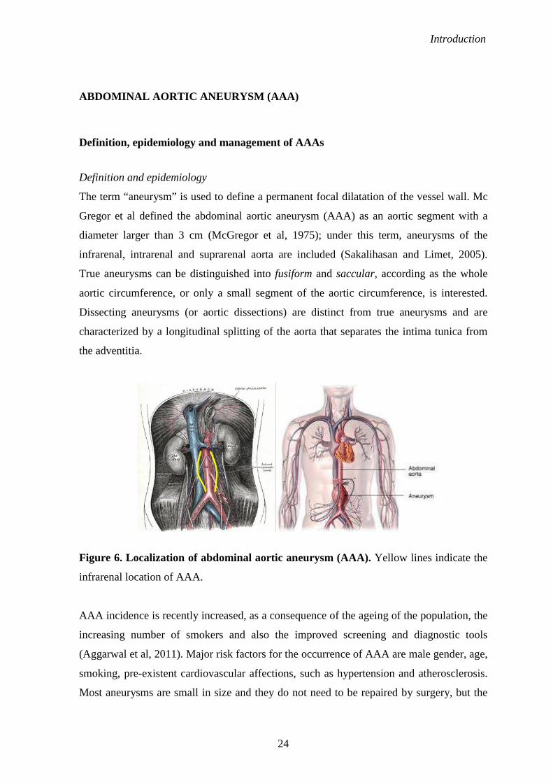
The term “aneurysm” is used to define a permanent focal dilatation of the vessel wall. Mc
Gregor et al defined the abdominal aortic aneurysm (AAA) as an aortic segment with a
diameter larger than 3 cm (McGregor et al, 1975); under this term, aneurysms of the
infrarenal, intrarenal and suprarenal aorta are included (Sakalihasan and Limet, 2005).
True aneurysms can be distinguished into fusiform and saccular, according as the whole
aortic circumference, or only a small segment of the aortic circumference, is interested.
Dissecting aneurysms (or aortic dissections) are distinct from true aneurysms and are
characterized by a longitudinal splitting of the aorta that separates the intima tunica from
the adventitia.
Figure 6. Localization of abdominal aortic aneurysm (AAA). Yellow lines indicate the
infrarenal location of AAA.
AAA incidence is recently increased, as a consequence of the ageing of the population, the
increasing number of smokers and also the improved screening and diagnostic tools
(Aggarwal et al, 2011). Major risk factors for the occurrence of AAA are male gender, age,
smoking, pre-existent cardiovascular affections, such as hypertension and atherosclerosis.
Most aneurysms are small in size and they do not need to be repaired by surgery, but the

Introduction
25
increase in the aortic diameter can lead to rupture, which can be fatal. The mortality rate
associated with AAA rupture is about 65-85%.
Atherosclerotic abdominal aortic aneurysm
Except in specific cases of aneurysms associated with inflammatory diseases, connective
tissue affections or traumatic event, the most common form of this dilatative affection rises
consequently to atherosclerotic plaque disease, although many researches suggest that
aneurysm formation is due to a combination of genetic, environmental and immune factors
(Sakalihasan and Limet, 2005).
The theory on the atherosclerotic origin of AAA has been based on the presence of
atherosclerosis pathogenesis features in patients affected by aortic aneurysm. Current
investigations and data about the specific nature of this relationship are still controversial;
in addition, the aetiology and the key mechanisms involved in aneurysm initiation and
expansion have not been completely elucidated. However, common features of aortic
aneurysms are inflammation, matrix degradation and smooth muscle cells depletion.
According to the most validated theory, the “atherosclerotic aneurysm” develops as a
complication to the pathological processes deriving from the atherosclerotic plaque
development: matrix remodelling, thrombus formation and release of pro-inflammatory
cytokines. An alternative theory considers aneurysm and atherosclerosis as independent
events, that result from the same environmental and genetic conditions, but different
dynamics are involved (Tonar et al, 2010). In any case, according to the current data
describing the type of association between the two arterial affections, the majority of AAA
are associated with atherosclerosis and elucidating this process would be crucial to develop
more specific and targeted therapies.
Inflammatory abdominal aortic aneurysm (IAAA): definition, incidence and pathogenesis
The concept of inflammatory aortic aneurysm (IAAA) was firstly defined by Walker et al
in 1972, observing unusual clinical features on a group of 19 patients. The presence of very
thick aortic wall, surrounded by a dense extended fibrosis of white aspect, involving the
adjacent organs were recognized as common features of a distinct form of AAA (Walker et
al, 1972).
At present, based on the original description by Walker, IAAAs are characterized by three
constant traits:

Introduction
26
• Thickened aortic wall;
• Extended peri-aneurysmal and retroperineal fibrosis;
• Dense adhesion of adjacent abdominal organs to the aortic wall (Pennel et al, 1985;
Crawford et al, 1985).
IAAAs represent the 3-10% of all aneurysm cases, with an higher prevalence rate among
males between 62 and 68 years; smoke and family history are additional risk factors (Tang
et al, 2005). A case control study by Nitecki et demonstrated a more elevated genetic
predisposition in IAAAs (17%) versus non non-inflammatory aneurysms (1.7%) (Nitecki
et al, 1996). The same work described the three main symptoms affecting IAAA patients:
back pain, weight loss and high erythrocyte sedimentation rate (ESR) (Nitecki et al, 1996).
Many histological evidences report the presence of a chronic inflammatory infiltrate,
composed of macrophages, T-lymphocytes and B-lymphocytes, in all AAA, mainly
interesting the adventitia layer (Pennel et al, 1985; Pasquinelli et al, 1993). A marked and
more extended infiltrate distinguishes IAAA from non-inflammatory aneurysms and some
clinical studies concord to consider IAAA as a progressive development from
atherosclerotic AAA (Pennel et al, 1985; Sterpetti et al, 1989; Latifi et al, 1992). Several
mechanism are responsible for the occurrence of inflammation, among which the immune
response to an unknown antigen is the most validated theory. Infection by a viral antigen
can also be involved; indeed, a more prominent presence of herpes simplex virus and
cytomegalovirus was observed in inflammatory aneurysm wall, compared to ordinary non-
inflammatory aneurysms.
Symptoms, diagnosis and management
Differently from IAAAs, non-inflammatory aneurysms are generally asymptomatic; only
in 8-18% of AAA cases, patients have reported symptoms. For this reason, unruptured
aneurysms are often recognized following clinical investigation in presence of other
cardiovascular diseases, or during screening test. Conversely, rupture of aneurysm is
accompanied by abdominal pain, shock and a pulsatile abdominal mass. Aneurysm
evolution and possible complications are generally evaluated by diameter measure, which
represent the major index of aortic rupture. The use of non-invasive techniques detecting
circulating levels of degradation peptides or proteins that actively participate to AAA
molecular pathogenesis would be more useful and secure to decide drug therapy or surgical
interventation. Matrix metalloproteinase 9 (MMP-9) may be represent one of the promising

Introduction
27
candidates, since it results higher in patients with AAA and its serum levels correlate with
aneurysm size and expansion (Lindholt et al, 2000).
About 30% of asymptomatic aneurysms are recognized by physical palpation as a pulsatile
abdominal mass, but this type of inspection conveys of the variability inter-operator.
Preferential diagnostic tool to identify aneurysm is represented by ultrasonography (UT),
also applied to follow-up, surveillance of asymptomatic aneurysms and screening. UT has
an high sensitivity and sensibility, without high costs. When the management of an
aneurysm is such to require surgical procedure, computational tomography (CT) is
performed and it can be helpful to define what type of surgical treatment is the best. CT
gives more detailed information about the aortic diameter, the shape of the aneurysm, the
thickness of intraluminal thrombus, together with the presence of blood within it. In
presence of an IAAA, CT also indicates the extent of inflammation. Magnetic resonance
Imaging (MRI) is an alternative and more accurate diagnostic procedure, but it is
expensive.
The treatment selection depends on the aortic size and diameter (Sakalihasan and Limet,
2005):
• aortic diameter less than 5 cm: follow-up through UT;
• aortic diameter between 5 and 5 cm: follow-up or surgery (the latter in case of
female patient, familial history, high serum markers)
• aortic diameter larger than 5.5 cm: surgery.
Surgical treatment of AAA is performed through open or endovascular repair. Open repair
has been performed since 1950s and consists of a large abdominal incision, followed by
the removal of the affected aortic segment and the insertion of a fabric graft (DuBost,
1952).
A less invasive option is given by the endovascular stenting, first described by Parodi in
1991 (Parodi et al, 1991) and consisting of the placement of a tubular graft within the
aneurysm sac, using a metallic stent to fix it to the normal aortic and iliac wall. The graft
acts like an artificial blood vessel, that excludes the damaged aortic segment from the
normal blood flow, and prevents aneurysm progression and rupture. This reparative
alternative does not require the surgical incision of the abdomen, thus avoiding
complications consequent to open surgery; in addition, it allows a shorter recovery time,
reduced hospital stay and implies a lesser blood loss (Paravastu et al, 2014). In some cases,
the blood can flow into the aneurysm sac, external to the graft, and this condition can lead

Introduction
28
to aneurysm expansion and rupture. This condition is defined by the term “endoleak” and it
is the major endovascular repair failure. This condition can be detected by angiography,
CT scan or duplex ultrasound imaging (White G. H. et al, 1997). Endoleaks can be
classified according to the timing of development: primary endoleak (early endoleak)
include all cases that occur during the 30 day perioperative period; secondary endoleak
(late endoleak) represent a late complication of the correct seal (White G. H. et al, 1997).
Endoleaks are further distinguished into: type I endoleak, when the seal between the ends
is not complete or ineffective, interesting the distal and proximal attachment sites; type II
endoleak, in presence of blood flow into the aneurysm sac from collateral arterial branches,
such as patent lumbar or inferior mesenteric artery, not depending on the graft seal; type III
endoleak, derived from a mechanical failure or a structural defect of the graft; type IV
endoleak, consequent to a graft wall porosity (White G. H. et al, 1998; Baum R.A. et al,
2003).
Figure 7. Open surgical repair versus endovascular repair (American Medical
Association, 2009).

Introduction
29
Histological and molecular features of AAAs
The human abdominal aorta is well organized into three concentric layers, or tunica,
displaced in the following order, starting from the lumen: intima, media and adventitia.
The histological composition, the cell types and the surrounding extracellular matrix have
been described in the previous chapter. The main fibrous component of the aortic wall are
represented by elastin and collagen, contributing to the aorta viscoelasticity and tensile
strength respectively, and assuring an equal distribution of the tensile stress along the
vessel. In this way, the healthy aorta is able to counteract the pulsatile pressure of the blood
flow. The typical laminar architecture of the aortic wall is lost in the AAA wall: analyzing
the AAA structure from the inner side, its characteristic hallmarks are the presence of the
intraluminal thrombus (ILT), tunica media degradation, marked by a decrease elastin
content and SMCs apoptosis, and adventitial inflammatory infiltrate, together with a
thickening of the intima. The presence of intimal lesions have supported the hypothesis of
aneurysm development from the atherosclerotic disease, as suggested by many laboratory
and clinical data. Currently, the link between aneurysm formation and atherosclerosis is
not completely defined.
Medial degradation: EMC remodelling and SMC apoptosis
The destruction of the lamellar architecture of the aortic media is the predominant
histopathological feature of aneurysm disease. As described in the previous chapter, ECM
represents the dynamic structure that supports the aortic wall, driving its main functions.
ECM remodelling is strictly regulated under physiological conditions and abnormal
expression and activity of the main ECM remodelling mediators are characteristic of many
vascular disorders. The elastin fragmentation represents an early event in AAA formation,
being cause of wall weakening; the degradation of the fibrillar collagen type I and III,
occurs in a later phase of the AAA expansion, leading to the loss of aortic tensile strength
and being a decisive step in the aortic rupture.
Many experimental data, obtained from tissue studies of human aneurysm specimens, as
well as from in vitro and in vivo animal models, have highlighted the MMP involvement
in ECM degradation, evidenced by the increased expression of these proteolytic enzymes
during aneurysm initiation and/or progression, together with a correlation with aneurysm
size. MMPs are secreted by medial SMCs, adventitial fibroblast and inflammatory cells;

Introduction
30
MMP hyper-expression, in terms of mRNA, protein as well as enzymatic activity, is not
only associated with an increased production and secretion, but is also the result of the
unbalanced MMPs/TIMPs ratio. MMP-2 and MMP-9 are the MMP members most
commonly associated with aneurysm disease and degrade both elastin and collagen. MMP-
2 is constitutively expressed in small aneurysm, suggesting a role in aneurysm initiation
and formation, whereas MMP-9 is more prevalent in large diameter aorta, indicating a
main participation to aneurysm progression and expansion. In addition, MMP-8 is a type I
collagenases and, together with MMP-9, was shown to be increased in ruptured aneurysm.
In addition to MMPs, other proteases localize at aneurysm wall: u-PA (urokinase-
Plasminogen activator) and t-PA (tissue-Plasminogen activator) convert plasminogen into
plasmin, which in turns activates MMPs; cathepsin S and K, with elastolytic action, and
cathepsin L, which degrade type IV and V collagen, laminin, elastin and proteoglycans.
Medial SMCs constitute the main vascular cell population that participate to aortic
structure, through the synthesis of ECM components as well as the secretion of ECM
remodelling mediators, like MMPs and their tissue inhibitors TIMPs. The importance of
SMC behaviour in the aneurysm development was explored by Lopez-Candavalez et al,
underlining an increased SMC apoptosis, marked by an increased expression of p53, which
is indicative of cell cycle arrest, in aneurysmal tissues versus non-aneurysmal and
atherosclerotic aortic tissues (Lopez-Candales et al).
The elastin degradation is tightly related to the abnormal hemodynamic shear stress that
influences aneurysm growth. The SMCs respond to the elastin degradation through an
increased tropoelastin synthesis; this is confirmed by increased mRNA levels of
tropoelastin in aneurysm tissue. This adaptation is not followed by the correct elastin
organization into the mature effective structures, resulting in the aortic wall inability to
respond to the increased shear stress.
Aortic inflammation and neo-angiogenesis
In addition to the loss of structural integrity, the aortic aneurysm is characterized by an
intensive transmural inflammation. The presence of the inflammatory infiltrate may be due
to the elastin fragmentation: this process releases soluble peptides exerting a chemotactic
effect that recruits inflammatory cells, which further secrete proteolytic enzymes, thus
amplifying the ECM degradation (Satta et al, 1998). Studies on the inflammatory infiltrate
invading the AAA wall revealed the presence of aggregating clusters of CD3+ T and

Introduction
31
CD19+ B lymphocytes, together with macrophages, and localized at the adventitial vasa
vasorum (Koch et al, 1990). In addition, an imbalance of the ratio between CD3+ T helper
and CD8+ T suppressor cells has been observed, with the predominance of T-helper cells
(Koch et al, 1990; Tang et al, 2005). Inflammatory cells activate a cascade of reactions
increasing inflammation environment and exacerbating the proteolysis process. CD3+ T
lymphocytes produce IL-4, -5, -8, -10 that attract other inflammatory cells, stimulate the
cytokine release by T cells and the formation of neovessels; in addition, CD3+ stimulate
the IFN-γ release, which in turn induces MMP expression.
The presence of cells presenting antigen in aneurysm wall takes to suggest an antigen-
driven T cell response as leading cause to aneurysm development. Matrix proteins, such as
elastin, collagen, aortic aneurysm antigenic protein-40 (AAAP-40) (Xia et al, 1996) and
exogenous agent (i.e. herpes simplex virus, cytomegalovirus) have been proposed as
potential antigen capable to induce T cells activation. Interestingly, the molecular mimicry
is mechanism of T cells activation, and it is supposed to be involved in aneurysm
pathogenesis. The molecular mimicry is based on the sharing of common sequences and
epitops between a foreign antigen (a microorganism) and self (host) antigen: the immune
reaction against a virus or bacteria, through a mechanism of cross reaction (Ozsvath et al,
1996; Oleszak et al, 2004) As consequence, the immune response inside the aortic wall is
exacerbated, thus complicating the proteolytic process. According to these findings, an
autoimmune origin of AAA has been proposed (Hirose and Tilson, 2001).
Macrophages actively participate to parietal remodelling and inflammation:
immunohistochemical and in-situ hybridization data revealed an intensive MMP-9
expression by aneurysm-infiltrating macrophages (Thompson et al, 1995); later works
demonstrated the macrophage-mediated release of cytokines, such as Il-1β, TNF-α, IL-8,
thus stimulating B-cell and cytotoxic T-cell differentiation, cytokine and protease
production and neoangiogenesis.
Neoangiogenesis consists in the formation of new blood vessels from pre-existing ones and
for this purpose it requires the ECM degradation by proteolytic enzymes to promote the
ECs migration from mature vessels; these steps are driven by specific growth factors, such
as VEGF, PDGF, EGF, TNF-α. The occurrence of neovascularisation in the AAA wall has
been demonstrated by Thompson et al (1995), evidenced by an increased number of neo-
vessels in aneurysmal tissues in comparison to normal aortic specimens; moreover, the
degree of neovascularisation was shown to be correlated with the inflammatory infiltrate

Introduction
32
amount (Holmes et al, 1995). These findings, together with immunolocalization studies
showing MMPs expression in correspondence of neo-vessels (Herron et al, 1991), support
a crucial role for the medial neo-vessels formation to the aneurysm pathogenesis. Not only,
increased neovascularisation and expression of angiogenic cytokines were detected at the
site of rupture, indicating an involvement of the angiogenic response into aneurysm rupture
(Choke et al, 2006).
Tumor Necrosis Factor-α (TNF-α)
TNF-α is a 17 kDa nonglycosilated soluble protein, derived by the cleavage of the 32 kDa
transmembrane precursor mediated by the TNF-α converting enzyme. TNF-α activates a
large spectrum of biological activities, among which the regulation of cell growth,
differentiation, programmed cell-death and it also drives the inflammatory cascade events
both under acute and chronic conditions (Pfeffer, 2003). As a pro-inflammatory cytokine,
TNF-α is involved in the pathogenesis of several inflammatory affections, such as Chron’s
disease (Danese et al, 2006) and rheumatoid arthritis (Matsuki et al, 2005). Many
investigations have demonstrated increased circulating and tissue levels of TNF-α in AAA
patients particularly in small versus large aneurysm (Juvonen et al, 1997; Hamano et al,
2003; Satoh et al, 2004). These observation suggested a role for TNF-α to early aneurysm
pathogenesis; this association was further confirmed by studies on animal models, showing
that the inhibition of TNF-α protein was able to prevent aortic dilatation (Hingorani et al,
1998).
Macrophages an lymphocytes are the main source of TNF-α, which exacerbates the
inflammatory response mediating the release of adhesion molecules, such as VCAM-1 and
ICAM-1, the lymphocyte proliferation and also stimulating the MMP secretion from both
inflammatory and resident vascular cells, including SMC (Saren et al, 1996 Cohen et al,
2006).
Biomechanical wall stress
Distinctive structural and hemodynamic features make the infrarenal aortic segment more
prone to the aneurysm development. As described in the previous chapter, the elastic
media of infrarenal aorta is not reached by adventitial vasa vasorum and, compared to
thoracic aorta, it receives a lesser nourishment. In addition, abdominal aortic media has a
decreased elastin content, corresponding to a shorter number of lamellar units. These

Introduction
33
anatomical parameters imply a major thickness and an increased wall tension per lamellar
unit. Many studies of the blood flow dynamics, revealed the presence of an oscillatory and
disturbed flow, together with an increased wall tension in the infrarenal aorta. These
factors contributes to aneurysm formation, as well as to accelerate its expansion and
rupture.
Intraluminal thrombus (ILT)
The ILT is considered as a neo-tissue, of various thickness, exerting a decisive role on
aneurysm fate and closely related to its rupture. The ILT structure is generally organized
into three layers (Michel et al, 2011):
• the luminal layer is in contact with the blood flow and has a red aspect, due to the
high content of erythrocytes; moreover, it is the site of platelet aggregation,
leucocytes and fibrin network;
• the middle layer has a white-yellow aspect and it is generally devoid of intact
erythrocytes, rarely it contains leukocyte infiltration;
• the abluminal layer is brown, lacking of alive cells and is adjacent to the aneurysm
wall; it is characterized by degraded fibrin and a weak gelatinous substance
(Wilson et al, 2013).
The origin of the ILT is mainly ascribed to the abnormal hemodynamic characteristic of
aneurysm disease; compared to the normal aorta, the blood flow along aneurysm lesion is
disturbed and turbulent, creating vortices and recirculation zones with high shear stress that
promotes platelet activation. Along the most dilated areas of the aneurysm, the shear stress
is lower than normal aorta, facilitating the platelet adhesion to the endothelium (Biasetti et
al, 2010; Folkesson et al, 2011).
Many studies addressing the composition and molecular behaviour of aneurysms
containing or not ILT, highlighted different aspects between them, suggesting the ILT
participation to the aneurysm development and rupture. At first, the presence of ILT
resulted associated with a thinner aortic wall, a decreased elastin content as well as an
enhanced fragmentation, accompanied by a prominent inflammation and increased
depletion of SMCs, showing a synthetic phenotype (Kazi et al, 2003). In addition, the
presence of ILT reduces the oxygen delivery to the aneurysm wall and generates an
hypoxic environment, which in turn exacerbates inflammation and neo-vascularisation,
increasing the risk of wall rupture (Vorp et al, 2001; Kazi et al, 2003). The mural thrombus

Introduction
34
constitutes a source of circulating inflammatory cells that invade the aneurysm wall and
enhance the ECM remodelling through the production and release of proteases, like MMP-
9 and urokinase-plasminogen activator (u-PA) (Fontaine et al, 2002).
Figure 8. Schematic representation of ILT contribution to inflammation and
proteolytic process in aneurysm wall (Michel et al, 2010).
Oxidative stress
The existence of oxygen and nitrogen reactive species (ROS, RNS) in the aneurysm wall
has been widely demonstrated. The oxidative stress include a series of reactions are
mediated by the release of ROS and RNS by different cell types, inducing cell and tissue
damage. These conditions depend on the increased ROS and RNS production, and/or the
impaired activity of anti-oxidant systems, such as superoxide dismutases (McCormick et
al, 2007).
First evidence of ROS and RNS implication in AAA pathogenesis derive from works of
Dubick et al, that demonstrated a reduced activity of superoxide dismutase in aneurysm
tissues, versus normal aorta. A comparative study between the aneurysm and the adjacent
non-aneurysmal portion of patients undergoing surgical repair revealed a marked oxidative
stress in the aneurysm segment evidenced by the increased expression of superoxide and
NADPH oxidase, in comparison to the adjacent unaffected segment; moreover, the O2-

Introduction
35
increase was associated with inflammation, SMC depletion and elastin degradation (Miller
et al, 2002).
The cells of the inflammatory infiltrate constitute the main source of oxidative stress,
especially macrophages generating large amounts of superoxide (O2-) and hydrogen
peroxide (H2O2), thus further contributing to the inflammation and the progression of the
disease. Another source of reactive oxygen species is represented by the vascular cells,
including ECs, SMCs and adventitial fibroblasts. In particular, the NADPH oxidase seems
to play a key role in generating ROS and its activity may be influenced by several and
different patwhays, including cytokines released by inflammatory cells, lipid mediators,
such as leucotryenes, oxidized LDL and growth factors. In addition, the mechanical stretch
also exert a decisive role, since it can stimulate the SMC production of ROS through the
activity of NADPH oxidase (McCormick et al, 2007). Oxidative stress can activate many
biological responses promoting the aortic wall remodelling and dilatation, including
osteopontin upregulation by ECs; MMP activation; SMC apoptosis.
The molecular mechanisms participating to AAA pathogenesis are summarized in Figure
9.
Figure 8. Molecular pathways leading to aneurysm formation and progression. (Kotze
and Ahmed 2011).

Introduction
36
STEM CELLS
Stem cells: definition, properties and classification
Stem cells are defined as undifferentiated cells capable to indefinitely self-replicate, giving
origin to an identical daughter cell at the same undifferentiated status. Under the
appropriate condition and stimulation, stem cells can also give rise to differentiated mature
cells, of all the organism types (multilineage differentiation). The property of a stem cell to
give origin to two daughter cells with different fates, goes under the termn of asymmetric
division.
Stem cells can be distinguished according to their differentiation potential, which is
functional to the development stage of individual life, and classified as totipotent,
pluripotent, multipotent and unipotent (Wagers and Weissman, 2004).
Totipotent stem cells have the ability to form cells of the whole organism and represent the
top of the stem cell hierarchy. The fertilized egg is a totipotent stem cell, leading to the
formation of germ layers (endoderm, mesoderm and ectoderm) and throphoblast,
necessary to support and protect the embryo. Pluripotent stem cells reside in the inner
mass of blastocyst and retain the capacity to differentiate into the cells of the three germ
layers (endoderm, ectoderm, mesoderm), but not the extra-embryonic components.
Embryonic stem cells (ESCs) and embryonic germ cells (EGCs) belong to this category,
whereas stem cells residing in adult tissues and able to differentiate into a more restricted
subset of cell lineages are defined as multipotent.
Stem cells belonging to adult tissues and able to differentiate only into a specific mature
cell type are called unipotent.

Introduction
37
Figure 10. The stem cell hierarchy.
A major distinction may be done between embryonic and adult stem cells, according to the
tissue source and the developmental stage: embryonic stem cells are pluripotent and derive
from embryonic tissues, whereas the adult stem cells have a restricted differentiation
potential and are present in many adult tissues including skin, bone, fat, cartilage, intestine,
liver.
Figure 11 illustrates the main differences between embryonic and somatic stem cells.

Introduction
38
Figure 11. Major differences between Embryonic Stem Cells (ESCs) and Adult Stem
Cells (ASCs) (O’Connor, 2008).
Embryonic stem cells (ESCs) Blastocyst is an early stage of embryo life: it consists of 150-200 cells and is composed of
the Inner Cell Mass (ICM), source of Embryonic Stem Cells (ESCs), and the external
trophoblast, that forms the placenta. Differently from totipotent fertilized egg, ESCs
differentiate into the three embryo lines, but do not form extra-embryonal tissues. ESCs
have been characterized as immortal stem cells, capable to indefinitely replicate
themselves. Mouse ESCs were detected for the first time in 1981 (Evans and Kaufman,
1981; Martin et al, 1981) and, many years later, Thomson and his team of investigators
were able to isolate ESCs from non-human primate and human blastocyst (Thomson et al,
1995; Thomson et al, 1998).
As described by Thomson et al, human embryos, donated after in-vitro fertilization, were
cultured to the blastocyst stage and 14 inner mass cells were isolated. Five embryos were
obtained and ES cell lines were isolated from each of them. Morphological characteristics
were comparable to rhesus monkey ESCs previously isolated (Thomson et al, 1995):
human ESCs displayed high nucleus to cytoplasm ratio, prominent nucleoli and kept a
normal karyotype. ESCs also expressed high levels of telomerase activity, index of cell
line immortality. Indeed, telmorase is a ribonucleoprotein which protect chromosome
ending with repeated telomeric sequence, thus preventing DNA rupture for each replicative
cycle and cell senescence. ESCs were established in in vitro cultures, showing prolonged
growing rates. ESCs showed the ability to self-renew and, under the appropriate culture

Introduction
39
condition, to aggregate into typical embryoid bodies and differentiate into many cell types,
like nerve cells, muscle cells and pancreatic isle cells.
Surface markers identificative of ESCs include: Stage Specific Associated Antigen (SSEA-
4 and SSEA-3); keratane sulphate antigens TRA-1-60 and TRA-1-8; alkaline phosphatises
(Thomson et al, 1998; Chambers et al, 2003).
ESC molecular profile was also investigated and revealed the presence of a transcriptional
core, composed of Nanog, Oct-4 and Sox-2, regulating the expression of downstream
genes. Genome scale location studies demonstrated that these three transcription factors are
physically associated and occupy the promoter region of many genes regulating the
developmental pathway (Boyer et al, 2005). This regulatory core may activate genes
involved in pluripotency and self-renewal, and repress genes promoting differentiation and
development of extra-embryonic, endoderm, mesoderm and ectoderm lineages.
Figure 12. ESC development and differentiation; embryoid bodies; transcriptional
profile of ESCs (adapted by Meshorer and Misteli, 2006; Nikishawa et al, 2007).
The high proliferative potential and differentiation ability make ESCs an important
resource for many research aims, from the transplantation medicine to toxicological
studies. However, major factors, including ethical questions, limit the use of ESCs for
clinical applications. Indeed, ESCs have been shown to induce, after transplatantion, the
formation of teratoma, which is a tumor originating from all the germ line cells. In addition
ESCs have displayed high immunogenicity, which could activate immune rejection in
heterologous transplant (Reubinoff et al, 2000).

Introduction
40
Adult stem cells (or Somatic stem cells)
The ethical debate and the disease risk associated with the clinical use of ESCs, have
addressed the regenerative medicine toward the study and the application of Adult Stem
Cells (ASCs).
ASCs, or Somatic Stem Cells, are undifferentiated cells, owning the self-renewal and
differentiation properties, like other stem cells. Unlike ESCs, ASCs are tissue-specific and
reside in post-natal differentiated tissues, but their origin is not completely identified and
this constitutes research field under current investigation. ASCs have been detected in
many post-natal organs and tissues, including bone-marrow, brain, peripheral blood,
vascular wall, muscle, skin and liver. ASCs reside inside a stem cell niche, which provides
support and drives stem cell fate and behaviour through many pathways and signalling
molecules. ASCs can differentiate into the specific mature cell types of the organ or tissue
they belong to; this property ensures the normal tissue homeostasis, as well as the tissue
repair when injured. Research on ASCs started in 1960 and, currently, the best
characterized ASC populations are represented by Hemopoietic Stem Cells (HSCs)
(Spangrude et al, 1988) and the Mesenchymal Stem Cells (MSCs) (Friedenstein, 1970),
both isolated from bone marrow.
Recently, many investigative studies have challenged the adult stem cell biology dogma,
introducing the concept of ASC plasticity. Indeed, different experimental findings suggest
that multipotent ASCs can overtake the lineage specific barrier and give origin to cell types
distinct from the origin tissue (Goodell, 2005). In this context, HSCs were shown to
differentiate into nonhematopoietic cells, such as liver cells; moreover, cultured
neurosphere cells (CNS-SC) were shown to contribute to hematopoiesis, when injected
into lethally irradiated mice. Also muscle mononuclear cells demonstrated hematopoietic
activity (Lakshmipathy and Verfaillie, 2005).
The possible explanations of ASC plasticity that have been proposed can be summarized in
the following mechanisms (Kapp and Mertelsmann, 2001):
• Direct or indirect transdifferentiation: a committed cell can differentiate into a cell
type specific of a distinct tissue through the direct activation of an alternative
differentiation lineage, or through a dedifferentiation program, which consists in

Introduction
41
the specialized stem cell return to a more primitive phenotype and consequent
change in lineage commitment.
• Coexistance of multiple stem cells within an adult organ or tissue, each of one
owns a different lineage conversion.
• Presence of a pluripotent stem cell in bone marrow and other tissues, able to
differentiate into all organism cell types; this theory has been supported by the
isolation of Multipotent Adult Progenitor Cells (MAPCs) from mouse and human
bone marrow, able to generate mesoderm, neuroectoderm and endoderm cells in
vitro; these cells, which have also been proved to give rise to most of somatic
tissues after implanation into early blasocyst, were also discovered in brain and
muscle tissues (Jang et al, 2002a, 2002b).
• Cell-cell fusion, leading to an heterokaryon formation, whose differentiation
pattern is the result of the two fused cells. This process has been observed at a very
low frequency and it has been mainly associated with a reparative function on
tissue injury.
Data supporting the ASC plasticity are still controversial, since they lack of standardized
detection method and are of difficult reproducibility between different experimental
models as well as different laboratories. The limits in using ASCs as substitutes of ESCs
for transplantation medicine are due to the restricted multilineage poltential and the lesser
content of ASCs in adult tissues; in addition, ESCs are more easily to be expanded in vitro,
with an higher proliferative potential, which is essential to guarantee a cell number
sufficient for transplantation uses. Thus, optimizing and standardizing the detection
methods and the experimental models, together with the understanding of cellular and
molecular pathways driving the ASC plasticity, are necessary to extend the knowledge on
ASCs and improve their clinical applications.
Stem cell niche
The hypothesis on the existence of the stem cell niche was introduced by Schofield in 1978
(Schofield, 1971) who proposed that stem cells reside in a defined structure designated to
control the stem cell behaviour. The niche is an anatomical structure which regulates many
stem cell functions, including proliferation and differentiation, and prevent cell death.
These activities are elaborated trhough surface molecules and signalling transduction
pathways. Distinct and specialized stem cell niches have been detected in many tissues

Introduction
42
and an unbalance in the niche regulation or structure may be responsible of tissue damage
with the occurrence of different diseases (Jones and Wagers, 2008).
Beside the variability existing between niche belonging to different tissues, the niche
generally comprises the following components:
• Stromal support cells, appointed to communicate with stem cells trough the
expression of specific surface molecules, gap junctions and the release of soluble
factors.
• ECM, which exerts a structural support and a regulatory role of the signalling
mechanisms inside the niche.
• Vascular network and neural inputs.
The hypothetical scheme of how is organized the stem cell niche is illustrated in Figure 13.
Figure 13. Scheme of the hypothetical structure of the stem cell niche; example of a
stem cell niche: HSC niche (Jones and Wagers, 2008).
The stem cell niche has been detected and characterized at the level of the intestinal cript,
hair follicle, in the subventricular zone of the hippocampus and olfactory bulb (Jones and
Wagers, 2008), and recently also at the vascular level (Zengin et al, 2006; Ergun et al,
2011). Indeed, the research of the last few years has reversed the idea of the vessel wall as
passive and quiescent layer, introducing the new concept about a functional participation to
tissue homeostasis and vascular processes, like post-natal angiogenesis. The vasculogenic
niche has been identified at the adventitia level, along its border with the media and closely

Introduction
43
to the vasa-vasorum network, which ensures the niche contact with the peripheral
circulation. The vascular stem cell niche includes distinct cell subsets: MSCs,
myofibroblasts, endothelial, hematopoietic and smooth muscle progenitors (Psaltis et al,
2011). Vascular progenitors participate to cell turnover and tissue homeostasis during
physiological conditions and, on the other hand, may regulate or exacerbate pathological
remodelling and neovascularisation.
Figure 14. Hypothetical structure of the vasculogenic niche (Zengin et al, 2006).

Introduction
44
Mesenchymal Stem Cells (MSCs)
Definition, in vitro phenotype and differentiation properties
Bone marrow has been considered as a source of stem cells belonging to the hematopoietic
lineage for many years and the existence of non-hematopoietic stem cells, as stromal
components of the HSC niche, was detected in 1970. Friedenstein and his co-workers, after
plating whole bone marrow on plastic dishes, using growing media supplemented with
10% Fetal Calf Serum (FCS), observed the presence of plastic adherent cells able to
proliferate and, after passaging in culture, assume a fibroblast-like morphology.
Interestingly, these adherent cells were shown to form colonies of fibroblastoid cells, and
were identified as Colony Forming Unit-fibroblasts (CFU-f); these CFU-f were also able to
form bone and cartilage (Friedenstein et al, 1970; Friedenstein et al, 1976). The following
workers by Castro-Malaspina (Castro-Malaspina et al, 1980) and Caplan (Caplan, 1991),
confirmed the CFU-f ability to differentiate into the mesoderm lineage, giving origin to
adipocytes, osteocytes, chondrocytes and myoblasts. Caplan in 1991 proposed the
nomenclature Mesenchymal Stem Cells (MSCs), due to their differentiation ability into
mesenchymal-type cells, and stromal, since they were considered as support cells to bone
marrow HSC niche.
MSCs have been isolated from different species, including human and mice, as well as
different tissues beside bone-marrow, including peripheral blood, umbilical cord blood
(Erices et al, 2000), Wharton’s jelly membrane (McElreavey et al, 1991), adipose tissue
(Zuk et al, 2002), synovial membrane (De Bari, 2001), synovial fluid (Karystinou et al,
2009). The high proliferative rate and the multilineage potential that characterize MSCs
make them promising candidates for clinical applications, such as cell-based therapies and
organ transplantation. Meanwhile, MSCs constitute a cell population with high degree of
heterogeneity, due to several aspects. First of all, the characteristics of the source tissue,
including tissue type and age; another important element derives from the coexistence, in
the same MSC population, of different subsets of cells at different development stages.
Moreover, MSCs are heterogeneous in terms of proliferative and differentiation potential,
thus representing an high complexity population; indeed, Pittenger et al showed that only
one third of the whole bone marrow population that they plated, demonstrated the ability to

Introduction
45
differentiate into the mesodermal lineages (Pittenger et al, 1999). As consequence, there is
not a specific marker, able to unequivocally individuate MSCs.
Therefore, an important result in the field of the MSC research would be the
standardization of isolation method and the identification of a specific signature for MSCs.
According to the International Society for Cell Therapy (ISCT), the minimal criteria for
defining human MSCs are as follows (Dominici et al, 2006):
• Adherence to the tissue culture substrate;
• Specific surface antigen (Ag) expression;
• Multilineage differentiation potential.
Figure 15. MSC properties: self-renewal and differentiation along the mesodermal
lineage; potential plasticity of MSCs (Uccelli et al, 2008).
MSC surface antigens
According to the ISCT, the expression of CD90, CD73 and CD105 Ag on cell surface,
performed by flow cytometry, define the MSC population:
• CD105, or Endoglin, is a type I homodimeric transmembrane glycoprotein,
belonging to the TGF-β receptor family and links with high affinity TGF-β1 and 3;
it has been shown to be expressed on connective stromal cells and also on
endothelial cells and monocytes.

Introduction
46
• CD90, or Thy-1, a 25-37 kDa heavily glycosilated glycophosphatididynil (GPI)-
anchored cell surface protein. It was identificated for the first time on thymocytes,
which are the T cells precursors in the thymus. It is expressed on MSCs, HSCs,
neurons, endothelium, myofibroblasts and its functions have not been clarified yet.
It may regulate cell-matrix interactions and regulate apoptosis, fibrosis,
inflammation and neuron regeneration processes.
• CD73, also known as ecto-5’-nucleotidase, a glycoprotein implied in AMP
conversion to adenosine; it is involved in B lymphocytes differentiation and it is
expressed on lymphocytes and monocytes.
• CD44, a cell surface glycoprotein, involved in intercellular interactions, cell
adhesion and migration. It acts as receptor for hyaluronic acid, but also recognizes
osteopontin, collagen and MMPs.
In addition, the expression of hematopoietic markers needs to be excluded: for this reason,
antigens CD45, CD34, CD14 or CD11b, CD79a or CD19 and HLA class II may be
included in the antibody panel to detect human MSCs.
• CD34, a glycoprotein expressed on many cell types, like HSCs, blood vessel ECs,
endothelial progenitor cells (EPCs), mast cells; it acts as a cell-cell adhesion factor
and is required for T cells entry into lymph nodes.
• CD45, also known as protein tyrosine phosphatases receptor type C. It is a
transmembrane protein, exerting a regulatory role for many cell processes,
including growth, cell cycle, differentiation and oncogenesis. It is expressed on
differentiated hematopoietic cells and also in pathological conditions, such as
lymphoma and B-cells chronic lymphocytic leukemia.
• CD14, mainly expressed by macrophages and neutrophils, acts as a receptor for
bacterial lipopolysaccharide (LPS) detection.
• CD31, also known as platelet endothelial cell adhesion molecule (PECAM-1), is
present on surface of platelets, monocytes, neutrophils, belongs to the
immunoglobulin family and is involved in leucocyte transmigration, angiogenesis
and integrin activation.
Recently, the lack of MSC selective markers and standardized isolation protocols has lead
investigators to introduce additional and more specific methods to enrich cultures for
MSCs. In this context, the use of monoclonal antibodies could be useful to identify a cell
population with specific phenotype; the murine monoclonal antibody against Stro-1

Introduction
47
recognizes non-hematopoietic bone marrow stromal precursors and it can be used as CFU-
F marker, since it was demonstrated that Stro-1 negative cells were not able to form
colonies (Simmons and Torok-Storb, 1991).
Stro-1 has a limited use as an universal MSC marker, so it is best to be associated with
additional antigens, such as CD106, also known as Vascular Cell Adhesion Molecule-1
(VCAM-1). CD106 has been shown to be involved in many processes, like chemotaxis,
cell adhesion and signal transduction. In addition, a study by Majumadar et al (2003)
identified MSC surface molecules, such as integrins α1, α5 and β1, expressed at high levels
by MSCs and mediate homing and cell binding processes.
MSC differentiation potential
The third aspect to identify human MSCs, as suggested by the ISCT in 2006 (Dominici et
al, 2006) consists in the MSC in vitro differentiation into mesodermal lineages. Indeed,
under the influence of certain culture conditions and growth factors, human MSCs isolated
from multiple sources could differentiate into adipocytes, osteocytes and condhrocytes
(Barry and Murphy, 2004; Chamberlain et al, 2007).
Osteogenic induction requires the addition of β-glycerophosphate, dexametasone, ascorbic
acid and to FBS-supplemented growing media, resulting in the formation of calcium
deposit and MSC expression oh phasphatese alkaline. Cartilage formation, evidenced by
proteoglycans and collagen II expression, occurs under MSC exposure to TGF-β factor in
serum-free conditions. TGF- β3 was shown to be the more potent condhrogenic induction
factor, associated with the MSC synthesis of aggregan, fibromodulin, type II collagen and
other typical components of articular cartilage matrix (Barry et al, 2001). The lipid-
vacuoles indicative of adipogenic differentiation were observed after MSC exposure to
FBS-supplmented media in presence of dexamethasone, insulin, indomethacin and isobutyl
methyl xanthine. Culture conditions used for in vitro differentiation assay on human MSCs
are summarized in Table 1.

Introduction
48
Table 1. MSC differentiation culture conditions.
Beside the differentiation assay mentioned above, human MSCs displayed myogenic
(Taylor and Jones, 1982), neurogenic (Deng et al, 2001) and angiogenic potential (Davani
et al, 2003).

Introduction
49
MSCs: immunomodulatory properties and clinical applications
One of the most intriguing aspect related to the therapeutic applications of human MSCs
reside in their ability to evade immune recognition and suppress immune response (Barry
and Murphy, 2004; Chamberlain et al, 2007). The low immunogenicity of MSCs is mainly
due to the low expression rate of histocompatibility complex HLA-I and lack of the
complex HLA-II, together with the absence of co-stimulatory molecules CD40, CD80 and
CD86. On the other hand, the immunosuppressive function may be exerted at various
levels, acting on many aspects of the immune system. Indeed, it has been reported that
MSCs can suppress T cell proliferation, activating the fraction of regulatory T cells
(Tregs); moreover, MSCs act on dendritic cells (DCs), impairing their differentiation and
function, as well as on B cell and natural killer cells (NKs), limiting their proliferation and
cytolytic function respectively (Le Blanc and Ringdén, 2007). These properties have been
investigated on in vitro and in vivo experimental models, demonstrating an
immunoregulatory activity associated both to direct cell-cell interactions and soluble factor
secretion. Soluble factors include anti-inflammatory mediators, such as Transforming
Growth Factor- β1 (TGF-β1), Inteurlekin-10, (IL-10), Indoleamine 2,3-dioxygenase (IDO),
Hepatocyte Growth Factor (HGF), Prostaglandin E2 (PGE2). In most of cases, Interferon-γ
enhances MSC immunosuppression, supporting the idea that the inflammatory
environment contributes to MSC activation and following establishment of an
immunosuppressive niche trough the release of soluble factors (Ryan et al, 2007).
A brief description of the immune cell system origin from HSCs and its functional
organization are represented in Figure 16.

Introduction
50
Figure 16. Immune cell system and hierarchical organization from stem cell
precursor.
MSC-mediated immunosuppression: mechanisms and molecular mediators
Many evidences obtained from human, baboon and mouse models have showed a
decreased T cell proliferation, under the influence of MSCs. More specifically, this process
was induced by a cell cycle arrest during the quiescent phase G0/G1, together with reduced
cyclin D2 and increased p27 expression levels (Glennie et al, 2005; Shi et al, 2011).
Several molecular signalling may be involved in MSC-mediated T cell suppression. Di
Nicola et al proposed the secretion of TGF-β1 and HGF, as active molecules against T cell
proliferation (Di Nicola et al, 2002). This was confirmed by the use of neutralizing
antibodies against TGF-β1 and HGF, able to restore T lymphocyte cell cycle. PGE2 is a
cytokine constitutively expressed by MSCs, undergoing an increased production in
presence of peripheral blood mononuclear cells (PBMCs), and inhibits the T cell
proliferation and the release of inflammatory cytokines T cells dependent. IDO represents
a further mediator of MSC suppressive activity on T cells: its expression increased under
the presence of IFN-γ, usually expressed during inflammatory process. Indeed, IDO
promoter contains two IFN-stimulated response elements (ISREs) and an IFN-γ activated
site (GAS) (Ryan et al, 2007). Increased IDO correspond to an enhanced tryptophan
catabolism, resulting in the release of the kyneurine degradation intermediates that block
lymphocyte proliferation. Nitric oxide (NO) signalling is also involved in

Introduction
51
immunomodulatory mechanism, depending on STAT-5. STAT-5 is a transcription factor
which is crucial for T cell activation and proliferation; the inhibition of STAT-5
phosphorylation, NO-dependent, mediates T cell cycle arrest (Sato et al, 2007).
Another mechanism by which MSCs regulate immune response consists in the activation
of Tregs, designated to suppress T cell proliferation and cytokine release. Treg population
comprises different subsets, the major part of which is defined on the basis on CD4 and
CD25 positive profile, together with the expression of the transcriptional factor FoxP3 and
the functional secretion of IL-10 and TGF-β (Singer and Caplan, 2011). The MSC-
mediated release of TGF-β1 and HLA-G5, contribute to Treg induction.
HLA-G is a non-classical HLA class I, identified in cytothrophoblast (McMaster et al,
1995), where regulates the maternal-fetus tolerance (Rouas-Freiss et al, 1997), and results
to be up regulated under specific conditions, such as tumorigenesis and organ
transplatantation. It has been suggested as a crucial mediator to MSC immunosuppression,
since it regulate many immune processes and beside Treg induction, HLA-G inhibits NK
cytotoxicity and DC maturation (Selmani et al, 2007; LeRonde et al, 2006). The B cell
inhibition mediated by MSCs was observed on murine models and later confirmed in
human MSCs. B cells resulted affected on proliferative as well as functional aspects,
including differentiation and chemotaxis.
A summary of the MSC-mediated immunosuppression is illustrated in the Figure 17,
which underlines each mentioned molecular mechanism and the involved mediators.

Introduction
52
Figure 17. MSC-mediated immunosuppressive mechanisms.
The high proliferative and differentiation potential, together with the homing to damaged
tissues, make MSCs really useful for therapeutic applications. The autologous or
allogeneic MSC transplantation can be practiced through systemic or local delivery and it
can be applied to several clinical fields, such as cardiovascular, neurodegenerative
diseases, spinal cord injury or bone and cartilage affections, requiring tissue repair. For
example, in 2001, bone marrow MSCs were shown to de novo regenerate myocardium and,
in 2002, Sanchez-Ramow demonstrated the differentiation of bone marrow MSCs into
neuronal lineage both in vitro and in vivo, using mice and rat models (Sanchez-Ramos,
2002).
The systemic delivery of cultured MSCs was performed to treat children affected by
Osteogenesis Imperfecta, which is a defective type I collagen disease resulting in
osteopenia, fractures, and bone malformations. After 3 months, surprising results were
observed, highlighted by increase bone formation and reduced bone fracture frequency
(Horwitz et al, 1999).
The hypoimmunogenic nature of MSCs and the above described immunoregulatory
mechanisms allows MSC use to regulate immune reaction in the context of transplantation

Introduction
53
and autoimmune diseases with an inflammatory origin, such as graft versus host disease
(GVHD), Crohn’s disease, systemic lupus erythematosus.

Introduction
54
Stem cells in disease: a pathogenetic role? Research on MSC clinical applications should also addresses the potential side effects
deriving from MSC use, as well as the environment influence on MSC behaviour. Under
specific tissue conditions, especially in presence of degenerative affections, stem cells can
be altered or do not function in a proper manner. This impaired function may result in a
worsening of the clinical disease conditions. An exemplificative situation is those
represented by osteoarthritis, a disease caused by a progressive cartilage loss. Murphy et al
(2002) demonstrated that MSCs isolated from patients had a lower proliferative potential,
together with a reduced adipogenic and chondrogenic differentiation ability. Moreover,
MSCs isolated from patients affected by idiopathic thrombocytopenia and systemic lupus
erythematosus display an abnormal function.
Beside an impaired growth and differentiation potential, MSCs can undergo an alteration
of the immunomodulatory function, thus creating or exacerbating the inflammatory
process. Some evidences report that MSCs secrete also pro-inflammatory cytokines and it
has been suggested the role of Toll-Like Receptors (TLR) as potential mediators of a MSC
pro-inflammatory phenotype.
TLRs are a conserved family of receptors that after recognition of pathogen associated
molecular patterns (PAMPs) activate immune cells. TLRs have been showed to be linked
with the growth and differentiation of adipose-derived MSCs (ASCs), but current data are
still controversial.
Bunnel et al suggested a mechanism involving TLR in MSC acquisition of a pro-
inflammatory or an immunosuppressive phenotype: according to their model, a short-term
and a low level exposure to TLR4 takes MSCs to a pro-inflammatory phenotype,
contributing to tissue injury; on the other hand, TLR3 stimulates MSCs toward an anti-
inflammatory, thus resolving the tissue damage (Bunnel et al, 2010). Moreover, this
functional divergence corresponds to a differential cytokine and chemokine expression
pattern, between the two MSC phenotypes (Waterman et al, 2010; Bunnel et al, 2010).
A study by Li et al demonstrated an impaired immunosuppressive activity for bone-
marrow MSCs isolated by multiple myeloma (MM-MSCs) patients, reflected on an
aberrant T cell function (Li et al, 2010). The alteration of MSC immunosuppressive
property was also documented in a mouse model of collagen-induced arthritis (CIA).
Indeed, MSCs exposed to an inflammatory environment, as in the CIA model, did not

Introduction
55
result beneficial and in vitro experiments showed that consequently to TNF-α addition,
MSCs lost the ability to regulate T cell proliferation (Djouad et al, 2005).
Based on these findings, it is clear that MSCs represent a great promise in the regenerative
therapy field, and many other issues must be explored to completely elucidate their
behaviour contextually to the tissue damage. Most importantly, the molecular mechanisms
triggering MSC attitude toward disease progression, especially as regards inflammation
and autoimmune disorders, needs to be investigated.

Aim of the thesis
56
AIM OF THE THESIS

Aim of the thesis
57
AIM OF THE THESIS AND EXPERIMENTAL DESIGN
The existence of the vascular stem cell niche has been proved in many laboratories (Zengin
et al, 2006; Ergun et al, 2011; Psaltis et al, 2011; Gómez-Gaviro et al, 2012; ). Our
research team has a ten-year experience on MSC isolation from the vascular wall of
different arterial segments obtained from healthy donors of various age and clinical
characteristics (Pasquinelli et al, 2007; Pasquinelli et al, 2010; Valente et al, 2014). More
specifically, a cell population expressing mesenchymal antigens, stemness transcriptional
factors and able to differentiate into the adipogenic, osteogenic and chondrogenic lineage
was isolated. Moreover, this cell population, named Vascular Wall MSCs (VW-MSCs),
was shown to form capillary-like structures under the influence of VEGF soluble factor
added to cell cultures. In addition to these phenotypical and functional characteristics, VW-
MSCs were also tested for the immunomodulatory activity, which represents one of the
most interesting MSC properties for their clinical and therapeutic use. Indeed, VW-MSCs,
as the other tissue source human MSCs, were able to negatively modulate the immune
response, through a decreasing effect on PBMC proliferation, acting on the cell cycle arrest
in G0/G1 phase. On the other hand, VW-MSCs demonstrated a pronounced expression of
HLA-G, a typical MSC non-classical antigen that inhibits immune reaction suppressing T
cell and Natural Killer cell function and improving the expression of T regs.
In addition, we recently succeed to obtain a consistent cell number with a high proliferative
capacity also from 5 years cryopreserved arteries, belonging to distinct vascular districts
(common carotid, subclavian artery, aortic arch, thoracic aorta). These cells were shown to
resist to nitrogen liquid temperature for long term (Valente et al, 2014).
Based on these findings, we planned to explore the presence as well as the functionality of
the MSC niche in adverse pathological conditions, as occurs during abdominal aortic
aneurysm (AAA) pathogenesis.
AAA is a focal aortic dilatation, necessary to be under monitoring since it has no sign or
symptoms and the silent progressive enlargement of the aortic diameter can lead to rupture
which can be fatal, carrying a 70-80% mortality risk. Currently, there is not an established
pharmacological approach able to restore the normal vessel wall condition, and large
aneurysms are treated only through surgical repair. AAA pathogenesis is associated with
the arterial medial degradation, mediated by the proteolytic activity of enzymes secreted by

Aim of the thesis
58
both vascular resident and inflammatory cells. In this context, MMPs represent the
molecular mediators mainly involved in the ECM degradation. Meanwhile, inflammation
invades the aortic wall and exacerbates the tissue remodelling, since inflammatory cells,
such as T and B lymphocytes and macrophages migrate at the injured tissue, release
cytokines (IL-1β, IL-6, TNF-α, etc) and MMPs.
Many investigators have supposed an immune-cell mediated origin for aneurysm initiation,
based on the immune response mediated by T cells against an endogenous or exogenous
antigen (like herpes simplex, CMV). Moreover, an autoimmune origin was also suggested.
Beyond inflammatory cells, MMPs are produced and released by MSCs, at different levels,
as demonstrated by several research works (Silva et al, 2003; Mannello et al, 2006).
Indeed, MMPs especially MMP-2 and MT-MMP1 are essential to MSC migration and
invasive abilities (Ries et al, 2007).
These background considerations lead us to suppose a concrete contribution of vascular
MSCs to pathological conditions involved in AAA disease. The aim of this study was so
organized on different levels:
AIM 1: MSC isolation from aneurysm and characterization
• MSC isolation from vascular specimens collected by patients affected by AAA
MSC.
• MSC characterization according to the classical criteria: morphology, mesenchymal
antigens (Flow cytometry) and stemness transcriptional factors (RT-PCR).
AIM 2: MSC participation to pathological processes inside aortic wall
• MSC characterization for MMP expression: MMP-2, MMP-9, TIMP-1, TIMP-2
and EMMPRIN (mRNA, protein and activity evaluation).
• MSC immunomodulation: ability to regulate immune reaction, trough co-colture of
MSC and Peripheral Blood Mononuclear Cells (PBMCs).
• Evaluation of MMP modulation mechanism, through an anti-inflammatory
approach: use of healthy vascular MSCs; AAA-MSC exposure to IL-10.
The following diagram provides a schematic description of the experimental design
followed in the present thesis.

Aim of the thesis
59
Figure 18. Aim of the thesis and experimental design.

Materials and methods
60
MATERIALS AND METHODS

Materials and methods
61
STUDY PROTOCOL AND TISSUE COLLECTION The study protocol was approved by the Local Ethic Committee (ethic protocol number
APP-13-01) and written informed consent was obtained from patients.
Aneurysm tissues of the abdominal aortic segment were provided by the Vascular Surgery
Unit, University Hospital S. Orsola-Malpighi, Bologna. AAA tissues were collected from
12 patients (males, mean age= 69.2±5.0) subjected to surgical repair; demographical and
clinical data were reported on a database.
Thoracic aortic wall tissues derived from 3 healthy donors (female:male=2:1, mean
age=25±6.5) were provided from the Cardiovascular Tissue Bank, University Hospital S.
Orsola-Malpighi, Bologna. The vascular tissues included in our study derive from the
arterial segments discarded from the Bank and were used as healthy controls for our study,
with the approval of the Local Ethic Committee (ethic protocol number APP-13-01). We
selected the healthy aortic segments according to two specific criteria: age between 18 and
55 years, to exclude ageing process interference with our analysis; cause of death not
associated with cardiovascular pre-existent affections, or brain hemorrhage, to compare
patient group with healthy subjects in absence of cardiovascular risk.
This study was developed on different levels, evaluating both the histological and cellular
analysis. For this purpose, pathological and healthy aortic samples were in part fixed for
histological and immunostaining applications and in part treated with an enzymatic
solution to allow cell isolation.
ANEURYSM-AFFECTED AORTA: HISTOPATHOLOGICAL FEATURES
Hematoxylin/Eosin staining
AAA tissues were in part reserved for histological exam, to characterize aortic wall and
identify typical aneurysm features. Briefly, AAA tissues were fixed with a 10%
formaldehyde solution, washed and dehydrated by passing the tissue through a series of
increasing alcohol concentrations (70%-96%-100%) and embedded in paraffin to obtain
four µm-thick sections. Each section was cut trough the microtome and stained with
hematoxylin & eosin (H&E) for traditional histopathological analysis according to the
histological procedures. After staining, slides were observed with light micsroscope (LM)
and analyzed using the Image-Pro Plus® 6.

Materials and methods
62
Immunohistochemical staining
Immunohistochemistry was performed on aneurysm and healthy specimens to characterize
MSC population before enzymatic digestion, using a non-biotin amplified method
(Novolink, Newcastle, UK). Mesenchymal antigens (CD44 and CD90) were used for the in
situ detection of human MSCs. In addition, MMP-9 staining was performed to evaluate the
contribution of the different cell types inside the vessel wall to pathological arterial
remodeling.
Briefly, four µm-thick sections of formalin-fixed and paraffin embedded tissues were
deparaffinized and rehydrated through a series of graded ethanol and rinsed in distilled
water. Endogenous peroxidases activity was blocked in 0.3 % H2O2 in absolute methanol
for 10 minutes at room temperature; antigen retrieval was performed using citrate buffer
(pH 6) in microwave (750w) for 20 minutes and after cooling slides were washed with Tris
Buffered Saline (TBS). Aortic sections were subsequently incubated with primary
antibodies against CD44 (1:100, BD Biosciences) and CD90 (1:100, BD Biosciences) and
MMP-9 (1:325, Cell Signaling) in a moist chamber at 4°C o/n, then they were incubated
with NovoLink Polymer for 30 minutes at room temperature and exposed to the
substrate/chromogen 3,3′-diaminobenzidine (DAB) prepared from Novocastra DAB
Chromogen and NovoLink DAB buffer. Nuclei were counterstained with Mayer’s
hematoxylin. Samples were dehydrated, coverslipped and observed under light microscope
using the Image-Pro Plus program.
ANALYSIS ON MSCs ISOLATED FROM ANEURYSM-AFFECTED AORTA
MSC isolation and culture
The protocol used for MSC isolation from vascular segments consists of an adjustment of
the previously described technique (Pasquinelli et al, 2010).
Two cm2 sections of healthy and aneurysm-affected aortic walls were digested in an
enzymatic solution containing 0,3 mg/mL Liberase type II (Liberase TM Researche Grade,
Roche) in serum-free Dulbecco's modified Eagle's medium (DMEM; SIGMA Aldrich) at
37°C o/n in a rotor apparatus. The resulting tissue homogenate was filtered using cell
strainers at decreasing diameter (100-70-40µm) to eliminate not-digested tissue residuals
and centrifuged at 1200 rpm. Cell viability was assessed by Trypan Blue exclusion. MSCs
isolated from aneurysm tissue were identified with the acronym AAA-MSCs, whereas
MSCs isolated from healthy aorta were defines as control MSCs (cMSCs). AAA-MSCs
and cMSCs were cultured according to the standard culture conditions (37° C incubator,

Materials and methods
63
5% CO2) in DMEM enriched with 20% Fetal Bovine Serum (FBS; SIGMA Aldrich) and
in-vitro expanded.
MSC immunophenotype: flow cytometer analysis
Flow cytometer analysis was performed on AAA-MSCs and cMSCs at passage 3 to
investigate the expression of typical mesenchymal markers, exploring eventual different
expression patterns of the investigated antigens.
Briefly, cells grown on monolayer when arrived at passage 3 were trypsinized and, after
centrifuge at 1200 rpm for 5 minutes, the pellet was fixed with the Fixation Kit (Beckman-
Coulter). Then, samples were stored at 4°C until use. Fixed MSCs were incubated for 20
minutes with each antibody; for our purpose to identify MSC populations, an extensive
conjugated panel of antibodies was used: anti-CD44-fluorescein isothiocyanate (FITC),
anti-CD73-phycoerythrin (PE), anti-CD90-phycoerythrin-cyanine 5, anti-CD14-FITC,
anti-CD34-FITC, anti-CD45-allophycocyanin, anti-CD146-PE, anti-platelet-derived
growth factor receptor beta (PDGFR-β) (R&D Systems, Inc., Minneapolis, MN, USA).
Flow cytometry analysis also included the expression of HLA-G, associated with the MSC
immunomodulatory activity. For this purpose, HLA-G detection was performed trough the
incubation with anti-HLA-G-FITC antibody (Abcam, Cambridge, UK).
Negative controls were performed using appropriate conjugated irrelevant antibodies.
Samples were analyzed using a Navios FC equipped with two lasers for data acquisition
(Beckman-Coulter). Results were analyzed with Kaluza FC Analysis software (Beckman-
Coulter).
MSC MOLECULAR PROFILE: STEMNESS MARKERS, AAA MEDIATORS AND IMMUNOMODULATORY FACTORS
Total RNA extraction and Reverse-Transcription Total RNA was extracted from cMSCs and AAA-MSCs at passage 3, by using TRIreagent
(TRIzol reagent, Invitrogen, Italy) according to the manufacturer’s instructions.
Total RNA was extracted from cMSCs and AAA-MSCs at passage 3, by using TRI
Reagent (TRIzol reagent, Invitrogen, Italy) according to the manufacturer’s instructions.
Briefly, cultured cells were homogenized in TRI Reagent solution and incubated at room
temperature for 5 minutes. After lysis, the chloroform was added to homogenate samples
and left at room temperature for 10 minutes, to allow the phase separation: after centrifuge,
RNA contained in the aqueous phase was collected and transferred into a new tube. A step

Materials and methods
64
of RNA precipitation by addition of isopropyl alcohol, incubated at room temperature for
10 minutes, was performed; the visible RNA pellet was finally washed with 75% alcohol,
centrifuged and air-dried for 10 minutes. RNA was dissolved in RNAse free water and
stored at 20°C until use.
At the end of the extraction protocol, RNA integrity and concentration were evaluated by
using ND-1000 spectrophotometer (NanoDrop, Fisher Thermo, Wilmington, DE, USA).
Reverse transcriptase PCR was performed on all RNA samples with an absorbance
(260/280) ratio between 1.8-2.2. One µg of total RNA was reverse transcribed in a 20 µL
reaction volume using High Capacity Reverse Transcription Kit (Life Technologies).
Stemness marker evaluation by RT-PCR
RT-PCR was performed on AAA-MSCs to evaluate the stemness genes Nanog, Oct-4 and
Sox-2 expression; primers sequences (SIGMA Aldrich) are listed in Table 2.
Table 2. Primer sequences used to detect ESC transcriptional factors AAA-MSCs. bp:
base pair.
Glyceraldehyde-3-phosphate dehydrogenase (GAPDH) was used as housekeeping gene to
value the cDNA quality. RT-PCR was performed according to the reaction program
described in Table 3, using a Thermocycler with gradient function to allow the
simultaneous amplification assay for each gene.

Materials and methods
65
Table 3. Scheme of PCR protocol reaction.
All samples were loaded on a 2% agarose gel with Tris-acetate-EDTA buffer 1X (TAE)
and a 100 bp DNA ladder was used to allow amplicone size identification. The gel was
subjected to electrophoresis at a constant 100 V and observed under ultraviolet light,
through ethidium bromide incorporation.
Quantitative Real-Time Polymerase Chain Reaction (qPCR)
Semi-quantitative Real Time PCR was performed to evaluate stemness transcriptional
factors, AAA molecular mediators and anti-inflammatory cytokines mRNA levels in
human MSCs isolated from aneurysm and healthy aorta.
qPCR was carried out in a Gene Amp 7000 Sequence Detection System (Applied
Biosystems) using the TaqMan approach for the gene β-glucoronidase (GUS) (Applied
Biostystems) and the SYBR green approach for other genes, using specific couples of
primers (SIGMA Aldrich; Table 4). Each assay was executed in triplicate and target gene
expression was normalized to GUS. The final results were determined by the comparative
2^-∆∆Ct method (Livak and Schmittgen, 2001) where ∆∆CT = [CT Target − CT GUS]
AAA-MSCs -[CT Target − CT GUS] cMSCs. Results were expressed as fold changes
relative to cMSCs as controls.

Materials and methods
66
Table 4. Primer sequences of genes measured trough Real Time PCR.
MMP-9 protein detection by Western Blot analysis
Western Blot was performed on AAA-MSCs and cMSCs to evaluate MMP-9 expression.
Total cellular proteins were extracted by AAA-MSCs and cMSCs using lysys buffer
(KH2PO4 0.1 M pH 7.5, NP-40 1%, 0.1 mM β-glycerolphosphate, supplemented with
complete protease inhibitors cocktail, Roche Diagnostics) and quantified
spectrophometrically with the Bio-Rad Protein Assay (Bio-Rad Laboratories, Hempstead,
UK). Thirty µg proteins were subjected to 8% SDS-PAGE and transferred to nitrocellulose
membrane (GE Healthcare Life Sciences, Amersham) at 30 mA for 2.5 h. The membrane
was blocked with 5% non-fat dry milk in TBS-tween (TBS-T) for 1h at room temperature
and incubated with primary antibodies against MMP-9 (1:1000, Cell Signaling
Technology, Beverly, MA, USA) and β-actin (clone AC-74, Sigma-Aldrich) at 4°C o/n.
Incubation with secondary antibodies human anti rabbit/mouse horseradish peroxidise-
conjugated (GE Healthcare Milan, Italy) was performed at room temperature for 1h.
Protein signal was detected using Westar ηC chemiluminescent substrate (Cyanagen) and
band intensities were quantified by densitometric analysis using ImageJ software (NIH,
USA).

Materials and methods
67
Metalloproteinase activity assay: gelatin zymography
Briefly, substrate-specific zymography is a simple technique used to analyze MMP activity
trough the electrophoretic separation of samples on SDS-PAGE, containing the substrate
specific for the MMP member on study; in our work, we used gelatin, as MMP-2 and
MMP-9 substrate (Grzela et al, 2011).
MSCs isolated from healthy and AAA tissues at 80% confluence were cultured in serum-
free DMEM; after 24h, conditioned media from both healthy and pathological cells were
recovered and stored at -80°C until use. Equal aliquots of each sample were loaded on 10%
SDS-PAGE containing 0,1% gelatin, in non-reducing conditions. Human colon cancer cell
line (HCT116) was used as positive control. After electrophoresis, the gel was washed in
Triton X-100, a detergent used to remove SDS and partially allow MMP reactivation, at
room temperature for 1 h. Then, the gel was incubated at 37°C in zymography buffer,
which contains Ca2+ ions necessary to reactivate MMPs (NaCl, CaCl2*2H2O, Tris-HCl,
pH=8). After 18h, the gel was stained with Coomassie blu and the gelatinase activity was
detected in correspondence of unstained bands on dark area and quantified by densitometry
using ImageJ software (NIH, USA). Figure 14 summarizes the main steps of this
technique.
Figure 19. Gelatin zymography.
EVALUATION OF AAA-MSC IMMUNOMODULATORY ACTIVITY
Co-culture assay of Peripheral Blood Mononuclear Cells (PBMCs) and MSCs
Human AAA and healthy MSCs at passage 3 were seeded at 2.5x10⁵ cells in a six-well
plate in DMEM 20% FBS. After 24h, peripheral blood mononuclear cells (PBMCs) were

Materials and methods
68
isolated by discarded buffy coats of healthy donors and provided by the Service of
Transfusional Medicine, University Hospital S. Orsola-Malpighi, Bologna. PBMCs were
obtained using a Ficoll-Histopaque density gradient centrifugation. Diluted peripheral
blood (1:3 dilution with PBS) was gently stratified on Ficoll-Histopaque solution and later
centrifuged at 1500 rpm for 30’ (Figure 15). Blood components resulted to be separated
according to their density: plasma, platelets and lymphocytes concentrate above the Ficoll,
whereas erythrocytes and polymorphonucleated cells, in virtue of a higher density,
concentrate below the Ficoll.
The white cell ring was recovered and, after two washes with PBS, a step of adhesion to
plastic flask at 37°C for 1h was performed to avoid monocytes contamination. PBMCs in
suspension were recovered and plated at a density of 2.5×10⁶ cells/well on MSC feeder
layer in RPMI 1640 (Lonza, Walkersville, MD, USA). Phytohemagglutin (PHA; Sigma
Aldrich) at the concentration of 5 µg/ml was added to cultures to activate PBMCs and
stimulate their proliferation. Positive control was represented by activated PBMCs cultured
in absence of MSCs. Negative control was represented by not-activated PBMCs, lacking of
PHA. Schematic illustration of the PBMC/MSC co-culture system is represented in Figure
15.
After 72h co-culture, PBMCs were recovered and 1x10⁵ cells were transferred in a 96-well
plate for the 5-bromo-2′-deoxyuridine (BrdU) incorporation assay. BrdU was added to
each culture condition and after 4h evaluation of PBMC proliferation was executed using
the BrdU-Assay kit (Roche Applied Science) and according to the manufacturer’s protocol.
The remaining part of PBMCs following the co-culture with MSCs, was fixed with 75%
ethanol at 4°C, stained with Propidium Iodide (Beckman Coulter) at room temperature for
10 min for cell cycle analysis by flow cytometer.
Regarding MSC feeder layer, AAA-MSCs and cMSCs were trypsinized and one part was
processed for HLA-G cytofluorimetric analysis, while the other one was designated to
RNA extraction, cDNA reverse transcription and qPCR for IL-10 and HLA-G mRNA, as
described above.

Materials and methods
69
.
Figure 20. PBMC isolation based on Ficoll-Histopaque density gradient separation.
MMP TRANSCRIPTIONAL REGULATION IN AAA-MSC
In vitro co-cultures of AAA-MSCs and cMSCs
According to the reparative MSC property, we investigated on the potential contribution of
MSCs derived from healthy aortic wall to MMP transcription by pathological AAA-MSCs.
For this purpose we tested two culture conditions: 1) AAA-MSCs and cMSCs seeded at the
density ratio 1:1/ well in a six-well plate; 2) AAA-MSCs cultured using conditioned media
recovered by cMSC cultures, evaluating also a paracrine mechanism. After 72h, total RNA
extraction, reverse-transcription were performed from each culture conditions, as described
above, and qPCR was assessed to measure MMP-2 and MMP-9 mRNA levels.
MMP-9 regulation by IL-10 addition to AAA-MSC cultures
IL-10 is an anti-inflammatory cytokine and has been demonstrated to down-regulate
MMP-9 (Roth and Fisher, 1999). For this reason, we investigated IL-10 effects also on
human AAA-MSCs.
AAA-MSCs belonging to three different samples were seeded at a density of 60000/well in
a 6-well plate and after 24h they were exposed to soluble IL-10 (SIGMA Aldrich, Italy) at
0-10-20 ng/mL, using serum-free DMEM. After 24h total RNA extraction, cDNA
synthesis and qPCR were performed to quantify MMP-9 transcription.

Materials and methods
70
STATISTICAL ANALYSIS Data analysis and graphical representation were performed by using GraphPad Prism 5
statistical software (GraphPad Software Inc). Results are expressed as mean ± standard
deviation and statistical analysis was assessed using t test (for comparison between two
groups), one-way and two-way ANOVA test followed by Bonferroni post hoc test (for
comparison between more than two groups). Results were considered statistically
significant at the 95% confidence level (p value < 0,05).

Results
71
RESULTS

Results
72
STUDY SUBJECTS: AAA PATIENTS AND CONTROL GROUP
The analysis of our study involved two main groups, represented by 1) aortic tissues
derived from twelve-male patients underwent surgical repair for abdominal aortic
aneurysm and provided by the Vascular Surgery Unit of S. Orsola-Malpighi Hospital
(Bologna); 2) aortic tissues derived from three-healthy donors collected from the
Cardiovascular Tissue Bank of S. Orsola-Malpighi Hospital (Bologna). Clinical
characteristics of AAA patients were reported on a database, including demographical
data, cardiovascular risk, therapies and pre-existing diseases. Table 5 summarizes patient
clinical data.
Table 5. Clinical data of AAA patient group.
Abbreviations: DAAA, abdominal aortic aneurysm diameter; LDL, low density
lipoprotein; HDL, high density lipoprotein; CAD, coronary artery disease; COPD, chronic
obstructive pulmonary disease; CVD, cerebro-vascular chronic disease; PAOD, peripheral
arterial obstructive disease; CRF, chronic renal failure.
Regarding the control group, we selected aortic tissues deriving from healthy donors of
arterial segments and valves, under discard by the Cardiovascular Tissue Bank. Selection

Results
73
criteria excluded vascular segments affected by calcifications and included cause of death
not associated with cardiovascular affections. Table 6 summarizes donor data.
Table 6. Basic characteristics of healthy donors of vascular segments, selected as
control group.
*Donors dead following haemorrhage were excluded since it can involve MMPs and
interfere with our analysis.
ABDOMINAL AORTIC ANEURYSM: HISTOLOGICAL CHARACTERISTICS OF THE INJURED AORTA
Histological features of aneurysm-affected abdominal aorta
Hematoxilin/Eosin staining was performed on 4 µm sections of aortic tissues, collected
from patients subjected to open repair for aneurysm, and highlighted the typical
histological features of the diseased aorta.
Distinctive trait of aneurysm aorta is represented by the complete loss of the vessel wall
architecture, so that the three wall layers could not be distinguished; this condition is due to
the loss of muscular tone and the exaggerate proteolytic activity, which promotes an
extensive medial degradation. Tunica media destruction is another typical histological
feature of AAA, as it can be observed in Figure 22. The triad of AAA histo-pathological
characteristics is completed with the high degree inflammatory infiltrate: this aspect can be
revealed by the presence of many immune cells, including lymphocytes and macrophages.
In addition, the lipid plaque affecting the AAA wall can be identified and this feature is
common among aneurysms associated with atherosclerotic lesions.

Results
74
Figure 21. Schematic representation of healthy aortic wall and imaging of AAA.
Figure 22. Representative H/E staining on aneurysm aortic sections.
Referring to the illustration shown in Figure 21, as an exemplified illustration of ordinary
arterial wall, H/E performed on AAA tissues highlights the main distinctive traits of
aneurysm aorta: (a) disorganized vascular layers and medial degradation; (b) inflammatory
infiltrate and neovessel formation (arrow); (c) presence of lipid plaque within intima layer.
IC: Inflammatory Cells; LP: Lipid Plaque.
MSCs AND VASCULAR DISEASES: PRIMARY IDENTIFICATION AT THE LEVEL OF ANEURYSM-AFFECTED ABDOMINAL AORTA
In situ detection of MSCs positive to CD44 and CD90 antigens
Previously to the enzymatic digestion and cell isolation from aortic tissues, we evaluated
MSC distribution within the diseased arterial wall: for this purpose, we performed an
immunohistochemical staining for typical mesenchymal markers on 4µm aortic tissues,
collected from AAA patients as described in the above section. CD44 and CD90 positive
cells were identified in correspondence of vasa vasorum, thus indicating the existence of a
cell population with mesenchymal origin and its localization in the perivascular niche.

Results
75
CD44 also stained inflammatory cells. Slides representing CD44 and CD90
immunostaining are represented in Figure 23.
Figure 23. MSC in situ detection in aneurysm-affected aorta.
CD44+ (a, c) and CD90+ (b, d) cell populations can be observed in AAA tissue; beside
inflammatory mononuclear cells, CD44 is expressed by spindle cells of the perivascular
niche that also express CD90 molecule. (Scale bars indicates 50 µm).
MMP-9 expression within healthy and AAA wall
MMP-9 involvement in pathological aortic remodeling contextually to AAA, was
demonstrated by mRNA quantification by Real Time qPCR. As shown in Figure 24e,
MMP-9 transcription was increased in AAA tissue, compared with healthy aorta (33.2 ± 27
fold increased in AAA aorta, in comparison to healthy sample) even if not significant,
possibly related to the high variability rate existing between different samples and their
clinical characteristics (Table 5).
Aortic wall affected by aneurysm, as we know from literature and histological analysis,
contains many cell types as possible sources of MMP-9. For this reason, we performed
immunostaining on 4 µm aortic tissues, collected from patient and control groups, using
antibody against MMP-9 protein to identify vascular cell contribute to the expression of
this MMP member.

Results
76
Consistent with literature data, an extensive vascular wall positivity to MMP-9 protein
could be observed on AAA sections, whereas healthy aortic tissue resulted negative and no
signal was detected. In more detail, MMP-9 could be easily identified in correspondence of
inflammatory infiltrate, as seen in Figure 24b. The high concentration of inflammatory
cells is due to the transmural migration of circulating cells in response to chemoattractive
factors; moreover, vascular resident cells greatly contribute to MMP secretion, and MMP-9
too. Indeed, we could notice this aspect in Figure 24b-c, where positive signal was detected
at the level of the perivascular niche located at the vasa vasorum; furthermore, spindle-
shaped stromal were also shown to be positive to MMP-9 labeling, as demonstrated by
Figure 24d.
Figure 24. MMP-9 detection within AAA wall: mRNA and protein expression. (a)
MMP-9 mRNA transcription by healthy and AAA tissue, performed by using qPCR.
Results are shown as fold changes compared to healthy aortic tissue. GUS gene was used
as housekeeping. MMP-9 protein expression at the level of aortic wall: (b) healthy aortic
tissue; (c), (d), (e) AAA tissue (Scale bar indicates 10 µm)
The perivascular localization of MMP-9, together with the detection of fusiform cells
belonging to the stromal tissue of the aortic wall, constitute a clear premise regarding the
existence of vascular wall MSCs (more evidenced by CD44 and CD90 expression) and
their hypothetical participation to the parietal disease processes.

Results
77
MSC ISOLATION FROM HUMAN HEALTHY AND ANEURYSM-AFFECTED AORTA: CELL RECOVERY, CHARACTERIZATION AND FUNCTION ASSAYS
MSC isolation from human healthy and aneurismal aorta
As described in the Material and Method section, aortic tissue was subjected to an
enzymatic digestion, resulting in an homogenous tissue compound, through the loss of gap
and adherent junctions that join together vessel wall cells. An amount of about 8x10⁵ cells
were recovered from the enzymatic digestion of 2 cm2 sections of aneurismal aorta. This
cell recovery was successfully obtained from the tissues of all the patients enrolled. Cells
were seeded in DMEM 20% FBS and after 24-48 h the appearance of cells adherent to the
plastic culture flask was observed. From this point we will refer to this cell population as
Abdominal Aortic Aneurysm-derived Mesenchymal Stromal/Stem Cells (MSCs). AAA-
MSCs in culture displayed a typical fibroblast-like morphology and exhibited a growing
kinetic similar to what we previously observed in MSCs isolated from fresh and
cryopreserved aortas belonging to healthy volunteers (Valente et al, 2014). MSCs isolated
from cryopreserved aorta have been used as healthy controls in this study and we define
them as control MSCs (cMSCs). AAA-MSCs at seven days from seeding reached
confluence and they have been in vitro expanded for at least 12-13 passages. Moreover
AAA-MSCs, as cMSCs, showed a heterogeneous aspect at p0, consisting of different
subpopulations, and after 1-2 passages in culture they started to assume a more
homogenous phenotype. These morphological features are shown in Figure 25.

Results
78
Figure 25. AAA-MSC in vitro cultures.
(a), (b) Heterogeneous primary cultures of AAA-MSCs displaying a typical fibroblast-like
morphology and a with long cytoplasmatic pseudopodia (a, 100x magnification; b, 200x
magnification); (c), (d) AAA-MSCs nearly confluence at passage 2 showed an
homogenous aspect (c, 100x magnification; d, 200x magnification).
MSC immunophenotype and molecular profile
AAA-MSCs were grown on a monolayer and when they reached the third culture passage,
they were processed for phenotypic and molecular characterization. Immunophenotyping
was evaluated by using an antibody panel addressed against specific surface antigens:
CD44, CD73, CD90, CD146 and PDGFR-β (mesenchymal origin markers); CD14, CD34
and CD45 (endothelial and hematopoietic markers).
AAA-MSC immunophenotype resulted specular to our previous observation on healthy
cMSCs (Valente et al, 2014); indeed, as revealed by flow cytometry analysis, AAA-MSCs
expressed a typical mesenchymal signature, with a positive profile to CD44, CD73, CD90,
CD146 and PDGFR-β. On the other hand, AAA-MSCs resulted negative to CD14, CD34
and CD45, thus excluding the vascular MSC belonging to the hematopoietic and
endothelial class. Elaboration of flow cytometry results are shown in Figure 26.

Results
79
Figure 26. Representative flow cytometry assay on cMSCs (a) and AAA-MSCs (b).
The panels representing flow cytometry elaborations evidence a shared surface antigen
expression between healthy and pathological MSCs, suggesting a common origin for the
two cell populations.
AAA-MSCs were also characterized for Nanog, Oct-4 and Sox-2 genes. These are peculiar
ES transcriptional factors and, as documented by literature works, constitute a
transcriptional unit which regulates genes associated with cell development, activating
self-renewal and pluripotency pathways. Figure 27a reports a representative
electrophoresis gel image following ES gene amplification by RT-PCR performed on our
AAA cell model: AAS-MSCs expressed all the investigated genes, especially Nanog and
Oct-4, while a weaker signal was detected in correspondence of Sox-2 band.
In addition, to best evaluate the effect of vascular disease on MSCs we isolated from
patient tissues, a semi-quantitative analysis of ES genes was performed through qPCR on
healthy and AAA-MSCs. As observed in Figure 27b, ES gene expression pattern was quite
similar between healthy and AAA MSCs, since semi-quantitative analysis did not reveal
significant differences.

Results
80
Figure 27. Genetic profile of AAA-MSCs.
(a) Nanog (326 bp), Oct-4 (380-402 bp) and Sox-2 (208 bp) expression in AAA-MSCs by
RT-PCR. (b) Semi-quantitative analysis of stemness genes AAA-MSCs compared to
cMSCs. Results are expressed as fold change relative to the control group. Values are
represented as mean ± standard deviation and are representative of at least three
independent experiments carried out in triplicate (*p<0.05, unpaired t test).
The preliminary analysis on AAA-MSC phenotype indicates the maintenance of the basic
characteristics (morphology, growth dynamic under in vitro cultures, immunophenotype
and genetic profile) in respect to MSCs isolated from healthy aortic samples, thus
suggesting that the arterial degradation and the high degree inflammation did not affect the
MSC phenotype.
Expression of AAA molecular players: MMPs, TIMPS and EMMPRIN
MMP-2 and MMP-9 are known to be up-regulated contextually to pathologic vascular
remodeling, so we explored this aspect in our cell model directly isolated from aneurysm
tissue. MMP-2, MMP-9 and their respective tissue inhibitors TIMP-2 and TIMP-1 were
analyzed in AAA-MSCs compared to cMSCs (Figure 28a). Semi-quantitative analysis of
MMP-2 and MMP-9 mRNA by qPCR showed that MMP-9 was really hyper-expressed in
AAA-MSCs, in comparison to cMSCs (449.2 fold higher, compared to healthy aortic
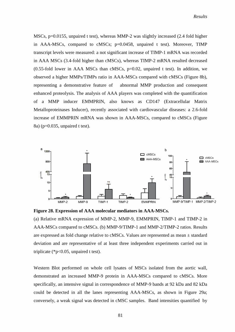
Results
81
MSCs, p=0.0155, unpaired t test), whereas MMP-2 was slightly increased (2.4 fold higher
in AAA-MSCs, compared to cMSCs; p=0.0458, unpaired t test). Moreover, TIMP
transcript levels were measured: a not significant increase of TIMP-1 mRNA was recorded
in AAA MSCs (3.4-fold higher than cMSCs), whereas TIMP-2 mRNA resulted decreased
(0.55-fold lower in AAA MSCs than cMSCs, p=0.02, unpaired t test). In addition, we
observed a higher MMPs/TIMPs ratio in AAA-MSCs compared with cMSCs (Figure 8b),
representing a demonstrative feature of abnormal MMP production and consequent
enhanced proteolysis. The analysis of AAA players was completed with the quantification
of a MMP inducer EMMPRIN, also knows as CD147 (Extracellular Matrix
Metalloproteinases Inducer), recently associated with cardiovascular diseases: a 2.6-fold
increase of EMMPRIN mRNA was shown in AAA-MSCs, compared to cMSCs (Figure
8a) (p=0.035, unpaired t test).
Figure 28. Expression of AAA molecular mediators in AAA-MSCs.
(a) Relative mRNA expression of MMP-2, MMP-9, EMMPRIN, TIMP-1 and TIMP-2 in
AAA-MSCs compared to cMSCs. (b) MMP-9/TIMP-1 and MMP-2/TIMP-2 ratios. Results
are expressed as fold change relative to cMSCs. Values are represented as mean ± standard
deviation and are representative of at least three independent experiments carried out in
triplicate (*p<0.05, unpaired t test).
Western Blot performed on whole cell lysates of MSCs isolated from the aortic wall,
demonstrated an increased MMP-9 protein in AAA-MSCs compared to cMSCs. More
specifically, an intensive signal in correspondence of MMP-9 bands at 92 kDa and 82 kDa
could be detected in all the lanes representing AAA-MSCs, as shown in Figure 29a;
conversely, a weak signal was detected in cMSC samples. Band intensities quantified by

Results
82
densitometry were normalized to cMSCs, considered as reference: MMP-9 expression by
AAA-MSCs resulted 149 times higher than cMSCs (Figure 29b).
These results on protein expression by AAA-MSCs are consistent with the above described
analysis on AAA tissue revealing MMP-9 positive signal in correspondence of
perivascular and stromal cells.
Figure 29. MMP-9 protein expression in AAA-MSCs.
(a) Immunoblot on AAA-MSCs and cMSCs cell lysates and bands quantification by
ImageJ densitometric software.
Figure 30a is a representative gelatin zymography performed on cMSC and AAA-MSC
conditioned media, to test MMP-2 and MMP-9 activity. MMP-9 secretion was mainly
detected in AAA-MSCs (lanes 1-4), as reflected by the digestive activity in
correspondence of 92 kDa, even if lane 4 displayed a weaker signal. Densitometry analysis
indicated a significant increase of MMP-9 activity in AAA-MSC conditioned media, in
comparison to cMSC samples (Figure 30a). Regarding MMP-2 secretion, we did not
observe relevant differences between healthy and AAA samples; indeed, the degradative
activity of MMP-2 could be broadly observed in AAA-MSCs and cMSCs. Band intensities
quantified by densitometry were normalized to cMSCs (Figure 30b).

Results
83
Figure 30. MMP-2 and MMP-9 activity assay on MSCs.
(a) Gelatin zymography performed on AAA-MSC and cMSC conditioned media: MMP-9
at 92 kDa could be detected only in AAA-MSC lanes (Lanes 1-4), whereas MMP-2 was
broadly identified in correspondence of 72 and 62 kDa, in all the lanes, included cMSCs
(Lanes 5-6); Lane 7 refers to HCT116, used as reference to identify MMP bands.
IMMUNOMODULATORY ACTIVITY OF AAA-MSCs: IN VITRO CO-CULTURE
WITH PHA-ACTIVATED PBMCs
HLA-G and IL-10 expression following MSC cultured under inflammatory conditions
MSC immunosuppressive function was performed under inflammatory conditions, created
trough a co-colture of MSCs seeded on culture flask as feeder layer and PHA-activated
PBMCs. Real Time PCR performed on MSCs after 72 h co-cultured with PHA-PBMCs
revelead a reduced immunomodulatory cytokine expression in AAA-MSCs, compared to
cMSCs.
At first, we analyzed IL-10 mRNA, which resulted 3-fold decreased in AAA-MSCs
(p=0.0386, unpaired t test) (Figure 31).

Results
84
Figure 31. IL-10 mRNA in AAA-MSCs after co-culture with PHA-PBMCs,
normalized to cMSCs.
Results are expressed as fold change relative to controls (cMSCs co-cultured with activated
PBMCs). Values are represented as mean ± standard deviation and are representative of at
least three independent experiments (*p<0.05; unpaired t test).
Beside IL-10, flow cytometer analysis was assayed to detect HLA-G expression on MSC
surface. Figure 32a that confirmed that HLA-G was expressed on cMSC surface, as
expected; interestingly, its expression resulted even before co-colture with inflammatory
cells. Conversely, HLA-G was not detected on AAA-MSC surface (Figure 32b).
Accordingly with this analysis, HLA-G was evaluated also through qPCR, resulting in a
decreased, even if not significant, expression of HLA-G mRNA in AAA-MSCs (4-fold
decreased in AAA-MSCs; no significant differences were observed), in comparison to
cMSCs exposed to the same inflammatory condition (Figure 32c).

Results
85
Figure 32. HLA-G expression in AAA-MSCs versus cMSCs.
HLA-G expression on AAA-MSCs and cMSCs, in basal condition, (a) without and (b)
after 72 h co-culture with activated PBMCs. Purple histograms refer to AAA-MSCs, while
white histograms are referred to cMSCs. Results are shown as a representative example of
at least three independent experiments. (c) HLA-G mRNA expression in AAA-MSCs
compared with cMSCs.
MSC effects on PBMC cell cycle
Among MSC clinical applications, many evidences support an effective
immunomodulatory activity and contextually to the aneurysm disease, as object of our
study, we tested this ability also on our cell model. As expected, cMSCs were able to
inhibit PHA-PBMC DNA synthesis, conversely to AAA-MSCs. As observed in BrDU
assay PHA-stimulated PBMCs cultured on cMSC feeder layer showed a lower
proliferation rate (p<0.05, one-way ANOVA test), whereas the proliferative rate measured
on PHA-PBMCs grown on AAA-MSCs was higher (p<0.05, one-way ANOVA test) and
quite comparable to PHA-PBMCs cultured alone (Figure 33).
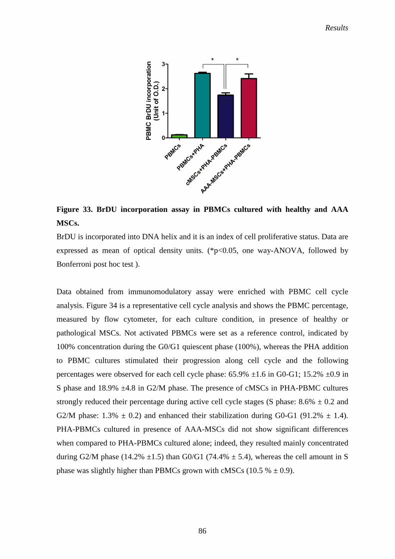
Results
86
Figure 33. BrDU incorporation assay in PBMCs cultured with healthy and AAA
MSCs.
BrDU is incorporated into DNA helix and it is an index of cell proliferative status. Data are
expressed as mean of optical density units. (*p<0.05, one way-ANOVA, followed by
Bonferroni post hoc test ).
Data obtained from immunomodulatory assay were enriched with PBMC cell cycle
analysis. Figure 34 is a representative cell cycle analysis and shows the PBMC percentage,
measured by flow cytometer, for each culture condition, in presence of healthy or
pathological MSCs. Not activated PBMCs were set as a reference control, indicated by
100% concentration during the G0/G1 quiescent phase (100%), whereas the PHA addition
to PBMC cultures stimulated their progression along cell cycle and the following
percentages were observed for each cell cycle phase: 65.9% ±1.6 in G0-G1; 15.2% ±0.9 in
S phase and 18.9% ±4.8 in G2/M phase. The presence of cMSCs in PHA-PBMC cultures
strongly reduced their percentage during active cell cycle stages (S phase: 8.6% ± 0.2 and
G2/M phase: 1.3% ± 0.2) and enhanced their stabilization during G0-G1 (91.2% ± 1.4).
PHA-PBMCs cultured in presence of AAA-MSCs did not show significant differences
when compared to PHA-PBMCs cultured alone; indeed, they resulted mainly concentrated
during G2/M phase (14.2% ±1.5) than G0/G1 (74.4% ± 5.4), whereas the cell amount in S
phase was slightly higher than PBMCs grown with cMSCs (10.5 % ± 0.9).

Results
87
Figure 34. PBMC cell cycle analysis following co-culture with PHA-PBMCs.
(a) PBMC distribution along cell cycle phases; (b) summary of cell percentage in each cell
cycle phase, calculated for all the culture conditions performed (**p<0.01, ***p<0.001;
one way-ANOVA and two way-ANOVA test, followed by Bonferroni post hoc test ).
Taken together, the results obtained from immunomodulatory assays, revealed a
compromised function in AAA-MSCs, that lost the property to regulate the immune
response as it can be observed for cMSCs, suggesting that inflammation may be more than
a histopathological feature in AAA pathogenesis and it may constitute a key process
influencing vascular resident MSC ordinary activities.
MMP-9 HYPER EXPRESSION BY MSCs UNDER PATHOLOGICAL CONDITIONS: POTENTIAL MECHANISMS TO MODULATE ITS EXPRESSION AT THE TRANSCRIPTIONAL LEVEL
cMSC influence on MMP transcription
MMPs are hyper expressed under an inflammatory context and many anti-inflammatory
approach can be applied to regulate their expression. For this purpose, we investigated on
the healthy cMSC potential to regulate MMP transcription by AAA-MSCs, appealing to
the secretion of immunomodulatory molecules. MMP transcription was evaluated by qPCR
and MMP-9 was shown to be reduced following co-culture of healthy and AAA MSCs (a
80% reduction observed in comparison to AAA-MSCs control; p=0.004, paired t test) and,
in a less evident measure, after AAA-MSC culture in presence of cMSC conditioned media
(0.8 fold lower than AAA-MSCs grown in conventional DMEM; p=0.0043, paired t test)

Results
88
(Figure 35b). Conversely, MMP-2 mRNA was not modulated by cMSCs (Figure 354a),
supporting the constitutive MMP-2 expression by cells of mesenchymal nature.
Inhibitory effect of anti-inflammatory cytokines on MMP-9 expression: the example of IL-
10
IL-10 is an anti-inflammatory cytokine, ordinarily expressed by human MSCs and
involved into immunomodulatory mechanism. We observed that the addition of soluble IL-
10 to AAA-MSC cultures, at the concentration of 20 ng/ml, for 24 h, resulted in the
reduced MMP-9 transcription (0.2 fold decreased, relative to untreated control, p=0.04,
paired t test) (Figure 35c).
Therefore, an anti-inflammatory approach, addressed toward vascular MSCs, could be
developed to restore their impaired function.
Figure 35. MMP modulation at the transcriptional level.
(a) MMP-2 and (b) MMP-9 transcription following 1) AAA-MSCs co-cultured with
cMSCs and 2) AAA-MSCs cultured in presence of cMSC conditioned media. (c) MMP-9
transcript decrease in AAA-MSCs exposed to anti-inflammatory cytokine IL-10. Results
are expressed as fold change relative to untreated AAA-MSCs. Values are represented as
mean ± standard deviation and are representative of three indipendent experiments
(*p<0.05, **p<0.01, ***p<0.001; paired t test). CM: conditioned media.

Results
89

Discussion
90
DISCUSSION

Discussion
91
The human vessel wall represents a dynamic component of the blood vessel system, being
made of distinct cell types, each of one contribute in a different manner to tissue
homeostasis. An altered working of specific cellular and molecular functions can occur, in
some cases as consequence to genetic defects or environmental factors, leading to vascular
disorders.
Blood vessel diseases display a heterogeneous localization along the arterial tree,
according to the structural characteristics of the vascular wall, that in turn depends on the
heterogeneity of the resident cells and their reactions to pathological alterations. Some
vascular segments are more susceptible to develop specific diseases and the large-diameter
vessel aorta represents an example of how structural heterogeneity influences disease
localization. Human aorta is composed of three segments: aortic arch, ascending aorta,
descending thoracic aorta and descending abdominal aorta. Abdominal aortic media has
peculiar morphological characteristics, consisting in a decreased elastin content
correspondent to a reduced number of lamellar units (28-32), in comparison to the thoracic
segment (55-60). This aspect, together with a poorer nutrient to the SMCs, makes
abdominal aorta more susceptible to aneurysm occurrence (Ruddy et al, 2008).
Abdominal aortic aneurysms (AAAs) are degenerative aortic affections, ascribed to an
extensive ECM proteolysis, which leads to the decrease of elastin and collagen proteins,
responsible of vessel wall tensile strength. The ECM turnover is a physiologic process,
necessary for the maintenance of tissue homeostasis and mediated by proteolytic enzymes,
among which MMPs are the most well characterized and studied (Delay et al, 2007). MMP
hyper expression and hyper activity can result into an exaggerate ECM degradation that,
together with the SMC depletion, reduce the vessel wall tone. The presence of an
inflammatory infiltrate, composed of T cells, B cells and macrophages, has been reported
in aneurysm-affected tissues. Generally, AAAs are distinguished between atherosclerotic
and inflammatory, basing on the different etiologic factor. On the basis of this
classification, IAAAs are primarily associated with a more extensive inflammatory process
and more evident signs and symptoms accompany their natural history. Non-inflammatory
aneurysms are mostly related to atherosclerotic complications, even if current researches
are investigating upon the nature of this relationship (Tonar et al, 2010). According to the
most validated theory, these aneurysms arise as consequence to intimal atherosclerotic
lesions. Beside these differences, the inflammatory infiltrate is extensively represented
among all the aneurysm forms and recent investigators raised the possibility of an immune-

Discussion
92
mediate origin for aneurysm initiation. One of the mechanisms supporting this view is the
so-called molecular mimicry, that is the sharing of common epitopes between foreign and
self antigens, taking to T cell activation also against self antigen (Ozsvath et al, 1996;
Oleszak et al, 2004). The same vessel wall components, such elastin, collagen or the
AAAP-40, have been proposed as antigen capable to induce T cell activation. In this
regard, an antigen-driven T cell response has been suggested as leading cause to aneurysm
development; moreover, aneurysm has been proposed as autoimmune disease (Hirose and
Tilson, 2001).
In this view, targeting the inflammatory disease affecting the vessel wall, could represent a
promising strategy to monitor aneurysm expansion and limit rupture cases, since wall
rupture following the aortic progression is responsible of the 65-85% death cases
(Sakalihasan and Limet, 2005).
Scientific research addressing the behaviour of human MSCs and their properties has
gained great interest and important findings, overcoming the ethical question regarding the
use of embryonic tissues as source of stem cells able to regenerate damaged tissues (ESCs)
(Reubinoff et al, 2000). Indeed, MSCs can be easily isolated from different source tissues,
including bone-marrow (Friednstein et al, 1970; Castro-Malaspina et al, 1980), fat (Zuk et
al, 2002), umbilical cord blood (Erices et al, 2000), vascular wall (Ergün et al, 2011) and
others. MSCs can be easily from human tissues and to expanded in in vitro cell cultures.
The well-known MSC properties of multilineage differentiation and proliferative potential,
can be exploited to treat many human pathological states, including cardiovascular
diseases, brain and spinal cord injury, connective tissue disorders (Barry and Murphy,
2004). Moreover, MSCs are hypoimmunogenic, representing an important resource for
transplantation medicine applications, such as GVHD additional therapeutic applications of
human MSCs can be extended to the affections with an inflammatory origin, (i.e. Chron’s
disease, autoimmune pathologies, etc) since MSCs possess the capacity to negatively
modulate immune reaction, exerting an anti-inflammatory function (Barry and Murphy,
2004; Chamberlain et al, 2007). This property is performed through different mechanisms,
mediated by the release of anti-inflammatory cytokines (TGF, IL-10), and enzymes (IDO,
NO), leading to the anergy and cell cycle blocking of T and B cells, inhibition of DC
maturation, inhibition of NK cell cytotoxicity (Le Blanc and Ringdén, 2007; Ryan et al,
2007).

Discussion
93
It is clear that MSCs could be effectively employed for many clinical applications, even if
optimization and standardization of isolation protocols have not been established yet.
Different tissue sources as well as different donor age determine a high heterogeneity
degree, so that MSC preparations contain distinct subsets of cells in terms of differentiation
and proliferative status (Hart, 2014). This may influence the outcome of MSC
administration as therapeutic tools.
Less is known about the potential side effects or, more particularly, the MSC participation
to disease initiation and progression. Some literature works have raised this issue,
suggesting that MSCs, under specific conditions, can undergo an impairment of their basic
functions, referring to their proliferative potential, differentiation and immunomodulation.
In these conditions, MSC behaviour can evolve into a pathologic direction, leading to the
initiation or the exacerbation of tissue damage. For example, MSCs isolated from patients
affected by osteoarthritis, determined by a progressive cartilage deficiency, demonstrated
low proliferative potential and reduced adipogenic and chondrogenic differentiation
(Murphy et al, 2002). Evidences on an ineffective immunomodulation have been reported
in a mouse model of collagen-induced arthritis (Li et al, 2010) and also in multiple
myeloma patients (Djouad et al, 2005). Few works addressing this topic suggest that a
defective MSC activity and immunosuppression could exert a crucial role in inflammatory
affections.
Given that many arterial diseases have an inflammatory basis, we found interesting the
study of the MSCs resident within the vascular wall. It has been discovered that endothelial
progenitors and cell populations with mesenchymal properties localize at the adventitia
level, occupying the boundary with media tunica (Zengin et al, 2006). Our research group
achieved an experience in this field, isolating MSC populations from vessel wall of healthy
donors (Pasquinelli et al, 2007; Pasquinelli et al, 2010). Interestingly, these cells displayed
high resistance to stressful conditions, indeed in our previous work (Valente et al, 2014)
the MSC isolation from cryopreserved arteries was successfully performed. Arteries
belonging to healthy donors were collected after 5-years store in liquid nitrogen; cells were
cultured according to standard conditions (37 °C; 5% CO2) and did not show signs of
sufferance, conversely they were characterized, expressing typical mesenchymal markers.
These Vascular-Wall MSCs (VW-MSCs) displayed adipogenic, osteogenic, chondrogenic
and angiogenic potential, clonogenic property and immunoregulatory activity. Exploring

Discussion
94
the MSC resistance and management of vascular diseased condition represents the next
step of our studies.
In this regard, AAA constitutes an ideal pathological model to perform vascular MSC
evaluation of their basic characteristics and functionality. We therefore collected twelve
aortic tissues from patients subjected to open repair for abdominal aortic aneurysm,
provided by the Vascular Surgery Unit of S. Orsola-Malpighi Hospital (Bologna). The first
observation derives from the in situ detection of MSCs, evaluated trough an
immunostaining for mesenchymal antigens on AAA sections. Beside the great amount of
inflammatory cells invading the AAA wall, cells positive to CD44 and CD90 could be
localized at the level of the perivascular niche.
Later, we focused on the MSC component of the aortic wall. Healthy aortic MSCs
(cMSCs) and whereas aneurysm-derived MSCs (AAA-MSCs) were compared on different
levels: in vitro morphological features, immunophenotype and molecular profile. A
consistent number of AAA-MSCs could be obtained from all the tissues we collected and
all the cell samples revealed basic characteristics comparable to our previous observations
on cMSCs.
AAA-MSCs displayed a typical fibroblastic shape and adherence to the plastic culture
substrate, representing primary peculiarity of MSC identification. Then, AAA-MSCs
showed a typical mesenchymal antigen expression pattern on their surface, as
demonstrated by flow cytometry: mesenchymal and perycite markers CD44, CD90, CD73,
CD146 and PDGFR-beta were broadly expressed on AAA-MSC surface, whereas
hematopoietic and endothelial antigens CD34, CD14 and CD44 were not detected. AAA-
MSC immunophenotype was totally specular to cMSC, as we observed for stemness
markers. We analyzed typical embryonic stem cell marker, represented by Nanog, Oct.4
and Sox-2, which posses a regulatory role in determining stem cell properties: these genes
compose a transcriptional core unit that activates downstream genes involved in
pluripotency and self-renewal of stem cells and, on the other hand, repress genes regulating
cell developmental processes (Chambers et al, 2003). This group of stemness markers
resulted to be expressed by AAA-MSCs, and the semi-quantitative analysis by Real Time
qPCR showed that no significant differences existed between healthy and pathological
MSCs.
Definitely, the pathological arterial remodeling and the inflammatory process affecting the
aortic wall, did not influence the basic properties of vascular resident MSCs.

Discussion
95
We therefore explored AAA-MSCs from a functional point of view, investigating on the
MMP expression and the ability to counteract the immune reaction. MSC mobilization
from bone marrow and their homing to diseased tissues, are mediated by MMPs that,
cleaving specific ECM substrates, allow MSC migration. In particular, among MMP
members, MMP-2 and MT-MMP-1 are constitutively expressed by human bone marrow
MSCs; a work by Ries et al (2007) showed the different MMP expression pattern in bone
marrow MSCs and how their expression and activity are influenced under specific
stimulations. In this regard, MMP-9 is known to be an inducible protein, indeed its
expression usually depends on external factors, like TNF-α. Contextually to aneurysm
initiation and progression, MMP-2 and MMP-9 have been demonstrated to play a decisive
role into ECM degradation (Tamarina et al, 1997; Longo et al, 2002). Based on these
premises, we evaluated expression levels of MMP-2, MMP-9 and their respective tissue
inhibitors, TIMP-1 and TIMP-2, in AAA-MSCs compared to healthy cMSCs. The most
relevant results were observed for MMP-9 expression, both at the mRNA and protein
levels: a prominent mRNA transcription (400-fold higher than cMSCs) and protein
production were observed in AAA-MSCs; MMP-2 and TIMP levels were quite similar
between the two groups of our study, whereas EMMPRIN, or CD147, was shown to be up-
regulated at the mRNA level in AAA-MSCs. According to literature studies, EMMPRIN
have been associated with atherosclerotic plaque (Yoon et al, 2005) and aneurysm
pathogenesis (Chen et al, 2009), as inducer of MMPs. Interestingly, EMMPRIN is
produced by stromal cells, so its upregulation supports the view of a MSC defective
functionality under pathological conditions. Further confirmation of data obtained on
vascular MSCs, derived from the tissue analysis of MMP-9 expression. An increased
MMP-9 mRNA was recorded in AAA tissue, which may contain different cell sources of
MMPs, especially inflammatory cells, like macrophages. Effectively, immunostaining for
MMP-9 labeled many inflammatory cells, but MSC-like populations positive to MMP-9
were also detected between perivascular cells of vasa vasorum district and stromal cells.
In order to elucidate the mechanism leading to this MSC degradative and pathological
phenotype, we tested AAA-MSCs for their immunomodulatory activity. The premise to the
second phase of the present study, derives from the extensive inflammatory infiltrate
observed inside aortic wall and the hypothesis that AAA-MSCs could have lost their innate
capacity to modulate the immune reaction. BrdU incorporation and cell cycle assays on
PHA-stimulated PBMCs cultured in presence of AAA-MSCs, indicated inflammatory cell

Discussion
96
ongoing along the cell cycle; PHA-PBMCs did not arrest at the Go/G1 stage, as occur in
presence of cMSCs.
The continuous PHA-PBMC proliferation could mean that AAA-MSCs behave like
quiescent cells, unable to arrest the proliferation of PHA-PBMCs, possibly explained with
the reduced expression of IL-10 and HLA-G, well-documented cytokines that mediate
some of the MSC-dependent immunosuppressive mechanisms.
The defective MSC immunomodulation could represent a critical step into AAA evolution;
MMPs can be regulated at many levels, including transcription, so we evaluated possible
regulatory mechanisms for MMP-9 transcript levels. Co-culture experiments of healthy
MSCs and pathological MSCs took to a significant reduction of MMP-9 transcription,
following direct contact between healthy and AAA-MSCs; a decrease, even if less evident,
was observed in AAA-MSCs cultured with cMSC conditioned media, suggesting that
cMSCs may regulate pathological mediators at the mRNA level, also in a paracrine
manner. Thus we supposed the involvement of IL-10, hat has been shown to inhibit MMP-
9 transcription in human placental trophoblast (Roth and Fischer, 1999). In fact,
preliminary data showed a negative MMP-9 transcriptional regulation in AAA-MSC
exposed to IL-10, indicating that IL-10 administration could represent a key strategy
of MMP-9 regulation, in addition to restore MSC immunomodulation (Rizzo et al, 2011).
Conclusions
The present study explored the basic and functional characteristics of a MSC population
isolated from aortic well affected by aneurysm. The molecular mechanisms involved in
arterial remodeling and chronic inflammation, characteristic of AAA disease, do not affect
the survival/self-renewal properties of the vascular MSC niche. Meanwhile, AAA-MSCs
displayed defective functions, in terms of ECM turnover and immunomodulation. Indeed,
AAA-MSCs showed to hyper express MMP-9 and its inducer EMMPRIN, in comparison
to cMSCs isolated from healthy aortic wall. Moreover, AAA-MSCs behaved in a weak
manner with respect to activated immune cells, which in turn show to proceed along the
cell cycle. This is opposed to what has been documented among MSCs isolated from
healthy aorta, suggesting a clear MSC dysfunction under pathological context. On the
other hand, both healthy MSCs that soluble factors associated with anti-inflammatory
abilities, showed to negatively modulate MMP-9 at the transcriptional level.

Discussion
97
These preliminary data suggest that AAA-MSC dysfunction can be resolved through an
anti-inflammatory approach; based on these observations, it would be of great importance
exploring the mechanisms and the molecular targets able to restore and improve MSC
immunomodulation. An approach focused on MSC function, aimed at potentiating their
therapeutic abilities could represent novel promising strategies to monitor aneurysm
progression, as well as other degenerative diseases in the cardiovascular medicine field.
Limits of our study
Limitations of our study reside in the selection of the control subjects, specifically the
limited number (3 healthy controls vs 12 AAA patients) and the absence of sex and age-
matching with patients. Regarding the age gap, we cannot exclude that our data on MMP-9
mRNA hyper-expression and activity could be a result of the physiologic vascular aging
process. Preliminary data on total RNA extracted from aortic tissue of two male subjects,
aged between 56 and 70, age-matched with AAA patients collected, showed a 9-fold
increased MMP-9 mRNA transcription, in comparison to healthy aortic tissues used in our
study (data not shown). Conversely, the MSCs isolated through enzymatic digestion
behaved like cMSCs from young donors, as revealed by results on MMP-9 mRNA and
immunomodulatory ability (data not shown). They were not used to extend the control
group in our study because their clinical data (i.e. cause of death associated with cerebral
hemorrhage, presence of vascular calcifications) were not in accordance with the inclusion
criteria. Thus, the age of control subjects does not significantly influence the differences
between our study subject groups, as the presence of the aortic dilation does. Our controls
can be considered as healthy subjects with no cardiovascular risk and as negative controls
for AAA molecular mediators expression in human perivascular MSCs.

Bibliography
BIBLIOGRAPHY

Bibliography
99
BIBLIOGRAPHY
Aggarwal, S., Qamar, A., Sharma, V., & Sharma, A. (2011). Abdominal aortic aneurysm:
A comprehensive review. Exp Clin Cardiol 16(1), 11–15.
Barry, F. P., & Murphy, J. M. (2004). Mesenchymal stem cells: clinical applications and
biological characterization. Int J Biochem Cell Biol 36, 568–584.
doi:10.1016/j.biocel.2003.11.001.
Barry, F., Boynton, R. E., Liu, B., & Murphy, J. M. (2001). Chondrogenic differentiation
of mesenchymal stem cells from bone marrow: differentiation-dependent gene expression
of matrix components. Exp Cell Res. 268, 189–200.
Bazzoni, G., & Dejana, E. (2004). Endothelial cell-to-cell junctions: molecular
organization and role in vascular homeostasis. Physiol Rev 84, 869–901.
doi:10.1152/physrev.00035.2003.
Biasetti J., Gasser T.C, Auer M., Hedin U., Labruto F. (2010). Hemodynamics of the
normal aorta compared to fusiform and saccular abdominal aortic aneurysms with
emphasis on a potential thrombus formation mechanism. Ann Biomed Eng, 38: 380–390.
Boyer, Laurie A. et al. (2005). Core Transcriptional Regulatory Circuitry in Human
Embryonic Stem Cells. Cell 122(6) , 947 – 956.
Brooks, A.R., Lelkes, P.I., Rubanyi, G.M. (2002). Gene expression profiling of human
aortic endothelial cells exposed to disturbed flow and steady laminar flow. Physiol
Genomic 9, 27–41.
Bunnell, B. a, Betancourt, A. M., & Sullivan, D. E. (2010). New concepts on the immune
modulation mediated by mesenchymal stem cells. Stem Cell Res Ther 1, 34.
doi:10.1186/scrt34.
Caplan AI. (1991). Mesenchymal stem cells. J Orthop Res 9:641–650.

Bibliography
100
Castro-Malaspina, B. H., Gay, E., Resnick, G., Broxmeyer, H. E., Meyers, P., & Moore, A.
S. (1980). Characterization of human bone marrow fibroblast colony-forming cells (CFU-
F) and their progeny. Blood 56(2).
Chamberlain, G., Fox, J., Ashton, B., & Middleton, J. (2007). Concise review:
mesenchymal stem cells: their phenotype, differentiation capacity, immunological features,
and potential for homing. Stem Cells 25, 2739–2749. doi:10.1634/stemcells.2007-0197.
Chambers, I., Colby, D., Robertson, M., Nichols, J., Lee, S., Tweedie, S., Smith A. (2003).
Functional expression cloning of Nanog, a pluripotency sustaining factor in embryonic
stem cells. Cell 113, 643–655.
Chen XF, Wang JA, Hou J, Gui C, Tang LJ, Chen XQ et al. (2009). Extracellular matrix
metalloproteinase inducer (EMMPRIN) is present in smooth muscle cells of human
aneurysmal aorta and is induced by angiotensin II in vitro. Clin Sci 116(11):819-826, doi:
10.1042/CS20080235.
Cohen, M., A. Meisser, L. Haenggeli, P. Bischof. (2006). Involvement of MAPK pathway
in TNF-α -induced MMP-9 expression in human trophoblastic cells. Mol Hum Reprod 12:
225–232.
Cook CL, Weiser MCM, Schwartz PE, Jones CL, Majack RA. (1994). Developmentally
timed expression of an embryonic growth phenotype in vascular smooth muscle cells. Circ
Res 79:189 –196.
Crawford JL, Stowe CL, Safi HJ, Hallman CH, Crawford ES. (1985). Inflammatory
aneurysms of the aorta. J Vasc Surg 2:113–124.
Daley, W. P., Peters, S. B., & Larsen, M. (2008). Extracellular matrix dynamics in
development and regenerative medicine. J Cell Sci 121, 255–264. doi:10.1242/jcs.006064.
Danese, S., M. Sans, F. Scaldaferri, A. Sgambato, S. Rutella, A. Cittadini, J. M. Pique, J.
Panes, J. A. Katz, A. Gasbarrini, and C. Fiocchi. (2006). TNF-α blockade down-regulates

Bibliography
101
the CD40/CD40L pathway in the mucosal microcircu- lation: a novel anti-inflammatory
mechanism of infliximab in Crohn’s disease. J Immunol 176: 2617–2624.
Davani, S., Marandin, A., Mersin, N., Royer, B., Kantelip, B., Herve, P., Etievent, J. P., &
Kantelip, J. P. (2003). Mesenchymal progenitor cells differentiate into an endothelial
phenotype, enhance vascular density, and improve heart function in a rat cellular
cardiomyoplasty model. Circulation 108, 11253– 11258.
De Bari C, Dell’Accio F, Tylzanowski P, Luyten FP. (2000). Multipotent mesenchymal
stem cells from adult human synovial membrane. Arthritis Rheum 44:1928-42.
Deng, W., Obrocka, M., Fischer, I., & Prockop, D. J. (2001). In vitro differentiation of
human marrow stromal cells into early progenitors of neural cells by conditions that
increase intracellular cyclic AMP. Biochem Biophys Res Commun 282, 148–152.
Di Nicola M, Carlo-Stella C, MagniMet al. (2002). Humanbonemarrow stromal cells
suppress T lymphocyte proliferation induced by cellular or nonspecific mitogenic stimuli.
Blood 99:3838– 43.
Djouad, F., Fritz, V., Apparailly, F., Louis-Plence, P., Bony, C., Sany, J et al. (2005).
Reversal of the immunosuppressive properties of mesenchymal stem cells by tumor
necrosis factor alpha in collagen-induced arthritis. Arthritis Rheum 52(5), 1595–1603.
doi:10.1002/art.21012.
Dominici M ,Le Blanc K, Mueller I, Slaper-Cortenbach I, Marini F, Krause D et al. (2006).
Minimal criteria for defining multipotent mesenchymal stromal cells. The International
Society for Cellular Therapy position statement. Cytotherapy 8(4):315-7.
Dubost C, Allary M, Oeconomos SN. (1952) Resecetion of an aneurysm of the abdominal
aorta: Reestablishment of the Continuity by a Preserved Human Arterial Graft, with Result
After Five Months. AMA Arch Surg 64(3):405-408.
doi:10.1001/archsurg.1952.01260010419018.

Bibliography
102
Ergün, S., Tilki, D., & Klein, D. (2011). Vascular wall as a reservoir for different types of
stem and progenitor cells. Antioxid Redox Signal 15(4), 981–995.
doi:10.1089/ars.2010.3507.
Erices A, Conget P, Minguell JJ. (2000). Mesenchymal progenitor cells in human
umbilical cord blood. Br J Haematol 109(1):235-42.
Eyre ER, Paz MA, Gallop PM. (1984). Cross-linking in collagen and elastin. Annu Rev
Biochem 53: 717–748.
Fini, ME, Cook, JR.,Mohan, R., Brinckerhoff, CE. (1998). Regulation of matrix
metalloproteinase gene expression. In: Parks, WC.; Mecham, RP., editors. Matrix
Metalloproteinases. New York: Academic; p. 299-356.
Friedenstein AJ, Chailakhjan RK, Lalykina KS. (1970). The development of fibroblast
colonies in monolayer cultures of guinea-pig bone marrow and spleen cells. Cell Tissue
Kinet 3:393–403.
Friedenstein AJ, Gorskaja JF, Kulagina NN. (1976). Fibroblast precursors in normal and
irradiated mouse hematopoietic organs. Exp Hematol 4:267–274.
G. Martin. (1981).Proc. Natl. Acad. Sci. U.S.A. 78, 7634.
Gabison, E. E., Hoang-Xuan, T., Mauviel, A., & Menashi, S. (2005). EMMPRIN/CD147,
an MMP modulator in cancer, development and tissue repair. Biochimie 87, 361–368.
doi:10.1016/j.biochi.2004.09.023.
Gasser, T. C., Ogden, R. W., & Holzapfel, G. a. (2006). Hyperelastic modelling of arterial
layers with distributed collagen fibre orientations. J R Soc Interface 15–35.
doi:10.1098/rsif.2005.0073.
Glennie S, Soeiro I, Dyson PJ, Lam EW, Dazzi F. (2005). Bone marrow mesenchymal
stem cells induce division arrest anergy of activated T cells. Blood 105:2821–7.

Bibliography
103
Gomez DE, Alonso DF, Yoshiji H, Thorgeirsson UP. (1997). Tissue inhibitors of
metalloproteinases: structure, regulation and biological functions. Eur J Cell Biol. 74:111–
122.
Gómez-Gaviro MV, Lovell-Badge R, Fernández-Avilés F, Lara-Pezzi E. (2012). The
vascular stem cell niche. J Cardiovasc Transl Res 5(5):618-30.
Goodell, M.A. (2003). Stem-cell “plasticity”: befuddled by the muddle. Curr. Opin.
Hematol. 10, 208–213.
Hamano, K., T. S. Li, M. Takahashi, T. Kobayashi, B. Shirasawa, H. Ito, and N. Zempo.
(2003). Enhanced tumor necrosis factor- expression in small sized abdominal aortic
aneurysms. World J Surg 27: 476–480.
Hart, D.A. (2014). Why Mesenchymal Stem/Progenitor Cell Heterogeneity in Specific
Ennvironments? –Implications for Tissue Engineering Applications Following Injury or
Degeneration of Connective Tissues. J. Biomed Sci Eng. 7, 526-532.
Hingorani, A., E. Ascher, M. Scheinman, W. Yorkovich, P. DePippo, C. T. Ladoulis, and
S. Salles-Cunha. (1998). The effect of tumor necrosis factor binding protein and
interleukin-1 receptor antagonist on the development of ab- dominal aortic aneurysms in a
rat model. J Vasc Surg 28: 522–526.
Hirose, H. and Tilson, M. D. (2001). Abdominal Aortic Aneurysm As an Autoimmune
Disease. Ann N Y Acad Sci 947: 416–418. doi: 10.1111/j.1749-6632.2001.tb03976.x.
Holzapfel, G. A, Gasser, T. C., Ogden, R. W. (2000). A new constitutive framework for
arterial wall mechanics and a comperative study of material models. J Elasticity, 61, 1–48.
Horwitz, E. M., Prockop, D. J., Fitzpatrick, L. A., Koo, W. W., Gordon, P. L., Neel, M.,
Sussman, M., Orchard, P., Marx, J. C., Pyeritz, R. E., & Brenner, M. K. (1999).
Transplantability and therapeutic effects of bone marrow-derived mesenchymal cells in
children with osteogenesis imperfecta. Nat Med 5, 309–313.

Bibliography
104
Jacob, M. P., Badier-Commander, C., Fontaine, V., Benazzoug, Y., Feldman, L., Michel, J.
B. (2001). Extracellular matrix remodeling in the vascular wall. Pathologie-Biologie,
49(01), 326–332. doi:10.1016/S0369-8114(01)00151-1.
Jiang, Y., Jahagirdar, B.N., Reinhardt, R.L., Schwartz, R.E., Keene, C.D., Ortiz-Gonzalez,
X.R. et al. (2002a). Pluripotency of mesenchymal stem cells derived from adult marrow.
Nature 418, 41–49.
Jiang, Y., Vaessen, B., Lenvik, T., Blackstad, M., Reyes, M., Verfaillie, C.M. (2002b).
Multipotent progenitor cells can be isolated from postnatal murine bone marrow, muscle,
and brain. Exp. Hematol. 30, 896–904.
Jones, D. L., Wagers, A. J. (2008). No place like home: anatomy and function of the stem
cell niche. Nat Rev Mol Cell 9,11–21. doi:10.1038/nrm2319.
Juvonen, J., H. M. Surcel, J. Satta, A. M. Teppo, A. Bloigu, H. Syrjala, J. et al. (1997).
Elevated circulating levels of inflammatory cytokines in patients with abdominal aortic
aneurysm. Arterioscler Thromb Vasc Biol 17: 2843–2847.
Kapp, U., Mertelsmann, R. (2001). Plasticity of stem cells. Ernst Schering Res Found
Workshop 1–17.
Karystinou A, Dell'Accio F, Kurth TB, Wackerhage H, Khan IM, Archer CW et al. (2009).
Distinct mesenchymal progenitor cell subsets in the adult human synovium. Rheumatology
48(9):1057-64. doi: 10.1093/rheumatology/kep192. Epub 2009 Jul 14.
Koc, O. N., Day, J., Nieder, M., Gerson, S. L., Lazarus, H. M., & Krivit, W. (2002).
Allogeneic mesenchymal stem cell infusion for treatment of metachromatic
leukodystrophy (MLD) and Hurler syndrome (MPS-IH). Bone Marrow Transplant 30,
215–222.

Bibliography
105
Koch AE, Haines GK, Rizzo RJ, et al. (1990) Human abdominal aortic aneurysms-
immunophenotypic analysis suggesting an immunemediated response. Am J Path
137:1199-1213.
Kotze C. W. and Ahmed I. G. (2011). Etiology and Pathogenisis of Aortic Aneurysm,
Etiology, Pathogenesis and Pathophysiology of Aortic Aneurysms and Aneurysm Rupture,
Prof. Reinhart Grundmann (Ed.), ISBN: 978-953-307-523-5, InTech, doi: 10.5772/18257.
Lakshmipathy, U., Verfaillie, C. (2005). Stem cell plasticity. Blood Reviews 19, 29–38.
doi:10.1016/j.blre.2004.03.001.
Latifi HR, Heiken JP. (1992). CT of inflammatory abdominal aortic aneurysm:
development from an uncomplicated atherosclerotic aneurysm. J Comput Assist Tomogr
16,484-486.
Le Blanc, K., Ringdén, O. (2007). Immunomodulation by mesenchymal stem cells and
clinical experience. J Intern Med 262, 509–525. doi:10.1111/j.1365-2796.2007.01844.x
Le Rond S, Azema C, Krawice-Radanne I et al. (2006). Evidence to support the role of
HLA-G5 in allograft acceptance through induction of immuno-suppressive/regulatory
suppressive/regulatory T cells. J Immunol 176, 3266–3276.
Lehoux, S., Castier, Y., Tedgui, A. (2006). Molecular mechanisms of the vascular
responses to haemodynamic forces. J Int Med 259, 381–392. doi:10.1111/j.1365-
2796.2006.01624.x.
Li, B., Fu, J., Chen, P., Zhuang, W. (2010). Impairment in Immunomodulatory Function of
Mesenchymal Stem Cells from Multiple Myeloma Patients. Arch Med Res 41(8), 623–633.
doi:10.1016/j.arcmed.2010.11.008.
Li, Y.-S. J., Haga, J. H., & Chien, S. (2005). Molecular basis of the effects of shear stress
on vascular endothelial cells. J Biomech 38, 1949–1971.
doi:10.1016/j.jbiomech.2004.09.030.

Bibliography
106
Lindholt JS, Vammen S, Fasting H, Henneberg EW, Heickendorff L. (2000). The plasma
level of matrix metalloproteinase 9 may predict the natural history of small abdominal
aortic aneurysms: a preliminary study. Eur J Vasc Endovasc Surg 20: 281–85.
Livak KJ. Schmittgen TD. (2001) Analysis of relative gene expression data using real-time
quantitative PCR and the 2(-Delta Delta C(T)) Method. Methods 25 (4):402-8.
Longo GM, Xiong W, Greiner TC, Zhao Y, Fiotti N, Baxter BT. (2002) Matrix
metalloproteinases 2 and 9 work in concert to produce aortic aneurysms. J Clin Invest
110(5):625-632.
Lu, P., Takai, K., Weaver, V. M., & Werb, Z. (2011). Extracellular Matrix degradation and
remodeling in development and disease. Cold Spring Harb Perspect Biol 3, 1–24.
doi:10.1101/cshperspect.a005058.
M. Evans and M. Kaufman. (1981). Nature 292, 154.
Malek, A.M., Alper, S.L., Izumo, S. (1999). Hemodynamic shear stress and its role in
atherosclerosis. JAMA 282, 2035–2042.
Mannello F. (2006). Multipotent mesenchymal stromal cell recruitment, migration, and
differentiation: what have matrix metalloproteinases got to do with it? Stem Cells.
24:1904-1907.
Matsuki, T., K. Isoda, R. Horai, A. Nakajima, Y. Aizawa, K. Suzuki, F. et al (2005).
Involvement of tumor necrosis factor in the development of T cell-dependent aortitis in
interleukin-1 receptor antagonist-deficient mice. Circulation 112: 1323–1331.
McElreavey, K.D., Irvine, A.I., Ennis, K.T., McLean, W.H. (1991). Isolation, culture and
characterisation of fibroblast-like cells derived from the wharton's jelly portion of human
umbilical cord. Biochem. Soc. Trans 19, 29S.

Bibliography
107
McGregor JC, Pollock JG, Anton HC. (1975). The value of ultrasonography in the
diagnosis of abdominal aortic aneurysm. Scott Med J 20: 133–37.
McMaster MT, Librach CL, Zhou Y et al. (1995). Human placental HLA-G expression is
restricted to differentiated cytotrophoblasts. J Immunol 154:3771–3778.
Meshorer E., Misteli T. (2006). Chromatin in pluripotent embryonic stem cells and
differentiation. Nat Rev Mol Cell Biol 7(7):540-6.
Michel, J. B., Martin-Ventura, J. L., Egido, J., Sakalihasan, N., Treska, V., Lindholt, J. et
al. (2011). Novel aspects of the pathogenesis of aneurysms of the abdominal aorta in
humans. Cardiovasc Res 90, 18–27. doi:10.1093/cvr/cvq337.
Murphy, J. M., Dixon, K., Beck, S., Fabian, D., Feldman, A., & Barry, F. (2002). Reduced
chondrogenic and adipogenic activity of mesenchymal stem cells from patients with
advanced osteoarthritis. Arthritis Rheum 46(3), 704–713. doi:10.1002/art.10118
Nishikawa D.C, Martin Jakt L., Era T. (2007) Embryonic stem-cell culture as a tool for
developmental cell biology. Nat Rev MolCell Biol 8, 502-507 doi:10.1038/nrm2189
Nitecki SS, Hallett JW Jr, Stanson AW, et al. (1996). Inflammatory abdominal aortic
aneurysms: new clinical implications from a case control study. J Vasc Surg 23:860-869.
O'Connor, T. P.,Crystal, R. G. (2006). Genetic medicines: treatment strategies for
hereditary disorders, Nat Rev Gen 7, 261–276 doi:10.1038/nrg182
Oleszak EL, Chang JR, Friedman H, Katsetos CD, Platsoucas CD. (2004). Theiler’s virus
infection: a model for multiple sclerosis. Clin Microbiol Rev 17:174 –207.
Ozsvath K. J., Hirose, H., Xia, S. and Tilson, M. D. (1996). Molecular Mimicry in Human
Aortic Aneurysmal Diseases. Ann N Y Acad Sci, 800: 288–293. doi: 10.1111/j.1749-
6632.1996.tb33335.x.

Bibliography
108
Paravastu SCV, Jayarajasingam R, Cottam R, Palfreyman SJ, Michaels JA, Thomas SM.
(2014). Endovascular repair of abdominal aortic aneurysm. Cochrane Database Syst Rev,
Issue 1. Art. No.: CD004178. doi: 10.1002/14651858.CD004178.pub2.
Parodi JC, Palmaz JC, Barone HD. (1991). Transfemoral intraluminal graft implantation
for abdominal aortic aneurysms. Ann Vasc Surg 5: 491–99.
Parks WC, Wilson CL, Lopez-Boado YS. (2004) Matrix metalloproteinases as modulators
of inflammation and innate immunity. Nat Rev Immunol. 4:617–629.
Pasquinelli G, Preda P, Gargiulo M, et al. (1993). An immunohistochemical study of
inflammatory abdominal aortic aneurysms. J Submicrosc Cytol Pathol 25:103-112.
Pasquinelli G, Tazzari PL, Vaselli C, Foroni L, Buzzi M, Storci G et al. (2007). Thoracic
aortas from multiorgan donors are suitable for obtaining resident angiogenic mesenchymal
stromal cells. Stem Cells 25: 1627–1634.
Pasquinelli, G., Tazzari, P. L., Vaselli, C., Foroni, L., Buzzi, M., Storci, G et al. (2007).
Thoracic aortas from multiorgan donors are suitable for obtaining resident angiogenic
mesenchymal stromal cells. Stem Cells 25, 1627–1634. doi:10.1634/stemcells.2006-0731.
Pennell RC, Hollier LH, Lie JT, Bernatz PE, Joyce JW, Pairolero PC, Cherry KJ, Hallett
JW. (1985). Inflammatory abdomi- nal aortic aneurysms: a thirty-year review. J Vasc Surg
2(6):859–869.
Pfeffer, K. (2003). Biological functions of tumor necrosis factor cytokines and their
receptors. Cytokine Growth Factor Rev. 14: 185–191.
Pittenger, M. F., Mackay, a M., Beck, S. C., Jaiswal, R. K., Douglas, R., Mosca, J. D. et al.
(1999). Multilineage potential of adult human mesenchymal stem cells. Science 284, 143–
147. doi:10.1126/science.284.5411.143
Praillet C, Grimaud JA, Lortat-Jacob H. (1998). Les protéoglycannes. I. Molécules aux
multiples fonctions ... futures molécules thérapeu- tiques ? M/S 14: 412-20.
Bibliography
109
Psaltis, P. J., Harbuzariu, A., Delacroix, S., Holroyd, E. W., Simari, R. D. (2011). Resident
vascular progenitor cells - diverse origins, phenotype and function. J Cardiovasc Transl
Res 4(2), 161–176. doi:10.1007/s12265-010-9248-9.
Raffetto, J. D., & Khalil, R. A. (2008). Matrix metalloproteinases and their inhibitors in
vascular remodeling and vascular disease. Biochem Pharmacol 75(2), 346–359.
doi:10.1016/j.bcp.2007.07.004.
Resnick, N., Yahav, H., & Shay-Salit, a. (2003). Fluid shear stress and the vascular
endothelium: for better and for worse. Prog Biophys Mol Biol 81, 177–199.
Reubinoff, B.E., Pera, M.F., Fong, C.Y., Trounson, A., Bongso, A. (2000). Embryonic
stem cell lines from human blastocysts: somatic differentiation in vitro. Nat. Biotechnol.
18, 399–404.
Ries, C., Egea, V., Karow, M., Kolb, H., Jochum, M., Neth, P. (2007). MMP-2, MT1-
MMP, and TIMP-2 are essential for the invasive capacity of human mesenchymal stem
cells: Differential regulation by inflammatory cytokines. Blood 109, 4055–4063.
doi:10.1182/blood-2006-10-051060.
Ross R. (1973).The elastic fiber. J Histochem Cytochem 21, 199–208.
Roth, I., & Fisher, S. J. (1999). IL-10 is an autocrine inhibitor of human placental
cytotrophoblast MMP-9 production and invasion. Dev Biol 205(1), 194–204.
doi:10.1006/dbio.1998.9122.
Rouas-Freiss N, Goncalves RM, Menier C et al. (1997). Direct evidence to support the role
of HLA-G in protecting the fetus from maternal uterine natural killer cytolysis. Proc Natl
Acad Sci 94: 11520–11525.
Ruddy, J. M., Jones, J. a., Spinale, F. G., Ikonomidis, J. S. (2008). Regional heterogeneity
within the aorta: Relevance to aneurysm disease. Thorac Cardiovasc Surg d 136(5), 1123–
1130. doi:10.1016/j.jtcvs.2008.06.027.

Bibliography
110
Ryan, J. M., Barry, F., Murphy, J. M., & Mahon, B. P. (2007). Interferon-γ does not break,
but promotes the immunosuppressive capacity of adult human mesenchymal stem cells.
Clin Exp Immunol 149, 353–363. doi:10.1111/j.1365-2249.2007.03422.x.
S.M. Arribas, A. Hinek, M.C. Gonzalez. (2006) Elastic fibres and vascular structure in
hypertension. Pharmacol Ther 111 771–791.
Sakalihasan N, Limet R, Defawe OD. (2005). Abdominal aortic aneurysm. Lancet 6;
365(9470):1577-89.
Sanchez-Ramos, J. R. (2002). Neural cells derived from adult bone marrow and umbilical
cord blood. J Neurosci Res 69, 880–893.
Sarén P, Welgus HG, Kovanen PT. (1996). TNF-alpha and IL-1beta selectively induce
expression of 92-kDa gelatinase by human macrophages. J Immunol 157(9):4159-65.
Sasaguri Y, Murahashi N, Sugama K, Kato S, Hiraoka K, Satoh T, Isomoto H, Morimatsu
M. (1994). Development-related changes in matrix metalloproteinase expression in human
aortic smooth muscle cells. Lab Invest 71:261–269.
Sato K, Ozaki K, Oh I et al. (2007). Nitric oxide plays a critical role in suppression of T-
cell proliferation by mesenchymal stem cells. Blood 109:228–34.
Satoh, H., M. Nakamura, M. Satoh, T. Nakajima, H. Izumoto, C. Maesawaet al. (2004).
Expression and localization of tumour necrosis factor-α and its converting enzyme in
human abdominal aortic aneurysm. Clin. Sci. 106: 301–306.
Schofield, R. (1978). The relationship between the spleen colony-forming cell and the
haemopoietic stem cell. Blood Cells 4, 7–25.
Selmani, Z., Naji, A., Zidi, I., Favier, B., Gaiffe, E., Obert, L. et al. (2008). Human
leukocyte antigen-G5 secretion by human mesenchymal stem cells is required to suppress

Bibliography
111
T lymphocyte and natural killer function and to induce CD4+CD25highFOXP3+
regulatory T cells. Stem Cells 26, 212–222. doi:10.1634/stemcells.2007-0554.
Shi, M., Liu, Z. W., & Wang, F. S. (2011). Immunomodulatory properties and therapeutic
application of mesenchymal stem cells. Clin Exp Immunol 164, 1–8. doi:10.1111/j.1365-
2249.2011.04327.x
SilvaWAJr, Covas DT, Panepucci RA, et al. (2003). The profile of gene expression of
human marrow mesenchymal stem cells. Stem Cells 21:661- 669.
Simmons, P. J., Torok-Storb, B. (1991). Identification of stromal cell precursors in human
bone marrow by a novel monoclonal antibody, STRO-1. Blood 78, 55–62.
Singer, N.G.; Caplan, A.I.(2011). Mesenchymal stem cells: Mechanisms of inflammation.
Annu Rev Pathol 6, 457–478.
Sternlicht, M., & Werb, Z. (2009). How Matrix metalloproteinases regulate cell behavior.
Annu Rev Cell Biol 463–516. doi:10.1146/annurev.cellbio.17.1.463.
Sterpetti AV, Cucina A, D’Angelo LS, Cardillo B, Cavallaro A. (1993). Shear stress
modulates the proliferation rate, protein synthesis, and mitogenic activity of arterial
smooth muscle cells. Surgery 113, 691–699.
Sterpetti AV, Hunter WJ, Feldhaus RJ, et al. (1989). Inflammatory aneurysms of the
abdominal aorta: incidence, pathologic, and etiologic considerations. J Vasc Surg 9:643-
650.
Tamarina NA, McMillan WD, Shively VP, Pearce WH. (1997). Expression of matrix
metalloproteinases and their inhibitors in aneurysms and normal aorta. Surgery
122(2):264-272.
Tang, T., Boyle, J. R., Dixon, a K., Varty, K. (2005). Inflammatory abdominal aortic
aneurysms. Eur J Vasc Endovasc Surg 29, 353–362. doi:10.1016/j.ejvs.2004.12.009.

Bibliography
112
Taylor, S. M., Jones, P. A. (1982). Changes in phenotypic expression in embryonic and
adult cells treated with 5-azacytidine. J Cell Physiol 111, 187–194.
Thomson, J. a, Itskovitz-Eldor, J., Shapiro, S. S., Waknitz, M. a, Swiergiel, J. J., Marshall,
V. S., Jones, J. M. (1998). Embryonic stem cell lines derived from human blastocysts.
Science 282 1145–1147. doi:10.1126/science.282.5391.1145.
Thomson, J. a, Kalishman, J., Golos, T. G., Durning, M., Harris, C. P., Becker, R. a, &
Hearn, J. P. (1995). Isolation of a primate embryonic stem cell line. Proc Natl Acad Sci
92, 7844–7848. doi:PMC41242.
Tonar, Z., Witter, K., Křížková, V., Eberlová, L., Kočová, J., Moláček, J., et al. (2010).
Stereological tools for quantitative microscopy of the aortic wall with focus on the
abdominal aortic aneurysm. Microscopy: Science, Technology, Applications and
Education 926–935.
Uccelli A., Moretta L., Pistoia V. (2008) Mesenchymal stem cells in health and disease.
Nat Rev Immy 8, 726-736. doi:10.1038/nri2395
Valente S., Alviano F., Ciavarella C., Buzzi M., Ricci F., Tazzari P. L et al. (2014). Human
cadaver multipotent stromal/stem cells isolated from arteries stored in liquid nitrogen for 5
years. Stem Cell Res Ther 5(1), 8.
Wagenseil J. E., & Mecham R. P. (2009). Vascular extracellular matrix and arterial
mechanics. Physiol Rev 89, 957–989. doi:10.1152/physrev.00041.2008.
Walker DI, Bloor K, Williams G, Gillie I. (1972). Inflammatory aneurysms of the
abdominal aorta. Br J Surg 59:609-614.
Waterman RS, Tomchuck SL, Henkle SL, Betancourt AM. (2010). A new mesenchymal
stem cell (MSC) paradigm: polarization into a pro-infl ammatory MSC1 or an
Immunosuppressive MSC2 phenotype. PLoS One 5:e10088.

Bibliography
113
Visse, R., Nagase, H. (2003). Matrix metalloproteinases and tissue inhibitors of
metalloproteinases: Structure, function, and biochemistry. Circ Res 92, 827–839.
doi:10.1161/01.RES.0000070112.80711.3D.
Xia S, Ozsvath K, Hirose H, Tilson MD. (1996). Partial amino acid sequence of a novel
40-kDa human aortic protein, with vitronectin-like, fibrinogen-like, and calcium binding
domains: aortic aneurysm associated protein-40 (AAAP-40) [human MAGP-3, proposed].
Biochem Biophys Res Commun 219:36 –39.
Yoon YW, Kwon HM, Hwang KC, Choi EY, Hong BK, Kim D et al. (2005). Upstream
regulation of matrix metalloproteinase by EMMPRIN; extracellular matrix
metalloproteinase inducer in advanced atherosclerotic plaque. Atherosclerosis 180 37–44.
Yoshida Y, Sue W, Okano M, Oyama T, Yamane T, Mitsumata M. (1990). The effects of
augmented hemodynamic forces on the progression and topography of atherosclerotic
plaques. Ann. NY Acad. Sci. 598, 256–273. (doi:10.1111/j.1749-6632. 1990.tb42298.x).
Zengin, E., Chalajour, F., Gehling, U. M., Ito, W. D., Treede, H., Lauke, H et al. (2006).
Vascular wall resident progenitor cells: a source for postnatal vasculogenesis.
Development 133, 1543–1551. doi:10.1242/dev.02315.
Zuk PA, Zhu M, Ashjian P, De Ugarte DA, Huang JI, Mizuno H et al. 2002. Human
adipose tissue is a source of multipotent stem cells. Mol Biol Cell 13(12):4279-95.