Chronology of asbestos cancer discoveries: · PDF fileChronology of Asbestos Cancer...
14
American Journal of Industrial Medicine 27593-606 (1995) COMMENTARY Chronology of Asbestos Cancer Discoveries: Experimental Studies of the Saranac Laboratory Gerrit W.H. Schepers, MD, SCD This commentary challenges a recently published perception that Dr. Le Roy Upson Gardner had not actually discovered in 1942 that inhaled chrysotile fibers could induce malignant neoplasia in mice. The handwritten laboratory notes and some of Dr. Gard- ner's slides have recently been found. They verify that the tumors he saw in the mice included truly malignant neoplasms. Gardner had by then also accumulated 11 cases of human lung cancer (tw&'mesotheliomas) derived from Quebec asbestos miners and millers. An inhalation study designed by Dr. Gardner and conducted between 1951 and 1954, using cancer-insusceptible mice, yielded neoplasia risk ratio of 5.7 compared with control animals. The studies also showed that the primary effect of chrysotile is to cause epithelialproliferation in alveoli adjacent to bronchioles. Chrysotile type asbestos bodies were shown to remain only transiently ferruginous, but even though invisible in direct light they can be visualized at high magnification through use of phase contrast and polarized light micrography . 0 1995 Wiley-Liss, Inc. Key words: Saranac laboratory, asbestos, history of medicine, experimental studies INTRODUCTION In a recent Letter to the Editor, Dr. Enterline [1993] seems to question whether Drs. Gardner, Vorwald, or Schepers, as directors of the former Saranac Laboratory, ever discovered, through animal experimentation, whether chrysotile asbestos dust is capable of inducing neoplasia. His interpretation of those documents he had available seems to be that Dr. Gardner had found in 1942 only autogenous adenomas in the lungs of mice that had inhaled chrysotile long fiber dust. Perhaps Dr. Enterline, who appears well informed on many matters, has nevertheless not yet read all that is available on this subject. Gardner had written letters and reports designating some of the mouse tumors as malignant and he attributed the tumors to inhaled chrysotile asbestos fibers, even though the mice had not developed asbestosis. In a companion discussion by Drs. Egilman and Hardy [1993], allusion to corruption of the early scientific literature on asbestos cancers appears. This causes Institute of Industrial and Forensic Medicine, McLean, VA. Address reprint requests to Dr. Gemt W.H. Schepers, Institute of Industrial and Forensic Medicine, 6527 Sunny Hill Court, McLean, VA 22101. Accepted for publication June 22, 1994. 0 1995 Wiley-Liss, Inc.
Transcript of Chronology of asbestos cancer discoveries: · PDF fileChronology of Asbestos Cancer...

American Journal of Industrial Medicine 27593-606 (1995)
COMMENTARY
Chronology of Asbestos Cancer Discoveries: Experimental Studies of the Saranac Laboratory Gerrit W.H. Schepers, MD, SCD
This commentary challenges a recently published perception that Dr. Le Roy Upson Gardner had not actually discovered in 1942 that inhaled chrysotile fibers could induce malignant neoplasia in mice. The handwritten laboratory notes and some of Dr. Gard- ner's slides have recently been found. They verify that the tumors he saw in the mice included truly malignant neoplasms. Gardner had by then also accumulated 11 cases of human lung cancer (tw&'mesotheliomas) derived from Quebec asbestos miners and millers. An inhalation study designed by Dr. Gardner and conducted between 1951 and 1954, using cancer-insusceptible mice, yielded neoplasia risk ratio of 5.7 compared with control animals. The studies also showed that the primary effect of chrysotile is to cause epithelialproliferation in alveoli adjacent to bronchioles. Chrysotile type asbestos bodies were shown to remain only transiently ferruginous, but even though invisible in direct light they can be visualized at high magnification through use of phase contrast and polarized light micrography . 0 1995 Wiley-Liss, Inc.
Key words: Saranac laboratory, asbestos, history of medicine, experimental studies
INTRODUCTION In a recent Letter to the Editor, Dr. Enterline [1993] seems to question whether
Drs. Gardner, Vorwald, or Schepers, as directors of the former Saranac Laboratory, ever discovered, through animal experimentation, whether chrysotile asbestos dust is capable of inducing neoplasia. His interpretation of those documents he had available seems to be that Dr. Gardner had found in 1942 only autogenous adenomas in the lungs of mice that had inhaled chrysotile long fiber dust. Perhaps Dr. Enterline, who appears well informed on many matters, has nevertheless not yet read all that is available on this subject. Gardner had written letters and reports designating some of the mouse tumors as malignant and he attributed the tumors to inhaled chrysotile asbestos fibers, even though the mice had not developed asbestosis.
In a companion discussion by Drs. Egilman and Hardy [1993], allusion to corruption of the early scientific literature on asbestos cancers appears. This causes
Institute of Industrial and Forensic Medicine, McLean, VA. Address reprint requests to Dr. Gemt W.H. Schepers, Institute of Industrial and Forensic Medicine, 6527 Sunny Hill Court, McLean, VA 22101. Accepted for publication June 22, 1994.
0 1995 Wiley-Liss, Inc.

594 Schepers
me to wonder whether Gardner is posthumously and 50 years later accused of evil doing. Dr. Vorwald is likewise cast in a rather unfavorable light, but this is more difficult to judge, as he actually contributed no personal work to the literature on asbestos’ health effects. A further implication may be that Dr. Gardner was an incompetent who could not distinguish a benign adenoma from a malignant neo- plasm, and that persons who never studied these cancers are in a better position to assess their true importance than the man who discovered them. Most of these suppositions are questionable. Dr. Le Roy Upson Gardner was the doyen research pioneer and wholly incorruptible scientist of American experimental pneumoconiol- ogy. He also was an experienced and very thorough, but conservative, pathologist who never made any judgment unless absolutely convinced of his facts. Most of his studies were repeated many times with multiple variations to get at the truth.
My interpretation is that Gardner had found in 1942 that the majority of the mouse tumors were truly malignant. This is fairly clearly recorded in Dr. Gardner’s handwritten notes, the originals of which presently are entrusted to me by court order. His ipsissima verba are summarized presented in the accompanying EXHZBZT. Fuller details are provided in my (to be published) manuscript on the “Thirty Years of Asbestos Research of the Saranac Laboratory between the years 1928 and 1958.”
Dr. Enterline also interprets descriptions published by Schepers on animals that had inhaled KAYLO (asbestos-containing thermal insulation) dust, as indicating that he had said they had developed lung adenomas [Schepers et al., 19551. This is erroneous. In the referenced paper, it was stated only that animals which had inhaled the KAYLO dust for up to 3 years, between 1946 and 195 1, progressively developed peribronchiolar adenomatoid epithelialization. This descriptive terminology was newly coined for the paper, as there was no precedent guidance on what to call such lesions. From about 1930 onwards, Dr. Gardner had described similar dramatic and unique responses of the animal lungs to inhaled chrysotile dust. No nomenclature then existed in standard texts of pathology, this tissue response being something new. Gardner, as the first scientist to study these phenomena, had to devise his own varied terminology, using phrases such as ‘ ‘peribronchiolar dust reactions in lateral alveoli” or “glandlike atelectatic air spaces around bronchioles. ” These terms, with some minor variations, also appear in quarterly reports to research sponsors over the period 1932-1946. Dr. Gardner did not publish these research findings in technical journals, for his work was performed on behalf of industrial sponsors, from whom he did not have permission to publish. His terminology therefore did not again appear in public media after his initial published report on his studies with ‘‘Kings Floats” [Gardner and Cummings, 19311 until Dr. Vorwald published an abbreviated version of parts of Dr. Gardner’s 1946 “Monograph on Asbestos,” in which Gardner’s descriptions were copied across almost verbatim, except that the mouse cancer data are not mentioned [Vorwald et al., 19511. The next time this very distinctive chrysotilic reaction was published was in the 1955 Schepers report on the KAYLO experiments. Since I had studied the Gardner experimental data previously, I was able immediately to recognize the similarity of the KAYLO lesions to those which chrysotile had evoked in Dr. Gardner’s other experiments.
Figures 1-4 provide examples of the abnormalities which inhaled chrysotile evokes in animal lungs. These photos were recently made from lung tissue sections of guinea pigs prepared in 1940. These animals had inhaled long fiber chrysotile dust for up to 34 months in experiment 770 of the Saranac Laboratory, which ran from
Tom
Highlight
Tom
Highlight

Saranac Laboratory Asbestos Cancer Discoveries 595
1936 to 1942. The low power view shows that the cellular proliferative reactions in the lungs are focal or patchy and centered on terminal ramifications of the bronchial tree and interspersed between emphysematous, but otherwise unaltered, lung paren- chyma. Higher magnification shows that the lesions consist of prodigious prolifera- tion of epithelial cells lining several tiers of alveoli adjacent to the terminal bronchi- oles. Asbestos particulates are clustered together in desquamated cells in the centers of the abnormal alveolar spaces where fermginous asbestos bodies can be identified. Fermginosity of these bodies disappeared within a few months from cessation of dust exposure, but these chrysotile asbestos bodies could be demonstrated by phase con- trast micrography , and tremolite-actinolite fibers in them by polarized light microg- raphy. It is the presence of this foreign matter which differentiates these lesions from neoplasia. The epithelial proliferations look like adenocarcinoma, except for the presence of the causative agent. Identical lesions were evoked in the KAYLO study, of which asbestos was only a 15% ingredient. That these epithelial proliferations should be classified as neoplastic was strongly considered by me in 1955 when the KAYLO paper was written, but at that time I felt that this could not yet be postulated, because the KAYLO particulates were still present and I had no means to know whether these adenomatoid epithelialization reactions would be permanent or tran- sient reactions to the KAYLO dust. I did not call these lesions either adenomata or carcinomata. Had I judged them to be adenomata or carcinomata I would have said so. I did suspect that these lesions would be precursive to malignant neoplasia. Had they persisted and enlarged after cessation of dust exposure I would have called them embryonating carcinomata. This is how chrysotile carcinoma often starts in human and animal lungs. The KAYLO study was, however, not designed to probe the issue of carcinogenesis. For that purpose the animals should have been allowed to live their full life spans in the KAYLO dust environment with one group taken out halfway and allowed to survive in normal air until death. This is not what the Saranac Laboratory undertook to do. The study sponsor, Owens Illinois Glass Company, merely wished to know whether KAYLO dust would be harmful to inhale by their staff or by end users. Dr. Gardner’s endeavor therefore was to discover how soon the lung tissues would react to the test substance and how any tissue reaction evolved and whether the responses would likely be permanent. For this purpose he had animal clusters sacri- ficed at intervals of 3 months, leaving very few at the end, when cancer likely might ensue. His task was to immediately warn industrial sponsors if any adverse reaction occurred, so that they could take immediate needed steps to combat identified prob- lems. His objective was not to find what the worst effects of the test material would be, but how quickly one could detect an adverse response. Cancer was not therefore an issue. Tuberculosis then still was highly relevant and a mycobacterial infection phase study was included, which fortunately yielded rather favorable results.
Did either Dr. Gardner or Dr. Vorwald discover that chrysotile causes cancer? Enterline seems to suggest that they did not. They both most assuredly did! How did I discover this? In 1949 I spent 3 months at the Saranac Laboratory on assignment by Dr. Anthony Lanza, director of the Institute of Industrial Medicine of New York University, where I then pursued post-graduate studies as a Commonwealth Fellow. I was also there as a representative of the Pneumoconiosis Bureau of the South African Government and my assignment from the Bureau was to ascertain whether chrysotile caused human cancer as we already knew amosite and crocidolite did. The South African Government was considering legislation to make cancers of all asbes-
Tom
Highlight
Tom
Highlight
Tom
Highlight

Fig. 1. Photomicrograph of a midcoronal section of a guinea pig lung. Appearance at the end of 34 months of inhalation exposure to an aerosol of chrysotile at about 5 million particles per cubic foot of air at cage level. The dark structures are bronchioles with peribronchiolar cellular proliferation around them. The light areas are lung alveoli, many of which are overdistended. The bronchus shown at X appears unaffected, but a definitive judgment would require higher magnification. (Reduced 1.85 X from original photo.)
Fig. 2. 750 X power view of guinea pig lung tissue reveals the dense tissue shown in Figure 1 to consist of tiers of alveoli adjacent to the bronchioles, whose surfaces are covered by sheets of large epithelial cells. The lesion is largely an epithelial proliferative response to the inhaled chrysotile fibers. There is mild associated interstitial reaction. (Reduced 1.85 X from original photo.)

Fig. 3. A: Fermginous asbestos bodies stained blue by the femcyanide. Bodies are visible at x 2,500 magnification. B: Ferricyanide no longer reveals fermginosity of the asbestos bodies, the guinea pig having lived in normal air for 3 months following inhalation exposure to chrysotile dust aerosol for 27 months. The body outline still is visible at high magnification ( X 2,500) because of oxidized ferritin. C: An asbestos body is again rendered visible despite being afemginous, phase contrast micrography at high magnification ( X 2,500) having been utilized to reveal it. This guinea pig had been in normal air for 7 months following 27 months of inhalation exposure to chrysotile dust aerosol. D: An asbestos fiber has been rendered visible through light polarization at high magnification ( X 2,500). This is a tremolite- actinolite fiber. (Reduced 1.85 X from original photo.)

598 Schepers
tos workers a compensable disease. The advice received from England following publication of Dr. E.R.A. Merewether’s 1947 report on 230 cases of lung and pleural cancers in asbestos workers was that the British Government was drafting new work- men’s compensation rules concerning asbestos cancers. Dr. Merewether was not a mere laboratorian writing cryptically on cancer for the edification of his colleagues. He was a doctor and lawyer and His Majesty’ Chief Inspector of Factories, a respon- sibility equivalent here to the present directorship of a combined OSHA-NIOSH. His report went officially to the British Parliament. After he had determined that 13.2% of asbestotics had developed lung and pleural cancers (as compared to 1.32% cancers in persons with silicosis), the parliamentarians were constrained to take notice and devise laws to compensate the affected workers and make regulations to lessen further health havoc by asbestos. At that time South Africa was still a Commonwealth of the British Empire. The governor general (Sir Patrick Duncan) received advance notice of what was transpiring in London. The South African Pneumoconiosis Bureau had, up to then, been the largest and most experienced world institution coping with pneumoconiosis problems of industrial workers, but the Bureau’s neoplasia experi- ence had been limited to cancer cases from amosite or crocidolite asbestos industries. We needed to know what chrysotile likely would do with respect to lung cancer. Implementation of health conservation measures at the Swaziland-Barberton chrysotile industry had only just begun and I had personally, on behalf of the Pneu- moconiosis Bureau, conducted health surveys of all workers in these asbestos indus- tries. At the Penge-Egnep amosite facility I found that past dust inhalation had created such disastrous health havoc that the facility had to be shut down for several years to re-engineer less dusty production methods. By contrast, the chrysotile industry had just started operations, so that in 1949 there were not yet any mill workers who had been exposed to chrysotile dust for more than 4 years. I therefore was asked to inquire in the United States and in Canada what was known about the ability of chrysotile to produce cancer.
Dr. Anthony Lanza, who had published Asbestosis and Silicosis in 1938, told me that, although he had experience with chrysotile asbestosis, he had not personally seen any cancers. However, he had read about cancer findings by Dr. Lynch and Dr. Gloyne, whom he knew, and by Germans unknown to him. He sent me to Saranac Lake and Quebec to find more information.
At the Saranac Laboratory no systematic animal research had yet been done to probe the cancer issue, but there was a proposed study protocol which Dr. Gardner had drafted to test the carcinogenicity of lifetime inhalation of long fiber chrysotile
~~ ~~
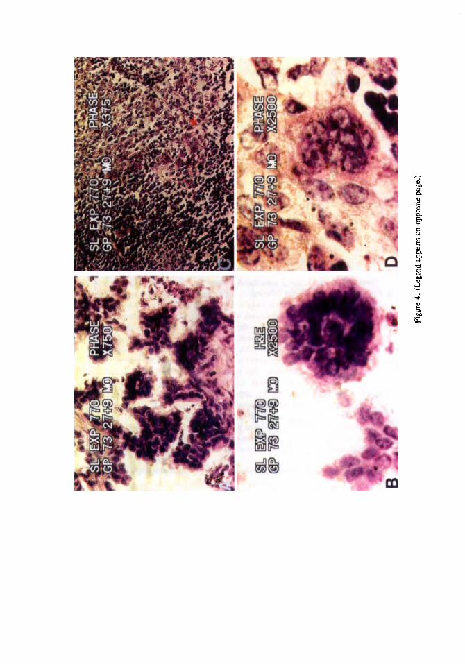
Fig. 4. A: An apparent early carcinomatous response to inhaled chrysotile. The guinea inhaled the aerosol for 27 months and then was allowed to survive in normal air for 9 months before it was killed. There is luxuriant papillomatoid epithelial proliferation into alveolar spaces. X 750. B: A portion of one of the papillary structures at higher magnification. Note the morular pattern of the abnormal cells and their cohesive nature. X 2,500. C: Accumulation of abnormal cells in a bronchial lymph node of this guinea pig. Asbestos structures are not discernible and much of the original lymphatic tissue had been replaced by abnormal cells. X 375. D: A cluster of abnormal cells in the lymph nodes at higher magnification. Lymphocytes have dark nuclei and very little cytoplasm. The abnormal cells are pleomorphic, have abundant cohesive cytoplasm, and assume a morular pattern. They have destroyed the host lymphocytes. X 2,500. (Reduced 1.85 X from original photos.)
Tom
Highlight
Tom
Highlight
Figu
re 4
. (Le
gend
app
ears
on
oppo
site p
age.
)

600 Schepers
dust on cancer resistant mice. Manfred Bowditch, one of Dr. Lanza’s professors, apprised me of this protocol and he told me that Dr. Vorwald had been reluctant to get it going. Bowditch had been the Chief of Industrial Hygiene at the Saranac Laboratory, its deputy, and later interim director until Dr. Vorwald took over in 1947. I asked Vorwald what cancer research was conducted at the Laboratory on chrysotile asbestos. He said none had been attempted, none was contemplated, and none was needed, for in his opinion pneumoconiosis did not cause cancer, as stated in a 1938 paper by Dr. Vorwald and Dr. Kark. However, this paper was on the noncarcinogenic action of siliceous dusts and not on asbestos effects. The negative findings for neo- plasia accorded with my own epidemiological findings on silicotic subjects , but provided no enlightenment on chrysotile carcinogenicity.
While reviewing Dr. Gardner’s accumulated research data at the Saranac Lab- oratory, I came across a set of slides of mice, some of them with malignant neo- plasms. These mice had been exposed to chrysotile asbestos, but showed no asbes- tosis, just cancers. I also found nine human lung cancer and two mesothelioma cases in a file identified as “Quebec Asbestos Workers.” Dr. Vorwald gave no explanation for these items. Soon after these observations I visited Quebec. Through conversa- tions with Drs. Kenneth Smith and Paul Cartier, the medical directors of the Johns Manville and Thetford asbestos industrial clinics in eastern Quebec, I gathered that these two doctors did not think their chrysotile asbestos caused cancer, since only six lung cancers were known to them in circa 6,000 current asbestos workers. I then told them about the 11 cases at Saranac Lake, which astonished them, for they did not know of their existence. Later I ascertained from Mr. Ivan Sabourin, the chief attorney for the Quebec Asbestos Mining Association (QAMA), that he had confi- dentially hand carried those case materials to Dr. Gardner during the period 1944- 1946. He said he had since then accumulated several additional cases of men who had developed and died of lung cancers after they retired from service at the asbestos mines and mills. He clarified that Drs. Smith and Cartier had responsibility only for health problems of the active employees. Mr. Sabourin was collecting the post em- ployment cases , because the asbestos workers had begun to agitate for compensation for lung cancer in the same manner that gold miners of South Africa received compensation for contracting tuberculosis. Canada also then was a Commonwealth of the British Empire. Adding the 11 Sabourin-Saranac cases to the 6 current employees with cancers, there then were actually 17 known cancers in the chrysotile group. (Such a large number of cases in such a small and well-defined group of industrial employees suggested a significant problem.) At this point I mentioned Dr. Gardner’s mice with the cancers, which seemed confirmatory. Smith, Cartier, and Sabourin were all much impressed since they had no knowledge of this aspect of the problem.
When I returned to the Saranac Laboratory about a month later after consulta- tions with officials of Canadian industries, university medical schools, and govern- ment departments, Dr. Vorwald took me to task for mentioning the Laboratory cancer data to Smith, Cartier, and Sabourin. This was to me wholly incomprehensible, as the Saranac Laboratory was a consultant to QAMA, whose officers they were. I also found that all the cancerous mouse slides had been lifted from the files. Dr. Vorwald said that Lanza, Smith, Cartier, and Sabourin all had homed in on him immediately after my visit to Canada. Vorwald said he was reprimanded for being derelict in letting me, “a foreigner,” see the patently sensitive data on chrysotile carcinogenic-
Tom
Highlight
Saranac Laboratory Asbestos Cancer Discoveries 601
ity, without being warned not to talk about what I observed. I complied thereafter in the United States.
During my oral examination for a New York University diploma, I commented to Professors Lanza and Bowditch that I did not understand why there was so much secrecy and circumlocution on the chrysotile cancer issue. Next Dr. Lanza took me to Mr. Vandiver Brown, the chief attorney for Johns Manville and also the asbestos consortium which had sponsored Gardner’s asbestos research up to the point where he mentioned cancer. Mr. Brown had the original of my university thesis, a copy of which doubled as my report to the South African Government. Brown asked me to withdraw this. I told him I could not and that I already had mailed my report to Johannesburg. Mr. Brown flew immediately to South Africa to retrieve my report from the Department of Mines officials. This merely heightened their interest in what I had to say.
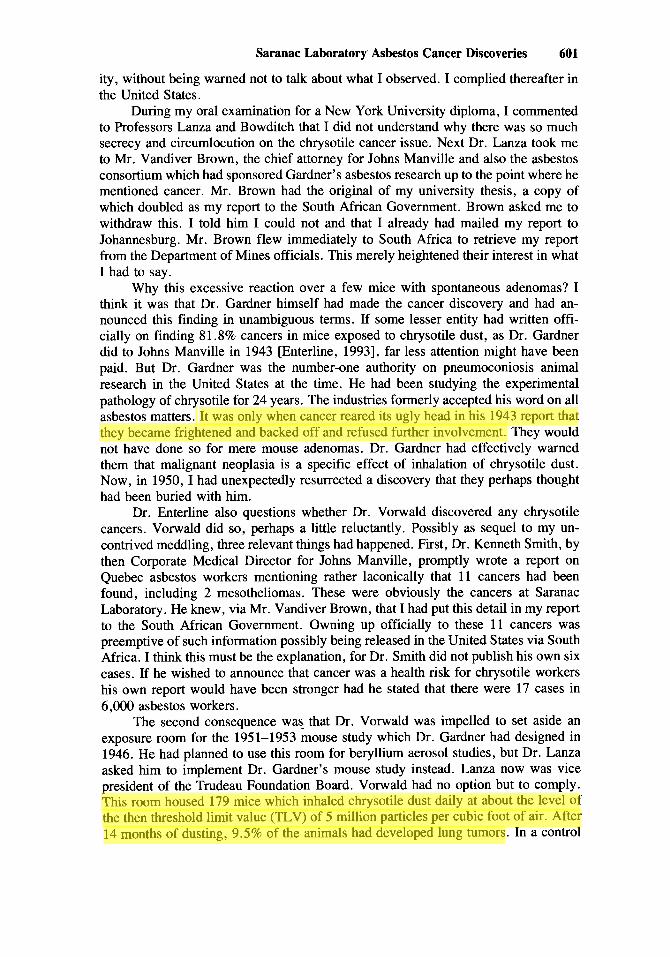
Why this excessive reaction over a few mice with spontaneous adenomas? I think it was that Dr. Gardner himself had made the cancer discovery and had an- nounced this finding in unambiguous terms. If some lesser entity had written offi- cially on finding 81.8% cancers in mice exposed to chrysotile dust, as Dr. Gardner did to Johns Manville in 1943 [Enterline, 19931, far less attention might have been paid. But Dr. Gardner was the number-one authority on pneumoconiosis animal research in the United States at the time. He had been studying the experimental pathology of chrysotile for 24 years. The industries formerly accepted his word on all asbestos matters. It was only when cancer reared its ugly head in his 1943 report that they became frightened and backed off and refused further involvement. They would not have done so for mere mouse adenomas. Dr. Gardner had effectively warned them that malignant neoplasia is a specific effect of inhalation of chrysotile dust. Now, in 1950, I had unexpectedly resurrected a discovery that they perhaps thought had been buried with him.
Dr. Enterline also questions whether Dr. Vorwald discovered any chrysotile cancers. Vorwald did so, perhaps a little reluctantly. Possibly as sequel to my un- contrived meddling, three relevant things had happened. First, Dr. Kenneth Smith, by then Corporate Medical Director for Johns Manville, promptly wrote a report on Quebec asbestos workers mentioning rather laconically that 1 1 cancers had been found, including 2 mesotheliomas. These were obviously the cancers at Saranac Laboratory. He knew, via Mr. Vandiver Brown, that I had put this detail in my report to the South African Government. Owning up officially to these 11 cancers was preemptive of such information possibly being released in the United States via South Africa. I think this must be the explanation, for Dr. Smith did not publish his own six cases. If he wished to announce that cancer was a health risk for chrysotile workers his own report would have been stronger had he stated that there were 17 cases in 6,000 asbestos workers.
The second consequence was- that Dr. Vorwald was impelled to set aside an exposure room for the 1951-1953 mouse study which Dr. Gardner had designed in 1946. He had planned to use this room for beryllium aerosol studies, but Dr. Lanza asked him to implement Dr. Gardner’s mouse study instead. Lanza now was vice president of the Trudeau Foundation Board. Vorwald had no option but to comply. This room housed 179 mice which inhaled chrysotile dust daily at about the level of the then threshold limit value (TLV) of 5 million particles per cubic foot of air. After 14 months of dusting, 9.5% of the animals had developed lung tumors. In a control
Tom
Highlight
Tom
Highlight

602 Schepers
group of 181 mice which had not been exposed to chrysotile, 5.5% had developed tumors. There thus was a risk ratio (RR) of 1.73. At that point (June 30, 1952), Dr. Vorwald’s 5-year contract with the Saranac Laboratory expired. On Dr. Lanza’s recommendation, he was allowed to continue working there for another year until the QAMA mouse cancer study would be completed at the 24th month of exposure. Vorwald did not finish the report on this study. Understandably, his mind now was on his future elsewhere.
When I succeeded Vorwald as Director of Saranac Laboratory, one of the first items Dr. Lanza asked me to tackle was finalization of this study on the chrysotile mouse cancers. By then all the mice had died. Mr. Stanley Kaszer, the statistician for the laboratory, presented me with a report on the mathematical findings. The neo- plasia response to chrysotile fibers was strongly positive, the cumulative RR for neoplasm having trebled to 5.7 in the final months of the mouse lives. I also tried to check what kinds of tumors these were, but all the animal protocols and slides had been taken to Wayne State University by Dr. Vorwald. I therefore called on him and presented him with Kaszer’s statistics and urged him to publish the data, since he now was free to do so, no longer being bound by contract with the QAMA sponsor. No publication on this very important matter ever followed. I also urged Dr. Vorwald to publish all the accumulated human cancer case data. He did not. He died in 1976 still mute on these topics.
The third consequence of my meddling perhaps was publication of the modified version of Dr. Gardner’s monograph in 1951. When I was at the Saranac Laboratory from 1949 to 1950, no one was working on this task. I was never told by Dr. Vorwald of the existence of the monograph even though I had been sent to the Laboratory by Dr. Lanza to study all asbestos matters. I had become Vorwald’s house guest during all the time I was there and came to know him and his activities very well. He was not working on this project. The monograph had been lying fallow for 5 years. Mr. Tom Durkan, who wrote most of the Laboratory technical reports for Dr. Vorwald, was not working on this monograph either. I would have discovered, since I used his office as my base. He knew of my interest in the former chrysotile research, as did everyone else. Gardner’s manuscript was somewhere hidden in a box, which would now be in a forgotten place, if I had not come there to meddle with the past. Perhaps after my exit visit with Mr. Brown, an executive decision was made to get on with the task which seemed to have been dropped after 1948 to judge by the chronology of interactions between Drs. Packard, Lanza, Lynch, and Vorwald.
Did Dr. Vorwald discover that chrysotile caused lung cancer? He most assur- edly did, both in mice and men. The fact that he never published anything on such an important matter may be a mystery, but not if one considers that for almost three decades he had served as a defense litigation expert for the asbestos industries. It would hardly have been of value to his sponsors to permit release in scientific publications of the very incriminating data he commanded.
A few years later Braun and Truan began recounting the Quebec human cancer numbers for QAMA. In their 1958 report they said they had identified 19 cancers in miners. This was a threefold jump over the 10 years since Smith and Cartier had counted only six cases. These 19 cancers again excluded the Saranac cases, which had grown to over 70. However, the Braun-Truan report was limited to the epide- miology of lung cancers in working miners. They likely did not know of the accu-
Tom
Highlight

Saranac Laboratory Asbestos Cancer Discoveries 603
mulated post-retirement cases at the Saranac Laboratory. Neither Dr. Vorwald nor Mr. Sabourin would have had any reason to tell them and they never came to inquire at the Saranac Laboratory, at least not while I was there.
Did Dr. Schepers discover anything new about the ability of chrysotile to cause cancer? There was not much left for Schepers to discover, for the animal cancer mine had been worked out by Gardner and Vorwald. Schepers did provide the QAMA sponsors of Saranac Laboratory research with Kaszer’s statistics and continued to report periodically to them and to their AT1 (Asbestos Textile Institute) confreres, on the occurrence and nature of cancers in persons with evidence of asbestos exposure. Some of these case reports were later discovered during the asbestos litigation, which started two decades later, some through raids on residual files and others via sub- poenas of company records.
There was only one animal cancer study which Schepers managed to launch to probe whether chrysotile caused cancer directly or as a complication of asbestosis. This was funded partly by the Damon Runyon Foundation. In this study, rats served as test subjects. One group of rats was exposed to chrysotile dust for 6 months and then to beryllium sulfate dust for 6 months, whereafter they were transferred to normal air. In a second group the beryllium and asbestos exposures occurred in reverse order. There also was a 6-month beryllium-only and another six-month chrysotile-only exposure group, while a fifth group of rats with no dust exposure served as a further control. All the animals were killed at 18 months to identify cancers. Through prior and concurrent studies, the Saranac Laboratory had demon- strated that beryllium sulfate would induce a predictable yield of lung cancers within 18 months after 6 months exposure to 1 part per million of BeSO, aerosol. The theorem to be tested by brief sequential exposures to chrysotile and beryllium, was whether chrysotile would increase the yield of beryllium cancers within 18 months. Had this occurred, it would have been possible to conclude that chrysotile acts as a primary carcinogen, without first inducing asbestosis, which was one of the then current theories of how asbestos causes cancer. Terminating all the animals at 18 months had as a purpose to exclude the possibly confounding factor of asbestosis, which would only manifest in the second year of life following 6 months of dust exposure. This study yielded inconclusive results, since the beryllium interfered with what asbestos did to the lungs and vice versa. The study did not advance knowledge on the carcinogenicity of chrysotile. It seemed not worth publishing, apart from a brief reference [ Schepers , 197 11.
I also wrote a proposal for a study of cocarcinogenicity of cigarette smoke and chrysotile. This was presented to the National Cancer Institute (NCI) for funding, which it declined. To my knowledge no such study has ever been accomplished. There is considerable physician conviction that concurrent cigarette smoking and asbestos inhalation increase the risk of human bronchogenic cancer. Dr. Enterline suggested that one should not accept that chrysotile causes cancer without published animal confirmation. He did not explain how it is possible to accept the cocarcino- genicity of cigarette smoking and chrysotile asbestos without experimental validation. In vitro experiments recently done in this area yielded rather vague and inconclusive data in comparison with evidence an inhalation study might have accomplished. Apart from the foregoing, it can be safely concluded that Schepers did not indepen- dently discover whether chrysotile is carcinogenic for animals. He did not have to. The problem had already been resolved by Dr. Gardner.

604 Schepers
In the discussions by Dr. Enterline, emphasis has been placed on whether Dr. Gardner had merely seen some autogenous mouse adenomas , having erroneously misdiagnosed these as chrysotile-induced malignant neoplasms. Although of great practical significance to human victims, it makes little scientific difference whether chrysotile causes adenoma or carcinoma or sarcoma. An adenoma is also a cancer although not malignant. Dr. Gardner did not describe merely spontaneous adenomas in the mice. He was clearly astonished at finding tumors of a more malignant aspect. The slides vanished, so that we cannot currently verify what it is that he saw. I do however have a few of the slides for the guinea pigs of experiment 770, which ran concurrently with the mouse study. Two of these guinea pigs seem to show early phases of carcinoma formation in one of the sites of adenomatoid epithelialization (Fig. 4). This supports Gardner’s judgment on the mice.
A question is raised whether Dr. Vorwald expertly judged the cancers differ- ently from the way Gardner did. I do not see how he could have. He had not studied the slides before 1949 and the slides vanished thereafter and are not mentioned at all in his 1951 report on Gardner’s asbestos research. None of the animal protocols shows any participation by him in the microscopy reviews of the lung sections during the period 1933-1941, at which point he had joined the Navy. On his return to the Saranac Laboratory, Dr. Vonvald served mainly as the administrator of the Trudeau Foundation and performed no histology tasks. All the photomicrographs in the 1951 paper had been made prior to 1946 by Mr. Edward Gockler, the laboratory photog- rapher, under the direction of Dr. Gardner.
Did Dr. Kenneth Lynch judge the Saramac Laboratory chrysotile cancers to be mere autogenous mouse adenomas? He was briefly involved. Soon after Dr. Gard- ner’s death in November 1946, Dr. Edward Packard, who served as temporary Medical Director of the Saranac Laboratory, mailed Dr. Gardner’s manuscript and the animal protocols to Dr. Lynch for editorial refinements. Lynch also came to the laboratory, perhaps to look at the slides. Lynch was an important connoisseur of asbestos matters having published one of the first three American asbestos cancer case reports in 1935 [Lynch and Smith, 19351 and later describing 40 examples of the pathology of asbestosis [Lynch and Cannon, 19431 in deceased workers of asbestos textile factories. Dr. Lynch recommended that Dr. Gardner’s manuscript be published unmodified. If he had judged that Dr. Gardner had made a grievous interpretive mistake in calling some mouse tumors malignant, when they should have been clas- sified as benign adenomas, he would have performed a disservice to the science of pathology, of which he was then a leading expert as Professor of Pathology at the University of South Carolina. He seems not to have questioned Dr. Gardner’s eval- uation of the tumors, but did express puzzlement over Dr. Vonvald’s disinclination to release Gardner’s cancer data in the planned posthumous publication of the Gardner monograph. This manuscript never was published despite Dr. Lynch’s recommen- dation and even though the Laboratory Board had set aside funds to have it published as a memorial to Dr. Gardner. It is also to be noted that Dr. Lynch initiated his own animal experimentation studies on mice to test for carcinogenicity of chrysotile. These ran for a number of years. By 1957, Dr. Lynch and colleagues had found that chrysotile evoked many tumors, but his paper did not reveal whether these were malignant. However, in 1962, Dr. Lynch invited me to his department in Charleston to advise his staff on how to conduct animal studies in such a manner that “the same malignant neoplasia results” that had been achieved at the Saranac Laboratory could

Saranac Laboratory Asbestos Cancer Discoveries 605
also be accomplished there. I found that they had not fully fiberized and adequately aerosolized their asbestos, so that their animals had inhaled relatively large chrysotile aggregates rather than separated asbestos fibers. I wrote a detailed methodology for him, but nothing came of this, the pathology department not having engineering staff and facilities to launch a full inhalation study such as had been customary at the Saranac Laboratory. Despite this frustration, Lynch’s actions seem to reflect clearly a conviction that the Saranac Laboratory had truly induced malignant cancers in experimental animals. He based this persuasion on what Dr. Gardner had accom- plished by 1943 and what Vonvald and I had reported to QAMA in 1952 and 1954, for nothing after that had been published from the Saranac Laboratory. Dr. Lynch, as a research consultant to Raybestos Corporation, would have received copies of re- ports on the Saranac Laboratory research data. Raybestos was one of the QAMA sponsors of the mouse investigations by Dr. Gardner.
Dr. Enterline suggests that because Gardner sought funding from the NCI to extend the mouse study, this can be interpreted as signifying that he did not believe his own findings on malignant neoplasia. I do not follow this argument as it implies that every time a scientist undertakes a new study of any subject he thereby declares that all he had discovered up to then was invalid. Every scientific study tends to reveal new avenues of inquiry that may be worthwhile. Dr. Gardner specifically had wished to conduct a study in which cancer-insusceptible mice (as compared to cancer-prone mice) would be used, perhaps because human beings are cancer-insusceptible crea- tures, absent enviromental causes. He also needed a current control group, so that his numerical calculations would be more valid. This does not negate the value of his prior work, but instead reveals his conviction that there was a cogent reason for additional research. This was also 1943, 50 years ago, and all that we know today about cancer and mice and asbestos was not evident then. It was extraordinary for Gardner to attempt such a cancer study. Dr. Gardner had tremendous concurrent research responsibilities on silicosis and tuberculosis, which then were major killer diseases (over 80,000 deaths per year), while the havoc of lung cancer was still of comparatively miniscule dimensions. That he was prepared to deviate from these responsibilities when short of staff during war time and commit scarce resources, indicates how seriously he took the matter. That he should have spent so much effort, as a dying man, to further probe scientific validation of chrysotilic carcinogenicity, was laudatory and consistent with his known objectivity and devotion to the pursuit of truth. He deserves praise instead of the iconoclasm seemingly implied in current comments.
REFERENCES Braun DC, Truan TD (1958): An epidemiological study of lung cancer in asbestos miners. A.M.A. Arch.
Egilman, DS, Hardy, HL (1993): Manipulation of early animal research on asbestos cancer. Am J Ind
Enterline, PE (1993): Early animal research on asbestos cancer. Am J Ind Med 24:783-785. Gardner LU, Cummings DE (193 1): Studies on experimental pneumoconiosis:lV-Inhalation of asbestos
Lanza AJ (1938): “Silicosis and Asbestosis.” New York: Oxford University Press. Lynch KM, Cannon WM (1943): Asbestosis: Analysis of forty necropsy cases. Dis Chest 14:874-885. Lynch KM, Smith WA (1935): Pulmonary asbestosis 111. Carcinoma of the lung in asbestos-silicosis. Am
Ind. Health. 17:634-653.
Med 24:787-791.
dust-Its effects on primary tuberculous infection. J Ind Hyg 13:65-81.
J Cancer 2456-64.

606 Schepers
Lynch KM, McIver FA, Lain JK (1957): Pulmonary tumors in mice exposed to asbestos dust. Arch Ind
Merewether ERA (1947): “Chief 1nspec.tor of Factories report for 1947.” London: His Majesty’s Sa-
Schepers GWH (1971): Lung tumors of primates and rodents. Ind Med Surg 31:l-70. Schepers GWH, Durkan TM, Delahant A (1955): Effect of inhaled commercial hydrous calcium silicate
Vorwald AJ, Kark JW (1938): Pneumoconiosis and pulmonary carcinoma. Am J Pathol 14:49-52. Vonvald AJ, Durkan TM, Pratt PC (1951): Experimental studies of asbestosis. Arch Ind Hyg Occup Med
Health 15:207-2 14.
tionery Office, pp 79-81.
dust on animal tissues. Arch Ind Health 12348-360.
3: 1-43.
APPENDIX Typescript Copies of Dr. Gardner’s Handwritten Notes
Tumor findings in 11 mice exposed to chrysotile aerosol Mouse no. 2-12 months. “Tumor mass in right lung-no metastases. Debat-
able whether a chronic abscess has resulted in acute or chronic organizing pneumo- nia-epithelial metaplasia or whether there is a true tumor. It is different at least from the ordinary mouse adenoma.”
Mouse no. 3-12 months. “Fibrosarcoma? of mediastinal tissue with retro- grade extension into the lungs. ”
Mouse no. 4-18 months. “Well marked epithelial hyperplasia in terminal bronchiole. One small adenoma. ”
Mouse no. 5-19 months. “A large tumor mass involving almost the entire left lung. Large papillary adenocarcinoma, replacing a lobe of the left lung. Undifferen- tiated sarcomatoid tissue in the lung next to the tumor and metastases to the liver and spleen. ’ ’
Mouse no. 6-20 months. “Partial collapse of hind legs. Small localized ade- noma in right lung. ” No microscopy description.
Mouse no. 7-24 months. “Scattered nodules of tumor-round and white in color. Rather large localized adenomatous nodules involving pulmonary parenchyma and bronchi. ”
Mouse no. 8-24 months. “Focal areas of cellular proliferation in walls of respiratory bronchioles. ”
Mouse no. 15-18 months. “Small localized tumor-apex of lower lobe- adenoma. ”
Mouse no. 17-19 months. No tumor described. Mouse no. 18-20 months. “One tumor about 2 X 3 cm in upper portion of
lower lobe on right. Abdomen-Scattered throughout are many tumor masses of varying size-one of which seems to have surrounded and obstructed the large bowel.”
Mouse no. 19-24 months. “One large tumor-like mass in middle third of left lung. Well circumscribed and measuring about 6 mm in diameter. Adenoleiomy- oma?”
Since Dr. Gardner judged 9 of the 1 1 mice to have developed neoplasia, he calculated an incidence of 81.8%, which was Six times more prevalent than tumors usually encountered in this mouse species at the Saranac Laboratory when used in experiments other than with chrysotile asbestos.