CHRONIC WOUNDS Based on a presentation by Dr. David Thomas at the AMDA Convention.
52
CHRONIC WOUNDS Based on a presentation by Dr. David Thomas at the AMDA Convention
-
Upload
brooke-carnal -
Category
Documents
-
view
213 -
download
0
Transcript of CHRONIC WOUNDS Based on a presentation by Dr. David Thomas at the AMDA Convention.

CHRONIC WOUNDS
Based on a presentation by
Dr. David Thomas at the AMDA Convention
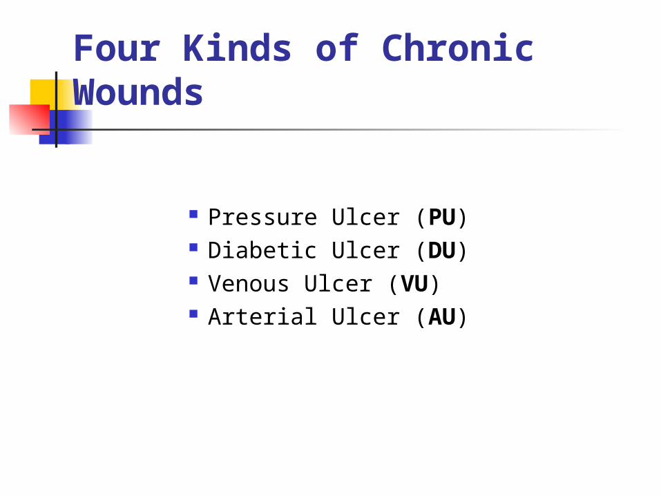
Four Kinds of Chronic Wounds
Pressure Ulcer (PU) Diabetic Ulcer (DU) Venous Ulcer (VU) Arterial Ulcer (AU)
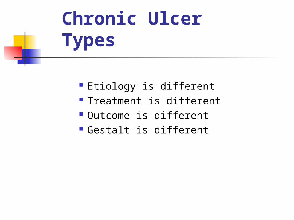
Chronic Ulcer Types
Etiology is different Treatment is different Outcome is different Gestalt is different
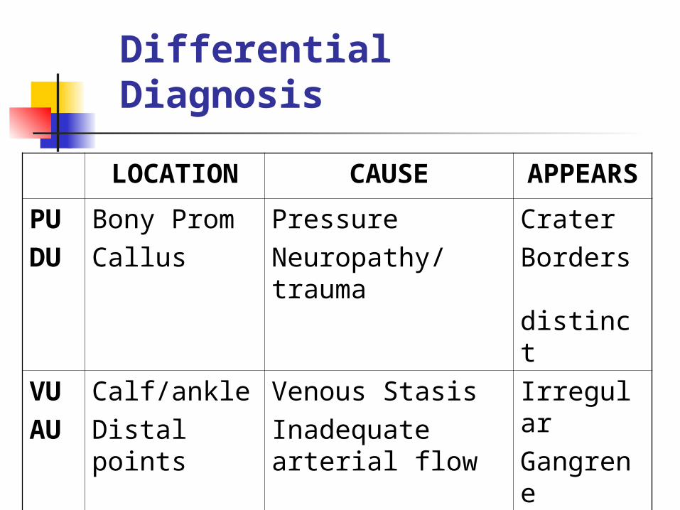
Differential Diagnosis
LOCATION CAUSE APPEARS
PUDU
Bony PromCallus
PressureNeuropathy/trauma
CraterBorders distinct
VUAU
Calf/ankleDistal points
Venous StasisInadequate arterial flow
IrregularGangrene
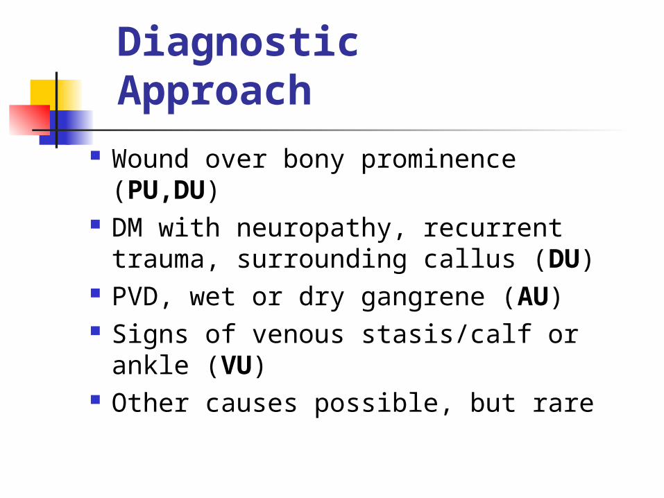
Diagnostic Approach
Wound over bony prominence (PU,DU)
DM with neuropathy, recurrent trauma, surrounding callus (DU)
PVD, wet or dry gangrene (AU) Signs of venous stasis/calf or
ankle (VU) Other causes possible, but rare
Pain in Chronic Ulcers
DU: no or diminished pain, sensation VU: little pain, intact sensation PU: intermittent pain AU: constant pain
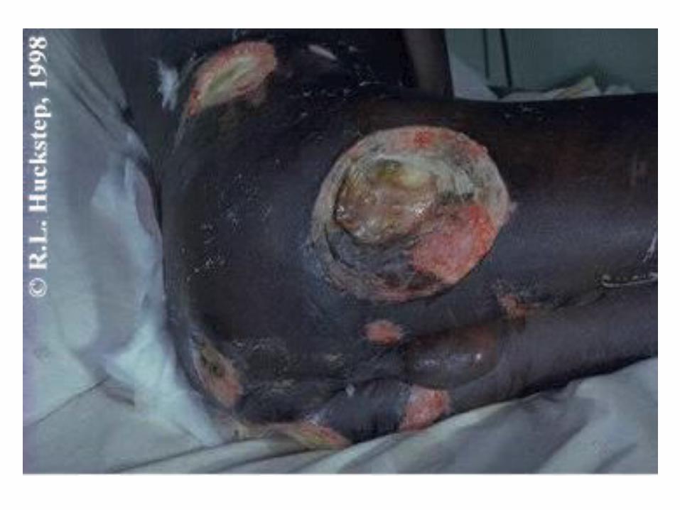

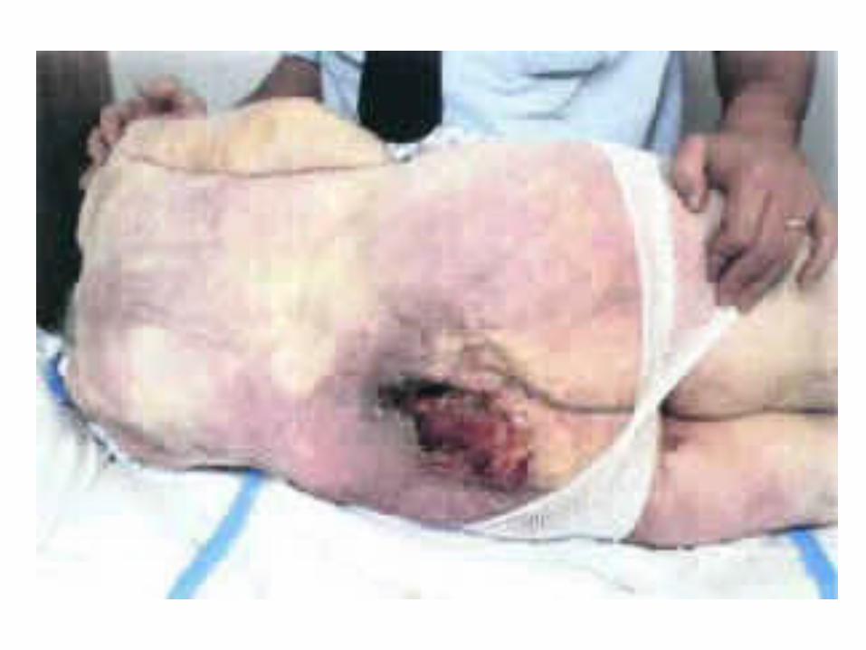
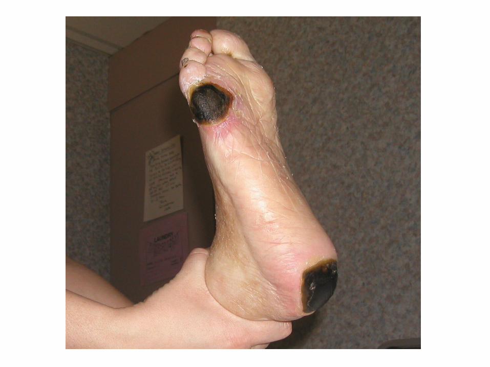
Pressure Ulcers
Visible evidence of pathological interruption of blood flow to dermal tissues
Chief cause: sustained pressure
Most commonly over sacrum, hip

Pressure Ulcers:
What Works
Must relieve pressure or it won’t heal.
Must use moist dressing or it won’t heal.
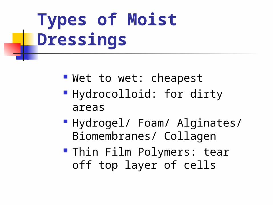
Types of Moist Dressings
Wet to wet: cheapest Hydrocolloid: for dirty areas Hydrogel/ Foam/ Alginates/
Biomembranes/ Collagen Thin Film Polymers: tear off
top layer of cells


Problems
Most doctors treat few pressure ulcers.
Very few good studies; none for most treatments.
Treatment modalities for pressure ulcers are considered devices: only safety, NOT efficacy, must be proved.
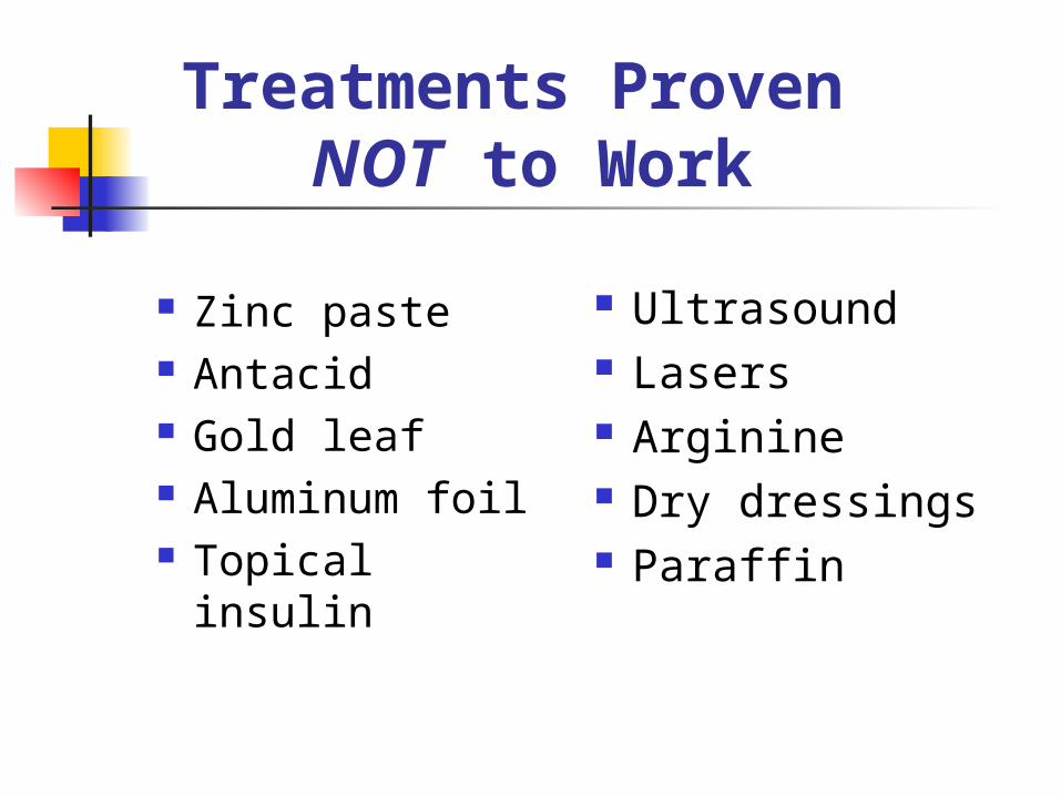
Treatments Proven NOT to Work
Zinc paste Antacid Gold leaf Aluminum foil Topical insulin
Ultrasound Lasers Arginine Dry dressings Paraffin

Treatments with No Data
Magnet therapy
Honey/ Sugar
“Skin equivalents”

Treatments With Very Flawed Data
Vitamin C Patient’s serum mixed
with proprietary gel Vacuum therapy Electrical stimulation Topical Phenytoin Cytokine growth factors

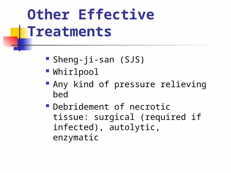
Other Effective Treatments
Sheng-ji-san (SJS) Whirlpool Any kind of pressure relieving
bed Debridement of necrotic
tissue: surgical (required if infected), autolytic, enzymatic
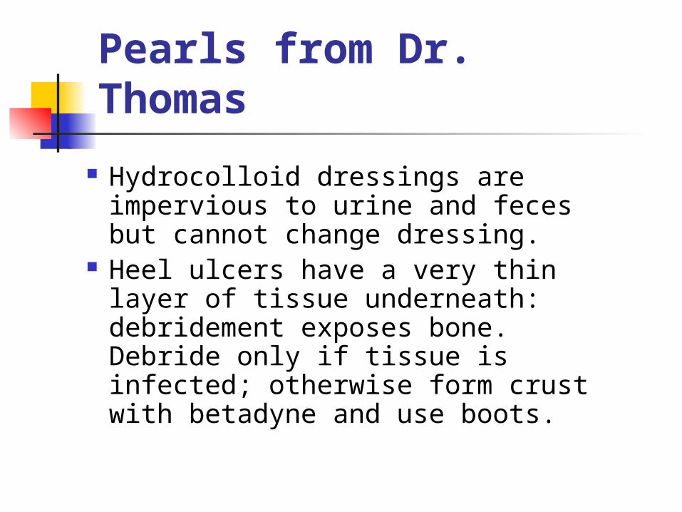
Pearls from Dr. Thomas
Hydrocolloid dressings are impervious to urine and feces but cannot change dressing.
Heel ulcers have a very thin layer of tissue underneath: debridement exposes bone. Debride only if tissue is infected; otherwise form crust with betadyne and use boots.
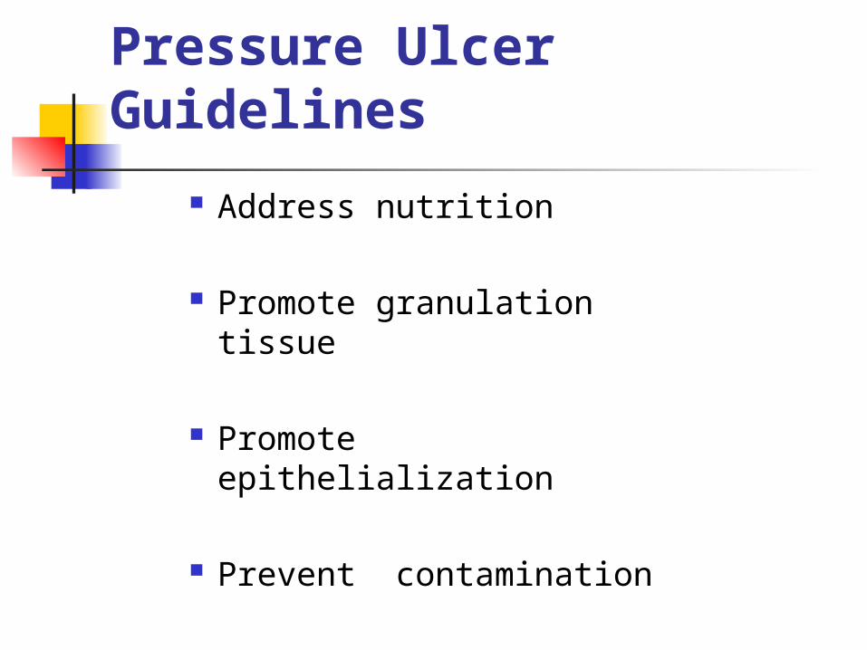
Pressure Ulcer Guidelines
Address nutrition
Promote granulation tissue
Promote epithelialization
Prevent contamination

Dressings
Stage I: Thin film polymer Stage II: Moist gauze (wet-to-wet) or
hydrocolloid Stage III/ IV with dead space/
exudate: hydrogel, wet-to-wet, or hydrocolloid with synthetic absorption dressing below.
Stage III/ IV with necrosis: debride, then treat as III/ IV above.

Nursing Home Pearl
Home health nursing and nursing home care plans of ulcers tend to call for improved nutrition and healing; if pressure ulcers have occurred because the patient is dying/ not eating, make sure the care plan reflects that (for liability and survey purposes).

Venous Stasis Ulcers
An area of discontinuity of the epidermis, persisting for 4 weeks or more, occurring as a result of venous hypertension and calf muscle pump insufficiency.
Must exclude arterial disease, neuropathy, diabetes, rheumatoid arthritis, hemoglobinopathies, and carcinoma.
Biopsy if long-standing or looks weird.
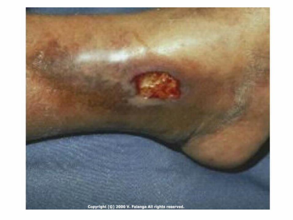

Diagnosis of Venous Ulcers
Location on the calf Bronzing (lipodermatosclerosis) Exclusion of arterial insufficiency by
bounding DP pulses, or ABI > 0.8 Tend to be slow-healing (~90% heal
by one year), irregular, and associated with edema and sloughing
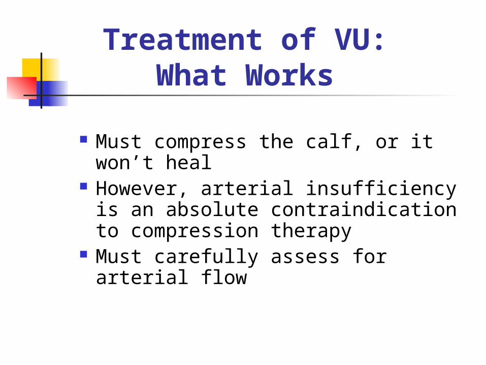
Treatment of VU:What Works
Must compress the calf, or it won’t heal
However, arterial insufficiency is an absolute contraindication to compression therapy
Must carefully assess for arterial flow
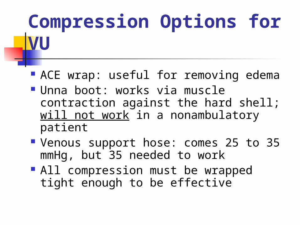
Compression Options for VU
ACE wrap: useful for removing edema Unna boot: works via muscle
contraction against the hard shell; will not work in a nonambulatory patient
Venous support hose: comes 25 to 35 mmHg, but 35 needed to work
All compression must be wrapped tight enough to be effective
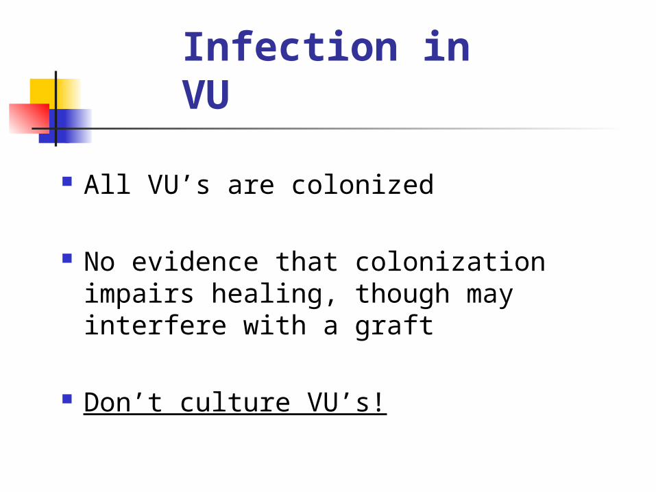
Infection in VU
All VU’s are colonized
No evidence that colonization impairs healing, though may interfere with a graft
Don’t culture VU’s!

Recognition of Infectionin VU’s
Fever Increased pain Increased skin erythema Lymphangitis Ulcer rapidly becomes larger If infected, treat with
systemic AB’s

VU Treatments
Hydrocolloid dressing Cadexomer iodine
topically Trental (anticytokine)
and compression Artificial skin Skin graft TGF-B2

Ineffective VU Treatments
(RCT’s) Antibiotics, including
Bactroban Elase Zinc Stanozolol Ifetroban Silver sulfadiazine

Secondary Prevention
in VU’s
Recurrence in ~57% Reflux in deep veins in 50 to 71% Prior DVT causes 95% of DV reflux Venous support hose may reduce
recurrence rate (unpublished data)
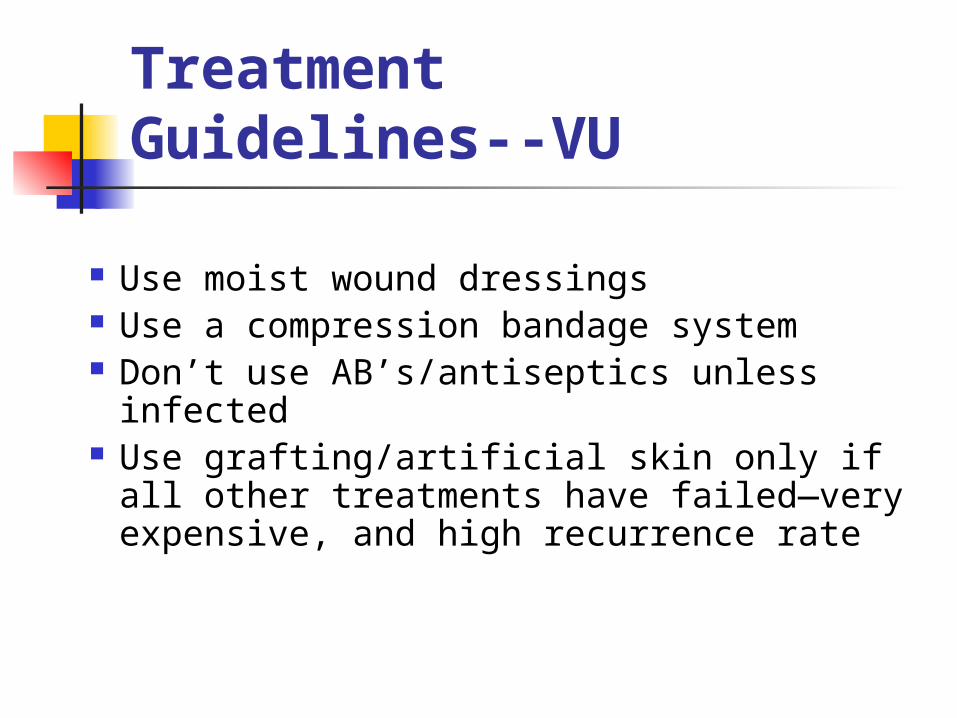
Treatment Guidelines--VU
Use moist wound dressings Use a compression bandage system Don’t use AB’s/antiseptics unless
infected Use grafting/artificial skin only if all
other treatments have failed—very expensive, and high recurrence rate
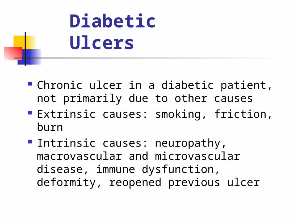
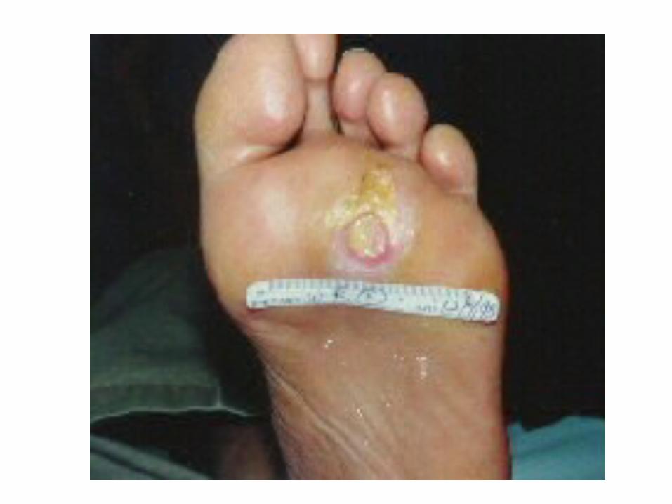
Diabetic Ulcers
Chronic ulcer in a diabetic patient, not primarily due to other causes
Extrinsic causes: smoking, friction, burn
Intrinsic causes: neuropathy, macrovascular and microvascular disease, immune dysfunction, deformity, reopened previous ulcer

Neuropathy in DU
Use monofilament for 5 seconds or less, to avoid triggering propioceptors
Also assess temperature sensation—may use reflex hammer
Can test pinprick and 2-point discrimination

Co-Morbidity in DU
Peripheral vascular disease occurs in 11% of diabetic patients
Peripheral neuropathy occurs in 42% of diabetic patients
PVD is associated with delayed ulcer healing and increased rates of amputation

Treatment of DU:
What Works
Must surgically debride ulcer to allow healing: the wound edges are dead
Weekly debridement down to healthy bleeding tissue gives best results
Must keep pressure off the ulcers to allow healing
Pressure Reduction Off DU
Orthopedic shoes: drop recurrence rate from 83% to 17%
Sandals Splints Crutches/wheelchairs Total contact casting
Total Contact Casting
Worsens the ulcer if not applied perfectly
Need to find a consultant for this task on whom you can rely
Other PossiblyHelpful Treatments
Moist dressings (clearly better than dry) Hyperbaric O2 Dermagraft (cultured skin—human) Platelet-derived growth factor Antibiotics (ineffective if
uncomplicated) Questionable effectiveness: U/S,
electrical stimulation
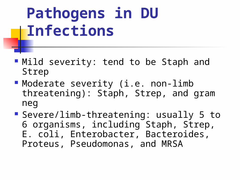
Pathogens in DU Infections
Mild severity: tend to be Staph and Strep Moderate severity (i.e. non-limb
threatening): Staph, Strep, and gram neg Severe/limb-threatening: usually 5 to 6
organisms, including Staph, Strep, E. coli, Enterobacter, Bacteroides, Proteus, Pseudomonas, and MRSA
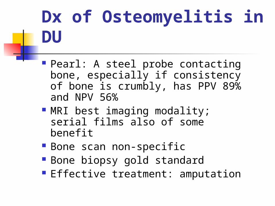
Dx of Osteomyelitis in DU Pearl: A steel probe contacting
bone, especially if consistency of bone is crumbly, has PPV 89% and NPV 56%
MRI best imaging modality; serial films also of some benefit
Bone scan non-specific Bone biopsy gold standard Effective treatment: amputation

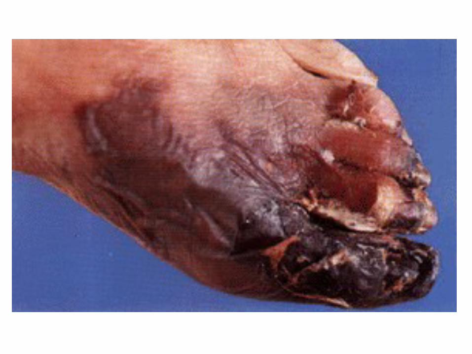
Arterial Ulcers--AU
Tend to occur on distal areas Diminished/absent pulses Punched-out appearance, or
gangrene Requires either salvage
revascularization, or amputation—usually the latter
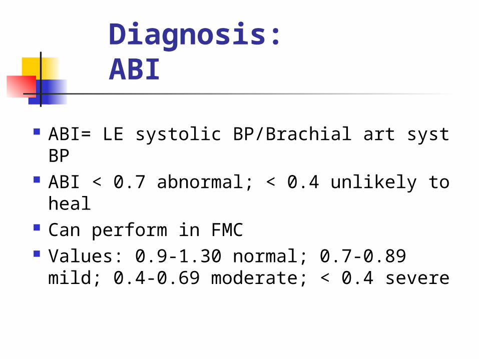
Diagnosis: ABI
ABI= LE systolic BP/Brachial art syst BP ABI < 0.7 abnormal; < 0.4 unlikely to heal Can perform in FMC Values: 0.9-1.30 normal; 0.7-0.89 mild;
0.4-0.69 moderate; < 0.4 severe

Medical Treatment of AU
Control DM and HTN Moderate exercise Smoking cessation Dry dressings (dry gangrene
preferable) ? Pletal, gingko biloba

What Works: AU
Amputation/revascularization/hospice if ABI < 0.4
Do not compress if ABI < 0.7