Chronic tendon pathology: molecular basis and therapeutic ...
25
Accession information: DOI: 10.1017/S1462399405008963; Vol. 7; Issue 5; 24 March 2005 ©2005 Cambridge University Press http://www.expertreviews.org/ Chronic tendon pathology: molecular basis and therapeutic implications 1 expert reviews in molecular medicine Chronic tendon pathology: molecular basis and therapeutic implications Graham Riley Graham Riley Head of Soft Tissue Injury and Repair Group, Rheumatology Research Unit, Box 194, Addenbrooke’s Hospital, Hills Road, Cambridge, CB2 2QQ, UK. Tel: +44 (0)1223 217458; Fax: +44 (0)1223 217838; E-mail: [email protected] Institute URL: http://www.addenbrookes.org.uk Tendons are frequently affected by chronic pain or rupture. Many causative factors have been implicated in the pathology, which until relatively recently was under-researched and poorly understood. There is now a greater knowledge of the molecular basis of tendon disease. Most tendon pathology (tendinopathy) is associated with degeneration, which is thought to be an active, cell-mediated process involving increased turnover and remodelling of the tendon extracellular matrix. Degradation of the tendon matrix is mediated by a variety of metalloproteinase enzymes, including matrix metalloproteinases and ‘aggrecanases’. Neuropeptides and other factors released by stimulated cells or nerve endings in or around the tendon might influence matrix turnover, and could provide novel targets for therapeutic intervention. Tendons are dense, fibrous connective tissues that connect muscle to bone and are essential for the transmission of force and the generation of movement at a joint. They are highly ordered composite materials consisting of collagens, proteoglycans and various glycoproteins, many of which have been poorly characterised. Tendon problems such as tendon rupture and chronic tendon pain are common, although the underlying pathology is not well understood and the conditions are often difficult to treat (Ref. 1). Terms such as tendonitis (or tendinitis) are traditionally used to describe a painful tendon, the name implying an inflammatory condition. This is contrary to the evidence from most histopathological studies, which describe a degenerative condition without inflammation that has been called tendinosis (Refs 2, 3, 4, 5, 6, 7, 8). In this review, the term tendinopathy is used for all forms of chronic tendon pathology, because it does not assume any knowledge of the underlying pathology. Factors implicated in tendinopathy It is increasingly recognised that most tendinopathies are not associated with any single factor, and tendon degeneration might result from various causes. Indeed, there is some evidence to suggest that the nature of the degenerative process varies at different sites (Ref. 3). Tendons at certain sites are more commonly affected, particularly the supraspinatus, extensor carpi radialis brevis, patellar and Achilles (at the shoulder, elbow, knee and ankle, respectively) (Refs 9, 10). These tendons
Transcript of Chronic tendon pathology: molecular basis and therapeutic ...
Accession information: DOI: 10.1017/S1462399405008963; Vol. 7; Issue 5; 24 March 2005 ©2005 Cambridge University Press
http://www.expertreviews.org/
Ch
ron
ic t
end
on
pat
ho
log
y: m
ole
cula
r b
asis
and
th
erap
euti
c im
plic
atio
ns
1
expert reviewsin molecular medicine
Chronic tendon pathology: molecular
basis and therapeutic implications
Graham Riley
Graham RileyHead of Soft Tissue Injury and Repair Group, Rheumatology Research Unit, Box 194, Addenbrooke’sHospital, Hills Road, Cambridge, CB2 2QQ, UK. Tel: +44 (0)1223 217458; Fax: +44 (0)1223 217838;E-mail: [email protected]
Institute URL: http://www.addenbrookes.org.uk
Tendons are frequently affected by chronic pain or rupture. Many causativefactors have been implicated in the pathology, which until relatively recentlywas under-researched and poorly understood. There is now a greater knowledgeof the molecular basis of tendon disease. Most tendon pathology (tendinopathy)is associated with degeneration, which is thought to be an active, cell-mediatedprocess involving increased turnover and remodelling of the tendonextracellular matrix. Degradation of the tendon matrix is mediated by a varietyof metalloproteinase enzymes, including matrix metalloproteinases and‘aggrecanases’. Neuropeptides and other factors released by stimulated cellsor nerve endings in or around the tendon might influence matrix turnover, andcould provide novel targets for therapeutic intervention.
Tendons are dense, fibrous connective tissues thatconnect muscle to bone and are essential for thetransmission of force and the generation ofmovement at a joint. They are highly orderedcomposite materials consisting of collagens,proteoglycans and various glycoproteins, manyof which have been poorly characterised. Tendonproblems such as tendon rupture and chronictendon pain are common, although theunderlying pathology is not well understood andthe conditions are often difficult to treat (Ref. 1).Terms such as tendonitis (or tendinitis) aretraditionally used to describe a painful tendon,the name implying an inflammatory condition.This is contrary to the evidence from mosthistopathological studies, which describe adegenerative condition without inflammation that
has been called tendinosis (Refs 2, 3, 4, 5, 6, 7, 8). Inthis review, the term tendinopathy is used for allforms of chronic tendon pathology, because it doesnot assume any knowledge of the underlyingpathology.
Factors implicated in tendinopathyIt is increasingly recognised that mosttendinopathies are not associated with any singlefactor, and tendon degeneration might result fromvarious causes. Indeed, there is some evidence tosuggest that the nature of the degenerative processvaries at different sites (Ref. 3). Tendons at certainsites are more commonly affected, particularly thesupraspinatus, extensor carpi radialis brevis,patellar and Achilles (at the shoulder, elbow, kneeand ankle, respectively) (Refs 9, 10). These tendons
Accession information: DOI: 10.1017/S1462399405008963; Vol. 7; Issue 5; 24 March 2005 ©2005 Cambridge University Press
http://www.expertreviews.org/
Ch
ron
ic t
end
on
pat
ho
log
y: m
ole
cula
r b
asis
and
th
erap
euti
c im
plic
atio
ns
2
expert reviewsin molecular medicine
are all exposed to relatively high mechanicaldemands, although additional factors are thoughtto be important. The majority of patients presentin late middle age, often with no memory of anyacute injury or trauma (Refs 10, 11). The conditionusually has an insidious onset, with paindeveloping during or shortly after exercise.Tendon ruptures often occur during physicalactivity, typically in sports such as badminton(Ref. 12). Many cases of tendinopathy are ascribedto ‘overuse’, thought to result from repeatedmicrostrain below the failure threshold, analogousto the fatigue failure that affects most materialsplaced under repetitive loading (Refs 10, 13, 14, 15).
It is generally assumed that tendon damageis the primary event, overwhelming the abilityof the tendon cells (generally referred to astenocytes) to repair structural defects in the tendonextracellular matrix (ECM). Alternatively, there isthought to be a failure to adapt to a change inphysical demands. Tenocytes have a central rolein the repair and maintenance of the tendon ECM,synthesising new proteins and producing theenzymes that degrade them. This continualprocess of matrix turnover is normally in balance,and changes in this activity in response to alteredpatterns of loading, for example, might precedeany physical lesion or ‘micro-injury’. Aside frommicrotrauma and hypoxia, factors that couldpotentially affect the tenocyte activity include age,temperature, drugs and the local activity ofbiochemical mediators produced by the residentcells. The potential roles of some of these factorsare discussed in this review, which emphasisesthe importance of ECM turnover and matrix-degrading enzymes in tendon health and disease.
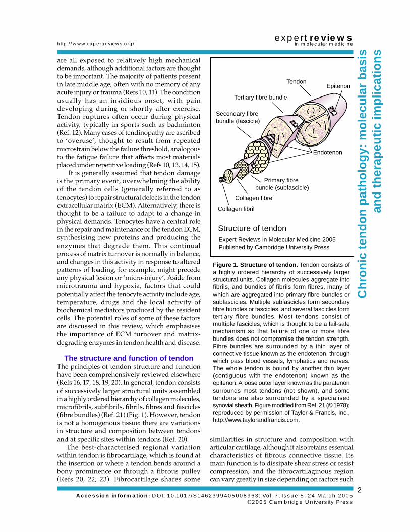
The structure and function of tendonThe principles of tendon structure and functionhave been comprehensively reviewed elsewhere(Refs 16, 17, 18, 19, 20). In general, tendon consistsof successively larger structural units assembledin a highly ordered hierarchy of collagen molecules,microfibrils, subfibrils, fibrils, fibres and fascicles(fibre bundles) (Ref. 21) (Fig. 1). However, tendonis not a homogenous tissue: there are variationsin structure and composition between tendonsand at specific sites within tendons (Ref. 20).
The best-characterised regional variationwithin tendon is fibrocartilage, which is found atthe insertion or where a tendon bends around abony prominence or through a fibrous pulley(Refs 20, 22, 23). Fibrocartilage shares some
similarities in structure and composition witharticular cartilage, although it also retains essentialcharacteristics of fibrous connective tissue. Itsmain function is to dissipate shear stress or resistcompression, and the fibrocartilaginous regioncan vary greatly in size depending on factors such
Figure 1. Structure of tendon. Tendon consists ofa highly ordered hierarchy of successively largerstructural units. Collagen molecules aggregate intofibrils, and bundles of fibrils form fibres, many ofwhich are aggregated into primary fibre bundles orsubfascicles. Multiple subfascicles form secondaryfibre bundles or fascicles, and several fascicles formtertiary fibre bundles. Most tendons consist ofmultiple fascicles, which is thought to be a fail-safemechanism so that failure of one or more fibrebundles does not compromise the tendon strength.Fibre bundles are surrounded by a thin layer ofconnective tissue known as the endotenon, throughwhich pass blood vessels, lymphatics and nerves.The whole tendon is bound by another thin layer(contiguous with the endotenon) known as theepitenon. A loose outer layer known as the paratenonsurrounds most tendons (not shown), and sometendons are also surrounded by a specialisedsynovial sheath. Figure modified from Ref. 21 (© 1978);reproduced by permission of Taylor & Francis, Inc.,http://www.taylorandfrancis.com.
Structure of tendon
Epitenon
Endotenon
Tendon
Tertiary fibre bundle
Secondary fibre bundle (fascicle)
Primary fibre bundle (subfascicle)
Collagen fibre
Collagen fibril
Expert Reviews in Molecular Medicine 2005 Published by Cambridge University Press

Accession information: DOI: 10.1017/S1462399405008963; Vol. 7; Issue 5; 24 March 2005 ©2005 Cambridge University Press
http://www.expertreviews.org/
Ch
ron
ic t
end
on
pat
ho
log
y: m
ole
cula
r b
asis
and
th
erap
euti
c im
plic
atio
ns
3
expert reviewsin molecular medicine
as the range of movement and the angle of insertion(Refs 24, 25). Fibrocartilage is also thought to playa role in the migration of the insertion duringskeletal growth, and to prevent narrowing of thestretched tendon at the interface with bone (Ref. 20).
The cell population in tendon is poorlydefined, and there is no single marker of tenocytes(Refs 26, 27). The majority of cells have theappearance of fibroblasts, although there are alsochondrocyte-like cells (fibrochondrocytes) withinfibrocartilaginous zones, and a small number ofcapillary endothelial cells, smooth muscle cellsand nerve cells, depending on the degree ofvascularity and innervation (Ref. 18). There areregional differences in cell morphology andactivity within tendons. Synovial-like cells foundin the endotenon and epitenon surrounding themain fibre bundles (Ref. 28) possess a greaterproliferative capacity and a different matrix-
synthesising activity compared with the tenocyteswithin the fibres, and are the first cells to respondafter acute tendon injury (Refs 28, 29, 30, 31, 32).Fibrochondrocytes from the fibrocartilaginouszones synthesise different matrix components (seebelow), an activity that is stimulated andmaintained by the application of compressive load(Refs 33, 34, 35). A small proportion of cells arethought to be mesenchymal stell cells, capable ofdifferentiating into chondrogenic, osteogenic andadipogenic cells when cultured in the appropriateconditions (Ref. 27).
The molecular composition of tendonTendon is a highly ordered composite materialconsisting predominantly of collagen, withsmaller amounts of various proteoglycans andglycoproteins, many of which are relativelypoorly characterised (Table 1). Although many
Table 1. Molecular composition of tendon extracellular matrix
Molecule Structure/type Location and function
CollagenType I Fibril-forming Main constituent of tendon (~95% of total collagen)Type II Fibril-forming Restricted to fibrocartilage; forms less-organised meshworkType III Fibril-forming Normally restricted to endotenon; forms smaller, less-organised fibrilsType IV Forms meshwork Basement membrane of blood vesselsType V Fibril-forming Core of type I collagen fibril; forms template for fibrillogenesisType VI Beaded filaments Cell-associated; found in ‘seams’ between fibrilsType IX FACIT Mediates cell–matrix interactions with type II collagen fibril surfaceType X Forms meshwork Restricted to insertion fibrocartilage; associated with mineralisation?Type XI Fibril-forming Core of type II collagen fibril; forms template for fibrillogenesisType XII FACIT Mediates cell–matrix interactions with type I collagen fibril surfaceType XIV FACIT Mediates cell–matrix interactions with type I collagen fibril surface
ProteoglycanDecorin SLRP Binds collagen, affects collagen-fibril formation, binds growth factorsBiglycan SLRP Binds collagen, affects collagen-fibril formation, binds growth factorsFibromodulin SLRP Binds collagen, affects collagen-fibril formation, binds growth factorsLumican SLRP Binds collagen, affects collagen-fibril formationAggrecan Hyalectan Resists compression; most prominent in fibrocartilageVersican Hyalectan Lubricates boundary between adjacent fibrils?
GlycoproteinElastin Branched network Forms elastic fibres; provides elastic properties of tissueFibrillin Linear arrays Forms elastic fibres; provides elastic properties of tissueTenascin-C Branched molecule Mediates cell–matrix interactions; forms ‘seams’ with versicanCOMP Branched molecule Mediates cell–matrix interactions; role in fibril formation?Fibronectin Modular protein Mediates cell–matrix interactions; role in tendon healingLaminin Modular protein Component of basement membranesLink protein Globular protein Stabilises proteoglycan–hyaluronan interactionsThrombospondin Modular protein Mediates cell–matrix interactions
Abbreviations: COMP, cartilage oligomeric matrix protein; FACIT, fibril-associated collagen with interruptedtriple helix; SLRP, small leucine-rich repeat proteoglycan.

Accession information: DOI: 10.1017/S1462399405008963; Vol. 7; Issue 5; 24 March 2005 ©2005 Cambridge University Press
http://www.expertreviews.org/
Ch
ron
ic t
end
on
pat
ho
log
y: m
ole
cula
r b
asis
and
th
erap
euti
c im
plic
atio
ns
4
expert reviewsin molecular medicine
constituents of tendon are present in very smallamounts, they have important roles in the ECM,including modulating the formation of fibrilsand mediating cell–ECM interactions. It isimportant to emphasise that the ECM does notmerely fulfil a passive, structural role: many ofits constituents also have an impact on tenocyteactivity. Tendon is not an inert material, as theECM can be continuously synthesised andreplaced throughout life, although the rate ofmetabolism is much lower than in tissues such asmuscle and bone (Ref. 36). There is also evidenceof variation in the rate of turnover at different sitesand in different tendons (Refs 37, 38). This activityis likely to be influenced by both internal andexternal factors, and is potentially a major factorin the development of tendinopathy.
CollagenExtensive reviews of the synthesis, structure andfunction of collagen, particularly of collagen typesI–XIX, have been published elsewhere (Refs 39,40, 41, 42, 43, 44). In brief, each collagen consistsof three polypeptide α-chains, which combinetogether as a homotrimer (three identical α-chains)or a heterotrimer (with two or three differentα-chains). To date, 42 vertebrate α-chains havebeen sequenced, several of which can bedifferentially spliced, and these combine to format least 27 different collagens (Refs 39, 40, 44). Eachα-chain forms an extended left-handed helix, andcontains a variable length of the repeated aminoacid motif Gly-X-Y, where X and Y are commonlyproline and hydroxyproline. This amino acidcomposition is an absolute requirement for theformation of the right-handed triple helix, adefining characteristic of collagenous (COL)protein domains. The collagens also possessglobular, noncollagenous (NC) domains ofvariable size, number and location.
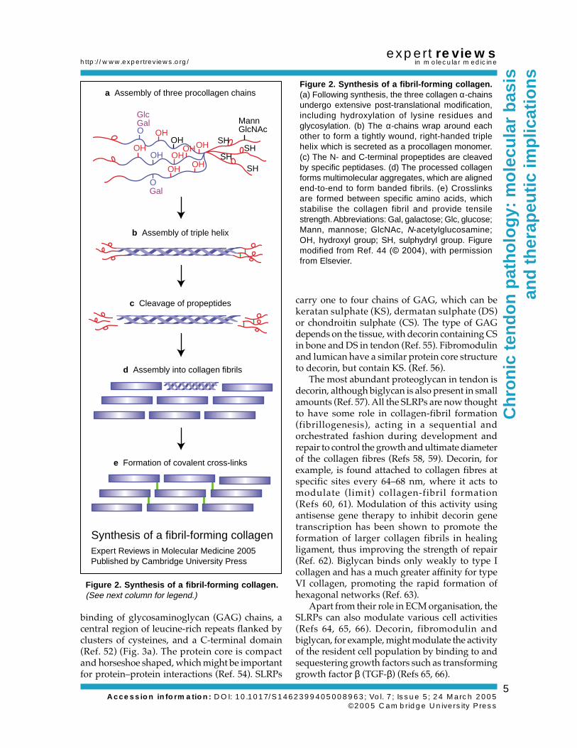
Although all collagens form highly organisedpolymers, the different collagen types can begrouped according to whether they form fibrilsor other structures such as extended sheets orlattices. The classic fibril-forming collagens (typesI, II, III, V and XI) comprise a single COL domainfor almost the entire length of the molecule, withsmall NC domains at each end (Fig. 2). The newlydiscovered collagen types XXIV and XXVII havea similar molecular structure (Refs 45, 46). Thenonfibrillar collagens are a heterogenous groupwith a variety of structures. Collagen types IV, VIIIand X form extensive networks, whereas type VI
collagen forms beaded microfilaments. Type VIIcollagen is essentially restricted to basementmembranes where it forms anchoring filaments.Several collagens have transmembrane domains,including types XIII, XVII, XXIII and XXV, whichmediate interactions between the cell and itsexternal environment. Some collagens, designatedFACIT (for ‘fibril-associated collagens withinterrupted triple helix’), are associated with thesurface of fibrils and have several interruptionsin the triple-helical structure. Collagen types IX,XII and XIV are the archetypal FACITs, althoughthe more recently described collagen types XVI,XIX, XX, XXI, XXII and XXVI are thought to berelated members.
The fibril-forming type I collagen is the majorcomponent of tendon; it is generally estimated torepresent 95% of the total collagen, although it isdifficult to be precise because of its insolubility,particularly in ageing tendon specimens (Ref. 47).Type III collagen is the next most abundantcollagen in tendon, forming around 3% of the totalin human supraspinatus and biceps brachiitendons (Ref. 47). In normal tendon, most type IIIcollagen is found in the endotenon and epitenon(Ref. 48), although it is also found intercalated intothe type I collagen fibril bundles, particularly inageing tendons and at the insertion (Ref. 49).Other minor constituents of tendon are collagentypes IV, V, VI, XII and XIV. Collagen types II, IX,X and XI, once thought to be restricted to cartilage,are found in the fibrocartilaginous regions oftendons and ligaments, where they are presumedto function to help resist compression and shearforces at these sites (Refs 50, 51).
ProteoglycansECM proteoglycans have been classified into twosubfamilies: the small leucine-rich repeatproteoglycans (SLRPs) and the large modularproteoglycans. The latter are further divided intotwo subgroups: those that do not bindhyaluronan; and the ‘hyalectans’, which bind bothhyaluronan and lectin (Refs 40, 52, 53). Only theSLRP and hyalectans are considered in this review,since these are the most abundant proteoglycansin the tendon ECM.
SLRPsSLRPs are found in most connective tissues andinclude decorin, biglycan, fibromodulin andlumican. They all possess a small protein core(36–42 kDa), with an N-terminal domain for
Accession information: DOI: 10.1017/S1462399405008963; Vol. 7; Issue 5; 24 March 2005 ©2005 Cambridge University Press
http://www.expertreviews.org/
Ch
ron
ic t
end
on
pat
ho
log
y: m
ole
cula
r b
asis
and
th
erap
euti
c im
plic
atio
ns
5
expert reviewsin molecular medicine
binding of glycosaminoglycan (GAG) chains, acentral region of leucine-rich repeats flanked byclusters of cysteines, and a C-terminal domain(Ref. 52) (Fig. 3a). The protein core is compactand horseshoe shaped, which might be importantfor protein–protein interactions (Ref. 54). SLRPs
carry one to four chains of GAG, which can bekeratan sulphate (KS), dermatan sulphate (DS)or chondroitin sulphate (CS). The type of GAGdepends on the tissue, with decorin containing CSin bone and DS in tendon (Ref. 55). Fibromodulinand lumican have a similar protein core structureto decorin, but contain KS. (Ref. 56).
The most abundant proteoglycan in tendon isdecorin, although biglycan is also present in smallamounts (Ref. 57). All the SLRPs are now thoughtto have some role in collagen-fibril formation(fibrillogenesis), acting in a sequential andorchestrated fashion during development andrepair to control the growth and ultimate diameterof the collagen fibres (Refs 58, 59). Decorin, forexample, is found attached to collagen fibres atspecific sites every 64–68 nm, where it acts tomodulate (limit) collagen-fibril formation(Refs 60, 61). Modulation of this activity usingantisense gene therapy to inhibit decorin genetranscription has been shown to promote theformation of larger collagen fibrils in healingligament, thus improving the strength of repair(Ref. 62). Biglycan binds only weakly to type Icollagen and has a much greater affinity for typeVI collagen, promoting the rapid formation ofhexagonal networks (Ref. 63).
Apart from their role in ECM organisation, theSLRPs can also modulate various cell activities(Refs 64, 65, 66). Decorin, fibromodulin andbiglycan, for example, might modulate the activityof the resident cell population by binding to andsequestering growth factors such as transforminggrowth factor β (TGF-β) (Refs 65, 66).
Figure 2. Synthesis of a fibril-forming collagen.(a) Following synthesis, the three collagen α-chainsundergo extensive post-translational modification,including hydroxylation of lysine residues andglycosylation. (b) The α-chains wrap around eachother to form a tightly wound, right-handed triplehelix which is secreted as a procollagen monomer.(c) The N- and C-terminal propeptides are cleavedby specific peptidases. (d) The processed collagenforms multimolecular aggregates, which are alignedend-to-end to form banded fibrils. (e) Crosslinksare formed between specific amino acids, whichstabilise the collagen fibril and provide tensilestrength. Abbreviations: Gal, galactose; Glc, glucose;Mann, mannose; GlcNAc, N-acetylglucosamine;OH, hydroxyl group; SH, sulphydryl group. Figuremodified from Ref. 44 (© 2004), with permissionfrom Elsevier.
Figure 2. Synthesis of a fibril-forming collagen.(See next column for legend.)
Synthesis of a fibril-forming collagen
OH
OHOH
OHOHOH
OHSH
SHSH
SH
Gal
GlcGal Mann
GlcNAc
a Assembly of three procollagen chains
b Assembly of triple helix
OHOH
O
O
c Cleavage of propeptides
d Assembly into collagen fibrils
e Formation of covalent cross-links
Expert Reviews in Molecular Medicine 2005 Published by Cambridge University Press

Accession information: DOI: 10.1017/S1462399405008963; Vol. 7; Issue 5; 24 March 2005 ©2005 Cambridge University Press
http://www.expertreviews.org/
Ch
ron
ic t
end
on
pat
ho
log
y: m
ole
cula
r b
asis
and
th
erap
euti
c im
plic
atio
ns
6
expert reviewsin molecular medicine
The ‘hyalectans’The hyalectan subgroup of large proteoglycanscomprises aggrecan, versican, brevican andneurocan (Refs 40, 52). Neurocan and brevicanare thought to be restricted to brain and neuraltissues. The hyalectans possess a large proteincore (100–370 kDa) consisting of a C-terminaldomain with epidermal growth factor (EGF)-likerepeats, a central domain carrying the majorityof GAG chains, and an N-terminal hyaluronan-binding domain (Ref. 56) (Fig. 3b).
Aggrecan, the major proteoglycan of articularcartilage but also found in tendon, formsmultimolecular aggregates with the nonsulphatedGAG hyaluronan. Aggrecan is reported to bepresent throughout tendon, although it isgenerally thought to be more abundant in regionsof fibrocartilage. Aggrecan has three globulardomains (G1, G2 and G3) and contains many GAGchains (CS and KS) attached to specific sites inthe GAG-binding domain between the G2 and G3domains (Ref. 67). The high fixed negative chargeof the GAG attracts counter-ions and functions tohold water within the tissue. Swelling of thetendon is restrained by the collagen meshwork,and the resulting turgor functions to resist
Proteoglycans in tendonExpert Reviews in Molecular Medicine C 2005 Cambridge University Press (part b only)
a SLRPs
b Hyalectans
Decorin
Biglycan
Fibromodulin
Lumican
Versican
LRRCore protein
CS/DS KS N-glycan Cys TyrSO4
G1
G1 G2 G3
G3
GAGα GAGβ
AggrecanCS2CS1KS
IGD
Figure 3. Proteoglycans in tendon. (a) Smallleucine-rich repeat proteoglycans (SLRPs). SLRPsin tendon include decorin, biglycan, fibromodulinand lumican. They share a common core proteinstructure with ten leucine-rich repeats (LRRs) and atleast one chain of glycosaminoglycan (GAG).Decorin in tendon has one dermatan sulphate (DS)chain, whereas biglycan has two chondroitinsulphate (CS) chains. Fibromodulin and lumicanhave up to four keratan sulphate (KS) chains andsome tyrosine (Tyr) residues are sulphated. Part aof figure reproduced, with permission, from theGlycoforum website (http://www.glycoforum.gr.jp).(b) Hyalectans. The hyalectans in tendon are versicanand aggrecan, with versican predominant in tensile-loaded regions and aggrecan in compressed,fibrocartilaginous regions. All hyalectans contain aG1 domain, a GAG-attachment region and a G3domain that has similarities with selectin. Aggrecanalso has a G2 domain, separated from G1 by aninterglobular domain (IGD), and the moleculecontains approximately 100 CS chains and 30 KSchains. Versican contains up to 21 CS chains,clustered within two GAG domains known as GAGαand GAGβ. One or both of the GAG domains mightbe removed by alternative splicing of the mRNAtranscript, forming four versican splice variants.
Figure 3. Proteoglycans in tendon. (See nextcolumn for legend.)

Accession information: DOI: 10.1017/S1462399405008963; Vol. 7; Issue 5; 24 March 2005 ©2005 Cambridge University Press
http://www.expertreviews.org/
Ch
ron
ic t
end
on
pat
ho
log
y: m
ole
cula
r b
asis
and
th
erap
euti
c im
plic
atio
ns
7
expert reviewsin molecular medicine
compressive load. Thus, the expression ofaggrecan in compressed regions of tendon, orareas subjected to shear forces such as theinsertion, is thought to be a functional adaptationthat protects the tissue from damage.
Versican has been identified in many softconnective tissues and has a similar structure toaggrecan, although it lacks a G2 domain andcontains much less GAG, all of which is CS(Ref. 68). Its precise role in tendon is unknown,although it is found associated with seams ofmicrofibrils between fibres and it mightfunction to facilitate the sliding of adjacent fibrebundles.
GlycoproteinsThe noncollagenous components of tendon arerelatively poorly characterised. Elastin, thoughtto make up less than 2% of the tendon dry weight,is a component of the elastic fibres that arethought to maintain the tendon crimp and to beresponsible for the elastic properties of the ECM(Refs 13, 69). Fibrillin is also a major componentof the elastic microfibril, and is thought to form atemplate for tropoelastin and the assembly of theelastin multimer (Refs 70, 71). Elastic microfibrilsare present in most connective tissues and containpolymers of fibrillin-1 and fibrillin-2 (Refs 70, 71).Mutations in fibrillin-1 have been linked toMarfan’s syndrome and associated disorders ofvarious connective tissues (Ref. 72). Fibrillin-1 isassociated with type XVI collagen in the dermis(but not in cartilage), and other proteins such asversican, fibulin, matrix-associated glycoprotein(MAGP)-1, MAGP-2 and emilin (Refs 73, 74, 75, 76).The microfibril structures that are formed havebeen divided into essentially three types on the basisof structure and appearance – oxytalan, elauninand elastic – which differ in the relative amountsof elastin (from lowest to highest, respectively).Elastic fibres, predominantly oxytalan, arereportedly more homogenous and abundant indeveloping tendon, less common in adult tendonand absent from fibrocartilage (Ref. 77).
Fibronectin mediates cell interactions withthe ECM, and affects a range of cell functionsincluding cell adhesion, cell migration,differentiation, haemostasis, phagocytosis andchemotaxis (Refs 40, 78). Present at low levels innormal tendon, fibronectin is massively increasedafter tendon injury and consequently has beenimplicated in cell adhesion, migration anddifferentiation at the site of injury (Refs 79, 80, 81).
Tenascin-C is a disulphide-linked hexamericprotein with subunits of 200–300 kDa in humans,created by alternative splicing of a single genetranscript (Refs 82, 83). In normal fibrous tendon,tenascin-C might have a role in maintaining theinterface between fibrils and adjacent structures(Ref. 84). In fibrocartilaginous regions of tendon,tenascin-C is predominantly cell-associated(similar to type VI collagen) and is implicated inthe development of the chondrocyte cell phenotype(the expression of type II collagen and aggrecan)in response to compressive load (Ref. 84).Tenascin-C is transiently increased after tendoninjury, and is thought to modulate cell activitiesin the developing scar (Refs 85, 86).
Cartilage oligomeric matrix protein (COMP),despite its name, is not restricted to cartilage andis a major component of tendon, representing upto 3% of the dry weight (Ref. 87). A member of thethrombospondin gene family (thrombospondin 5),it is a large (524 kDa) pentameric moleculecomposed of five disulphide-bonded subunits(Ref. 88). Like tenascin-C and the otherthrombospondins, it is thought to have both astructural role and an interactive role with the cellpopulation. There is a strong positive correlationof COMP expression with the levels of mechanicalload, with higher levels in flexor tendonscompared with extensors (Ref. 89). Levels ofCOMP increase with age up to skeletal maturity,although only in weight-bearing tendons (Ref. 89).The structural importance of COMP is shown bythe condition of pseudoachondroplasia, a geneticdisorder caused by a mutation in the COMP gene,resulting in short stature, lax joints and early-onsetosteoarthritis (Ref. 90).
In addition to serum proteins such as albumin,other glycoproteins in tendon include: laminin,which is found as a major constituent of basementmembranes; link protein, which stabiliseshyalectan–hyaluronan interactions (Refs 6, 91);and other multidomain adhesive glycoproteins,including members of the thrombospondinfamily, that, like COMP, tenascin and fibronectin,mediate cell–matrix interactions in normal andinjured tissues (Refs 92, 93, 94).
The molecular pathology of chronictendinopathy
Histopathological studies of tendinopathy haveshown an absence of inflammatory cell infiltrationand the presence of many features thought to becharacteristic of ECM degeneration. These include

Accession information: DOI: 10.1017/S1462399405008963; Vol. 7; Issue 5; 24 March 2005 ©2005 Cambridge University Press
http://www.expertreviews.org/
Ch
ron
ic t
end
on
pat
ho
log
y: m
ole
cula
r b
asis
and
th
erap
euti
c im
plic
atio
ns
8
expert reviewsin molecular medicine
a loss of fibre organisation, decreased fibrildiameter, changes in cell density (both increasedand decreased), cell rounding, GAG accumulation,lipid accumulation and calcification (Refs 3, 4, 5,7, 95) (Fig. 4). Although similar changes arecommonly found in normal tendons, they aregenerally less severe, and it is assumed that ECMdegeneration precedes the onset of the clinicalcondition (Refs 3, 7, 96).
There have been few biochemical studies ofchronic tendinopathy. In ruptured supraspinatustendons there was a small but significant decreasein the total collagen content, and an increasedproportion of type III collagen relative to type I
collagen (Ref. 47). The collagen had a highcontent of hydroxylysine and there were greaterthan normal levels of the mature collagencrosslinks hydroxylyslpyridinoline (HP) andlysylpyridinoline (LP) (Ref. 97). These and othercrosslinks greatly affect the solubility of thecollagen, making it difficult to assess the collagentype, even using chemical methods such ascyanogen bromide peptide mapping. However,the changes in the tendon ECM are characteristicof scar tissue, and the levels of type III collagenand HP tend to diminish as healing proceeds,although high levels often persist because ofincomplete remodelling (Ref. 98). Similar ECM
Figure 4. Histopathology of tendinopathy. (a) Normal flexor tendon histology, showing organised parallelfibre bundles and long thin tenocytes dispersed throughout the matrix [stained with hematoxylin and eosin(H&E)]. (b) Ruptured supraspinatus tendon, showing hyaline (glassy) appearance, loss of matrix organisationand rounded, shrunken nuclei (H&E). (c) Glycosaminoglycan (GAG) accumulation (‘mucoid degeneration’) insupraspinatus tendon, showing GAG (blue) surrounding rounded cells in the matrix (Alcian Blue and H&E).(d) ‘Angiofibroblastic’ change in painful Achilles tendinopathy, showing increase in cell number and bloodvessels (H&E). Part c of figure reprinted from Ref. 4 (© 1994); reproduced with permission from the BMJPublishing Group.
Histopathology of tendinopathy
a b
c d
Expert Reviews in Molecular Medicine C 2005 Cambridge University Press (parts a, b and d only)

Accession information: DOI: 10.1017/S1462399405008963; Vol. 7; Issue 5; 24 March 2005 ©2005 Cambridge University Press
http://www.expertreviews.org/
Ch
ron
ic t
end
on
pat
ho
log
y: m
ole
cula
r b
asis
and
th
erap
euti
c im
plic
atio
ns
9
expert reviewsin molecular medicine
changes have since been reported in studies ofposterior tibialis tendon dysfunction and Achillestendon rupture (Refs 99, 100). These data areconsistent with the gradual accumulation of typeIII collagen over a relatively long time period,allowing the maturation of collagen crosslinksand stabilisation of the ECM. The gradualincorporation of type III collagen into the mainfibre bundles is consistent with a reduction in theaverage fibril diameter (Ref. 101), a change that isthought to weaken the tendon and to precedetendon rupture (Ref. 102).
Other biochemical studies have shown anincrease in the amount of hyaluronan and variousproteoglycans in degenerate tendons, althoughthe latter have not yet been fully characterised(Refs 103, 104). An increased water content,typically 10% above normal, is commonly foundin tendinopathy, associated with the increasedproteoglycan content. Consequently, the specimendry weight or collagen (hydroxyproline) contentis a better reference point for composition studiesthan the tissue wet weight. Differences in thesugar moieties expressed in ruptured tendonscompared with normal were revealed bychanges in lectin-staining properties (Ref. 105).Glycoproteins such as tenascin-C were increasedin ruptured supraspinatus, with differentprotein isoforms expressed as well as numeroussmall peptide fragments, the latter consistentwith enzyme-mediated cleavage in the tissue(Ref. 84). Fibronectin immunostaining wassignificantly increased in ruptured tendon, andthere was accumulation of necrotic tissue andfibrin (Ref. 106).
Several studies have shown differences in theexpression of various genes encoding matrixproteins in tendinopathy (Refs 107, 108, 109). Inone study, for example, 23 genes were found tobe upregulated and 17 genes were downregulatedin degenerate Achilles tendon (Ref. 107). Althoughpotentially very informative, relatively few ofthese changes have been confirmed by morerigorous techniques, and the levels of cognateproteins for many of these genes have not beeninvestigated. There were no changes detected inthe expression of cytokines and cytokinereceptors, consistent with the absence of anyongoing inflammatory process, as confirmed byan analysis of the fluids surrounding painfultendons (Ref. 110). However, these data do notrule out the involvement of inflammation atearlier stages of the disease. It is possible, for
example, that the tendon fails to recover after theinflammatory reaction has subsided.
In summary, the ECM changes described inchronic tendinopathy are consistent with a cell-mediated remodelling process occurring indegenerate tendon, without inflammation. Theprocess is similar in many respects to the laterstages of a wound-healing response, albeit withimpaired or incomplete remodelling, rather thanany functional adaptation. The evidence generallysupports the hypothesis that there is gradualdeterioration in the quality of the ECM, whichpredisposes to tendon pathology. A key elementof this process is thought to be the cellularexpression of proteolytic activities and their effectson tendon ECM turnover.
Role of MMPs in tendon collagendegradation and tendinopathy
Proteolytic activity is an essential component oftissue maintenance and repair. After injury,proteolysis is required to remove any damagedECM and remodel the newly formed scar sothat it more closely resembles the normal tissue.Some collagen in tendon is probably degradedintracellularly after phagocytosis, with fibroblastsand macrophages engulfing collagen moleculesthat are then digested by lysosomal enzymes,comprising mainly cysteine and aspartateproteases (Refs 111, 112). This is a major activityin the rapidly remodelling peridontal ligament,although few (if any) studies have investigatedthe function of lysosomal enzymes and serineproteases in tendon matrix turnover. Mostpublished studies have focused on collagendegradation occurring in the extracellularenvironment and mediated by secretedmetalloenzymes known as the MMPs.
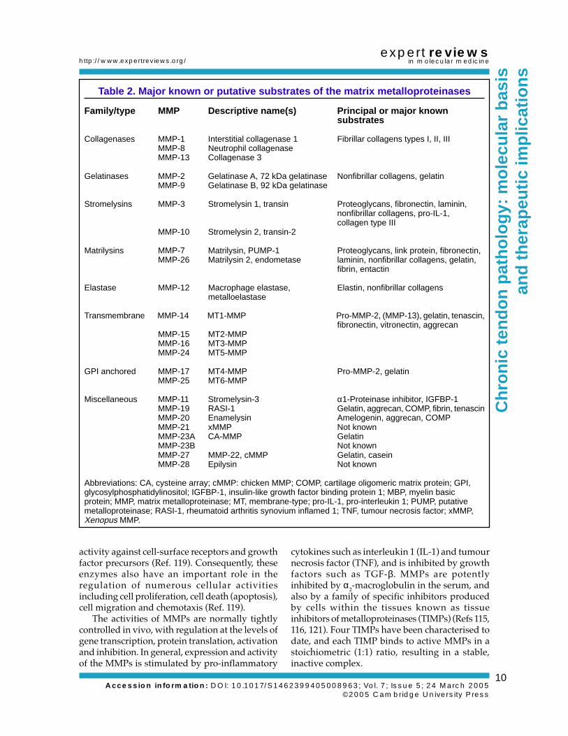
MMPs: a brief overviewComprehensive reviews of the MMPs have beenpublished elsewhere and only salient points arereviewed here (Refs 113, 114, 115, 116, 117, 118,119, 120). MMPs are members of the ‘MB’ clan ofmetallopeptidases, generically referred to as‘metzincins’ because they contain zinc at the activesite and a conserved methionine eight residuesdownstream. There are 23 MMPs found inhumans, each comprising a multidomainstructure and with activity at neutral pH againsta broad spectrum of different ECM substrates(Refs 114, 116, 118) (Table 2; Fig. 5a). Althoughimportant in ECM degradation, MMPs also have
Accession information: DOI: 10.1017/S1462399405008963; Vol. 7; Issue 5; 24 March 2005 ©2005 Cambridge University Press
http://www.expertreviews.org/
Ch
ron
ic t
end
on
pat
ho
log
y: m
ole
cula
r b
asis
and
th
erap
euti
c im
plic
atio
ns
10
expert reviewsin molecular medicine
activity against cell-surface receptors and growthfactor precursors (Ref. 119). Consequently, theseenzymes also have an important role in theregulation of numerous cellular activitiesincluding cell proliferation, cell death (apoptosis),cell migration and chemotaxis (Ref. 119).
The activities of MMPs are normally tightlycontrolled in vivo, with regulation at the levels ofgene transcription, protein translation, activationand inhibition. In general, expression and activityof the MMPs is stimulated by pro-inflammatory
cytokines such as interleukin 1 (IL-1) and tumournecrosis factor (TNF), and is inhibited by growthfactors such as TGF-β. MMPs are potentlyinhibited by α2-macroglobulin in the serum, andalso by a family of specific inhibitors producedby cells within the tissues known as tissueinhibitors of metalloproteinases (TIMPs) (Refs 115,116, 121). Four TIMPs have been characterised todate, and each TIMP binds to active MMPs in astoichiometric (1:1) ratio, resulting in a stable,inactive complex.
Table 2. Major known or putative substrates of the matrix metalloproteinases
Family/type MMP Descriptive name(s) Principal or major knownsubstrates
Collagenases MMP-1 Interstitial collagenase 1 Fibrillar collagens types I, II, IIIMMP-8 Neutrophil collagenaseMMP-13 Collagenase 3
Gelatinases MMP-2 Gelatinase A, 72 kDa gelatinase Nonfibrillar collagens, gelatinMMP-9 Gelatinase B, 92 kDa gelatinase
Stromelysins MMP-3 Stromelysin 1, transin Proteoglycans, fibronectin, laminin,nonfibrillar collagens, pro-IL-1,collagen type III
MMP-10 Stromelysin 2, transin-2
Matrilysins MMP-7 Matrilysin, PUMP-1 Proteoglycans, link protein, fibronectin,MMP-26 Matrilysin 2, endometase laminin, nonfibrillar collagens, gelatin,
fibrin, entactin
Elastase MMP-12 Macrophage elastase, Elastin, nonfibrillar collagensmetalloelastase
Transmembrane MMP-14 MT1-MMP Pro-MMP-2, (MMP-13), gelatin, tenascin,fibronectin, vitronectin, aggrecan
MMP-15 MT2-MMPMMP-16 MT3-MMPMMP-24 MT5-MMP
GPI anchored MMP-17 MT4-MMP Pro-MMP-2, gelatinMMP-25 MT6-MMP
Miscellaneous MMP-11 Stromelysin-3 α1-Proteinase inhibitor, IGFBP-1MMP-19 RASI-1 Gelatin, aggrecan, COMP, fibrin, tenascinMMP-20 Enamelysin Amelogenin, aggrecan, COMPMMP-21 xMMP Not knownMMP-23A CA-MMP GelatinMMP-23B Not knownMMP-27 MMP-22, cMMP Gelatin, caseinMMP-28 Epilysin Not known
Abbreviations: CA, cysteine array; cMMP: chicken MMP; COMP, cartilage oligomeric matrix protein; GPI,glycosylphosphatidylinositol; IGFBP-1, insulin-like growth factor binding protein 1; MBP, myelin basicprotein; MMP, matrix metalloproteinase; MT, membrane-type; pro-IL-1, pro-interleukin 1; PUMP, putativemetalloproteinase; RASI-1, rheumatoid arthritis synovium inflamed 1; TNF, tumour necrosis factor; xMMP,Xenopus MMP.
Accession information: DOI: 10.1017/S1462399405008963; Vol. 7; Issue 5; 24 March 2005 ©2005 Cambridge University Press
http://www.expertreviews.org/
Ch
ron
ic t
end
on
pat
ho
log
y: m
ole
cula
r b
asis
and
th
erap
euti
c im
plic
atio
ns
11
expert reviewsin molecular medicine
MMPs in tendonThe MMPs are implicated in both the physiologicaland pathological turnover of the tendon ECM.Pieces of normal tendon placed in explant cultureproduce collagenase and gelatinase activities,degrading the collagen matrix after two to threeweeks in culture, and this process is stimulatedby the addition of inflammatory cytokines suchas IL-1 (Refs 122, 123, 124, 125). MMPs are also
implicated in the remodelling of tendon thatfollows immobilisation (Refs 126, 127, 128, 129).MMP-1 is thought to be one of the key mediatorsof tendon fibrillar collagen degradation, at leastin explant culture, and this activity can beinhibited by the application of cyclical strain, aneffect though to be mediated via the tenocytecytoskeleton (Refs 130, 131, 132, 133, 134). Isolatedtenocytes respond to strain and shear forces by
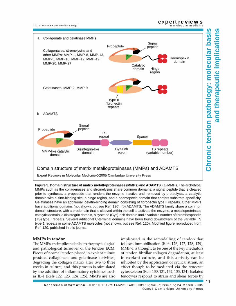
Figure 5. Domain structure of matrix metalloproteinases (MMPs) and ADAMTS. (a) MMPs. The archetypalMMPs such as the collagenases and stromelysins share common domains: a signal peptide that is cleavedprior to synthesis, a propeptide that renders the enzyme inactive until removed by proteolysis, a catalyticdomain with a zinc-binding site, a hinge region, and a haemopexin domain that confers substrate specificity.Gelatinases have an additional, gelatin-binding domain consisting of fibronectin type II repeats. Other MMPshave additional domains (not shown, but see Ref. 120). (b) ADAMTS. The ADAMTS family share a commondomain structure, with a prodomain that is cleaved within the cell to activate the enzyme, a metalloproteinasecatalytic domain, a disintegrin domain, a cysteine (Cys)-rich domain and a variable number of thrombospondin(TS) type I repeats. Several additional C-terminal domains have been found downstream of the variable TStype 1 repeats in some ADAMTS molecules (not shown, but see Ref. 120). Modified figure reproduced fromRef. 120, published in this journal.
Zn2+
b ADAMTS
TSrepeat Spacer
TS repeats(variable number)
Domain structure of matrix metalloproteinases (MMPs) and ADAMTSExpert Reviews in Molecular Medicine C 2005 Cambridge University Press
Propeptide
Disintegrin-likedomain
Signalpeptide
MMP-like catalyticdomain
Cys-richregion
Collagenases, stromelysins and other MMPs: MMP-1, MMP-8, MMP-13, MMP-3, MMP-10, MMP-12, MMP-19, MMP-20, MMP-27
Zn2+
C
Gelatinases: MMP-2, MMP-9 Zn2+
C
a Collagenate and gelatinase MMPs
PropeptideSignalpeptide
Catalyticdomain Hinge
region
Haemopexindomain
Type IIfibronectin
repeats
Accession information: DOI: 10.1017/S1462399405008963; Vol. 7; Issue 5; 24 March 2005 ©2005 Cambridge University Press
http://www.expertreviews.org/
Ch
ron
ic t
end
on
pat
ho
log
y: m
ole
cula
r b
asis
and
th
erap
euti
c im
plic
atio
ns
12
expert reviewsin molecular medicine
upregulation of MMP, demonstrating thatmechanical strain is likely to be a major stimulusfor ECM remodelling in tendon (Refs 135, 136).MMP expression was stimulated in rabbittendon in vivo by extended periods of cyclicalloading, although there was no sign of injury asassessed by histological examination (Ref. 137).Intense physical training was also shown toincrease MMP-2 and MMP-9 activities in the fluidsurrounding human Achilles tendon (Ref. 138).
Several MMPs have been identified in acutetendon injuries, with differences in the timing andlocation of expression that suggest different rolesin the healing process. Levels of MMP-9 andMMP-13 peaked 7–14 days after injury, whereasMMP-2, MMP-3 and MMP-14 increased and weremaintained at high levels until at least day 28(Ref. 139). Thus, it appears that MMP-9 andMMP-13 are involved in the degradation ofcollagen in the initial inflammatory phase,whereas MMP-2, MMP-3 and MMP-14 have a rolein the remodelling of the scar tissue.
A comparison of human tendons showedevidence of substantial differences in the rate ofcollagen turnover between tendons from differentsites (Ref. 38). There was very little collagenturnover in normal biceps brachii tendons,which contained no significant levels of MMPactivity and a linear accumulation of pentosidinecrosslinks with increasing age (Ref. 38). Bycontrast, supraspinatus tendons obtained fromnormal shoulders showed relatively high levels ofcollagen turnover, and there were correspondinglyhigh levels of MMP-1, MMP-2 and MMP-3activity (Ref. 38). In ruptured supraspinatustendon, there was increased activity of MMP-1,reduced activity of MMP-2 and MMP-3, andevidence of increased turnover of the collagennetwork (Ref. 38). Levels of expression of MMP-1and MMP-3 in shoulder fluids were shown tocorrelate with the size of the tendon tear, andMMP-1 was expressed by cells within theruptured tendon (Refs 140, 141).
Studies of painful Achilles tendons usingcDNA arrays have identified several differencesin MMP gene expression, although many of thesefindings need to be confirmed by RT-PCR analysis(Refs 107, 108). The absence of MMP-1 and MMP-8expression was consistent with an absence ofinflammation, and there was a small but variableincrease in MMP-2 and MMP-14 in degeneratetendons, whereas MMP-9 and MMP-13 weredetected only in ruptured tendons (Refs 107, 108).
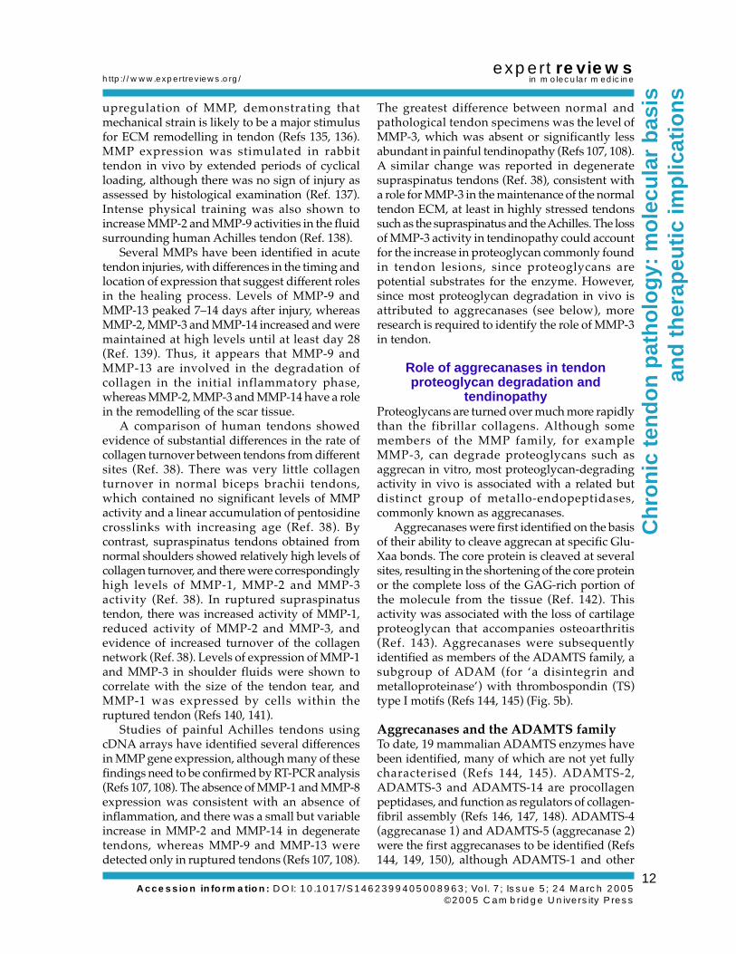
The greatest difference between normal andpathological tendon specimens was the level ofMMP-3, which was absent or significantly lessabundant in painful tendinopathy (Refs 107, 108).A similar change was reported in degeneratesupraspinatus tendons (Ref. 38), consistent witha role for MMP-3 in the maintenance of the normaltendon ECM, at least in highly stressed tendonssuch as the supraspinatus and the Achilles. The lossof MMP-3 activity in tendinopathy could accountfor the increase in proteoglycan commonly foundin tendon lesions, since proteoglycans arepotential substrates for the enzyme. However,since most proteoglycan degradation in vivo isattributed to aggrecanases (see below), moreresearch is required to identify the role of MMP-3in tendon.
Role of aggrecanases in tendonproteoglycan degradation and
tendinopathyProteoglycans are turned over much more rapidlythan the fibrillar collagens. Although somemembers of the MMP family, for exampleMMP-3, can degrade proteoglycans such asaggrecan in vitro, most proteoglycan-degradingactivity in vivo is associated with a related butdistinct group of metallo-endopeptidases,commonly known as aggrecanases.
Aggrecanases were first identified on the basisof their ability to cleave aggrecan at specific Glu-Xaa bonds. The core protein is cleaved at severalsites, resulting in the shortening of the core proteinor the complete loss of the GAG-rich portion ofthe molecule from the tissue (Ref. 142). Thisactivity was associated with the loss of cartilageproteoglycan that accompanies osteoarthritis(Ref. 143). Aggrecanases were subsequentlyidentified as members of the ADAMTS family, asubgroup of ADAM (for ‘a disintegrin andmetalloproteinase’) with thrombospondin (TS)type I motifs (Refs 144, 145) (Fig. 5b).
Aggrecanases and the ADAMTS familyTo date, 19 mammalian ADAMTS enzymes havebeen identified, many of which are not yet fullycharacterised (Refs 144, 145). ADAMTS-2,ADAMTS-3 and ADAMTS-14 are procollagenpeptidases, and function as regulators of collagen-fibril assembly (Refs 146, 147, 148). ADAMTS-4(aggrecanase 1) and ADAMTS-5 (aggrecanase 2)were the first aggrecanases to be identified (Refs144, 149, 150), although ADAMTS-1 and other
Accession information: DOI: 10.1017/S1462399405008963; Vol. 7; Issue 5; 24 March 2005 ©2005 Cambridge University Press
http://www.expertreviews.org/
Ch
ron
ic t
end
on
pat
ho
log
y: m
ole
cula
r b
asis
and
th
erap
euti
c im
plic
atio
ns
13
expert reviewsin molecular medicine
phylogenetically related enzymes (ADAMTS-8,ADAMTS-9, ADAMTS-15 and ADAMTS-20)might also have aggrecanase activity (Refs 151,152, 153, 154). Best known for their activity againstaggrecan, ADAMTS-1 and ADAMTS-4 are alsocapable of cleaving other ECM proteoglycans suchas versican and brevican (Ref. 155), andglycoproteins such as COMP (Ref. 156), at leastin vitro. Although inhibition of ADAMTS-4 andADAMTS-5 can prevent cartilage degradation intissue culture models (Ref. 157), the enzymesresponsible for proteoglycan degradation inosteoarthritis and other diseases of connectivetissues have yet to be identified.
Aggrecanase activity is thought to be regulatedat multiple levels, although the mechanism iscurrently poorly understood. Differentialregulation of ADAMTS mRNAs has beendeduced from analysis of their expression in celland explant cultures, albeit with considerablevariation between studies (Refs 157, 158, 159, 160,161). A study of human tendon cells has reportedsmall and variable effects of IL-1 on ADAMTS-4expression (Ref. 161).
In addition to regulation at the level of genetranscription, the activities of ADAMTS enzymesare also subject to post-translational regulation.The noncatalytic ancillary domains of ADAMTS-4are required for both catalytic activity andsubstrate specificity (Refs 162, 163). Full-lengthenzyme is sequestered in the ECM via GAG-binding sequences in the spacer domain, andsulphated GAGs attached to the aggrecan coreprotein are required for ADAMTS-4 activity(Ref. 163). Deletion of the C-terminal spacerdomain increased the efficiency of hydrolysis ofaggrecan at Glu373-Ala374 bonds, and revealednew activities against fibromodulin, decorin anda general protein substrate (Ref. 163). Several shortforms of ADAMTS-4, thought to be generated byautocatalytic C-terminal truncation, are found incartilage, and these potentially contribute to thedegradation of a broad range of protein substratesin addition to proteoglycans (Refs 162, 163). Theenzymes are thought to be secreted in an activeform after cleavage of the prodomain withinthe cell by furin, which might be followed byC-terminal truncation by MMP-17 at the cell surface(Refs 164, 165). Aggrecanases such as ADAMTS-4are inhibited by the general proteinase inhibitorα2-macroglobulin and by the specific endogenousinhibitor TIMP-3, but not by other TIMPs(TIMP-1, -2 and -4) (Ref. 166).
Aggrecanase activities in tendonStudies of normal (bovine) tendon have shownthat proteoglycans are constitutively turned overrelatively rapidly, with hyalectans breaking downmore rapidly than the SLRPs (Refs 167, 168, 169).Proteolytic fragments of proteoglycans such asaggrecan were present in both young and maturetendon, in both tensional and compressed regions.Much of the aggrecan in tendon appears to lackthe G1 domain, but the molecule might beretained in the tissue by interactions of the G3domain with an unidentified matrix component.Cultured tendon explants released cleavageproducts into the culture medium and there wasno significant stimulation of this activity by IL-1(Ref. 167). There was no evidence of MMP-mediated proteoglycan turnover, althoughaggrecan turnover did not directly correlate withthe levels of expression of either ADAMTS-4 orADAMTS-5 mRNA. However, gene expressionmight play a relatively minor role in the regulationof aggrecanase activity, and further studies arerequired to identify the enzyme activitiespresent in human tendon and their role intendinopathy. Since levels of proteoglycan areincreased in the degenerate tendon lesion (unlikeosteoarthritic cartilage), it will be interesting todetermine whether this is caused by an increasein proteoglycan synthesis or a decrease inproteoglycan degradation.
Clinical implicationsAnti-inflammatory drugs: do they have arole in the treatment of tendinopathy?The absence of inflammatory cells, at least at laterstages of the disease, would suggest that there isno rational basis for the treatment of chronictendinopathy with anti-inflammatories such asnonsteroidal anti-inflammatory drugs (NSAIDs)and corticosteroid injections, both of which arecommonly used even though there are limiteddata to demonstrate their effectiveness (Ref. 1).However, it has recently been argued that thedefinition of inflammation should be reappraised,since inflammatory mediators can be producedby a variety of cell types, and not just byinfiltrating leukocytes (Ref. 170). Degenerativeconditions such as osteoarthritis can be consideredinflammatory diseases on this basis, with theexpression of cytokines and nitric oxide bychondrocytes and/or synovial cells (Refs 170, 171,172). In a similar fashion, it is possible thatmediators of inflammation might be involved in
Accession information: DOI: 10.1017/S1462399405008963; Vol. 7; Issue 5; 24 March 2005 ©2005 Cambridge University Press
http://www.expertreviews.org/
Ch
ron
ic t
end
on
pat
ho
log
y: m
ole
cula
r b
asis
and
th
erap
euti
c im
plic
atio
ns
14
expert reviewsin molecular medicine
chronic tendinopathy, although produced bythe resident cell population or surroundingconnective tissues (Refs 173, 174, 175, 176). Forexample, prostaglandins and other inflammatorymediators were found to be increased in the fluidssurrounding tendons following vigorous exerciseand are implicated at least in the adaptiveresponse of the tissue (Refs 177, 178, 179).Increased expression of the induciblecyclooxygenase (COX-2) in patellar tendinosissuggests some involvement of prostaglandins inthe disease (Ref. 175). Several studies have shownIL-1 was more abundant in the endotenon,vascular endothelium and synovial tissuessurrounding affected tendons (Refs 174, 176).Inflammatory mediators might also be transientlyproduced by the tenocytes, which can be inducedto express IL-1β and COX-2 under the influenceof mechanical strain, providing a theoretical basisfor the development of ‘overuse’ tendinopathy(Refs 180, 181, 182, 183). However, most molecularstudies of tendinopathy have so far provided noconclusive evidence for the involvement ofinflammatory mediators and cytokines such asprostaglandin E2 and IL-1 (Refs 107, 108, 184, 185).
Enzymes as potential targets for therapyIn degenerative conditions such as osteoarthritis,one of the targets for drug therapy is the increasedcartilage ECM degradation mediated by enzymessuch as MMPs and ADAMTS (Refs 118, 186).Various therapeutic approaches have beenattempted, including selective inhibition ofcollagenases or broad-spectrum inhibition ofmany different MMPs, although none has yet beensuccessful in clinical trials (Ref. 118).
The pathology of tendinopathy is evidentlydifferent from osteoarthritis, with increased cellactivity, neovascularisation, and accumulation ofproteoglycan within the lesion, in addition toincreased turnover of the matrix collagen. Indeed,it would appear that some ECM turnover isrequired for maintaining the health of the tendon,at least in highly stressed tendons such as thesupraspinatus and Achilles. This hypothesis isconsistent with data obtained from clinical trialswith broad-spectrum MMP inhibitors, which werefound to cause an unwanted side effect describedas a ‘musculo-skeletal syndrome’ in the tendonsof the patients’ shoulders and hands, whichrecovered after the cessation of therapy (Refs 187,188). Compounds selective for collagenases orgelatinases did not induce the condition, and
the precise cause of the syndrome remainsunidentified. Additional activity against‘sheddases’ (members of the ADAM family ofmetalloproteases involved in the processing ofcell-surface receptors) might explain thedevelopment of the syndrome, although there isno consensus of opinion. Because there are manydifferent enzymes, a solution to this problem willrequire an analysis of all the metalloproteinaseenzymes that are expressed and active in healthyand degenerate tendon.
Neuropeptides and tendon ECM turnover:a novel therapeutic target for tendinopathy?If inflammation is not associated with thedevelopment of chronic tendinopathy, othercauses of tendon pain and ECM degenerationmust be considered. Recent studies havesuggested that nerves, and small peptides(‘neuropeptides’) produced by nerve endings,might have a role in tendinopathy, similar to thatdescribed in intervertebral disc degeneration (Refs189, 190).
In studies of ‘tennis elbow’ lesions, nerveendings and neuropeptides were found at the siteof the lesion, although it was unclear if thisdistribution was different to normal (Ref. 191).Studies of fluids obtained from around painfultendons using a microdialysis technique haveshown that levels of the neurotransmitterglutamate were significantly increased intendinopathy relative to controls (Refs 110, 184,192). Both free glutamate and glutamate receptors(NMDAR1) have also been detected withinAchilles tendons, located to nerve fibres, both intendinopathy specimens and in controls (Ref. 193).Since glutamate is a potent mediator of pain inthe central nervous system, it was suggested thatNMDAR1 antagonists might be useful in thetreatment of tendon pain.
It has also been reported that substance P,another neuropeptide associated with the sensationof pain, is increased in the subacromical bursa inpatients with rotator cuff tendinopathy (Ref. 194).The amount of substance P was shown to correlatewith the degree of motion pain as assessed by avisual analogue scale. Whether this was due toan increase in the release of substance P or anincrease in the number of nerve fibres was notclear, although immunohistochemistry showedmore nerve fibres in bursal tissues of patients witha perforated rotator cuff (Ref. 194). Apart from themodulation of pain, substance P and other

Accession information: DOI: 10.1017/S1462399405008963; Vol. 7; Issue 5; 24 March 2005 ©2005 Cambridge University Press
http://www.expertreviews.org/
Ch
ron
ic t
end
on
pat
ho
log
y: m
ole
cula
r b
asis
and
th
erap
euti
c im
plic
atio
ns
15
expert reviewsin molecular medicine
neuropeptides might have additional effects,regulating the local circulation and stimulatingneurogenic inflammation in and around thetendon (Refs 195, 196).
Hart et al. have proposed that regulatory unitscomposed of nerve endings and mast cells residein and around the tendon (Ref. 197). The releaseof neurotransmitters stimulates mast-celldegranulation, releasing a variety of mediatorsincluding growth factors that influence oedema,angiogenesis, fibroblast proliferation and manyother aspects of cell activity. Biomechanicalstimulation of these nerve–mast-cell units mightform part of the normal regulatory system,maintaining the tissue and also contributing tothe adaptive response to load. Excessivestimulation of neural–mast-cell units mightcontribute to overuse tendinopathy. Since theextent of innervation and vascularisation variesbetween different tendons, the potential for thedevelopment of neurogenic dysfunction alsovaries. This theory potentially links mechanicalstimulation of the paratenon, which is morerichly innervated, and tissue changes in thetendon mid-substance. The association ofneuropeptides with tissue remodelling has notbeen conclusively proved, although substance Pand calcitonin-gene-related peptide (CGRP) wereshown to modulate directly the expression ofMMP-1 and MMP-3, at least in vitro (Refs 195,198). Thus innervation, and the stimulation ofneuropeptide release by strain or friction at thetendon surface, is thought to be important for bothnormal tendon function and tendinopathy,affecting remodelling events in the tissue. Sincepeptide antagonists of substance P are alreadyavailable, having been used in clinical trials forthe treatment of pain, emesis and depression, it ispossible that they could prove useful for thetreatment of chronic tendinopathy.
Concluding remarksMost tendinopathy is associated with degeneration,which is thought to be an active, cell-mediatedprocess involving increased turnover andremodelling of the tendon ECM. There is a gradualtransformation in the quantity and quality of theECM that precedes tendon rupture. However,some ECM turnover might be required tomaintain the health of the tendon, particularly atsites exposed to high mechanical strain, such asthe shoulder and ankle. Degradation of the tendonECM is mediated by a variety of metalloproteinase
enzymes, including MMPs and aggrecanases.Some enzymes are thought to be responsible forrepair and maintenance of the tendon, and othersare implicated in the pathological destruction ofthe ECM: these enzymes need to be identified sothat new drugs can be developed. There are avariety of factors that might influence ECMturnover in tendon, although a major factor inmost tendinopathy is thought to be repeatedminor mechanical strain or ‘overuse’. Althoughinfiltrating inflammatory cells are probably notinvolved in chronic tendinopathy, neuropeptidesand other mediators of pain and inflammationproduced in or around the tendon might beimplicated, and could provide novel targets fortherapeutic intervention.
Acknowledgements and fundingThe author is grateful for financial support fromthe Arthritis Research Campaign, The IsaacNewton Trust, REMEDI, The Wishbone Trust, TheDunhill Medical Trust, The Sybil Eastwood Trustand the Cambridge Arthritis Research Endeavour(CARE). Acknowledgements are due to many pastand present members of the RheumatologyResearch Unit, in particular Dr Brian Hazleman,Professor Tim Cawston, Dr Michael Chard, DrSteven Fenwick, Dr Anthony Corps, Mrs ValCurry and Ms Rebecca Harrall. Thanks also to thesurgeons who have provided human tendonspecimens, including Mr Chris Constant, MrAndrew Robinson, Mr Roger Hackney and MrGraham Holloway.
References1 Almekinders, L.C. and Temple, J.D. (1998)
Etiology, diagnosis, and treatment of tendonitis:an analysis of the literature. Med Sci Sports Exerc30, 1183-1190, PubMed: 9710855
2 Puddu, G., Ippolito, E. and Postacchini, F. (1976)A classification of Achilles tendon disease. Am JSports Med 4, 145-150, PubMed: 984291
3 Kannus, P. and Jozsa, L. (1991) Histopathologicalchanges preceding spontaneous rupture of atendon. A controlled study of 891 patients. J BoneJoint Surg Am 73, 1507-1525, PubMed: 1748700
4 Chard, M.D. et al. (1994) Rotator cuffdegeneration and lateral epicondylitis: acomparative histological study. Ann Rheum Dis53, 30-34, PubMed: 8311552
5 Astrom, M. and Rausing, A. (1995) ChronicAchilles tendinopathy. A survey of surgical andhistopathologic findings. Clin Orthop 151-164,
Accession information: DOI: 10.1017/S1462399405008963; Vol. 7; Issue 5; 24 March 2005 ©2005 Cambridge University Press
http://www.expertreviews.org/
Ch
ron
ic t
end
on
pat
ho
log
y: m
ole
cula
r b
asis
and
th
erap
euti
c im
plic
atio
ns
16
expert reviewsin molecular medicine
PubMed: 76346996 Jarvinen, M. et al. (1997) Histopathological
findings in chronic tendon disorders. Scand JMed Sci Sports 7, 86-95, PubMed: 9211609
7 Riley, G.P., Goddard, M.J. and Hazleman, B.L.(2001) Histopathological assessment andpathological significance of matrix degenerationin supraspinatus tendons. Rheumatology(Oxford) 40, 229-230, PubMed: 11257166
8 Khan, K.M. et al. (2002) Time to abandon the“tendinitis” myth. Bmj 324, 626-627, PubMed:11895810
9 Leadbetter, W.B. (1992) Cell-matrix response intendon injury. Clin Sports Med 11, 533-578,PubMed: 1638640
10 Józsa, L. and Kannus, P. (1997) Overuse injuriesof tendons. In Human Tendons: Anatomy,Physiology and Pathology (Józsa, L. and Kannus,P., eds), pp. 164-253, Champaign, IL
11 Kannus, P. (1997) Etiology and pathophysiologyof chronic tendon disorders in sports. Scand JMed Sci Sports 7, 107-112, PubMed: 9211611
12 Fahlstrom, M., Lorentzon, R. and Alfredson, H.(2002) Painful conditions in the Achilles tendonregion: a common problem in middle-agedcompetitive badminton players. Knee SurgSports Traumatol Arthrosc 10, 57-60, PubMed:11819023
13 Butler, D.L. et al. (1978) Biomechanics ofligaments and tendons. Exerc Sport Sci Rev 6,125-181, PubMed: 394967
14 Ker, R.F., Wang, X.T. and Pike, A.V. (2000) Fatiguequality of mammalian tendons. J Exp Biol 203 Pt8, 1317-1327, PubMed: 10729280
15 Ker, R.F. (2002) The implications of the adaptablefatigue quality of tendons for their construction,repair and function. Comp Biochem Physiol AMol Integr Physiol 133, 987-1000, PubMed:12485688
16 Oakes, B. (1994) Tendon-ligament basic science.In Oxford Textbook of Medicine (Harries, M. etal., eds), pp. 493-511, Oxford University Press,Oxford
17 Józsa, L. and Kannus, P. (1997) Functional andmechanical behaviour of tendons. In HumanTendons: Anatomy, Physiology and Pathology(Józsa, L. and Kannus, P., eds), pp. 98-113,Champaign, IL
18 Józsa, L. and Kannus, P. (1997) Structure andmetabolism of normal tendons. In Humantendons: anatomy, physiology and pathology(Józsa, L.and Kannus, P., eds), pp. 46-95,Champaign, IL,
19 Kannus, P. (2000) Structure of the tendonconnective tissue. Scand J Med Sci Sports 10, 312-320, PubMed: 11085557
20 Benjamin, M. (2004) The structure and functionof tendons. In Soft Tissue Rheumatology(Hazleman, B.L., Riley, G.P. and Speed, C.A.,eds), pp. 9-19, Oxford University Press, Oxford
21 Kastelic, J., Galeski, A. and Baer, E. (1978) Themulticomposite structure of tendon. ConnectTissue Res 6, 11-23, PubMed: 149646
22 Cooper, R.R. and Misol, S. (1970) Tendon andligament insertion. A light and electronmicroscopic study. J Bone Joint Surg Am 52, 1-20,PubMed: 4189231
23 Benjamin, M., Evans, E.J. and Copp, L. (1986) Thehistology of tendon attachments to bone in man.J Anat 149, 89-100, PubMed: 3693113
24 Evans, E.J., Benjamin, M. and Pemberton, D.J.(1990) Fibrocartilage in the attachment zones ofthe quadriceps tendon and patellar ligament ofman. J Anat 171, 155-162, PubMed: 2081702
25 Benjamin, M. et al. (1991) Quantitativedifferences in the histology of the attachmentzones of the meniscal horns in the knee joint ofman. J Anat 177, 127-134, PubMed: 1769887
26 Schweitzer, R. et al. (2001) Analysis of the tendoncell fate using Scleraxis, a specific marker fortendons and ligaments. Development 128, 3855-3866, PubMed: 11585810
27 Salingcarnboriboon, R. et al. (2003)Establishment of tendon-derived cell linesexhibiting pluripotent mesenchymal stem cell-like property. Exp Cell Res 287, 289-300, PubMed:12837285
28 Banes, A.J. et al. (1988) Cell populations oftendon: a simplified method for isolation ofsynovial cells and internal fibroblasts:confirmation of origin and biologic properties. JOrthop Res 6, 83-94, PubMed: 3334741
29 Gelberman, R.H. et al. (1986) Flexor tendonrepair. J Orthop Res 4, 119-128, PubMed: 3950804
30 Garner, W.L. et al. (1989) Identification of thecollagen-producing cells in healing flexortendons. Plast Reconstr Surg 83, 875-879,PubMed: 2652163
31 Gelberman, R.H. et al. (1991) Fibroblastchemotaxis after tendon repair. J Hand Surg[Am] 16, 686-693, PubMed: 1880367
32 Khan, U., Edwards, J.C. and McGrouther, D.A.(1996) Patterns of cellular activation after tendoninjury. J Hand Surg [Br] 21, 813-820, PubMed:8982936
33 Vogel, K.G. et al. (1986) Proteoglycan synthesis

Accession information: DOI: 10.1017/S1462399405008963; Vol. 7; Issue 5; 24 March 2005 ©2005 Cambridge University Press
http://www.expertreviews.org/
Ch
ron
ic t
end
on
pat
ho
log
y: m
ole
cula
r b
asis
and
th
erap
euti
c im
plic
atio
ns
17
expert reviewsin molecular medicine
by fibroblast cultures initiated from regions ofadult bovine tendon subjected to differentmechanical forces. Eur J Cell Biol 41, 102-112,PubMed: 3792332
34 Koob, T.J. et al. (1992) Compression loading invitro regulates proteoglycan synthesis by tendonfibrocartilage. Arch Biochem Biophys 298, 303-312, PubMed: 1524441
35 Evanko, S.P. and Vogel, K.G. (1993) Proteoglycansynthesis in fetal tendon is differentiallyregulated by cyclic compression in vitro. ArchBiochem Biophys 307, 153-164, PubMed: 7694546
36 Vailas, A.C. et al. (1978) Physical activity andhypophysectomy on the aerobic capacity ofligaments and tendons. J Appl Physiol 44, 542-546, PubMed: 205528
37 Robbins, J.R. and Vogel, K.G. (1994) Regionalexpression of mRNA for proteoglycans andcollagen in tendon. Eur J Cell Biol 64, 264-270,PubMed: 7813514
38 Riley, G.P. et al. (2002) Matrix metalloproteinaseactivities and their relationship with collagenremodelling in tendon pathology. Matrix Biol 21,185-195, PubMed: 11852234
39 Brown, J.C. and Timpl, R. (1995) The collagensuperfamily. Int Arch Allergy Immunol 107, 484-490, PubMed: 7620364
40 Aumailley, M. and Gayraud, B. (1998) Structureand biological activity of the extracellular matrix.J Mol Med 76, 253-265, PubMed: 9535559
41 Prockop, D.J. and Kivirikko, K.I. (1995)Collagens: molecular biology, diseases, andpotentials for therapy. Annu Rev Biochem 64,403-434, PubMed: 7574488
42 Myllyharju, J. and Kivirikko, K.I. (2001)Collagens and collagen-related diseases. AnnMed 33, 7-21, PubMed: 11310942
43 Gelse, K., Poschl, E. and Aigner, T. (2003)Collagens—structure, function, and biosynthesis.Adv Drug Deliv Rev 55, 1531-1546, PubMed:14623400
44 Myllyharju, J. and Kivirikko, K.I. (2004)Collagens, modifying enzymes and theirmutations in humans, flies and worms. TrendsGenet 20, 33-43, PubMed: 14698617
45 Koch, M. et al. (2003) Collagen XXIV, a vertebratefibrillar collagen with structural features ofinvertebrate collagens: selective expression indeveloping cornea and bone. J Biol Chem 278,43236-43244, PubMed: 12874293
46 Boot-Handford, R.P. et al. (2003) A novel andhighly conserved collagen (pro(alpha)1(XXVII))with a unique expression pattern and unusual
molecular characteristics establishes a new cladewithin the vertebrate fibrillar collagen family. JBiol Chem 278, 31067-31077, PubMed: 12766169
47 Riley, G.P. et al. (1994) Tendon degeneration andchronic shoulder pain: changes in the collagencomposition of the human rotator cuff tendons inrotator cuff tendinitis. Ann Rheum Dis 53, 359-366, PubMed: 8037494
48 Duance, V.C. et al. (1977) The location of threecollagen types in skeletal muscle. FEBS Lett 79,248-252, PubMed: 330230
49 Kumagai, J., Sarkar, K. and Uhthoff, H.K. (1994)The collagen types in the attachment zone ofrotator cuff tendons in the elderly: animmunohistochemical study. J Rheumatol 21,2096-2100, PubMed: 7869316
50 Fukuta, S. et al. (1998) Identification of types II,IX and X collagens at the insertion site of thebovine achilles tendon. Matrix Biol 17, 65-73,PubMed: 9628253
51 Sagarriga Visconti, C.S. et al. (1996) Biochemicalanalysis of collagens at the ligament-boneinterface reveals presence of cartilage-specificcollagens. Arch Biochem Biophys 135-142,PubMed: 8638922
52 Iozzo, R.V. and Murdoch, A.D. (1996)Proteoglycans of the extracellular environment:clues from the gene and protein side offer novelperspectives in molecular diversity and function.Faseb J 10, 598-614, PubMed: 8621059
53 Iozzo, R.V. (1999) The biology of the smallleucine-rich proteoglycans. Functional networkof interactive proteins. J Biol Chem 274, 18843-18846, PubMed: 10383378
54 Scott, J.E. (1990) Proteoglycan:collageninteractions and subfibrillar structure in collagenfibrils. Implications in the development andageing of connective tissues. J Anat 169, 23-35,PubMed: 2384335
55 Hardingham, T.E. and Fosang, A.J. (1992)Proteoglycans: many forms and many functions.Faseb J 6, 861-870, PubMed: 1740236
56 Iozzo, R.V. (1998) Matrix proteoglycans: frommolecular design to cellular function. Annu RevBiochem 67, 609-652, PubMed: 9759499
57 Vogel, K.G. and Heinegard, D. (1985)Characterization of proteoglycans from adultbovine tendon. J Biol Chem 260, 9298-9306,PubMed: 4019475
58 Ameye, L. et al. (2002) Abnormal collagen fibrilsin tendons of biglycan/fibromodulin-deficientmice lead to gait impairment, ectopicossification, and osteoarthritis. Faseb J 16, 673-

Accession information: DOI: 10.1017/S1462399405008963; Vol. 7; Issue 5; 24 March 2005 ©2005 Cambridge University Press
http://www.expertreviews.org/
Ch
ron
ic t
end
on
pat
ho
log
y: m
ole
cula
r b
asis
and
th
erap
euti
c im
plic
atio
ns
18
expert reviewsin molecular medicine
680, PubMed: 1197873159 Ameye, L. and Young, M.F. (2002) Mice deficient
in small leucine-rich proteoglycans: novel in vivomodels for osteoporosis, osteoarthritis, Ehlers-Danlos syndrome, muscular dystrophy, andcorneal diseases. Glycobiology 12, 107R-116R,PubMed: 12213783
60 Scott, J.E., Orford, C.R. and Hughes, E.W. (1981)Proteoglycan-collagen arrangements indeveloping rat tail tendon. An electronmicroscopical and biochemical investigation.Biochem J 195, 573-581, PubMed: 6459082
61 Hedbom, E. and Heinegard, D. (1993) Binding offibromodulin and decorin to separate sites onfibrillar collagens. J Biol Chem 268, 27307-27312,PubMed: 8262971
62 Nakamura, N. et al. (2000) Decorin antisensegene therapy improves functional healing ofearly rabbit ligament scar with enhancedcollagen fibrillogenesis in vivo. J Orthop Res 18,517-523, PubMed: 11052486
63 Wiberg, C. et al. (2002) Biglycan organizescollagen VI into hexagonal-like networksresembling tissue structures. J Biol Chem 277,49120-49126, PubMed: 12354766
64 Ruoslahti, E. et al. (1992) Extracellular matrix/growth factor interactions. Cold Spring HarbSymp Quant Biol 57, 309-315, PubMed: 1339667
65 Hildebrand, A. et al. (1994) Interaction of thesmall interstitial proteoglycans biglycan, decorinand fibromodulin with transforming growthfactor beta. Biochem J 302 ( Pt 2), 527-534,PubMed: 8093006
66 Schlessinger, J., Lax, I. and Lemmon, M. (1995)Regulation of growth factor activation byproteoglycans: what is the role of the low affinityreceptors? Cell 83, 357-360, PubMed: 8521464
67 Hardingham, T.E. and Fosang, A.J. (1995) Thestructure of aggrecan and its turnover incartilage. J Rheumatol Suppl 43, 86-90, PubMed:7752148
68 Margolis, R.U. and Margolis, R.K. (1994)Aggrecan-versican-neurocan familyproteoglycans. Methods Enzymol 245, 105-126,PubMed: 7539091
69 Elliott, D.H. (1965) Structure and Function ofMammalian Tendon. Biol Rev Camb Philos Soc40, 392-421, PubMed: 14340913
70 Ramirez, F. and Pereira, L. (1999) The fibrillins.Int J Biochem Cell Biol 31, 255-259, PubMed:10216958
71 Bax, D.V. et al. (2003) Cell adhesion to fibrillin-1molecules and microfibrils is mediated by alpha
5 beta 1 and alpha v beta 3 integrins. J Biol Chem278, 34605-34616, PubMed: 12807887
72 Dietz, H.C. et al. (1991) Marfan syndrome causedby a recurrent de novo missense mutation in thefibrillin gene. Nature 352, 337-339, PubMed:1852208
73 Reinhardt, D.P. et al. (1996) Fibrillin-1 andfibulin-2 interact and are colocalized in sometissues. J Biol Chem 271, 19489-19496, PubMed:8702639
74 Kassner, A. et al. (2003) Discrete integration ofcollagen XVI into tissue-specific collagen fibrilsor beaded microfibrils. Matrix Biol 22, 131-143,PubMed: 12782140
75 Visconti, R.P. et al. (2003) Codistribution analysisof elastin and related fibrillar proteins in earlyvertebrate development. Matrix Biol 22, 109-121,PubMed: 12782138
76 Timpl, R. et al. (2003) Fibulins: a versatile familyof extracellular matrix proteins. Nat Rev Mol CellBiol 4, 479-489, PubMed: 12778127
77 Ritty, T.M., Ditsios, K. and Starcher, B.C. (2002)Distribution of the elastic fiber and associatedproteins in flexor tendon reflects function. AnatRec 268, 430-440, PubMed: 12420291
78 Labat-Robert, J., Bihari-Varga, M. and Robert, L.(1990) Extracellular matrix. FEBS Lett 268, 386-393, PubMed: 2166694
79 Jozsa, L. et al. (1989) Fibronectin and laminin inAchilles tendon. Acta Orthop Scand 60, 469-471,PubMed: 2683566
80 Lehto, M. et al. (1990) Fibronectin in the rupturedhuman Achilles tendon and its paratenon. Animmunoperoxidase study. Ann Chir Gynaecol 79,72-77, PubMed: 2201247
81 Amiel, D. et al. (1991) Fibronectin in healingflexor tendons subjected to immobilization orearly controlled passive motion. Matrix 11, 184-189, PubMed: 1870449
82 Chiquet, M. (1992) Tenascin: an extracellularmatrix protein involved in morphogenesis ofepithelial organs. Kidney Int 41, 629-631,PubMed: 1374137
83 Gulcher, J.R. et al. (1991) Structure of the humanhexabrachion (tenascin) gene. Proc Natl Acad SciU S A 88, 9438-9442, PubMed: 1719530
84 Riley, G.P. et al. (1996) Tenascin-C and humantendon degeneration. Am J Pathol 149, 933-943,PubMed: 8780397
85 Mackie, E.J., Halfter, W. and Liverani, D. (1988)Induction of tenascin in healing wounds. J CellBiol 107, 2757-2767, PubMed: 2462568
86 Whitby, D.J. and Ferguson, M.W. (1991) The
Accession information: DOI: 10.1017/S1462399405008963; Vol. 7; Issue 5; 24 March 2005 ©2005 Cambridge University Press
http://www.expertreviews.org/
Ch
ron
ic t
end
on
pat
ho
log
y: m
ole
cula
r b
asis
and
th
erap
euti
c im
plic
atio
ns
19
expert reviewsin molecular medicine
extracellular matrix of lip wounds in fetal,neonatal and adult mice. Development 112, 651-668, PubMed: 1724421
87 DiCesare, P. et al. (1994) Cartilage oligomericmatrix protein (COMP) is an abundantcomponent of tendon. FEBS Lett 354, 237-240,PubMed: 7957930
88 Oldberg, A. et al. (1992) COMP (cartilageoligomeric matrix protein) is structurally relatedto the thrombospondins. J Biol Chem 267, 22346-22350, PubMed: 1429587
89 Smith, R.K. et al. (1997) The distribution ofcartilage oligomeric matrix protein (COMP) intendon and its variation with tendon site, ageand load. Matrix Biol 16, 255-271, PubMed:9501326
90 Briggs, M.D. et al. (1995) Pseudoachondroplasiaand multiple epiphyseal dysplasia due tomutations in the cartilage oligomeric matrixprotein gene. Nat Genet 10, 330-336, PubMed:7670472
91 Oldberg, A. et al. (1990) Structure and function ofextracellular matrix proteoglycans. Biochem SocTrans 18, 789-792, PubMed: 2083676
92 Sage, E.H. and Bornstein, P. (1991) Extracellularproteins that modulate cell-matrix interactions.SPARC, tenascin, and thrombospondin. J BiolChem 266, 14831-14834, PubMed: 1714444
93 Bornstein, P. (1992) Thrombospondins: structureand regulation of expression. Faseb J 6, 3290-3299, PubMed: 1426766
94 Miller, R.R. and McDevitt, C.A. (1991)Thrombospondin in ligament, meniscus andintervertebral disc. Biochim Biophys Acta 1115,85-88, PubMed: 1958708
95 Tallon, C., Maffulli, N. and Ewen, S.W. (2001)Ruptured Achilles tendons are significantly moredegenerated than tendinopathic tendons. MedSci Sports Exerc 33, 1983-1990, PubMed: 11740288
96 Chard, M.D., Gresham, A. and Hazleman, B.L.(1989) Age-related changes in the rotator cuff. BrJ Rheum 28, 19 (Abstract)
97 Bank, R.A. et al. (1999) Lysylhydroxylation andnon-reducible crosslinking of humansupraspinatus tendon collagen: changes with ageand in chronic rotator cuff tendinitis. Ann RheumDis 58, 35-41, PubMed: 10343538
98 Bailey, A.J. et al. (1975) Characterization of thecollagen of human hypertrophic and normalscars. Biochim Biophys Acta 405, 412-421,PubMed: 1180964
99 Eriksen, H.A. et al. (2002) Increased content oftype III collagen at the rupture site of human
Achilles tendon. J Orthop Res 20, 1352-1357,PubMed: 12472252
100 Goncalves-Neto, J. et al. (2002) Changes incollagen matrix composition in human posteriortibial tendon dysfunction. Joint Bone Spine 69,189-194, PubMed: 12027311
101 Lapiere, C.M., Nusgens, B. and Pierard, G.E.(1977) Interaction between collagen type I andtype III in conditioning bundles organization.Connect Tissue Res 5, 21-29, PubMed: 141359
102 Magnusson, S.P. et al. (2002) Collagen fibril sizeand crimp morphology in ruptured and intactAchilles tendons. Matrix Biol 21, 369-377,PubMed: 12128074
103 Riley, G.P. et al. (1994) Glycosaminoglycans ofhuman rotator cuff tendons: changes with ageand in chronic rotator cuff tendinitis. Ann RheumDis 53, 367-376, PubMed: 8037495
104 Birch, H.L., Bailey, A.J. and Goodship, A.E. (1998)Macroscopic ‘degeneration’ of equine superficialdigital flexor tendon is accompanied by a changein extracellular matrix composition. Equine Vet J30, 534-539, PubMed: 9844973
105 Maffulli, N., Waterston, S.W. and Ewen, S.W.(2002) Ruptured Achilles tendons show increasedlectin stainability. Med Sci Sports Exerc 34, 1057-1064, PubMed: 12131241
106 Tillander, B., Franzen, L. and Norlin, R. (2002)Fibronectin, MMP-1 and histologic changes inrotator cuff disease. J Orthop Res 20, 1358-1364,PubMed: 12472253
107 Ireland, D. et al. (2001) Multiple changes in geneexpression in chronic human Achillestendinopathy. Matrix Biol 20, 159-169, PubMed:11420148
108 Alfredson, H. et al. (2003) cDNA-arrays and real-time quantitative PCR techniques in theinvestigation of chronic Achilles tendinosis. JOrthop Res 21, 970-975, PubMed: 14554207
109 Corps, A.N. et al. (2004) Versican splice variantmessenger RNA expression in normal humanAchilles tendon and tendinopathies.Rheumatology (Oxford) 43, 969-972, PubMed:15138331
110 Alfredson, H. et al. (2001) In vivo microdialysisand immunohistochemical analyses of tendontissue demonstrated high amounts of freeglutamate and glutamate NMDAR1 receptors,but no signs of inflammation, in Jumper’s knee. JOrthop Res 19, 881-886, PubMed: 11562137
111 Everts, V. et al. (1996) Phagocytosis andintracellular digestion of collagen, its role inturnover and remodelling. Histochem J 28, 229-

Accession information: DOI: 10.1017/S1462399405008963; Vol. 7; Issue 5; 24 March 2005 ©2005 Cambridge University Press
http://www.expertreviews.org/
Ch
ron
ic t
end
on
pat
ho
log
y: m
ole
cula
r b
asis
and
th
erap
euti
c im
plic
atio
ns
20
expert reviewsin molecular medicine
245, PubMed: 8762055112 Creemers, L.B. et al. (1998) Gelatinase A (MMP-2)
and cysteine proteinases are essential for thedegradation of collagen in soft connective tissue.Matrix Biol 17, 35-46, PubMed: 9628251
113 Matrisian, L.M. (1990) Metalloproteinases andtheir inhibitors in matrix remodeling. TrendsGenet 6, 121-125, PubMed: 2132731
114 Nagase, H. (1994) Matrix metalloproteinases. Amini-review. Contrib Nephrol 107, 85-93,PubMed: 8004978
115 Murphy, G. et al. (1994) Regulation of matrixmetalloproteinase activity. Ann N Y Acad Sci 732,31-41, PubMed: 7978800
116 Cawston, T.E. (1995) Proteinases and inhibitors.Br Med Bull 51, 385-401, PubMed: 7552071
117 Birkedal-Hansen, H. (1995) Proteolyticremodeling of extracellular matrix. Curr OpinCell Biol 7, 728-735, PubMed: 8573349
118 Clark, I.M. and Parker, A.E. (2003)Metalloproteinases: their role in arthritis andpotential as therapeutic targets. Expert OpinTher Targets 7, 19-34, PubMed: 12556200
119 McCawley, L.J. and Matrisian, L.M. (2001) Matrixmetalloproteinases: they’re not just for matrixanymore! Curr Opin Cell Biol 13, 534-540,PubMed: 11544020
120 Lafleur, M.A., Handsley, M.M. and Edwards,D.R. (2003) Metalloproteinases and theirinhibitors in angiogenesis. Expert Rev Mol Med2003, 1-39, PubMed: 14585170
121 Murphy, G. and Willenbrock, F. (1995) Tissueinhibitors of matrix metalloendopeptidases.Methods Enzymol 248, 496-510, PubMed: 7674941
122 Vater, C.A., Mainardi, C.L. and Harris, E.D., Jr.(1979) Inhibitor of human collagenase fromcultures of human tendon. J Biol Chem 254, 3045-3053, PubMed: 218961
123 Porat, S. et al. (1985) Increased collagenolyticactivity in severed and sutured tendonsfollowing topical application of exogenouscollagen in chickens. J Orthop Res 3, 43-48,PubMed: 2984391
124 Piening, C. and Riederer-Henderson, M.A. (1989)Neutral metalloprotease from tendons. J OrthopRes 7, 228-234, PubMed: 2537397
125 Harper, J., Amiel, D. and Harper, E. (1988)Collagenase production by rabbit ligaments andtendon. Connect Tissue Res 17, 253-259, PubMed:2850134
126 Harper, J., Amiel, D. and Harper, E. (1989)Collagenases from periarticular ligaments andtendon: enzyme levels during the development
of joint contracture. Matrix 9, 200-205, PubMed:2550751
127 Harper, J., Amiel, D. and Harper, E. (1992)Inhibitors of collagenase in ligaments andtendons of rabbits immobilized for 4 weeks.Connect Tissue Res 28, 257-261, PubMed:1304441
128 Amiel, D. et al. (1983) Stress deprivation effect onmetabolic turnover of the medial collateralligament collagen. A comparison between nine-and 12-week immobilization. Clin Orthop 265-270, PubMed: 6821994
129 Akeson, W.H. et al. (1987) Effects ofimmobilization on joints. Clin Orthop 28-37,PubMed: 3581580
130 Majima, T. et al. (2000) In-vitro cyclic tensileloading of an immobilized and mobilizedligament autograft selectively inhibits mRNAlevels for collagenase (MMP-1). J Orthop Sci 5,503-510, PubMed: 11180909
131 Arnoczky, S.P. et al. (2004) Ex vivo static tensileloading inhibits MMP-1 expression in rat tailtendon cells through a cytoskeletally basedmechanotransduction mechanism. J Orthop Res22, 328-333, PubMed: 15013092
132 Arnoczky, S.P. et al. (2002) Activation of stress-activated protein kinases (SAPK) in tendon cellsfollowing cyclic strain: the effects of strainfrequency, strain magnitude, and cytosoliccalcium. J Orthop Res 20, 947-952, PubMed:12382958
133 Lavagnino, M. et al. (2003) Effect of amplitudeand frequency of cyclic tensile strain on theinhibition of MMP-1 mRNA expression in tendoncells: an in vitro study. Connect Tissue Res 44,181-187, PubMed: 14504039
134 Cawston, T.E. et al. (1998) The role of oncostatinM in animal and human connective tissuecollagen turnover and its localization within therheumatoid joint. Arthritis Rheum 41, 1760-1771,PubMed: 9778217
135 Archambault, J.M. et al. (2002) Rabbit tendoncells produce MMP-3 in response to fluid flowwithout significant calcium transients. J Biomech35, 303-309, PubMed: 11858805
136 Archambault, J. et al. (2002) Stretch andinterleukin-1beta induce matrixmetalloproteinases in rabbit tendon cells in vitro.J Orthop Res 20, 36-39, PubMed: 11853088
137 Archambault, J.M., Hart, D.A. and Herzog, W.(2001) Response of rabbit Achilles tendon tochronic repetitive loading. Connect Tissue Res 42,13-23, PubMed: 11696985

Accession information: DOI: 10.1017/S1462399405008963; Vol. 7; Issue 5; 24 March 2005 ©2005 Cambridge University Press
http://www.expertreviews.org/
Ch
ron
ic t
end
on
pat
ho
log
y: m
ole
cula
r b
asis
and
th
erap
euti
c im
plic
atio
ns
21
expert reviewsin molecular medicine
138 Koskinen, S.O. et al. (2004) Physical exercise caninfluence local levels of matrixmetalloproteinases and their inhibitors intendon-related connective tissue. J Appl Physiol96, 861-864, PubMed: 14506093
139 Oshiro, W. et al. (2003) Flexor tendon healing inthe rat: a histologic and gene expression study. JHand Surg [Am] 28, 814-823, PubMed: 14507513
140 Yoshihara, Y. et al. (2001) Biochemical markers inthe synovial fluid of glenohumeral joints frompatients with rotator cuff tear. J Orthop Res 19,573-579, PubMed: 11518264
141 Gotoh, M. et al. (1997) Significance of granulationtissue in torn supraspinatus insertions: animmunohistochemical study with antibodiesagainst interleukin-1 beta, cathepsin D, andmatrix metalloprotease-1. J Orthop Res 15, 33-39,PubMed: 9066524
142 Sandy, J.D. et al. (1991) Catabolism of aggrecan incartilage explants. Identification of a majorcleavage site within the interglobular domain. JBiol Chem 266, 8683-8685, PubMed: 2026585
143 Sandy, J.D. et al. (1992) The structure of aggrecanfragments in human synovial fluid. Evidence forthe involvement in osteoarthritis of a novelproteinase which cleaves the Glu 373-Ala 374bond of the interglobular domain. J Clin Invest89, 1512-1516, PubMed: 1569188
144 Kaushal, G.P. and Shah, S.V. (2000) The new kidson the block: ADAMTSs, potentiallymultifunctional metalloproteinases of the ADAMfamily. J Clin Invest 105, 1335-1337, PubMed:10811839
145 Cal, S. et al. (2002) Cloning, expression analysis,and structural characterization of seven novelhuman ADAMTSs, a family ofmetalloproteinases with disintegrin andthrombospondin-1 domains. Gene 283, 49-62,PubMed: 11867212
146 Colige, A. et al. (1999) Human Ehlers-Danlossyndrome type VII C and bovinedermatosparaxis are caused by mutations in theprocollagen I N-proteinase gene. Am J HumGenet 65, 308-317, PubMed: 10417273
147 Fernandes, R.J. et al. (2001) Procollagen II aminopropeptide processing by ADAMTS-3. Insightson dermatosparaxis. J Biol Chem 276, 31502-31509, PubMed: 11408482
148 Colige, A. et al. (2002) Cloning andcharacterization of ADAMTS-14, a novelADAMTS displaying high homology withADAMTS-2 and ADAMTS-3. J Biol Chem 277,5756-5766, PubMed: 11741898
149 Tortorella, M.D. et al. (1999) Purification andcloning of aggrecanase-1: a member of theADAMTS family of proteins. Science 284, 1664-1666, PubMed: 10356395
150 Abbaszade, I. et al. (1999) Cloning andcharacterization of ADAMTS11, an aggrecanasefrom the ADAMTS family. J Biol Chem 274,23443-23450, PubMed: 10438522
151 Kuno, K. et al. (2000) ADAMTS-1 cleaves acartilage proteoglycan, aggrecan. FEBS Lett 478,241-245, PubMed: 10930576
152 Somerville, R.P. et al. (2003) Characterization ofADAMTS-9 and ADAMTS-20 as a distinctADAMTS subfamily related to Caenorhabditiselegans GON-1. J Biol Chem 278, 9503-9513,PubMed: 12514189
153 Yamaji, N. et al. (2001) Novel metalloproteasehaving aggrecanase activity. Patent WO0134785
154 Collins-Racie, L.A. et al. (2004) ADAMTS-8exhibits aggrecanase activity and is expressed inhuman articular cartilage. Matrix Biol 23, 219-230, PubMed: 15296936
155 Sandy, J.D. et al. (2001) Versican V1 proteolysis inhuman aorta in vivo occurs at the Glu441-Ala442bond, a site that is cleaved by recombinantADAMTS-1 and ADAMTS-4. J Biol Chem 276,13372-13378, PubMed: 11278559
156 Dickinson, S.C. et al. (2003) Cleavage of cartilageoligomeric matrix protein (thrombospondin-5)by matrix metalloproteinases and a disintegrinand metalloproteinase with thrombospondinmotifs. Matrix Biol 22, 267-278, PubMed:12853037
157 Tortorella, M.D. et al. (2001) The role of ADAM-TS4 (aggrecanase-1) and ADAM-TS5(aggrecanase-2) in a model of cartilagedegradation. Osteoarthritis Cartilage 9, 539-552,PubMed: 11520168
158 Caterson, B. et al. (1999) Mechanisms ofproteoglycan metabolism that lead to cartilagedestruction in the pathogenesis of arthritis.Drugs Today (Barc) 35, 397-402, PubMed:12973442
159 Vankemmelbeke, M.N. et al. (2001) Expressionand activity of ADAMTS-5 in synovium. Eur JBiochem 268, 1259-1268, PubMed: 11231277
160 Koshy, P.J. et al. (2002) The modulation of matrixmetalloproteinase and ADAM gene expression inhuman chondrocytes by interleukin-1 andoncostatin M: a time-course study using real-time quantitative reverse transcription-polymerase chain reaction. Arthritis Rheum 46,961-967, PubMed: 11953973
Accession information: DOI: 10.1017/S1462399405008963; Vol. 7; Issue 5; 24 March 2005 ©2005 Cambridge University Press
http://www.expertreviews.org/
Ch
ron
ic t
end
on
pat
ho
log
y: m
ole
cula
r b
asis
and
th
erap
euti
c im
plic
atio
ns
22
expert reviewsin molecular medicine
161 Tsuzaki, M. et al. (2003) IL-1 beta induces COX2,MMP-1, -3 and -13, ADAMTS-4, IL-1 beta and IL-6 in human tendon cells. J Orthop Res 21, 256-264, PubMed: 12568957
162 Flannery, C.R. et al. (2002) Autocatalytic cleavageof ADAMTS-4 (Aggrecanase-1) reveals multipleglycosaminoglycan-binding sites. J Biol Chem277, 42775-42780, PubMed: 12202483
163 Kashiwagi, M. et al. (2004) Altered proteolyticactivities of ADAMTS-4 expressed by C-terminalprocessing. J Biol Chem 279, 10109-10119,PubMed: 14662755
164 Wang, P. et al. (2004) Proprotein convertase furininteracts with and cleaves pro-ADAMTS4(Aggrecanase-1) in the trans-Golgi network. JBiol Chem 279, 15434-15440, PubMed: 14744861
165 Gao, G. et al. (2004) ADAMTS4 (aggrecanase-1)activation on the cell surface involves C-terminalcleavage by glycosylphosphatidyl inositol-anchored membrane type 4-matrixmetalloproteinase and binding of the activatedproteinase to chondroitin sulfate and heparansulfate on syndecan-1. J Biol Chem 279, 10042-10051, PubMed: 14701864
166 Kashiwagi, M. et al. (2001) TIMP-3 is a potentinhibitor of aggrecanase 1 (ADAM-TS4) andaggrecanase 2 (ADAM-TS5). J Biol Chem 276,12501-12504, PubMed: 11278243
167 Rees, S.G. et al. (2000) Catabolism of aggrecan,decorin and biglycan in tendon. Biochem J 350 Pt1, 181-188, PubMed: 10926842
168 Samiric, T., Ilic, M.Z. and Handley, C.J. (2004)Characterisation of proteoglycans and theircatabolic products in tendon and explant culturesof tendon. Matrix Biol 23, 127-140, PubMed:15246111
169 Samiric, T., Ilic, M.Z. and Handley, C.J. (2004)Large aggregating and small leucine-richproteoglycans are degraded by differentpathways and at different rates in tendon. Eur JBiochem 271, 3612-3620, PubMed: 15317597
170 Attur, M.G. et al. (2002) Osteoarthritis orosteoarthrosis: the definition of inflammationbecomes a semantic issue in the genomic era ofmolecular medicine. Osteoarthritis Cartilage 10,1-4, PubMed: 11795977
171 Abramson, S.P. et al. (2001) Nitric oxide andinflammatory mediators in the perpetuation ofosteoarthritis. Curr Rheumatol Rep 3, 535-541,PubMed: 11709117
172 Amin, A.R. et al. (2000) COX-2, NO, and cartilagedamage and repair. Curr Rheumatol Rep 2,447-453, PubMed: 11123096
173 Gotoh, M. et al. (2000) Perforation of rotator cuffincreases interleukin 1beta production in thesynovium of glenohumeral joint in rotator cuffdiseases. J Rheumatol 27, 2886-2892, PubMed:11128681
174 Gotoh, M. et al. (2001) Interleukin-1-inducedsubacromial synovitis and shoulder pain inrotator cuff diseases. Rheumatology (Oxford) 40,995-1001, PubMed: 11561109
175 Fu, S.C. et al. (2002) Increased expression oftransforming growth factor-beta1 in patellartendinosis. Clin Orthop 174-183, PubMed:12072760
176 Hosaka, Y. et al. (2002) Localization of cytokinesin tendinocytes of the superficial digital flexortendon in the horse. J Vet Med Sci 64, 945-947,PubMed: 12419874
177 Kjaer, M. et al. (2000) In vivo studies ofperitendinous tissue in exercise. Scand J Med SciSports 10, 326-331, PubMed: 11085559
178 Langberg, H. et al. (2002) Exercise-inducedincrease in interstitial bradykinin and adenosineconcentrations in skeletal muscle andperitendinous tissue in humans. J Physiol 542,977-983, PubMed: 12154194
179 Langberg, H. et al. (2002) Substantial elevation ofinterleukin-6 concentration in peritendinoustissue, in contrast to muscle, following prolongedexercise in humans. J Physiol 542, 985-990,PubMed: 12154195
180 Tsuzaki, M. et al. (2003) ATP modulates load-inducible IL-1beta, COX 2, and MMP-3 geneexpression in human tendon cells. J Cell Biochem89, 556-562, PubMed: 12761889
181 Wang, J.H. et al. (2003) Cyclic mechanicalstretching of human tendon fibroblasts increasesthe production of prostaglandin E2 and levels ofcyclooxygenase expression: a novel in vitromodel study. Connect Tissue Res 44, 128-133,PubMed: 14504032
182 Wang, J.H. et al. (2004) Repetitively stretchedtendon fibroblasts produce inflammatorymediators. Clin Orthop 243-250, PubMed:15187863
183 Li, Z. et al. (2004) Inflammatory response ofhuman tendon fibroblasts to cyclic mechanicalstretching. Am J Sports Med 32, 435-440,PubMed: 14977670
184 Alfredson, H., Thorsen, K. and Lorentzon, R.(1999) In situ microdialysis in tendon tissue: highlevels of glutamate, but not prostaglandin E2 inchronic Achilles tendon pain. Knee Surg SportsTraumatol Arthrosc 7, 378-381, PubMed:

Accession information: DOI: 10.1017/S1462399405008963; Vol. 7; Issue 5; 24 March 2005 ©2005 Cambridge University Press
http://www.expertreviews.org/
Ch
ron
ic t
end
on
pat
ho
log
y: m
ole
cula
r b
asis
and
th
erap
euti
c im
plic
atio
ns
23
expert reviewsin molecular medicine
10639657185 Alfredson, H. and Lorentzon, R. (2002) Chronic
tendon pain: no signs of chemical inflammationbut high concentrations of the neurotransmitterglutamate. Implications for treatment? CurrDrug Targets 3, 43-54, PubMed: 11899264
186 Cawston, T.E. and Rowan, A. (1998) Preventionof cartilage breakdown by matrixmetalloproteinase inhibition—a realistictherapeutic target? Br J Rheumatol 37, 353-356,PubMed: 9619881
187 Whittaker, M. et al. (1999) Design andtherapeutic application of matrixmetalloproteinase inhibitors. Chem Rev 99, 2735-2776, PubMed: 11749499
188 Drummond, A.H. et al. (1999) Preclinical andclinical studies of MMP inhibitors in cancer. AnnN Y Acad Sci 878, 228-235, PubMed: 10415734
189 Brown, M.F. et al. (1997) Sensory andsympathetic innervation of the vertebralendplate in patients with degenerative discdisease. J Bone Joint Surg Br 79, 147-153,PubMed: 9020464
190 Freemont, A.J. et al. (1997) Nerve ingrowth intodiseased intervertebral disc in chronic back pain.Lancet 350, 178-181, PubMed: 9250186
191 Sanchis-Alfonso, V., Rosello-Sastre, E. andSubias-Lopez, A. (2001) Neuroanatomic basis forpain in patellar tendinosis (“jumper’s knee”): aneuroimmunohistochemical study. Am J KneeSurg 14, 174-177, PubMed: 11491428
192 Alfredson, H. et al. (2000) In vivo investigation ofECRB tendons with microdialysis technique—nosigns of inflammation but high amounts of
glutamate in tennis elbow. Acta Orthop Scand 71,475-479, PubMed: 11186404
193 Alfredson, H. et al. (2001) Glutamate NMDAR1receptors localised to nerves in human Achillestendons. Implications for treatment? Knee SurgSports Traumatol Arthrosc 9, 123-126, PubMed:11354854
194 Gotoh, M. et al. (1998) Increased substance P insubacromial bursa and shoulder pain in rotatorcuff diseases. J Orthop Res 16, 618-621, PubMed:9820287
195 Hart, D.A., Kydd, A. and Reno, C. (1999) Genderand pregnancy affect neuropeptide responses ofthe rabbit Achilles tendon. Clin Orthop 237-246,PubMed: 10627708
196 Hart, D.A. et al. (1998) Gender and neurogenicvariables in tendon biology and repetitivemotion disorders. Clin Orthop 44-56, PubMed:9646746
197 Hart, D.A., Frank, C.B. and R.C., B. (1995)Inflammatory processes in repetitive motion andoveruse syndromes:potential role of neurogenicmechanisms in tendons and ligaments. InRepetitive Motion Disorders of the UpperExtremity (Gordon, S.L., Blair, S.J. and Fine, L.J.,eds), pp. 247-262, American Academy ofOrthopaedic Surgeons, Rosemont, IL
198 Hart, D.A. and Reno, C. (1998) Pregnancy altersthe in vitro responsiveness of the rabbit medialcollateral ligament to neuropeptides: effect onmRNA levels for growth factors, cytokines,iNOS, COX-2, metalloproteinases and TIMPs.Biochim Biophys Acta 1408, 35-43, PubMed:9784599

Accession information: DOI: 10.1017/S1462399405008963; Vol. 7; Issue 5; 24 March 2005 ©2005 Cambridge University Press
http://www.expertreviews.org/
Ch
ron
ic t
end
on
pat
ho
log
y: m
ole
cula
r b
asis
and
th
erap
euti
c im
plic
atio
ns
24
expert reviewsin molecular medicine
Further reading, resources and contacts
Tendon structure and functionKannus, P. (2000) Structure of the tendon connective tissue. Scand J Med Sci Sports 10, 312-320, PubMed:
11085557Benjamin, M. (2004) The structure and function of tendons. In Soft Tissue Rheumatology (Hazleman, B.L.,
Riley, G.P. and Speed, C.A., eds), pp. 9-19, Oxford University Press, OxfordOakes, B. (1994) Tendon-ligament basic science. In Oxford Textbook of Medicine (Harries, M. et al., eds),
pp. 493-511, Oxford University Press, Oxford
Tendon pathologyKannus, P. (1997) Etiology and pathophysiology of chronic tendon disorders in sports. Scand J Med Sci
Sports 7, 78-85, PubMed: 9211608Józsa, L. and Kannus, P. (1997) Overuse injuries of tendons. In Human Tendons: Anatomy, Physiology and
Pathology (Józsa, L. and Kannus, P., eds), pp. 164-253, Champaign, ILRiley, G. (2004) The pathogenesis of tendinopathy. A molecular perspective. Rheumatology (Oxford) 43,
131-142, PubMed: 12867575Riley, G.P. (2004) Tendon and ligament biochemistry and pathology. In Soft Tissue Rheumatology
(Hazleman, B.L., Riley, G.P. and Speed, C.A., eds), pp. 20-53, Oxford University Press, OxfordLeadbetter, W.B. (1992) Cell-matrix response in tendon injury. Clin Sports Med 11, 533-578, PubMed:
1638640
Treatment of tendinopathyAlmekinders, L.C. and Temple, J.D. (1998) Etiology, diagnosis, and treatment of tendonitis: an analysis of
the literature. Med Sci Sports Exerc 30, 1183-1190, PubMed: 9710855el Hawary, R., Stanish, W.D. and Curwin, S.L. (1997) Rehabilitation of tendon injuries in sport. Sports Med
24, 347-358, PubMed: 9368280
CollagenMyllyharju, J. and Kivirikko, K.I. (2004) Collagens, modifying enzymes and their mutations in humans, flies
and worms. Trends Genet 20, 33-43, PubMed: 14698617
ProteoglycanIozzo, R.V. (1998) Matrix proteoglycans: from molecular design to cellular function. Annu Rev Biochem 67,
609-652, PubMed: 9759499
Extracellular matrix in generalAumailley, M. and Gayraud, B. (1998) Structure and biological activity of the extracellular matrix. J Mol Med
76, 253-265, PubMed: 9535559
Matrix metalloproteinasesClark, I.M. and Parker, A.E. (2003) Metalloproteinases: their role in arthritis and potential as therapeutic
targets. Expert Opin Ther Targets 7, 19-34, PubMed: 12556200McCawley, L.J. and Matrisian, L.M. (2001) Matrix metalloproteinases: they’re not just for matrix anymore!
Curr Opin Cell Biol 13, 534-540, PubMed: 11544020
AggrecanasesApte, S.S. (2004) A disintegrin-like and metalloprotease (reprolysin type) with thrombospondin type 1 motifs:
the ADAMTS family. Int J Biochem Cell Biol 36, 981-985, PubMed: 15094112
Recent related article in Expert Reviews in Molecular Medicine:Lafleur, M.A., Handsley, M.M. and Edwards, D.R. (2003) Metalloproteinases and their inhibitors in
angiogenesis. Expert Rev Mol Med 5(23), 1-39, PubMed: 14585170
Accession information: DOI: 10.1017/S1462399405008963; Vol. 7; Issue 5; 24 March 2005 ©2005 Cambridge University Press
http://www.expertreviews.org/
Ch
ron
ic t
end
on
pat
ho
log
y: m
ole
cula
r b
asis
and
th
erap
euti
c im
plic
atio
ns
25
expert reviewsin molecular medicine
Features associated with this article
FiguresFigure 1. Structure of tendon.Figure 2. Synthesis of a fibril-forming collagen.Figure 3. Proteoglycans in tendon.Figure 4. Histopathology of tendinopathy.Figure 5. Domain structure of matrix metalloproteinases (MMPs) and ADAMTS.
TableTable 1. Molecular composition of tendon extracellular matrix.Table 2. Major known or putative substrates of the matrix metalloproteinases.
Citation details for this article
Graham Riley (2005) Chronic tendon pathology: molecular basis and therapeutic implications. Expert Rev.Mol. Med. Vol. 7, Issue 5, 24 March, DOI: 10.1017/S1462399405008963