![Skin Inflammation, [Acute, Suppurative, Chronic, Chronic ... · Skin – Inflammation, [Acute, Suppurative, Chronic, Chronic Active, Granulomatous] presence of mononuclear cells (lymphocytes,](https://static.fdocuments.us/doc/165x107/5f0eb0c97e708231d44075f1/skin-inflammation-acute-suppurative-chronic-chronic-skin-a-inflammation.jpg)
Chronic Obstructive Pulmonary Disease ystic Fibrosis ronchitis – Chronic sthma ronchiectasis...
35
-
Upload
annice-gilbert -
Category
Documents
-
view
216 -
download
0
Transcript of Chronic Obstructive Pulmonary Disease ystic Fibrosis ronchitis – Chronic sthma ronchiectasis...
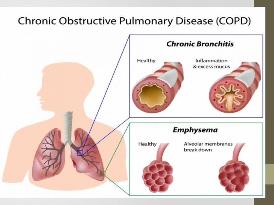
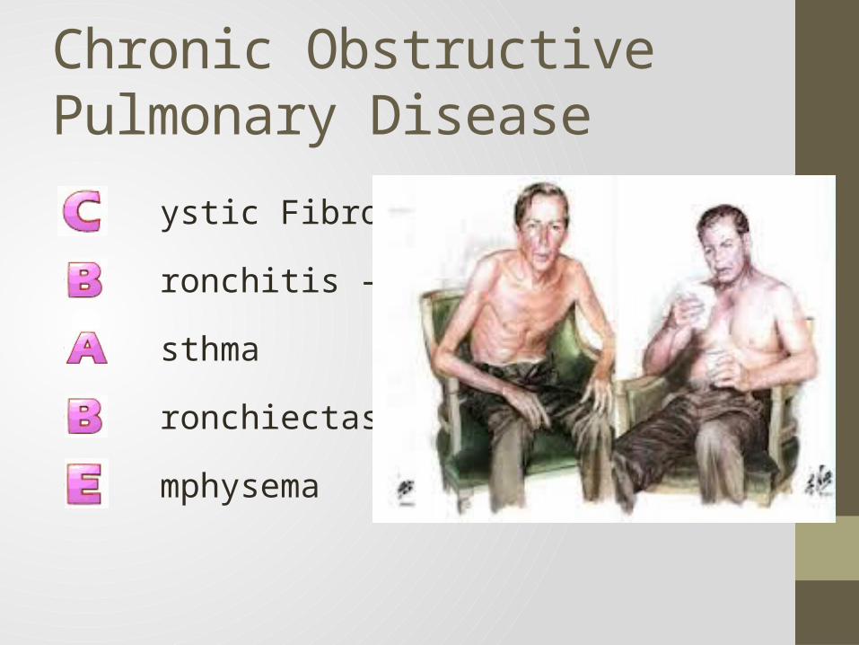
Chronic Obstructive Pulmonary Disease
• ystic Fibrosis
• ronchitis – Chronic
• sthma
• ronchiectasis
• mphysema

Chronic Obstructive Pulmonary Disease• Smoking
• #1 cause of COPD
• Increased mucous production
• Inhibition of mucociliary clearance
• Toxicity of inhaled gases and particulates
• Bronchospasm
• Decrease in macrophage activity
• Disruption of the alveolar wall and capillary endothelium

Epidemiology• Some 16 Million Americans are affected
• COPD is the 3rd leading cause of death in the U.S.
• COPD causes millions of hospital admissions per year
• Total health expenditure of $32.1 Billion in 2000
• Most common form of COPD is Chronic Bronchitis

Risk Factors for COPD
1. Cigarette smoking/passive smoking
2. Pollution
3. Occupational exposure to dust and fumes
4. Recurrent lung infections
5. Hereditary factors
6. Allergies
7. Socioeconomic factors
8. Alcohol ingestion
9. Age

General Manifestations of COPD
1. Small airways ( < 2mm) are most susceptible to airway obstruction in COPD2. Diagnosed by Pulmonary Function Testing, clinical signs and symptoms3. Early to middle manifestations of COPD include:
I. Changes in pulmonary function testingII. Shortness of breath with exertionIII. Changes in CXRIV. Increases in sputum productionV. CoughVI. Recurrent pulmonary infectionsVII. Wheezing
4. Late manifestations of COPD include:I. Accessory muscle usageII. Edema from Cor PulmonaleIII. Mental status changes from chronic hypoxia/hypercapneaIV. Clubbing of fingersV. Barrel Chest or Increased A-P Diameter
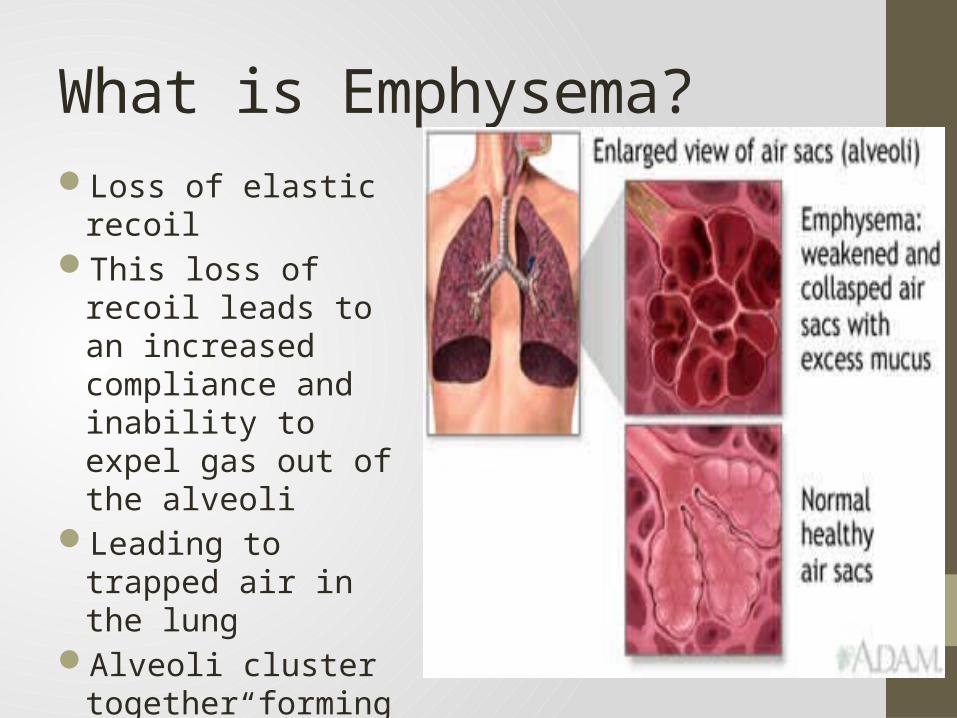
What is Emphysema?Loss of elastic recoil This loss of recoil leads to
an increased compliance and inability to expel gas out of the alveoli
Leading to trapped air in the lung
Alveoli cluster together forming “bullae”
What is Emphysema Cont…Damage occurs to the tiny airways in the lungs called
bronchioles. Bronchioles are joined to alveoli, tiny grape-like clusters of sacs in the lungs where oxygen from the air is exchanged for carbon dioxide from the body. The elastic properties of the lung reside in the tissue around the alveoli
Because the lungs lose elasticity they become less able to retract
This prevents the alveoli from deflating completely, and the person has difficulty exhaling.
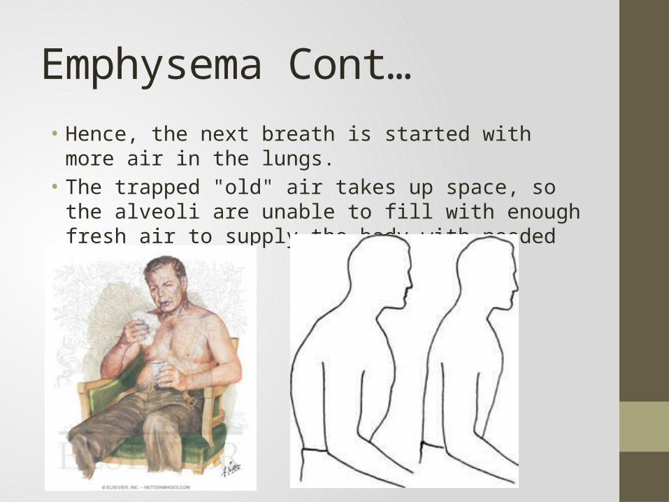
Emphysema Cont…• Hence, the next breath is started with more air in the lungs. • The trapped "old" air takes up space, so the alveoli are unable
to fill with enough fresh air to supply the body with needed oxygen.

Pulmonary Emphysema
• Centrilobular emphysema
• Abnormal weakening and enlargement of the respiratory bronchioles in the proximal portion of the acinus
• Primary changes occur in upper lobes
• High correlation with smoking

Pulmonary Emphysema• Bullous emphysema
• Changes seen at both respiratory bronchiole and alveolar levels
• Prominent bullae formation (air spaces greater than 1 cm in diameter)
Emphysema
Typically, symptoms of emphysema appear only after 30 to 50 percent of lung tissue is lost.
Emphysema rates are highest for men age 65 and older.
More people in the Midwest have emphysema than in any other region in the country.
Emphysema is an irreversible disease that can be slowed but not reversed or stopped.
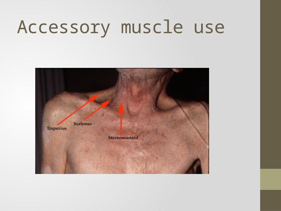
Accessory muscle use
Causes• Generally, lungs become damaged because of
reactions to irritants entering the airways and alveoli. Researchers continue to investigate the factors that may make some people more susceptible to emphysema than others. But there are some clear causes for emphysema:• Cigarette smoking • Alpha-1 antitrypsin deficiency

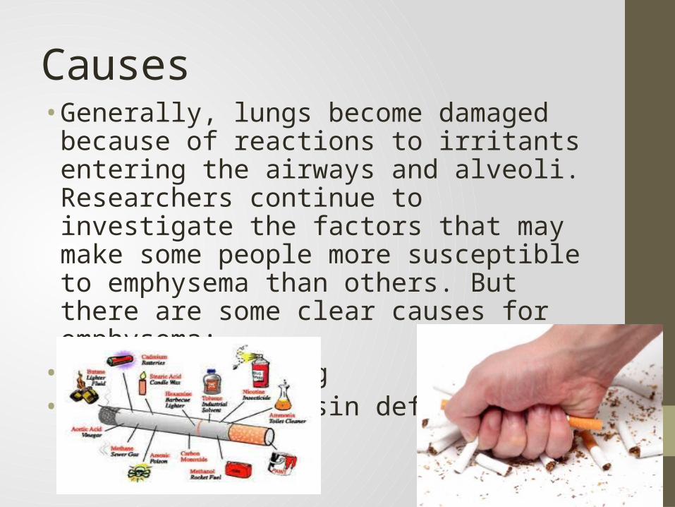
CausesCigarette smoking is the major cause of emphysema. When exposed
to cigarette smoke, the air sacs of the lungs produce defensive cells, called macrophages, which "eat" the inhaled particles. But macrophages are stimulated to release materials which can destroy the proteins that let the lungs expand and contract, called elastin and collagen .
Cigarette smoke also damages the cilia, tiny hair-like projections in the bronchi that "sweep" foreign bodies and bacteria out of the lungs

Other Cause
Alpha-1 Antitrypsin Deficiency• People who a deficiency of a protein called alpha-1 antitrypsin
(AAT) are at a higher risk of developing severe emphysema. Alpha-1 antitrypsin deficiency (AAT deficiency) is an inherited condition and occurs in varying degrees

AAT
• AAT is thought to protect against some of the damage caused by macrophages. In AAT deficiency-related emphysema, the walls of the bronchial tubes and the alveoli are both damaged, often leading to severe disease. • About 2 out of every 1,000 people have an
alpha-1 antitrypsin deficiency. People who smoke and have AAT deficiency are almost certain to develop emphysema.

Symptoms The first sign of emphysema is shortness of breath during
exertion. Eventually, this shortness of breath occurs while at rest
As the disease progresses, the following symptoms which are related to one of the other major lung diseases also caused by smoking - bronchitis - may occur:
• Difficulty breathing (dyspnea)• Coughing (with or without sputum) • Wheezing (this can also be caused by emphysema itself) • Excess mucus production • A bluish tint to the skin (cyanosis) • Hypoxemia• Tachycardia• Polytcythemia (increased hemoglobin levels)

More Symptoms• Clubbed fingers (chronic hypoxia)• Right Heart Failure• Stained yellow fingers, teeth

Diagnosis
History And Physical Examination
Smoking history (calculate pack years, # packs smoked times # years smoked)
Working environment- Breathing in any harmful chemicals?A physical examination will include an examination of your
chest and breathing patterns; prolonged expiratory timesNasal flaring, accessory muscle usage (due to loss of
diaphragm recoil from air trapping)

Diagnosis ContinuedX-Ray and/or CT of the ChestChest x-rays are a very useful tool to evaluate anatomy of
the lung. In emphysema, there is evidence of increased air in the chest and destruction of some of the lung tissue. Bronchitis can be suspected on a chest x-ray by presence of thickening of the tissue around the large airways (bronchi). Chest x-rays are also useful as screening for lung cancer and heart disease.
Computerized axial tomography or CAT scans indicate lung anatomy in greater detail. In some cases, this information is needed to fully evaluate lung disease.
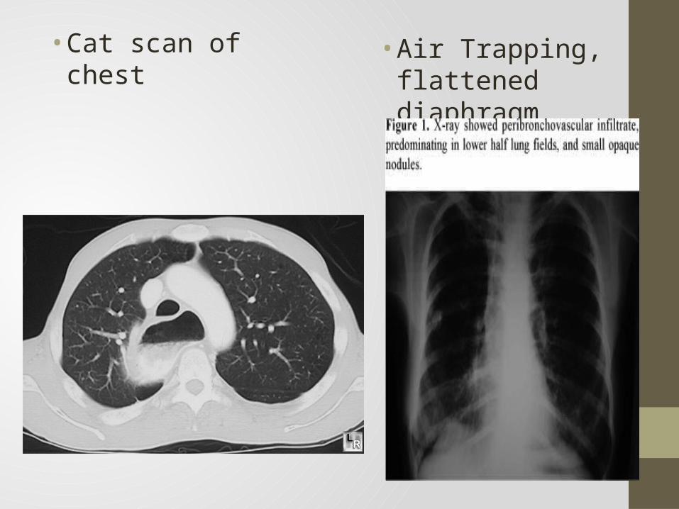
• Cat scan of chest • Air Trapping, flattened diaphragm
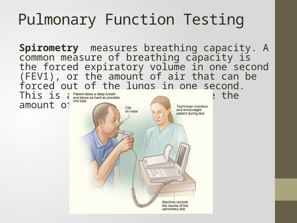
Pulmonary Function TestingSpirometry measures breathing capacity. A common measure of breathing capacity is the forced expiratory volume in one second (FEV1), or the amount of air that can be forced out of the lungs in one second. This is a common way to determine the amount of airway obstruction.

Pulmonary Function Testing• Spirometry and body plethysmography be
repeated after administration of an inhaled bronchodilator• This test will help your physician determine if
there is an asthmatic component present• Lung Volumes measures the amount of air in the
lungs. This increases markedly in emphysema.

Arterial Blood Gas• Patient’s with emphysema have chronic CO2 retention due to
the inability to expel gas. Their blood reflects higher levels of CO2 than normal people; CO2 is acidic in nature
• Over time their body compensates for this higher CO2 by creating more buffer in the blood in the form of Bicarb from the Kidney

Treatment
• There is no cure for emphysema. The goal of treatment is to slow the development of disabling symptoms. The most important step to take is to stop smoking.• Treatments for emphysema caused by smoking include
medication, breathing retraining, and surgery.• People with inherited emphysema due to alpha-1 antitrypsin
deficiency can receive alpha 1-proteinase inhibitor (A1PI), which slows lung tissue destruction.

Breathing TechniquesDiaphragmatic Breathing
• The diaphragm is a major muscle used in breathing and is located beneath the lowest two ribs. At rest, the diaphragm muscle is bell shaped. During inspiration, it lowers and flattens out.
• Optimizing the use of the diaphragm is beneficial because it pulls air into the lower lobes of the lungs where more gas exchange takes place. Not only is the diaphragm the most efficient of all respiratory muscles, but using it tends to be very relaxing and calming.
• Along with our diaphragm, we use intercostal and abdominal muscles in the work of breathing. The intercostals (muscles between the ribs) pull to lift the rib cage up and out. This causes the lungs to open in all directions and air can be pulled down the airways. To exhale, the muscles that have been pulling relax and air is forced out.
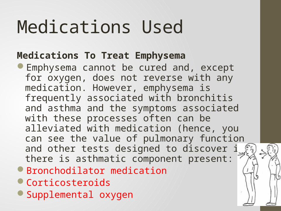
Medications UsedMedications To Treat EmphysemaEmphysema cannot be cured and, except for oxygen,
does not reverse with any medication. However, emphysema is frequently associated with bronchitis and asthma and the symptoms associated with these processes often can be alleviated with medication (hence, you can see the value of pulmonary function and other tests designed to discover if there is asthmatic component present:
Bronchodilator medication Corticosteroids Supplemental oxygen

Bronchodilator Medication• Bronchodilator medication may be prescribed for airway
tightness. Bronchodilators react similar to norepinephrine through the sympathetic nervous system • The most commonly prescribed bronchodilators are beta2
agonists , the anti-cholinergic drug ipatropium bromide, and theophylline• Anti-cholinergics block musacaric receptors which normally
respond to acetylcholine and cause bronchoconstriction

Corticosteroids• The potent anti-inflammatory medications known as corticosteroids
- commonly called steroids - may be used to help lessen the inflammation that often accompanies emphysema. These may be taken by mouth or inhaled.
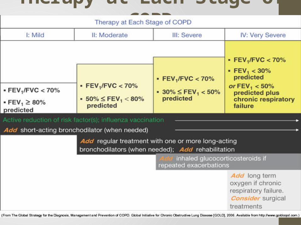
Therapy at Each Stage of COPD
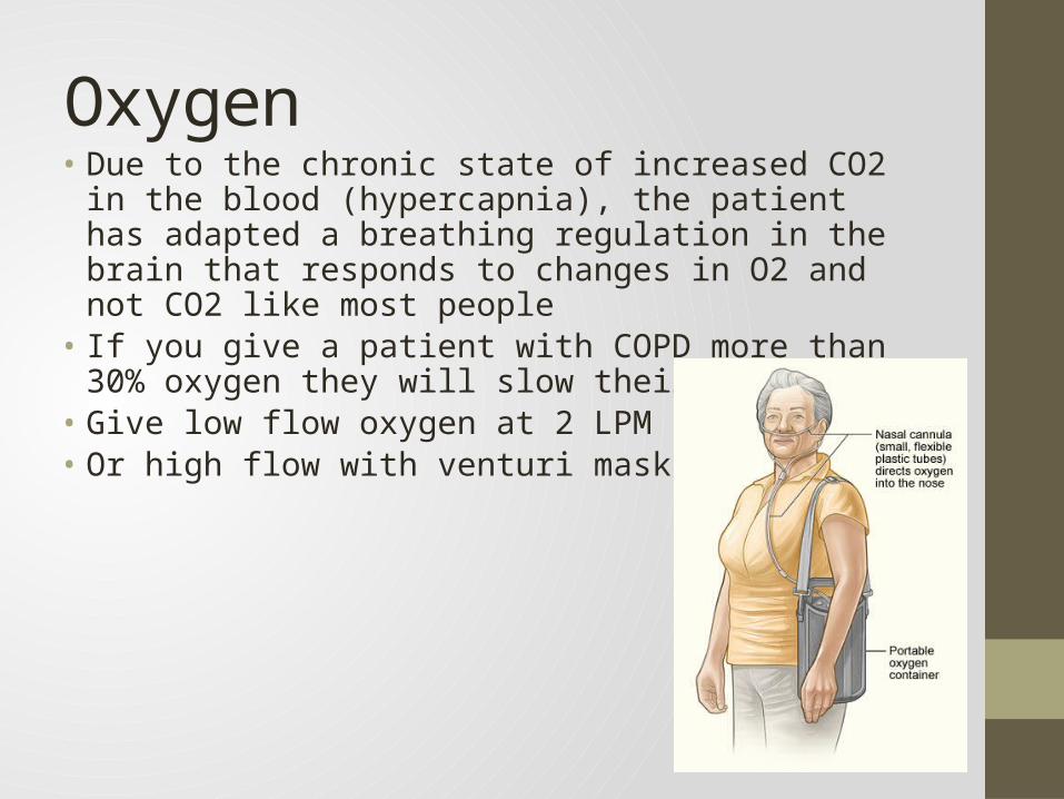
Oxygen • Due to the chronic state of increased CO2 in the blood
(hypercapnia), the patient has adapted a breathing regulation in the brain that responds to changes in O2 and not CO2 like most people
• If you give a patient with COPD more than 30% oxygen they will slow their breathing
• Give low flow oxygen at 2 LPM by NC• Or high flow with venturi mask at 22-30%

Lung Reduction
A surgical procedure called lung reduction may improve symptoms for people with certain types of emphysema. During the procedure, part of the lung is cut out, giving healthy lung tissue more room to expand.
Lung reduction may eliminate the need for supplemental oxygen and make it much easier for the person to breathe. Early studies show that it reduces the volume of the over-inflated lungs. This improves the ability of the lung and chest wall to spring back during exhalation. This more-elastic lung appears to be the biggest reason that emphysema sufferers experience relief.

ConclusionEmphysema is a chronic disease that takes years to
progress; usually as a result of heavy cigarette smoking but also can be caused by inherited Alpha-1 antitrypsin deficiency
It destroys the stability of the alveoli and bronchioles leaving them over compliant
This leads to air trapping and an accumulation of CO2 and decrease in O2
The air trapping leads to dyspneaDiagnose with symptoms, ABG, CXR, PFT and historyTreatment consists of stop smoking, medications and
lung reduction surgery or transplant