Chronic Kidney Disease Sarah Callison Chelsea Irving Anneli Robinaugh Tiffany Willard.
88
Chronic Kidney Disease Sarah Callison Chelsea Irving Anneli Robinaugh Tiffany Willard
-
Upload
erica-moore -
Category
Documents
-
view
216 -
download
0
Transcript of Chronic Kidney Disease Sarah Callison Chelsea Irving Anneli Robinaugh Tiffany Willard.
Chronic Kidney DiseaseSarah CallisonChelsea IrvingAnneli RobinaughTiffany Willard
Physiology
Main Kidney Functions- Balancing solute and water
transport
- Excreting metabolic waste products
- Conserving nutrients
- Regulate acid base balance
Endocrine FunctionSecretion of:
1. Renin (regulation of blood pressure)
2. Erythropoietin (erythrocyte production)
3. Vitamin D3 (calcium metabolism)
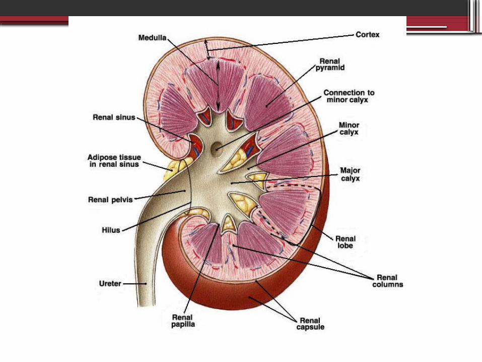
StructureMajor components:1. Outer Renal Cortex
• Contains all glomeruli and portions of the tubules
2. Inner Renal Medulla• Formed by segments of the proximal
and distal tubules, as well as the collecting ducts
• Consists of renal pyramids
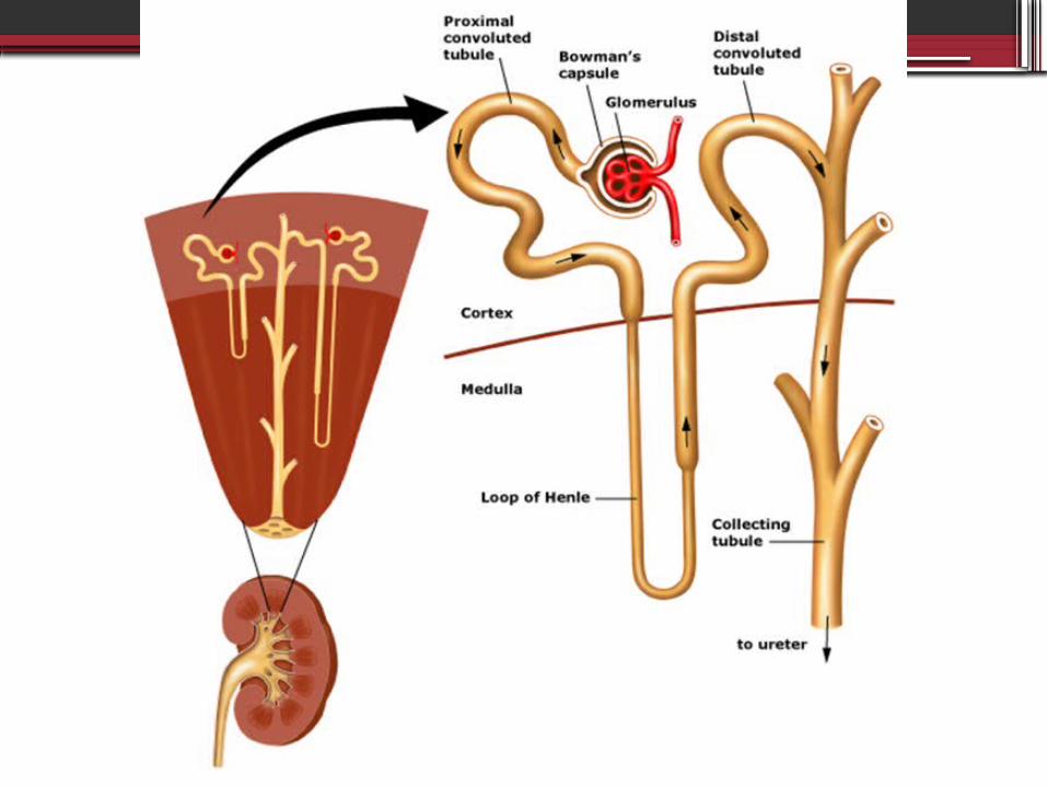
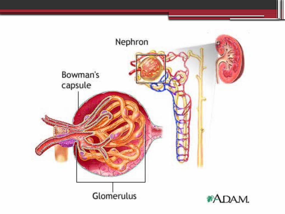
Nephron Function-Filters plasma at the glomerulus and then reabsorbs and secretes different substances
-Function = form a filtrate of protein free plasma
-Regulates filtrate to maintain: 1. body fluid volume2. electrolyte composition 3. pH
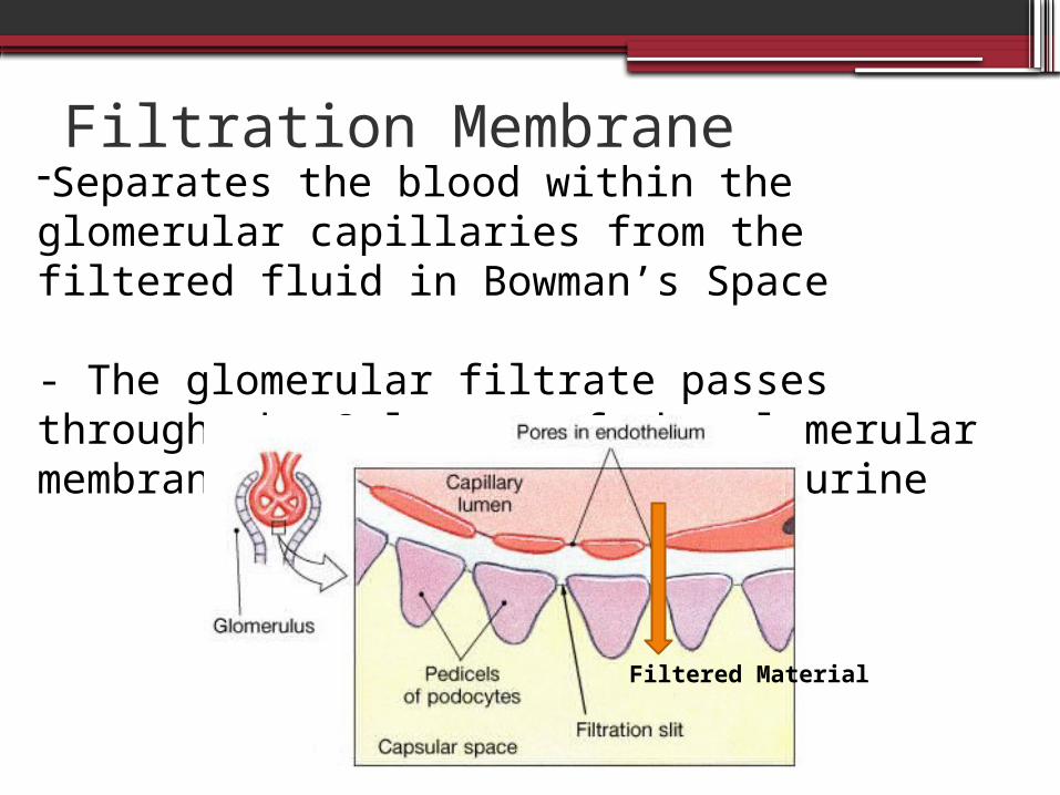
Filtration Membrane-Separates the blood within the glomerular capillaries from the filtered fluid in Bowman’s Space
- The glomerular filtrate passes through the 3 layers of the glomerular membrane and forms the primary urine
Filtered Material
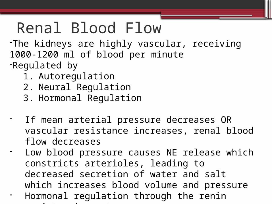
Renal Blood Flow-The kidneys are highly vascular, receiving 1000-1200 ml of blood per minute-Regulated by
1. Autoregulation2. Neural Regulation3. Hormonal Regulation
- If mean arterial pressure decreases OR vascular resistance increases, renal blood flow decreases
- Low blood pressure causes NE release which constricts arterioles, leading to decreased secretion of water and salt which increases blood volume and pressure
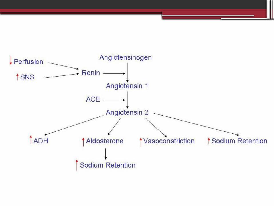
- Hormonal regulation through the renin angiotensin system
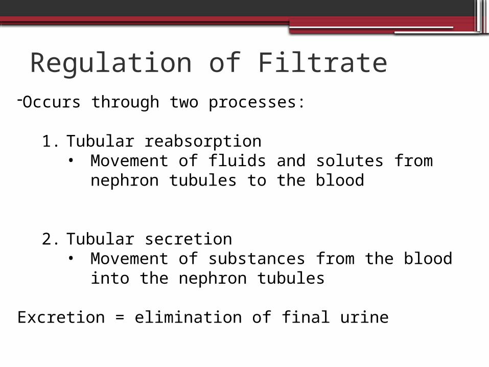
Regulation of Filtrate-Occurs through two processes:
1. Tubular reabsorption• Movement of fluids and solutes from
nephron tubules to the blood
2. Tubular secretion• Movement of substances from the blood
into the nephron tubules
Excretion = elimination of final urine
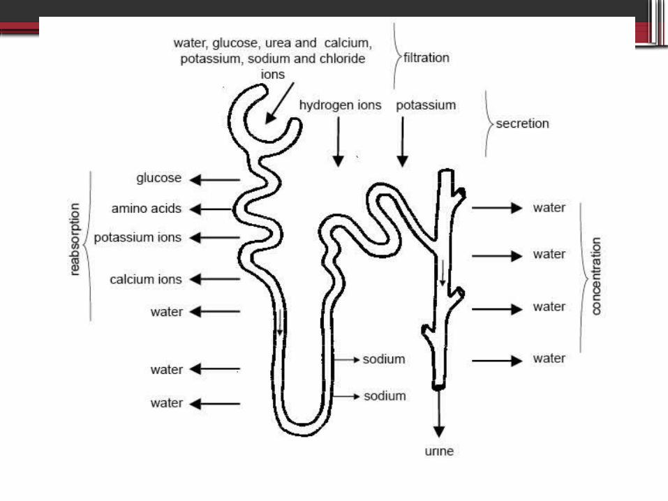
Figure 35-12, pr 1354
Urine Formation- From the proximal tubules, Na and glucose are
reabsorbed into peritubular capillaries by active transport
- Water reabsorption follows by osmosis
- From the distal tubules Na is reabsorbed by active transport
- Osmotic reabsorption of H2O occurs when ADH is present
- Secretion of ammonia and hydrogen occurs from peritubular capillaries into distal tubules by active transport

Helpful Clips
•http://www.youtube.com/watch?v=uo-NOr-P49I
•http://www.youtube.com/watch?v=aQZaNXNroVY&feature=related
•http://www.youtube.com/watch?v=AdlfxBooqIA&feature=related
Vitamin D- Inactive vitamin D (from diet or sunlight) require
two hydroxylations to become metabollically active
- The first hydroxilation occurs in the liver, the second occurs in the KIDNEY
- This process is stimulated by parathyroid hormone
- End result = active vitamin D3

Pathophysiology
Chronic Kidney Disease
The progressive loss of renal function associated
with systemic disease such as hypertension and
diabetes mellitus or intrinsic kidney disease
including glomerulonephritis, chronic
pyelonephritis, obstructive uropathies, or
vascular disorders
Chronic Pyelonephritis – persistent or recurrent infection of the kidney leading to kidney scarring
Glomerulonephritis – inflammation of the glomerulus caused by factors such as infection, immunologic abnormalities, ischemia, free radicals, drugs, toxins, vascular diseases, and systemic diseases such as diabetes mellitus and lupus erythmatosis
Obstructive uropathies – anatomic changes in the urinary system cause by an obstruction
Definitions
CKD- CKD decreases GFR and tubular functions
with changes manifested in ALL organ systems
- Kidney damage is defined as GFR less than 60 ml/min/1.73m for 3 months or more, irrespective of cause
- Other terms: Renal Insufficiency Chronic Renal Failure
2
Pathophysiology
- Kidneys can adapt to loss of nephron mass
- Symptomatic changes resulting from increased creatinine, urea, potassium, and alterations in salt and water balance do not become apparent until renal function declines to less than 25% of normal when adaptive renal reserves are exhausted
Intact Nephron Hypothesis
- Loss of nephron mass causes remaining nephrons to
hypertrophy/hyperfunction their rates of filtration,
reabsorption, and secretion in order to maintain a
constant rate of excretion and normal kidney
function in the presence of overall declining GFR
- This explains the adaptive changes in solute and water regulation that occur with advancing renal failure
Trade Off Hypothesis
- The continual loss of functioning nephrons
and the adaptive hyperfiltration/increased
glomerular pressure causes
glomerulosclerosis which contributes to
uremia and end stage renal failure.
Location of Kidney Damage- The location of kidney damage influences the
loss of kidney function
- Tubular interstitial diseases damage the tubular or medullary parts of the nephron- causing renal tubular acidosis, salt wasting, and
difficulty and diluting or concentrating urine
- Vascular or Glomerular damage- Proteinuria, hematuria, and nephrotic syndrome
Progression
Progression of CKD
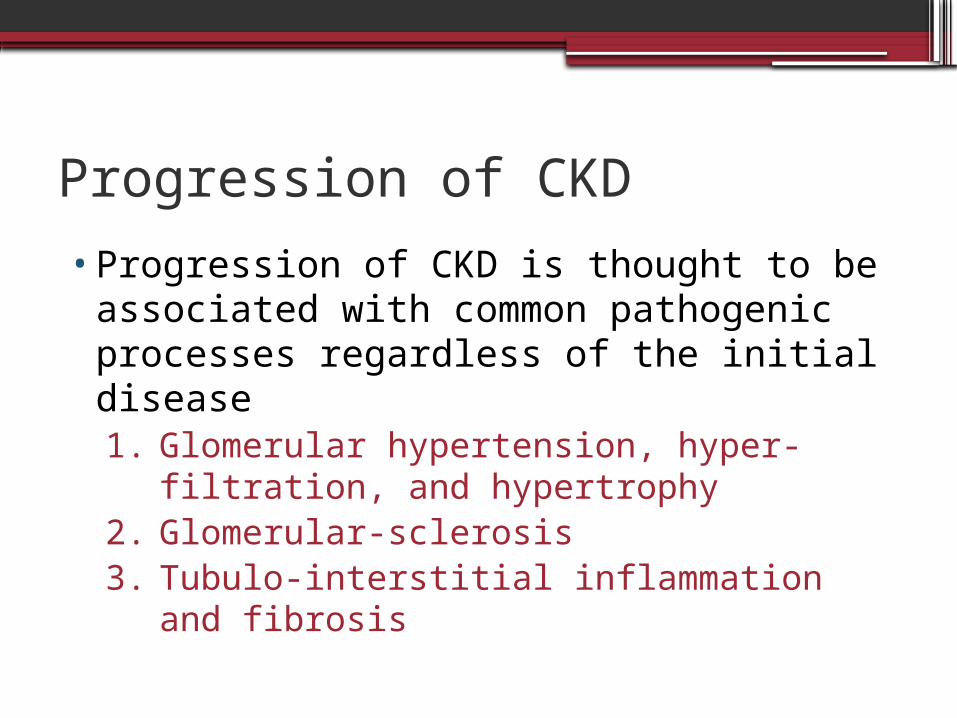
•Progression of CKD is thought to be associated with common pathogenic processes regardless of the initial disease1. Glomerular hypertension, hyper-filtration,
and hypertrophy2. Glomerular-sclerosis3. Tubulo-interstitial inflammation and
fibrosis
Progression of CKD
•Two factors are consistently recognized with advanced renal disease▫Proteinuria▫Elevated angiotensin II
Proteinuria
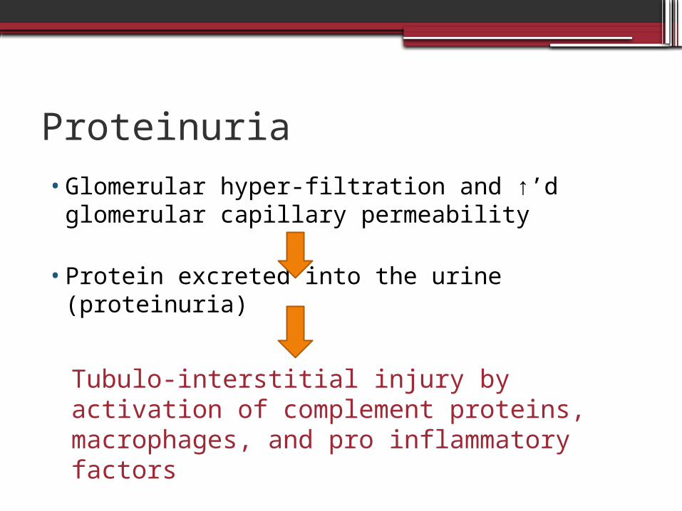
•Glomerular hyper-filtration and ↑’d glomerular capillary permeability
•Protein excreted into the urine (proteinuria)
Tubulo-interstitial injury by activation of complement proteins, macrophages, and pro inflammatory factors
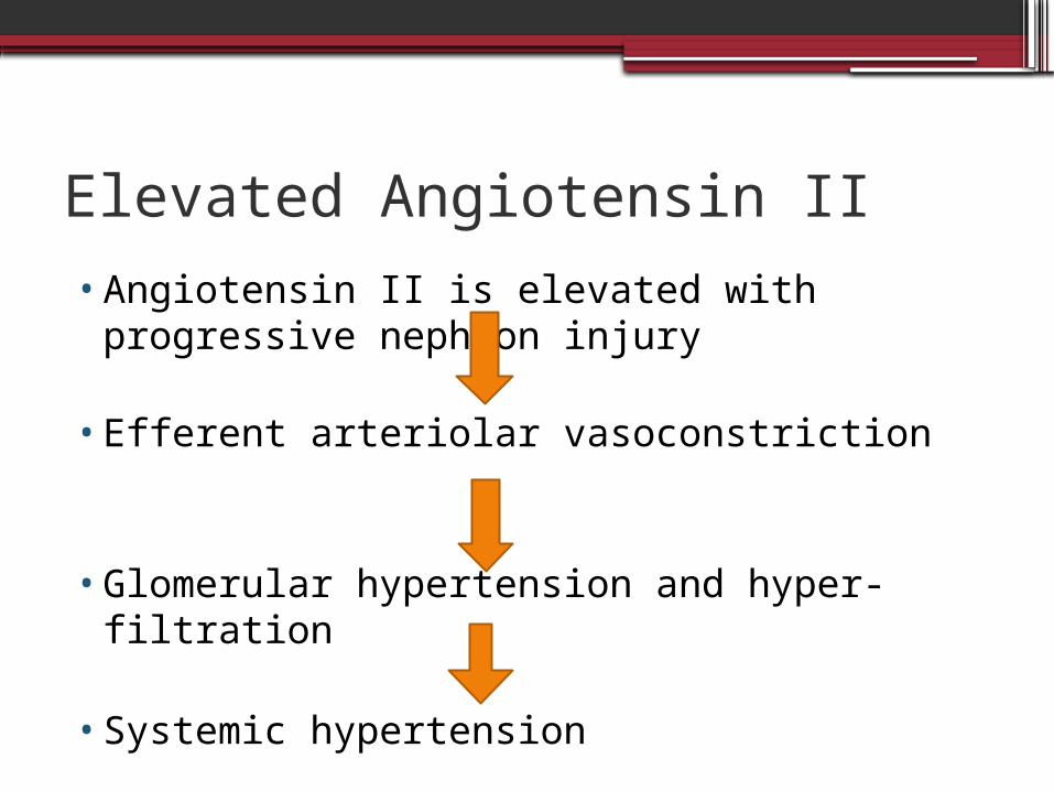
Elevated Angiotensin II
•Angiotensin II is elevated with progressive nephron injury
•Efferent arteriolar vasoconstriction
•Glomerular hypertension and hyper-filtration
•Systemic hypertension
Angiotensin II
•This high intra-glomerular pressure can cause proteinuria as well
•may also promote pro inflammatory cells the cause fibrosis and scarring of the tissue.
Risk Factors
Risk Factors•1. Diabetes Mellitus
▫33% of CKD is due to diabetes mellitus
•2. Hypertension•Family History of CKD
Risk Factors Cont.
•Ethnicity▫African Americans: 3.8x▫Native Americans: 2.0x▫Asians: 1.3x▫Others: Hispanics and Pacific Islanders▫Due to higher rates of developing diabetes mellitus and hypertension
•Age•Polycystic kidney disease
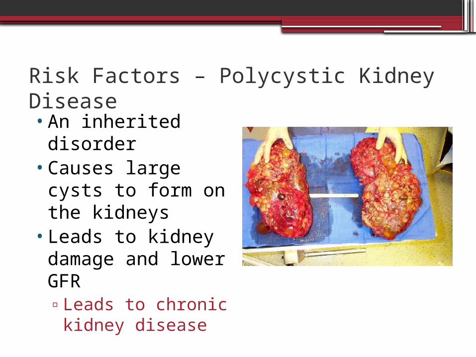
Risk Factors – Polycystic Kidney Disease•An inherited
disorder•Causes large cysts
to form on the kidneys
•Leads to kidney damage and lower GFR▫Leads to chronic
kidney disease
Signs and Symptoms
Signs and Symptoms
•Change in urination▫Pressure during urination▫More frequently▫At night▫Foamy or bubbly
•Swelling▫In hands, feet, ankles, legs, or face▫Kidney can’t filter fluid properly so more
gets retained
Signs and Symptoms Cont.
•Skin rashes and itching•Metallic taste in mouth•Nausea and vomiting
▫Due to the build up of urea and other waste products in the blood

Signs and Symptoms Cont.
•Fatigue•Feeling cold•Dizziness•Shortness of breath
▫Due to the reduced amount of erythropoietin being produced by the kidney Can lead to anemia
Diagnosis and Staging
Diagnosis – Glomerular Filtration Rate
•Kidneys’ rate of fluid filtration of the blood▫Regulated by:
Hydrostatic pressures in blood and Bowman’s space Pulls water out of the blood
Oncotic pressures in blood and Bowman’s space Pressure from protein – pulls water into the
blood Thickness of glomerular membrane Area of glomerular membrane
Diagnosis – GFR cont.
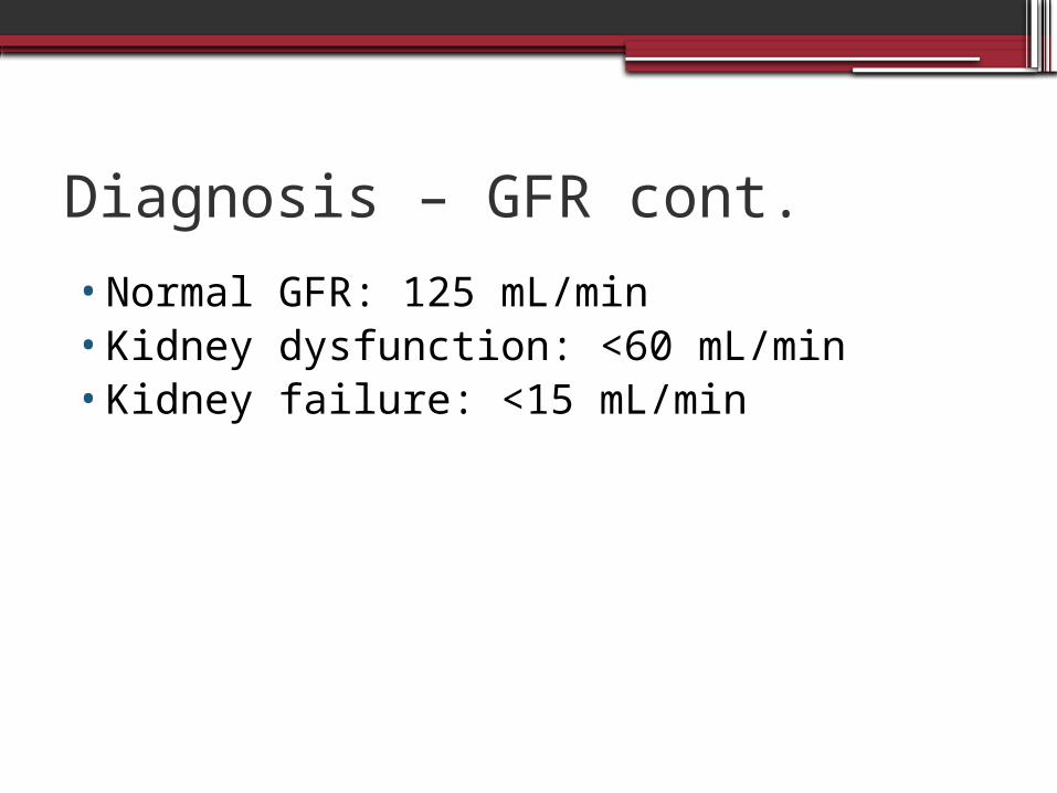
•Normal GFR: 125 mL/min•Kidney dysfunction: <60 mL/min•Kidney failure: <15 mL/min
Diagnosis – GFR cont.
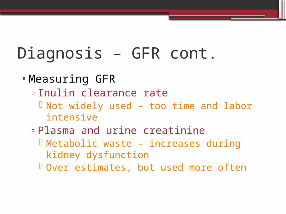
•Measuring GFR▫Inulin clearance rate
Not widely used – too time and labor intensive
▫Plasma and urine creatinine Metabolic waste – increases during kidney
dysfunction Over estimates, but used more often
Diagnosis - Urinalysis
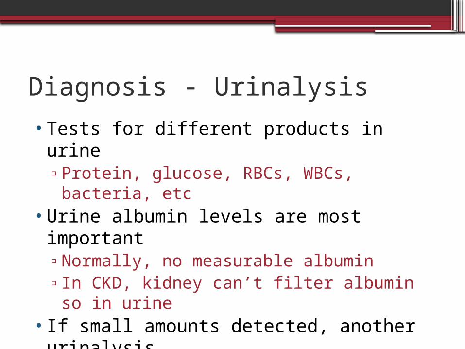
•Tests for different products in urine▫Protein, glucose, RBCs, WBCs, bacteria, etc
•Urine albumin levels are most important▫Normally, no measurable albumin▫In CKD, kidney can’t filter albumin so in
urine•If small amounts detected, another
urinalysis•If large amounts detected, 24-hour urine
collection
Diagnosis – Urinalysis cont.
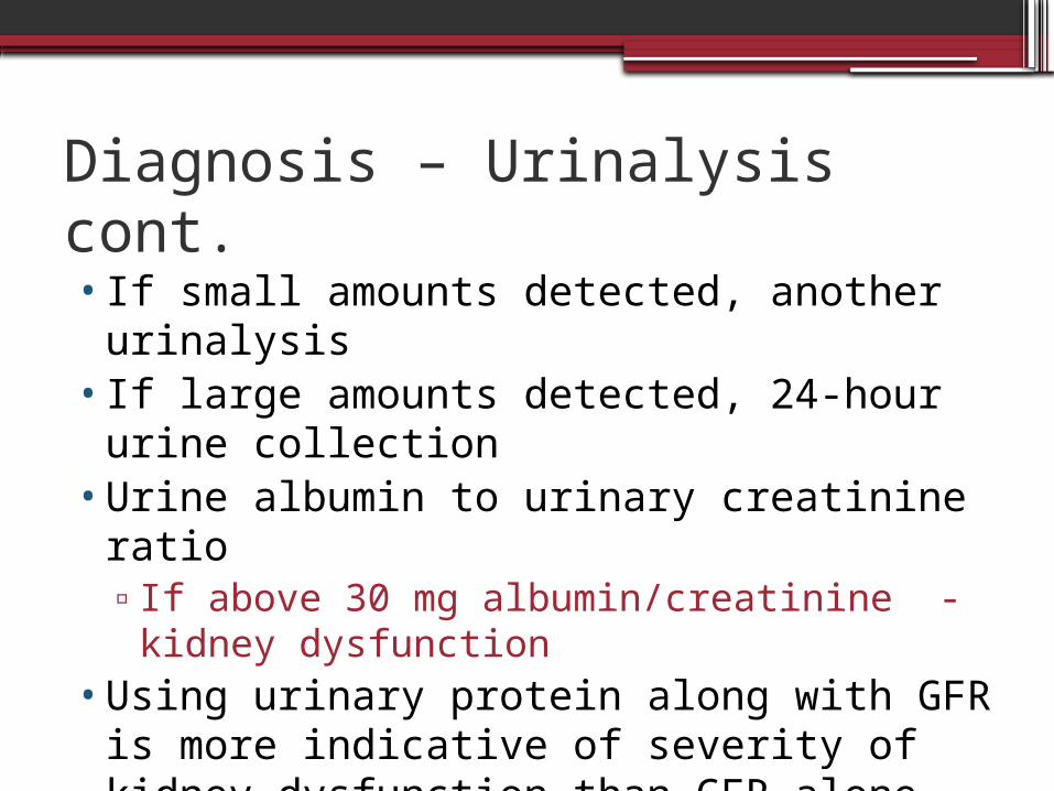
•If small amounts detected, another urinalysis
•If large amounts detected, 24-hour urine collection
•Urine albumin to urinary creatinine ratio▫If above 30 mg albumin/creatinine - kidney
dysfunction•Using urinary protein along with GFR is
more indicative of severity of kidney dysfunction than GFR alone
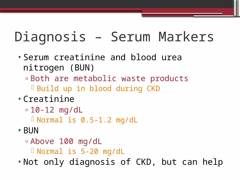
Diagnosis – Serum Markers•Serum creatinine and blood urea nitrogen
(BUN)▫Both are metabolic waste products
Build up in blood during CKD•Creatinine
▫10-12 mg/dL Normal is 0.5-1.2 mg/dL
•BUN▫Above 100 mg/dL
Normal is 5-20 mg/dL•Not only diagnosis of CKD, but can help
Terms
•Azotemia: when nitrogenous waste products build up in the blood▫Urea, creatinine, etc
•Uremia: when waste products build up in blood and cause clinical side effects▫Itching, rashes, metallic taste in food, etc
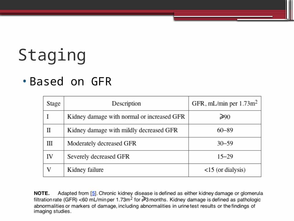
Staging
•Based on GFR
Comorbidities
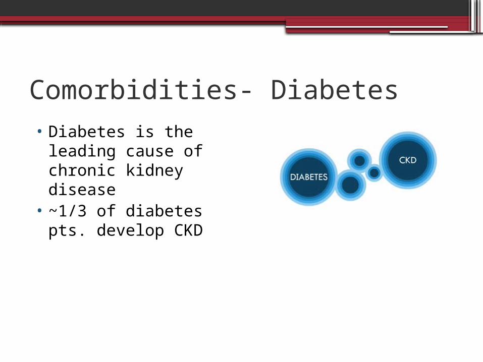
Comorbidities- Diabetes • Diabetes is the
leading cause of chronic kidney disease
• ~1/3 of diabetes pts. develop CKD
Diabetes
•Earliest sign of kidney damage from diabetes: albumin in the urine.
•Other signs include▫ HBP ▫ Swelling in ankles and legs▫ Increased urination
Diabetes- Mechanism• High blood glucose causes damage to blood vessels
in the kidney. • This decreases kidney function• Buildup of waste products in the blood (they aren’t
excreted in the urine)• Changes in the kidney affect blood vessels and
protein is excreted into the urine.• Damage to nerves (from high glucose) can damage
nerves- making it harder to empty the bladder. • Pressure from this can cause infection or injure the
kidney further
Anemia and CKD
•Normally…▫Low 02 content of kidney cells stimulate
them to produce erythropoietin (EPO)▫Erythropoietin signals the bone marrow to
produce mature red blood cells. Without EPO, the ability of the body to
produce RBC is greatly decreased
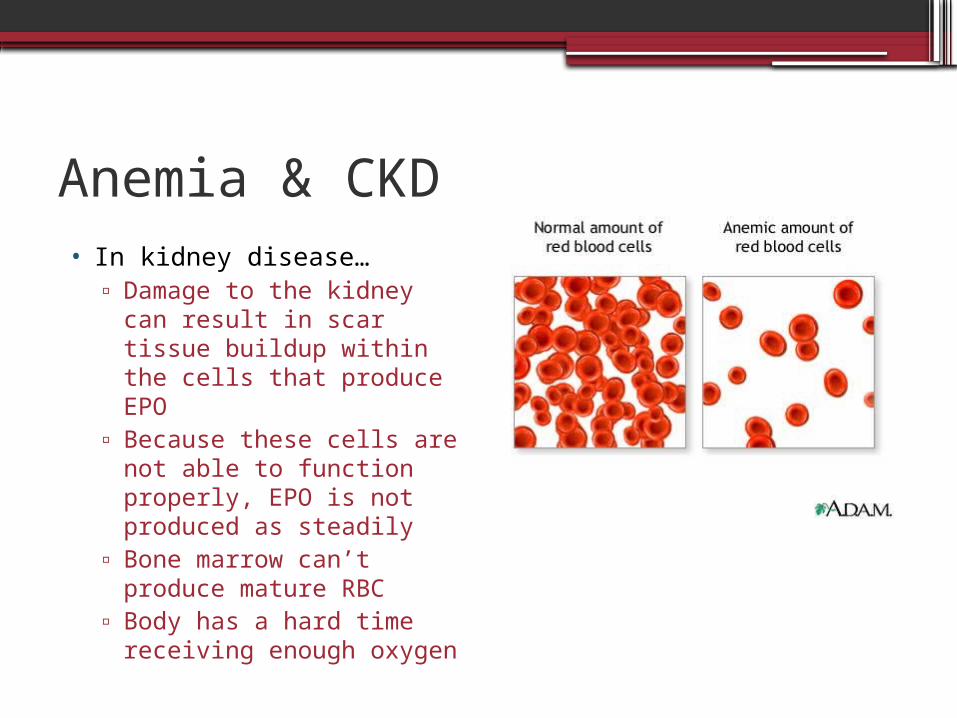
Anemia & CKD• In kidney disease…
▫ Damage to the kidney can result in scar tissue buildup within the cells that produce EPO
▫ Because these cells are not able to function properly, EPO is not produced as steadily
▫ Bone marrow can’t produce mature RBC
▫ Body has a hard time receiving enough oxygen
Anemia and CKD•Anemia is usually evident in the 3rd stage of
kidney disease- long before dialysis is needed
•As kidney disease gets worse, so does anemia•Symptoms:
▫Fatigue, weakness▫Less energy and enthusiasm▫Harder to concentrate▫Dizziness- if anemia is severe▫Pale skin
Anemia & CKD •Usual cause of anemia in CKD is lack of EPO
▫Normochromic, normocytic•Anemia can also result from
▫Low iron▫Chronic inflammation▫Blood loss from GI tract (ulcers, tumors,
gastrits)▫Menstruation▫Other uterine bleeding
•Blood tests can determine cause
Anemia Therapy
•Injections of synthetic EPO•Can be given every week, 2 weeks, or
month
Heart Disease and CKD
Heart disease is the major cause of morbidity and mortality for patients with CKD
Heart Disease- Manifestations
•Manifestations: ▫Left ventricular hypertrophy▫Cardiomyopathy▫Ischemic heart disease
Hypertension Dysrhythmias Accelerated atherosclerosis Chest pain
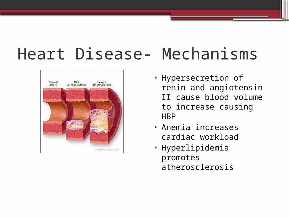
Heart Disease- Mechanisms• Hypersecretion of renin
and angiotensin II cause blood volume to increase causing HBP
• Anemia increases cardiac workload
• Hyperlipidemia promotes atherosclerosis
Medical Nutrition Therapy in CKD
PRO Restriction and Kidney Function
•Studies determined protein restriction may decrease renal decline▫Modification of diet in renal disease study▫Protein increases glomerular pressure
•Prompted conference with National Institute of Diabetes and Kidney and Digestive National Institutes of Health to determine protein recommendations
Current PRO Recommendations
•Results of conference:▫Determined by GFR▫GFR >55 ml/min - .8 g/kg▫GFR 25-55 ml/min - .6 g/kg▫GFR <25ml/min - .6 g/kg & 35 kcals/kg
▫If .6 g/kg is inadequate, intake may be increased to .75 g/kg
Protein Restriction vs. Malnutrition
• Despite conference, protein recommendations are controversial▫Benefits in renal function are seen with PRO
restriction▫High PRO contributes to the health of
patient by preventing PRO malnutrition▫Balance must be found between PRO
restriction and PRO malnutrition Current recommendations restrict PRO
▫ If the benefits of PRO restriction outweigh malnutrition risk, PRO is restricted Patient must be carefully monitored
Effective PRO Restriction
•Hypertension must be controlled as it also contributes to renal decline
•In patients with DM, blood glucose control is more important than PRO restriction in preventing renal decline
High Biological Value Protein
•Needed with intake less than .8 g/kg•50 % of intake should be HBV•Provides all of the essential amino acids•Sources:
▫Animals products such as meat, poultry, fish, eggs, cheese, yogurt, milk
•Plant sources such as grains, legumes, and nuts are considered low biological value
Na+ and Fluid Recommendations
•Sodium intake is reduced to decrease thirst, fluid gain, and hypertension
•2,000 – 3,000 mg/day•No...
▫Added salt in cooking or at the table▫Convenience/processed foods▫Cured meats
•There is rarely a fluid restriction pre-dialysis
K+ Recommendations
•Reduced to 2.3 – 3.1 g/day▫Normal intake for most Americans is 3 – 4
g/day•Avoid foods high in K+ such as avocado,
banana, cantaloupe, spinach, tomato, milk chocolate▫Fruits and vegetables may be leached to
lower K+ content•Avoid potassium chloride as a salt
substitute in low sodium foods
Phosphorus Recommendations
•Limit intake to 17 mg/kg or 1200 mg/day•Processed foods and metabolically active
foods▫Metabolically active foods contain ATP
•High phosphorus foods include fast food pancakes and hamburgers, mac’n cheese, 2% milk, cheese, meats
•Intake of PRO contributes to the intake of P▫The following equation estimates the PRO
impact 128 + 14 * g of PRO in diet = mg P/day
Phosphate Binders• Usually dietary restrictions is not enough• Calcium carbonate, acetate, lactate, gluconate
▫Risk of hypercalcemia▫Used in lower amounts along with aluminum
binders Basaljel and Amophojel, although aluminum toxicity is a risk
• Renagel▫Phosphate binding resin▫Does not raise calcium levels▫High dosage puts pill burden on patients
• Fosrenol is also used
Side Effects of Phosphate Binders
•GI distress•Constipation
▫Can lead to intestinal impaction▫Important to treat constipation so patients
will stay on binders
Anemia
•Treated with recombinant human EPO (rHuEPO)▫Given when serum ferritin <100 ng/ml
•Initial treatment can cause a rise in K+•Treatment should cause a rise in
hematocrit▫Increased need for iron▫Cochrane review: Effective in pre dialysis
patients
Iron Supplementation• Iron is usually replaced IV or intramuscularly •Oral iron is not sufficient to replenish stores• Iron dextran, iron gluconate, iron sucrose are
available to patients with an allergy to IV iron▫Oral iron is available for the small minority of
patients who can’t tolerate any other form▫Should be taken between meals and w/o large
amounts of Vit C to avoid kidney stone formation
Blood Transfusions
•NOT recommended for the following reasons:▫Depresses EPO levels in bone marrow▫Risk of over expanding blood volume▫Risk of blood borne pathogens▫The additional K+ load▫Risk of hemochromatosis and
hemosiderosis
Incidence & Prevalence
•Incidence:▫111,000 U.S residents began new CKD
treatment in 2007•Prevalence:
▫26 million people in the US have CKD▫Millions more at increased risk
Quality of life•Depends on stage of CKD and presence of
comorbidities•Knowledge and awareness of CKD will help
patients cope•Self- empowerment: a positive attitude can
help patients feel better and reduce complications
•Proper care and self management slow progression of disease▫Good blood glucose control▫Maintaining blood pressure
Case Study
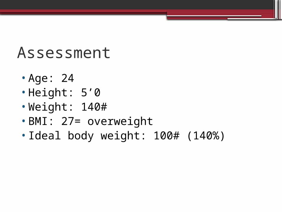
Assessment
•Age: 24•Height: 5’0•Weight: 140#•BMI: 27= overweight•Ideal body weight: 100# (140%)
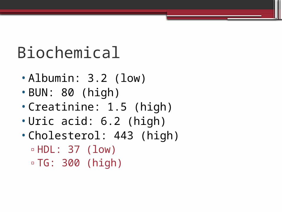
Biochemical
•Albumin: 3.2 (low)•BUN: 80 (high)•Creatinine: 1.5 (high)•Uric acid: 6.2 (high)•Cholesterol: 443 (high)
▫HDL: 37 (low)▫TG: 300 (high)
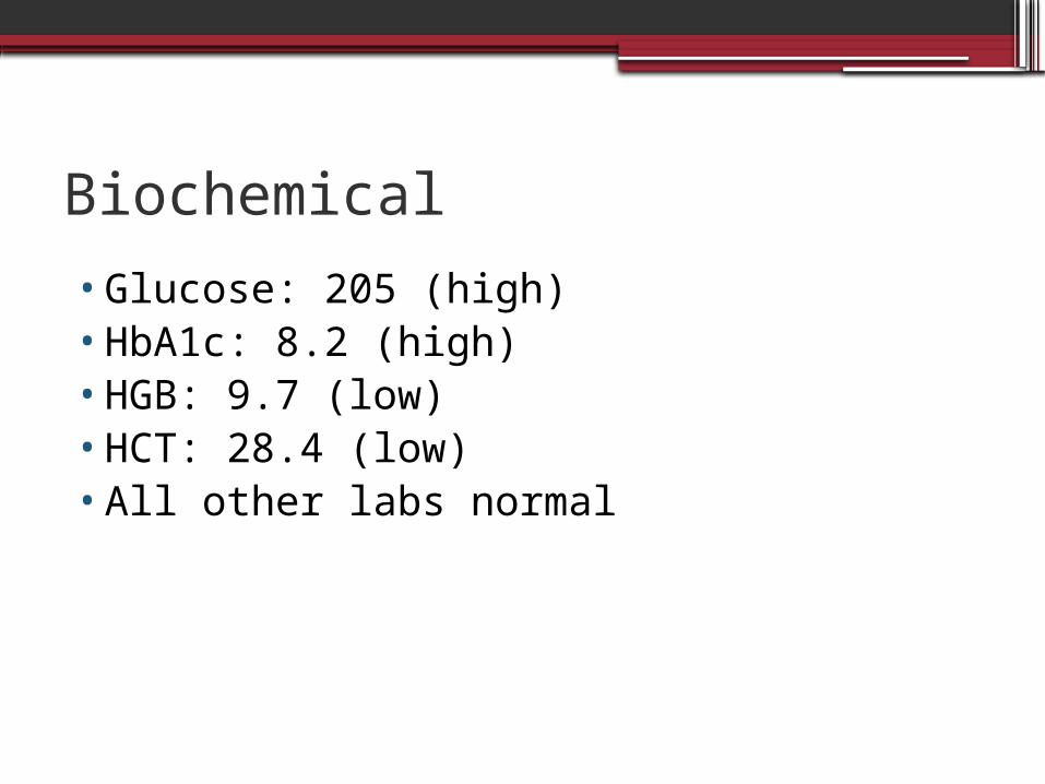
Biochemical
•Glucose: 205 (high)•HbA1c: 8.2 (high)•HGB: 9.7 (low)•HCT: 28.4 (low)•All other labs normal
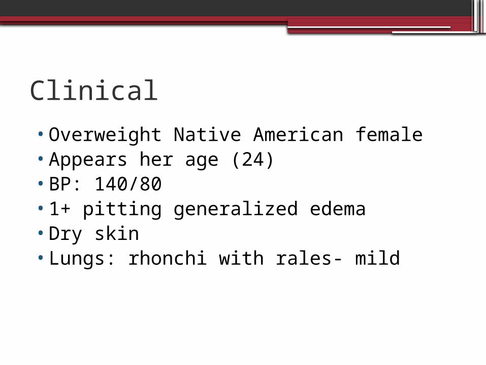
Clinical
•Overweight Native American female•Appears her age (24)•BP: 140/80•1+ pitting generalized edema•Dry skin•Lungs: rhonchi with rales- mild
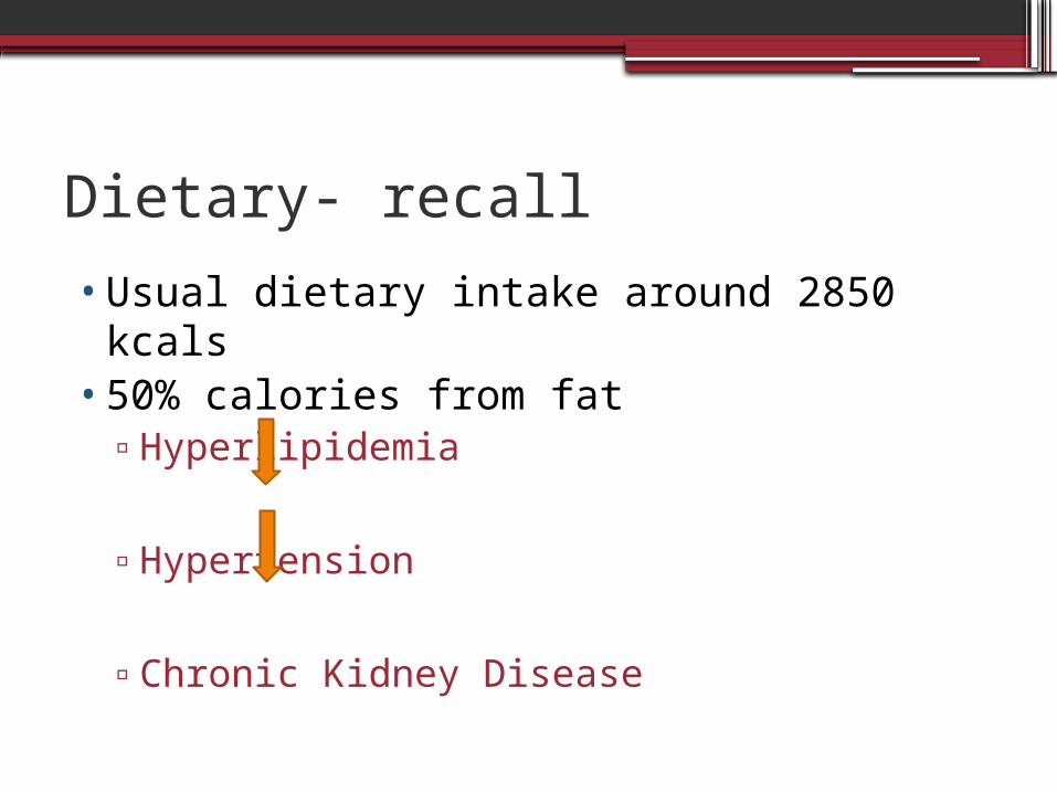
Dietary- recall
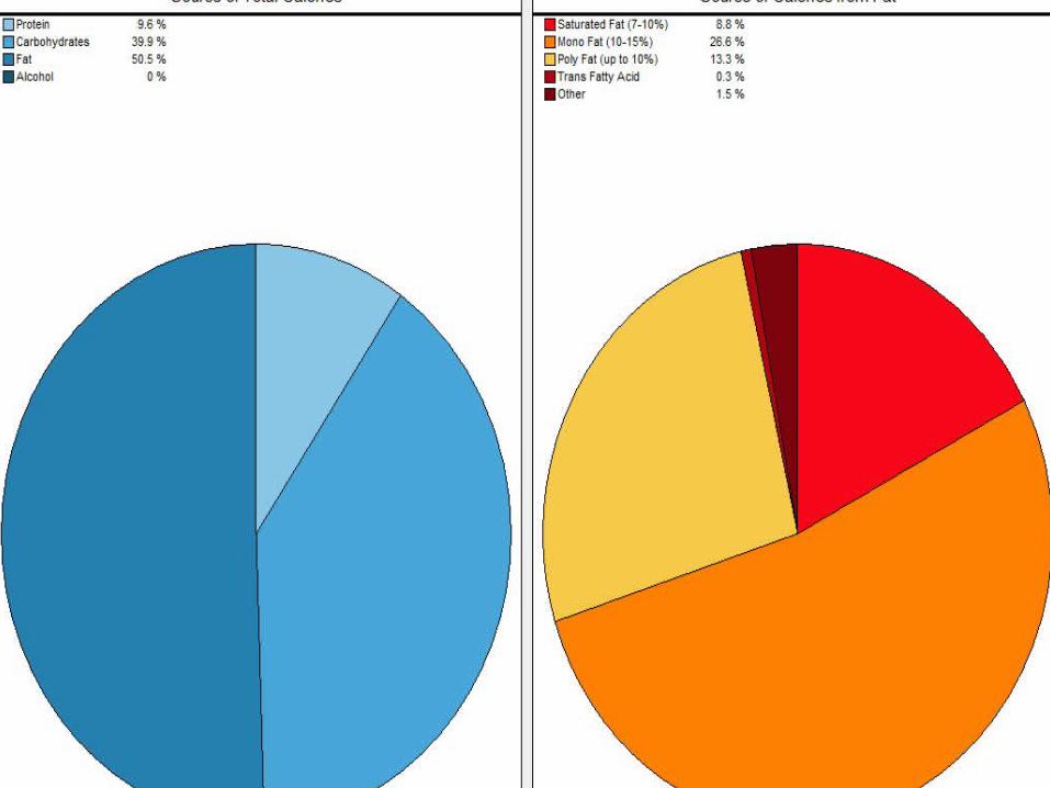
•Usual dietary intake around 2850 kcals•50% calories from fat
▫Hyperlipidemia
▫Hypertension
▫Chronic Kidney Disease
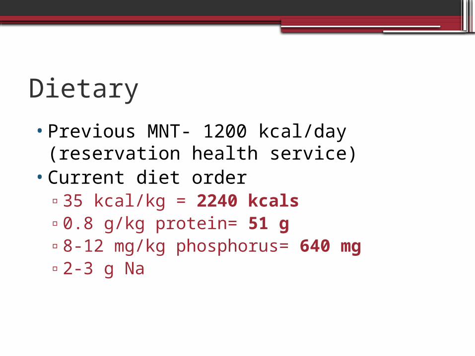
Dietary
•Previous MNT- 1200 kcal/day (reservation health service)
•Current diet order ▫35 kcal/kg = 2240 kcals ▫0.8 g/kg protein= 51 g▫8-12 mg/kg phosphorus= 640 mg▫2-3 g Na
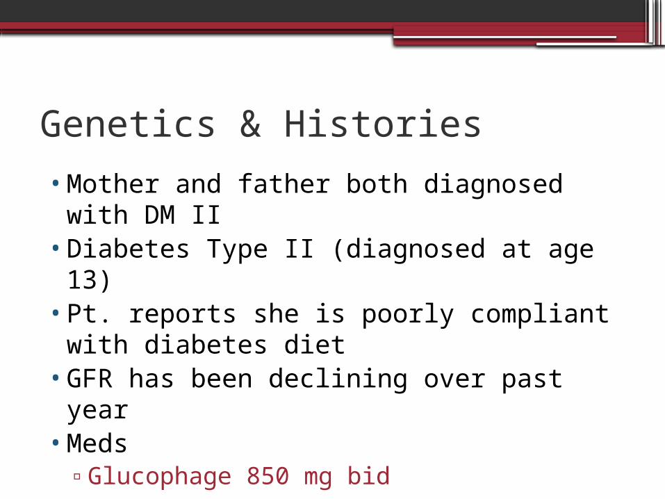
Genetics & Histories
•Mother and father both diagnosed with DM II
•Diabetes Type II (diagnosed at age 13)•Pt. reports she is poorly compliant with
diabetes diet•GFR has been declining over past year•Meds
▫Glucophage 850 mg bid
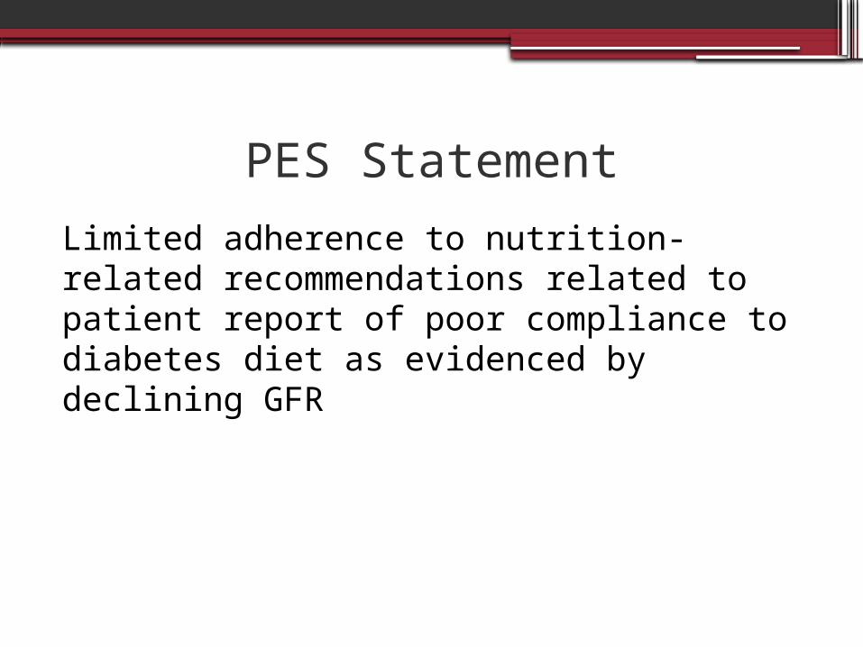
PES Statement
Limited adherence to nutrition-related recommendations related to patient report of poor compliance to diabetes diet as evidenced by declining GFR
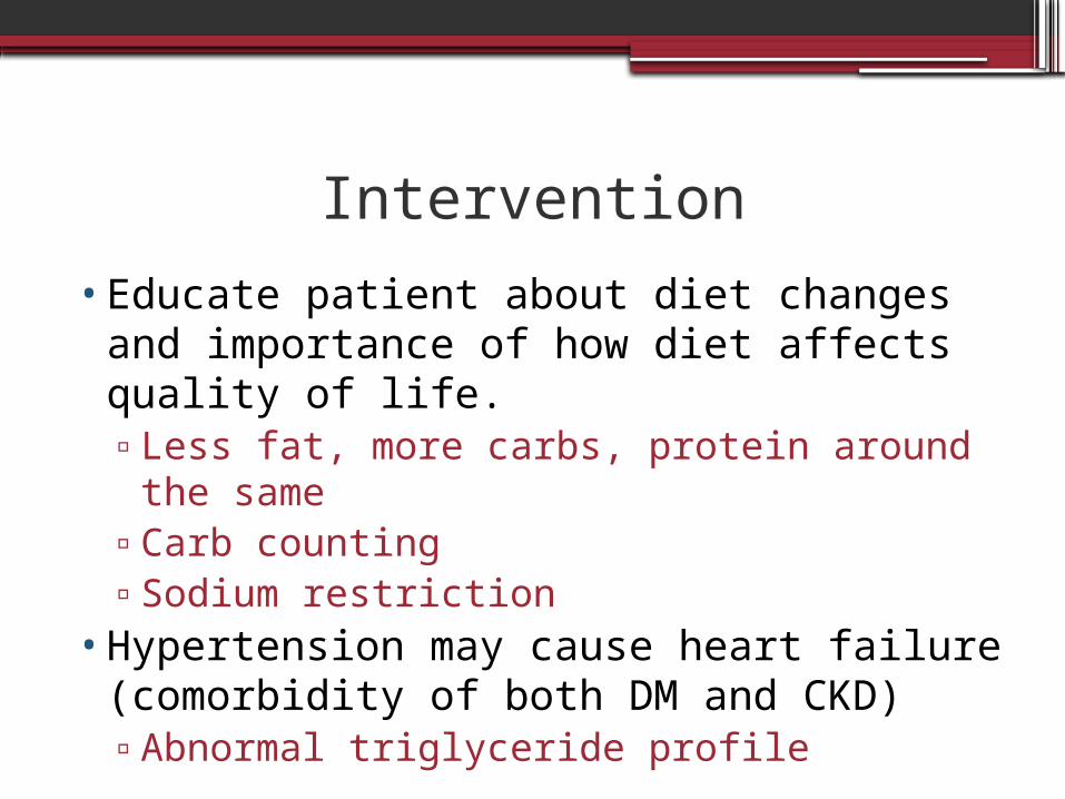
Intervention
•Educate patient about diet changes and importance of how diet affects quality of life.▫Less fat, more carbs, protein around the
same▫Carb counting▫Sodium restriction
•Hypertension may cause heart failure (comorbidity of both DM and CKD)▫Abnormal triglyceride profile
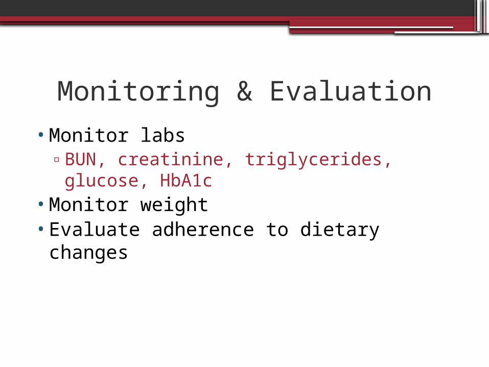
Monitoring & Evaluation
•Monitor labs ▫BUN, creatinine, triglycerides, glucose,
HbA1c•Monitor weight•Evaluate adherence to dietary changes