Chronic Inflammation Caused by Lymphotoxin Is Lymphoid ...
12
Chronic Inflammation Caused by Lymphotoxin Is Lymphoid Neogenesis By Alexander Kratz, Antonio Campos-Neto, Matthew S. Hanson, and Nancy H. Ruddle From the Department of Epidemiology and Public Health, Yale University School of Medicine, New Haven, Connecticut 06520-8034 Summary In presenting a unifying concept for chronic inflammation and lymphoid organogenesis, we suggest that lymphotoxin's (LT, LT-ot, TNF-J3) crucial role in these processes is pivotal and similar. Chronic inflammatory lesions that developed in the kidney and pancreas at the sites of transgene expression in rat insulin promoter-LT (RIP-LT) mice resembled lymph nodes with regard to cellular composition (T cells, B cells, plasma cells, and antigen-presenting cells), de- lineated T and B cell areas, primary and secondary follicles, characteristic morphologic and an- tigenic (ICAM-1, VCAM-1, MAdCAM-1, and PNAd) features of high endothelial venules, and ability to respond to antigen and undergo Ig class switching when obtained from mice im- munized with SRBC. The vascular changes, with the exception of PNAd, appear to be the di- rect consequence of transgene derived LT expression, as they were also observed in RIP-LT mice lacking mature T and B cells. These data show that LT-induced chronic inflammation has the characteristics of organized lymphoid tissue. C hronic inflammation, a complex pathophysiologic process characterized by an accumulation of mononu- clear cells, is associated with the response to invading patho- gens, neoplastic transformation, or autoimmune recognition of self-antigens. This is usually initiated by events involving macrophages and T cells and is perpetuated through re- cruitment of additional T cells, B cells, and macrophages. The process can lead to elimination of the pathogen or tu- mor, but frequently results in tissue damage. The cells in the infiltrate influence the severity of clinical signs and, in addition to the elimination of the causative stimulus, can perpetuate the lesion, resulting in exacerbation in diseases such as multiple sclerosis and rheumatoid arthritis. They can also contribute to resolution of the lesion through sup- pression of inflammatory activity (1). Although the classical definition of chronic inflammation includes tissue destruc- tion and loss of function (2), several instances of chronic in- flammation have been described in which cellular infiltra- tion occurs without tissue destruction. These include insulitis in male non obese diabetic (NOD) 1 mice (3) and thyroidi- tis in biobreeding (BB) rats (4). In several other situations, chronic, long lasting inflammation can eventually lead to ] Abbrem'ations used in this paper: BB, biobreeding; EAE, experimental al/er- gic encephalomyelitis', HEV, high endothelialvenules;LT, lymphotoxin; NOD, non obese diabetic; PFC, plaque-formingcell; PLP, periodate/ lysine/paraformaldehyde; RIP, rat insulin promoter. tissue damage. Such is the case with female NOD mice, in whom insulitis begins as early as four weeks of age, but tis- sue damage and islet destruction is not seen until the age of three months. In rat insulin promoter-TNF-~t (RIP-TNF- or), RIP-lymphotoxin (RIP-LT), or RIP-IL-2 mice, in- flammation is apparent in very young mice (before three weeks of age) but tissue damage occurs after costimulatory signals or upon antigenic stimulation (5-8). The development and organization of chronic inflamma- tion is just beginning to be understood. Though several cy- tokines and chemokines have been implicated, a clear un- derstanding of the role of the individual factors and their mechanism of action in the inflammatory process awaits elucidation. Although the expression "tertiary lymphoid tissue" has been used to describe any tissue that can be in- filtrated (9), the process of chronic inflammation had been considered to be completely different from that of lym- phoid organ development. Lymphotoxin (LT, also known as LT-ot or TNF-[~), a product of CD4Thl, CD8, and early B cells, has been im- plicated in the inflammatory process though it has many other activities as well (reviewed in 10). A major role in in- flammation is implied from its ability to activate endothelial cells in vitro for the expression of the adhesion molecules ICAM-1 and VCAM-1 (11, 12). Studies with T cell clones indicate its importance (with TNF-ot) in the transfer of ex- perimental allergic encephalomyelitis (EAE) (13, 14). The 1461 j. Exp. Med. 9 The Rockefeller University Press 9 0022-1007/96/04/1461/12 $2.00 Volume 183 April 1996 1461-1472 on February 3, 2018 jem.rupress.org Downloaded from
Transcript of Chronic Inflammation Caused by Lymphotoxin Is Lymphoid ...
Chronic Inflammation Caused by Lymphotoxin Is Lymphoid Neogenesis By Alexander Kratz, Antonio Campos-Neto, Matthew S. Hanson, and N a n c y H. R u d d l e
From the Department of Epidemiology and Public Health, Yale University School of Medicine, New Haven, Connecticut 06520-8034
Summary In presenting a unifying concept for chronic inflammation and lymphoid organogenesis, we suggest that lymphotoxin's (LT, LT-ot, TNF-J3) crucial role in these processes is pivotal and similar. Chronic inflammatory lesions that developed in the kidney and pancreas at the sites of transgene expression in rat insulin promoter-LT (RIP-LT) mice resembled lymph nodes with regard to cellular composition (T cells, B cells, plasma cells, and antigen-presenting cells), de- lineated T and B cell areas, primary and secondary follicles, characteristic morphologic and an- tigenic (ICAM-1, VCAM-1, MAdCAM-1, and PNAd) features of high endothelial venules, and ability to respond to antigen and undergo Ig class switching when obtained from mice im- munized with SRBC. The vascular changes, with the exception of PNAd, appear to be the di- rect consequence of transgene derived LT expression, as they were also observed in RIP-LT mice lacking mature T and B cells. These data show that LT-induced chronic inflammation has the characteristics of organized lymphoid tissue.
C hronic inflammation, a complex pathophysiologic process characterized by an accumulation of mononu-
clear cells, is associated with the response to invading patho- gens, neoplastic transformation, or autoimmune recognition of self-antigens. This is usually initiated by events involving macrophages and T cells and is perpetuated through re- cruitment of additional T cells, B cells, and macrophages. The process can lead to elimination of the pathogen or tu- mor, but frequently results in tissue damage. The cells in the infiltrate influence the severity of clinical signs and, in addition to the elimination of the causative stimulus, can perpetuate the lesion, resulting in exacerbation in diseases such as multiple sclerosis and rheumatoid arthritis. They can also contribute to resolution of the lesion through sup- pression of inflammatory activity (1). Although the classical definition of chronic inflammation includes tissue destruc- tion and loss of function (2), several instances of chronic in- flammation have been described in which cellular infiltra- tion occurs without tissue destruction. These include insulitis in male non obese diabetic (NOD) 1 mice (3) and thyroidi- tis in biobreeding (BB) rats (4). In several other situations, chronic, long lasting inflammation can eventually lead to
] Abbrem'ations used in this paper: BB, biobreeding; EAE, experimental al/er- gic encephalomyelitis', HEV, high endothelial venules; LT, lymphotoxin; NOD, non obese diabetic; PFC, plaque-forming cell; PLP, periodate/ lysine/paraformaldehyde; RIP, rat insulin promoter.
tissue damage. Such is the case with female N O D mice, in whom insulitis begins as early as four weeks of age, but tis- sue damage and islet destruction is not seen until the age of three months. In rat insulin promoter-TNF-~t (RIP-TNF- or), RIP-lymphotoxin (RIP-LT), or RIP-IL-2 mice, in- flammation is apparent in very young mice (before three weeks of age) but tissue damage occurs after costimulatory signals or upon antigenic stimulation (5-8).
The development and organization of chronic inflamma- tion is just beginning to be understood. Though several cy- tokines and chemokines have been implicated, a clear un- derstanding of the role of the individual factors and their mechanism of action in the inflammatory process awaits elucidation. Although the expression "tertiary lymphoid tissue" has been used to describe any tissue that can be in- filtrated (9), the process of chronic inflammation had been considered to be completely different from that of lym- phoid organ development.
Lymphotoxin (LT, also known as LT-ot or TNF-[~), a product of CD4Thl , CD8, and early B cells, has been im- plicated in the inflammatory process though it has many other activities as well (reviewed in 10). A major role in in- flammation is implied from its ability to activate endothelial cells in vitro for the expression of the adhesion molecules ICAM-1 and VCAM-1 (11, 12). Studies with T cell clones indicate its importance (with TNF-ot) in the transfer of ex- perimental allergic encephalomyelitis (EAE) (13, 14). The
1461 j. Exp. Med. �9 The Rockefeller University Press �9 0022-1007/96/04/1461/12 $2.00 Volume 183 April 1996 1461-1472
on February 3, 2018
jem.rupress.org
Dow
nloaded from

ability o f LT and TNF-ot to influence EAE is due in large part to the upregulation o f VCAM-1 in the CNS (15) and the extent o f inflammation is positively correlated with ex- pression of that adhesion molecule. Inhibition o f LT and TNF-ot in this situation prevents the upregulation o f VCAM-1 and the subsequent recruitment o f additional T cells, B cells, and macrophages (15).
LT induces inflammation at sites o f targeted expression in transgenic animals. We have previously described mice transgenic for LT under the control o f the rat insulin pro- moter (RIP-LT mice). This somewhat leaky promoter is expressed in the pancreatic islets o f Langerhans, proximal convoluted tubules in the kidney (16, 17), and skin. The expression of LT in these circumstances results in an infil- trate consisting o f T cells, B cells, and macrophages at the sites o f cytokine expression and results in a ruffled hair phe- notype (18). A similar inflammatory process is seen in islets o f RIP-TNF-e t mice (18, 19). Tissue damage does not oc- cur spontaneously in either R I P - L T or 1LIP-TNF-et mice, but can be induced by coexpression in the islets o f the B7-1 costimulatory molecule which activates infiltrating T cells resulting in beta cell destruction and diabetes (5, 7). The mechanism of the LT-induced inflammatory process has not been elucidated, nor have the relative contributions of the LT transgene and products o f the cells in the infiltrates.
LT, in addition to its role in inflammation, plays a crucial and unique role in lymphoid development and tissue orga- nization. This is apparent from our analysis o f mice made selectively deficient in LT expression through the process of homologous recombination (20). Such LT knockout mice are devoid o f all peripheral and mesenteric lymph nodes and Peyer's patches. Splenic organization is disrupted with the loss o f usual compartmentalization into T and B cell ar- eas. Since LT-deficient mice produce normal levels o f TNF-cx, it is apparent that this function in lymphoid or- ganogenesisis is peculiar to LT. The mechanism of LT's ef- fect in lymphoid organogenesis has not been investigated.
Here we report the results o f studies in which we evalu- ated R I P - L T mice to test the hypothesis that chronic in- flammation represents lymphoid neo-organogenesis and that LT plays a crucial and identical role in both. We found that the mononuclear accumulations in pancreata and kidneys are similar if not identical to organized lymphoid tissue with regard to cellular composition, compartmentahzation, a specialized vascular system with expression of markers characteristic o f vessels in lymph nodes and ultrastructural appearance o f high endothelial venules, and reactivity to an exogenous antigen. We show that even R I P - L T mice that lack the recombination activating gene 2 protein (RIP-LT R A G - 2 - / - mice) and thus any additional cytokines de- rived from mature T or B cells express endothelial adhesion markers, VCAM-1, ICAM-1, and MAdCAM-1 , that are likely to initiate mononuclear accumulation. Therefore, we propose that LT-induced chronic inflammation is actually lymphoid neogenesis and that the cytokine recapitulates in the adult its activities in embryogenesis, in part through its ability to induce changes in the vasculature. This provides a unifying model for lymphoid development and inflamma-
tion and has implications for determinant spreading and clinical exacerbation in autoimmune disease.
Materials and Methods
Mice. The generation of RIP-LT transgenic mice has been described (17). Animals were backcrossed to C57BL/6 (The Jack- son Laboratory, Bar Harbor, ME) or to mice lacking the RAG-2 gene (21). A breeding nucleus of the latter was kindly provided by Dr. Frederick Alt and maintained under specific pathogen-free conditions. These mice fail to produce mature B or T lympho- cytes. The presence of the RIP-LT transgene and of the RAG-2 deletion was determined by Southern blot hybridization as de- scribed (17, 21). FACS analysis for the presence of CD3 + cells in peripheral blood was performed to ascertain the absence of ma- ture lymphocytes in the R_AG-2 - / - animals. To achieve maximal transcription from the insulin promoter, some animals were kept on a high-fat diet and given 10% sucrose in drinking water.
Primary Antibodies for FACS and Immunohistochemistry. The fol- lowing primary antibodies were used for immunohistochemistry or for flow cytometry: YN 1/1.7 (specific for ICAM-1 [22]), MK/2 (specific for VCAM-1 [23]), RA3-6B2 (specific for the B-cell restricted determinant of B220; Ca/tag Laboratories, South San Francisco, CA); F4/80 (specific for macrophages); anti-CD8a- Phycoerythrin (PE; GIBCO BRL, Gaithersburg, MD); anti- CD4-PE (GIBCO BRL); FITC-labeled anti-mouse imnmno- globulin (Sigma); N418 (specific for the p150/90 132 integrin ex- pressed on dendritic cells (24); a kind gift of Dr. Ralph Steinman); FDC-M1 (specific for mouse follicular dendritic cells (25); a kind gift of Dr. M.H. Kosco-Vilbois); PE-anti-mouse CD3-epsilon (Pharmingen, San Diego, CA). MECA-367 (specific for MAd- CAM-1 [26]), MECA-79 (specific for PNAd [27]), and MJ 7/18 (specific for endoglin [28]) were generously provided by Dr. Eu- gene Butcher.
Flow Cytometry. Cells were isolated from lymph nodes and kid- neys by gentle pressure with microscope slides and washed three times in Hanks' medium. Lymphocytes were purified from the kid- ney preparations by centrifugation over Histopaque-1090 (Ficoll/ Hypaque; Sigma). Cells were washed once with staining buffer (5% fetal calf serum, 10 mM sodium azide in PBS), and incubated with the appropriate biotinylated primary antibodies in 3% mouse serum (Sigma) in staining buffer for one hour. For B cell staining, fetal bovine serum was used instead of mouse serum. After three washes in staining buffer, incubation with streptavidin-labeled FITC was performed. For double staining, cells were incubated with a second primary antibody and a PE-labeled secondary anti- body specific for immunoglobulin of the species of the second primary antibody, washed three times and fixed in 2% paraform- aldehyde (Sigma). Samples were analyzed with a FACScan | flow cytometer (Becton Dickinson, Mountain View, CA) for single stained cells and with a FACStarplus | flow cytometer (Becton Dickinson) for double stained cells using LYSYS software. 25,000-50,000 cells were analyzed per sample. For FITC-stained cells, a 530/30 filter was used; for PE-labeled cells, a 575/26 filter was used.
Immunohistochemistry. Tissues were fixed in periodate/lysine/ paraformaldehyde (PLP) (29) fixative overnight, processed through three consecutive sucrose solutions (10, 20, and 30%), and snap frozen in Tissue Tek compound (Miles, Elkhart, IN). Tissue sec- tions were cut with a Cryocut 1800 cryostat (Reichert Young, Deerfield, IL) at 7 p~m and stored at -20~ Sections were air dried and rehydrated for 10 Inin in wash buffer (0.1 M phosphate buffer, pH 7.3, with 0.01% Triton X). To prevent background
1462 Lymphotoxin Causes Lymphoid Neogenesis
on February 3, 2018
jem.rupress.org
Dow
nloaded from

staining from endogenous peroxidase and nonspecific binding of antibodies, sections were treated with 0.3% I-I202 in methanol and with 10% normal goat serum (Pierce Chem. Co., Rockford, IL). Incubation with the primary antibody was performed for 1-3 h. After three rinses with wash buffer, an appropriate biotinylated species-specific secondary antibody (GIBCO BRL, Gaithersburg, MD) was applied. Then, after incubation with horseradishperoxi- dase-streptavidin (Vector Laboratories, Burlingame, CA), DAB (Sigma) was used for the color reaction. Slides were counterstained with methyl green (Sigma) and coverslipped with Permount (Fisher Scientific, Pittsburgh, PA).
Fluorescence Microscopy. Tissues were fixed in 2% paraformal- dehyde/5% sucrose for 30 min, infused with 20% sucrose for 1.5 h, and snap frozen in Tissue-Tek compound. Tissue sections were cut with a Cryocut 1800 cryostat at 5 I.LM and stored at -20~ Sections were air dried, fixed in acetone for 10 s, and re- hydrated in wash buffer (PBS, pH 7.4) for 10 rain. For identifica- tion ofT and B lymphocytes sections were incubated for 1 h with directly conjugated monoclonal antibodies 145-2C11-FITC (anti- CD3) and RA3-6B2-PE (anti-B220) (PharMingen, San Diego, CA) in staining buffer (PBS, 2.5% Blotto (Pierce), 10% rabbit se- rum (Sigma). Plasma cells were stained as above with FITC-la- beled anti-mouse immunoglobulin. Stained sections were rinsed three times in wash buffer, coverslipped with Gel/Mount (Bio- meda Corp., Foster City, CA) and observed by UV-light micros- copy using a Zeiss Axioskop microscope (Carl Zeiss, Inc., Thorn- wood, NY). Kodak Ektachrome P1600 film was used.
Hematoxylin-eosin Stains. Tissues were fixed in neutral buffered zinc-formalin, embedded in para~in, sectioned at 5 Ixm, and stained with hematoxylin and eosin following standard techniques.
Electron Microscopy. Animals were euthanized by inhalation of Metofane (Methoxyflurane; Pitman-Moore, Inc., Mundelein, IL) and perfused with 0.1 M phosphate buffer and PLP. Tissues were removed and held in PLP for 1 h. After three changes of phos- phate buffer, tissues were washed in phosphate buffer overnight. Tissues were postfzxed with 1% glutaraldehyde in 0.1 M sodium cacodylate for an additional one hour, and washed three times with 0.1 M sodium cacodylate. After postfixation in 1.33% osmium tetroxide for 1 h, tissues were washed three times with 0.1 M s-collidine, dehydrated in a graded alcohol chain, and embedded in Epon. After sectioning at 60 nm, the tissue sections were stained with uranyl acetate and lead citrate. Sections were viewed on a Zeiss (Oberkochen, Germany) EM10B electron microscope at 80KV.
Plaque Forming Ceil Assay. Mice were immunized with 2 • 108 sheep red blood cells (SR_BC) i.v. and boosted 10 d later with the same number of i.p. SRBC. 6 d after the second immuniza- tion, the animals were killed by cervical dislocation and lymph nodes, spleens, and kidneys were removed. The organs were teased and ceils were washed three times in Hanks' balanced salt solution. Lymphocytes were purified from the kidney prepara- tions by centrifugation over Histopaque-1090 (Ficoll/Hypaque; Sigma). SRBC were washed three times, mixed with agarose and lymphocytes, and spread onto microscope slides precoated with agarose. After incubation in Hanks' balanced salt solution for 90 rain at 37~ complement (Rockland, Gilbertsville, PA) was added to the medium and plaques (IgM) were counted 1 h later. For in- direct plaques (IgG), an anti-mouse IgG antibody was added be- fore incubation with complement (30).
Results
L T Expressing Tissues Have the Morphologic Appearance of Lymphoid Organs. W e previously reported that R I P - L T
1463 Kratz et al.
mice express the transgene in the pancreatic beta cells and the proximal tubuli o f the kidney (17, 18), as had been re- ported for other RIP-transgenic mice (16). The transgene is also expressed in the skin. We also reported that the ex- pression of the R I P - L T transgene leads to a remarkable in- flammation only at those local sites. Here we provide an in depth analysis o f the infiltrates induced by the local expres- sion in kidney and pancreas. Skin has not been further ana- lyzed. W e found that the infiltrates are similar to each other and to lymphoid organs.
There were obvious gross changes in the appearance o f the R I P - L T kidneys. Macroscopically they were pale with surface protrusions (Fig. 1 A). Kidney sections stained with hematoxylin and eosin showed that the protrusions on the kidney surface were due to massive accumulations o f m o n o - nuclear cells. Though these aggregates were most obvious immediately under the kidney capsule, they were also present deeper in the organ (Fig. 1, B and C). N o such mononuclear accumulation was present in kidneys oftrans- gene-negative litter mates (Fig. 1 B). There was no evi- dence o f tissue damage or cell death in pancreata or kidneys o f R I P - L T animals. Aggregations o f cells with the mor- phologic appearance o f primary and secondary follicles were apparent in kidneys o f R I P - L T mice immunized with SRBC (Fig. 1 C).
The Cellular Composition and Organization of the Infiltrate Caused by RIP-L T Is Similar to That of a Lymph Node. Our previously reported immunohistochemical analysis o f the pancreatic R I P - L T infiltrate revealed the presence o f C D 4 § and CD8 + T cells, B220 +, IgM + B cells and F4/80 + mac- rophages (17, 18). Here we further evaluate the cellular composition and organization o f both the pancreatic and kidney infiltrates. We used primarily immunohistochemical techniques to analyze pancreatic infiltrates as this allowed us to distinguish between the islets and peripancreatic lymph nodes. FACS analysis was used primarily on kidney because o f ease in obtaining larger numbers o f lymphoid cells from that organ o f R I P - L T mice and the absence o f any lym- phocytes in and around kidneys o f normal mice.
A central characteristic o f lymphoid tissues is the com- partmentalization o f B cells and T cells and the organiza- tion o f B cells in primary follicles and germinal centers (sec- ondary follicles). In our previous studies o f R I P - L T mice we reported the presence o f CD4 + and CD8 + T cells and B220 + B cells in infiltrates in the pancreata o f R I P - L T mice (17). Though not specifically noted in that publication, compartmentalization was apparent in the chronic inflam- mation caused by LT in that T cells and B cells occupied different areas. Here we show a similar compartmentaliza- tion o f T and B cells in R I P - L T kidneys (Fig. 2 A).
Antigen-presenting cells characteristic o f lymph nodes were detected in the peri-insular infiltrates o f R I P - L T mice. These included cells staining with N418, an antibody that stains the p150/90 [~2 integrin expressed on dendritic cells. Though this antigen is not unique to dendritic cells, its expression in R I P - L T islets and its absence in normal is- lets is consistent with their presence (Fig. 2, B and C). Cells staining with FDC-M1, an antibody specific for follicular
on February 3, 2018
jem.rupress.org
Dow
nloaded from

Figure 1. 1LIP-LT transgenic kidneys resemble lymphoid organs. (,'t) Macroscopically, the kidney of the non-transgenic animal is pale, with many surface protrusions (left). The kidney of the non-transgenic mouse is red, with no protrusions (right). (B) H- and E-stained paraffin embedded tissue sections show that the protrusions on the surface of the P, IP-LT transgenic kidney (left) are caused by massive cellular accumulations. No infiltrate is visible in a non-transgenic kidney (right) (3• (C) Higher magnification of a RIP-LT transgenic kidney (31 • Mononuclear infil- trating cells are clearly discernible, with the appearance of primary follicles and germinal centers.
dendritic cells, were also present in the infiltrate (Fig. 2 D). As follicular dendritic cells are thought to play an important role in the maturation o f B cells in lymph nodes and spleen, and interdigitating dendritic cells present antigen to T cells,
the presence of these two cell types in the infiltrate o f I<IP-LT animals is particularly notable. In addition, fully differentiated B cells (plasma cells) were detected in the in- filtrates in kidney (Fig. 2 E) and in pancreas (not shown) o f SRBC-imrnunized R I P - L T mice. This result is suggestive o f in loco maturation of the B cells.
To determine whether the infiltrating cells were present in the same proportions as in a conventional lymphoid or- gan, flow cytometric analysis was performed on cells iso- lated from R.IP-LT infiltrates. W e have previously com- pared pancreatic infiltrates o f R.IP-TNF-e~ and N O D mice by FACS analysis (18). Such studies could be criticized for the possibility of contamination by cells from peripancreatic lymph nodes. Furthermore, the number of cells obtained in this way, particularly from P,1P-LT pancreata is quite small. Therefore, as we extended our study here we concentrated our analysis on lymphoid cells obtained from R.IP-LT kid- neys and compared them with the composition of a pool o f mesenteric and peripheral lymph nodes. Mononuclear cells were stained with antibodies specific for T cells, B cells, and macrophages. The cellular composit ion o f the kidney infiltrate was very similar to that o f lymph nodes of the same animal (Fig. 3). The percentage of B cells in lymph nodes and kidneys was virtually identical. There was a some- what lower proportion o f T cells present in kidney infil- trates, while there were slightly more F4/80-positive mac- rophages in 1LIP-LT kidneys than in lymph nodes, though they were still a small percentage of the total. As expected from the higher number o f C D 3 + cells in lymph nodes, the absolute numbers o f CD4 + and CD8 § cells was higher in lymph nodes than in kidneys. However , the ratio o f C D 4 + to CD8 + cells in the two organs was virtually identical. These data show that all cell types characteristic o f a lymph node can be found in a chronic infiltrate caused by LT, and that the proport ion o f the cells in the infiltrate is very simi- lar to that in lymph nodes.
RIP-LT Induced Inflammation Indudes Vessels With Mor- phologic and Antigenic Characteristics of Those in Lymph Nodes. To determine whether the presence of LT leads to the de- velopment of vessels characteristic o f lymph nodes, we eval- uated tissues from P, IP-LT mice and transgene-negative littermates morphologically in the electron microscope and histochemically for the expression of markers characteristic o f lymph node vessels. In these studies we concentrated on the pancreas as this allowed a comparison with previous studies of vessels in the pancreatic infiltrates in N O D mice (18, 31, 32). Furthermore, the location o f LT-expressing 13 cells and the infiltrates are more easily defined in the pan- creas than in the kidney. High endothefial venules (HEV) are specialized vessels, usually present in lymph nodes, which are the sites of migration of lymphocytes from the bloodstream into the lymphoid organ. Lymph node H E V have a characteristic ultrastructural morphology, with an increase in vessel wall thickness, protrusions into the lumen of the vessels, and enlarged, plump nuclei, and they express peculiar adhesion molecules called addressins.
Endothelial cells in vessels in the vicinity of endocrine cells in the pancreas of transgene-negative controls had the
1464 Lymphotoxin Causes Lymphoid Neogenesis
on February 3, 2018
jem.rupress.org
Dow
nloaded from

Figure 2. RIP-LT inflammatory lesions include the cells and organization characteristic of lymph nodes and the presence of dendritic cells, follicular dendritic cells, and plasma cells in the inflammatory lesions of RIP-LT mice. (A) Organization into B and T cell areas as revealed by immunofluorescence staining with anti-CD3-FITC (green; left) and anti-B220-PE (orange; right) (magnification 62• (B) Absence of staining with N418, an antibody specific for the p150/90 [32 integrin expressed on dendritic cells, in non-transgenic control tissue. (C) N 418 staining cells are present among the cells of the mononuclear infiltrate of RIP-LT mice. (D) Cells staining with FDC-M1, an antibody specific for follicular dendritic cells in the peri-insular infiltrate of a RIP-LT mouse. (E) Cells exhibiting cytoplasmic staining with anti-mouse immunoglobulin-FITC indicates the presence of plasma cells in the kidney of an immunized RIP-LT mouse. (125 • ).
morphology typical o f a non-inflamed pancreas. The vessel wall was smooth and lacked protrusions and exhibited elongated nuclei (Fig. 4 A). Blood cells are not obvious in the vessels o f these extensively perfused mice. Vessels in pancreata o f R.IP-LT mice showed the typical morphology o f HEV (12, 33) with an irregular and increased thickness o f the vessel wall, protrusions into the vessel lumen, and enlarged and plump nuclei (Fig. 4 B). These findings dem- onstrate that LT expression leads to the development o f high endothehal venules. These results therefore indicate that the expression o f LT at a local site results in the differ- entiation o f vessels into HEV. As HEV are the sites o f mi-
1465 Kratz et al.
gration o f lymphocytes from the bloodstream into lym- phoid organs, this suggests that changes in endothehal cells could contribute to the development o f the infiltrates in R I P - L T mice.
A complex system of vascular addressins is responsible for lymphocyte extravasation into lymphoid organs. We inves- tigated whether the adhesion molecules that are expressed on vessels in lymphoid tissues are also expressed in the re- gions o f LT-induced chronic inflammation. The adhesion molecules P N A d (peripheral node addressin) and MAd- CAM-1 (mucosal addressin-ceU adhesion molecule 1) are involved in the recirculation of lymphocytes into lymphoid
on February 3, 2018
jem.rupress.org
Dow
nloaded from

Lymph Node
16 ' " i l , " q i 2 ilJ i l"
I] G ANTI-CD4
I] I ANll-CD8
Kidney
~ - ~ ANTI-Ig
,be.
Lymph NO~ Kidney
B cells 38.3% 4 4 . 1 %
T cells 4 7 . 6 % 31.3% CD4/CD8 mUo 1.3 1.2 ~'Jlacrophages 0.73% 2.79%
Figure 3. Flow cytometric analysis for Ig, CD3, F4/80, CD4, and CD8+ Cells were obtained from a pool of mesenteric and peripheral lymph nodes and from the kidney of a RIP-LT transgenic animal and stained with the indicated antibodies as described in Experimental Pro- cedures. The intensity of staining and relative percentages of B cells, T cells, and macrophages were very similar in lymph node and transgenic kidney. The ratio ofCD4 + cells to CD8 + cells in the kidney is the same as in a lymph node. The somewhat higher percentage of F4/80 + cells in the kidney infiltrates could be due to staining ofrnesangial cells. Solid line indicates staining with an irrelevant control antibody; dotted hne indicates staining with anti-Ig, anti-CD3, anti-F4/80, anti-CD4, or anti-CD8.
regions and mucosal tissues (26-28). In the adult, P N A d is expressed predominantly on endothelial cells in peripheral lymph nodes; MAdCAM-1 is found mainly in mesenteric lymph nodes and Peyer's patches. To determine if these
adhesion molecules were expressed in the pancreata o f R I P - L T mice, immunohistochemical studies were per- formed. Though MAdCAM-1 was expressed constitutively at low levels in the exocrine pancreas o f RIP-LT-negat ive mice, it was clearly upregulated on endothelial cells o f ves- sels within the infiltrate in R I P - L T mice. This upregula- tion was restricted to vessels within the peri-insular infil- trate, and was not seen on vessels within the R I P - L T islets (Fig. 5, A and B). PNAd was not detected on any vessels in the pancreata o f non-transgenic mice (Fig. 5 C), but was present on vessels located within the chronic infiltrate o f the peri-insular lesions o f R I P - L T animals. As was true with MAdCAM-1 , PNAd expression was restricted to ves- sels within the infiltrate. Endothelial cells located within the islets o f k l P - L T animals did not express PNAd (Fig. 5 D). These studies also suggest that LT expression resulted in angiogenesis. For example, in Fig. 5 B, a larger vessel, that does not express MAdCAM-1 , is present in close proximity to an islet. A smaller vessel, highly positive for MAdCAM-1 , is growing out o f this larger vessel and branching out into the location o f the infiltrate. Immuno- histochemical analysis with an antibody specific for endog- lin, a marker expressed by most endothelial cells (28), also indicated angiogenesis in a peri-insular and intra-insular lo- cation in R I P - L T mice (data not shown).
The Cells of the Infiltrate Can Respond to an Exogenous An- tigen. While the presence o f LT was sufficient for the development o f the morphologic appearance and cellular composition o f a lymphoid organ, the central property o f organized lymphoid tissue is its ability to generate an im- mune response. We first studied whether cells isolated from R.IP-LT kidneys and pancreata could respond to T and B cell mitogens. A significant proliferative response was in- duced by the T cell mitogen Con A and the B cell mitogen LPS (data not shown). In order to investigate the ability o f the LT-induced infiltrate to generate an immune response to an exogenous antigen, R I P - L T mice were immunized with SRBC and lymphocytes were obtained from lymph nodes, spleens, and kidneys. (It was not possible to obtain sufficient cells from pancreata for these studies.) Direct plaque- forming cell (PFC) assays performed to evaluate the pres- ence o f cells producing IgM to SRJ3C revealed that in lym- phocytes obtained from kidneys from SRBC immunized R.IP-LT mice, the number o f PFC was comparable to that in lymph nodes (Table I). To determine whether IgG pro- duction and isotype class switching occurred in the lymphoid accumulations in R.IP-LT kidneys, indirect PFC assays were performed with cells from SRBC boosted R I P - L T mice. I gG-SRBC PFC were present in the kidneys o f R.IP-LT mice (Table I). These experiments show that the newly formed "lymphoid organs" in the kidneys of R I P - L T ani- mals are able to generate an immune response, as shown by their ability to produce SR_BC-specific IgM-producing cells and to provide T cell help to perform immunoglobu- lin class switching.
L T, Even in the Absence of Mature Lymphocytes, Induces Several Characteristics of a Lymphoid Organ. As indicated above, the expression of LT in tissues o f immunocompetent animals
1466 Lymphotoxin Causes Lymphoid Neogenesis
on February 3, 2018
jem.rupress.org
Dow
nloaded from

Figure 4. IMP-LT induced inflammation includes vessels with the morphologic characteristics of HEV. (A) In the electron microscope, vessels from a non-transgenic animals show even, thin vessel walls. No protrusions are seen. (B) Vessels from a KIP-LT transgenic animal show protrusions into the lu- men and thickening of the endothehal cell body. (t?, endocrine cells; *, pancreatic duct; arrows indicate endothehal cells.) (6,000 •
resulted in infiltrates consisting o f all cell types normally seen in a lymphoid organ. The development o f these in- flammatory lesions could be the direct effect o f LT, or could be partly mediated by factors derived from the infil- trating cells. To gain insight into the mechanism of LT's induction o f lymphoid cell accumulation in the absence o f lymphocytes, and to determine whether factors derived from mature lymphocytes were necessary for the induction of adhesion molecules to occur, we evaluated R I P - L T R A G - 2 - / - mice. These mice, shown to be homozygous for the R A G - 2 knockout mutation by Southern blot anal- ysis, showed a complete absence o f C D 3 + T cells in FACS analysis o f peripheral blood, Immunohistochemical analyses o f markers expressed on CD4 + and CD8 + T cells, B cells, macrophages, dendritic ceils and follicular dendritic cells were carried out on pancreatic tissue sections o f K I P - L T R A G - 2 - / - mice. Only a few inflammatory cells were present in the kidneys and pancreata o f R I P - L T R A G - 2 - / - mice. These were mainly dendritic cells, follicular dendritic cells, macrophages, and a few immature lymphocytes (data not shown). Therefore, the elicitation o f even a minimal infiltrate was not dependent on factors derived from mature lymphocytes. Because these studies establish that hardly any inflammatory cells are present at the sites o f LT expression in IMP-LT R A G - 2 - / - mice, these animals are appropriate to analyze the effects o f LT in the absence o f a major in- flammatory lesion.
R.IP-LT R A G - 2 - / - mice were investigated to deter- mine whether LT directly induces expression of adhesion molecules in vivo. Previous studies have demonstrated its abihty to induce V C A M - and ICAM-1 on endothehal cells in vitro (11, 12), and its close relative, TNF-o~ has been
1467 Kratz et al.
shown to also induce MAdCAM-1 in vitro (34). We have previously reported that the adhesion molecules ICAM-1 and VCAM-1 are upregulated in R I P - L T mice on vessels within islets and in the peri-insular space. However, ICAM-1 is also expressed on some of the infiltrating cells (18). The high levels o f l C A M - 1 and VCAM-1 on vessels in the islets even in areas lacking infiltrates suggests that the transgene- derived LT, rather than factors derived solely from infiltrat- ing cells, induces these molecules. To critically address the role of LT in ICAM-1 and VCAM-1 upregulation, adhesion molecules were evaluated in pancreata o f PdP-LT R A G - 2 - / - mice and their non-transgenic httermates. The low con- stitutive expression of ICAM-1 in non-transgenic R A G - 2 - / - mice was comparable to that observed in non-trans- genic immunocompetent animals. In R I P - L T R A G - 2 - / - animals, there was clear upregulation o f the adhesion mole- cule on the endothehal cells o f vessels throughout the islets, in the peri-insular space, and in the exocrine tissue in close vicinity to the islets (Fig. 6 A). The increased ICAM-1 ex- pression was comparable to that in immunocompetent PdP-LT mice. Almost no expression of VCAM-1 was ob- served in R A G - 2 - / - non-transgenic pancreatic tissues. This confirmed our previous report in non-transgenic im- munocompetent mice (18). Upregulation o f VCAM-1 was observed in RIP-LT-posi t ive R A G - 2 - / - pancreata (Fig. 6 /3). The distribution o f VCAM-1 was the same as that o f ICAM-1. In order to quantify the upregulation o f l C A M - 1 and VCAM-1, coded tissue sections stained for ICAM-1 and VCAM-1 were scored as showing normal or increased expression o f the adhesion molecules. Increased expression o f ICAM-1 was seen in 100% of the islets and VCAM-1 in 98% of the islets in 1LIP-LT-positive mice. The difference
on February 3, 2018
jem.rupress.org
Dow
nloaded from
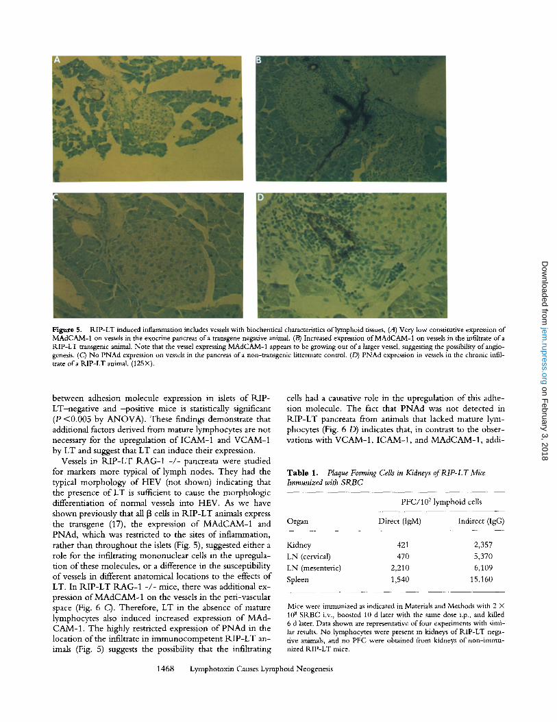
Figure 5. RIP-LT induced inflammation includes vessels with biochemical characteristics of lymphoid tissues, (A) Very low constitutive expression of MAdCAM-1 on vessels in the exocrine pancreas ofa transgene negative animal. (B) Increased expression of MAdCAM-1 on vessels in the infiltrate of a RIP-LT transgenic animal. Note that the vessel expressing MAdCAM-1 appears to be growing out of a larger vessel, suggesting the possibility of anglo- genesis. (C) No PNAd expression on vessels in the pancreas of a non-transgenic littermate control. (D) PNAd expression in vessels in the chronic infil- trate ofa RIP-LT animal. (125X).
between adhesion molecule expression in islets o f R I P - LT-negat ive and -pos i t ive mice is statistically significant (P <0 .005 by A N O V A ) . These findings demonstrate that additional factors derived from mature lymphocytes are not necessary for the upregulation o f ICAM-1 and VCAM-1 by LT and suggest that LT can induce their expression.
Vessels in R I P - L T R A G - 1 - / - pancreata were studied for markers more typical o f lymph nodes. They had the typical morphology o f HEV (not shown) indicating that the presence o f LT is suflficient to cause the morphologic differentiation o f normal vessels into HEV. As we have shown previously that all [3 cells in R I P - L T animals express the transgene (17), the expression o f M A d C A M - 1 and PNAd, which was restricted to the sites o f inflammation, rather than throughout the islets (Fig. 5), suggested either a role for the infiltrating mononuclear cells in the upregula- t ion o f these molecules, or a difference in the susceptibility o f vessels in different anatomical locations to the effects o f LT. In R I P - L T R A G - 1 - / - mice, there was additional ex- pression o f M A d C A M - 1 on the vessels in the peri-vascular space (Fig. 6 C). Therefore, LT in the absence of mature lymphocytes also induced increased expression o f M A d - CAM-1 . The highly restricted expression o f P N A d in the location o f the infiltrate in immunocompe ten t R I P - L T an- imals (Fig. 5) suggests the possibility that the infiltrating
cells had a causative role in the upregulation o f this adhe- sion molecule. The fact that P N A d was not detected in R I P - L T pancreata from animals that lacked mature lym- phocytes (Fig. 6 D) indicates that, in contrast to the obser- vations with V C A M - 1 , ICAM-1 , and M A d C A M - 1 , addi-
Table 1. Plaque Forming Cells in Kidneys of RIP-LT Mice Immunized with SRBC
PFC/107 lymphoid cells
Organ Direct (IgM) Indirect (IgG)
Kidney 421 2,357
LN (cervical) 470 5,370
LN (mesenteric) 2,210 6,109
Spleen 1,540 15,160
Mice were immunized as indicated in Materials and Methods with 2 • 10 s SRBC i.v., boosted 10 d later with the same dose i.p., and killed 6 d later. Data shown are representative of four experiments with simi- lar results. No lymphocytes were present in kidneys of RIP-LT nega- tive animals, and no PFC were obtained from kidneys of non-immu- nized RIP-LT mice.
1468 Lymphotoxin Causes Lymphoid Neogenesis
on February 3, 2018
jem.rupress.org
Dow
nloaded from

Figure 6. Upregulation oflCAM-1, VCAM-1, and MAdCAM-1, but not PNAd on vessels in and around islets of animals transgenic for R.IP-LT, but lacking mature lymphocytes. (A) ICAM-1 is upregulated in R.IP-LT RAG-2 -/- mouse on vessels throughout the islets, in the infiltrate, and even on some vessels in the exocrine pancreas in close proximity to the islets. (B) VCAM-1 is upregulated in the pancreas of R.IP-LT R.AG-2 -/- animal. Note that hke ICAM-1, VCAM-1 is upregulated on vessels throughout the islets, in the infiltrate, and even on some vessels in the exocrine pancreas in close proximity to the islets. (C) Expression of MAdCAM-1 on vessels in the peri-imular space ofa B.IP-LT RAG-2 -/- mouse. (D) No e~pression of PNAd on vessels ofa RIP-LT RAG-2 -/- animal. (125•
tional factors derived from mature lymphocytes are necessary for the upregulation o f this adhesion molecule.
Discuss ion
Our data support a unifying model for lymphoid organ development and chronic inflammation. W e show that the chronic inflammation caused by local expression of LT has all the characteristics o f functional lymphoid tissue. All cell types normally found in lymphoid organs, including T cells, B cells, plasma cells, follicular dendritic cells, and other an- tigen-presenting cells, were present in the R . IP-LT- induced infiltrates. The proportions o f these cells were very similar to those observed in a lymph node. In addition to T and B cell compartments, areas with the morphological ap- pearance o f primary and secondary follicles were present. The endothelial cells of the vessels in the infiltrates had the typical morphology o f high endothelial venules and ex- pressed ICAM-1 and VCAM-1, molecules usually associ- ated with chronic inflammation, and P N A d and MAd- C A M - l , molecules normally associated with vessels in peripheral or mesenteric lymph nodes. In addition to the morphologic characteristics o f organized lymphoid tissue, the cells o f the chronic infiltrate showed the functional at-
1469 Kratz et al.
tributes o f a lymphoid organ in their ability to generate a T cell--dependent antibody response and to induce class switching. Lymphocytes in the newly organized lymphoid accumulations in the pancreata and kidneys o f R.IP-LT mice are most likely derived from the recirculating pool o f mononuclear cells, just as they are in normal lymph nodes. The organization into germinal centers and the presence o f plasma cells points to a local activation o f these cells. Stud- ies with R.IP-LT transgenic mice that lack secondary lym- phoid organs are under way to further investigate this ques- tion.
The LT-induced upregulation o f the adhesion molecules ICAM-1, VCAM-1, and M A d C A M - I and the morpho- logic appearance o f HEV did not depend upon factors de- rived from mature lymphocytes. On the other hand, ma- ture lymphocytes, in addition to the transgene product, were necessary for the expression of PNAd. These findings suggest that many o f the effects o f LT in lymphoid neogen- esis are direct effects o f the cytokine, as suggested by in vitro studies (11, 34) or secondary to its expression, and not caused by factors derived from infiltrating lymphoid cells. Other effects most likely require chemokines and other cy- tokines derived from local and infiltrating cells and are sec- ondary or even independent o f LT.
on February 3, 2018
jem.rupress.org
Dow
nloaded from

Further studies are needed to explore the interactions of other cytokines and chemokines with LT in lymphoid neo-organogenesis. There are several transgenic models in which local expression of cytokines leads to the develop- ment of inflammatory lesions that in many respects are sim- ilar to those described here. Mice transgenic for TNF-Ix, IL-2, or IL-10 under the control of the insulin promoter show pancreatic inflammation, but do not develop sponta- neous diabetes (18, 19, 35, 36) and mice expressing IFN-o~ or IFN-', /under the same promoter develop both inflam- mation and diabetes (37-39). Mice transgenic for a modi- fied TNF-cl gene develop arthritis (40). It is not known whether the infiltrates in these systems also exhibit all the lymphoid organ characteristics that R.IP-LT mice do and whether and how LT participates. It is possible that LT and other cytokines induce converging pathways that contrib- ute to these inflammatory events. It is quite possible that LT plays a critical role in these systems, because of all the cytokine or cytokine receptor knockout mice (including those lacking the p55 and/or p75 TNF receptors), only LT-deficient mice lack lymph nodes. It is therefore very probable that LT's role in the induction of lymphoid or- gans is unique. Answers to these questions may be provided in the analysis of e.g., RIP-IL-2 LTo~-/- mice being gener- ated now in our laboratory.
Our work provides a functional implication and mecha- nism for the phenomena that have given rise to the term "tertiary lymphoid tissue." This designation has been sug- gested for all tissues because they can be induced to recruit unique subsets of lymphocytes in the setting of inflamma- tion (9). Consistent with this designation have been several previous reports that chronic inflammatory lesions resemble lymphoid tissues. Clearly delineated T and B cell areas can be seen in inflamed synovium of patients suffering from rheumatoid arthritis (41) and this tissue produces the same immunoglobulin isotypes as does a lymphoid organ (42). Vessels with the characteristics of HEV have been de- scribed in the lesions of experimental allergic encephalo- myelitis (43-46). Thyroiditis in BB rats is so similar to a secondary lymphoid organ that it has been suggested that this inflammation in which very little tissue destruction oc- curs should be called "thyroid associated lymphoid tissue" (4). Vessels in the insulitis of prediaberic NOD mice ex- press MAdCAM-1 and PNAd (31, 32). The characteristics of lymphoid neogenesis noted above in several different types of chronic inflammatory lesions indicate that this pro- cess occurs throughout life in adults and is not an isolated phenomenon peculiar to the transgenic system described here. The functional implication of the designation "ter- tiary lymphoid tissue," namely the possibility that antigen can be presented at a local inflammatory site and contribute to determinant spreading (47), is apparent from our work
and provides an appreciation that the mechanism of inflam- mation may be very similar to the process of fetal lymphoid organogenesis.
Our work provides a framework to consider common mechanisms for lymphoid organogenesis and inflammation. The first cells observed in lymphoid organ development are lymphocytes that migrate from veins to the sites of the fu- ture lymph nodes (48). Though most studies evaluating LT regulation have concentrated on CD4+Thl, CD8 +, or pre-B cells (49, 50), and virus infected cells (51), LT can also be made by a variety of other cell types, including NK cells and astrocytes (10). Therefore, there are several possi- ble sources of LT that could contribute to lymph node for- marion in embryonic development. LT in development (whatever its source) and inflammation (from T cells) could cause the infiltration ofmononuclear cells via upregulation of adhesion molecules and other changes in endothelial cells. The data presented here clearly show that LT alone is suffi- cient for the expression of the adhesion molecules ICAM-1, VCAM-1, and MAdCAM-1 and the morphologic changes characteristic of the high endothelial venules found in lym- phoid organs, and eventually leads to cellular and functional attributes of a lymphoid organ.
The findings reported here provide insight into mecha- nisms that lead to lymph node development and of those forms of chronic inflammation that lead to the formation of lymphoid tissues in inappropriate locations. They imply that one cytokine, LT, not only plays a crucial role in these processes, but that it is sufficient for many of their associ- ated characteristics. Our work revealing LT as an initiating factor in chronic inflammation provides a clearer under- standing of these events. An even better understanding will come from our ongoing studies of KIP-LT mice that lack any endogenous LT (LT-c~-/- mice) and from the elucida- tion of the role of receptors, signaling pathways, and other LT-induced proteins. Such studies could lead to highly specific therapies for a variety of diseases. For example, spe- cific LT-antagonists could be useful for the prevention of determinant spreading in autoimmune diseases. Engineered release of LT at sites of malignant lesions could lead to in- flammation and provide a local milieu to induce an im- mune response against a tumor. Elucidation of the mecha- nisms of lymphoid organogenesis could also be important for the therapy of other diseases as well. Patients who re- ceive whole-body irradiation often develop an atrophy of their lymphoid tissues; presently, few therapeutic modali- ties exist to influence the regeneration of these organs. It is possible that LT could be useful here. The lesions of pul- monary lymphoid hyperplasia seen in pediatric AIDS pa- tients have characteristics of a lymphoid organs (52, 53) hinting at a role of LT and therapeutic implications of the findings reported here for some infectious diseases.
We thank Irene Visintin and Dr. Peter Kima for technical advice, Tom Taylor for assistance with flow cyto- metric analysis, Tom Ardito for help with ultrastructural studies, and Drs. Marie-Kosco-Vilbois and Ralph
1470 Lymphotoxin Causes Lymphoid Neogenesis
on February 3, 2018
jem.rupress.org
Dow
nloaded from

Steimnan for their gifts of antibodies. We are particularly grateful to Dr. Eugene Butcher for his gift of anti- bodies and helpful comments.
This work was supported by National Institutes of Health grants RO1 CA 16885; RO1 AI 34404; T32 AI 07019 and DK 45735.
Address correspondence to Dr. Nancy H. Ruddle, Department of Epidemiology and Pubhc Health, Yale University School of Medicine, P.O. Box 208034, New Haven, CT 06520-8034.
Received for publication 14 November 1995 and in revised form 2 February 1996.
References
1. Chen, Y., V.K. Kuchroo, J.-I. Inobe, D.A. Hailer, and H.L. Weiner. 1994. Regulatory T-cell clones induced by oral tol- erance: suppression ofautoimmune encephalomyelitis. Science (Wash. DC). 265:1237-1240.
2. Cotran, R.Z., V. Kumar, S.L. Robbins, and F.J. Schoen. 1994. Inflammation and repair. In Robbin's Pathologic Basis of Disease. Fifth edition. W.B. Saunders, editor. Philadelphia. 51-92.
3. Pozzilli, P., A. Signore, A.J.K. Williams, and P.E. Beales. 1993. NOD mouse colonies around the world--recent facts and figures. Immunol. Today. 14:193-196.
4. Mooij, P., H.J.d. Wit, and H.A. Drexhage. 1993. An excess of dietary iodine accelerates the development of a thyroid-as- sociated lymphoid tissue in autoimmune prone BB rats. Clin. Immunol. lmmunopath. 69:189-198.
5. Guerder, S., D. Picarella, P.S. Linsley, and R.A. Flavell. 1994. Costimulator B7-1 confers antigen-presenting cell func- tion to parenchymal tissue and in conjunction with tumor necrosis factor alpha leads to autoirnmunity in transgenic mice. Proc. Natl. Acad. Sci. USA. 91:5138-5142.
6. Heath, W.R., J. Allison, M.W. Hoffmann, G. Schonrich, G. Harnmerling, B. Arnold, and J.F.A.P. Miller. 1992. Autoim- mune diabetes as a consequence of locally produced interleu- kin-2. Nature (Lond.). 359:587-549.
7. Herrera, P.L., D.M. Harlan, L. Fossati, S. Izui, J. Huarte, L. Orci, J.D. Vassalli, and P. Vassalli. 1994. A CD8+ T-lym- phocyte-mediated and CD4+ T-lymphocyte-independent autoimmune diabetes of early onset in transgenic mice. Dia- betologia. 37:1277-1279.
8. Ohashi, P., S. Oehen, P. Aichele, H. Pircher, B. Odermatt, P. Herrera, Y. Higuchi, K. Buerki, H. Hengartner, and R.M. Zinkernagel. 1993. Induction of diabetes is influenced by the infectious virus and local expression of Class I and TNF-al- pha.J. Immunol. 11:5185-5194.
9. Picker, L.J., and E.C. Butcher. 1992. Physiological and mo- lecular mechanisms of lymphocyte homing. Annu. Rev. Im- munol. 10:561-591.
10. Turetskaya, R.L., S.J. Fashena, N.L. Paul, and N.H. Ruddle. 1992. Genomic structure, induction, and production of TNF-~. In Tumor Necrosis Factors. Structure, Function, and Mechanism of Action. B.B. Aggawal and J. Vilcek, edi- tors. Marcel Dekker, Inc., New York. 35-60.
11. Pober, J.S., L.A. Lapierre, A.H. Stolpen, T.A. Brock, T.A. Springer, W. Fiers, M.P. Bevilacqua, D.L. Mendrick, and M.A. Gimbrone. 1987. Activation of cultured human endo- thelial cells by recombinant lymphotoxin: comparison with tumor necrosis factor and interleukin-1 species. J. lmmunol. 138:3319-3324.
12. Cavender, D.E., D. Edelhaum, and M. Ziff. 1989. Endothe- lial cell activation induced by tumor necrosis factor and lyre-
1471 Kratz et al.
photoxin. Am.]. Path. 134:551-560. 13. Powell, M.B., D. Mitchell. J. Lederman, J. Buckmeier, S.S.
Zamvil, M. Graham, N.H. Ruddle, and L. Steinman. 1990. Lymphotoxin and tumor necrosis factor-alpha production by myehn basic protein-specific T-cell clones correlates with en- cephalitogenicity. Internat. Immunol. 2:539-544.
14. Ruddle, N.H., C. Bergrnan, K.M. McGrath, E.G. Lingen- held, M.L. Grunnet, S.J. Padula, and R.B. Clark. 1990. An antibody to lymphotoxin and tumor necrosis factor prevents transfer of experimental allergic encephalomyelitis. J. Exp. Med. 172:1193-1200.
15. Barren, D., and N.H. Ruddle. 1993. Vascular cell adhesion molecule-1 modulation by tumor necrosis factor in experi- mental allergic encephalomyelitis. J. Neuroimmunol. 51:123--133.
16. Lo, D., L.C. Burkly, G. Widera, C. Cowing, R.A. Flavell, R.D. Palmiter, and R.L. Brinster. 1988. Diabetes and toler- ance in transgenic mice expressing class II MHC molecules in pancreatic beta cells. Cell. 53:159-168.
17. Picarella, D.E., A. Kratz, C.-b. Li, N.H. Ruddle, and R.A. Flavell. 1992. Insulitis in transgenic mice expressing TNF-13 (lymphotoxin) in the pancreas. Proc. Natl. Acad. Sci. USA. 89: 10036-10040.
18. Picarella, D.E., A. Kratz, C.-b. Li, N.H. Ruddle, and R.A. Flavell. 1993. Transgenic TNF-o~ production in islets leads to insulitis, not diabetes: distinct patterns of inflammation in TNF-ot and TNF-I3 transgenic mice. J. Immunol. 149:4136- 4150.
19. Higuchi, Y., P. Herrera, P. Muniesa, J. Huarte, D. Belin, P. Ohasi, P. Aichele, L. Orci, J.-D. Vassalli, and P. Vassalli, 1992. Expression of a tumor necrosis factor alpha transgene in murine pancreatic beta cells results in severe and permanent insulitis without evolution towards diabetes. J. Exp. Med. 176:1719-1731.
20. De Togni, P.D., J. Goellner, N.H. Ruddle, P.R. Streeter, A. Fick, S. Mariathasan, S.C. Smith, R. Carlson, L.P. Shomick, J. Strauss-Schoenberger et al. 1994. Abnormal development of peripheral lymphoid organs in mice deficient in lyrnpho- toxin. Science (Wash. DC). 264:703-707.
21. Shinkai, Y., G. Rathbun, K.-P. Lain, E.M. Oltz, V Stewart, M. Mendelsohn, J. Charron, M. Datta, F. Young, A.M. Stall, and F.W. Alt. 1992. RAG-2-deficient mice lack mature lym- phocytes owing to inability to initiate V(D)J rearrangement. Cell. 68:855-867.
22. Takei, F. 1985. Inhibition of mixed lymphocyte response by a rat monoclonal antibody to a novel murine lymphocyte ac- tivation antigen (MALA-2).J. Immunol. 134:1403-1407.
23. Miyake, K., K. Medina, K. Ishihara, M. Kimoto, R. Auer- bach, and P.W. Kincade. 1991. A VCAM-like adhesion mol- ecule on murine bone marrow stromal cells mediates binding of lymphocyte precursors in cu/ture.J. Cell Biol. 114:577-565.
on February 3, 2018
jem.rupress.org
Dow
nloaded from

24. Witmer-Pack, M.D., M.T. Crowley, K. Inaba, and R.M. Steinman. 1993. Macrophages, but not dendritic cells, accu- mulate colloidal carbon following administration in situ. J. Cell Sci. 105:965-973.
25. Kosco, M.H., E. Pflugfelder, and D. Gray. 1992. Follicular dendritic cell-dependent adhesion and proliferation orB-cells in vitro.J. Immunol. 148:2331-2339.
26. Streeter, P.R., E.L. Berg, B.T.N. Rouse, R.F. Bargatze, and E.C. Butcher. 1988. A tissue-specific endothelial cell mole- cule involved in lymphocyte homing. Nature (Lond.). 331: 41-46.
27. Streeter, P.R., B.T.N. Rouse, and E.C. Butcher. 1988. Im- munohistologic and functional characterization of a vascular addressin involved in lymphocyte homing into peripheral lymph nodes.J. Cell Biol. 107:1853-1862.
28. Ge, A.Z., and E.C. Butcher. 1994. Cloning and expression of a cDNA encoding mouse endoglin, an endothelial cell TGF- beta ligand. Gene (Amst.). 138:201-206.
29. McLean, I .W, and P.K. Makane. 1974. Periodate-lysine- paraformaldehyde fixative. A new fixative for immunoelec- tron microscopy.J. Histochem. Cytochem. 22:1077-1083.
30. Mishell, B.B., and S.M. Shiigi. 1980. Hemolytic plaque as- says. In Selected Methods in Cellular Immunology. W.H. Freeman and Company, San Francisco, CA. 69-77.
31. Faveeuw, C., M.-C. Gaguerault, and F. Lepault. 1994. Ex- pression of homing and adhesion molecules in infiltrated islets of Langerhans and salivary glands of nonobese diabetic mice. J. lmmunol. 152:5969-5978.
32. Hanninen, A., C. Taylor, P.R. Streeter, L.S. Stark, J.M. Sarte, J.A. Shizuru, O. Simell, and S.A. Michie. 1993. Vascu- lar addressins are induced on islet vessels during insulitis in nonobese diabetic mice and are involved in lymphoid cell binding to islet endothelium.J. Clin. Invest. 92:2509-2515.
33. Wilms-Kretschmer, K., M.H. Flax, and R.S. Cotran. 1967. The fine structure of the vascular response in hapten-specific delayed hypersensitivity and contact dermatitis. Lab. Invest. 17:334-349.
34. Sikorski, E.E., R. Hallmann, E.L. Berg, and E.C. Butcher. 1993. The Peyer's patch high endothelial receptor for lym- phocytes, the mucosal vascular addressin, is induced on a mu- rine endothelial cell line by tumor necrosis factor-alpha and IL-1.J. Immunol. 151:5239-5250.
35. Allison, J., L. Malcolm, N. Chosich, and J.F.A.P. Miller. 1992. Inflammation but not autoimmunity occurs in trans- genic mice expressing constitutive levels of interleukin-2 in islet beta cells. Eur.J, Immunol. 22:1115-1121.
36. Wogensen, L., X. Huang, and N. Sarvetnick. 1993. Leuko- cyte extravasation into the pancreatic tissue in transgenic mice expressing interleukin I0 in the islets of Langerhans. J. Exp. Med. I75:175-185.
37. Sarvemick, N., D. Liggitt, S.L. Pitts, S.E. Hansen, and T.A. Stewart. 1988. Insulin-dependent diabetes meflitus induced in transgenic mice by ectopic expression of class II MHC and interferon-gamma. Cell. 52:773-782.
38. Sarvetnick, N., J. Shizuru, D. Liggitt, L. Martin, B. Mcln- tyre, A. Gregory, T. Parslow, and T. Stewart. 1990. Loss of pancreatic islet tolerance induced by [3-cell expression of in- terferon-',/. Nature (Lond.). 346:844-847.
39. Stewart, T.A., B. Hultgren, X. Huang, S. Pitts-Meek, J. Hully, and N.J. MacLachlan. 1993. Induction of type I dia- betes by Interferon-alpha in transgenic mice. Science (Wash.
DC). 260:1942-1946. 40. Keffer, J., L. Probert, H. Cazlaris, S. Georgopoulos, E.
Kaslaris, D. Kioussis, and G. Kollias. 1991. Transgenic mice expressing human tumour necrosis factor: a predictive genetic model of arthritis. EMBO (Eur. Mol. Biol. Organ.)d. 10: 4025-4031.
41. Ziff, M., and D. Cavender. 1988. The role of endothelium in chronic inflammation. In Cellular and Molecular Aspects of Inflammation. G. Poste and S.T. Crooke, editors. Plenum Press, NY. 57-64.
42. Smiley, J.D., C. Sachs, and M. Ziff. 1968. In vitro synthesis of immunoglobulin by rheumatoid synovial membrane. J. Clin. Invest. 47:624-632.
43. Cannella, B., A.H. Cross, and C.S. Raine. 1990. Upregula- tion and co-expression of adhesion molecules correlate with relapsing autoimmune demyelination in the central nervous system.J. Exp. Med. 172:1521-1524.
44. Cannella, G., A.H. Cross, and C.S. Raine. 1991. Fluctuations in the expression of adhesion related molecules during relaps- ing experimental autoimmune encephalomyelitis. Lab. Invest. 65:23-31.
45. Cross, A.H., B. Cannella, C.F. Brosnan, and C.S. Raine. 1990. Homing to central nervous system vasculature by anti- gen specific lymphocytes. I. Localization of 14-C labeled cells during acute, chronic and relapsing allergic encephalomyeli- tis. Lab. Invest. 63:162-170.
4& Raine, C.S., B. Cannefla, A.M. Duijvestijn, and A.H. Cross. 1990. Homing to central nervous system vasculature by anti- gen-specific lymphocytes. II. Lymphocyte/endothelial cell adhesion during the initial stages of autoimmune demyelina- tion. Lab. Invest. 63:476-489.
47. Lehmann, P.V., E.E. Sercarz, T. Forsthuber, C.M. Dayan, and G. Gammon. 1993. Determinant spreading and the dy- namics of the autoimmune T-cell repertoire. Immunol. Today. 14:203-208.
48. Eikelenboom, P., J.J.J. Nassy, J. Post, J.C.M.B. Versteeg, and H.L. Langevoort. 1978. The histogenesis of lymph nodes in rat and rabbit. Anat. Rec. 190:201-216.
49. Laskov, R., G. Lancz, N.H. Ruddle, K.M. McGrath, S. Specter, T. Klein, J.Y. Djeu, and H. Friedman. 1990. Pro- duction of tumor necrosis factor (TNF-alpha) and lympho- toxin (TNF-beta) by routine pre-B and B cell lymphomas. J. ImmunoI. 144:3424-3430.
50. Fashena, S.J., R. Reeves, and N.H. Ruddle. 1992. A poly (dA-dT) upstream activating sequence binds high mobility group I protein and contributes to lymphotoxin (tumor ne- crosis factor-g) gene regulation. Mol. Cell. Biol. 12:894-903.
51. Paul, N.L., M.J. Lenardo, K.D. Novak, T. Sarr, W.-L. Tang, and N.H. Ruddle. 1990. Lymphotoxin activation by human T-cell leukemia virus type I infected cell lines: role for NFKB.J. Virol. 64:5412-5419.
52. Joshi, V.V. 1991. Pathologic findings associated with HIV in- fection in children. In Pediatric AIDS. The challenge of HIV infection in infants, children, and adolescents. P.A. Pizzo and C.M. Wilfert, editors. Williams & Wilkins, Baltimore, MD. 113-139.
53. Connor, E.M., J. Marquis, andJ.M. Oleske. 1991. Lymphoid interstitial pneumonitis. In Pediatric AIDS. The Challenge of HIV Infection in Infants, Children, and Adolescents. P.A. Pizzo and C.M. Wilfert, editors. Williams & Wilkins, Balti- more, MD. 343-354.
1472 Lymphotoxin Causes Lymphoid Neogenesis
on February 3, 2018
jem.rupress.org
Dow
nloaded from