Chromosomal Basis of Inheritance - BYJU'S · 2019-04-09 · 3.2 Linkage - Eye colour in Drosophila...
37
55 Chromosomal Basis of Inheritance 3.1 Chromosomal Theory of Inheritance G. J. Mendel (1865) studied the inheritance of well-defined characters of pea plant but for several reasons it was unrecognized till 1900. Three scientists (de Vries, Correns and Tschermak) independently rediscovered Mendel’s results on the inheritance of characters. Various cytologists also observed cell division due to advancements in microscopy. This led to the discovery of structures inside nucleus. In eukaryotic cells, worm-shaped structures formed during cell division are called chromosomes (colored bodies, visualized by staining). An organism which possesses two complete basic sets of chromosomes are known as diploid. A chromosome consists of long, continuous coiled piece of DNA in which genes are arranged in linear order. Each gene has a definite position (locus) on a chromosome. These genes are hereditary units. Chromosomal theory of inheritance states that Mendelian factors (genes) have specific locus (position) on chromosomes and they carry information from one generation to the next generation. 3.1.1 Historical development of chromosome theory e important cytological findings related to the chromosome theory of inheritance are given below. � Wilhelm Roux (1883) postulated that the chromosomes of a cell are responsible for transferring heredity. In the previous chapter you have learned about Mendelian genetics, now you are going to be study with deviations of concepts related to Mendelian genetics and chromosomal theory of inheritance. You must recall the structure of chromosome and cell division from eleventh standard. Learning Objectives e Learner will be able to Understand chromosomal theory of inheritance. Analyze the three- point test crosses and appreciate results in linkage map construction. Describe the sex determination in plants. Observe and calculate recombination frequency. Differentiate mutation types with examples. Explain DNA metabolism in Plants 3.1 Chromosomal theory of Inheritance 3.2 Linkage - Eye colour in Drosophila and Seed colour in Maize 3.3 Crossing over, Recombination and Gene mapping 3.4 Multiple alleles 3.5 Sex determination in plants. 3.6 Mutation-types, mutagenic agents and their significance. 3.7 DNA Metabolism in Plants Chapter outline UNIT VII: Genetics Chapter 3 Chromosomal Basis of Inheritance TN_GOVT_BOTANY_XII_PAGES_055-091 CH 03.indd 55 01-03-2019 16:59:00
Transcript of Chromosomal Basis of Inheritance - BYJU'S · 2019-04-09 · 3.2 Linkage - Eye colour in Drosophila...

55Chromosomal Basis of Inheritance
3.1 Chromosomal Theory of Inheritance
G. J. Mendel (1865) studied the inheritance of well-defined characters of pea plant but for several reasons it was unrecognized till 1900. Three scientists (de Vries, Correns and Tschermak) independently rediscovered Mendel’s results on the inheritance of characters. Various cytologists also observed cell division due to advancements in microscopy. This led to the discovery of structures inside nucleus. In eukaryotic cells, worm-shaped structures formed during cell division are called chromosomes (colored bodies, visualized by staining). An organism which possesses two complete basic sets of chromosomes are known as diploid. A chromosome consists of long, continuous coiled piece of DNA in which genes are arranged in linear order. Each gene has a definite position (locus) on a chromosome. These genes are hereditary units. Chromosomal theory of inheritance states that Mendelian factors (genes) have specific locus (position) on chromosomes and they carry information from one generation to the next generation.
3.1.1 Historical development of chromosome theory
Th e important cytological fi ndings related to the chromosome theory of inheritance are given below.� Wilhelm Roux (1883) postulated that the
chromosomes of a cell are responsible for transferring heredity.
In the previous chapter you have learned about Mendelian genetics, now you are going to be study with deviations of concepts related to Mendelian genetics and chromosomal theory of inheritance. You must recall the structure of chromosome and cell division from eleventh standard.
Learning Objectives
Th e Learner will be able to Understand
chromosomal theory of inheritance.
Analyze the three-point test crosses and appreciate results in linkage map construction.
Describe the sex determination in plants. Observe and calculate recombination
frequency. Diff erentiate mutation types with examples. Explain DNA metabolism in Plants
3.1 Chromosomal theory of Inheritance3.2 Linkage - Eye colour in Drosophila
and Seed colour in Maize 3.3 Crossing over, Recombination and
Gene mapping3.4 Multiple alleles3.5 Sex determination in plants.3.6 Mutation-types, mutagenic agents
and their signifi cance.3.7 DNA Metabolism in Plants
Chapter outline
UNIT VII: GeneticsChapter
3 Chromosomal Basis of Inheritance
TN_GOVT_BOTANY_XII_PAGES_055-091 CH 03.indd 55 01-03-2019 16:59:00
56 Chromosomal Basis of Inheritance
� Montgomery (1901) was first to suggest occurrence of distinct pairs of chromosomes and he also concluded that maternal chromosomes pair with paternal chromosomes only during meiosis.
� T. Boveri (1902) supported theidea that the chromosomes containgenetic determiners, and he waslargely responsible for developing thechromosomal theory of inheritance.
� W.S. Sutton (1902), a young Americanstudent independently recognized aparallelism (similarity) between thebehaviour of chromosomes and Mendelian factors during gamete formation.Sutton and Boveri (1903) independently
proposed the chromosome theory of inheritance. Sutton united the knowledge of chromosomal segregation with Mendelian principles and called it chromosomal theory of inheritance.
3.1.2 Salient features of the Chromosomal theory of inheritance� Somatic cells of organisms are derived
from the zygote by repeated cell division (mitosis). These consist of two identical sets of chromosomes. One set is received from female parent (maternal) and the other from male parent (paternal). These two chromosomes constitute the homologous pair.
� Chromosomes retain their structural uniqueness and individuality throughout the life cycle of an organism.
� Each chromosome carries specific determiners or Mendelian factors which are now termed as genes.
� The behaviour of chromosomes during the gamete formation (meiosis) provides evidence to the fact that genes or factors are located on chromosomes.
3.1.3 Support for chromosomal theory of heredity
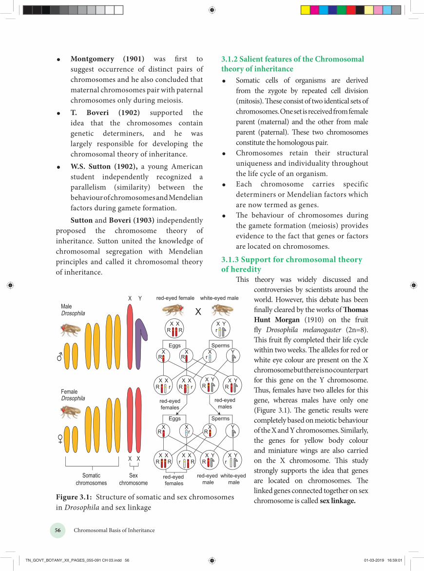
This theory was widely discussed and controversies by scientists around the world. However, this debate has been finally cleared by the works of Thomas Hunt Morgan (1910) on the fruit fly Drosophila melanogaster (2n=8). This fruit fly completed their life cycle within two weeks. The alleles for red or white eye colour are present on the X chromosome but there is no counterpart for this gene on the Y chromosome. Thus, females have two alleles for this gene, whereas males have only one (Figure 3.1). The genetic results were completely based on meiotic behaviour of the X and Y chromosomes. Similarly, the genes for yellow body colour and miniature wings are also carried on the X chromosome. This study strongly supports the idea that genes are located on chromosomes. The linked genes connected together on sex chromosome is called sex linkage.
X Xr R
X XR R
X XR r
X XrR
X YR
X YR
X YR
X Yr
XR
XR
YXr
RX
RX
rX Y
X XR R
X Yr
r
red-eyed females
red-eyed male
white-eyed male
Eggs Sperms
Eggs Sperms
red-eyedmales
red-eyedfemales
red-eyed female white-eyed male
R R
Figure 3.1: Structure of somatic and sex chromosomes in Drosophila and sex linkage
TN_GOVT_BOTANY_XII_PAGES_055-091 CH 03.indd 56 01-03-2019 16:59:01

57Chromosomal Basis of Inheritance
3.1. Comparison between gene and chromosome behaviourAround twentieth century cytologists established that, generally the total number of chromosomes is constant in all cells of a species. A diploid eukaryotic cell has two haploid sets of chromosomes, one set from each parent. All somatic cells of an organism carry the same genetic complement. The behaviour of chromosomes during meiosis not only explains Mendel’s principles but leads to new and different approaches to study about heredity.
Homologouschromosomes
Non homologous chromosomes
Chromosome 1 Chromosome 2 Chromosome 3Same genes, same order
Allele-A Allele-a
Genes
Different DNA sequences
GCATTCGAGTCCATAAGCGATGA GCATTCGAGTCTATAAGCGATGA
Genes
Figure 3.2: Comparison of chromosome and gene behaviour
Table 3.1: Parallelism between Mendelian factors and chromosomal behaviour.
Mendelian factors Chromosomes behaviour1. Alleles of a factor
occur in pairChromosomes occur in pairs
2. Similar or dissimilaralleles of a factorseparate during thegamete formation
The homologous chromosomes separate during meiosis
3. Mendelianfactors can assortindependently
The paired chromosomes can separate independently during meiosis but the linked genes in the same chromosome normally do not assort independently.
The important aspects to be remembered about the chromosome behaviour during cell division (meiosis) are as follows.� The alleles of a genotype are found in the
same locus of a homologous chromosome (A/a) (Figure 3.2).
� In the S phase of meiotic interphase each chromosome replicates forming two copies of each allele (AA/aa), one on each chromatid.
� The homologous chromosomes segregate in anaphase I, thereby separating two different alleles (AA) and (aa).
� In anaphase II of meiosis, separation of sister chromatids of homologous chromosomes takes place. Therefore, each daughter cell (gamete) carries only a single allele (gene) of a character (A), (A), (a) and (a).
Organism Number of chromosomes (2n)
Adder’s tongue fern (Ophioglossum) 1262
Horsetail (Equisetum) 216Giant sequoia 22Arabidopsis 10Sugarcane 80Apple 34Rice 24Potato 48Maize 20Onion 16Haplopappus gracilis 4
Table 3.2 : Number of Chromosomes
Thomas Hunt Morgan (1933) received Nobel Prize in Physiology or Medicine for his discoveries concerning the role played by chromosomes in heredity.
Fossil Genes: Some of the junk DNA is made up of pseudo genes, the sequences presence in that was once working genes.
They lost their ability to make proteins. They tell the story of evolution through fossilized parts.
TN_GOVT_BOTANY_XII_PAGES_055-091 CH 03.indd 57 01-03-2019 16:59:01

58 Chromosomal Basis of Inheritance
Genes located close together on the same chromosome and inherited together are called linked genes. But the two genes that are sufficiently far apart on the same chromosome are called unlinked genes or syntenic genes (Figure 3.3). Such condition is known as synteny. It is to be differentiated by the value of recombination frequency. If the recombination frequency value is more than 50 % the two genes show unlinked. when the recombination frequency value is less than 50 %, they show linked. Closely located genes show strong linkage, while genes widely located show weak linkages.
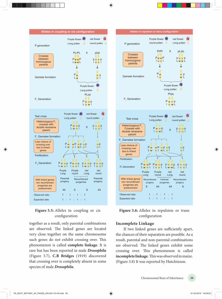
3.2.1 Coupling and Repulsion theoryThe two dominant alleles or recessive alleles occur in the same homologous chromosomes, tend to inherit together into same gamete are called coupling or cis configuration (Figure: 3.5 ). If dominant or recessive alleles are present on two different, but homologous chromosomes they inherit apart into different gamete are called repulsion or trans configuration (Figure: 3. 6).
Figure 3.4: Cis-Trans arrangement of genes
Cis AB/ab Trans Ab/aB
3.2.2 Kinds of LinkageT.H. Morgan found two types of linkage. They are complete linkage and incomplete linkage depending upon the absence or presence of new combination of linked genes.
Complete LinkageIf the chances of separation of two linked
genes are not possible those genes always remain
3.2 LinkageThe genes which determine the character of an individual are carried by the chromosomes. The genes for different characters may be present either in the same chromosome or in different chromosomes. When the genes are present in different chromosomes, they assort independently according to Mendel’s Law of Independent Assortment. Biologists came across certain genetic characteristics that did not assort out independently in other organisms after Mendel’s work. One such case was reported in Sweet pea (Lathyrus odoratus) by Willium Bateson and Reginald C. Punnet in 1906. They crossed one homozygous strain of sweet peas having purple flowers and long pollen grains with another homozygous strain having red flowers and round pollen grains. All the F1 progenies had purple flower and long pollen grains indicating purple flower long pollen (PL/PL) was dominant over red flower round pollen (pl/pl). When they crossed the F1 with double recessive parent (test cross) in results, F2 progenies did not exhibit in 1:1:1:1 ratio as expected with independent assortment. A greater number of F2 plants had purple flowers and long pollen or red flowers and round pollen. So they concluded that genes for purple colour and long pollen grain and the genes for red colour and round pollen grain were found close together in the same homologous pair of chromosomes. These genes do not allow themselves to be separated. So they do not assort independently. This type of tendency of genes to stay together during separation of chromosomes is called Linkage.
Linked genes Unlinked genes Unlinked genes
Gene 1
Gene 2
Gene 1
Gene 2
Gene 1
Gene 2
Figure 3.3: Arrangement of linked and unlinked genes on chromosome
TN_GOVT_BOTANY_XII_PAGES_055-091 CH 03.indd 58 01-03-2019 16:59:01
59Chromosomal Basis of Inheritance
Figure 3.5: Alleles in coupling or cis configuration
Figure 3.6: Alleles in repulsion or trans configuration
together as a result, only parental combinations are observed. The linked genes are located very close together on the same chromosome such genes do not exhibit crossing over. This phenomenon is called complete linkage. It is rare but has been reported in male Drosophila (Figure 3.7). C.B Bridges (1919) discovered that crossing over is completely absent in some species of male Drosophila.
Incomplete LinkageIf two linked genes are sufficiently apart,
the chances of their separation are possible. As a result, parental and non-parental combinations are observed. The linked genes exhibit some crossing over. This phenomenon is called incomplete linkage. This was observed in maize. (Figure 3.8) It was reported by Hutchinson.
TN_GOVT_BOTANY_XII_PAGES_055-091 CH 03.indd 59 01-03-2019 16:59:02

60 Chromosomal Basis of Inheritance
Figure 3.7: Complete linkage in male Drosophila
3.2.3 Linkage GroupsThe groups of linearly arranged linked genes on a chromosome are called Linkage groups. In any species the number of linkage groups corresponds to the number haploid set of chromosomes. Example:Name of organism Linkage groupsMucor 2Drosophila 4Sweet pea 7Neurospora 7Maize 10
Table 3.3 : Linkage groups in some organisms
C
P generation
F1 Generation
Gametes formation
Test Cross
Test cross result Colouredfull seed
colourlessfull seed
ColouredShrunken
seed
Recombinant progenies
Parentalprogeny
Parentalprogeny
Due to crossing over recombinantprogenies obtainedleast in number
Heterozygous F1crosses with doublerecessive parent
Crosses betweenhomozygousparents
Colouredfull seed
colourlessshrunken seedX
Coloured full seed
CS/CS cs/cs
CS/cs
CS/cs
CS/CS48%
Cs/Cs2% 2% 48%
cS/cS cs/cs
colourlessshrunken
seed
cs/cs
Colouredfull seed X
colourlessshrunken seed
S
c
s
C
S
c
s
c
s
C
S
c
s
C
S
c
s
c
s
Incomplete linkage in Maize seed
Figure 3.8: Incomplete linkage in Maize seed
Linkage and crossing over are two processes that have opposite effects. Linkage keeps particular genes together but crossing over mixes them. The differences are given below.
Linkage Crossing over
1. The genes present onchromosomestay closetogether
It leads to separation of linked genes
2. It involves samechromosomeof homologouschromosome
It involves exchange of segments between non-sister chromatids of homologous chromosome.
3. It reduces new genecombinations
It increases variability by forming new gene combinations. lead to formation of new organism
Table 3.4: Differences between linkage and crossing over
TN_GOVT_BOTANY_XII_PAGES_055-091 CH 03.indd 60 01-03-2019 16:59:03

61Chromosomal Basis of Inheritance
are called Chiasmata (singular-Chiasma). At chiasma, cross-shaped or X-shaped structures are formed, where breaking and rejoining of two chromatids occur. This results in reciprocal exchange of equal and corresponding segments between them. A recent study reveals that synapsis and chiasma formation are facilitated by a highly organised structure of filaments called Synaptonemal Complex (SC) (Figure 3.9). This synaptonemal complex formation is absent in some species of male Drosophila hence crossing over does not takes place.
Sister chromatid 1Sister chromatid 2
Sister chromatid 3Sister chromatid 4
Recombinationnodules+
+
Synaptonemalcomplex
Synaptonemalcomplex
Figure 3.9: Structure of Synaptonemal Complex
3.3 Crossing OverCrossing over is a biological process that produces new combination of genes by inter-changing the corresponding segments between non-sister chromatids of homologous pair of chromosomes. The term 'crossing over' was coined by Morgan (1912). It takes place during pachytene stage of prophase I of meiosis. Usually crossing over occurs in germinal cells during gametogenesis. It is called meiotic or germinal crossing over. It has universal occurrence and has great significance. Rarely, crossing over occurs in somatic cells during mitosis. It is called somatic or mitotic crossing over.3.3.1 Mechanism of Crossing OverCrossing over is a precise process that includes stages like synapsis, tetrad formation, cross over and terminalization.
(i) SynapsisIntimate pairing between two homologous
chromosomes is initiated during zygotene stage of prophase I of meiosis I. Homologous chromosomes are aligned side by side resulting in a pair of homologous chromosomes called bivalents. This pairing phenomenon is called synapsis or syndesis. It is of three types, 1. Procentric synapsis: Pairing starts from
middle of the chromosome.2. Proterminal synapsis: Pairing starts from
the telomeres.3. Random synapsis: Pairing may start from
anywhere.
(ii) Tetrad FormationEach homologous chromosome of a
bivalent begin to form two identical sister chromatids, which remain held together by a centromere. At this stage each bivalent has four chromatids. This stage is called tetrad stage.
(iii) Cross OverAfter tetrad formation, crossing over occurs inpachytene stage. The non-sister chromatids ofhomologous pair make a contact at one or morepoints. These points of contact between non-sister chromatids of homologous chromosomes Figure 3.10: Mechanism of crossing over
TN_GOVT_BOTANY_XII_PAGES_055-091 CH 03.indd 61 01-03-2019 16:59:03

62 Chromosomal Basis of Inheritance
2. Double cross over: Formation of twochiasmata and involves two or three orall four strands
3. Multiple cross over: Formation of morethan two chiasmata and crossing overfrequency is extremely low.
3.3.3 Importance of Crossing OverCrossing over occurs in all organisms like bacteria, yeast, fungi, higher plants and animals. Its importance is� Exchange of segments leads to new gene
combinations which plays an important role in evolution.
� Studies of crossing over reveal that genes are arranged linearly on the chromosomes.
� Genetic maps are made based on the frequency of crossing over.
� Crossing over helps to understand the nature and mechanism of gene action.
� If a useful new combination is formed it can be used in plant breeding.
3.3.4 RecombinationCrossing over results in the formation of new combination of characters in an organism called recombinants. In this, segments of DNA are broken and recombined to produce new combinations of alleles. This process is called Recombination. (Figure 3.12)
(iv) TerminalisationAfter crossing over, chiasma starts to movetowards the terminal end of chromatids. This isknown as terminalisation. As a result, completeseparation of homologous chromosomesoccurs. (Figure 4.10)
3.3.2 Types of Crossing OverDepending upon the number of chiasmata formed crossing over may be classified into three types. (Figure 3.11)
1. Single cross over: Formation of singlechiasma and involves only two chromatidsout of four.
Figure 3.11: Types of crossing over and its Recombination Frequency (RF)
RF = = 0%04
RF = = 0%04
RF = = 50%X100
X100
X100
X100
24
RF = = 50%24
RF = = 100%44
A
A
a
a
B
B
b
b
A
a
A
a
B
B
b
b
A
a
A
a
B
b
B
b
A
a
A
a
B
b
b
A b
A b
a B
B
a B
A
A
a
a
B
B
b
b
A
A
a
a
B
B
b
b
A
A
a
a
B
B
b
b
A
A
a
a
B
B
b
b
No cross over
Single cross over
Two strand double cross over
Three strand double cross over
Four strand double cross over
X100
Activity: Solve this
Consider two hypothetical recessive autosomal genes a and b, where a heterozygote is testcrossed to a double homozygous mutant. Predict the phenotypic ratios under the following conditions:(a) a and b are located on separate
autosomes.(b) a and b are linked on the same
autosome but are so far apart that acrossover occurs between them.
(c) a and b are linked on the sameautosome but are so close together thata crossover almost never occurs.
TN_GOVT_BOTANY_XII_PAGES_055-091 CH 03.indd 62 01-03-2019 16:59:04

63Chromosomal Basis of Inheritance
RF =Number of recombinants
Number of off springsx 100
= 6+644+6+6+44
x 100
= 12100
x 100
= 12%
Figure 3.14 Recombination frequency observation
Check your Grasp
Find out Recombination frequency value from the above figure.
Figure 3.12 : Recombination
The widely accepted model of DNA recombination during crossing over is Holliday’s hybrid DNA model. It was first proposed by Robin Holliday in 1964. It involves several steps. (Figure 3.13)
1. Homologous DNA molecules are paired sideby side with their duplicated copies of DNAs
2. One strand of both DNAs cut in one placeby the enzyme endonuclease.
3. The cut strands cross and join thehomologous strands forming the Hollidaystructure or Holliday junction.
4. The Holliday junction migrates away from theoriginal site, a process called branch migration, as a result heteroduplex region is formed.
5. DNA strands may cut along through thevertical (V) line or horizontal (H) line.
6. The vertical cut will result in heteroduplexes with recombinants.
7. The horizontal cut will result inheteroduplex with non recombinants.
Calculation of Recombination Frequency (RF)The percentage of recombinant progeny
in a cross is called recombination frequency. The recombination frequency (cross over frequency) (RF) is calculated by using the following formula. The data is obtained from alleles in coupling configuration (Figure 3.14)
A
(a) (b)
(c) (d)
(f) (g)
(e)
5’
5’
3’
5’
3’
3’
5’
3’B C A B C
A B C
A B C
A C
Aa
Bb
Cc a b c
a b ca b c
a b c
a b c
a b C
a b c
A5’
5’
3’
3’
5’
3’B c
A b c
A
Holliday junction
Vertical cut(along line V)and reseal
Horizontal cut(along line H)and reseal
Heteroduplex region
C
aB
B Cb
AA
c
cb
a
V
H
Heteroduplexesand recombinants
HeteroduplexesNo recombinants
5’
5’
3’
5’
3’
3’
5’
3’B C
Aa
bB
Cc
BA B CA b C
a B c
a b c
5’3’a B C
Figure 3.13: Holliday model showing Recombination
RF
TN_GOVT_BOTANY_XII_PAGES_055-091 CH 03.indd 63 01-03-2019 16:59:04

64 Chromosomal Basis of Inheritance
3.3.5 Genetic MappingGenes are present in a linear order along the chromosome. They are present in a specific location called locus (plural: loci). The diagrammatic representation of position of genes and related distances between the adjacent genes is called genetic mapping. It is directly proportional to the frequency of recombination between them. It is also called as linkage map. The concept of gene mapping was first developed by Morgan’s student Alfred H Sturtevant in 1913. It provides clues about where the genes lies on that chromosome.
Map distanceThe unit of distance in a genetic map is called
a map unit (m.u). One map unit is equivalent to one percent of crossing over (Figure 4. ). One map unit is also called a centimorgan (cM) in honour of T.H. Morgan. 100 centimorgan is equal to one Morgan (M). For example: A distance between A and B genes is estimated to be 3.5 map units. It is equal to 3.5 centimorgans or 3.5 % or 0.035 recombination frequency between the genes.
3.5 cM 7.5 cM 5.5 cM
A B C D
Genetic maps can be constructed from a series of test crosses for pairs of genes called two point crosses. But this is not efficient because double cross over is missed.
Three point test cross A more efficient mapping technique is to
construct based on the results of three-point test cross. It refers to analyzing the inheritance patterns of three alleles by test crossing a triple recessive heterozygote with a triple recessive homozygote. It enables to determine the distance between the three alleles and the order in which they are located on the chromosome. Double cross overs can be detected which will provide more accurate map distances.
Three-point test cross can be best understood by considering following an example.
In maize (corn), the three recessive alleles are 1. l for lazy or prostrate growth habit2. g for glossy leaf3. s for sugary endospermThese three recessive alleles (l g s) are crossedwith wild type dominant alleles (L G S).
Parents LGS / LGS x lgs / lgsGametes LGS x lgsF1 trihybrid LGS / lgsTest cross (Heterozygous F1 crosses with triple recessive alleles)
LGS / lgs x lgs / lgs
This trihybrid test cross produces 8 different types (23=8) of gametes in which 740 progenies are observed. The following table shows the result obtained from a test cross of corn with three linked genes.
The analysis of a three-point cross:S. no
Phenotype of test cross progeny
Gamete types
Number of progenies
1. Normal (wild type) L G S 2862. Lazy l G S 333. Glossy L g S 594. Sugary L G s 45. Lazy, glossy l g S 26. Lazy, sugary l G s 447. Glossy, sugary L g s 408. Lazy, glossy, sugary l g s 272
Total 740From the above result, we must be careful to
observe parental (P) and recombinant (R) types. First note that parental genotypes for the triple homozygotes are L G S and l g s, then analyse two recombinant loci at a time orderly L G/ l g, L S/ l s and G S/ g s. In this any combination other than these two constitutes a recombinant (R).
Let’s analyse the loci of two alleles at a time starting with L and G Since the L G and l g parental genotypes the recombinants will be L g and l G. The Recombinant frequency (RF) for these two alleles can be calculated as follows
TN_GOVT_BOTANY_XII_PAGES_055-091 CH 03.indd 64 01-03-2019 16:59:04
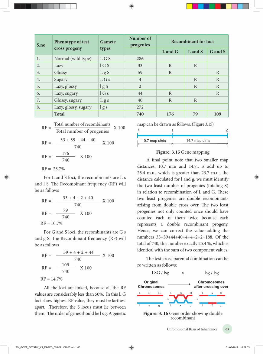
65Chromosomal Basis of Inheritance
RF =Total number of recombinantsTotal number of progenies
X 100
RF = X 10033 + 59 + 44 + 40740
RF = X 100176740
RF = 23.7%
For L and S loci, the recombinants are L s and l S. The Recombinant frequency (RF) will be as follows
RF = X 10033 + 4 + 2 + 40 740
RF = X 10079740
RF = 10.7%
For G and S loci, the recombinants are G s and g S. The Recombinant frequency (RF) will be as follows
RF = X 10059 + 4 + 2 + 44740
RF = X 100109740
RF = 14.7%
All the loci are linked, because all the RF values are considerably less than 50%. In this L G loci show highest RF value, they must be farthest apart. Therefore, the S locus must lie between them. The order of genes should be l s g. A genetic
map can be drawn as follows: (Figure 3.15)
10.7 map uints 14.7 map uints
s gl
Figure: 3.15 Gene mappingA final point note that two smaller map
distances, 10.7 m.u and 14.7., is add up to 25.4 m.u., which is greater than 23.7 m.u., the distance calculated for l and g. we must identify the two least number of progenies (totaling 8) in relation to recombination of L and G. These two least progenies are double recombinants arising from double cross over. The two least progenies not only counted once should have counted each of them twice because each represents a double recombinant progeny. Hence, we can correct the value adding the numbers 33+59+44+40+4+4+2+2=188. Of the total of 740, this number exactly 25.4 %, which is identical with the sum of two component values.
The test cross parental combination can be re written as follows:
LSG / lsg x lsg / lsg
S.noPhenotype of test cross progeny
Gamete types
Number of progenies
Recombinant for loci
L and G L and S G and S1. Normal (wild type) L G S 2862. Lazy l G S 33 R R3. Glossy L g S 59 R R4. Sugary L G s 4 R R5. Lazy, glossy l g S 2 R R6. Lazy, sugary l G s 44 R R7. Glossy, sugary L g s 40 R R8. Lazy, glossy, sugary l g s 272
Total 740 176 79 109
L S G
l s g
L S G
l s g
L s G
l S g
OriginalChromosomes
Chromosomesafter crossing over
Figure: 3. 16 Gene order showing double recombinant
TN_GOVT_BOTANY_XII_PAGES_055-091 CH 03.indd 65 01-03-2019 16:59:05

66 Chromosomal Basis of Inheritance
pollen from a plant is unable to germinate on its own stigma and will not be able to bring about fertilization in the ovules of the same plant. East (1925) observed multiple alleles in Nicotiana which are responsible for self-incompatibility or self-sterility. The gene for self-incompatibility can be designated as S, which has allelic series S1, S2, S3, S4 and S5 (Figure 3.17).
Style
Pollen tube
Ovary
S3
S1S2 S1S2 S1S2
S3 S2S4 S1 S1
Pollen
SS11SS22 SS11SS22SS SS11SS2222 SS
Figure: 3.17 The self-incompatibility in relation to its genotype in tobacco
The cross-fertilizing tobacco plants were not always homozygous as S1S1 or S2S2, but all plants were heterozygous as S1S2, S3S4, S5S6. When crosses were made between different S1S2 plants, the pollen tube did not develop normally. But effective pollen tube development was observed when crossing was made with other than S1S2 for example S3S4.
Female parent
(Stigma spot)
Male parent (Pollen source)
S1S2 S2S3 S3S4
S1S2
Self Sterile
S3S2
S3S1
S3S1
S3S2
S4S1
S4S2
S2S3
S1S2
S1S3
Self Sterile
S4S2
S4S3
S3S4
S1S3
S1S4
S2S3
S2S4
S2S3
S2S4
Self Sterile
Table: 3.5. Different combinations of progeny in self-incompatibility
Uses of genetic mapping� It is used to determine gene order, identify
the locus of a gene and calculate the distances between genes.
� They are useful in predicting results of dihybrid and trihybrid crosses.
� It allows the geneticists to understand the overall genetic complexity of particular organism.
3.4 Multiple allelesA given phenotypic trait of an individual depends on a single pair of genes, each of which occupies a specific position called the locus on homologous chromosome. When any of the three or more allelic forms of a gene occupy the same locus in a given pair of homologous chromosomes, they are said to be called multiple alleles.
Check your Grasp
There may be multiple alleles within the population, but individuals have only two of those alleles. Why?
3.4.1 Characteristics of multiple alleles• Multiple alleles of a series always occupy
the same locus in the homologouschromosome. Therefore, no crossing overoccurs within the alleles of a series.
• Multiple alleles are always responsible forthe same character.
• The wild type alleles of a series exhibitdominant character whereas mutanttype will influence dominance or anintermediate phenotypic effect.
• When any two of the mutant multiplealleles are crossed the phenotype is alwaysmutant type and not the wild type
3.4.2 Self-sterility in NicotianaIn plants, multiple alleles have been reported in association with self-sterility or self-incompatibility. Self-sterility means that the
TN_GOVT_BOTANY_XII_PAGES_055-091 CH 03.indd 66 01-03-2019 16:59:05

67Chromosomal Basis of Inheritance
When crosses were made between seed parents with S1S2 and pollen parents with S2S3, two kinds of pollen tubes were distinguished. Pollen grains carrying S2 were not effective, but the pollen grains carrying S3 were capable of fertilization. Thus, from the cross S1S2XS3S4, all the pollens were effective and four kinds of progeny resulted: S1S3, S1S4, S2S3 and S2S4. Some combinations are showed in the table-3.5.
3.5 Sex determination in plantsAbout 94% of all flowering plants have only one type of individual, which produces flowers with male organs (the stamens) and female organs (the carpels). Such plants are termed as sexually monomorphic. Some 6% of flowering plants which have two separate sexes are called dimorphic. Male plants produce flowers with stamens and female plants produce flowers with carpels only. Researchers are interested to study the mechanism of sex determination in plants.C.E. Allen (1917) discovered sex determination in plants. Sex determination is a complex process determined by genes, the environment and hormones.
Sex determination in Silene latifolia (Melandrium album) is of controlled by three distinct regions in a sex chromosome.
1. Y chromosome determines maleness
2. X specifies femaleness
3. X and Y show different segments
(I II III IV and V)
Does environment play a role on sex determination in plants?Yes. Horsetail plant
(Equisetum) grown under good conditions develop as female and those grown under stress condition develop into males.
3.5.1 Sex determination in papayaRecently researchers in Hawaii discovered sex chromosomes in Papaya (Carica papaya, 2n=36). Papaya has 17 pairs of autosomes and one pair of sex chromosomes. Male papaya plants have XY and female plants have XX. Unlike human sexchromosomes, papaya sex chromosomes look like autosomes and it is evolved from autosome. The sex chromosomes are functionally distinct because the Y chromosome carries the genes for male organ development and X bears the female organ developmental genes (Figure 3.18).
In papaya sex determination is controlled by three alleles. They are m, M1 and M2 of a single gene.
Genotype Dominant/recessive
Modification Sex
mm Homozygous recessive
Restrict maleness
Female
M1m Heterozygous Induces maleness
Male
M2m Heterozygous Induces both the sex
Bisexual (rare)
M1M1 or M2M2 or M1M2
Homozygous/ Heterozygous dominant
Inviable plants
Sterile
Table 3.6 : Sex determination in Papaya
3.5.2 Sex Determination in SphaerocarposSex determination was first described in the bryophyte Sphaerocarpos donnellii which has heteromorphic chromosomes. The gametophyte is haploid and heteromorphic. The male gametophyte as well as the female gametophyte is an haploid organism
XY
Papaya
Figure 3.18 : Sex chromosome of papaya
Bottle liverwort-Sphaerocarpus
TN_GOVT_BOTANY_XII_PAGES_055-091 CH 03.indd 67 01-03-2019 16:59:06

68 Chromosomal Basis of Inheritance
gibberellin biosynthesis. Gibberellins play an important role in the suppression of stamens in florets on the ears.
Genotype Dominant/recessive
Modification Sex
ba/ba ts/ts
Double recessive
Lacks silk on the stalk, but transformed tassel to pistil
Rudimentary female
ba/ba ts+/ts+
Recessive and dominant
Lacks silk and have tassel
Male
ba+/ba+ ts+/ts+
Double dominant
Have both tassel and cob
Monoecious
ba+/ba+ ts/ts
Dominant and recessive
Bears cob and lacks tassel
Normal female
Table 3.7: Sex determination in Maize (Superscript (+) denotes dominant character)
3.6 MutationGenetic variation among individuals provides the raw material for the ultimate source of evolutionary changes. Mutation and re c ombi n at i on are the two major processes responsible for genetic variation. A sudden change in the genetic material of an organisms is called mutation. The term mutation was introduced by Hugo de Vries (1901) while he has studying on the plant, evening primrose
(Oenothera lamarkiana) and proposed ‘Mutation theory’. There are two broad types of changes in genetic material. They are point mutation and chromosomal mutations.
with 8 chromosome (n=8). The diploid sporophyte is always heterogametic. Seven autosomes are similar in both male and female gametophyte. But the eighth chromosome of female is X which is larger than the seven autosomes. The eighth chromosome of male is Y which is comparatively smaller than autosomes. The sporophyte containing XY combination produces two types of meiospores, that is some with X and others with Y chromosomes. The meiospores with X chromosomes produce female gametophyte and those with Y chromosome produces male gametophyte.
3.5.3 Sex determination in maizeZea mays (maize) is an example for monoecious, which means male and female flowers are present on the same plant. There are two types of i n f l o r e s c e n c e . The terminal i n f l o r e s c e n c e which bears staminate florets develops from shoot apical meristem called tassel. The lateral i n f l o r e s c e n c e which develop pistillate florets from axillary bud is called ear or cob. Unisexuality in maize occurs through the selective abortion of stamens in ear florets and pistils in tassel florets. A substitution of two single gene pairs 'ba' for barren plant and 'ts' for tassel seed makes the difference between monoecious and dioecious (rare) maize plants. The allele for barren plant (ba) when homozygous makes the stalk staminate by eliminating silk and ears. The allele for tassel seed (ts) transforms tassel into a pistillate structure that produce no pollen. The table-3.7 is the resultant sex expression based on the combination of these alleles. Most of these mutations are shown to be defects in
Tassel
Ear
Inflorescence of Zea mays
Mutant Leaf
TN_GOVT_BOTANY_XII_PAGES_055-091 CH 03.indd 68 01-03-2019 16:59:07

69Chromosomal Basis of Inheritance
Mutational events that take place within individual genes are called gene mutations or point mutation, whereas the changes occur in structure and number of chromosomes is called chromosomal mutation. Agents which are responsible for mutation are called mutagens,
that increase the rate of mutation. Mutations can occur either spontaneously or induced. The production of mutants through exposure of mutagens is called mutagenesis, and the organism is said to be mutagenized.
Table 3.8: Major types of mutations
S.No Basis of classification
Major types of mutations Major features
1. Origin Spontaneous
Induced
Occurs in the absence of known mutagen
Occurs in the presence of known mutagen
2. Cell type Somatic
Germ-line
Occurs in non-reproductive cells
Occurs in reproductive cells3. Effect on
function Loss-of-function (knockout, null)
Hypomorphic(leaky)
Hypermorphic
Gain-of-function (ectopic expression)
Eliminates normal function
Reduces normal function
Increases normal function
Expressed at incorrect time or inappropriate cells
4. Molecular change
Nucleotide substitution
• Transition
A base pair in DNA duplex is replaced with a different base pairPurine to purine(A G)or pyrimidine to pyrimidine(T C)
• Transversion
• Insertion
• Deletion
Purine to pyrimidine(A T) or pyrimidine to purine(C G)
One or more extra nucleotides are present
One or more nucleotides are missing
5. Effect on translation
• Silent (synonymous)
• Missense (non-synonymous)
• Nonsense(termination)
• Frameshift
No change in amino acid encoded
Change in amino acid encoded
Creates translational termination codon (UAA, UAG, or UGA)
Shifts triplet reading of codons out of correct phase
TN_GOVT_BOTANY_XII_PAGES_055-091 CH 03.indd 69 01-03-2019 16:59:08
70 Chromosomal Basis of Inheritance
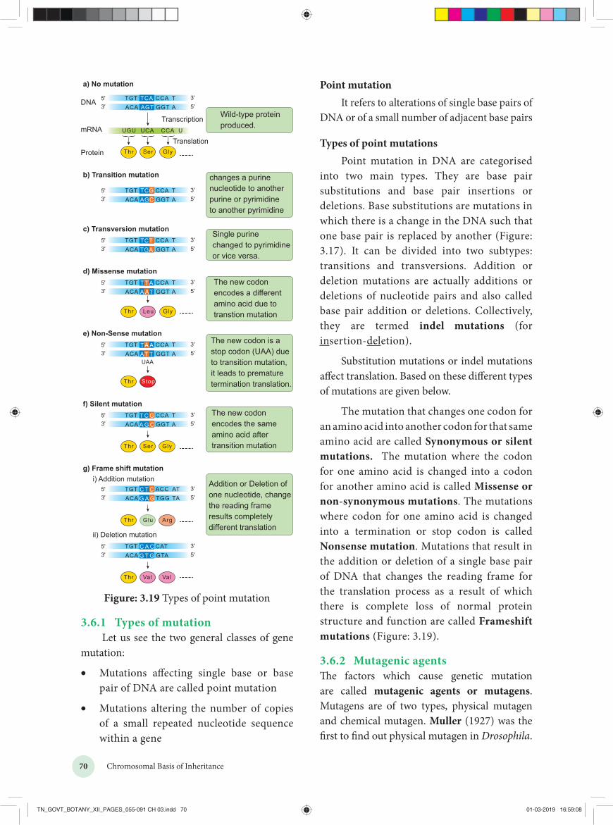
Point mutationIt refers to alterations of single base pairs of
DNA or of a small number of adjacent base pairs
Types of point mutationsPoint mutation in DNA are categorised
into two main types. They are base pair substitutions and base pair insertions or deletions. Base substitutions are mutations in which there is a change in the DNA such that one base pair is replaced by another (Figure: 3.17). It can be divided into two subtypes: transitions and transversions. Addition or deletion mutations are actually additions or deletions of nucleotide pairs and also called base pair addition or deletions. Collectively, they are termed indel mutations (for insertion-deletion).
Substitution mutations or indel mutations affect translation. Based on these different types of mutations are given below.
The mutation that changes one codon for an amino acid into another codon for that same amino acid are called Synonymous or silent mutations. The mutation where the codon for one amino acid is changed into a codon for another amino acid is called Missense or non-synonymous mutations. The mutations where codon for one amino acid is changed into a termination or stop codon is called Nonsense mutation. Mutations that result in the addition or deletion of a single base pair of DNA that changes the reading frame for the translation process as a result of which there is complete loss of normal protein structure and function are called Frameshift mutations (Figure: 3.19).
3.6.2 Mutagenic agentsThe factors which cause genetic mutation are called mutagenic agents or mutagens. Mutagens are of two types, physical mutagen and chemical mutagen. Muller (1927) was the first to find out physical mutagen in Drosophila.
a) No mutation
b) Transition mutation
c) Transversion mutation
DNA
Protein
Wild-type proteinproduced.
changes a purine nucleotide to anotherpurine or pyrimidineto another pyrimidine
The new codonencodes a differentamino acid due totranstion mutation
Single purine changed to pyrimidineor vice versa.
TCAAGT
TGTACA
CCA TGGT A
mRNATranscription
TranslationUCA UCCAUGU
3'5' 3'
5'
TCGAGC
TGTACA
CCA TGGT A3'
5' 3'5'
TGTACA
CCAGGT3'
5' 3'5'
Thr Ser Gly
Thr Ser Gly
Thr Leu Gly
TCTTGA
d) Missense mutationTGTACA
CCAGGT3'
5' 3'5'
T TAA AT
The new codon is a stop codon (UAA) dueto transition mutation,it leads to premature termination translation.Thr Stop
e) Non-Sense mutationTGTACA
CCAGGT3'
5' 3'5'
TA AAT TUAA
The new codon encodes the same amino acid after transition mutation
f) Silent mutationTGTACA
CCAGGT3'
5' 3'5'
T C GA G C
Thr Glu Arg
Addition or Deletion ofone nucleotide, changethe reading frameresults completely different translation
g) Frame shift mutationi) Addition mutation
TGTACA
ACCTGG3'
5' 3'5'
C T CG A G
Thr Val Val
ii) Deletion mutationTGTACA
CATGTA3'
5' 3'5'
C A CG T G
TA
TA
TA
TA
ATTA
Figure: 3.19 Types of point mutation
3.6.1 Types of mutationLet us see the two general classes of gene
mutation:
• Mutations affecting single base or basepair of DNA are called point mutation
• Mutations altering the number of copiesof a small repeated nucleotide sequencewithin a gene
TN_GOVT_BOTANY_XII_PAGES_055-091 CH 03.indd 70 01-03-2019 16:59:08

71Chromosomal Basis of Inheritance
Physical mutagens:
Scientists are using temperature and radiations such as X rays, gamma rays, alfa rays, beta rays, neutron, cosmic rays, radioactive isotopes, ultraviolet rays as physical mutagen to produce mutation in various plants and animals.
Temperature: Increase in temperature increases the rate of mutation. While rise in temperature, breaks the hydrogen bonds between two DNA nucleotides which affects the process of replication and transcription.
Radiation: The electromagnetic spectrum contains shorter and longer wave length rays than the visible spectrum. These are classified into ionizing and non-ionizing radiation. Ionizing radiation are short wave length and carry enough higher energy to ionize electrons from atom. X rays, gamma rays, alfa rays, beta rays and cosmic rays which breaks the chromosomes (chromosomal mutation) and chromatids in irradiated cells. Non-ionizing radiation, UV rays have longer wavelengths and carry lower energy, so they have lower penetrating power than the ionizing radiations. It is used to treat unicellular microorganisms, spores, pollen grains which possess nuclei located near surface membrane.
Sharbati Sonora
Sharbati Sonora is a mutant variety of wheat, which is developed from Mexican variety (Sonora 64) by irradiating of gamma rays. It is the work of Dr. M.S.Swaminathan who is known as ‘Father of Indian green revolution’ and his team.
Castor Aruna
Castor Aruna is mutant variety of castor which is developed by treatment of seeds with thermal neutrons in order to induce very early maturity (120 days instead of 270 days as original variety).
Chemical mutagens:
Chemicals which induce mutation are
called chemical mutagens. Some chemical mutagens are mustard gas, nitrous acid, ethyl and methyl methane sulphonate (EMS and MMS), ethyl urethane, magnous salt, formaldehyde, eosin and enthrosine. Example: Nitrous oxide alters the nitrogen bases of DNA and disturb the replication and transcription that leads to the formation of incomplete and defective polypeptide during translation.
Comutagens
The compounds which are not having own mutagenic properties but can enhance the effects of known mutagens are called comutagens.
Example: Ascorbic acid increase the damage caused by hydrogen peroxide.
Caffeine increase the toxicity of methotrexate
Mustard gas (Dichloro ethyl sulphide) used as chemical weapon in world war I.
H J Muller (1928) first time used X rays to induce mutations in fruit fly.
L J Stadler reported induced mutations in plants by using X rays and gamma rays.
Chemical mutagenesis was first reported by C. Auerback (1944).
3.6.3 Chromosomal mutationsThe genome can also be modified on a larger scale by altering the chromosome structure or by changing the number of chromosomes in a cell. These large-scale variations are termed as chromosomal mutations or chromosomal aberrations. Gene mutations are changes that take place within a gene, whereas chromosomal mutations are changes to a chromosome region consisting of many genes. It can be detected by microscopic examination, genetic analysis, or both. In contrast, gene mutations are never detectable microscopically. Chromosomal mutations are divided into two groups: changes in chromosome number and changes in chromosome structure.
TN_GOVT_BOTANY_XII_PAGES_055-091 CH 03.indd 71 01-03-2019 16:59:08

72 Chromosomal Basis of Inheritance
I. Changes in chromosome number
Each cell of living organisms possesses fixed number of chromosomes. It varies in different species. Even though some species of plants and animals are having identical number of chromosomes, they will not be similar in character. Hence the number of chromosomes will not differentiate the character of species from one another but the nature of hereditary material (gene) in chromosome that determines the character of species.
Sometimes the chromosome number of somatic cells are changed due to addition or elimination of individual chromosome or basic set of chromosomes. This condition in known as numerical chromosomal aberration or ploidy. There are two types of ploidy.
(i). Ploidy involving individual chromosomes within a diploid set (Aneuploidy)
(ii). Ploidy involving entire sets of chromosomes (Euploidy) (Figure 3.20)
(i) Aneuploidy
It is a condition in which diploid number is altered either by addition or deletion of one or more chromosomes. Organisms
showing aneuploidy are known as aneuploids or heteroploids. They are of two types, Hyperploidy and Hypoploidy (Figure 3.21).
1. Hyperploidy
Addition of one or more chromosomes to diploid sets are called hyperploidy. Diploid set of chromosomes represented as Disomy. Hyperploidy can be divided into three types. They are as follows,
(a) TrisomyAddition of single chromosome to diploid setis called Simple trisomy(2n+1). Trisomics werefirst reported by Blackeslee (1910) in Daturastramonium (Jimson weed). But later it wasreported in Nicotiana, Pisum and Oenothera.Sometimes addition of two individualchromosome from different chromosomalpairs to normal diploid sets are called Doubletrisomy (2n+1+1).
(b) TetrasomyAddition of a pair or two individual pairs ofchromosomes to diploid set is called tetrasomy(2n+2) and Double tetrasomy (2n+2+2)respectively. All possible tetrasomics areavailable in Wheat.
Ploidy
Aneuploidy Euploidy
Hyperploidy
Monosomy (2n-1)
Double Monosomy
(2n-1-1)
Double Nullisomy (2n-2-2)
Nullisomy (2n-2)
Hypoploidy
Haploidy (n) Diploidy (2n)
AllopolyploidyAutopolyploidy
AutotetraploidAutotriploid
Polyploidy (2n+n+n...)
Monoploidy (x)
Trisomy (2n+1)
Double Trisomy
(2n+1+1)
Tetrasomy (2n+2)
Double Tetrasomy (2n+2+2)
Pentasomy (2n+3)
Figure 3.20 Types of Ploidy
TN_GOVT_BOTANY_XII_PAGES_055-091 CH 03.indd 72 01-03-2019 16:59:08

73Chromosomal Basis of Inheritance
(c) Pentasomy
Addition of three individual chromosome from different chromosomal pairs to normal diploid set are called pentasomy (2n+3).
2. Hypoploidy
Loss of one or more chromosome from the diploid set in the cell is called hypoploidy. It can be divided into two types. They are
(a) Monosomy
Loss of a single chromosome from the diploid set are called monosomy(2n-1). However loss of two individual or three individual chromosomes are called double monosomy (2n-1-1) and triple monosomy (2n-1-1-1) respectively. Double monosomics are observed in maize.
(b) Nullisomy
Loss of a pair of homologous chromosomes or two pairs of homologous chromosomes from the diploid set are called Nullisomy (2n-2) and double Nullisomy (2n-2-2) respectively. Selfing of monosomic plants produce nullisomics. They are usually lethal.
Disomy(normal)
(2n)
Trisomy(2n + 1)
Monosomy(2n – 1)
DoubleMonosomy(2n – 1– 1)
DoubleTrisomy
(2n + 1 + 1)
Tetrasomy(2n + 2)
Pentasomy(2n + 3)
Nullisomy(2n – 2)
Figure 3.21 Types of aneuploidy
(ii) Euploidy
Euploidy is a condition where the organisms possess one or more basic sets of chromosomes. Euploidy is classified as monoploidy, diploidy and polyploidy. The condition where an organism or somatic cell has two sets of chromosomes are
called diploid (2n). Half the number of somatic chromosomes is referred as gametic chromosome number called haploid(n). It should be noted that haploidy (n) is different from a monoploidy (x). For example, the common wheat plant is a polyploidy (hexaploidy) 2n=6x=72 chromosomes. Its haploid number (n) is 36, but its monoploidy (x) is 12. Therefore, the haploid and diploid condition came regularly one after another and the same number of chromosomes is maintained from generation to generation, but monoploidy condition occurs when an organism is under polyploidy condition. In a true diploid both the monoploid and haploid chromosome number are same. Thus a monoploid can be a haploid but all haploids cannot be a monoploid.
Polyploidy
Polyploidy is the condition where an organism possesses more than two basic sets of chromosomes. When there are three, four, five or six basic sets of chromosomes, they are called triploidy (3x) tetraploidy (4x), pentaploidy (5x) and hexaploidy (6x) respectively. Generally, polyploidy is very common in plants but rarer in animals. An increase in the number of chromosome sets has been an important factor in the origin of new plant species. But higher ploidy level leads to death. Polyploidy is of two types. They are autopolyploidy and allopolyploidy
1. AutopolyploidyThe organism which possesses more than twohaploid sets of chromosomes derived fromwithin the same species is called autopolyploid.They are divided into two types. Autotriploidsand autotetraploids.
Autotriploids have three set of its own genomes. They can be produced artificially by crossing between autotetraploid and diploid species. They are highly sterile due to defective gamete formation. Example: The cultivated banana are usually triploids and are seedless having larger fruits than diploids. Triploid sugar beets have higher sugar content than diploids
TN_GOVT_BOTANY_XII_PAGES_055-091 CH 03.indd 73 01-03-2019 16:59:08
74 Chromosomal Basis of Inheritance
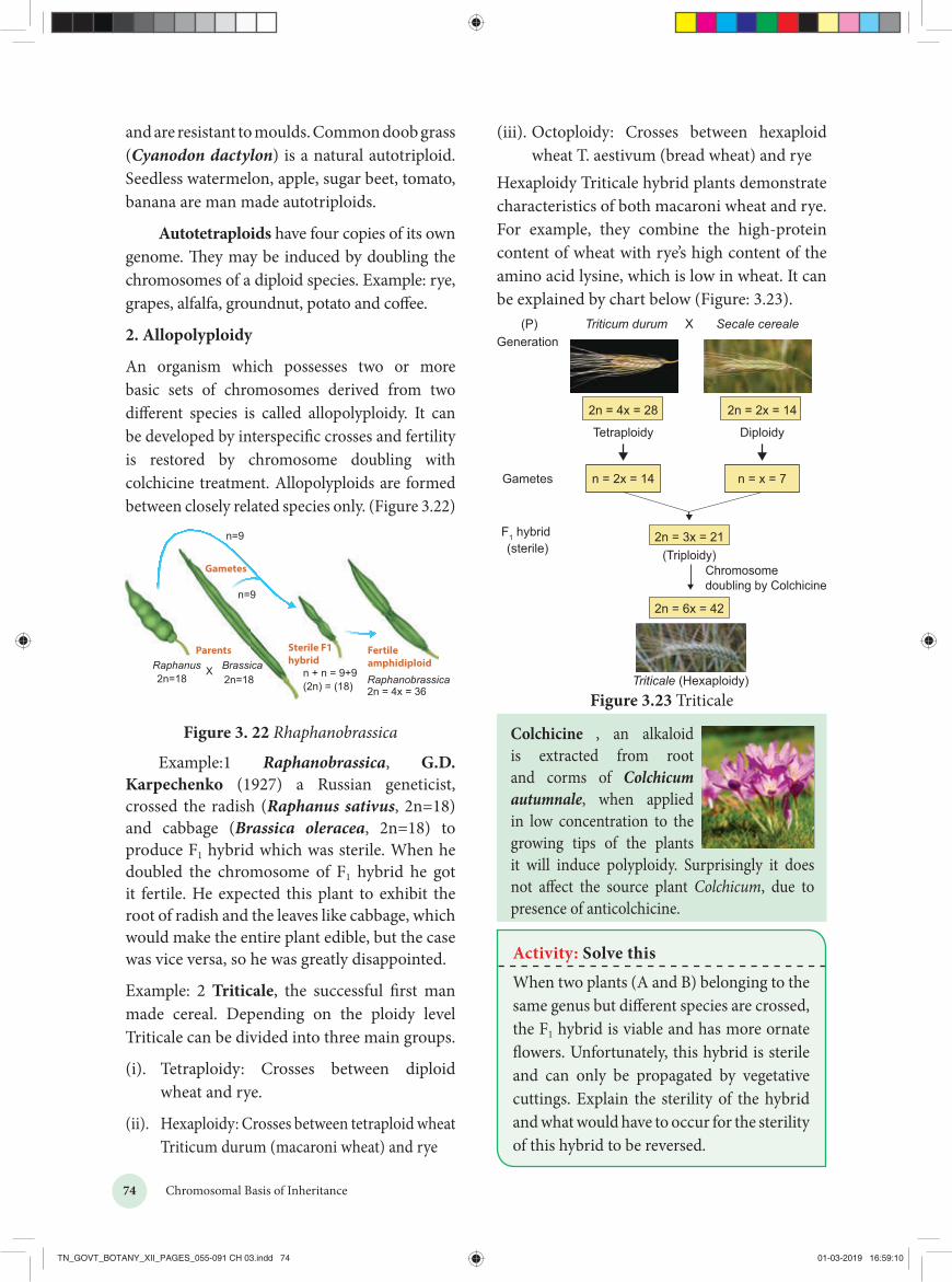
(iii). Octoploidy: Crosses between hexaploid wheat T. aestivum (bread wheat) and rye
Hexaploidy Triticale hybrid plants demonstrate characteristics of both macaroni wheat and rye. For example, they combine the high-protein content of wheat with rye’s high content of the amino acid lysine, which is low in wheat. It can be explained by chart below (Figure: 3.23).
Triticum durum Secale cereale(P)Generation
X
Gametes
F1 hybrid (sterile)
2n = 4x = 28
Tetraploidy
n = 2x = 14
2n = 2x = 14
Diploidy
n = x = 7
2n = 3x = 21
2n = 6x = 42
Chromosomedoubling by Colchicine
Triticale (Hexaploidy)
(Triploidy)
Figure 3.23 Triticale
Colchicine , an alkaloid is extracted from root and corms of Colchicum autumnale, when applied in low concentration to the growing tips of the plants it will induce polyploidy. Surprisingly it does not affect the source plant Colchicum, due to presence of anticolchicine.
and are resistant to moulds. Common doob grass (Cyanodon dactylon) is a natural autotriploid. Seedless watermelon, apple, sugar beet, tomato, banana are man made autotriploids.
Autotetraploids have four copies of its own genome. They may be induced by doubling the chromosomes of a diploid species. Example: rye, grapes, alfalfa, groundnut, potato and coffee.
2. Allopolyploidy
An organism which possesses two or more basic sets of chromosomes derived from two different species is called allopolyploidy. It can be developed by interspecific crosses and fertility is restored by chromosome doubling with colchicine treatment. Allopolyploids are formed between closely related species only. (Figure 3.22)
n=9
ParentsRaphanus Brassica
Sterile F1hybrid
n=9
2n=18 n + n = 9+9(2n) = (18)2n=18
XRaphanobrassica
Fertileamphidiploid
2n = 4x = 36
Gametes
Figure 3. 22 Rhaphanobrassica
Example:1 Raphanobrassica, G.D. Karpechenko (1927) a Russian geneticist, crossed the radish (Raphanus sativus, 2n=18) and cabbage (Brassica oleracea, 2n=18) to produce F1 hybrid which was sterile. When he doubled the chromosome of F1 hybrid he got it fertile. He expected this plant to exhibit the root of radish and the leaves like cabbage, which would make the entire plant edible, but the case was vice versa, so he was greatly disappointed.
Example: 2 Triticale, the successful first man made cereal. Depending on the ploidy level Triticale can be divided into three main groups.
(i). Tetraploidy: Crosses between diploid wheat and rye.
(ii). Hexaploidy: Crosses between tetraploid wheat Triticum durum (macaroni wheat) and rye
Activity: Solve this
When two plants (A and B) belonging to the same genus but different species are crossed, the F1 hybrid is viable and has more ornate flowers. Unfortunately, this hybrid is sterile and can only be propagated by vegetative cuttings. Explain the sterility of the hybrid and what would have to occur for the sterility of this hybrid to be reversed.
TN_GOVT_BOTANY_XII_PAGES_055-091 CH 03.indd 74 01-03-2019 16:59:10

75Chromosomal Basis of Inheritance
Significance of Ploidy• Many polyploids are more vigorous and
more adaptable than diploids.• Many ornamental plants are autotetraploids
and have larger flower and longer floweringduration than diploids.
• Autopolyploids usually have increase in freshweight due to more water content.
• Aneuploids are useful to determine thephenotypic effects of loss or gain of differentchromosomes.
• Many angiosperms are allopolyploids andthey play a role in an evolution of plants.
II Structural changes in chromosome (Structural chromosomal aberration)
Structural variations caused by addition or deletion of a part of chromosome leading to rearrangement of genes is called structural chromosomal aberration. It occurs due to ionizing radiation or chemical compounds. On the basis of breaks and reunion in chromosomes, there are four types of aberrations. They are classified under two groups.
A. Changes in the number of the gene loci1. Deletion or Deficiency2. Duplication or Repeat
B. Changes in the arrangement of gene loci3. Inversion4. Translocation
1. Deletion or DeficiencyLoss of a portion of chromosome is calleddeletion. On the basis of location of breakageon chromosome, it is divided into terminaldeletion and intercalary deletion. It occursdue to chemicals, drugs and radiations. It isobserved in Drosophila and Maize. (Figure 3.24)There are two types of deletion:i. Terminal deletion: Single break in any one
end of the chromosome.ii. Intercalary deletion or interstitial deletion:
It is caused by two breaks and reunion ofterminal parts leaving the middle.
Both deletions are observable during meiotic pachytene stage and polytene chromosome. The unpaired loop formed in the normal chromosomal part at the time of chromosomal pairing. Such loops are called as deficiency loops and it can be seen in meiotic prophase. Larger deletions may lead to lethal effect.
2. Duplication or RepeatThe process of arrangement of the same orderof genes repeated more than once in the same
A
B
C
D
E
FG
H
A
B
B
C
C
D
E
F
G
H
A
B
B
C
C
D
E
F
G
H
I
I I
Normal chromosome
Duplications
Tandem Reversetandem
Displacedtandem
D
E
F
G
HI
A
C
B
C
B
Figure 3.25 Duplication
A
B
C
D
E
FG
IH
Deletion ofsegment
D
Deletion ofsegment
A
Terminaldeletion
Intercalarydeletion
NormalChromosome
A
B
C
E
FG
IH
B
C
D
E
FG
IH
Figure 3.24 Deletion
TN_GOVT_BOTANY_XII_PAGES_055-091 CH 03.indd 75 01-03-2019 16:59:11

76 Chromosomal Basis of Inheritance
ii. Pericentric inversion: An inversion thatincludes the centromere.
Inversions lead to evolution of a new species.
4. Translocation
The transfer of a segment of chromosome to a non-homologous chromosome is called translocation. Translocation should not be confused with crossing over, in which an exchange of genetic material between homologous chromosome takes place. Translocation occurs as a result of interchange of chromosome segments in non-homologous chromosomes. There are three types
i. Simple translocationii. Shift translocationiii. Reciprocal translocation
i. Simple translocation
A single break is made in only one chromosome. The broken segment gets attached to one end of a non-homologous chromosome. It occurs very rarly in nature.
ii. Shift translocation
Broken segment of one chromosome gets inserted interstitially in a non-homologous chromosome.
iii. Reciprocal translocations
It involves mutual exchange of chromosomal segments between two non-homologous
chromosome is known as duplication. Due to duplication some genes are present in more than two copies. It was first reported in Drosophila by Bridges (1919) and other examples are Maize and Pea. It is three types.
i. Tandem duplicationThe duplicated segment is located immediatelyafter the normal segment of the chromosome inthe same order.
ii. Reverse tandem duplicationThe duplicated segment is located immediatelyafter the normal segment but the gene sequenceorder will be reversed.
iii. Displaced duplicationThe duplicated segment is located in the samechromosome, but away from the normalsegment. (Figure 3.25)
Duplications play a major role in evolution.
3. InversionA rearrangement of order of genes in achromosome by reversed by an angle 1800. Thisinvolve two chromosomal breaks and reunion.During this process there is neither gain nor lossbut the gene sequences is rearranged. Inversionwas first reported in Drosophila by Sturtevant(1926). There are two types of inversion,paracentric and pericentric (Figure 3.26).
i. Paracentric inversion: An inversion whichtakes place apart from the centromere
a) Paracentric inversion(does not include centromere)
b) Pericentric inversion(includes centromere)
A A
B
B
C C
D
D
D
B
C
D
E E
F F
G G
H
A
B
C
D
EE
F
F
D
E
F
G
H
A
B
C
G
HHI I II
Figure 3.26 Inversion Figure: 3.27 Translocation
TN_GOVT_BOTANY_XII_PAGES_055-091 CH 03.indd 76 01-03-2019 16:59:12

77Chromosomal Basis of Inheritance
chromosomes. It is also called illegitimate crossing over. It is further divided into two types (Figure 3.27).
a. Homozygous translocation: Both thechromosomes of two pairs are involved intranslocation. Two homologous of eachtranslocated chromosomes are identical.
b. Heterozygous translocation: Only oneof the chromosome from each pair of twohomologous are involved in translocation,while the remaining chromosome is normal.
Translocations play a major role in the formation of species.
3.7. DNA Metabolism in PlantsAs the repository of genetic information, DNA occupies a unique and central place among biological macromolecules. The structure of DNA is a marvelous device for the storage of genetic information. The term “DNA Metabolism” can be used to describe process by which copies of DNA molecules are made (replication) along with repair and recombination.
In this chapter we briefly discuss about the DNA metabolism in plants
DNA Replication: In the double helix the two parental strands of DNA separate and each parental strand synthesizes a new complementary strand. DNA replication is semiconservative, i.e each new DNA molecule conserves one original strand.
DNA Repair: How is genomic stability maintained in all living organisms? How do organisms on earth survive? What is essential for their survival?
DNA is unique because it is the only macromolecule where the repair system exists, which recognises and removes mutations. DNA is subjected to various types of damaging reactions such as spontaneous or environmental agents or natural endogenous threats. Such
damages are corrected by repair enzymes and proteins, immediately after the damage has taken place. DNA repair system plays a major role in maintaining the genomic / genetic integrity of the organism. DNA repair systems protect the integrity of genomes from genotoxic stresses.
Plants are sessile. How do they protect themselves from the exposure of sunlight throughout the day?
Plants have effective DNA repair mechanism to prevent UV damage from sunlight. They produce an enzyme called photolyase, which can repair the thymine dimers and restore the structure of DNA.
Recombination: In cells the genetic information within and among DNA molecule are re-arranged by a process called genetic recombination. Recombination is the result of crossing over between the pairs of homologous chromosomes during meiosis. In earlier classes you have learnt chromosomal recombination. In molecular level it involves breakage and reunion of polynucleotides.
3.7.1 Eukaryotic DNA replicationReplication starts at a specific site on a DNA sequence known as the Origin of replication. There are more than one origin of replication in eukaryotes. Saccharomyces cerevisiae (yeast) has approximately 400 origins of replication. DNA replication in eukaryotes starts with the assembly of a prereplication complex (preRC) consisting of 14 different proteins. Part of a preRC is a group of 6 proteins called the origin recognition complex (ORC) which acts as initiator in eukaryotic DNA replication. The origin of replication in yeast is called as ARS sites (Autonomously Replicating Sequences). In yeast, ORC was identified as a protein complex which binds directly to ARS elements.
TN_GOVT_BOTANY_XII_PAGES_055-091 CH 03.indd 77 01-03-2019 16:59:12

78 Chromosomal Basis of Inheritance
DNA Polymerase β does not play any role in the replication of normal DNA. Function - Removing incorrect bases from damagedDNA. It is involved in Base excision repair.
DNA Synthesis takes place in 5’ 3’ direction and it is semidiscontinuous. When DNA is synthesized in 5’ 3’ direction, only in the free 3’ end (OH end) DNA is elongated. In 1960s Reiji Okazaki and his colleagues found out that one of the new DNA strands is synthesized in short pieces called Okazaki fragments. In discontinuous strand where the Okazaki fragments are united by ligase is called Lagging strand where the replication direction is 5’ 3’ which is opposite to the direction of fork movement. . The continuous strand is called Leading strand where the replication direction is 5’ 3’ which is same to the direction to that of the replication fork movement. DNA ligase joins any nicks in the DNA by forming a phosphodiester bond between 3’ hydroxyl and 5’ phosphate group.
Arabidopsis telomere sequence - TTTAGGG
Plants Lacks Telomere ClockPlant meristematic cells produces telomerase so the meristematic cells has an unlimited ability to divide. You have already studied about the telomeres in Chapter 6 and 8 of Class XI. In plants telomeres do not shrink as in somatic cells of vertebrates. Telomerase levels are higher in root tips and seedlings (renewable tissue) which has a higher amount of meristematic cells than proliferative structures like leaves.
What is the special mechanism which replicates chromosomal ends?After the replication of the chromosomes, the enzyme telomerase adds several more repeats of DNA sequences to the telomeres. Telomerase
Figure: 3.28 Eukaryotic replication fork
Replication fork is the site (point of unwinding) of separation of parental DNA strands where new daughter strands are formed. Multiple replication forks are found in eukaryotes. The enzyme helicases are involved in unwinding of DNA by breaking hydrogen bonds holding the two strands of DNA and replication protein A (RPA) prevents the separated polynucleotide strand from getting reattached.
Topoisomerase is an enzyme which breaks DNAs covalent bonds and removes positive supercoiling ahead of replication fork. It eliminates the torsional stress caused by unwinding of DNA double helix.
DNA replication is initiated by an enzyme DNA polymerase α / primase which synthesizes short stretch of RNA primers on both leading strand (continuous DNA strand) and lagging strands (discontinuous DNA strand). Primers are needed because DNA polymerase requires a free 3’ OH to initiate synthesis. DNA polymerase covalently connects the nucleotides at the growing end of the new DNA strand.DNA Pol α (alpha), DNA Pol δ (delta) and DNA Pol ε (Epsilon) are the 3 enzymes involved in nuclear DNA replication.DNA Pol α – Synthesizes short primers of RNADNA Pol δ – Main Replicating enzyme of cell nucleusDNA Pol ε – Extend the DNA Strands in replication fork
Leading strand3"
5"
5"
3"
5"
3"Lagging strand
TN_GOVT_BOTANY_XII_PAGES_055-091 CH 03.indd 78 01-03-2019 16:59:12

79Chromosomal Basis of Inheritance
1. In the chromosome of first generation theradioactivity was found to be distributed toboth the chromatids because in the originalstrand of DNA double helix was labelledwith radioactivitiy and the new strand wasunlabelled.
2. In the chromosome of the second generation only one of the two chromatids in eachchromosome was radioactive (labelled).
The results proved the semiconservative method of DNA replication.
3.8 Protein synthesis in plants The process of protein synthesis consists of two major steps, they are Transcription and Translation.
Plant cell
Nucleus
DNATranscription
Translation
Processing3’
5’5’
5’
Polypeptide
Ribosome
Coding sequence
mRNA
3’
3’
Cell wall
Figure: 3.30 Protein synthesis in plants
3.8.1 TranscriptionTranscription is the process in which one strand of DNA acts as a template to generate mRNA with the bases complementary to the template strand. It is catalyzed by the enzymes called RNA polymerases.
Transcription and processing of RNA takes place in the nucleus, whereas the translation occurs in the ribosomes found in cytoplasm. In Eukaryotes, mRNA molecules are monocistronic with only one protein being derived from each mRNA.
use short RNA molecules as a template and add repeat sequences on to telomeres (DNA nucleotide polymerisation).
The Energetics of DNA Replication - Deoxyribonucleotides such as deoxyadenosine triphosphate dATP, dGTP, dCTP and dTTP provide energy for the synthesis of DNA. Purpose of Deoxyribonucelotides (1) acts as a substrate (2) provide energy for polymerisation.
3.7.2 Experimental evidence of DNA replication: Taylors ExperimentJ. Herbert Taylor, Philip Woods and WalterHughes demonstrated the semiconservativereplication of DNA in the root cells of Viciafaba. They labelled DNA with 3H Thymidine,a radioactive precursor of DNA and performedautoradiography. They grew root tips in amedium in the presence of radioactive labelledthymidine, so that the radioactivity wasincorporated into the DNA of these cells. Theoutline of this labelled chromosomes appears inthe form of scattered black dots of silver grainson a photographic film.
Figure: 3.29 Taylors Experiment on Vicia faba
The root tips with labelled chromosomes were placed in an unlabelled medium containing colchicine to arrest the culture at the metaphase and examine the chromosome by autoradiography. The observations were,
TN_GOVT_BOTANY_XII_PAGES_055-091 CH 03.indd 79 01-03-2019 16:59:13

80 Chromosomal Basis of Inheritance
are the proteins which recognise base sequences of DNA and controls transcription. Some transcription factors bind directly to the promoter.
Some transcription factors recognize the regulatory elements and bind to them to increase the rate of transcription, others inhibits transcription.
To start the process of transcription the Regulatory elements help the RNA polymerase to recognize core promoter. The two categories of regulatory elements are
1. Enhancer sequences – they are DNAsequences (activating sequences) whichhelp to influence transcription.
2. Silencer sequence – DNA sequencesthat inhibit transcription or decreasetranscription.
Consensus sequence – An ideal sequence in which each position represents the base which is found most often.
In addition to General transcription factors (GTF) and RNA Pol II, a mediator is required for transcription. The interactions between RNA polymerase II and regulatory TF that bind to enhancers or silencers are mediated by a mediator.
RNA Polymerases cannot bind directly to the DNA, first it binds to the transcription factor which recognizes the promoter sequences
which helps to find the protein coding regions of DNA.
RNA Polymerase with the promoter sequence will transcribe the gene. Transcription factor plays an important role in guiding RNA Polymerases to the promoter sequence. RNA Polymerases bind RNA nucleotides together
The transcription begins with unwinding of DNA double helix and the hydrogen bonds are broken at the site of the gene being transcribed.
Template Strand / Non-Coding Strand / Antisense Strand
The strand of DNA which is oriented in 3’ 5’ direction that serves as a template for the synthesis of mRNA is called template strand.
Coding strand / Non-template strand / Sense Strand
The other strand of DNA which is not transcribed is called the Coding Strand.
A specific sequence of DNA nucleotides called the Promoter is necessary for transcription to takes place. It consists of TATA box and transcription start site where transcription begins.
Termination sequences are the DNA sequences which tells when the RNA polymerase should stop producing RNA molecule.
Eukaryotic structural gene has 3 features in promoter1. Regulatory elements2. TATA box3. A transcriptional start siteThe transcription start site contains about 25 bp(basepairs) upstream, the sequence is TATAATknown as TATA or Hogness box which is presentin core promoter. General transcriptional factors
Figure: 3.31 Transcription
TN_GOVT_BOTANY_XII_PAGES_055-091 CH 03.indd 80 01-03-2019 16:59:13

81Chromosomal Basis of Inheritance
3. It regulates the mRNA export from thenucleus into the cytoplasm.
4. It helps in binding of mRNA to theribosome.
Tailing / Polyadenylation
The 3’ end of hnRNA is cleaved by an endonuclease and a string of adenine nucleotides is added to the 3’ end of hnRNA (pre mRNA) is known as Poly (A) tail - Polyadenylation. This process is called tailing or polyadenylation.
Purpose of Tailing
1. Translation of RNA transcript isfacilitated.
2. Helps in the synthesis of Polypeptides.3. It enhances the mRNA stability in the
cytoplasm.The protein coding regions are not continuous in eukaryotes. Split genes were independently discovered by Richard J Roberts and Phillip A. Sharp in 1977 and was awarded NobelPrize in 1993. Exons are the coding sequencesor expressed sequences contain biologicalinformations in the matured processed mRNA. Introns are intervening sequences, whichare non-coding sequences (non-amino acid- coding sequences) that should be removedfrom a gene before the mRNA product ismade. Introns do not code for any enzymeor structural protein or polypeptides. Theseexons and introns are known as Split Genes.
3.8.2 RNA Splicing in plantsRNA Splicing is a process which involves the cutting or removing out of introns and knitting of exons. This process takes place in spherical particles which is a multiprotein complex called SPLICISOMES. It is approximately 40 – 60 nm in diameter. The spliceosomes have many small nuclear ribonucleic acids (snRNAs) and small nuclear ribonuclear protein particles (snRNPs) which identify and helps in the removal of introns.
forming a growing strand in the 5’ 3’ direction. Transcription occurs in 5’ 3’ direction, RNA Polymerase catalyses the addition of nucleotides at the 3’ end of the growing chain of RNA.
In Eukaryotes, 3 different RNA Polymerases called RNA Polymerases I, II and III are found.
Enzyme SynthesisRNA Polymerase I
Large Ribosome RNAs except 5S rRNA
RNA Polymerase II
Precursors of mRNAs (hnRNAs)
RNA Polymerase III
tRNAs, 5S ribosomal RNA, snRNAs (small nuclear RNA)
The processing of pre-mRNA to mature mRNA / Molecular mechanism of RNA modification
In eukaryotes three major types of RNA, mRNA, tRNA and rRNA are produced from a precursor RNA molecule termed as the primary transcript or preRNA. The RNA polymerase II transcribes the precursor of mRNA, which are also called the heterogenous nuclear RNA or hnRNA which are processed in the nucleus before they are transported into the cytoplasm.
Capping
Modification at the 5’ end of the primary RNA transcript (hn RNA) with methylguanosine triphosphate is called capping.
Internal methylationApart from capping, the internal nucleotides in mRNA are also methylated.
Methylated sites are present in translated, untranslated regions, introns and exons.
Purpose of Capping1. Protects RNA from degradation.2. Capping plays an important role in
removal of first intron in pre mRNA.
TN_GOVT_BOTANY_XII_PAGES_055-091 CH 03.indd 81 01-03-2019 16:59:14

82 Chromosomal Basis of Inheritance
Process of translationThe following are major steps in translation process
1. InitiationThe translation begins with the AUG codon(start codon) of mRNA. Translation occurson the surface of the macromolecular arenacalled the ribosome. It is a nonmembranousorganelle. During the process of translationthe two subunits of ribosomes unite (combine)together and hold mRNA between them. Theprotein synthesis begins with the reading ofcodons of mRNA. The tRNA brings amino acidto the ribosome, a molecular machine whichunites amino acids into a chain according to theinformation given by mRNA. rRNA plays thestructural and catalytic role during translation.
A ribosome has one binding site for mRNA and two for tRNA. The two binding sites of tRNA are
i. P-Site – The peptidyl – tRNA binding siteis one of the tRNA binding site. At this sitetRNA is held and linked to the growing endof the polypeptide chain.
ii. A-Site – The Aminoacyl – tRNA bindingsite. This is another tRNA binding sitewhich holds the incoming amino acidscalled aminoacyl tRNA. The anticodons oftRNA pair with the codons of the mRNA inthese sites.
2. Elongation of polypeptide chainThe P and A sites are nearby, so that two tRNAform base pairs with adjacent codon. Thepolypeptide chain is formed by the pairingof codons and anticodons according to thenucleotide sequence of the mRNA.
Translators of the genetic code - tRNAThe tRNA translates the genetic code from the nucleic acid sequence to the amino acid sequence i.e from gene – Polypeptide. When an amino acid is attached to tRNA it is called aminoacylated or charged. This is an energy requiring process which
Figure: 3.32 RNA Splicing in plants
A spliceosome removes the introns with an enzyme ribozyme. Now the mature mRNA comes away from the spliceosomes through the nuclear pore and is transported out from the nucleus into cytoplasm, and gets attached to ribosome to carry out translation. The RNA and many proteins are transported through a nuclear pore complex by an energy dependent process.
3.8.3 TranslationThe genetic information in the DNA code is copied onto mRNA bound in ribosomes for making polypeptides. The mRNA nucleotide sequence is decoded into amino acid sequence of the protein which is catalyzed by the ribosome. This process is called translation.
Terminology in Protein synthesis Codon – DNA codes are referred to as triplet codes and those in mRNA is called as Codons. Each triplet specifies a particular amino acid. Codons present in mRNA are read in 5’ 3’ direction. There are 64 codons of which 61 codons codes for amino acids.
Start codon – AUG specified methionine
Stop or Termination codon – UAA – Ochre UAG – amber and UGA – Opal.
Anticodons – The triplet of bases in a tRNA molecule is known as anticodon. In tRNA sequence of three bases which is complementary to codons of mRNA are called anticodon.The codons of mRNA are recognized by the anticodons of tRNA which are oriented in 3’ 5’ direction.
TN_GOVT_BOTANY_XII_PAGES_055-091 CH 03.indd 82 01-03-2019 16:59:14

83Chromosomal Basis of Inheritance
uses the ATP for its energy requirement. Protein synthesis takes place as the next aminoacyl tRNA binds to the A-Site.
The translation begins with the AUG codon (start codon) of mRNA. The tRNA which carries first amino acid methionine attach itself to P-site of ribosome. The ribosome adds new amino acids to the growing polypeptides. The second tRNA molecules has anticodons which carries amino acid alanine pairs with the mRNA codon in the A-site of the ribosome. The aminoacids methionine and alanine are close enough so that a peptide bond is formed between them.
The bond between the first tRNA and methionine now breaks. The first tRNA leaves the ribosome and the P-site is vacant. The ribosome now moves one codon along the mRNA strand. The second t-RNA molecule now occupies the P-site. The third t-RNA comes and fills the A site (serine).Now a peptide bond is formed betweenalanine and serine. The mRNA then movesthrough the ribosome by three bases. Thisexpels deacylated / uncharged tRNA fromP-site and moves peptidyl tRNA into theP-site and empties the A-site.This movementof tRNA from A-site to P-site is said to betranslocation. The translocation requires thehydrolysis of GTP.
The ribosome (ribozyme - peptidyl transferase) catalyses the formation of peptide bond by adding amino acid to the growing polypeptide chain.
The ribosome moves from codon to codon along the mRNA in the 5’ to 3’ direction. Amino acids are added one by one translated into polypeptide as dictated by the mRNA. Translation is an energy intensive process. A cluster of ribosomes are linked together by a molecule of mRNA and forming the site of protein synthesis is called as polysomes or polyribosomes.
Methionine
tRNA
mRNA
Anticodon5'
3'
CodontRNA binds to matching codon
New peptide bond
mRNA shifts forward by a codon
Ribosome
Nextaminoacid
P A
5'
3'
P A
5'
3'
P A
5'
3'
P A
a
b
c
Polypeptide chain stillattached to tRNA
P site
Releasefactor
Stop codon at A site(UAG, UAA, or UGA)
mRNA
mRNA
Polypeptide
Free tRNA
Ribosomalsubunits
Releasefactor
3'
3'
5'
5'
d
e
f
Figure: 3.33 Translation
TN_GOVT_BOTANY_XII_PAGES_055-091 CH 03.indd 83 01-03-2019 16:59:14
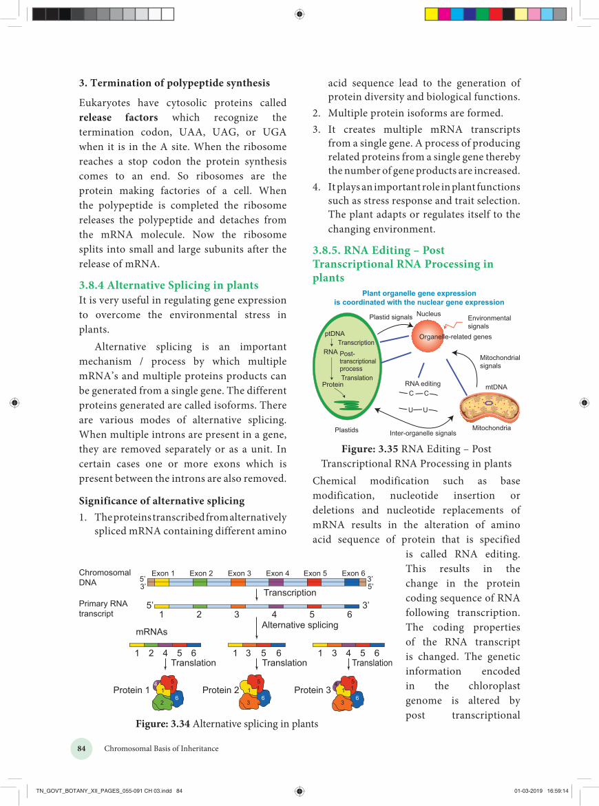
84 Chromosomal Basis of Inheritance
acid sequence lead to the generation of protein diversity and biological functions.
2. Multiple protein isoforms are formed.3. It creates multiple mRNA transcripts
from a single gene. A process of producingrelated proteins from a single gene therebythe number of gene products are increased.
4. It plays an important role in plant functions such as stress response and trait selection.The plant adapts or regulates itself to thechanging environment.
3.8.5. RNA Editing – Post Transcriptional RNA Processing in plants
Plant organelle gene expressionis coordinated with the nuclear gene expression
Plastid signals
Post-transcriptionalprocess
Transcription
Translation
ptDNA
RNA
Protein
Nucleus
Organelle-related genes
RNA editingC C
UU
Environmentalsignals
Mitochondrialsignals
mtDNA
MitochondriaInter-organelle signalsPlastids
Figure: 3.35 RNA Editing – Post Transcriptional RNA Processing in plants
Chemical modification such as base modification, nucleotide insertion or deletions and nucleotide replacements of mRNA results in the alteration of amino acid sequence of protein that is specified
is called RNA editing. This results in the change in the protein coding sequence of RNA following transcription. The coding properties of the RNA transcript is changed. The genetic information encoded in the chloroplast genome is altered by post transcriptional
3. Termination of polypeptide synthesis
Eukaryotes have cytosolic proteins called release factors which recognize the termination codon, UAA, UAG, or UGA when it is in the A site. When the ribosome reaches a stop codon the protein synthesis comes to an end. So ribosomes are the protein making factories of a cell. When the polypeptide is completed the ribosome releases the polypeptide and detaches from the mRNA molecule. Now the ribosome splits into small and large subunits after the release of mRNA.
3.8.4 Alternative Splicing in plantsIt is very useful in regulating gene expression to overcome the environmental stress in plants.
Alternative splicing is an important mechanism / process by which multiple mRNA’s and multiple proteins products can be generated from a single gene. The different proteins generated are called isoforms. There are various modes of alternative splicing. When multiple introns are present in a gene, they are removed separately or as a unit. In certain cases one or more exons which is present between the introns are also removed.
Significance of alternative splicing1. The proteins transcribed from alternatively
spliced mRNA containing different amino
Figure: 3.34 Alternative splicing in plants
TN_GOVT_BOTANY_XII_PAGES_055-091 CH 03.indd 84 01-03-2019 16:59:14

85Chromosomal Basis of Inheritance
Have you heard of Jumping Genes or Hopping Genes?
This is the nick name of transposable genetic elements. Transposons are the DNA sequences which can move from one position to another position in a genome. This was first reported in 1948 by American Geneticist Barbara McClintock as “mobile controlling element” in Maize. One of the most significant scientists of 20th century was Barbara McClintock because she gave a shift in gene organization. McClintock was awarded Nobel prize in 1983 for her work on transposons. Barbara McClintock when studying aleurone of single maize kernels, noted the unstable inheritance of the mosaic pattern of blue, brown and red spots due to the differential production of vacuolar anthocyanins.
In maize plant genome has AC / Ds transposon (AC = Activator, Ds = Dissociation). The activity of AC element is very distinct in maize plant. The transposition in somatic cells results in the changes in gene expression such as variegated pigmentation in maize kernels. Maize genome has transposable elements which regulated the different colour pattern of kernels.
McClintock’s findings concluded that Ds and AC genes were mobile controlling elements. We now call it as transposable elements, a term coined by maize geneticist, Alexander Brink. McClintock gave the first direct experimental evidence that genomes are not static but are highly plastic entities.
phenomenon which is site – specific (C U) in chloroplast of higher plants – RNA editing occurs in plant mitochondria and chloroplast.
In plant cells RNA editing by pyrimidine transitions occurs in mitochondria and plastids (chloroplast). There are two main types of RNA editing. (1) Substitution editing – Alteration of individual nucleotide bases.Mitochondria and chloroplast RNA in plants.(2) Insertion / Deletion editing – Nucleotidesare added or deleted from the total numberof bases.
Significance of RNA Editing
1. In higher plant chloroplast, it helps torestore the codons for conserved aminoacids which include initiation andtermination codon.
2. It regulates Organellar gene expression inplants.
3. RNA editing results in the restoration ofcodons for phylogenetically conservedamino acid residues.
3.8.6 Jumping Genes
Figure: 3.36 Barbara McClintock
Year Editing type Organelle in Plant cell Target Inference / Result
1989 C U Plant mitochondria mRNA For conserved amino acids, multiple changes in codon takes place
1990 U C Plant mitochondria mRNA First report on editing (U C)
1991 C U Plant chloroplast mRNA First report in chloroplast
Table 3.9: RNA Editing types
TN_GOVT_BOTANY_XII_PAGES_055-091 CH 03.indd 85 01-03-2019 16:59:14

86 Chromosomal Basis of Inheritance
the first flowering plant, where its entire genome is sequenced.
3. The two regions of the nucleolar organiserribosomal DNA which codes for theribosomal RNA are present at the extremity of chromosomes 2 and 4
4. It is Diploid plant having small genomewith 2n = 10 chromosomes. Severalgenerations can be produced in one year.So it facilitates rapid genetic analysis. Thegenome has low repetitive DNA, over 60%of the nuclear DNA have protein codingfunctions.
5. The plant is small, self fertilizes, annuallong-day plant with short-life cycle (only6 weeks), large numbers of seeds areproduced and they are easy to be grown inlaboratory. It is easy to induce mutations.It has many genomic resources and thetransformation can be done easily.
6. In 1982, Arabidopsis has successfullycompleted its life cycle in Microgravityi.e. space. This shows that Human SpaceMissions with plant companions may bepossible.
Significance of transposons
1. They contribute to many visible mutationsand mutation rate in an Organism.
2. In evolution, they contribute to geneticdiversity.
3. In genetic research transposons arevaluable tools which are used as mutagens,as cloning tags, vehicles for insertingforeign DNA into model organism.
Plant genome – The word genome is defined as the full complement of DNA (including all the genes and the intergenic regions) present in an organism It specifies the entire biological information of an organism. There are three distinct genomes in eukaryotic cells and they are (1) The nuclear genome (2) The mitochondrial genome and (3) Thechloroplast genome present only in plants.
1
2
34
5ribosomal DNA
Centromere
The five chromosomes of Arabidopsisthaliana (the region of chromosome 2 and 4
coding for ribosomal RNA)
Heterochromaticregion
Figure: 3.37 Arabidopsis thaliana
Arabidopsis thaliana – Thale cress, Mouse-ear cress
1. It is a model plant for the study ofgenetic and molecular aspects of plantdevelopment.
2. It belongs to mustard family and it is
SUMMARYChromosomal theory of inheritance states that Mendelian factors have specific locus on chromosomes and they carry information from one generation to the next generation. Genes located close together on the same chromosome and inherited together are called linked genes the phenomenon is called Linkage. Two types of linkage are complete linkage and incomplete linkage. The groups of linearly arranged linked genes are called Linkage groups. Crossing over is biological a process that produces new combination of genes by inter-changing the corresponding
TN_GOVT_BOTANY_XII_PAGES_055-091 CH 03.indd 86 01-03-2019 16:59:15

87Chromosomal Basis of Inheritance
segments between non-sister chromatids of homologous pair of chromosomes. In this segment of DNA are broken and recombined to produce new combinations of alleles a process is called Recombination. The diagrammatic representation of distances between the adjacent genes which is directly proportional to the frequency of recombination between them is called genetic mapping. When any of the three or more allelic forms of a gene occupy the same locus in a given pair of homologous chromosomes, they are said to be multiple alleles. Papaya sex determination is controlled by three alleles. They are m, M1 and M2 of a single gene. Mutational events that take place within individual genes are called gene mutations or point mutation, whereas the changes occur in structure and number of chromosomes is called chromosomal mutation. The agents which are responsible for mutation is called mutagens.
DNA metabolism includes replication, repair and recombination. Protein synthesis in eukaryotes is unique due to the capping, tailing and splicing. Transcription takes place in nucleus and translation in cytoplasm. AUG codes for methionine and it is monocistronic. Alternative splicing is a mechanism to overcome stress in plants. RNA editing takes place in chloroplast and mitochondria of plants which is of phylogenetic importance. Controlling elements gave a major shift in the gene organisation in plants to prove that DNA is not static but plastic entities. The first genome sequenced is Arabidopsis thaliana, which is a potential genetic tool to study the development and metabolism in plants.
Evaluation1. An allohexaploidy
contains
a) Six different genomesb) Six copies of three
different genomesc) Two copies of three different genomesd) Six copies of one genome
2. The A and B genes are 10 cM apart on achromosome. If an AB/ab heterozygoteis testcrossed to ab/ab, how many of eachprogeny class would you expect out of 100total progeny?
a) 25 AB, 25 ab, 25 Ab, 25 aBb) 10 AB, 10 abc) 45 AB, 45 abd) 45 AB, 45 ab, 5 Ab, 5aB
3. Match list I with list II
List I List IIA. A pair of
chromosomes extrawith diploid
i) monosomy
B. One chromosomeextra to the diploid
ii) tetrasomy
C. One chromosomeloses from diploid
iii) trisomy
D. Two individualchromosomes losefrom diploid
iv) doublemonosomy
a) A-i, B-iii, C-ii, D-iv b) A-ii, B-iii, C-iv, D-ic) A-ii, B-iii, C-i, D-iv d) A-iii, B-ii, C-i, D-iv
4. Which of the following sentences are correct?
1. The offspring exhibit only parentalcombinations due to incomplete linkage
2. The linked genes exhibit some crossingover in complete linkage
3. The separation of two linked genes arepossible in incomplete linkage
4. Crossing over is absent in complete linkage
TN_GOVT_BOTANY_XII_PAGES_055-091 CH 03.indd 87 01-03-2019 16:59:15

88 Chromosomal Basis of Inheritance
a) 1 and 2 b) 2 and 3c) 3 and 4 d) 1 and 4
5. Accurate mapping of genes can be done bythree point test cross because increases
a) Possibility of single cross overb) Possibility of double cross overc) Possibility of multiple cross overd) Possibility of recombination frequency
6. Due to incomplete linkage in maize, theratio of parental and recombinants are
a) 50:50 b) 7:1:1:7 c) 96.4: 3.6 d) 1:7:7:1
7. Genes G S L H are located on samechromosome. The recombinationpercentage is between L and G is 15%, Sand L is 50%, H and S are 20%. The correctorder of genes is
a) GHSL b) SHGL c) SGHL d) HSLG
8. The point mutation sequence for transition,transition, transversion and transversion inDNA are
a) A to T, T to A, C to G and G to Cb) A to G, C to T, C to G and T to Ac) C to G, A to G, T to A and G to Ad) G to C, A to T, T to A and C to G
9. If haploid number in a cell is 18. The doublemonosomic and trisomic number will be
a) 35 and 37 b) 34 and 35c) 37 and 35 d) 17 and 19
10. Changing the codon AGC to AGA representsa) missense mutation b) nonsense mutationc) frameshift mutation d) deletion mutation
11. Assertion (A): Gamma rays are generallyuse to induce mutation in wheat varieties.
Reason (R): Because they carry lowerenergy to non-ionize electrons from atom
a) A is correct. R is correct explanation of Ab) A is correct. R is not correct explanation of Ac) A is correct. R is wrong explanation of A
d) A and R is wrong
12. How many map units separate two alleles Aand B if the recombination frequency is 0.09?a) 900 cM b) 90 cM c) 9 cM d) 0.9 cM
13. Which one of the following pairs of codonsis correctly matched with their function orthe signal for the particular amino acid?a) UUA, UCA - Leucineb) GUU, GCU - Alaninec) UAG, UGA - Stopd) AUG, ACG – Start / Methionine
14. Removal of introns and joining of exons in adefined order during transcription is calleda) Splicing b) Loopingc) Inducing d) Slicing
15. If one strand of DNA has the nitrogenousbase sequence as ATCTS, what would be thecomplementary RNA strand sequence?a) ATCGU b) TTAGUc) UAGAC d) AACTG
16. Removal of RNA polymerase III nucleoplasm will affect the synthesis ofa) rRNA b) tRNAc) hnRNA d) mRNA
17. DNA dependent RNA polymerase catalyzestranscription on one strand of the DNAwhich is called thea) Alpha strand b) Anti strandc) Template strand d) Coding strand
18. Which of the following correctly representsthe flow of genetic information?a) DNA RNA Proteinb) RNA DNA Proteinc) RNA Protein DNAd) Protein RNA DNA
19. Initiation codon isa) UUU b) UGAc) AUG d) UAG
20. A eukaryotic gene contains two kinds of base
TN_GOVT_BOTANY_XII_PAGES_055-091 CH 03.indd 88 01-03-2019 16:59:15

89Chromosomal Basis of Inheritance
sequences which of these plays an important role in protein synthesis?a) Introns b) Exonsc) Both a and b d) None of the above
21. Codon – anticodon interactions occur bya) Covalent bondb) Electrostatic interactionsc) Hydrogen bondsd) Hydrophobic interaction
22. Which of the following RNA polymerases isresponsible for the transcription of proteincoding genes in eukaryotes?a) RNA Pol I b) RNA Poly IIc) RNA Pol III d) RNA Pol IV
23. How are RNA molecules transported out ofthe nucleusa) Passive diffusion through the membraneb) Through membrane pores in an energy
independent processc) Through membrane pores in an energy
dependent processd) Through a channel in the membrane that
leads to the endoplastic reticulation
24. During translation the codon in mRNA isactually “read” bya) The A site in the ribosomesb) The P site in the ribosomesc) The anticodon in at RNAd) The anticodon is an amino acid
25. A complex of ribosome attached to a singlestrand of RNA is known asa) Polysome b) Polymerc) Polypeptide d) Okazaki fragment
26. Which of the following is the start codona) AUG b) UGAd) UAA d) UAG
27. What is true about tRNA?
a) It binds with an amino acid at its 3’ end
b) It has 5 double stranded regions
c) It has a codon at one end which recognizes the anticodon of mRNA
d) It looks like clover leaf in the three Dstructure
28. Which one of the following hydrolysisinternal phosphodiester bonds in apolynucleotide chain?a) Lipase b) Exonucleasec) Endonuclease d) Protease
29. DNA element with ability to change position is calleda) Cistron b) Transposonc) Intron d) Recon
30. Spliceosomes are not found in cells ofa) Plants b) Fungic) Animals d) Bacteria
31. During DNA replication Okazaki fragmentsare used to elongatea) The leading strand towards replication
forkb) The lagging strand towards replication
forkc) The leading strand away from replication
forkd) The lagging strand away from replication
fork32. When two different genes came from same
parent they tend to remain together.
i) What is the name of this phenomenon?ii) Draw the cross with suitable example.iii) Write the observed phenotypic ratio.
33. If you cross dominant genotype PV/PVmale Drosophila with double recessivefemale and obtain F1 hybrid. Now you crossF1 male with double recessive female.
i) What type of linkage is seen?ii) Draw the cross with correct genotype.iii) What is the possible genotype in F2
generation?
TN_GOVT_BOTANY_XII_PAGES_055-091 CH 03.indd 89 01-03-2019 16:59:15

90 Chromosomal Basis of Inheritance
34.
S. no Gamete types Number of progenies
1. ABC 3492. Abc 1143. abC 1244. AbC 55. aBc 46. aBC 1167. ABc 1288. abc 360
i) What is the name of this test cross?ii) How will you construct gene mapping
from the above given data?iii) Find out the correct order of genes.
35. What is the difference between missenseand nonsense mutation?
36. A B B
C
C
D E F G H I
From the above figure identify the type ofmutation and explain it.
37. Write the salient features of Sutton andBoveri concept.
38. Explain the mechanism of crossing over.
39. Write the steps involved in molecularmechanism of DNA recombination withdiagram.
40. How is Nicotiana exhibit self-incompatibility. Explain its mechanism.
41. How sex is determined in monoeciousplants. write their genes involved in it.
42. What is gene mapping? Write its uses.
43. Draw the diagram of different types ofaneuploidy.
44. Mention the name of man-made cereal.How it is formed?
45. What is DNA repair?
46. What is replication fork?
47. Write about the energetics of DNAreplication.
48. What is TATA box?
49. What is alternative splicing?
50. What is coding strand?
51. What are the enzymes involved in DNAreplication in eukaryotes?
52. Differentiate coding and non coding strand.
53. What are splicesomes?
54. What is meant by capping and tailing?
55. What is RNA editing?
56. Explain the DNA replication in eukaryotes.
57. With reference to the given diagram correctly match the following pairs.
Column I Column II
A Transcribed region
B Regulation of initiation of transcription
C Protein-encoding sequence
D Termination of transcription
E Gene
58. What attributes make Arabidopsis a suitablemodel plant for molecular genetic research?
59. Describe the molecular mechanism of RNAmodification.
60. Explain ribosomal translocation in proteinsynthesis.
61. Describe transposons.
62. Describe RNA editing in plants.
TN_GOVT_BOTANY_XII_PAGES_055-091 CH 03.indd 90 01-03-2019 16:59:15

91Chromosomal Basis of Inheritance
Promoter: A specific nucleotide sequence to which RNA polymerase attaches to initiate transcription of mRNA from a gene.
Self incompatibility: A genetic mechanism which prevent self fertilization thus encourage outcross.
Synapsis: The pairing of two homologous chromosomes that occurs during meiosis.
Tassel seed: Feminization of the tassel
Terminalisation: The movement of transverse bonds between paired chromosomes in meiosis from their points of origin toward the ends of the chromosomes.
Termination codon: A stop codon
Trans configuration: An arrangement in which the dominant allele of one pair of genes and the recessive allele of another pair are on the same chromosome
Transcript: The DNA is said to be transcribed into RNA and RNA is call as Transcript.
Transesterification: A reaction that breaks and makes chemical bonds in a coordinated transfer, so that no energy is required.
Transposon: A DNA sequence capable of transposition.
Vestigial: Rudimentary organ of body become functionless in the course of evolution
Glossary
Antisense strand: It is also called Template strand. It is the strand of DNA that is used as template for RNA synthesis
Branch Migration: The process in which base pairs on homologous strands are consequently exchanged at a Holliday junction, moving the branch up or down the DNA sequence.
Cis configuration: The presence of dominant alleles of two or more pairs on one chromosome and the recessive alleles on the homologous chromosome.
Exons: A segment of DNA that is both transcribed into RNA and translated into protein.
Feminizing Masculinizing: To induce female characteristics in male To induce male characteristics in female
Heteroduplex: A double stranded molecule of nucleic acid originated through genetic recombination from different sources
Introns: Eukaryotes have non-amino acid coding sequences called Introns.
Monocistronic: Eukaryotic mRNAs contain amino acid coding information from just one gene.
Okazaki fragment: A short segment of DNA produced by discontinuous replication elongating in the 5’ → 3’ direction away from the replication.
Primase: It is a type of RNA polymerase an enzyme that catalyzes the polymerization of ribonucleotides to RNA. It creates a primer for DNA synthesis.
TN_GOVT_BOTANY_XII_PAGES_055-091 CH 03.indd 91 01-03-2019 16:59:15