CHROMagar a NewDifferential Isolation Medium for ... · ential reduction of complex bismuth salts...
7
JOURNAL OF CLINICAL MICROBIOLOGY, Aug. 1994, p. 1923-1929 Vol. 32, No. 8 0095-1137/94/$04.00+0 Copyright © 1994, American Society for Microbiology CHROMagar Candida, a New Differential Isolation Medium for Presumptive Identification of Clinically Important Candida Species FRANK C. ODDS* AND RIA BERNAERTS Department of Bacteriology and Mycology, Janssen Research Foundation, B-2340 Beerse, Belgium Received 7 February 1994/Returned for modification 31 March 1994/Accepted 21 April 1994 CHROMagar Candida is a novel, differential culture medium that is claimed to facilitate the isolation and presumptive identification of some clinically important yeast species. We evaluated the use of this medium with 726 yeast isolates, including 82 isolated directly on the medium from clinical material. After 2 days of incubation at 37°C, 285 C. albicans isolates gave distinctive green colonies that were not seen with any of 441 other yeast isolates representing 21 different species. A total of 54 C. tropicalis isolates also developed distinctive dark blue-gray colonies with a halo of dark brownish purple in the surrounding agar. C. krusei isolates (n = 43) also formed highly characteristic rough, spreading colonies with pale pink centers and a white edge that was otherwise encountered only rarely with isolates of C. norvegensis. Trichosporon spp. (n = 34) formed small, pale colonies that became larger and characteristically rough with prolonged incubation. Most of the other 310 yeasts studied formed colonies with a color that ranged from white to pink to purple with a brownish tint. The only exceptions were found among isolates identified as Geotrichum sp. or Pichia sp., some of which formed colonies with a gray to blue color and which in two instances formed a green pigment or a dark halo in the agar. The specificity and sensitivity of the new medium for the presumptive identification of C. albicans, C. krusei, and C. tropicalis exceeded 99%o for all three species. A blinded reading test involving four personnel and 57 yeast isolates representing nine clinically important species confirmed that colonial appearance after 48 h of incubation on CHROMagar Candida afforded the correct presumptive recognition of C. albicans, C. tropicalis, C. krusei, and Trichosporon spp. None of nine bacterial isolates grew on CHROMagar Candida within 72 h, and bacteria (Escherichia coli) grew from only 4 of 104 vaginal, 100 oral, and 99 anorectal swabs. The new medium supported the growth of 19 of 23 dermatophyte fungi tested and 41 of 43 other molds representing a broad range of fungal pathogens and contaminants. In parallel cultures of 348 clinical specimens set up on Sabouraud agar and CHROMagar Candida, both media grew yeasts in the same 78 instances. CHROMagar Candida is recommended as a useful isolation medium capable of the presumptive identification of the yeast species most commonly isolated from clinical material and facilitating recognition of mixed yeast cultures. The medium most widely used for the isolation of Candida and other yeast species from clinical specimens is Sabouraud glucose agar (7), a general-purpose medium that supports the growth of most pathogenic fungi. Sabouraud agar is not a differential medium, and colonies of different pathogenic yeast species grown on this agar cannot be easily distinguished from each other. Careful observers are often able to recognize mixtures of different yeast species when they occur on a single plate, but the absence of any differential indicator property in Sabouraud agar means that there is no guarantee that mixed yeast cultures will be detected. Candida albicans is the yeast species most often isolated from clinical material, and most clinical laboratories approach yeast identification by applying rapid tests such as the property of germ tube formation to distinguish C. albicans from other species, which require more extensive testing for proper identification. The high overall prevalence of yeast isolations in clinical laboratories has led earlier to the design of at least three isolation media intended to differentiate C. albicans from other yeasts on the basis of colony color. However, these media have rarely or never gone into routine laboratory use. Nickerson's medium (4, 5), which is essentially the same as * Corresponding author. Mailing address: Department of Bacteriol- ogy and Mycology, Janssen Research Foundation, B-2340 Beerse, Belgium. Phone: 32 14-603004. Fax: 32 14-602841. the commercially available BiGGY agar, relies on the differ- ential reduction of complex bismuth salts to give light- and dark-colored colonies, and its use has been described in several reports (3, 6). Pagano et al. (9) added triphenyl tetrazolium chloride as an indicator to Sabouraud agar; on this medium, C. albicans isolates give pale-colored colonies, while other yeast species develop various shades of pink. The medium has been used successfully to reveal mixed yeast species in clinical isolates (12). Costa and de Lourdes Branco (2) devised a phosphomolybdate agar on which C. albicans colonies are green and those of other species are blue. At least one other study confirmed the efficacy of this medium (1). Despite the independent studies confirming the value of these differential isolation media, none of them has achieved wide acceptance for use in the routine isolation of clinically important yeasts. The Pagano-Levin and phosphomolybdate agars are not currently available from commercial sources, and Pagano-Levin medium in practice yields a high rate of both false-positive and false-negative results when used to differen- tiate species (11). Bismuth-based media do not adequately differentiate yeast species from each other, or from bacteria, since most organisms form colonies with a brown to black color on this substrate (10). Nevertheless, the concept of a reliable routine isolation medium that can facilitate the presumptive differentiation at least of C. albicans from other yeasts remains attractive. The present study describes an evaluation of a new 1923
Transcript of CHROMagar a NewDifferential Isolation Medium for ... · ential reduction of complex bismuth salts...
JOURNAL OF CLINICAL MICROBIOLOGY, Aug. 1994, p. 1923-1929 Vol. 32, No. 80095-1137/94/$04.00+0Copyright © 1994, American Society for Microbiology
CHROMagar Candida, a New Differential Isolation Mediumfor Presumptive Identification of Clinically
Important Candida SpeciesFRANK C. ODDS* AND RIA BERNAERTS
Department of Bacteriology and Mycology, Janssen Research Foundation, B-2340 Beerse, Belgium
Received 7 February 1994/Returned for modification 31 March 1994/Accepted 21 April 1994
CHROMagar Candida is a novel, differential culture medium that is claimed to facilitate the isolation andpresumptive identification of some clinically important yeast species. We evaluated the use of this medium with726 yeast isolates, including 82 isolated directly on the medium from clinical material. After 2 days ofincubation at 37°C, 285 C. albicans isolates gave distinctive green colonies that were not seen with any of 441other yeast isolates representing 21 different species. A total of 54 C. tropicalis isolates also developeddistinctive dark blue-gray colonies with a halo of dark brownish purple in the surrounding agar. C. kruseiisolates (n = 43) also formed highly characteristic rough, spreading colonies with pale pink centers and a whiteedge that was otherwise encountered only rarely with isolates of C. norvegensis. Trichosporon spp. (n = 34)formed small, pale colonies that became larger and characteristically rough with prolonged incubation. Mostof the other 310 yeasts studied formed colonies with a color that ranged from white to pink to purple with abrownish tint. The only exceptions were found among isolates identified as Geotrichum sp. or Pichia sp., someofwhich formed colonies with a gray to blue color and which in two instances formed a green pigment or a darkhalo in the agar. The specificity and sensitivity of the new medium for the presumptive identification of C.albicans, C. krusei, and C. tropicalis exceeded 99%o for all three species. A blinded reading test involving fourpersonnel and 57 yeast isolates representing nine clinically important species confirmed that colonialappearance after 48 h of incubation on CHROMagar Candida afforded the correct presumptive recognition ofC. albicans, C. tropicalis, C. krusei, and Trichosporon spp. None of nine bacterial isolates grew on CHROMagarCandida within 72 h, and bacteria (Escherichia coli) grew from only 4 of 104 vaginal, 100 oral, and 99 anorectalswabs. The new medium supported the growth of 19 of 23 dermatophyte fungi tested and 41 of 43 other moldsrepresenting a broad range of fungal pathogens and contaminants. In parallel cultures of 348 clinicalspecimens set up on Sabouraud agar and CHROMagar Candida, both media grew yeasts in the same 78instances. CHROMagar Candida is recommended as a useful isolation medium capable of the presumptiveidentification of the yeast species most commonly isolated from clinical material and facilitating recognitionof mixed yeast cultures.
The medium most widely used for the isolation of Candidaand other yeast species from clinical specimens is Sabouraudglucose agar (7), a general-purpose medium that supports thegrowth of most pathogenic fungi. Sabouraud agar is not adifferential medium, and colonies of different pathogenic yeastspecies grown on this agar cannot be easily distinguished fromeach other. Careful observers are often able to recognizemixtures of different yeast species when they occur on a singleplate, but the absence of any differential indicator property inSabouraud agar means that there is no guarantee that mixedyeast cultures will be detected. Candida albicans is the yeastspecies most often isolated from clinical material, and mostclinical laboratories approach yeast identification by applyingrapid tests such as the property of germ tube formation todistinguish C. albicans from other species, which require moreextensive testing for proper identification. The high overallprevalence of yeast isolations in clinical laboratories has ledearlier to the design of at least three isolation media intendedto differentiate C. albicans from other yeasts on the basis ofcolony color. However, these media have rarely or never goneinto routine laboratory use.
Nickerson's medium (4, 5), which is essentially the same as
* Corresponding author. Mailing address: Department of Bacteriol-ogy and Mycology, Janssen Research Foundation, B-2340 Beerse,Belgium. Phone: 32 14-603004. Fax: 32 14-602841.
the commercially available BiGGY agar, relies on the differ-ential reduction of complex bismuth salts to give light- anddark-colored colonies, and its use has been described in severalreports (3, 6). Pagano et al. (9) added triphenyl tetrazoliumchloride as an indicator to Sabouraud agar; on this medium, C.albicans isolates give pale-colored colonies, while other yeastspecies develop various shades of pink. The medium has beenused successfully to reveal mixed yeast species in clinicalisolates (12). Costa and de Lourdes Branco (2) devised aphosphomolybdate agar on which C. albicans colonies aregreen and those of other species are blue. At least one otherstudy confirmed the efficacy of this medium (1).
Despite the independent studies confirming the value ofthese differential isolation media, none of them has achievedwide acceptance for use in the routine isolation of clinicallyimportant yeasts. The Pagano-Levin and phosphomolybdateagars are not currently available from commercial sources, andPagano-Levin medium in practice yields a high rate of bothfalse-positive and false-negative results when used to differen-tiate species (11). Bismuth-based media do not adequatelydifferentiate yeast species from each other, or from bacteria,since most organisms form colonies with a brown to black coloron this substrate (10). Nevertheless, the concept of a reliableroutine isolation medium that can facilitate the presumptivedifferentiation at least of C. albicans from other yeasts remainsattractive. The present study describes an evaluation of a new
1923
1924 ODDS AND BERNAERTS
commercial product, CHROMagar Candida, that can be usedfor the isolation and presumptive identification of C. albicans,C. krusei, and C. tropicalis and the differentiation of thesespecies from other yeasts on the basis of strongly contrastedcolony colors produced by reactions of species-specific en-zymes with a proprietary chromogenic substrate. The mediumgreatly facilitates the detection of specimens containing mix-tures of yeast species.
MATERLALS AND METHODS
CHROMagar Candida, a proprietary product, was donatedfor evaluation by the CHROMagar Company, Paris, France.The medium comprised (per liter) peptone (10 g), glucose (20g), agar (15 g), chloramphenicol (0.5 g), and "chromogenicmix" (2 g). It was supplied as a white powder in preweighedbatches for the preparation of 1,000-ml volumes and wasprepared according to the manufacturer's instructions. Thisentailed stirring the powder into distilled water and heating themixture to the boiling point, with continuous stirring, todissolve the powder. The medium, which does not requiresterilization by autoclaving, was dispensed in petri dishes afterbeing allowed to cool slightly.A total of 726 isolates representing 22 yeast species were
screened for their abilities to grow and for their colony colorson CHROMagar Candida. (The isolates included five identi-fied as "Geotrichum sp.", a taxon with some yeast-like proper-ties but often regarded as a mold rather than a typical yeastspecies.) Of the 726 isolates tested, 233 had been freshlycultured from clinical samples within the 3 months prior to thestudy, including 78 that were directly isolated from clinicalsamples on CHROMagar Candida. The remaining yeasts wereselected from the stock collection at the Janssen ResearchFoundation. The yeasts were maintained and subcultured onSabouraud glucose agar (Oxoid, Basingstoke, United King-dom). They were identified according to their morphologies onrice-cream agar inoculated by the Dalmau technique, theirformation of germ tubes in serum, and, for isolates that werechlamydospore negative and/or germ tube negative, theirassimilation patterns, which were determined with the APIID32C yeast identification panel. Isolates identified as Geotni-chum sp. or Trichosporon sp. were not further identified to thespecies level.
For tests with CHROMagar Candida, the yeasts were eitherpipetted onto the agar as 10-pl drops of suspensions containing4 x 107 cells per ml (8) or streaked out to single colonies fromyeast suspensions or clinical samples. (No difference in colonycolor or form was discernible between those colonies that grewfrom streak inocula and those that grew from drops ofsuspensions.) In a preliminary study, 197 cultures onCHROMagar Candida were incubated at 37°C and wereexamined at 1, 2, and 3 days after inoculation. The extent ofgrowth was scored subjectively as "none," "weak" (small,partially grown colonies), or "positive" (colonies of normal,raised appearance characteristic of yeasts), and colony colorswere described with reference to the Pantone Color FormulaGuide. The presence or absence of a colored halo in the agarimmediately surrounding the colony was also noted, as werecolony forms that departed from the most common "smooth,entire" appearance of yeasts. These experiments were carriedout twice, with a different manufacturer's lot of CHROMagarCandida used on each occasion. The remaining yeast isolateswere grown for 48 h at 37°C on CHROMagar Candida, andcolony colors and unusual colony forms were recorded.For evaluation of the selectivity of CHROMagar Candida, a
panel of 9 bacteria and 66 molds was inoculated onto the
medium and the plates were incubated at 30 or 37°C; theincubation temperature depended on the species concerned.The panel of molds tested included 18 species of dermato-phytes (22 isolates), Aspergillus spp. (3 isolates), Mucorales (6isolates), and dematiaceous molds (5 isolates) as well as a
range of opportunistically pathogenic and laboratory contam-inant genera represented by species of Acremonium, Chaeto-mium, Cunninghamella, Cutrvularia, Fusarium, Hendersonula,Madurella, Paecilomyces, Penicillium, Phoma, Pseudallescheria,Scopulariopsis, Scytalidiuim, Sporothrix, Trichoderma, Ustilago,and Verticillium.To determine how effectively CHROMagar Candida per-
formed as a yeast isolation medium, a total of 348 clinicalspecimens from 115 patients, comprising mainly vaginal, oral,and anorectal swabs, eight nail clippings, and one sample ofskin scrapings, were inoculated in parallel on CHROMagarCandida and Sabouraud glucose agar containing chloramphen-icol (50 mg/liter) and gentamicin (20 mg/liter).To assess the ease with which yeast species could be
recognized by their colony colors on CHROMagar Candida, apanel of 57 yeast isolates representing the species C. albicans(22 isolates), C. (Torulopsis) glabrata (5 isolates), C. guillier-mondii (3 isolates), C. kefyr (3 isolates), C. krusei (5 isolates), C.lusitaniae (3 isolates), C. parapsilosis (6 isolates), C. tropicalis (5isolates), and Trichosporon sp. (5 isolates) was coded randomlyand inoculated onto the new medium, and the plates wereexamined after 24 and 48 h of incubation by four individualswho were not otherwise involved with the evaluation. The fourreaders separately recorded their presumptive identificationsof the yeasts by comparing the growth of the strains coded ina single-blind fashion with that of a panel of referenceCHROMagar Candida plates labelled with the name of thespecies.To confirm that growth on CHROMagar Candida did not
adversely affect the yeasts, all 726 yeasts in the main experi-ment plus an additional 30 yeast clones grown on CHROMa-gar Candida were subcultured onto Sabouraud glucose agarand were incubated at 37°C to confirm their viabilities.
Colony appearances on CHROMagar Candida were ana-lyzed in terms of sensitivity (number of true positives/numberof true positives + number of false negatives)] and specificity[number of true negatives/(number of true negatives + num-ber of false positives)] to determine their likely usefulness inthe clinical laboratory setting.
RESULTS
Appearance of yeast colonies on CHROMagar Candida. Allof the yeast isolates tested grew on CHROMagar Candida.After 24 h of incubation at 37°C, the majority of yeasts testedhad grown well, forming colonies of 1 to 5 mm in diameter;however, growth and colony color development were inconsis-tent after 24 h of incubation, and color readings were thereforemade only after 48 h of incubation, as specified in themanufacturer's instructions. At this time, 3 of the 24 Crypto-coccus neoformans isolates tested and 2 of 5 Geotrichum sp.isolates had formed only pinpoint colonies, and these isolateswere therefore evaluated for colony appearance after anadditional 24 h of incubation.A variety of colony colors was seen, some of which were
apparently species specific (Table 1). Colors generally deep-ened slightly after 72 h of incubation (data not shown), but,except for those for the five slowly growing isolates alreadymentioned, they were clear and distinguishable after 48 h. Theedges of all colored colonies were paler than the coloredcenters; the descriptions of colors that follow refer only to the
J. CLIN. MICROBIOL.

NEW DIFFERENTIAL MEDIUM FOR CANDIDA SPP. 1925
TABLE 1. Growth and colony colors of 726 yeast isolates incubated for 2 days on CHROMagar Candida at 37°C
Species Total no. No. of isolates Range of colony colors Color referenceof isolates <3 mo old described number(s)"
Candida albicans 285 117 Green 3258/338Candida famata 15 12 White, light pink, pink 435/436Candida (Torulopsis) glabrata 84 44 White, pink, purple 5135/5155Candida guilliermondii 13 2 Pale pink, purple 435/436Candida humnicola 3 0 Light gray, pink 3/435Candida inconspicuia 3 0 White, pink 435Candida kefyr 15 0 Pink, purple 435/257Candida krusei 43 27 Pale pink, purple (rough with spreading, pale edges) 435/5025Candida lambica 5 0 Pink 5135Candida lusitaniae 21 4 Pink, grayish purple 5135/5155Candida norvegensis 5 0 White, pale pink (slightly rough with spreading, pale 435
edges)Candida parapsilosis 74 2 White, pale pink 435Candida pelliculosa 7 0 Pink, pale purple 434Candida tropicalis 54 6 Dark blue to blue-gray, with dark halo in agar 548/549Candida utilis 2 0 Pink, purple 434/5155Cryptococcus laurenttii 1 0 Pink-purple 434Cryptococcus neoformans 23 1 Gray, pale pink 2/434Debaryomyces polymorphus 2 0 Pink 434Geotrichum spp. 5 1 Pale to pink (green in agar in two cases) or purple, 5405
fimbriatePichia spp. 10 1 Highly variable: pink, gray-purple, purple with green
edges, dark agar halo in two strainsSacclcaromyces cerevisiae 22 6 White-purple 435/5155Trichosporon spp. 34 10 Variable; small, pale, "dirty pink" or "dirty gray- 5165/5635-5645
green" (becoming darker and rough on prolongedincubation)
"Color reference numbers are from the Pantone color guide. Colonies of yeasts that gave colonies described as "pale to pink to purple" usually had a dirty, brownishcolor and a narrow, paler edge (Fig. 1).
dominant, central colors. Nearly all isolates of all of the yeastspecies tested gave colonies with colors described as rangingfrom white through grayish or brownish pink to grayish orbrownish purple after 48 h of incubation at 37°C (Table 1; Fig.IA and B). However, some species gave different colony colorsthat were well demarcated from such hues.
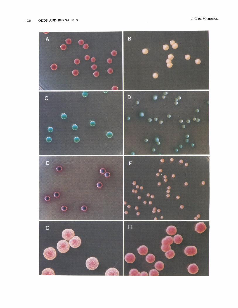
All 285 C. albicans isolates formed yellow-green to blue-green colonies on CHROMagar Candida (Fig. 1C). This greencolor was particularly distinctive for the species, and among theother species tested, only two Geotrichum isolates formed asimilar hue. However, in the case of these Geotrichum sp.isolates, the colonies themselves were light pink and of muchsmaller diameter than those of C. albicans, and the greenpigmentation was present in the agar beneath the colonies(Fig. ID), an appearance never seen with C. albicans, thecolonies themselves of which were green. From these data, thesensitivity and specificity of a green colony color for recogni-tion of C. albicans were calculated as 100%.
C. tropicalis isolates (n = 54) all developed a distinctive darkblue-gray central color after 48 h of incubation, and theircolonies were surrounded by a dark brown to purple halo inthe agar surrounding the colony (Fig. IE). Although a colonycolor approximately similar to that of C. tropicalis was observedwith some other species, the dark halo in the agar was seenonly with isolates of C. tropicalis plus 2 of 12 isolates identifiedas "Pichia sp." among the yeasts tested. The sensitivity andspecificity of the dark blue-gray colony color and brown-purpleagar halo for C. tropicalis were therefore >99%.The color of Trichosporon sp. colonies after 48 h was also
distinctive but variable. The isolates formed small colonieswith a pale color, subjectively described as "dirty pink" to"dirty gray-green" (Fig. 1F); these colonies became darker andacquired a characteristically rough appearance after 72 h or
more of incubation. Isolates identified as Pichia spp. on thebasis of their API ID32D assimilation patterns also formedconspicuously atypical (but variable) colonies after 48 h ofincubation on CHROMagar Candida. (It should be stressedthat no ascospore formation has been seen for any of theseisolates, so their true identifications remain uncertain). TwoPichia sp. isolates gave an appearance indistinguishable fromthat of C. tropicalis, as already mentioned. One formed colo-nies with a dark purple center and a green edge. The remainingeight isolates gave less remarkable colony colors ranging frombrownish pink to grayish purple, without the formation of agarhaloes.
All 43 isolates of C. krusei tested formed colonies that were
typically pale, flat, papillate, and spreading with broad whiteedges (Fig. 1G). After 48 h, C. krusei colonies were easilydistinguishable from those of other yeasts that formed smooth,brownish pink to brownish purple colonies on CHROMagarCandida. The characteristic appearance of C. krusei was seen
only with the five isolates of C. norvegensis among the otheryeasts tested (Fig. 1H). The specificity and sensitivity of thepale pink, spreading, rough colony form for the presumptiveidentification of C. krusei were therefore >99 and 100%,respectively.When different yeast species were mixed in a single suspen-
sion and plated out on CHROMagar Candida, the distinctionsin colony colors and forms were extremely easy to recognize(Fig. 2).
Viabilities of yeasts grown on CHROMagar Candida. All726 yeast isolates plus 30 additional yeast clones were subcul-tured onto Sabouraud agar after 48 or 72 h of incubation at370C on CHROMagar Candida. All of the isolates grew well inthe subcultures, demonstrating that viability was not lost by
VOL. 32, 1994
1926 ODDS AND BERNAERTS J. CLIN. MICROBIOL.

NEW DIFFERENTIAL MEDIUM FOR CANDIDA SPP. 1927
FIG. 1. (A) Dark pink colonies (paler edges) of C. glabrata grown for 48 h on CHROMagar Candida at 37°C. (B) Pale colonies of C. parapsilosisgrown for 48 h on CHROMagar Candida at 37C. (C) Green colonies (paler edges) of C. albicans grown for 48 h on CHROMagar Candida at37°C. (D) Colonies of a Geotrichumn sp. isolate grown for 48 h on CHROMagar Candida at 37°C. Unlike C. albicans, this isolate formed small, pale,rough colonies, and the green color is a halo in the agar. (E) Colonies of C. tropicalis grown for 48 h on CHROMagar Candida at 37°C. The purplehalo in the agar surrounding the dark blue-gray colonies (paler, pinker edges) was observed only with this species. (F) Colonies of a Trichosporonsp. isolate grown for 48 h on CHROMagar Candida at 37°C. This isolate formed small, "dirty pink" colonies; other isolates also formed smallcolonies, but with a pale gray-green hue. (G) Colonies of C. knisei grown for 48 h on CHROMagar Candida at 37°C. The large, rough, spreadingcolonies with broad, pale edges were formed by all isolates tested. (H) Colonies of C. norvegensis grown for 48 h on CHROMagar Candida at 37°C.This species was the only 1 of 21 yeasts tested that formed colonies resembling those of C. krusei on this medium. Magnifications, X2.
growth in the presence of the chromophore present inCHROMagar Candida.
Presumptive identification of yeast species by four readersin a single-blind experiment. The 57 yeast isolates coded forsingle-blind presumptive identification by four independentreaders represented nine different yeast species. The fourindividuals who attempted to identify the isolates from theircolonial appearances were asked to record their identificationsby reference to single labelled examples of each species. The22 C. albicans isolates were correctly identified by all fourobservers after 24 and 48 h of incubation. For the other specieswith distinctive appearances after growth on CHROMagarCandida, all required 48 h of incubation to ensure a correctidentification. At this time, all isolates of C. tropicalis, C. krusei,and Trichosporon spp. were correctly identified. None of theother species was correctly identified by all readers at 24 or 48h, but none was misidentified as C. albicans, C. tropicalis, C.krusei, or Trichosporon sp. These observations indicate that theCHROMagar Candida medium serves principally to differen-tiate C. albicans, C. krusei, C. tropicalis, and Trichosporon spp.from other yeast species.
Selectivity ofCHROMagar Candida for yeasts and fungi. To
determine the selectivity of CHROMagar Candida as anisolation medium for general-purpose use in medical mycolog-ical practice, a panel of single isolates representing severalbacterial and mold species was inoculated on the new medium.None of the test bacteria, namely, a Salmonella sp., Escherichiacoli, Staphylococcus aurelus, Streptococcus pyogenes, Staphylo-cocculs epidermidis, Enterococcus faecalis, Pseudomonas aerugi-nosa, Morganella morganii, and Streptomyces albidus, hadgrown after 72 h of incubation on CHROMagar Candida at37°C. Among 116 vaginal swabs, 113 oral swabs, and 110anorectal swabs inoculated onto CHROMagar Candida, bac-teria grew from just 4 anorectal swabs. In all four instances thebacteria isolated were identified as E. coli, and they all gavepink to purple colonies that were recognizable from their sizesand appearances as being typical of bacteria rather than ofyeasts.
Tests with 66 mold isolates indicated that CHROMagarCandida supported the growth of most molds. The exceptionsincluded some dermatophytes, 4 of 23 isolates of which failedto grow after 3 weeks of incubation at 25°C, and isolates of aVerticillium sp. and a Phoma sp., both of which showed nogrowth after 2 weeks of incubation. The remaining 60 isolates
FIG. 2. Colonies plated out from a mixed suspension of four different Candida species and incubated for 48 h at 37°C on CHROMagarCandida. All four species can be distinguished by their colony appearances: two C. glabrata colonies are pink, two C. tropicalis colonies arebluish-purple (the characteristic agar haloes are indistinctly visible under the lighting conditions used), four C. albicans colonies are green, and thetwo large, pale pink, rough colonies are C. krusei. Magnification, x5.5.
VOL. 32, 1994
1928 ODDS AND BERNAERTS
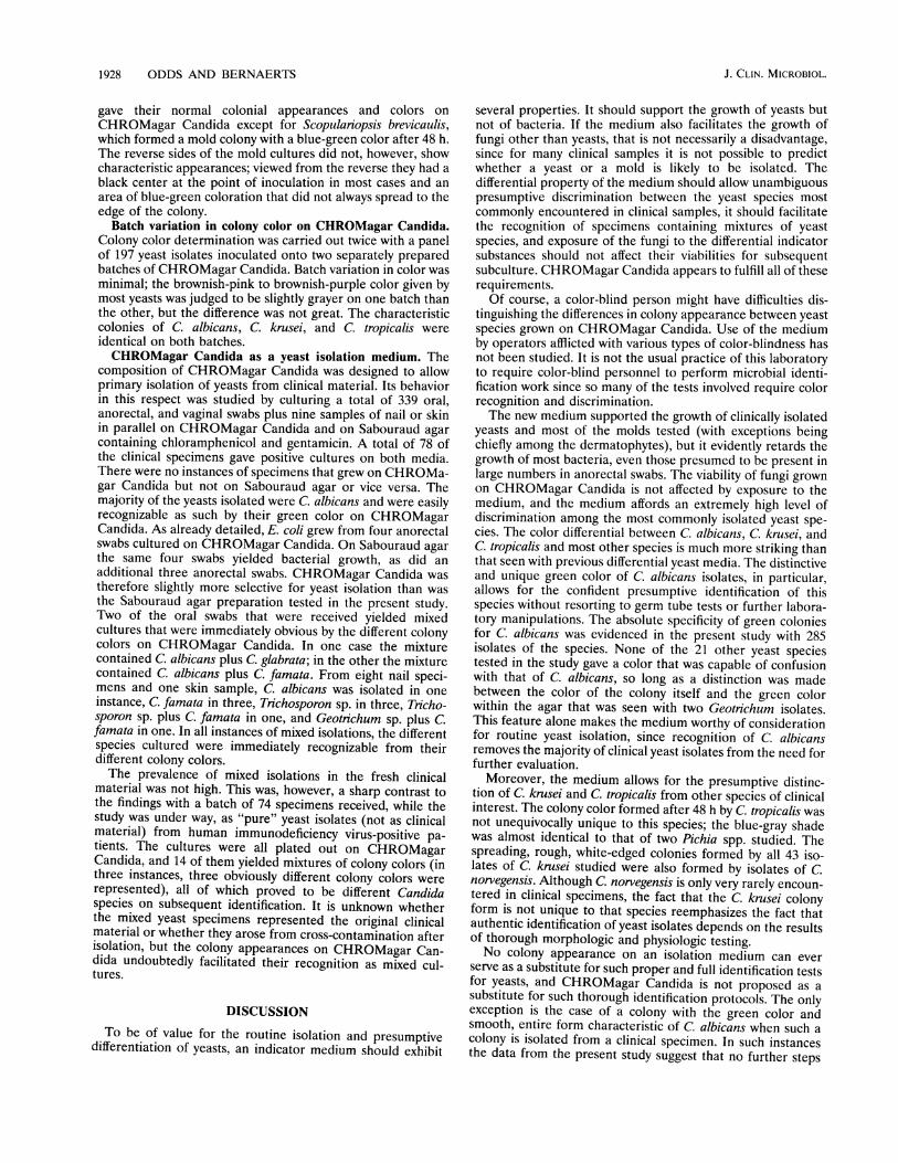
gave their normal colonial appearances and colors onCHROMagar Candida except for Scopulariopsis brevicaulis,which formed a mold colony with a blue-green color after 48 h.The reverse sides of the mold cultures did not, however, showcharacteristic appearances; viewed from the reverse they had ablack center at the point of inoculation in most cases and anarea of blue-green coloration that did not always spread to theedge of the colony.
Batch variation in colony color on CHROMagar Candida.Colony color determination was carried out twice with a panelof 197 yeast isolates inoculated onto two separately preparedbatches of CHROMagar Candida. Batch variation in color wasminimal; the brownish-pink to brownish-purple color given bymost yeasts was judged to be slightly grayer on one batch thanthe other, but the difference was not great. The characteristiccolonies of C. albicans, C. krusei, and C. tropicalis wereidentical on both batches.CHROMagar Candida as a yeast isolation medium. The
composition of CHROMagar Candida was designed to allowprimary isolation of yeasts from clinical material. Its behaviorin this respect was studied by culturing a total of 339 oral,anorectal, and vaginal swabs plus nine samples of nail or skinin parallel on CHROMagar Candida and on Sabouraud agarcontaining chloramphenicol and gentamicin. A total of 78 ofthe clinical specimens gave positive cultures on both media.There were no instances of specimens that grew on CHROMa-gar Candida but not on Sabouraud agar or vice versa. Themajority of the yeasts isolated were C. albicans and were easilyrecognizable as such by their green color on CHROMagarCandida. As already detailed, E. coli grew from four anorectalswabs cultured on CHROMagar Candida. On Sabouraud agarthe same four swabs yielded bacterial growth, as did anadditional three anorectal swabs. CHROMagar Candida wastherefore slightly more selective for yeast isolation than wasthe Sabouraud agar preparation tested in the present study.Two of the oral swabs that were received yielded mixedcultures that were immediately obvious by the different colonycolors on CHROMagar Candida. In one case the mixturecontained C. albicans plus C. glabrata; in the other the mixturecontained C. albicans plus C. famata. From eight nail speci-mens and one skin sample, C. albicans was isolated in oneinstance, C. famata in three, Trichosporon sp. in three, Tricho-sporon sp. plus C. famata in one, and Geotrichum sp. plus C.famata in one. In all instances of mixed isolations, the differentspecies cultured were immediately recognizable from theirdifferent colony colors.The prevalence of mixed isolations in the fresh clinical
material was not high. This was, however, a sharp contrast tothe findings with a batch of 74 specimens received, while thestudy was under way, as "pure" yeast isolates (not as clinicalmaterial) from human immunodeficiency virus-positive pa-tients. The cultures were all plated out on CHROMagarCandida, and 14 of them yielded mixtures of colony colors (inthree instances, three obviously different colony colors wererepresented), all of which proved to be different Candidaspecies on subsequent identification. It is unknown whetherthe mixed yeast specimens represented the original clinicalmaterial or whether they arose from cross-contamination afterisolation, but the colony appearances on CHROMagar Can-dida undoubtedly facilitated their recognition as mixed cul-tures.
DISCUSSIONTo be of value for the routine isolation and presumptive
differentiation of yeasts, an indicator medium should exhibit
several properties. It should support the growth of yeasts butnot of bacteria. If the medium also facilitates the growth offungi other than yeasts, that is not necessarily a disadvantage,since for many clinical samples it is not possible to predictwhether a yeast or a mold is likely to be isolated. Thedifferential property of the medium should allow unambiguouspresumptive discrimination between the yeast species mostcommonly encountered in clinical samples, it should facilitatethe recognition of specimens containing mixtures of yeastspecies, and exposure of the fungi to the differential indicatorsubstances should not affect their viabilities for subsequentsubculture. CHROMagar Candida appears to fulfill all of theserequirements.Of course, a color-blind person might have difficulties dis-
tinguishing the differences in colony appearance between yeastspecies grown on CHROMagar Candida. Use of the mediumby operators afflicted with various types of color-blindness hasnot been studied. It is not the usual practice of this laboratoryto require color-blind personnel to perform microbial identi-fication work since so many of the tests involved require colorrecognition and discrimination.The new medium supported the growth of clinically isolated
yeasts and most of the molds tested (with exceptions beingchiefly among the dermatophytes), but it evidently retards thegrowth of most bacteria, even those presumed to be present inlarge numbers in anorectal swabs. The viability of fungi grownon CHROMagar Candida is not affected by exposure to themedium, and the medium affords an extremely high level ofdiscrimination among the most commonly isolated yeast spe-cies. The color differential between C. albicans, C. krusei, andC. tropicalis and most other species is much more striking thanthat seen with previous differential yeast media. The distinctiveand unique green color of C. albicans isolates, in particular,allows for the confident presumptive identification of thisspecies without resorting to germ tube tests or further labora-tory manipulations. The absolute specificity of green coloniesfor C albicans was evidenced in the present study with 285isolates of the species. None of the 21 other yeast speciestested in the study gave a color that was capable of confusionwith that of C. albicans, so long as a distinction was madebetween the color of the colony itself and the green colorwithin the agar that was seen with two Geotrichum isolates.This feature alone makes the medium worthy of considerationfor routine yeast isolation, since recognition of C. albicansremoves the majority of clinical yeast isolates from the need forfurther evaluation.
Moreover, the medium allows for the presumptive distinc-tion of C. krusei and C. tropicalis from other species of clinicalinterest. The colony color formed after 48 h by C. tropicalis wasnot unequivocally unique to this species; the blue-gray shadewas almost identical to that of two Pichia spp. studied. Thespreading, rough, white-edged colonies formed by all 43 iso-lates of C. krusei studied were also formed by isolates of C.norvegensis. Although C. norvegensis is only very rarely encoun-tered in clinical specimens, the fact that the C. krusei colonyform is not unique to that species reemphasizes the fact thatauthentic identification of yeast isolates depends on the resultsof thorough morphologic and physiologic testing.No colony appearance on an isolation medium can ever
serve as a substitute for such proper and full identification testsfor yeasts, and CHROMagar Candida is not proposed as asubstitute for such thorough identification protocols. The onlyexception is the case of a colony with the green color andsmooth, entire form characteristic of C. albicans when such acolony is isolated from a clinical specimen. In such instancesthe data from the present study suggest that no further steps
J. CLIN. MICROBIOL.

NEW DIFFERENTIAL MEDIUM FOR CANDIDA SPP. 1929
are necessary, not even the germ tube test, to confirm theidentification of the species. The particular value of the newisolation medium is that it facilitates enormously the recogni-tion of mixtures of yeast species on a single isolation plate,and it offers, for certain yeast species, a strong indication ofthe likely subsequent identification. In single-blind testswith individuals not involved with the laboratory study,CHROMagar Candida could be used successfully to differen-tiate isolates of C. albicans, C. tropicalis, C. krusei, and Tricho-sporon spp.The performance of CHROMagar Candida exactly paral-
leled that of Sabouraud glucose agar in terms of its abilityto support the isolation of yeasts from clinical samples.CHROMagar Candida was slightly superior to the Sabouraudagar used for comparison in terms of its ability to suppressbacterial growth. Its overall superiority has been self-evident inour hands in its ability to reveal mixtures of yeast speciespresent in cultures (although the clinical material directlycultured on CHROMagar Candida happens so far to haveyielded only two instances of mixed yeast infections). We haveso far experienced several instances of cultures submitted as"pure" yeast isolates, but which were shown to contain mix-tures when plated out on CHROMagar Candida.CHROMagar Candida appears to be a medium well-suited
for medical mycological use. It can serve as a primary iso-lation and differentiation medium for clinical specimenslikely to contain yeasts and also as an adjunctive differentialmedium for the identification of yeasts isolated on othermedia.
ACKNOWLEDGMENTS
The technical assistance of Peter De Backker, Marc Van der Flaes,and Luc Van Nuffel and the skilled photographic help of HansHenderickx and Lambert Leijssen are gratefully acknowledged.
REFERENCES
1. Bump, C. M., and L. J. Kunz. 1968. Routine identification ofyeasts with the aid of molybdate-agar medium. Appl. Microbiol.16:1503-1506.
2. Costa, S. O., and C. de Lourdes Branco. 1964. Evaluation of amolybdenum culture medium as selective and differential foryeasts. J. Pathol. Bacteriol. 87:428-431.
3. Mendel, E. B., S. Haberman, and D. K. Hall. 1960. Isolation ofCandida from clinical specimens. Comparative study of Pagano-Levin and Nickerson's culture media. Obstet. Gynecol. 16:180-184.
4. Nickerson, W. J. 1953. Reduction of inorganic substances byyeasts. I. Extracellular reduction of sulfite by species of Candida. J.Infect. Dis. 93:45-56.
5. Nickerson, W. J. 14 September 1954. Culture medium containingbismuthyl polyhydroxy polysulfite. U.S. patent 2,689,204.
6. O'Brien, J. R. 1964. Nickerson's medium in the diagnosis ofvaginal moniliasis. Can. Med. Assoc. J. 90:1073-1074.
7. Odds, F. C. 1991. Sabouraud('s) agar. J. Med. Vet. Mycol.29:355-359.
8. Odds, F. C. 1991. Antifungal susceptibility testing of Candida spp.by relative growth measurement at single concentrations of anti-fungal agents. Antimicrob. Agents Chemother. 36:1727-1737.
9. Pagano, J., J. D. Levin, and W. Trejo. 1958. Diagnostic medium fordifferentiation of species of Candida. Antibiot. Ann. 1957-1958:137-143.
10. Schnell, J. D. 1982. Investigations into the pathoaetiology anddiagnosis of vaginal mycoses. Chemotherapy (Basel) 28 (Suppi.1):14-21.
11. Sinski, J. T., L. M. Kelley, and G. L. Reed. 1975. Pagano-LevinCandida test medium: evaluation using vaginal samples. J. Clin.Microbiol. 1:206-211.
12. Yamane, N., and Y. Saitoh. 1985. Isolation and detection ofmultiple yeasts from a single clinical sample by use of Pagano-Levin agar medium. J. Clin. Microbiol. 21:276-277.
VOL. 32, 1994


