Cholesterol is accumulated by mycobacteria but its ... · Cholesterol is accumulated by...
6
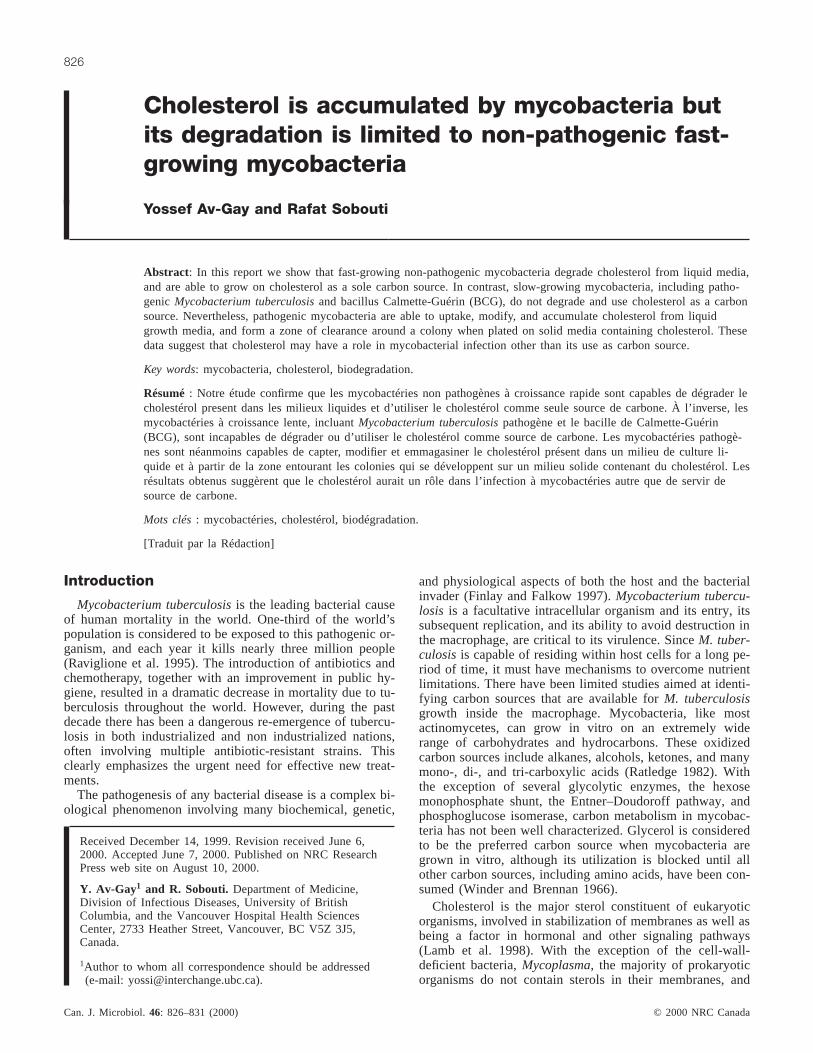
Cholesterol is accumulated by mycobacteria but its degradation is limited to non-pathogenic fast- growing mycobacteria Yossef Av-Gay and Rafat Sobouti Abstract: In this report we show that fast-growing non-pathogenic mycobacteria degrade cholesterol from liquid media, and are able to grow on cholesterol as a sole carbon source. In contrast, slow-growing mycobacteria, including patho- genic Mycobacterium tuberculosis and bacillus Calmette-Guérin (BCG), do not degrade and use cholesterol as a carbon source. Nevertheless, pathogenic mycobacteria are able to uptake, modify, and accumulate cholesterol from liquid growth media, and form a zone of clearance around a colony when plated on solid media containing cholesterol. These data suggest that cholesterol may have a role in mycobacterial infection other than its use as carbon source. Key words: mycobacteria, cholesterol, biodegradation. Résumé : Notre étude confirme que les mycobactéries non pathogènes à croissance rapide sont capables de dégrader le cholestérol present dans les milieux liquides et d’utiliser le cholestérol comme seule source de carbone. À l’inverse, les mycobactéries à croissance lente, incluant Mycobacterium tuberculosis pathogène et le bacille de Calmette-Guérin (BCG), sont incapables de dégrader ou d’utiliser le cholestérol comme source de carbone. Les mycobactéries pathogè- nes sont néanmoins capables de capter, modifier et emmagasiner le cholestérol présent dans un milieu de culture li- quide et à partir de la zone entourant les colonies qui se développent sur un milieu solide contenant du cholestérol. Les résultats obtenus suggèrent que le cholestérol aurait un rôle dans l’infection à mycobactéries autre que de servir de source de carbone. Mots clés : mycobactéries, cholestérol, biodégradation. [Traduit par la Rédaction] Av-Gay and Sobouti 831 Introduction Mycobacterium tuberculosis is the leading bacterial cause of human mortality in the world. One-third of the world’s population is considered to be exposed to this pathogenic or- ganism, and each year it kills nearly three million people (Raviglione et al. 1995). The introduction of antibiotics and chemotherapy, together with an improvement in public hy- giene, resulted in a dramatic decrease in mortality due to tu- berculosis throughout the world. However, during the past decade there has been a dangerous re-emergence of tubercu- losis in both industrialized and non industrialized nations, often involving multiple antibiotic-resistant strains. This clearly emphasizes the urgent need for effective new treat- ments. The pathogenesis of any bacterial disease is a complex bi- ological phenomenon involving many biochemical, genetic, and physiological aspects of both the host and the bacterial invader (Finlay and Falkow 1997). Mycobacterium tubercu- losis is a facultative intracellular organism and its entry, its subsequent replication, and its ability to avoid destruction in the macrophage, are critical to its virulence. Since M. tuber- culosis is capable of residing within host cells for a long pe- riod of time, it must have mechanisms to overcome nutrient limitations. There have been limited studies aimed at identi- fying carbon sources that are available for M. tuberculosis growth inside the macrophage. Mycobacteria, like most actinomycetes, can grow in vitro on an extremely wide range of carbohydrates and hydrocarbons. These oxidized carbon sources include alkanes, alcohols, ketones, and many mono-, di-, and tri-carboxylic acids (Ratledge 1982). With the exception of several glycolytic enzymes, the hexose monophosphate shunt, the Entner–Doudoroff pathway, and phosphoglucose isomerase, carbon metabolism in mycobac- teria has not been well characterized. Glycerol is considered to be the preferred carbon source when mycobacteria are grown in vitro, although its utilization is blocked until all other carbon sources, including amino acids, have been con- sumed (Winder and Brennan 1966). Cholesterol is the major sterol constituent of eukaryotic organisms, involved in stabilization of membranes as well as being a factor in hormonal and other signaling pathways (Lamb et al. 1998). With the exception of the cell-wall- deficient bacteria, Mycoplasma, the majority of prokaryotic organisms do not contain sterols in their membranes, and Can. J. Microbiol. 46: 826–831 (2000) © 2000 NRC Canada 826 Received December 14, 1999. Revision received June 6, 2000. Accepted June 7, 2000. Published on NRC Research Press web site on August 10, 2000. Y. Av-Gay 1 and R. Sobouti. Department of Medicine, Division of Infectious Diseases, University of British Columbia, and the Vancouver Hospital Health Sciences Center, 2733 Heather Street, Vancouver, BC V5Z 3J5, Canada. 1 Author to whom all correspondence should be addressed (e-mail: [email protected]).
Transcript of Cholesterol is accumulated by mycobacteria but its ... · Cholesterol is accumulated by...
Cholesterol is accumulated by mycobacteria butits degradation is limited to non-pathogenic fast-growing mycobacteria
Yossef Av-Gay and Rafat Sobouti
Abstract: In this report we show that fast-growing non-pathogenic mycobacteria degrade cholesterol from liquid media,and are able to grow on cholesterol as a sole carbon source. In contrast, slow-growing mycobacteria, including patho-genic Mycobacterium tuberculosisand bacillus Calmette-Guérin (BCG), do not degrade and use cholesterol as a carbonsource. Nevertheless, pathogenic mycobacteria are able to uptake, modify, and accumulate cholesterol from liquidgrowth media, and form a zone of clearance around a colony when plated on solid media containing cholesterol. Thesedata suggest that cholesterol may have a role in mycobacterial infection other than its use as carbon source.
Key words: mycobacteria, cholesterol, biodegradation.
Résumé: Notre étude confirme que les mycobactéries non pathogènes à croissance rapide sont capables de dégrader lecholestérol present dans les milieux liquides et d’utiliser le cholestérol comme seule source de carbone. À l’inverse, lesmycobactéries à croissance lente, incluantMycobacterium tuberculosispathogène et le bacille de Calmette-Guérin(BCG), sont incapables de dégrader ou d’utiliser le cholestérol comme source de carbone. Les mycobactéries pathogè-nes sont néanmoins capables de capter, modifier et emmagasiner le cholestérol présent dans un milieu de culture li-quide et à partir de la zone entourant les colonies qui se développent sur un milieu solide contenant du cholestérol. Lesrésultats obtenus suggèrent que le cholestérol aurait un rôle dans l’infection à mycobactéries autre que de servir desource de carbone.
Mots clés: mycobactéries, cholestérol, biodégradation.
[Traduit par la Rédaction] Av-Gay and Sobouti 831
Introduction
Mycobacterium tuberculosisis the leading bacterial causeof human mortality in the world. One-third of the world’spopulation is considered to be exposed to this pathogenic or-ganism, and each year it kills nearly three million people(Raviglione et al. 1995). The introduction of antibiotics andchemotherapy, together with an improvement in public hy-giene, resulted in a dramatic decrease in mortality due to tu-berculosis throughout the world. However, during the pastdecade there has been a dangerous re-emergence of tubercu-losis in both industrialized and non industrialized nations,often involving multiple antibiotic-resistant strains. Thisclearly emphasizes the urgent need for effective new treat-ments.
The pathogenesis of any bacterial disease is a complex bi-ological phenomenon involving many biochemical, genetic,
and physiological aspects of both the host and the bacterialinvader (Finlay and Falkow 1997).Mycobacterium tubercu-losis is a facultative intracellular organism and its entry, itssubsequent replication, and its ability to avoid destruction inthe macrophage, are critical to its virulence. SinceM. tuber-culosisis capable of residing within host cells for a long pe-riod of time, it must have mechanisms to overcome nutrientlimitations. There have been limited studies aimed at identi-fying carbon sources that are available forM. tuberculosisgrowth inside the macrophage. Mycobacteria, like mostactinomycetes, can grow in vitro on an extremely widerange of carbohydrates and hydrocarbons. These oxidizedcarbon sources include alkanes, alcohols, ketones, and manymono-, di-, and tri-carboxylic acids (Ratledge 1982). Withthe exception of several glycolytic enzymes, the hexosemonophosphate shunt, the Entner–Doudoroff pathway, andphosphoglucose isomerase, carbon metabolism in mycobac-teria has not been well characterized. Glycerol is consideredto be the preferred carbon source when mycobacteria aregrown in vitro, although its utilization is blocked until allother carbon sources, including amino acids, have been con-sumed (Winder and Brennan 1966).
Cholesterol is the major sterol constituent of eukaryoticorganisms, involved in stabilization of membranes as well asbeing a factor in hormonal and other signaling pathways(Lamb et al. 1998). With the exception of the cell-wall-deficient bacteria,Mycoplasma, the majority of prokaryoticorganisms do not contain sterols in their membranes, and
Can. J. Microbiol.46: 826–831 (2000) © 2000 NRC Canada
826
Received December 14, 1999. Revision received June 6,2000. Accepted June 7, 2000. Published on NRC ResearchPress web site on August 10, 2000.
Y. Av-Gay1 and R. Sobouti. Department of Medicine,Division of Infectious Diseases, University of BritishColumbia, and the Vancouver Hospital Health SciencesCenter, 2733 Heather Street, Vancouver, BC V5Z 3J5,Canada.
1Author to whom all correspondence should be addressed(e-mail: [email protected]).

hapanoid is believed to fulfill the role of sterol in higher or-ganisms (Taylor 1984). Nevertheless, cholesterol can be me-tabolized by a wide range of microorganisms as a carbonand energy source (Schatz et al. 1949; Fukuda et al. 1973; Liand Beitz 1996). The ability of actinomycetes, specificallymycobacteria, to bioconvert sterol is well documented, andhas been utilized by the pharmaceutical industry to synthe-size novel steroid compounds (Schoemer and Martin 1980).In human macrophages, cholesterol is available as an inte-gral part of the cell membrane, including the phagosomalmembrane. Since mycobacteria reside in macrophages, animportant issue is whether pathogenic mycobacteria can uti-lize the cholesterol located in the phagosomal membranethat surrounds the tubercle bacilli. Although it has been pro-posed that pathogenic mycobacteria use lecithin and notcholesterol (Kondo and Kanai 1976b) in vivo as carbonsource, a few reports indicate that cholesterol might be con-sumed by pathogenic mycobacteria. In addition, it has beenshown that cholesterol is required for growth of pathogenicmycobacteria such asM. scrofulaceum, isolated from lep-rous tissues (Buki et al. 1969; Kato 1979). Interestingly, thecomplete genomic sequence ofM. tuberculosissuggests thatit contains sterol biosynthetic enzymes as well as two puta-tive cholesterol degradation enzymes (Cole et al. 1998;Bellamine et al. 1998). Supportive evidence for the ability ofmycobacteria to synthesize cholesterol appeared in a recentreport which demonstrates thatM. smegmatisis able to syn-thesize trace amounts of cholesterol (Lamb et al. 1998).Nevertheless, the physiological role of cholesterol and theenzymes involved in its biodegradation by bacteria, and spe-cifically mycobacteria, remain unknown. This study exam-ines and compares the ability of various mycobacteria totake up and utilize cholesterol supplied in their growth me-dia in an attempt to determine whether mycobacteria usecholesterol as a carbon source.
Materials and methods
Strains, growth media, and sample preparationCultures of M. smegmatisMC2155 (a kind gift of William
Jacobs),M. fortuitum (ATCC 6841), andM. phlei (ATCC 354)were grown in Modified Dubos Medium (MDM).MycobacteriumtuberculosisH37Rv (ATCC 27294),M. tuberculosisH37Ra (ATCC25177), M. avium (ATCC 25291), M. intracellulare (ATCC13950), andM. bovis BCG strain Pasteur (ATCC 35734) weregrown in MDM media, or in minimal medium containing the inor-ganic components of Proskauer-Beck medium without any glycerolas a carbon source. In each case, cholesterol (Sigma) was added tothe media when required. The cholesterol was suspended in 5 mLTween-80, heated briefly, and added to the media (1 g/L). For platepreparation, cholesterol was either suspended in 10 mL of 95%ethanol or in 20 mL of warm Tween-80. The solution was filteredthrough a Millex 0.22µm filter and added to the media. Plateswere made by adding pure agarose (FMC) to a final concentrationof 15% as a solidifying agent. After growth and five-fold expan-sion over two weeks, cells were harvested by centrifugation forfive minutes at 3750 ×g. Cell culture media was filtered twice with0.22-µm filter units (Nunc) and stored at –70°C until assayed. Cellpellets were washed once in lysis buffer (20 mM Tris pH 7.5,2 mM EDTA, 150 mM NaCl, 0.2 mM PMSF, 1µg/mL pepstatin A,1 µg/mL aprotonin, 0.5 mM sodium vanadate, 0.2 mM sodiummolybdate), suspended in the same buffer, then subjected to me-chanical bead breakage (Mini-Beadbeater, Biospec Products) for
five minutes using 100µm zirconia beads (Sigma) at room temper-ature. Cell debris was removed by centrifugation at 10 000 ×g for5 min. The resulting supernatant was filter-sterilized (Millex0.22 µm, Millipore) and stored in aliquots at –70°C.
Cholesterol assayCholesterol levels were determined colorimetrically using a com-
mercially available kit (Boehringer Manneheim Cat. No. 139050).
Thin layer chromatographyThin layer chromatography of sterols was performed according
to Stahl (Stahl 1969). Briefly, lipids, sterols, and sterol derivativeswere extracted with a solution of chloroform-methanol 1:1 (v/v). A40-µL volume of each phase was applied to silica gel thin layerchromatography (TLC) plates (Whatman 25) and separated usingcyclohexane/chloroform 1:1 (v/v). Development and detection wereperformed by spraying with o-phosphoric acid/H2O, 1:1 (v/v), fol-lowed by baking at 120°C for 15 min and spraying with freshlyprepared 50 mg/mL phosphomolibdic acid ethanolic solution.Spots were stabilized after 5 min by heating plates at 85°C.
HPLC analysisA 14 mL volume of media was passed through silica columns
(abeselut NEXUS sorbent-Varian), rinsed with 1 mL H2O, fol-lowed by vacuum drying. Hydrophobic compounds were elutedwith 1 mL methanol. HPLC analysis was performed on BondapakC-18 column (Waters). A 100-µL volume of the eluate from sorbentcolumn was injected, and an isocratic acetonitrile/isopropanol1:1 (v/v) wash was used to separate sterols. Cholesterol was de-tected at 210 nm.
Results
Growth on cholesterol as a sole carbon source is aproperty of non-pathogenic mycobacteria
To test whether mycobacteria are able to grow on choles-terol, a number of slow- and fast-growing mycobacterialstrains were plated on minimal media containing cholesterolas a sole carbon source; all fast-growing mycobacterialstrains tested were able to grow on cholesterol plates. As il-lustrated in Fig. 1A,M. smegmatiswas able to grow on min-imal medium containing cholesterol as a sole carbon source.The same was observed forM. phleiandM. fortuitum(data notshown). In contrast, slow-growing pathogenic-mycobacteria,such asM. tuberculosis,grew on this medium only whenTween-80 was used as the organic solvent (which by itselfcan be used as a carbon source). When left to grow for sixweeks on solid media, slow-growing pathogenic mycobacte-ria such asM. avium, M. intracellulare, and two strains ofM. tuberculosis(Ra and Rv) cleared cholesterol from theplates (Fig. 1B), while fast growers such asM. fortiutumandM. smegmatisdid not. The clearing of cholesterol was dem-onstrated by the creation of a clear zone around the colony.
Cholesterol contribution to mycobacterial growth inliquid media
As seen in Fig. 2, growth analysis of bothM. smegmatisand M. phlei demonstrates that cholesterol enhances theirgrowth. BothM. smegmatisandM. phlei reached stationaryphase after 48 h of growth in media containing cholesterol,while in minimal media, at least 96 h were needed to reachstationary phase. Furthermore, in the presence of cholesterol,the stationary phase of both cultures lasts longer when com-
© 2000 NRC Canada
Av-Gay and Sobouti 827

pared to the minimal media growth curve. On the otherhand, whenM. bovisBCG was examined as a representativeof the slow-growing pathogenic mycobacteria, BCG grow-ing in media containing cholesterol grew slower than thatgrown in minimal media lacking cholesterol (Fig. 2C). Fur-thermore, we noticed that higher levels of cholesterol in thegrowth media inhibited BCG growth.
Cholesterol accumulation and degradation bymycobacteria
Three different experimental approaches were used to de-termine cholesterol utilization by mycobacteria, specificallycolorimetric determination of cholesterol levels, TLC, andHPLC analysis. Examination of cholesterol utilization dur-ing mycobacterial growth using the coupled cholesterol-oxidase and catalase colorimetric assay showed thatM. phleiand M. smegmatisconsumed cholesterol from the growthmedia throughout their growth cycle (Fig. 2A and 2B). Ex-amination of cholesterol utilization by BCG during itsgrowth curve (Fig. 2C) showed clearly that BCG did not re-move cholesterol from its growth medium.
The use of a second approach, TLC analysis of choles-terol levels, was used in order to rule out the possibility thatmycobacteria accumulate cholesterol from the growth mediarather than degrade it, and to determine whether the smalldecrease in cholesterol levels during the first two days ofBCG growth can be explained by its accumulation-depo-sition into the mycobacterial cell wall. As seen in Fig. 3,
M. smegmatis(A) and M. phlei (B) growth media and cell-free-extracts TLC showed (i) initial accumulation of choles-terol inside cells, and (ii ) that cholesterol is disappearing
© 2000 NRC Canada
828 Can. J. Microbiol. Vol. 46, 2000
Fig. 1. (A) Growth of mycobacteria on cholesterol as a sole car-bon source. A defined agarose based minimal medium without(–) or with (+) cholesterol as a carbon source was used to growM. smegmatisfor 48 h at 30°C. (B) Growth of mycobacteria onTween-cholesterol plates. Slow growers:M. tuberculosis(2 and3), M. intracellulare (1 and 6) andM. avium (4 and 5). Fastgrowers:M. fortuitum (7) andM. smegmatis(8).
Fig. 2. Growth curve and cholesterol utilization by(A) M. smegmatis, (B) M. phlei, and (C) BCG in liquid media.Results are representative of three independent experiments.Solid squares represent growth (A 600 nm) on MDM media andsolid triangles are growth in the presence of 0.5 mg/mL choles-terol in MDM media for the fast growers and 0.25 mg/mL forBCG. Solid circles represent amount of cholesterol in the media.Initial cholesterol levels were titrated to give optimal growth ofeach strain.

from both growth media and the cell extract. Further quanti-tative analysis of media and cell samples from late station-ary phase ofM. phlei andM. smegmatis(Fig. 3C), grown inthe presence or absence of cholesterol, showed clearly thatcholesterol is degraded. Furthermore, cholesterol degrada-tion sterol by-products can be detected in cultures grown inthe presence of cholesterol, but not in medium from cellsthat grew without cholesterol. On the other hand, TLC anal-ysis of M. tuberculosisH37Rv and BCG growth media, andcell free extracts, showed that cholesterol is present in thegrowth media, and that there is an accumulation of choles-terol within M. tuberculosiscells (Fig. 3D). Cholesterol did
not disappear from the growth media even when the culturewas left to grow for over six months. Identical results wereobtained with cultures of BCG. Furthermore, using a radio-active assay, we were not able to identify degradation prod-ucts of cholesterol inM. tuberculosis, M. avium, or BCGcultures (data not shown).
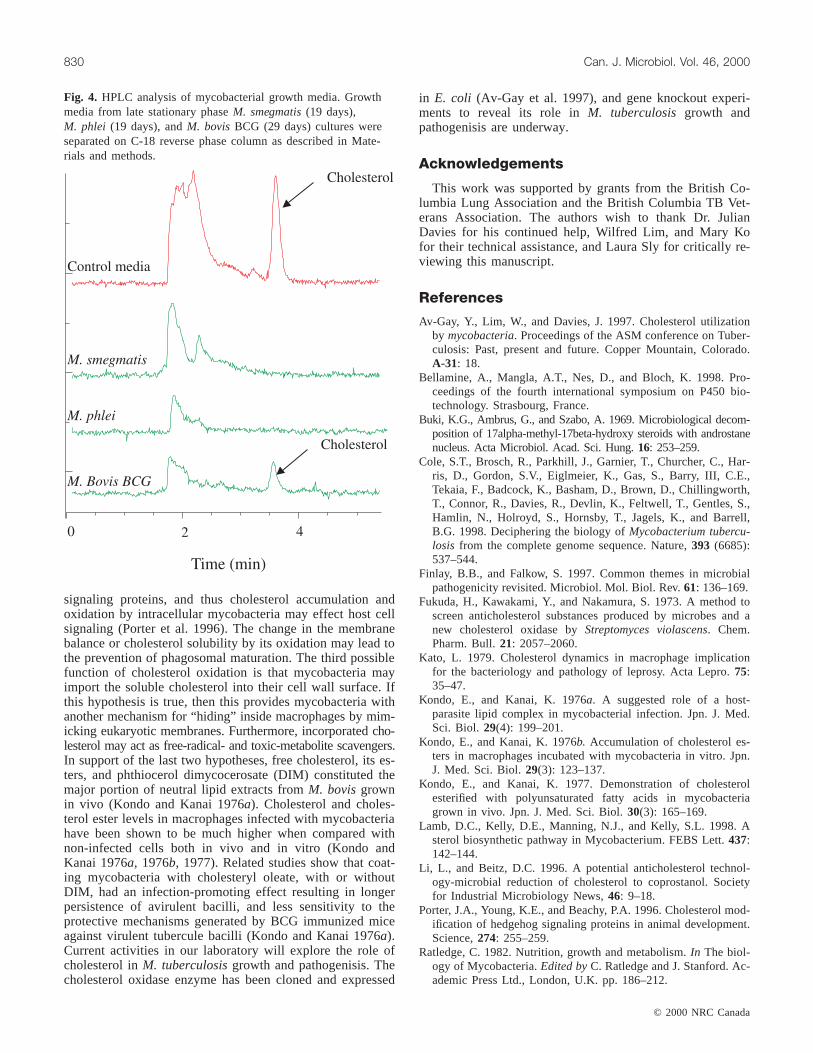
HPLC analysis is probably the most sensitive method forthe detection of trace amounts of sterols. As a confirmationto the above results, analysis of late stationary phase growthmedia, (Fig. 4), showed clearly that cholesterol is com-pletely depleted fromM. smegmatisand M. phlei culturemedia, while it can be detected inM. bovisBCG culture me-dia even after approximately four weeks of growth.
Discussion
In this report, we demonstrate that fast-growing non-pathogenic mycobacteria are able to degrade and utilize cho-lesterol when it is supplied as a sole carbon source. On theother hand, slow-growing mycobacteria, including those thatare pathogenic, utilize cholesterol in a different manner. Wehave shown thatM. tuberculosisand BCG accumulate cho-lesterol, but do not seem to metabolize it. The ability to de-grade cholesterol has been shown for several speciesincluding Streptomyces, Brevibacterium, and several non-pathogenic fast-growing mycobacteria (Schoemer and Martin1980). Biochemical descriptions of cholesterol degradativeenzymes, such as cholesterol oxidase, exist for a wide varietyof microorganisms including Gram-negative enterococci (Liand Beitz 1996) and Gram-positive bacteria (Solaiman andSomkuti 1991). In mammals, there is no complete degrada-tion of cholesterol. Cholesterol is modified by either 7-α-hydroxylation to form bile acids, or by oxidation of the 3-β-hydroxyl group with additional hydroxylation and oxidationof the side chain to form cholic acid or steroid hormones. Inthese cells, steroids are hydrolysed by mixed function oxi-dases that utilize NADPH and O2. All of these reactions in-volve cytochrome P450. In contrast to mammals, bacteriaare able to degrade cholesterol, with CO2 and H2O as the fi-nal products. Two different bacterial pathways for choles-terol degradation are represented by two different families ofthe cholesterol oxidase enzymes (Wilmanska et al. 1995).Fast-growing mycobacteria andRhodococcuscholesteroloxidases have a cascade of enzymes that take part in steroidbiodegradation, while the producers of extracellular choles-terol oxidases such asStreptomycesspp., andArthrobacterwere suggested to be incapable of decomposing sterols(Wilmanska et al. 1995). The above classification is inagreement with our results which show clearly that fast-growing mycobacteria degrade cholesterol, but do not clearit from their growth media. Our demonstration that slow-growing pathogenic mycobacteria clear cholesterol fromtheir growth media, probably via secretion of the enzymecholesterol oxidase, is further support for the hypothesis thatcholesterol oxidases are different in organisms that fail todegrade cholesterol after its uptake.
By ruling out the nutritional role of cholesterol for patho-genic mycobacteria, one needs to provide reasons for the ob-servation that cholesterol is accumulated in the cells. A fewhypotheses may be considered. Cholesterol has been shownto play a regulatory role in several systems by modifying
© 2000 NRC Canada
Av-Gay and Sobouti 829
Fig. 3. Cholesterol degradation in mycobacteria. TLC analysis ofcholesterol utilization by(A) M. smegmatisand (B) M. phlei dur-ing their growth curve. Culture volumes of 1.5 mL were frac-tionated by centrifugation to supernatant (S) and cells (P) beforeapplying to TLC. (C) TLC analysis of sterols, marked by arrows,in M. smegmatisand M. phlei. M , control media + cholesterol.1, Growth media from 16-day-old culture containing cholesterol;2, cell-free extract from 16-day-old culture grown in the pres-ence of cholesterol;3, growth media from 16-day-old culturewithout cholesterol;4, cell-free extract from 16-day-old culturegrown without cholesterol. (D) TLC analysis of sterols inM. tu-berculosisH37Rv. M , Media containing cholesterol;1, mediafrom six-month-old culture grown in the presence of cholesterol;2, cell-free extract from six-month-old culture grown in the pres-ence of cholesterol. No spots were observed when cholesterolwas omitted from the growth media.
signaling proteins, and thus cholesterol accumulation andoxidation by intracellular mycobacteria may effect host cellsignaling (Porter et al. 1996). The change in the membranebalance or cholesterol solubility by its oxidation may lead tothe prevention of phagosomal maturation. The third possiblefunction of cholesterol oxidation is that mycobacteria mayimport the soluble cholesterol into their cell wall surface. Ifthis hypothesis is true, then this provides mycobacteria withanother mechanism for “hiding” inside macrophages by mim-icking eukaryotic membranes. Furthermore, incorporated cho-lesterol may act as free-radical- and toxic-metabolite scavengers.In support of the last two hypotheses, free cholesterol, its es-ters, and phthiocerol dimycocerosate (DIM) constituted themajor portion of neutral lipid extracts fromM. bovisgrownin vivo (Kondo and Kanai 1976a). Cholesterol and choles-terol ester levels in macrophages infected with mycobacteriahave been shown to be much higher when compared withnon-infected cells both in vivo and in vitro (Kondo andKanai 1976a, 1976b, 1977). Related studies show that coat-ing mycobacteria with cholesteryl oleate, with or withoutDIM, had an infection-promoting effect resulting in longerpersistence of avirulent bacilli, and less sensitivity to theprotective mechanisms generated by BCG immunized miceagainst virulent tubercule bacilli (Kondo and Kanai 1976a).Current activities in our laboratory will explore the role ofcholesterol inM. tuberculosisgrowth and pathogenisis. Thecholesterol oxidase enzyme has been cloned and expressed
in E. coli (Av-Gay et al. 1997), and gene knockout experi-ments to reveal its role inM. tuberculosis growth andpathogenisis are underway.
Acknowledgements
This work was supported by grants from the British Co-lumbia Lung Association and the British Columbia TB Vet-erans Association. The authors wish to thank Dr. JulianDavies for his continued help, Wilfred Lim, and Mary Kofor their technical assistance, and Laura Sly for critically re-viewing this manuscript.
References
Av-Gay, Y., Lim, W., and Davies, J. 1997. Cholesterol utilizationby mycobacteria. Proceedings of the ASM conference on Tuber-culosis: Past, present and future. Copper Mountain, Colorado.A-31: 18.
Bellamine, A., Mangla, A.T., Nes, D., and Bloch, K. 1998. Pro-ceedings of the fourth international symposium on P450 bio-technology. Strasbourg, France.
Buki, K.G., Ambrus, G., and Szabo, A. 1969. Microbiological decom-position of 17alpha-methyl-17beta-hydroxy steroids with androstanenucleus. Acta Microbiol. Acad. Sci. Hung.16: 253–259.
Cole, S.T., Brosch, R., Parkhill, J., Garnier, T., Churcher, C., Har-ris, D., Gordon, S.V., Eiglmeier, K., Gas, S., Barry, III, C.E.,Tekaia, F., Badcock, K., Basham, D., Brown, D., Chillingworth,T., Connor, R., Davies, R., Devlin, K., Feltwell, T., Gentles, S.,Hamlin, N., Holroyd, S., Hornsby, T., Jagels, K., and Barrell,B.G. 1998. Deciphering the biology ofMycobacterium tubercu-losis from the complete genome sequence. Nature,393 (6685):537–544.
Finlay, B.B., and Falkow, S. 1997. Common themes in microbialpathogenicity revisited. Microbiol. Mol. Biol. Rev.61: 136–169.
Fukuda, H., Kawakami, Y., and Nakamura, S. 1973. A method toscreen anticholesterol substances produced by microbes and anew cholesterol oxidase byStreptomyces violascens. Chem.Pharm. Bull.21: 2057–2060.
Kato, L. 1979. Cholesterol dynamics in macrophage implicationfor the bacteriology and pathology of leprosy. Acta Lepro.75:35–47.
Kondo, E., and Kanai, K. 1976a. A suggested role of a host-parasite lipid complex in mycobacterial infection. Jpn. J. Med.Sci. Biol. 29(4): 199–201.
Kondo, E., and Kanai, K. 1976b. Accumulation of cholesterol es-ters in macrophages incubated with mycobacteria in vitro. Jpn.J. Med. Sci. Biol.29(3): 123–137.
Kondo, E., and Kanai, K. 1977. Demonstration of cholesterolesterified with polyunsaturated fatty acids in mycobacteriagrown in vivo. Jpn. J. Med. Sci. Biol.30(3): 165–169.
Lamb, D.C., Kelly, D.E., Manning, N.J., and Kelly, S.L. 1998. Asterol biosynthetic pathway in Mycobacterium. FEBS Lett.437:142–144.
Li, L., and Beitz, D.C. 1996. A potential anticholesterol technol-ogy-microbial reduction of cholesterol to coprostanol. Societyfor Industrial Microbiology News,46: 9–18.
Porter, J.A., Young, K.E., and Beachy, P.A. 1996. Cholesterol mod-ification of hedgehog signaling proteins in animal development.Science,274: 255–259.
Ratledge, C. 1982. Nutrition, growth and metabolism.In The biol-ogy of Mycobacteria.Edited byC. Ratledge and J. Stanford. Ac-ademic Press Ltd., London, U.K. pp. 186–212.
© 2000 NRC Canada
830 Can. J. Microbiol. Vol. 46, 2000
Fig. 4. HPLC analysis of mycobacterial growth media. Growthmedia from late stationary phaseM. smegmatis(19 days),M. phlei (19 days), andM. bovisBCG (29 days) cultures wereseparated on C-18 reverse phase column as described in Mate-rials and methods.

© 2000 NRC Canada
Av-Gay and Sobouti 831
Raviglione, M.C., Snider, D.E., and Kochi, A. 1995. Global epide-miology of tuberculosis. Morbidity and mortality of a world-wide epidemic. J. Am. Med. Assoc.273: 220–226.
Schatz, A., Savard, K., and Pinter, I.J. 1949. The ability of soil mi-croorganisms to decompose steroids. J. Bacteriol.58: 117–125.
Schoemer, U., and Martin, C.K.A. 1980. Microbial transformationof sterols. Biotechnol. Bioeng.22: 11–25.
Solaiman, D.K., and Somkuti, G.A. 1991. Expression ofstreptomycetes cholesterol oxidase inEscherichia coli. J. Ind.Microbiol. 8: 253–258.
Stahl, E. 1969. Thin-layer chromatography: A laboratory hand-book. Springer, New York. pp. 249–503.
Taylor, R.F. 1984. Bacterial triterpenoids. Microbiol. Rev.48:537–544.
Wilmanska, D., Dziadek, J., Sajduda, A., Milczarek, K., Jaworski,A., and Murooka, Y. 1995. Identification of cholesterol oxidasefrom fast-growing mycobacterial strains andRhodococcussp. J.Ferment. Bioeng.79: 119–124.
Winder, F.G., and Brennan, P.J. 1966. Initial steps in the metabo-lism of glycerol byM. tuberculosis. J. Bacteriol.92: 1846–1847.