Choanal Atresia and Craniosynostosis: Development and …Choanal atresia is typically suspected in...
13
Copyright © 2017 American Society of Plastic Surgeons. Unauthorized reproduction of this article is prohibited. www.PRSJournal.com 156 W e present a review of case reports that link craniosynostosis and choanal atre- sia to highlight the uncertainty of a choanal atresia diagnosis in pediatric cranio- synostosis patients and provide anatomical data from human and mouse to more fully define choanal and associated dysmorphologies. The lack of a precise definition of choanal atresia in the current craniosynostosis literature results in an unclear set of standards for the diagnosis of choanal dysmorphologies. The developmental genetic significance of the association of choanal atresia and craniosynostosis and the implications for developing appropriate therapeutics require a clear understanding of these anomalies. THE HUMAN CHOANAE In humans, the choanae are defined in sev- eral ways. Osteologically, the choanae are the posterior openings of the right and left nasal pas- sages that are bordered medially by the posterior border of the vomer, superiorly by the sphenoid body, laterally by the medial pterygoid plates, and inferiorly by the horizontal plate of the palatine bones 1 (Fig. 1). An anatomical definition includes these osteologic borders of the choanae, or pos- terior nares, while incorporating the surrounding Disclosure: The authors have no financial relationships relevant to this article to disclose. Copyright © 2017 by the American Society of Plastic Surgeons DOI: 10.1097/PRS.0000000000003928 Kate M. Lesciotto, M.S. Yann Heuzé, Ph.D. Ethylin Wang Jabs, M.D. Joseph M. Bernstein, M.D. Joan T. Richtsmeier, Ph.D. University Park, Pa.; New York, N.Y.; and Pessac, France Summary: A number of textbooks, review articles, and case reports highlight the potential comorbidity of choanal atresia in craniosynostosis patients. How- ever, the lack of a precise definition of choanal atresia within the current cra- niosynostosis literature and widely varying methods of detection and diagnosis have produced uncertainty regarding the true coincidence of these conditions. The authors review the anatomy and embryologic basis of the human choanae, provide an overview of choanal atresia, and analyze the available literature that links choanal atresia and craniosynostosis. Review of over 50 case reports that describe patients diagnosed with both conditions reveals inconsistent descrip- tions of choanal atresia and limited use of definitive diagnostic methodologies. The authors further present preliminary analysis of three-dimensional medical head computed tomographic scans of children diagnosed with craniosynostosis syndromes (e.g., Apert, Pfeiffer, Muenke, and Crouzon) and typically develop- ing children and, although finding no evidence of choanal atresia, report the potentially reduced nasal airway volumes in children diagnosed with Apert and Pfeiffer syndromes. A recent study of the Fgfr2c +/C342Y Crouzon/Pfeiffer syndrome mouse model similarly found a significant reduction in nasal air- way volumes in littermates carrying this FGFR2 mutation relative to unaffected littermates, without detection of choanal atresia. The significant correlation between specific craniosynostosis syndromes and reduced nasal airway volume in mouse models for craniosynostosis and human pediatric patients indicates comorbidity of choanal and nasopharyngeal dysmorphologies and craniosyn- ostosis conditions. Genetic, developmental, and epidemiologic sources of these interactions are areas particularly worthy of further research. (Plast. Reconstr. Surg. 141: 156, 2018.) From the Department of Anthropology, Pennsylvania State University; the Departments of Genetics and Genomic Sci- ences and Otolaryngology, Icahn School of Medicine at Mount Sinai; and the University of Bordeaux, Bordeaux Archaeological Sciences Cluster of Excellence. Received for publication March 30, 2017; accepted August 8, 2017. Presented in part at the 38th Annual Meeting of the Soci- ety for Craniofacial Genetics and Developmental Biology, in Baltimore, Maryland, October 5 through 6, 2015. Choanal Atresia and Craniosynostosis: Development and Disease SPECIAL TOPIC
Transcript of Choanal Atresia and Craniosynostosis: Development and …Choanal atresia is typically suspected in...

Copyright © 2017 American Society of Plastic Surgeons. Unauthorized reproduction of this article is prohibited.
www.PRSJournal.com156
We present a review of case reports that link craniosynostosis and choanal atre-sia to highlight the uncertainty of a
choanal atresia diagnosis in pediatric cranio-synostosis patients and provide anatomical data from human and mouse to more fully define choanal and associated dysmorphologies. The lack of a precise definition of choanal atresia in the current craniosynostosis literature results in an unclear set of standards for the diagnosis of
choanal dysmorphologies. The developmental genetic significance of the association of choanal atresia and craniosynostosis and the implications for developing appropriate therapeutics require a clear understanding of these anomalies.
THE HUMAN CHOANAEIn humans, the choanae are defined in sev-
eral ways. Osteologically, the choanae are the posterior openings of the right and left nasal pas-sages that are bordered medially by the posterior border of the vomer, superiorly by the sphenoid body, laterally by the medial pterygoid plates, and inferiorly by the horizontal plate of the palatine bones1 (Fig. 1). An anatomical definition includes these osteologic borders of the choanae, or pos-terior nares, while incorporating the surrounding
Disclosure: The authors have no financial relationships relevant to this article to disclose.Copyright © 2017 by the American Society of Plastic Surgeons
DOI: 10.1097/PRS.0000000000003928
Kate M. Lesciotto, M.S.Yann Heuzé, Ph.D.
Ethylin Wang Jabs, M.D.Joseph M. Bernstein, M.D.Joan T. Richtsmeier, Ph.D.
University Park, Pa.; New York, N.Y.; and Pessac, France
Summary: A number of textbooks, review articles, and case reports highlight the potential comorbidity of choanal atresia in craniosynostosis patients. How-ever, the lack of a precise definition of choanal atresia within the current cra-niosynostosis literature and widely varying methods of detection and diagnosis have produced uncertainty regarding the true coincidence of these conditions. The authors review the anatomy and embryologic basis of the human choanae, provide an overview of choanal atresia, and analyze the available literature that links choanal atresia and craniosynostosis. Review of over 50 case reports that describe patients diagnosed with both conditions reveals inconsistent descrip-tions of choanal atresia and limited use of definitive diagnostic methodologies. The authors further present preliminary analysis of three-dimensional medical head computed tomographic scans of children diagnosed with craniosynostosis syndromes (e.g., Apert, Pfeiffer, Muenke, and Crouzon) and typically develop-ing children and, although finding no evidence of choanal atresia, report the potentially reduced nasal airway volumes in children diagnosed with Apert and Pfeiffer syndromes. A recent study of the Fgfr2c+/C342Y Crouzon/Pfeiffer syndrome mouse model similarly found a significant reduction in nasal air-way volumes in littermates carrying this FGFR2 mutation relative to unaffected littermates, without detection of choanal atresia. The significant correlation between specific craniosynostosis syndromes and reduced nasal airway volume in mouse models for craniosynostosis and human pediatric patients indicates comorbidity of choanal and nasopharyngeal dysmorphologies and craniosyn-ostosis conditions. Genetic, developmental, and epidemiologic sources of these interactions are areas particularly worthy of further research. (Plast. Reconstr. Surg. 141: 156, 2018.)
From the Department of Anthropology, Pennsylvania State University; the Departments of Genetics and Genomic Sci-ences and Otolaryngology, Icahn School of Medicine at Mount Sinai; and the University of Bordeaux, Bordeaux Archaeological Sciences Cluster of Excellence.Received for publication March 30, 2017; accepted August 8, 2017.Presented in part at the 38th Annual Meeting of the Soci-ety for Craniofacial Genetics and Developmental Biology, in Baltimore, Maryland, October 5 through 6, 2015.
Choanal Atresia and Craniosynostosis: Development and Disease
SPECIAL TOPIC

Copyright © 2017 American Society of Plastic Surgeons. Unauthorized reproduction of this article is prohibited.
Volume 141, Number 1 • Choanal Atresia and Craniosynostosis
157
soft tissues: the choanae are the pair of poste-rior apertures of the nasal cavity that open into the nasopharynx. Each choana can be defined functionally, as an internal nostril, connecting the nasal air space and the posterior roof of the pharyngeal cavity (Fig. 2). Study of extant jawed fishes and fossil vertebrates shows that choanae evolved from a condition in which anterior and posterior external nostrils functioned without a connection between the nasal sac and the oral
cavity.2 The tetrapod choanae (“internal nostrils”) are homologous to the posterior external nostrils of jawed fishes2 and are a key feature of the evolu-tion of tetrapods, a group that includes, reptiles, mammals, and humans. The tetrapod respiratory system appeared with the evolution of the palate separating the nasal and oral respiratory systems. Only tetrapods possess choanae.2
Embryogenesis of the choanae is complex, characterized by several distinct developmental
Fig. 1. Three-dimensional computed tomographic reconstruction of the cranium of a typically developing child viewed from below showing the osteologic borders of the choanae: vomer (blue), sphe-noid body (pink), medial pterygoid plates (red), and horizontal plates of the palatine bones (purple).
Fig. 2. Midsagittal section of an adult human showing the position of the cho-anae relative to the human nasal, oral, and pharyngeal airways.

Copyright © 2017 American Society of Plastic Surgeons. Unauthorized reproduction of this article is prohibited.
158
Plastic and Reconstructive Surgery • January 2018
periods, each requiring the precise spatiotempo-ral coordination of the development of diverse tissues and functioning spaces before the final structure and function are reached (Fig. 3). At the end of the seventh week of prenatal ontogeny, the medial nasal prominences fuse,3 providing the foundation for the primary palate.3,4 The posterior portion of the intermaxillary process becomes the oro-olfactory, oronasal, or nasobuccal membrane, which separates the developing olfactory sac from the oral cavity.3,5 When this membrane ruptures, the primary choanae are formed, permitting com-munication between the nasal and oral cavities.3,6 At this stage, the lateral palatal shelves are still oriented vertically.3,6 As these shelves transition downward to their final horizontal position, the remnants of the primary choanae become the incisive foramen, the primary palate fuses to the secondary palate posteriorly, the right and left lat-eral shelves of the secondary palate fuse along the midline, and the posterior or secondary choanae are formed and shifted posteriorly following this pro-gressive fusion.3,5–8 During this time, the nasal sep-tum has formed from the roof of the nasal cavity to meet the superior surfaces of the primary and secondary palates along the midline, dividing the left and right nasal cavities.3 The completion of this process results in separation of the right and left nostrils and separation of the nasal and oral cavities, with the secondary choanae defining the posterior aspect of the left and right nasal cavities immediately rostral to the nasopharynx. For the purposes of this article, the secondary choanae are referred to generally as the choanae.
CHOANAL ATRESIA: DEFINITION, DEVELOPMENT AND DIAGNOSIS
Errors in timing, organization, or develop-ment of the palate can give rise to numerous dys-morphic conditions, including various degrees of clefting of the hard and/or soft palate.4 Choanal atresia is a less common, though medically sig-nificant, anomaly associated with errors of devel-opment of the nasal cavity and palate. Choanal atresia is defined as the complete obstruction of the posterior nasal apertures (choanae) by osse-ous tissue, either alone or in combination with nonosseous tissue.1,9–11 This blockage may occur unilaterally or bilaterally and results in a lack of communication of the nasal cavity with the pha-ryngeal cavity by means of the nasopharynx,1 thereby preventing inhalation and exhalation of air through the affected nasal passage(s). Two major osteologic deformities have been described in choanal atresia: (1) a medialization of the medial pterygoid plates and (2) a thickening of the posterior vomer.9,10,12,13 Either of these defor-mations can lead to a narrowing of the choanae, potentially resulting in complete obstruction of the choanae. Several developmental theories are commonly cited in the formation of choanal atresia: (1) persistence of the buccopharyngeal membrane from the foregut; (2) persistence or abnormal location of mesoderm-forming adhe-sions in the nasochoanal region; (3) persistence of the nasobuccal membrane of Hochstetter; and (4) misdirection of neural crest cell migration and subsequent flow of mesoderm.1,5,9–11,14 How-ever, none of these provides a precise explanation
Fig. 3. Formation of the secondary palate and choanae. Inferior view of the forming palate showing (left) vertically oriented palatal shelves, (center) the palatal shelves as they rotate downward into a horizontal position and begin to approximate one another to form the primary choanae, and (right) fused palatal shelves in their final orientation, with the incisive foramen at the intersection of the primary and secondary palate and the secondary, or definitive, choanal openings at the posterior end of the palate. (Adapted from Jankowski R. The Evo-Devo Origin of the Nose, Anterior Skull Base and Midface. Paris: Springer Paris; 2013.3)

Copyright © 2017 American Society of Plastic Surgeons. Unauthorized reproduction of this article is prohibited.
Volume 141, Number 1 • Choanal Atresia and Craniosynostosis
159
for obstruction or minimization of the size of the choanal openings by developmental processes, and to date, there has been no definitive evidence supporting one theory over the others.
Significantly, choanal atresia must be differen-tiated from choanal stenosis, a diagnosis defined as the narrowing of the posterior choanae with-out complete obstruction,15 and from nasal pyri-form aperture stenosis, which involves narrowing of the skeletal borders of the anterior nasal cav-ity.1,16,17 Precise definitions are required to correct common errors that incorporate narrowing or incomplete obstruction of the choanae within the definition of choanal atresia or that conflate cho-anal atresia with choanal stenosis (e.g., Corrales and Koltai,10 Ramsden et al.,18 Meyers et al.,19 and Wilkes et al.20). The potential for the misdiagnosis of choanal atresia has been recognized in pedi-atric patients with major craniofacial anomalies because these conditions routinely include some form of midfacial retrusion. Airway obstruction is common in craniofacial syndromes because of potential maldevelopment of the palate (floor of the pyriform aperture), the nasal airway, the naso-pharynx, or the entire midfacial skeleton in the production of midfacial dysmorphogenesis.1,21–23
Choanal atresia is typically suspected in infants exhibiting respiratory distress, particularly when feeding.12,13 Bilateral choanal atresia in neonates presents an emergent situation, as infants are obli-gate nasal breathers. Bilateral choanal atresia leads to cyclic cyanosis relieved by crying, which facilitates mouth breathing.1,10,11 Although truly complete obstruction of the posterior choanae can be con-firmed only through diagnostic imaging or endos-copy, choanal atresia is often diagnosed by the inability to cannulate the nasal passage with a small catheter, a procedure that cannot definitively dis-tinguish partial stenosis from complete obstruction of the choanae.9,12,17,22,24 The incidence of choanal atresia ranges from one in 5000 to 8000 live births, with a 2:1 higher occurrence in females.9,10,13,24,25 Unilateral choanal atresia is slightly more common than bilateral atresia, whereas bilateral atresia is more common when other craniofacial malforma-tions are present.9,10,13,24
In an early review, Durward and colleagues defined choanal atresia as a very rare condition and concluded that the association between cho-anal atresia and other syndromic craniofacial dysmorphologies was no more than spurious.26 Improvements in diagnostic imaging and neo-natal care have permitted researchers to make the explicit link between choanal atresia and a number of craniofacial disorders, most notably
CHARGE syndrome, with an estimated 7 to 29 percent of choanal atresia patients also being diagnosed with CHARGE syndrome.10 Syndromic craniosynostosis patients make up another core subset of patients diagnosed with choanal atresia, with specific associations made between choanal atresia and Antley-Bixler, Apert, Beare-Stevenson, Crouzon, Crouzonodermoskeletal (Crouzon with acanthosis nigricans), Jackson-Weiss, and Pfeiffer syndromes.1,10,15,17,18,27–29
CHOANAL ATRESIA AND SYNDROMIC CRANIOSYNOSTOSIS IN PEDIATRIC
PATIENTSCraniosynostosis is a condition with a complex
cause that always involves the premature fusion of one or multiple cranial sutures and includes vari-ous anomalies of the soft and hard tissues of the head.30 In cases of syndromic craniosynostosis, the closed suture occurs as part of a suite of symptoms or features, and mutations in a number of genes have been identified as being associated with these syndromes (e.g., Flaherty et al.,30 Heuzé et al.,31 and Lattanzi et al.32). The nearly 200 identified craniosynostosis syndromes account for approxi-mately 15 percent of all craniosynostosis cases.30 Recent work stresses the complexity of craniosyn-ostosis phenotypes even in cases of nonsyndromic (isolated) craniosynostosis, emphasizing that cra-niosynostosis conditions need to be defined not simply by premature suture closure, but more broadly as growth disorders that affect many dif-ferent cell and tissue lineages.30,31,33–35 Consequent to the broad developmental impact of the genes on which craniosynostosis-causing mutations are located (e.g., fibroblast growth factor receptors, TWIST), many craniofacial tissues are affected in craniosynostosis syndromes, including skeletal (bone and cartilage), muscular, neural, and circu-latory structures. Facial dysmorphologies poten-tially associated with craniosynostosis syndromes include maxillary dysmorphogenesis resulting in a reduced midface, hypertelorism, exophthalmos, depressed or low nasal bridge, mandibular prog-nathism, cleft palate, and highly arched and/or constricted palate.27,36–40 Any one of these struc-tural anomalies has the potential to contribute to altering the position, size, shape, or patency of the choanae.
Craniosynostosis has been explicitly linked with choanal atresia in one of the seminal texts on the diagnosis and evaluation of craniosynosto-sis, noting that atresia or stenosis is an “expected” clinical finding in craniosynostosis syndromes,

Copyright © 2017 American Society of Plastic Surgeons. Unauthorized reproduction of this article is prohibited.
160
Plastic and Reconstructive Surgery • January 2018
particularly where there have been structural rearrangements in the cranial base,27 a region of the skull that forms endochondrally from a com-plex series of cartilages that underlie the brain. Additional links between syndromic craniosynos-tosis and choanal atresia can be found in review and research articles throughout the clinical lit-erature (e.g., Adil,1 Corrales and Koltai,10 Lowe et al.,15 Keller and Kacker,17 Ramsden et al.,18 Hehr and Muenke,29 and Cunningham et al.37). Table 1 lists published case reports that have explicitly reported choanal atresia in patients diagnosed with syndromic craniosynostosis. Craniosynosto-sis cases reporting only choanal stenosis are not included.
Of the 54 case reports reviewed, none included a definition of choanal atresia, and several pro-vide descriptions suggesting that the condition may have more likely been choanal stenosis.19,20,79 For example, various authors reported (emphases added):
• “all four of our patients exhibited choanal atresia (narrowed nasal passage).”19
• “incomplete choanal atresia led to respiratory difficulties.”20
• condition was first labeled “choanal atre-sia” and later as “choanal hypoplasia,”79 the former being a diagnostic category and the latter being a description that suggests the developmental basis of this anomaly.
In addition, the methods of evaluation and diagnosis were often not indicated, and the fun-damental differences among diagnostic tools were not discussed by these authors. Only eight cases reported the use of computed tomographic imaging to confirm the choanal atresia diagno-sis,43,44,59,62,64,65,67,75 whereas others cited Doppler evidence,42 choanography,48 inability to pass a nasogastric tube through the posterior choanae,49 simple reference to “imaging,”44 and pharyngogra-phy66. Although computed tomographic imaging was mentioned in seven additional case reports, those reports did not include an indication of whether the scan was used in the choanal atresia diagnosis.54–56,66,76,77,80 Another nine reports men-tioned various types of surgical intervention in which direct visualization may have been possible, but no explicit description of the surgical evalu-ation was given.45,50,53,54,61,68,71,73,76 These reports also varied widely in the detail of the description of co-occurring facial anomalies that might con-tribute to respiratory difficulties. It is important to note that, unless the above-referenced case
reports included images of the diagnostic scans, it is impossible to say for certain whether the sug-gested choanal atresia was correctly diagnosed.
LOOKING FORWARDThe clear implication of the case reports
(Table 1) is the need for a consistent applica-tion of an invariant clinical definition of choanal atresia that is distinct from choanal stenosis. The term “choanal atresia” was used in a number of these case reports, yet the condition described may actually be choanal stenosis. Without review of each described patient’s medical records and associated diagnostic images and results, we are only able to note that the diagnosis is not well sup-ported based on the published information and cannot definitively state whether any of these cho-anal atresia diagnoses are truly erroneous. Other reports simply group the conditions together and report a finding of “choanal stenosis/atresia.” Although options may be similar from a treat-ment perspective, understanding choanal steno-sis and atresia as potentially different pathologic conditions with distinct causes requires more precise descriptions and further research. While several craniofacial textbooks and journal articles provide clear definitions of choanal atresia,1,9–11,16 many authors either omit a definition from pub-lished case reports or fail to explicitly match a given definition to their clinical observations and reports. In addition, although medical computed tomography is acknowledged to be the gold stan-dard for the diagnosis of choanal atresia,1,11,15,17,18,81 the vast majority of published case reports either fail to report the use of this preferred diagnostic methodology or use less reliable techniques that may erroneously lead to a choanal atresia diag-nosis when choanal stenosis or an other choanal or nasal dysmorphology is present. Sculerati and colleagues’ previous study of over 250 pediatric patients with major craniofacial anomalies pro-duced results that support our observations, find-ing that choanal atresia was often misdiagnosed when respiratory difficulties were actually being caused by nasal obstructions secondary to midfa-cial retrusion.21 In addition to the need for a better understanding of the facial dysmorphologies asso-ciated with midfacial retrusion (e.g., hypoplasia, flattening, dysgenesis), further research should be directed toward the investigation of the rela-tionship between choanal stenosis and choanal atresia and whether they are distinct abnormali-ties or represent unique conditions along a con-tinuum of choanal dysmorphogenesis. Given the

Copyright © 2017 American Society of Plastic Surgeons. Unauthorized reproduction of this article is prohibited.
Volume 141, Number 1 • Choanal Atresia and Craniosynostosis
161
Tabl
e 1.
Cas
e Re
port
s Tha
t Exp
licit
ly D
iagn
ose
Synd
rom
ic C
rani
osyn
osto
sis
and
Choa
nal A
tres
ia
Gen
eM
utat
ion
Dia
gnos
isC
alva
rial
Sut
ures
Cho
anal
Atr
esia
/Ste
nosi
sM
etho
d of
E
valu
atio
nC
itat
ion
NR
NR
An
tley
-Bix
ler
Lam
bdoi
dU
nila
tera
l atr
esia
NR
41
NR
NR
An
tley
-Bix
ler
Met
opic
Ch
oan
al a
tres
iaD
oppl
er; a
utop
sy42
NR
NR
An
tley
-Bix
ler
NR
Un
ilate
ral r
igh
t atr
esia
an
d le
ft s
ten
osis
CT
43
NR
NR
An
tley
-Bix
ler
Cor
onal
Bila
tera
l atr
esia
CT
44
NR
NR
An
tley
-Bix
ler
Cor
onal
Un
ilate
ral a
tres
iaIm
agin
g44
NR
NR
An
tley
-Bix
ler
Cor
onal
, met
opic
Bila
tera
l atr
esia
Surg
ical
inte
rven
tion
45
NR
NR
Ape
rtC
oron
alU
nila
tera
l atr
esia
an
d st
enos
isN
R46
NR
NR
Ape
rtC
oron
alB
ilate
ral a
tres
iaN
R47
NR
NR
Ape
rtC
oron
alU
nila
tera
l atr
esia
Ch
oan
ogra
phy;
sur
gica
l in
terv
enti
on48
NR
NR
Ape
rtM
ulti
ple
Bila
tera
l atr
esia
Inab
ility
to c
ann
ulat
e49
FGFR
2N
RA
pert
Cor
onal
Bila
tera
l atr
esia
Surg
ical
inte
rven
tion
50
FGFR
2S2
52W
Ape
rtN
R“C
hoa
nal
ste
nos
is/a
tres
ia”
NR
51
FGFR
2S2
52W
Ape
rtC
oron
al, l
ambd
oid
Ch
oan
al a
tres
iaN
R52
FGFR
2S2
52W
Ape
rtN
RC
hoa
nal
atr
esia
NR
52
FGFR
2S2
52W
Ape
rtN
RC
hoa
nal
atr
esia
NR
52
FGFR
2P2
53R
Ape
rtN
RC
hoa
nal
atr
esia
NR
52
FGFR
3P2
53R
Ape
rtN
R“C
hoa
nal
ste
nos
is/a
tres
ia”
NR
47
FGFR
2A
lu in
sert
ion
Ape
rtN
RC
hoa
nal
atr
esia
Surg
ical
inte
rven
tion
53
NR
NR
Bea
re-S
teve
nso
nC
oron
al, l
ambd
oid,
squ
amou
sC
hoa
nal
atr
esia
NR
54
NR
NR
Bea
re-S
teve
nso
nC
oron
al, l
ambd
oid,
sag
itta
lB
ilate
ral a
tres
iaSu
rgic
al in
terv
enti
on54
NR
NR
Bea
re-S
teve
nso
nC
oron
al, l
ambd
oid,
sag
itta
lC
hoa
nal
atr
esia
NR
54
NR
NR
Bea
re-S
teve
nso
nC
oron
al, l
ambd
oid,
sag
itta
lC
hoa
nal
atr
esia
NR
54
NR
NR
Bea
re-S
teve
nso
nC
love
rlea
fC
hoa
nal
atr
esia
NR
55
NR
NR
Bea
re-S
teve
nso
nC
love
rlea
fC
hoa
nal
atr
esia
NR
56
NR
NR
Bea
re-S
teve
nso
nN
RC
hoa
nal
atr
esia
NR
57
NR
NR
Bea
re-S
teve
nso
nN
RC
hoa
nal
atr
esia
NR
58
FGFR
2Ty
r375
Cys
Bea
re-S
teve
nso
nC
love
rlea
fB
ilate
ral a
tres
iaC
T59
FGFR
2Ty
r375
Cys
Bea
re-S
teve
nso
nC
love
rlea
fU
nila
tera
l atr
esia
NR
59
FGFR
2Ty
r375
Cys
Bea
re-S
teve
nso
nC
love
rlea
fC
hoa
nal
atr
esia
NR
60
FGFR
2Ty
r375
Cys
Bea
re-S
teve
nso
nC
oron
al, s
agit
tal,
lam
bdoi
dB
ilate
ral a
tres
iaSu
rgic
al in
terv
enti
on61
FGFR
2Ty
r375
Cys
Bea
re-S
teve
nso
nC
love
rlea
fC
hoa
nal
ste
nos
is w
ith
pos
sibl
e at
resi
aC
T62
FGFR
2Ty
r375
Cys
Bea
re-S
teve
nso
nM
ulti
ple
Ch
oan
al a
tres
iaN
R63
FGFR
2Ty
r375
Cys
Bea
re-S
teve
nso
nN
one
Bila
tera
l atr
esia
CT
64
NR
NR
Cro
uzon
NR
Un
ilate
ral r
igh
t atr
esia
an
d le
ft s
ten
osis
CT
65
NR
NR
Cro
uzon
Clo
verl
eaf
Bila
tera
l atr
esia
Phar
yngi
ogra
phy
66
NR
NR
Cro
uzon
NR
Bila
tera
l atr
esia
CT
67
NR
NR
Cro
uzon
NR
Un
ilate
ral r
igh
t atr
esia
an
d le
ft s
ten
osis
Surg
ical
inte
rven
tion
68
FGFR
2C
ys34
2Tyr
Cro
uzon
Cor
onal
, lam
bdoi
d, s
agit
tal
Un
ilate
ral a
tres
iaN
R69
FGFR
2C
ys34
2Tyr
Cro
uzon
Cor
onal
Ch
oan
al a
tres
iaN
R63
FGFR
2Se
r351
Cys
Cro
uzon
Clo
verl
eaf
Bila
tera
l atr
esia
NR
70
NR
NR
Cro
uzon
wit
h a
can
thos
is n
igri
can
sC
oron
al, s
agit
tal
Bila
tera
l atr
esia
Surg
ical
inte
rven
tion
71
NR
NR
Cro
uzon
wit
h a
can
thos
is n
igri
can
sC
oron
alC
hoa
nal
atr
esia
NR
72
NR
NR
Cro
uzon
wit
h a
can
thos
is n
igri
can
sC
oron
al, s
agit
tal
Ch
oan
al a
tres
iaN
R72
FGFR
3A
la39
1Glu
Cro
uzon
wit
h a
can
thos
is n
igri
can
sC
oron
al“I
nco
mpl
ete”
ch
oan
al a
tres
iaN
R20
FGFR
3A
la39
1Glu
Cro
uzon
wit
h a
can
thos
is n
igri
can
sN
RC
hoan
al a
tres
ia (
narr
owed
nas
al p
assa
ge)
NR
19
FGFR
3A
la39
1Glu
Cro
uzon
wit
h a
can
thos
is n
igri
can
sC
love
rlea
f/m
ulti
ple
sutu
res
Un
ilate
ral a
tres
ia a
nd
sten
osis
Surg
ical
inte
rven
tion
73
(Con
tinue
d )
Copyright © 2017 American Society of Plastic Surgeons. Unauthorized reproduction of this article is prohibited.
162
Plastic and Reconstructive Surgery • January 2018
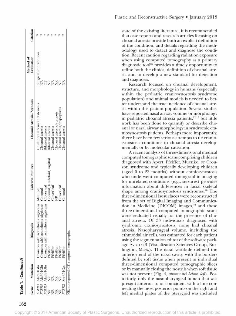
state of the existing literature, it is recommended that case reports and research articles focusing on choanal atresia provide both an explicit definition of the condition, and details regarding the meth-odology used to detect and diagnose the condi-tion. Recent caution regarding radiation exposure when using computed tomography as a primary diagnostic tool82 provides a timely opportunity to refine both the clinical definition of choanal atre-sia and to develop a new standard for detection and diagnosis.
Research focused on choanal development, structure, and morphology in humans (especially within the pediatric craniosynostosis syndrome population) and animal models is needed to bet-ter understand the true incidence of choanal atre-sia within this patient population. Several studies have reported nasal airway volume or morphology in pediatric choanal atresia patients,12,13 but little work has been done to quantify or describe cho-anal or nasal airway morphology in syndromic cra-niosynostosis patients. Perhaps more importantly, there have been few serious attempts to tie cranio-synostosis conditions to choanal atresia develop-mentally or by molecular causation.
A recent analysis of three-dimensional medical computed tomographic scans comprising children diagnosed with Apert, Pfeiffer, Muenke, or Crou-zon syndrome and typically developing children (aged 0 to 23 months) without craniosynostosis who underwent computed tomographic imaging for unrelated conditions (e.g., seizures) provides information about differences in facial skeletal shape among craniosynostosis syndromes.40 The three-dimensional isosurfaces were reconstructed from the set of Digital Imaging and Communica-tion in Medicine (DICOM) images,40 and these three-dimensional computed tomographic scans were evaluated visually for the presence of cho-anal atresia. Of 33 individuals diagnosed with syndromic craniosynostosis, none had choanal atresia. Nasopharyngeal volume, including the ethmoidal air cells, was estimated for each patient using the segmentation editor of the software pack-age Avizo 6.3 (Visualization Sciences Group, Bur-lington, Mass.). The nasal vestibule defined the anterior end of the nasal cavity, with the borders defined by soft tissue when present in individual three-dimensional computed tomographic slices or by manually closing the nostrils when soft tissue was not present (Fig. 4, above and below, left). Pos-teriorly, only the nasopharyngeal lumen that was present anterior to or coincident with a line con-necting the most posterior points on the right and left medial plates of the pterygoid was included FG
FR3
Ala
391G
luC
rouz
on w
ith
aca
nth
osis
nig
rica
ns
Mul
tipl
eU
nila
tera
l atr
esia
an
d st
enos
isSu
rgic
al in
terv
enti
on73
FGFR
3A
la39
1Glu
Cro
uzon
wit
h a
can
thos
is n
igri
can
sC
oron
alC
hoa
nal
atr
esia
NR
74
FGFR
3A
la39
1Glu
Cro
uzon
wit
h a
can
thos
is n
igri
can
sC
love
rlea
fB
ilate
ral a
tres
iaC
T75
NR
NR
Pfei
ffer
Cor
onal
, lam
bdoi
d, s
agit
tal
Bila
tera
l atr
esia
NR
76
NR
NR
Pfei
ffer
Clo
verl
eaf
Ch
oan
al a
tres
iaN
R77
NR
NR
Pfei
ffer
NR
Ch
oan
al a
tres
ia/s
ten
osis
NR
78
NR
NR
Pfei
ffer
Clo
verl
eaf
Ch
oan
al a
tres
ia/h
ypop
lasi
aN
R79
FGFR
2Se
r351
Cys
Pfei
ffer
Cor
onal
Ch
oan
al a
tres
iaN
R80
NR
, not
rep
orte
d; C
T, c
ompu
ted
tom
ogra
phy.
Tabl
e 1.
(Co
ntinue
d)
Gen
eM
utat
ion
Dia
gnos
isC
alva
rial
Sut
ures
Cho
anal
Atr
esia
/Ste
nosi
sM
etho
d of
E
valu
atio
nC
itat
ion

Copyright © 2017 American Society of Plastic Surgeons. Unauthorized reproduction of this article is prohibited.
Volume 141, Number 1 • Choanal Atresia and Craniosynostosis
163
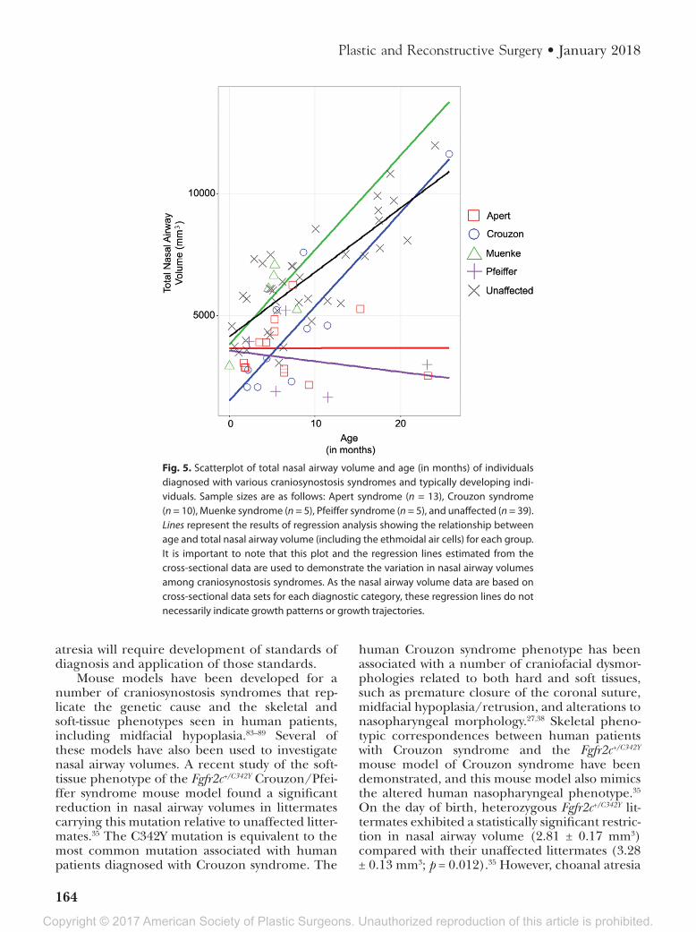
in the segmented volume (Fig. 4, below, right). Comparisons between unaffected children and those diagnosed with syndromic craniosynostosis reveal potentially reduced nasal airway volumes in children diagnosed with Apert and Pfeiffer syn-dromes (Figs. 5 and 6). Analysis of cross-sectional data representing nasal airway volumes of varying groups from birth to approximately 30 months of age shows that children diagnosed with Apert and Pfeiffer syndromes appear to share similar nasal airway volumes with children diagnosed with Muenke syndromes and their typically developing peers at birth. Although the sample size is small, the results also indicate that children diagnosed with Crouzon syndrome may have reduced nasal airway volumes at birth. Based on this analysis using limited samples, children diagnosed with Apert and Pfeiffer syndromes may experience an early postnatal developmental divergence that results in smaller overall nasal airways within the first year of life (Fig. 5).
The distinction between true choanal atre-sia and more diffuse nasal airway stenosis that is often present in syndromic craniosynostosis is important for both clinical and basic research reasons. Although it is essential that researchers in the field have a clear understanding of the correct terminology to ensure appropriate com-munication and reporting, there are also poten-tial clinical ramifications to consider. Choanal atresia in the newborn is a condition that is very amenable to early surgical intervention, which can often obviate the need for tracheostomy, prolonged neonatal intensive care unit hospital-ization, and continued respiratory monitoring. Nasal airway obstruction in the newborn with syndromic craniosynostosis may not be as read-ily surgically correctable in early life. Incorrect terminology may lead a surgeon down an errant pathway and may lead the child’s family to have unrealistic expectations. Knowledge of asso-ciations between craniosynostosis and choanal
Fig. 4. Nasal airway segmented from a computed tomographic image of a typically developing child at 10 months, as an example of how the nasal airway volume data presented in Figure 5 were collected. (Above, left) Axial com-puted tomographic image at the level of the orbits indicating area of close-up (red box) for three additional ana-tomical levels, including (above, right) the nasal cavity (red) with soft tissue of the nose bordering the anterior nares at the level of the maxillary sinuses; (below, left) midnares level with partial soft-tissue border of the anterior nares; and (below, right) at the level of the alveolar processes of the maxillae showing the nasopharyngeal lumen (red) anterior to a line (blue) connecting the most posterior points on the medial plates of the pterygoid bone.
Copyright © 2017 American Society of Plastic Surgeons. Unauthorized reproduction of this article is prohibited.
164
Plastic and Reconstructive Surgery • January 2018
atresia will require development of standards of diagnosis and application of those standards.
Mouse models have been developed for a number of craniosynostosis syndromes that rep-licate the genetic cause and the skeletal and soft-tissue phenotypes seen in human patients, including midfacial hypoplasia.83–89 Several of these models have also been used to investigate nasal airway volumes. A recent study of the soft-tissue phenotype of the Fgfr2c+/C342Y Crouzon/Pfei-ffer syndrome mouse model found a significant reduction in nasal airway volumes in littermates carrying this mutation relative to unaffected litter-mates.35 The C342Y mutation is equivalent to the most common mutation associated with human patients diagnosed with Crouzon syndrome. The
human Crouzon syndrome phenotype has been associated with a number of craniofacial dysmor-phologies related to both hard and soft tissues, such as premature closure of the coronal suture, midfacial hypoplasia/retrusion, and alterations to nasopharyngeal morphology.27,38 Skeletal pheno-typic correspondences between human patients with Crouzon syndrome and the Fgfr2c+/C342Y mouse model of Crouzon syndrome have been demonstrated, and this mouse model also mimics the altered human nasopharyngeal phenotype.35 On the day of birth, heterozygous Fgfr2c+/C342Y lit-termates exhibited a statistically significant restric-tion in nasal airway volume (2.81 ± 0.17 mm3) compared with their unaffected littermates (3.28 ± 0.13 mm3; p = 0.012).35 However, choanal atresia
Fig. 5. Scatterplot of total nasal airway volume and age (in months) of individuals diagnosed with various craniosynostosis syndromes and typically developing indi-viduals. Sample sizes are as follows: Apert syndrome (n = 13), Crouzon syndrome (n = 10), Muenke syndrome (n = 5), Pfeiffer syndrome (n = 5), and unaffected (n = 39). Lines represent the results of regression analysis showing the relationship between age and total nasal airway volume (including the ethmoidal air cells) for each group. It is important to note that this plot and the regression lines estimated from the cross-sectional data are used to demonstrate the variation in nasal airway volumes among craniosynostosis syndromes. As the nasal airway volume data are based on cross-sectional data sets for each diagnostic category, these regression lines do not necessarily indicate growth patterns or growth trajectories.

Copyright © 2017 American Society of Plastic Surgeons. Unauthorized reproduction of this article is prohibited.
Volume 141, Number 1 • Choanal Atresia and Craniosynostosis
165
Fig. 6. Three-dimensional computed tomographic reconstruction of a typically developing child (left) showing superim-posed segmentations of skin surface (beige), brain surface (gray), and upper airway lumen (blue). At right are “virtual endo-casts” of the nasopharynx of a child with Apert syndrome (pink, left) and a typically developing child (maroon, right) as segmented from high resolution three-dimensional computed tomographic reconstruction. Superimposition of the two virtual endocasts (second from right) shows local areas of greatest shape difference. This comparison is not a statistical com-parison of the nasopharyngeal anatomy of patients with Apert syndrome and typically developing individuals; rather, this superimposition provides an example of how nasopharyngeal morphology of craniosynostosis patients may differ from typically developing individuals.
Fig. 7. Three-dimensional computed tomographic reconstruction of the cranium of a 6-week-old C57BL/6J mouse showing the bones that form the osteologic borders of the choanae in the human skull: vomer (blue), basisphenoid (pink), medial pterygoid plates (red), and horizontal plates of the palatine bones (purple). The vomer (which is ghosted in this illustration) lies deep to the maxillae and so is hidden in an inferior view. The presphenoid is shown in green. Note the anatomi-cal separation of these bones compared to the human skull in Figure 1. The black arrow indicates the position of the choanae in mice at the soft-tissue intersection of the posterior nasal cavity and nasopharynx.

Copyright © 2017 American Society of Plastic Surgeons. Unauthorized reproduction of this article is prohibited.
166
Plastic and Reconstructive Surgery • January 2018
was not reported in any of the mice studied. As of this date, there have been no published reports of definitive choanal atresia in any mouse models of syndromic craniosynostosis.
While murine data provide valuable infor-mation about the molecular and developmental mechanisms that produce the choanae, significant differences in human and murine craniofacial anatomy and development must be taken into con-sideration when evaluating the comorbidity of cho-anal atresia and craniosynostosis using cross-species comparisons. Because of the rostral-caudal elonga-tion of the murine premaxillae, maxillae, palatine bones, and the soft palate, different osteologic and soft-tissue boundaries define the murine choanae relative to humans. In humans, choanal atresia has been attributed to a combination of a thickening of the posterior vomer with medialization of the pterygoid plates of the sphenoid. Although use-ful murine models of choanal atresia have recently been produced and will be critical to determining the molecular and developmental basis of choanal atresia,90 species-specific differences including the anatomical separation of the vomer and pterygoid plates along the rostrocaudal axis in mice suggests an alternate structural foundation for murine choanal atresia (Fig. 7). While mouse models are an excellent tool for understanding the cause of human craniofacial disorders such as craniosyn-ostosis, given the tremendous number of genetic mutations implicated in craniosynostosis condi-tions, each model can represent only a single development pathway to the craniosynostosis phe-notype. We propose that there are potentially as many ways to produce choanal atresia.
The significant correlation between specific craniosynostosis syndromes and reduced nasal air-way volume in mouse models for craniosynostosis and human pediatric patients indicates comorbid-ity of choanal and nasopharyngeal dysmorpholo-gies and craniosynostosis conditions. Genetic, developmental, and epidemiologic sources of these interactions are areas particularly worthy of further research.
Joan T. Richtsmeier, Ph.D.Department of AnthropologyPennsylvania State University
409 Carpenter BuildingUniversity Park, Pa. 16802
ACKNOWLEDGMENTSResearch presented here was funded in part by
the National Institutes of Health (National Insti-tute of Dental and Craniofacial Research, National
Institute of Child Health and Human Develop-ment, and American Recovery and Reinvestment Act) (R01-DE018500, R01-DE018500-S1, R01-DE022988, and P01HD078233) and the National Science Founda-tion (BCS-0725227). The authors thank members of the Bernstein, Jabs, and Richtsmeier laboratories for ongoing discussions about the development, anatomy, and dysmor-phology of the choanae that informed this study, includ-ing Susan Motch Perrine, Kevin Flaherty, Kazuhiko Kawasaki, Mizuho Kawasaki, Greg Holmes, James Azzi, and Sameep Kadakia. Use of the computed tomographic images was approved by the institutional review boards of the Pennsylvania State University and the participating institutions, and the images were acquired in accordance with institutional guidelines. All collected images were anonymized and no information other than sex, age at the time of the computed tomographic examination, and causative mutation were available.
REFERENCES 1. Adil E, Huntley C, Choudhary A, Carr M. Congenital nasal
obstruction: Clinical and radiologic review. Eur J Pediatr. 2012;171:641–650.
2. Zhu M, Ahlberg PE. The origin of the internal nostril of tet-rapods. Nature 2004;432:94–97.
3. Jankowski R. The Evo-Devo Origin of the Nose, Anterior Skull Base and Midface. Paris: Springer Paris; 2013.
4. Sperber GH, Sperber SM, Guttmann GD. Craniofacial Embryogenetics and Development. Shelton, Conn: People’s Medical; 2010.
5. Hengerer AS, Brickman TM, Jeyakumar A. Choanal atresia: Embryologic analysis and evolution of treatment. A 30-year experience. Laryngoscope 2008;118:862–866.
6. Kim CH, Park HW, Kim K, Yoon JH. Early development of the nose in human embryos: A stereomicroscopic and histo-logic analysis. Laryngoscope 2004;114:1791–1800.
7. Tamarin A. The formation of the primitive choanae and the junction of the primary and secondary palates in the mouse. Am J Anat. 1982;165:319–337.
8. Mankarious LA, Goudy SL. Craniofacial and upper airway development. Paediatr Respir Rev. 2010;11:193–198.
9. Kwong KM. Current updates on choanal atresia. Front Pediatr. 2015;3:52.
10. Corrales CE, Koltai PJ. Choanal atresia: Current concepts and controversies. Curr Opin Otolaryngol Head Neck Surg. 2009;17:466–470.
11. Harney MS, Russell J. Choanal atresia. In: PuriP, HöllwarthM, eds. Pediatric Surgery. Berlin, Heidelberg: Springer Berlin Heidelberg; 2009:223–227.
12. Aslan S, Yilmazer C, Yildirim T, Akkuzu B, Yilmaz I. Comparison of nasal region dimensions in bilateral cho-anal atresia patients and normal controls: A computed tomographic analysis with clinical implications. Int J Pediatr Otorhinolaryngol. 2009;73:329–335.
13. Cheung R, Prince M. Comparison of craniofacial skeletal characteristics of infants with bilateral choanal atresia and an age-matched normative population: Computed tomography analysis. J Otolaryngol. 2001;30:173–178.
14. Bergstrom L, Owens O. Posterior choanal atresia: A syndro-mal disorder. Laryngoscope 1984;94:1273–1276.

Copyright © 2017 American Society of Plastic Surgeons. Unauthorized reproduction of this article is prohibited.
Volume 141, Number 1 • Choanal Atresia and Craniosynostosis
167
15. Lowe LH, Booth TN, Joglar JM, Rollins NK. Midface anoma-lies in children. Radiographics 2000;20:907–922.
16. Brown OE, Myer CM III, Manning SC. Congenital nasal pyri-form aperture stenosis. Laryngoscope 1989;99:86–91.
17. Keller JL, Kacker A. Choanal atresia, CHARGE association, and congenital nasal stenosis. Otolaryngol Clin North Am. 2000;33:1343–1351.
18. Ramsden JD, Campisi P, Forte V. Choanal atresia and cho-anal stenosis. Otolaryngol Clin North Am. 2009;42:339–352.
19. Meyers GA, Orlow SJ, Munro IR, Przylepa KA, Jabs EW. Fibroblast growth factor receptor 3 (FGFR3) transmem-brane mutation in Crouzon syndrome with acanthosis nigri-cans. Nat Genet. 1995;11:462–464.
20. Wilkes D, Rutland P, Pulleyn LJ, et al. A recurrent mutation, ala391glu, in the transmembrane region of FGFR3 causes Crouzon syndrome and acanthosis nigricans. J Med Genet. 1996;33:744–748.
21. Sculerati N, Gottlieb MD, Zimbler MS, Chibbaro PD, McCarthy JG. Airway management in children with major craniofacial anomalies. Laryngoscope 1998;108:1806–1812.
22. Buchman SR, Muraszko K. Syndromic craniosynostosis. In: Thaller S, Bradley JP, Garri JI, eds. Craniofacial Surgery. New York: CRC Press; 2007:103–126.
23. Allanson JE, Cunniff C, Hoyme HE, McGaughran J, Muenke M, Neri G. Elements of morphology: Standard terminology for the head and face. Am J Med Genet A 2009;149:6–28.
24. Neskey D, Eloy JA, Casiano RR. Nasal, septal, and turbi-nate anatomy and embryology. Otolaryngol Clin North Am. 2009;42:193–205.
25. Burrow TA, Saal HM, de Alarcon A, Martin LJ, Cotton RT, Hopkin RJ. Characterization of congenital anomalies in indi-viduals with choanal atresia. Arch Otolaryngol Head Neck Surg. 2009;135:543–547.
26. Durward A, Lord OC, Polson CJ. Congenital choanal atresia. J Laryngol Otol. 1945;60:461–500.
27. Cohen MM, MacLean RE, eds. Craniosynostosis: Diagnosis, Evaluation, and Management. New York: Oxford University Press; 2000.
28. Arnaud-López L, Fragoso R, Mantilla-Capacho J, Barros-Núñez P. Crouzon with acanthosis nigricans: Further delin-eation of the syndrome. Clin Genet. 2007;72:405–410.
29. Hehr U, Muenke M. Craniosynostosis syndromes: From genes to premature fusion of skull bones. Mol Genet Metab. 1999;68:139–151.
30. Flaherty K, Singh N, Richtsmeier JT. Understanding cranio-synostosis as a growth disorder. Wiley Interdiscip Rev Dev Biol. 2016;5:429–459.
31. Heuzé Y, Holmes G, Peter I, Richtsmeier JT, Jabs EW. Closing the gap: Genetic and genomic continuum from syndromic to nonsyndromic craniosynostoses. Curr Genet Med Rep. 2014;2:135–145.
32. Lattanzi W, Barba M, Di Pietro L, Boyadjiev SA. Genetic advances in craniosynostosis. Am J Med Genet A 2017;173:1406–1429.
33. Heuzé Y, Boyadjiev SA, Marsh JL, et al. New insights into the relationship between suture closure and craniofacial dysmorphology in sagittal nonsyndromic craniosynostosis. J Anat. 2010;217:85–96.
34. Heuzé Y, Martínez-Abadías N, Stella JM, et al. Unilateral and bilateral expression of a quantitative trait: Asymmetry and symmetry in coronal craniosynostosis. J Exp Zool B Mol Dev Evol. 2012;318:109–122.
35. Martínez-Abadías N, Motch SM, Pankratz TL, et al. Tissue-specific responses to aberrant FGF signaling in complex head phenotypes. Dev Dyn. 2013;242:80–94.
36. Cohen MM Jr, Kreiborg S. A clinical study of the craniofa-cial features in Apert syndrome. Int J Oral Maxillofac Surg. 1996;25:45–53.
37. Cunningham ML, Seto ML, Ratisoontorn C, Heike CL, Hing AV. Syndromic craniosynostosis: From history to hydrogen bonds. Orthod Craniofac Res. 2007;10:67–81.
38. Johnson D, Wilkie AO. Craniosynostosis. Eur J Hum Genet. 2011;19:369–376.
39. Nah HD, Koyama E, Agochukwu NB, Bartlett SP, Muenke M. Phenotype profile of a genetic mouse model for Muenke syndrome. Childs Nerv Syst. 2012;28:1483–1493.
40. Heuzé Y, Martínez-Abadías N, Stella JM, et al. Quantification of facial skeletal shape variation in fibroblast growth factor receptor-related craniosynostosis syndromes. Birth Defects Res A Clin Mol Teratol. 2014;100:250–259.
41. Kelley RI, Kratz LE, Glaser RL, Netzloff ML, Wolf LM, Jabs EW. Abnormal sterol metabolism in a patient with Antley-Bixler syndrome and ambiguous genitalia. Am J Med Genet. 2002;110:95–102.
42. Machado LE, Osborne NG, Bonilla-Musoles F. Antley-Bixler syndrome: Report of a case. J Ultrasound Med. 2001;20:73–77.
43. Hassell S, Butler MG. Antley-Bixler syndrome: Report of a patient and review of literature. Clin Genet. 1994;46:372–376.
44. Hosalkar HS, Shah HS, Gujar PS, Shaw BA. The Antley-Bixler syndrome: Two new cases. J Postgrad Med. 2001;47:252–255.
45. Adolphs N, Klein M, Haberl EJ, Graul-Neumann L, Menneking H, Hoffmeister B. Antley-Bixler-syndrome: Staged management of craniofacial malformations from birth to adolescence. A case report. J Craniomaxillofac Surg. 2011;39:487–495.
46. Kumar MR, Bhat BV, Bhatia BD. Apert syndrome with partial post-axial polydactyly and unilateral choanal atresia. Indian Pediatr. 1994;31:869–871.
47. al-Qattan MM, al-Husain MA. Classification of hand anoma-lies in Apert’s syndrome. J Hand Surg Br. 1996;21:266–268.
48. Pius S, Ibrahim HA, Bello M, Mbaya K, Ambe JP. Apert syn-drome: A case report and review of literature. Open J Pediatr. 2016;6:175–184.
49. Shrestha M, Adhikari N, Shah G. Apert syndrome (acroceph-alosyndactyly): A rare syndromic craniosynostosis. J Nepal Paediatr Soc. 2009;29:92–93.
50. Boynukalın K, Baykal C, Dolar O, Ozcan N. Prenatal diag-nosis of Apert syndrome: A case report. Basic Clin Sci. 2013;2:165–169.
51. Park WJ, Theda C, Maestri NE, et al. Analysis of phenotypic features and FGFR2 mutations in Apert syndrome. Am J Hum Genet. 1995;57:321–328.
52. Lajeunie E, Cameron R, El Ghouzzi V, et al. Clinical variability in patients with Apert’s syndrome. J Neurosurg. 1999;90:443–447.
53. Oldridge M, Zackai EH, McDonald-McGinn DM, et al. De novo alu-element insertions in FGFR2 identify a distinct pathological basis for Apert syndrome. Am J Hum Genet. 1999;64:446–461.
54. Hall BD, Cadle RG, Golabi M, Morris CA, Cohen MM Jr. Beare-Stevenson cutis gyrata syndrome. Am J Med Genet. 1992;44:82–89.
55. Hsu TY, Chang SY, Wang TJ, Ou CY, Chen ZH, Hsu PH. Prenatal sonographic appearance of Beare-Stevenson cutis gyrata syndrome: Two- and three-dimensional ultrasono-graphic findings. Prenat Diagn. 2001;21:665–667.
56. Ito S, Matsui K, Ohsaki E, et al. A cloverleaf skull syndrome probably of Beare-Stevenson type associated with Chiari mal-formation. Brain Dev. 1996;18:307–311.
57. Upmeyer S, Bothwell M, Tobias JD. Perioperative care of a patient with Beare-Stevenson syndrome. Paediatr Anaesth. 2005;15:1131–1136.

Copyright © 2017 American Society of Plastic Surgeons. Unauthorized reproduction of this article is prohibited.
168
Plastic and Reconstructive Surgery • January 2018
58. Too CW, Tang PH. Imaging findings of chronic subluxation of the os odontoideum and cervical myelopathy in a child with Beare-Stevenson cutis gyrata syndrome after surgery to the head and neck. Ann Acad Med Singapore 2009;38:832–834.
59. Barge-Schaapveld DQ, Brooks AS, Lequin MH, van Spaendonk R, Vermeulen RJ, Cobben JM. Beare-Stevenson syndrome: Two Dutch patients with cerebral abnormalities. Pediatr Neurol. 2011;44:303–307.
60. Fonseca RF, Costa-Lima MA, Pereira ET, Castilla EE, Orioli IM. Beare-Stevenson cutis gyrata syndrome: A new case of a c.1124C→G (Y375C) mutation in the FGFR2 gene. Mol Med Rep. 2008;1:753–755.
61. Vargas RA, Maegawa GH, Taucher SC, et al. Beare-Stevenson syndrome: Two South American patients with FGFR2 analy-sis. Am J Med Genet A 2003;121:41–46.
62. Wenger TL, Bhoj EJ, Wetmore RF, et al. Beare-Stevenson syn-drome: Two new patients, including a novel finding of tra-cheal cartilaginous sleeve. Am J Med Genet A 2015;167:852–857.
63. Yu JE, Jeong SY, Yang JA, Park MS, Kim HJ, Yoon SH. Genotypic and phenotypic analyses of Korean patients with syndromic craniosynostosis. Clin Genet. 2009;76:287–291.
64. Ron N, Leung S, Carney E, Gerber A, David KL. A case of Beare-Stevenson syndrome with unusual manifestations. Am J Case Rep. 2016;17:254–258.
65. Scheid SC, Spector AR, Luft JD. Tracheal cartilaginous sleeve in Crouzon syndrome. Int J Pediatr Otorhinolaryngol. 2002;65:147–152.
66. Alpers CE, Edwards MS. Hemangiomatous anomaly of bone in Crouzon’s syndrome: Case report. Neurosurgery 1985;16:391–394.
67. Dunmade AD, Ajayi I, Alabi BS, Mokuolu OA, Bolaji BO, Oyinloye OI. Intranasal endoscopic repair of bilateral cho-anal atresia in a male newborn with Crouzon’s syndrome. East Cent Afr J Surg. 2015;20:96–101.
68. Lo LJ, Chen YR. Airway obstruction in severe syndromic cra-niosynostosis. Ann Plast Surg. 1999;43:258–264.
69. Beck R, Sertie AL, Brik R, Shinawi M. Crouzon syndrome: Association with absent pulmonary valve syndrome and severe tracheobronchomalacia. Pediatr Pulmonol. 2002;34:478–481.
70. Okajima K, Robinson LK, Hart MA, et al. Ocular anterior chamber dysgenesis in craniosynostosis syndromes with a fibroblast growth factor receptor 2 mutation. Am J Med Genet. 1999;85:160–170.
71. Suslak L, Glista B, Gertzman GB, Lieberman L, Schwartz RA, Desposito F. Crouzon syndrome with periapical cemental dysplasia and acanthosis nigricans: The pleiotropic effect of a single gene? Birth Defects Orig Artic Ser. 1985;21:127–134.
72. Breitbart AS, Eaton C, McCarthy JG. Crouzon’s syndrome associated with acanthosis nigricans: Ramifications for the craniofacial surgeon. Ann Plast Surg. 1989;22:310–315.
73. Schweitzer DN, Graham JM Jr, Lachman RS, et al. Subtle radio-graphic findings of achondroplasia in patients with Crouzon syndrome with acanthosis nigricans due to an Ala391Glu sub-stitution in FGFR3. Am J Med Genet. 2001;98:75–91.
74. Di Rocco F, Collet C, Legeai-Mallet L, et al. Crouzon syn-drome with acanthosis nigricans: A case-based update. Childs Nerv Syst. 2011;27:349–354.
75. Sharda S, Panigrahi I, Gupta K, Singhi S, Kumar R. A new-born with acanthosis nigricans: Can it be Crouzon syndrome with acanthosis nigricans? Pediatr Dermatol. 2010;27:43–47.
76. Kroczek RA, Mühlbauer W, Zimmermann I. Cloverleaf skull associated with Pfeiffer syndrome: Pathology and manage-ment. Eur J Pediatr. 1986;145:442–445.
77. Oyamada MK, Ferreira HS, Hoff M. Pfeiffer Syndrome type 2: Case report. Sao Paulo Med J. 2003;121:176–179.
78. Stoler JM, Rosen H, Desai U, Mulliken JB, Meara JG, Rogers GF. Cleft palate in Pfeiffer syndrome. J Craniofac Surg. 2009;20:1375–1377.
79. Erten E, Çekmen N, Bilgin F, Orhan ME. Respiratory and cra-nial complications during anaesthesia in Pfeiffer syndrome. Brain Disord Ther. 2015;4. doi:10.4172/2168-975X.1000175.
80. Mathijssen IM, Vaandrager JM, Hoogeboom AJ, Hesseling-Janssen AL, van den Ouweland AM. Pfeiffer’s syndrome resulting from an S351C mutation in the fibroblast growth factor receptor-2 gene. J Craniofac Surg. 1998;9:207–209.
81. Brown OE, Smith T, Armstrong E, Grundfast K. The evalua-tion of choanal atresia by computed tomography. Int J Pediatr Otorhinolaryngol. 1986;12:85–98.
82. Chen JX, Kachniarz B, Gilani S, Shin JJ. Risk of malignancy associated with head and neck CT in children: A systematic review. Otolaryngol Head Neck Surg. 2014;151:554–566.
83. Chen L, Li D, Li C, Engel A, Deng CX. A Ser252Trp [corrected] substitution in mouse fibroblast growth fac-tor receptor 2 (Fgfr2) results in craniosynostosis. Bone 2003;33:169–178.
84. Holmes G. Mouse models of Apert syndrome. Childs Nerv Syst. 2012;28:1505–1510.
85. Mai S, Wei K, Flenniken A, et al. The missense mutation W290R in Fgfr2 causes developmental defects from aberrant IIIb and IIIc signaling. Dev Dyn. 2010;239:1888–1900.
86. Twigg SR, Healy C, Babbs C, et al. Skeletal analysis of the Fgfr3(P244R) mouse, a genetic model for the Muenke cra-niosynostosis syndrome. Dev Dyn. 2009;238:331–342.
87. Wang Y, Xiao R, Yang F, et al. Abnormalities in cartilage and bone development in the Apert syndrome FGFR2(+/S252W) mouse. Development 2005;132:3537–3548.
88. Zhou YX, Xu X, Chen L, Li C, Brodie SG, Deng CX. A Pro250Arg substitution in mouse Fgfr1 causes increased expression of Cbfa1 and premature fusion of calvarial sutures. Hum Mol Genet. 2000;9:2001–2008.
89. Eswarakumar VP, Horowitz MC, Locklin R, Morriss-Kay GM, Lonai P. A gain-of-function mutation of Fgfr2c demonstrates the roles of this receptor variant in osteogenesis. Proc Natl Acad Sci USA 2004;101:12555–12560.
90. Kurosaka H, Wang Q, Sandell L, Yamashiro T, Trainor PA. Rdh10 loss-of-function and perturbed retinoid signaling underlies the etiology of choanal atresia. Hum Mol Genet. 2017;26:1268–1279.