CHEST X-RAY SEGMENTATION to CALCULATE … · The calculation of pleural effusion index on patients...
4
International Journal of Innovative Research in Advanced Engineering (IJIRAE) ISSN: 2349-2163 Issue 07, Volume 4 (July 2017) www.ijirae.com DOI:/10.26562/IJIRAE.2017.JYAE10084 _________________________________________________________________________________________________ IJIRAE: Impact Factor Value – SJIF: Innospace, Morocco (2016): 3.916 | PIF: 2.469 | Jour Info: 4.085 | ISRAJIF (2016): 3.715 | Indexcopernicus: (ICV 2015): 47.91 IJIRAE © 2014- 17, All Rights Reserved Page -9 CHEST X-RAY SEGMENTATION to CALCULATE PLEURAL EFFUSION INDEX in PATIENT with DENGUE HEMORRHAGIC FEVER Arnefia Mei Yusnida * Magister of Physics, Faculty of Science and Mathematics, Diponegoro University, Indonesia [email protected] Catur Edi Widodo Department of Physics,Faculty of Science and Mathematics, Diponegoro university,Indonesia [email protected] Kusworo Adi Department of Physics,Faculty of Science and Mathematics, Diponegoro university,Indonesia [email protected] Manuscript History Number: IJIRAE/RS/Vol.04/Issue07/JYAE10084 DOI: 10.26562/IJIRAE.2017.JYAE10084 Received: 15, June 2017 Final Correction: 12, July 2017 Final Accepted: 19, July 2017 Published: July 2017 Citation: Mei, Y. A.; Edi, W. C. & Kusworo, A. (2017), 'CHEST X-RAY SEGMENTATION to CALCULATE PLEURAL EFFUSION INDEX in PATIENT with DENGUE HEMORRHAGIC FEVER', Master's thesis, Department of Physics, Faculty of Science and Mathematics, Diponegoro university,Indonesia . Editor: Dr.A.Arul L.S, Chief Editor, IJIRAE, AM Publications, India Copyright: ©2017 This is an open access article distributed under the terms of the Creative Commons Attribution License, Which Permits unrestricted use, distribution, and reproduction in any medium, provided the original author and source are credited. Abstract— A study of the calculation of pleural effusion index (PEI) in patient with dengue hemorrhagic fever (DHF) has been conducted. PEI calculation was done through matlab programming language. Some digital image processing methods used in this research were thresholding segmentation, morphology operation, and calculation of pixel number per column in image to get PEI value. PEI values generated from image processing can be an alternative to replace the manual calculations done by physicians and used to demonstrate the gravity level of DHF. Keywords— Dengue hemorrhagic fever, Pleural effusion index, Segmentation. I. INTRODUCTION Digital image processing technology has been progressing rapidly in various fields including the medical field. Through several stages of processing, an image can give us desired information. Image segmentation is one of the stages in image processing that is often used[1]. The success of the medical image processing system depends on the segmentation step. If the segmentation step is done properly the analysis will then give correct results [2] The main objective of segmentation is to divide the image into parts that have a strong correlation with the object in the image. Clear object boundary information will be obtained by the well-segmented medical image. This information is very helpful for medical personnel in an objective and accurate way to perform the analysis, diagnosis, treatment planning, and necessary medical action [3].
Transcript of CHEST X-RAY SEGMENTATION to CALCULATE … · The calculation of pleural effusion index on patients...

International Journal of Innovative Research in Advanced Engineering (IJIRAE) ISSN: 2349-2163 Issue 07, Volume 4 (July 2017) www.ijirae.com DOI:/10.26562/IJIRAE.2017.JYAE10084
_________________________________________________________________________________________________ IJIRAE: Impact Factor Value – SJIF: Innospace, Morocco (2016): 3.916 | PIF: 2.469 | Jour Info: 4.085 |
ISRAJIF (2016): 3.715 | Indexcopernicus: (ICV 2015): 47.91 IJIRAE © 2014- 17, All Rights Reserved Page -9
CHEST X-RAY SEGMENTATION to CALCULATE PLEURAL EFFUSION INDEX in PATIENT with DENGUE
HEMORRHAGIC FEVER Arnefia Mei Yusnida*
Magister of Physics, Faculty of Science and Mathematics, Diponegoro University, Indonesia [email protected]
Catur Edi Widodo Department of Physics,Faculty of Science and Mathematics, Diponegoro university,Indonesia
[email protected] Kusworo Adi
Department of Physics,Faculty of Science and Mathematics, Diponegoro university,Indonesia [email protected]
Manuscript History Number: IJIRAE/RS/Vol.04/Issue07/JYAE10084 DOI: 10.26562/IJIRAE.2017.JYAE10084 Received: 15, June 2017 Final Correction: 12, July 2017 Final Accepted: 19, July 2017 Published: July 2017 Citation: Mei, Y. A.; Edi, W. C. & Kusworo, A. (2017), 'CHEST X-RAY SEGMENTATION to CALCULATE PLEURAL EFFUSION INDEX in PATIENT with DENGUE HEMORRHAGIC FEVER', Master's thesis, Department of Physics, Faculty of Science and Mathematics, Diponegoro university,Indonesia . Editor: Dr.A.Arul L.S, Chief Editor, IJIRAE, AM Publications, India Copyright: ©2017 This is an open access article distributed under the terms of the Creative Commons Attribution License, Which Permits unrestricted use, distribution, and reproduction in any medium, provided the original author and source are credited.
Abstract— A study of the calculation of pleural effusion index (PEI) in patient with dengue hemorrhagic fever (DHF) has been conducted. PEI calculation was done through matlab programming language. Some digital image processing methods used in this research were thresholding segmentation, morphology operation, and calculation of pixel number per column in image to get PEI value. PEI values generated from image processing can be an alternative to replace the manual calculations done by physicians and used to demonstrate the gravity level of DHF.
Keywords— Dengue hemorrhagic fever, Pleural effusion index, Segmentation.
I. INTRODUCTION
Digital image processing technology has been progressing rapidly in various fields including the medical field. Through several stages of processing, an image can give us desired information. Image segmentation is one of the stages in image processing that is often used[1]. The success of the medical image processing system depends on the segmentation step. If the segmentation step is done properly the analysis will then give correct results [2]
The main objective of segmentation is to divide the image into parts that have a strong correlation with the object in the image. Clear object boundary information will be obtained by the well-segmented medical image. This information is very helpful for medical personnel in an objective and accurate way to perform the analysis, diagnosis, treatment planning, and necessary medical action [3].

International Journal of Innovative Research in Advanced Engineering (IJIRAE) ISSN: 2349-2163 Issue 07, Volume 4 (July 2017) www.ijirae.com DOI:/10.26562/IJIRAE.2017.JYAE10084
_________________________________________________________________________________________________ IJIRAE: Impact Factor Value – SJIF: Innospace, Morocco (2016): 3.916 | PIF: 2.469 | Jour Info: 4.085 |
ISRAJIF (2016): 3.715 | Indexcopernicus: (ICV 2015): 47.91 IJIRAE © 2014- 17, All Rights Reserved Page -10
In previous research, segmentation method was used to calculate the value of cardiothoracicratio (CTR) with an accuracy value of more than 90%. Several developmental methods of segmentation also used to detect lung abnormality automatically[4]. The segmentation of the Chest X-Ray (CXR) image can also be used for simple calculation of pleural effusion volume [5].
In this study, chest image segmentation would be used in the calculation of pleuraleffusion index (PEI) on denguehemorrhagic fever (DHF) patients with pleural effusion. PEI is the ratio between the maximum width of the pleural effusion and the maximum width of right hemitorax on chest radiographic examination of the right lateral decubitus position. The spot determined as the maximum width of right pleural effusion was the upper right diaphragm with the right lung because the effusion fluid will mostly collect in the right costofrenicus corner due to the influence of gravity. PEI assessment can be used as a predictor of the severity of DHF which is a risk factor for mortality[6]. Currently PEI calculation was still done manually by radiologist specialists. The image was printed in the film and measured in the required area using a ruler. The calculation of PEI from patients indicated by DHF must wait for the arrival of radiologist specialists who do not always stay in place. Based on the existing problems, the method of chest image segmentation is expected to be used as an alternative to calculate PEI value, so it can be used as a reference for handling patients with proper diagnosis of DHF.
II. MATERIALS AND METHOD 2.1 Materials The material used in this research was RLD projection of CXR with indicated effusions as the radiation result of X-ray machine (Toshiba Rotanode type DRX-1603B) with the maximum voltage of 150 kV. The example of the CXR used in this research is shown in Figure 2.1.
Fig 2.1 Chest X-Ray RLD projection with pleural effusion.. 2.2 Method
The calculation of pleural effusion index on patients with DHF were through several steps:
1. Inputting the CRX using matlab programming. 2. Determining the ROI that was the effusion in the right lung. 3. Determining the thresholding value to gain the binary image through the equation
T = T [x,y, p(x,y), f(x,y)] (2.1)
On the equation (2.1), T represents the threshold value. The variable of x, y is as the coordinate of threshold value, p (x, y), f (x, y) are the gray level of image pixel. The g (x, y) threshold image can be defined as follows:
g(x,y) = 1 jika f(x,y) > T g(x,y) = 0 jika f(x,y) ≤ T (2.2) 4. Performing a morphological operation (dilation) to obtain a firm boundary between the part of lung and
pleural with the indicated effusion. The dilation operation thickens the image. The extent of how much it should be thicken is based on the structuring element. The structuring element is a part of the image. The morphological transformation dilation (+) combines two sets using vector addition.The dilation operation can be done by performing vector addition of the pair of elements for both the sets p and q .
International Journal of Innovative Research in Advanced Engineering (IJIRAE) ISSN: 2349-2163 Issue 07, Volume 4 (July 2017) www.ijirae.com DOI:/10.26562/IJIRAE.2017.JYAE10084
_________________________________________________________________________________________________ IJIRAE: Impact Factor Value – SJIF: Innospace, Morocco (2016): 3.916 | PIF: 2.469 | Jour Info: 4.085 |
ISRAJIF (2016): 3.715 | Indexcopernicus: (ICV 2015): 47.91 IJIRAE © 2014- 17, All Rights Reserved Page -11
5. Calculating the pixels number of each line. The PEI value was obtained from the number of pixels at the
maximum pleural effusion width (A) and the right maximum hemitorax width (B) which shown in Figure 2.2.
Fig. 2.2 The Illustration of PEI calculation.PEI value was obtained from the calculation A/B*100%
III. RESULTS AND DISCUSSION
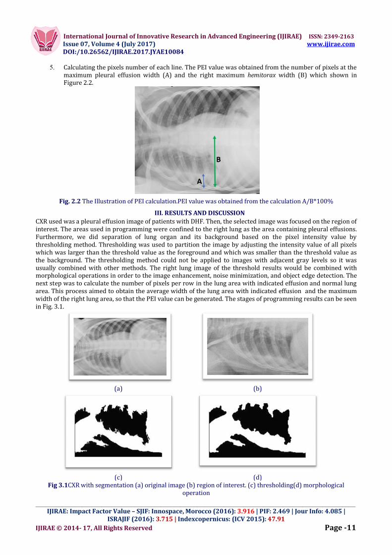
CXR used was a pleural effusion image of patients with DHF. Then, the selected image was focused on the region of interest. The areas used in programming were confined to the right lung as the area containing pleural effusions. Furthermore, we did separation of lung organ and its background based on the pixel intensity value by thresholding method. Thresholding was used to partition the image by adjusting the intensity value of all pixels which was larger than the threshold value as the foreground and which was smaller than the threshold value as the background. The thresholding method could not be applied to images with adjacent gray levels so it was usually combined with other methods. The right lung image of the threshold results would be combined with morphological operations in order to the image enhancement, noise minimization, and object edge detection. The next step was to calculate the number of pixels per row in the lung area with indicated effusion and normal lung area. This process aimed to obtain the average width of the lung area with indicated effusion and the maximum width of the right lung area, so that the PEI value can be generated. The stages of programming results can be seen in Fig. 3.1.
(a)
(b)
(c) (d)
Fig 3.1CXR with segmentation (a) original image (b) region of interest. (c) thresholding(d) morphological operation

International Journal of Innovative Research in Advanced Engineering (IJIRAE) ISSN: 2349-2163 Issue 07, Volume 4 (July 2017) www.ijirae.com DOI:/10.26562/IJIRAE.2017.JYAE10084
_________________________________________________________________________________________________ IJIRAE: Impact Factor Value – SJIF: Innospace, Morocco (2016): 3.916 | PIF: 2.469 | Jour Info: 4.085 |
ISRAJIF (2016): 3.715 | Indexcopernicus: (ICV 2015): 47.91 IJIRAE © 2014- 17, All Rights Reserved Page -12
THE RESULTS OF THE NUMBER OF SAMPLES CALCULATION CAN BE SEEN IN TABLE 3.1.
TABLE 3.1 CALCULATIONS OF PLEURAL EFFUSION INDEX VALUES
IV. CONCLUSION
The method offered in this research was the calculation of pleural effusion index value through digital image processing with segmentation as a substitute for manual method to assist the physicians and other medical officers to advance the medical action in patients with DHF and pleural effusion. We conclude that chest image segmentation can be used as one of the predictors of DHF severity.
REFERENCES
1. Hazlinger M, Ctvrtlik F, Langova K and Herman M, 2014, Quantification of pleural effusion on CT by simple measurement,Biomedical Papers 158(1): 107–111.
2. Itai Y, Kim H, Ishikawa S, Yamamoto A and Nakamura K, 2007, A segmentation method of lung areas by using snakes and automatic detection of abnormal shadow on the areas, International Journal of Innovative Computing, Information and Control 3(2): 277–284.
3. Mardhiyah A and Harjoko A, 2011, Method of Lung and Heart Segmentation on X-Ray Thorax Images. Ijeis 1(2): 35–44.
4. Setiati TE, Retnaningsih A, Supriatna M and Soemantri A (2005) Vascular leakage score as the early predictor of shock. Jurnal Kedokteran Brawijaya XXI(1): 16–21.
5. Soesanti I, Susanto A, Widodo TS and Tjokronagoro M., 2010, Computational Analyzes on Image Segmentation of Adaftif Based Fuzzy Optimized Logic, Forum 33: 89–96.
6. Tarambale MR and Lingayat NS (2013) Computer Based Performance Evaluation of Segmentation Methods for Chest X-Ray Image. International Journal of Bioscience, Biochemistry and Bioinformatics 3(6): 545–551.
Patient A B PEI (%)
I 51.7647 179 28.9188
II 19.8667 172 11.5504
III 48.3529 179 27.0128
IV 55.913 181 30.8912
V 46.1429 175 26.3673