Chest Tube and Water-seal Drainage
25
CHEST TUBE and WATER-SEAL DRAINAGE By: Ghada J. Al-Omaireen
Transcript of Chest Tube and Water-seal Drainage

CHEST TUBE and WATER-SEAL DRAINAGE
By: Ghada J. Al-Omaireen

Objectives
Introduction Chest drainage system Indications Chest drainage insertion Types of drainage system Nursing managements References
Normal Breathing Mechanism Operates on the principle of negative
pressure Pressure in the chest cavity is lower
than the atmosphere, causing air to move into the lungs during inspiration.
When chest is opened, there is loss of negative pressure which can collapse a lung.

Chest Drainage Systems
Crucial intervention for improving gas exchange and breathing
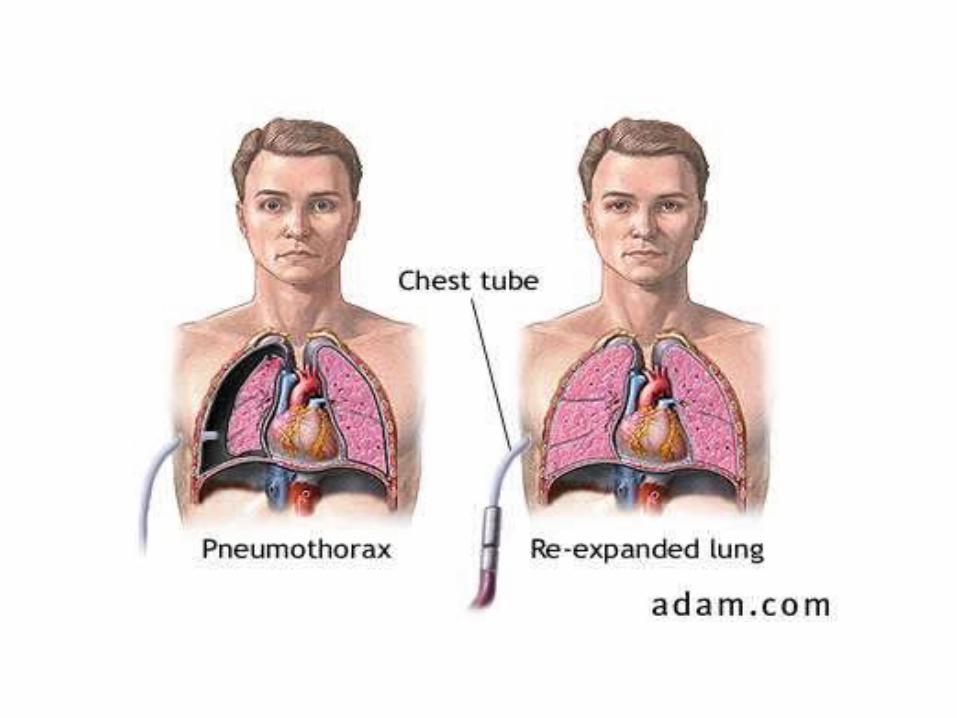
Chest drainage therapy involves the removal of air, blood, pus, or other secretions from the chest cavity.
Installing a chest drainage tube can be either an emergency or a planned procedure.
Removing air or fluids from the chest involves the insertion of a tube through the skin and the muscles between the ribs, and into the chest cavity.
This cavity is also called the pleural space.

Who May Need One
Conditions that may need to be treated by chest drainage therapy include:
Pleural effusion (excess fluid that accumulates in the pleura)
Tuberculosis spontaneous pneumothorax that causes
more than a 25% collapse of the lung. cancer that causes excessive secretions Hemothorax (blood in the thoracic cavity)

Who Needs One Continues
Empyema (pus in the thoracic cavity) Oftentimes an x ray is performed prior to
treatment to determine whether the problem is either fluid or air in the pleural space.

Types of Chest Tubes
Small-bore One way valve apparatus to prevent air
from moving back into the patient Inserted through small incision
Large-bore Usually connected to a chest drainage
system to collect any pleural fluid and monitor for air leaks.
Chest Drainage Insertion
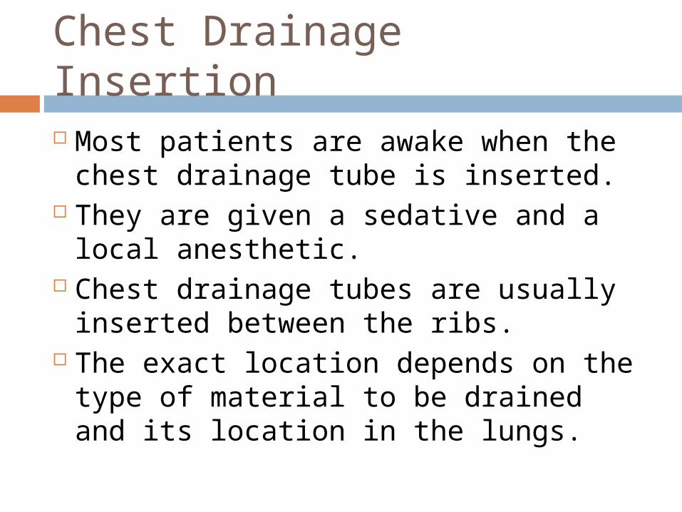
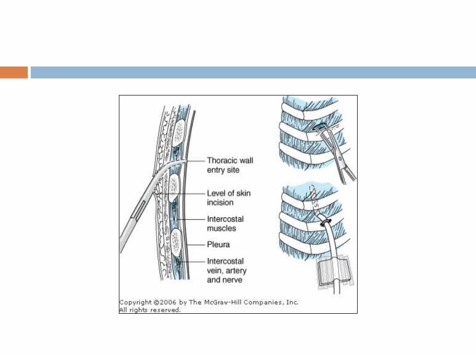
Most patients are awake when the chest drainage tube is inserted.
They are given a sedative and a local anesthetic.
Chest drainage tubes are usually inserted between the ribs.
The exact location depends on the type of material to be drained and its location in the lungs.

Continued
The chest drainage system must remain sealed to prevent air from entering the chest cavity through the tube.
One commonly used system is a water-seal drainage system, comprised of three compartments that collect and drain the fluid or air without allowing air to backflow into the tube.
Once the tube and drainage system are in place, a chest x ray is done to confirm that the tube is in the right location, and that it is working.
Types of Drainage Systems
Traditional water seal 3 chambers, collection, water seal
(middle) and wet suction control Requires sterile fluid be instilled into
water seal and suction chamber + and – pressure release valves Intermittent bubbling indicates
system is functioning properly

Types Continued
Dry suction water seal 3 chambers like traditional Requires sterile fluid be instilled in water
seal chamber at 2-cm level No need to fill suction chamber with fluid + and _ pressure release valves Indicator to signify suction pressure is
adequate Quieter than traditional

Types continued
Dry suction One way mechanical suction that allows air
to leave the chest and prevents from moving back into chest
Also referred to as one way valve system No need to fill suction chamber with fluid,
can be set up quickly in emergency Works even if knocked over, great for
ambulatory patients

Special Considerations
Newborns may develop multiple pneumothoraxes requiring multiple chest tubes.
Units have smaller collection chambers and finer calibrations to allow more accurate measurement of small drainage volumes.
The connecting tube has a narrower diameter to allow connection to the smaller chest tubes used in these patients.
Nursing Management
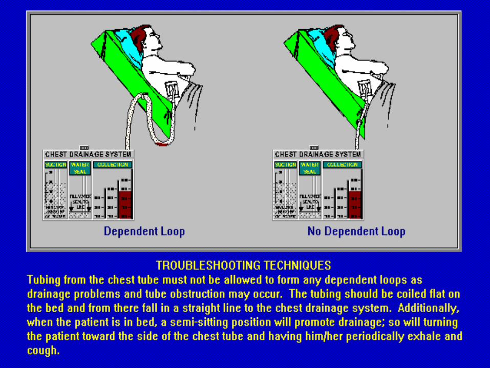
Ensure drainage tubing does not kink, loop or interfere with the patient’s movements to prevent fluid back up into the pleural space or impede drainage.
Assist patient with range of motion to reduce post op pain and prevent ankylosis of the shoulder.
Milk tubing in direction of drainage system as needed to prevent tubing from becoming obstructed by clots and fibrin.
Nursing Management Continued Make sure there is fluctuation of the fluid
level in the water seal chamber, shows effective connection
Fluid fluctuations in the water seal chamber or air leak indicator are will stop when Lung has reexpanded Tubing is obstructed Lop of tubing hangs below rest of tubing Suction motor or wall suction is not working
Nursing Management Continued Monitor for air leaks to prevent tension
pneumothorax. Notify MD if excessive bubbling in water
seal chamber not due to external leaks. Assess for rapid and shallow breathing,
cyanosis, pressure in chest, symptoms of hemorrhage to significant changes in VS
Encourage deep breathing a coughing to help aisle pleural raise and promotion of accumulated fluid in pleural space.
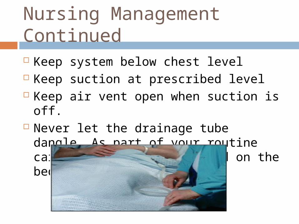
Nursing Management Continued Keep system below chest level Keep suction at prescribed level Keep air vent open when suction is off. Never let the drainage tube dangle. As
part of your routine care, make sure it's coiled on the bed.

Nursing Management Continued Pneumothorax, expect little if any output
because the tube is draining air, not fluid. Hemothorax, a lack of drainage may indicate
a clot obstructing the tube. If that occurs, try milking the tube: Starting at the proximal end, gently squeeze and release it between your fingers along the length of the tubing.
However, don't “strip” the chest tube, which means squeezing the length of the tube without releasing it. Once a common practice, stripping the tube causes a dangerous increase in intrathoracic pressure and doesn't lead to any significant increase in output.

Milking
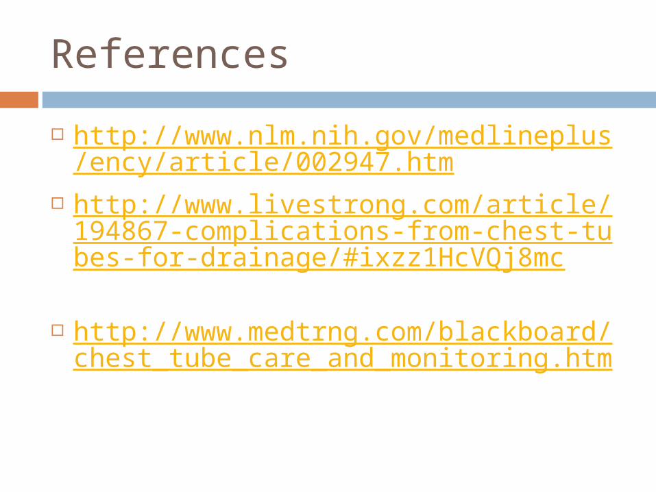
References
http://www.nlm.nih.gov/medlineplus/ency/article/002947.htm
http://www.livestrong.com/article/194867-complications-from-chest-tubes-for-drainage/#ixzz1HcVQj8mc
http://www.medtrng.com/blackboard/chest_tube_care_and_monitoring.htm

Thank You