Pulmonary Edema Pathophysiological Considerations Manifestations on Chest Radiography
CCCCCHESTHESTHESTHESTHEST RADIOGRAPHYRADIOGRAPHYRADIOGRAPHYRADIOGRAPHYRADIOGRAPHY ANDANDANDANDAND
ABDOMINALABDOMINALABDOMINALABDOMINALABDOMINAL ULULULULULTRASOUNDTRASOUNDTRASOUNDTRASOUNDTRASOUND
INININININ GENERALGENERALGENERALGENERALGENERAL PRACTICEPRACTICEPRACTICEPRACTICEPRACTICE
ANOUK MARIËLLE SPEETS
Chest radiography and abdominal ultrasound in general practice
Thesis University Utrecht, Faculty of Medicine - with a summary in DutchProefschrift Universiteit Utrecht - met een samenvatting in het Nederlands
ISBN-10 90-393-4225-3ISBN-13 978-90-393-4225-1Author Anouk Mariëlle SpeetsCover picture Mark Pinder, Newcastle, United KingdomCover design Roy SandersLay-out Haaring Automatisering, ScherpenzeelPrinted by Febodruk BV, Enschede
© A.M. Speets, 2006No part of this thesis may be reproduced or transmitted in any form or by anymeans, without prior permission of the author. The copyrigths on articles that havebeen published or accepted for publication have been transferred to the respectivejournals.
CHEST RADIOGRAPHY AND ABDOMINAL
ULTRASOUND IN GENERAL PRACTICE
X-THORAX EN BUIKECHOGRAFIE IN DE HUISARTSPRAKTIJK
(met een samenvatting in het Nederlands )
PROEFSCHRIFT
ter verkrijging van de graad van doctor aan de Universiteit Utrechtop gezag van de Rector Magnificus, Prof. dr. W.H. Gispen,
ingevolge het besluit van het College voor Promotiesin het openbaar te verdedigen op
vrijdag 12 mei 2006 des ochtends te 10.30 uur
door
ANOUK MARIËLLE SPEETS
geboren op 18 oktober 1978, te Harderwijk
Promotores Prof. dr. W.P.Th.M. MaliDepartment of RadiologyUniversity Medical Centre Utrecht, The Netherlands
Prof. dr. A.W. HoesJulius Centre for Health Sciences and Primary CareUniversity Medical Centre Utrecht, The Netherlands
Prof. dr. Y. van der GraafJulius Centre for Health Sciences and Primary CareUniversity Medical Centre Utrecht, The Netherlands
Co-promotor Dr. S. KalmijnJulius Centre for Health Sciences and Primary CareUniversity Medical Centre Utrecht, The Netherlands
Publication of this thesis was financially supported by the Julius Centre for HealthSciences and Primary Care.

Voor Hans en Milica Voor mijn ouders
MANUSCRIPTS BASED ON THE STUDIES PRESENTED IN THIS THESIS
CHAPTER 2Speets AM, Kalmijn S, Hoes AW, Van der Graaf Y, Smeets HM, Mali WPThM.Frequency of chest radiography and abdominal ultrasound in The Netherlands:1999-2003. Eur J Epidemiol. 2005;20(12):1031-1036.
CHAPTER 3Speets AM, Van der Graaf Y, Hoes AW, Kalmijn S, Sachs APE, Rutten MJCM,Gratama JWC, Montauban van Swijndregt AD, Mali WPThM. Chest radiography ingeneral practice: indications, diagnostic yield, and consequences for patientmanagement. Submitted.
CHAPTER 4Speets AM, Hoes AW, Van der Graaf Y, Kalmijn S, Sachs APE, Mali WPThM. Chestradiography in patients suspected of pneumonia in primary care: diagnostic yield,and consequences for patient management. In revision for Eur Respir J.
CHAPTER 5Speets AM, Kalmijn S, Hoes AW, Van der Graaf Y, Mali WPThM. The yield of abdominalultrasound in patients with abdominal pain referred by general practitioners.Submitted.
CHAPTER 6Speets AM, Hoes AW, Van der Graaf Y, Kalmijn S, De Wit NJ, Montauban vanSwijndregt AD, Gratama JWC, Rutten MJCM, Mali WPThM. Upper abdominalultrasound in general practice: indications, diagnostic yield, and consequences forpatient management. In revision for Fam Pract.
CHAPTER 7Speets AM, Van der Graaf Y, Hoes AW, Kalmijn S, De Wit NJ, Mali WPThM.Expected and unexpected gallstones in primary care. Submitted.
CONTENTS
CHAPTER 1 Introduction 1
CHAPTER 2 Frequency of chest radiography and abdominal ultrasound 9in The Netherlands
CHEST RADIOGRAPHY
CHAPTER 3 Indications and consequences of chest radiography in 21general practice
CHAPTER 4 Chest radiography in patients suspected of pneumonia 31in primary care: diagnostic yield, and consequences forpatient management
ABDOMINAL ULTRASOUND
CHAPTER 5 The value of abdominal ultrasound in patients with 43abdominal pain referred by general practitioners
CHAPTER 6 Indications and consequences of upper abdominal 53ultrasound in general practice
CHAPTER 7 Unexpected and expected gallstones in primary care 63
CHAPTER 8 General discussion: Evaluation of routinely used 75diagnostic tests
CHAPTER 9 Summary 83
Samenvatting 89
Dankwoord 95
Curriculum Vitae 99

Introduction
1

3
INTRODUCTION
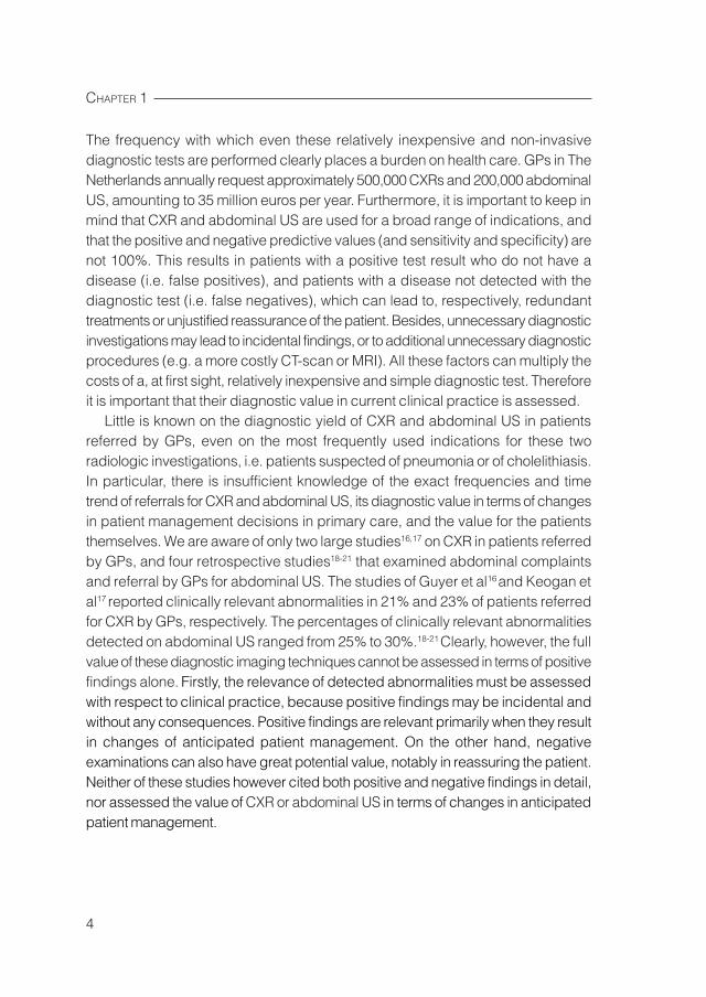
The diagnostic process is a multivariable and consecutive process of estimatingthe diagnostic probability of the presence of a particular disease given combinationsof test results. A diagnosis in clinical practice starts with obtaining basic informationof patients (e.g. age, gender and medical history), followed by history taking andphysical examination and, if necessary, by more invasive, patient burdening, timeconsuming and costly tests, such as laboratory or imaging investigations.1,2 Themain goal of a radiological examination is to provide information on the presenceor absence and nature of a certain disease. In addition, the course of a diseasecan be followed, by demonstration of the disease process itself or the effects of thedisease process on the normal anatomy.3 This thesis focuses on the first goal: thediagnostic value of imaging examinations. A valuable diagnostic investigation canbe defined as one in which the result will either alter the anticipated patientmanagement or increase confidence in the clinician’s diagnosis.4,5 In addition,diagnostic investigations can be very valuable for a patient’s uncertainty.
Chest radiography (CXR) and abdominal ultrasound (US) are two of the mostwidely used diagnostic imaging techniques in Western societies. CXR is an importantmethod for evaluation of the lower airways, pulmonary parenchyma and vessels,mediastinum, heart, pleura and chest wall.6 Diagnostic indications are signs andsymptoms potentially related to the respiratory, cardiovascular, upper gastrointestinal,and musculoskeletal system, evaluating diseases involving the thorax, and thestaging of extrathoracic as well as intrathoracic tumors.6 CXR is considered thereference standard for diagnosing pneumonia.7-10 In Western societies, on average236 CXRs per 1000 patients per year are performed and this technique accountsfor 25% of the annual total number of diagnostic imaging procedures. Thecorresponding numbers for The Netherlands are 120 CXRs per 1000 patients peryear, and they comprise 20% of all diagnostic imaging procedures.11 In TheNetherlands, approximately one quarter of all CXRs are requested by generalpractitioners (GPs).
Abdominal US is important for the evaluation of many structures in the abdomen,such as the liver, gallbladder, biliary tract, pancreas, aorta and kidneys. Indicationsinclude abdominal, flank and/or back pain, palpable abnormalities, abnormallaboratory values suggestive for abdominal pathology, follow-up of known orsuspected abnormalities and search for metastatic disease or occult primarydisease.12 Abdominal US is an important diagnostic imaging method for detectinggallstones, providing more than 95% sensitivity and specificity for the diagnosis ofgallstones greater than 2 mm in diameter.13-15 Annually, about 200,000 abdominal USare requested by GPs in The Netherlands; approximately 40% of all requested US.
CHAPTER 1
4
The frequency with which even these relatively inexpensive and non-invasivediagnostic tests are performed clearly places a burden on health care. GPs in TheNetherlands annually request approximately 500,000 CXRs and 200,000 abdominalUS, amounting to 35 million euros per year. Furthermore, it is important to keep inmind that CXR and abdominal US are used for a broad range of indications, andthat the positive and negative predictive values (and sensitivity and specificity) arenot 100%. This results in patients with a positive test result who do not have adisease (i.e. false positives), and patients with a disease not detected with thediagnostic test (i.e. false negatives), which can lead to, respectively, redundanttreatments or unjustified reassurance of the patient. Besides, unnecessary diagnosticinvestigations may lead to incidental findings, or to additional unnecessary diagnosticprocedures (e.g. a more costly CT-scan or MRI). All these factors can multiply thecosts of a, at first sight, relatively inexpensive and simple diagnostic test. Thereforeit is important that their diagnostic value in current clinical practice is assessed.
Little is known on the diagnostic yield of CXR and abdominal US in patientsreferred by GPs, even on the most frequently used indications for these tworadiologic investigations, i.e. patients suspected of pneumonia or of cholelithiasis.In particular, there is insufficient knowledge of the exact frequencies and timetrend of referrals for CXR and abdominal US, its diagnostic value in terms of changesin patient management decisions in primary care, and the value for the patientsthemselves. We are aware of only two large studies16,17 on CXR in patients referredby GPs, and four retrospective studies18-21 that examined abdominal complaintsand referral by GPs for abdominal US. The studies of Guyer et al16 and Keogan etal17 reported clinically relevant abnormalities in 21% and 23% of patients referredfor CXR by GPs, respectively. The percentages of clinically relevant abnormalitiesdetected on abdominal US ranged from 25% to 30%.18-21 Clearly, however, the fullvalue of these diagnostic imaging techniques cannot be assessed in terms of positivefindings alone. Firstly, the relevance of detected abnormalities must be assessedwith respect to clinical practice, because positive findings may be incidental andwithout any consequences. Positive findings are relevant primarily when they resultin changes of anticipated patient management. On the other hand, negativeexaminations can also have great potential value, notably in reassuring the patient.Neither of these studies however cited both positive and negative findings in detail,nor assessed the value of CXR or abdominal US in terms of changes in anticipatedpatient management.
5
INTRODUCTION
These considerations were the main justifications for the studies described in thisthesis. The main objective of this thesis was to determine the clinical effectivenessof CXR and upper abdominal US in general practice. The objectives of the studiespresented in the following chapters were:
1. To provide detailed information on the number and time trend of CXRs andabdominal US examinations by age, gender, referring physician and ethnicity inThe Netherlands (Chapter 2).
2. To assess the influence of CXR on the change in anticipated patient managementin general practice, and to evaluate the consequences of the CXR according tothe patient (Chapter 3).
3. To determine the effect of CXR on the probability estimation of pneumonia byGPs, the influence of CXR on patient management, and consequences of CXRaccording to the patient among primary care patients with a clinical suspicionof pneumonia (Chapter 4).
4. To examine the prevalence and determinants of significant abnormalities onabdominal US in patients with abdominal pain referred by GPs (Chapter 5).
5. To assess the influence of upper abdominal US on the change in anticipatedpatient management in general practice, and to evaluate the consequences ofthe abdominal US according to the patient (Chapter 6).
6. To assess the differences in signs and symptoms of primary care patients withexpected and unexpected gallstones referred for upper abdominal US (Chapter 7).
7. To outline the difficulties of diagnostic research assessing the value of diagnostictests that are routinely used in a wide variety of complaints (Chapter 8)
CHAPTER 1
6
References
1. Sackett DL, Haynes RB, Tugwell P. Clinical epidemiology: a basic science forclinical medicine. Boston: Little, Brown and Company, 1985.
2. Moons KG, Van Es GA, Deckers JW, Habbema JD, Grobbee DE. Limitations ofsensitivity, specificity, likelihood ratio, and Bayes’ theorem in assessing diagnosticprobabilities: a clinical example. Epidemiology. 1997;8(1):12-17.
3. American College of Radiology. ACR Standards. ACR standard for generalradiography. United States, 2000.
4. European Commission. Referral guidelines for imaging. Radiation protection 118.Luxembourg: Office for Official Publications of the European Communities, 2000.
5. Stolberg HO, Hynes DM, Rainbow AJ, Moran LA. Requesting diagnostic imagingexaminations: a position paper of the Canadian Association of Radiologists. CanAssoc Radiol J. 1997;48:89-91.
6. American College of Radiology. ACR Standards. ACR standard for the performanceof pediatric and adult chest radiography. United States, 2001.
7. Coblentz CL, Matzinger F, Samson LM, Scherer J, Stolberg HO, Weisbrod G. CARstandards for chest radiography. Canada: Canadian Association of Radiologists,2000.
8. Health Services Utilization and Research Commission. Chest radiography: asummary of the evidence supporting selective clinical practice guidelines andrecommendations for implementation. Saskatoon: Health Services Utilization andResearch Commission, 1997.
9. Stolberg HO, Buckley N, Coblentz CL, Cornett R, Feightner K, Killian K, et al.Guidelines for chest X-rays in asymptomatic populations. Canada: The College ofPhysicians and Surgeons of Ontario, 1999.
10. World Health Organization Scientific Group. Chest and cardiovascular system.Effective choices for diagnostic imaging in clinical practice. Geneva: World HealthOrganization, 1990.
11. United Nations Scientific Committee on the Effects of Atomic Radiation UNSCEAR2000 Report to the General Assembly, with Scientific Annexes. Sources and effectsof ionizing radiation, Volume I: Sources. Annex D Medical radiation exposures.New York: United Nations, 2000:293-495.
12. American College of Radiology. ACR Standards. ACR Standard for the performanceof an ultrasound examination of the abdomen or retroperitoneum. United States, 2001.
13. Ahmed A, Cheung RC, Keeffe EB. Management of gallstones and theircomplications. Am Fam Physician. 2000;61(6):1673-1680,1687-1688.
14. Bortoff GA, Chen MY, Ott DJ, Wolfman NT, Routh WD. Gallbladder stones: imagingand intervention. Radiographics. 2000;20(3):751-766.
15. Kalloo AN, Kantsevoy SV. Gallstones and biliary disease. Prim Care.2001;28(3):591-606.
7
INTRODUCTION
16. Guyer PB, Chalmers AG. Chest radiography for general practitioners- a low yieldinvestigation. J R Coll Gen Pract. 1983;33:477-479.
17. Keogan MT, Padhani AR, Flower CD. Chest radiography for general practitioners:scope for change? Clin Radiol. 1992;46:51-54.
18. Charlesworth CH, Sampsom MA. How do general practitioners compare with theoutpatient department when requesting upper abdominal examinations? ClinRadiol. 1994;49:343-345.
19. Colquhoun IR, Saywell WR, Dewburry KC. An analysis of referrals for primarydiagnostic abdominal ultrasound at a general X-ray department. Br J Radiol.1988;61:297-300.
20. Connor SEJ, Banerjee AK. General practitioner requests for upper abdominal ultra-sound: their effect on clinical outcome. Br J Radiol. 1998;73:1021-1025.
21. Mills P, Joseph AEA, Adam EJ. Total abdominal and pelvic ultrasound: incidentalfindings and a comparison between outpatient and general practice referrals in1000 cases. Br J Radiol. 1989;62:974-976.

Frequency of chest radiography andabdominal ultrasound in The Netherlands
Speets AM, Kalmijn S, Hoes AW, Van der Graaf Y, Smeets HM,Mali WPThM. Frequency of chest radiography and abdominalultrasound in The Netherlands: 1999-2003. Eur J Epidemiol.2005;20(12):1031-1036.
2
CHAPTER 2
10
AbstractBackground. Chest radiography (CXR) and abdominal ultrasound (US) are twowidely used diagnostic imaging techniques in Western societies. However, little isknown about the frequency of these examinations and its determinants. The aim ofthis descriptive study was to provide detailed information on the number of CXRand abdominal US examinations by age, gender, referring physician and ethnicity.Methods. We used data of approximately 3,000,000 sick fund insured persons ofthe Health Insurance Company Agis in The Netherlands from 1999 to 2003. Wecalculated annual numbers and corresponding 95% confidence intervals for differentage, gender and ethnicity categories.Results. The mean age of the population was 38±22 years and 46% were male.CXRs were ordered in 130 per 1000 persons per year and abdominal USexaminations in 39 per 1000 persons per year; these frequencies did not changenoticeable over the five-year period. CXR was performed more often in males (156versus 109 per 1000 persons/year in females; p<0.05) and abdominal US moreoften in females (43 versus 34 per 1000 persons/year in males; p<0.05). Frequencieswere highest in persons aged 70-79 years. Compared to medical specialists, generalpractitioners more frequently referred younger patients and females, especially forabdominal US. Up to the age of 60 years the frequencies of both CXR and abdominalUS were higher in Turks and Moroccans compared to other persons.Conclusion. This study showed marked differences in the frequencies of CXR andabdominal US according to age, gender and ethnicity in The Netherlands.
11
FREQUENCY OF CHEST RADIOGRAPHY AND ABDOMINAL ULTRASOUND
Introduction
The goal of a radiological examination is to provide information on the presence orabsence and nature of a certain disease or to follow its course, in addition toinformation obtained by the medical history, physical examination and otherdiagnostic tests.1 Ideally, there should always be a sufficient clinical suspicion towarrant a radiological examination, and a reasonable anticipation that the resultsof the imaging test, normal or abnormal, may influence the management of thepatient.2
There is little scientific literature on the exact frequency of regularly performedradiodiagnostic tests, especially imaging tests requested by general practitioners(GPs). Chest radiography (CXR) and abdominal ultrasound (US) are two of themost widely used diagnostic imaging techniques in Western societies.2-6 However,the frequency and determinants of CXR and abdominal US examinations are largelyunknown. The aim of this descriptive study was to provide detailed information onthe number of CXR and abdominal US examinations by age, gender, referringphysician and ethnicity in The Netherlands.
Methods
In this study we used information from a large dataset of the Health InsuranceCompany Agis located in Amersfoort in The Netherlands. In the Agis database allhealth care procedures of sick fund insured persons are documented in detail.Besides health care procedures, a number of personal characteristics aredocumented, such as date of birth and gender. Agis refunds only expenses claimsafter the computer has verified all data of the claim (e.g. name, birth date andaddress of the patient). Data are submitted to the database after this automatedverification, to ensure the validity of these data.
We used information of approximately 3,000,000 sick fund insured personsregistered in the Agis database from 1999 to 2003, encompassing almost one-fifthof the Dutch population. Of each registered person, information on gender, date ofbirth and personal insurance number was extracted. The age and gender distributionof our study population was similar to that of the Dutch population at large (dataprovided by Statistics Netherlands7). Thus, our study population is a good reflectionof the population in The Netherlands. All CXRs and abdominal US (including entireabdomen, upper abdominal and lower abdominal US; excluding pregnancy US)that were performed for in- and outpatients were registered. Data included the
CHAPTER 2
12
date of the radiological examination and the referring physician (medical specialistor GP). We could use only the data of 1999 to 2001 for the results on the referringphysicians, because Agis changed the coding for the applicants in 2002. In addition,Agis determined the ethnicity of Moroccan and Turkish people of all insured personsin 1999, based on the nationality of the first generation Moroccan and Turkishpeople and subsequently a match of their names with the remaining insured persons.8
This way we were able to investigate whether the frequency of CXRs and abdominalUS among Moroccans and Turks was different from the frequency among otherinsured persons in 1999.
We calculated the frequencies of CXR and abdominal US per year by age,gender and ethnicity. Because of the large differences between the age groups,the frequencies per 1000 persons per year for gender and ethnicity werestandardized for age.9 The distribution of the ten age groups of all insured personsin 1999 were handled as reference group. This way, trends in time would still bedetectable. We calculated 95% confidence intervals for the frequencies. Differencesbetween frequencies were considered significant when the confidence intervalsdid not overlap.9
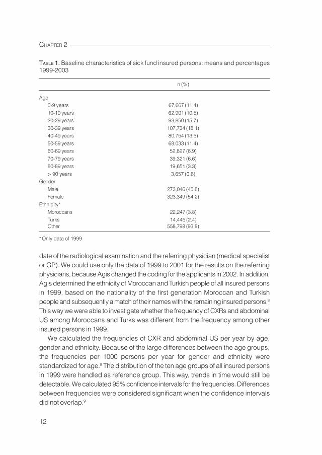
TABLE 1. Baseline characteristics of sick fund insured persons: means and percentages1999-2003
n (%)
Age0-9 years 67,667 (11.4)
10-19 years 62,901 (10.5)20-29 years 93,850 (15.7)
30-39 years 107,734 (18.1)40-49 years 80,754 (13.5)
50-59 years 68,033 (11.4)60-69 years 52,827 (8.9)
70-79 years 39,321 (6.6)80-89 years 19,651 (3.3)
> 90 years 3,657 (0.6)Gender
Male 273,046 (45.8)Female 323,349 (54.2)
Ethnicity*Moroccans 22,247 (3.8)
Turks 14,445 (2.4)Other 558,798 (93.8)
* Only data of 1999
13
FREQUENCY OF CHEST RADIOGRAPHY AND ABDOMINAL ULTRASOUND
Results
The mean number of sick fund insured persons from 1999 to 2003 was 596,395 peryear, 46% was male and the mean age was 38 years (SD 22 years). The agedistribution is shown in Table 1. In 1999 4% of the sick fund insured persons wereof Moroccan and 2% of Turkish origin.
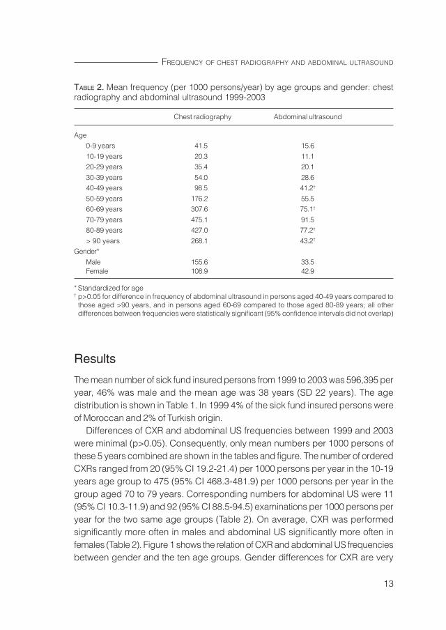
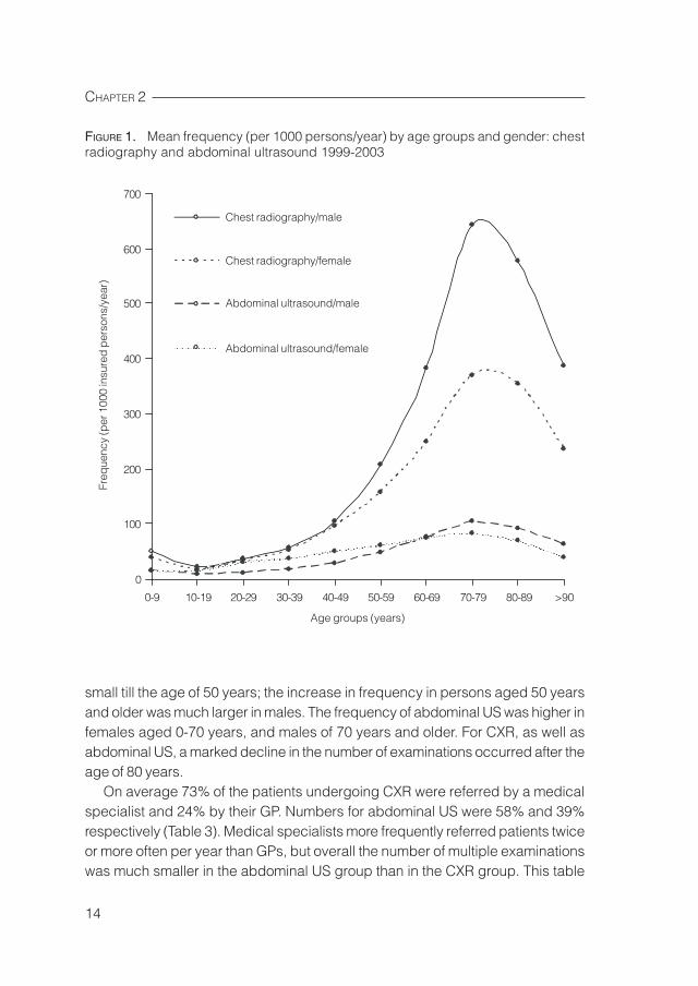
Differences of CXR and abdominal US frequencies between 1999 and 2003were minimal (p>0.05). Consequently, only mean numbers per 1000 persons ofthese 5 years combined are shown in the tables and figure. The number of orderedCXRs ranged from 20 (95% CI 19.2-21.4) per 1000 persons per year in the 10-19years age group to 475 (95% CI 468.3-481.9) per 1000 persons per year in thegroup aged 70 to 79 years. Corresponding numbers for abdominal US were 11(95% CI 10.3-11.9) and 92 (95% CI 88.5-94.5) examinations per 1000 persons peryear for the two same age groups (Table 2). On average, CXR was performedsignificantly more often in males and abdominal US significantly more often infemales (Table 2). Figure 1 shows the relation of CXR and abdominal US frequenciesbetween gender and the ten age groups. Gender differences for CXR are very
TABLE 2. Mean frequency (per 1000 persons/year) by age groups and gender: chestradiography and abdominal ultrasound 1999-2003
Chest radiography Abdominal ultrasound
Age0-9 years 41.5 15.6
10-19 years 20.3 11.120-29 years 35.4 20.1
30-39 years 54.0 28.640-49 years 98.5 41.2†
50-59 years 176.2 55.560-69 years 307.6 75.1†
70-79 years 475.1 91.580-89 years 427.0 77.2†
> 90 years 268.1 43.2†
Gender*
Male 155.6 33.5Female 108.9 42.9
* Standardized for age† p>0.05 for difference in frequency of abdominal ultrasound in persons aged 40-49 years compared to
those aged >90 years, and in persons aged 60-69 compared to those aged 80-89 years; all otherdifferences between frequencies were statistically significant (95% confidence intervals did not overlap)
CHAPTER 2
14
small till the age of 50 years; the increase in frequency in persons aged 50 yearsand older was much larger in males. The frequency of abdominal US was higher infemales aged 0-70 years, and males of 70 years and older. For CXR, as well asabdominal US, a marked decline in the number of examinations occurred after theage of 80 years.
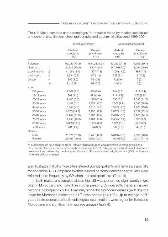
On average 73% of the patients undergoing CXR were referred by a medicalspecialist and 24% by their GP. Numbers for abdominal US were 58% and 39%respectively (Table 3). Medical specialists more frequently referred patients twiceor more often per year than GPs, but overall the number of multiple examinationswas much smaller in the abdominal US group than in the CXR group. This table
700
600
500
400
300
200
100
0
Freq
uenc
y (p
er 1
000
insu
red
per
sons
/yea
r)
Age groups (years)
0-9 10-19 20-29 30-39 40-49 50-59 60-69 70-79 80-89 >90
Chest radiography/male
Chest radiography/female
Abdominal ultrasound/male
Abdominal ultrasound/female
FIGURE 1. Mean frequency (per 1000 persons/year) by age groups and gender: chestradiography and abdominal ultrasound 1999-2003
15
FREQUENCY OF CHEST RADIOGRAPHY AND ABDOMINAL ULTRASOUND
TABLE 3. Mean numbers and percentages for requests made by medical specialistsand general practitioners: chest radiography and abdominal ultrasound 1999-2001
Chest radiography Abdominal ultrasound
Medical General Medical Generalspecialist practitioner specialist practitioner
n (%) n (%) n (%) n (%)
Referrals* 56,039 (73.3) 18,502 (24.2 ) 13,213 (57.8) 8,955 (39.1)Number of 1 20,616 (70.2) 14,327 (89.9) 9,749 (87.0) 8,056 (95.0)
examinations 2 4,128 (14.1) 1,237 (7.8) 1,105 (11.2) 386 (7.7)per insured 3 1,634 (5.6) 217 (1.3) 237 (2.1) 24 (0.3)
person 4 890 (3.0) 59 (0.4) 72 (0.6) 7 (0.1) >4 2,112 (7.1) 92 (0.6) 44 (0.4) 5 (0.1)
Age0-9 years 1,967 (3.5) 935 (5.0) 854 (6.5) 216 (2.4)
10-19 years 802 (1.4) 473 (2.6) 419 (3.2)† 250 (2.8)†
20-29 years 2,103 (3.8) 1,308 (7.1) 898 (6.8) 1,046 (11.7)
30-39 years 3,447 (6.1) 2,352 (12.7) 1,306 (9.9) 1,698 (18.9)40-49 years 5,049 (9.0) 2,716 (14.7) 1,527 (11.6) 1,701 (19.0)
50-59 years 8,253 (14.7) 3,295 (17.8) 2,087 (15.8) 1,527 (17.1)60-69 years 12,244 (21.8) 3,400 (18.4) 2,478 (18.8) 1,268 (14.1)
70-79 years 14,750 (26.3) 2,781 (15.0) 2,464 (18.7) 866 (9.7)80-89 years 6,660 (11.9) 1,119 (6.0) 1,075 (8.1) 342 (3.8)
> 90 years 761 (1.4) 124 (0.7) 104 (0.8) 42 (0.5)Gender
Male 28,571 (51.0) 8,746 (47.3) 5,619 (42.5) 2,584 (28.9)Female 27,467 (49.0) 9,756 (52.7) 7,594 (57.5) 6,370 (71.1)
* Percentages do not add up to 100%: remaining percentages were unknown referring physicians† P>0.05; all other differences between the frequency of chest radiography and abdominal ultrasound
examinations ordered by medical specialists and GPs were statistically significant (95% confidenceintervals did not overlap)
also illustrates that GPs more often referred younger patients and females, especiallyfor abdominal US. Compared to other insured persons Moroccans and Turks werereferred more frequently by GPs than medical specialists (Table 4).
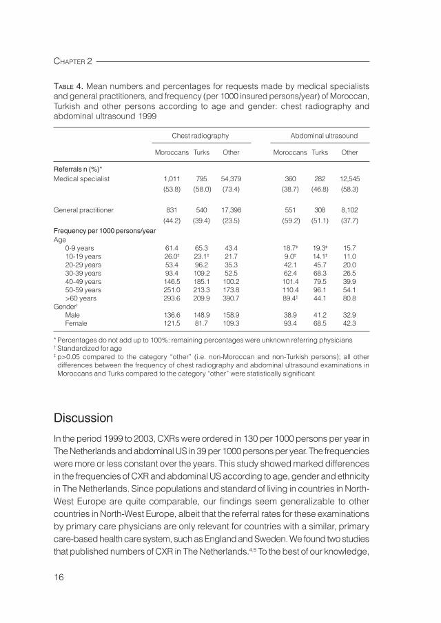
In both males and females abdominal US was performed significantly moreoften in Moroccans and Turks than in other persons. Compared to the other insuredpersons the frequency of CXR was only higher for Moroccan females (p<0.05), butlower for Moroccan males and all Turkish people (p<0.05). Up to the age of 60years the frequencies of both radiological examinations were higher for Turks andMoroccans and significant in most age groups (Table 4).
CHAPTER 2
16
Discussion
In the period 1999 to 2003, CXRs were ordered in 130 per 1000 persons per year inThe Netherlands and abdominal US in 39 per 1000 persons per year. The frequencieswere more or less constant over the years. This study showed marked differencesin the frequencies of CXR and abdominal US according to age, gender and ethnicityin The Netherlands. Since populations and standard of living in countries in North-West Europe are quite comparable, our findings seem generalizable to othercountries in North-West Europe, albeit that the referral rates for these examinationsby primary care physicians are only relevant for countries with a similar, primarycare-based health care system, such as England and Sweden. We found two studiesthat published numbers of CXR in The Netherlands.4,5 To the best of our knowledge,
TABLE 4. Mean numbers and percentages for requests made by medical specialistsand general practitioners, and frequency (per 1000 insured persons/year) of Moroccan,Turkish and other persons according to age and gender: chest radiography andabdominal ultrasound 1999
Chest radiography Abdominal ultrasound
Moroccans Turks Other Moroccans Turks Other
Referrals n (%)*Medical specialist 1,011 795 54,379 360 282 12,545
(53.8) (58.0) (73.4) (38.7) (46.8) (58.3)
General practitioner 831 540 17,398 551 308 8,102
(44.2) (39.4) (23.5) (59.2) (51.1) (37.7)Frequency per 1000 persons/yearAge
0-9 years 61.4 65.3 43.4 18.7‡ 19.3‡ 15.710-19 years 26.0‡ 23.1‡ 21.7 9.0‡ 14.1‡ 11.020-29 years 53.4 96.2 35.3 42.1 45.7 20.030-39 years 93.4 109.2 52.5 62.4 68.3 26.540-49 years 146.5 185.1 100.2 101.4 79.5 39.950-59 years 251.0 213.3 173.8 110.4 96.1 54.1>60 years 293.6 209.9 390.7 89.4‡ 44.1 80.8
Gender†
Male 136.6 148.9 158.9 38.9 41.2 32.9Female 121.5 81.7 109.3 93.4 68.5 42.3
* Percentages do not add up to 100%: remaining percentages were unknown referring physicians† Standardized for age‡ p>0.05 compared to the category “other” (i.e. non-Moroccan and non-Turkish persons); all otherdifferences between the frequency of chest radiography and abdominal ultrasound examinations inMoroccans and Turks compared to the category “other” were statistically significant
17
FREQUENCY OF CHEST RADIOGRAPHY AND ABDOMINAL ULTRASOUND
there is no scientific literature on annual numbers of abdominal US, so it is notpossible to compare the numbers of our study with those of other studies.
The United Nations Scientific Committee on the Effects of Atomic Radiationregularly monitored the medical use of radiation. Their most recent report waspublished in 2001.4 The mean annual number of CXR examinations from 1991-1996in The Netherlands was 120 per 1000 population and CXR contributed 20% to theannual total numbers of diagnostic research. The mean frequency of 130 CXRs per1000 persons per year in our study is somewhat higher.
Beentjes and Timmermans published numbers for the distribution of age andgender of patients undergoing CXR in The Netherlands in the period 1984-1985.5
The shape of their age distribution corresponds well with our figures. The numberof CXRs for males was 171 per 1000 males and 142 per 1000 females. The annualnumbers in our study are lower, 156 and 109 per 1000 sick fund insured persons,respectively. The long period of almost 20 years may explain part of this differencein prevalence, such as the development of new alternative diagnostic imagingtechniques (e.g. CT-scan).
Before discussing the differences in frequencies of CXR and abdominal USfound in our study we emphasize that many factors (e.g. psychological andeconomical) may have influenced the observed differences. It is beyond the scopeof this study to thoroughly investigate all causes for these differences. Furtherstudies, including more detailed information on relevant determinants, are neededto explain the observed differences in frequencies.
The larger annual numbers of CXR in males could be partly explained by thehigher frequency of smoking and a higher prevalence of more severe lung diseases,such as lung carcinoma and chronic obstructive pulmonary disease, in the malepopulation.10-12 However, trends in the prevalence of cigarette smoking are decliningamong males and increasing among females and concurrently opposite trends areobserved for lung diseases.10-12 This could be a reason for the smaller genderdifferences in the younger age groups. Females more often have abdominalcomplaints, can suffer from gynecological problems and the prevalence of abdominaldiseases, such as infectious gastrointestinal diseases, is higher.15,16 One of thedeterminants for the higher frequency of abdominal US in males aged 70 yearsand older could be the frequency of abdominal aortic aneurysms (AAA), becausemale gender and increasing age are important risk factors for AAA.13,14 AAA is tentimes more common in 65- to 75-year-old men compared to women of the sameage.13 A marked decline in the number of CXR and abdominal US examinationsoccurred after the age of 80 years. One can only speculate on the underlyingreasons. Perhaps the number of examinations is low because patient management
CHAPTER 2
18
is unlikely to change in these very old people or because the patient burden is toohigh. Further research is required to address this issue.
Up to the age of 60 years both CXR and abdominal US were ordered morefrequently in Turks and Moroccans compared to other persons. A community surveyamong the Amsterdam population showed that Moroccan and Turkish personsreported a poorer personal health and higher use of health care compared to theindigenous population of the same age and gender.17 Data of the Dutch NationalSurvey on Morbidity and Interventions in General Practice showed that the frequencyof gastrointestinal and lung diseases was higher for Moroccans and Turks incomparison to the Dutch population.18 Possible explanations for the higher frequencyof health problems in the Moroccan and Turkish population in The Netherlands areadverse social and economic position of ethnic minority groups, differences incongenital predisposition, cultural factors, living conditions and diet.8,17,18
Furthermore, communication problems could result in a higher referral frequencyof Moroccans and Turks by GPs for CXR and abdominal US shown in this study.8,17,18
As expected, medical specialists more frequently referred patients for two ormore examinations per year than GPs. The number of multiple examinations wasmuch smaller in the abdominal US group than in the CXR group. In The Netherlands,routine radiological investigations, such as CXR, are performed often when patientsare submitted to the hospital. Each patient at the intensive care ward is dailyexamined with CXR and this increases the number of multiple CXRs requested bymedical specialists.
In conclusion, in the period 1999 to 2003 CXRs were ordered in 130 per 1000persons per year and abdominal US in 39 per 1000 persons per year in TheNetherlands. These frequencies did not change noticeable over the five-year period.This study showed marked differences in the frequencies of these examinationsaccording to age, gender and ethnicity.
19
FREQUENCY OF CHEST RADIOGRAPHY AND ABDOMINAL ULTRASOUND
References
1. American College of Radiology. ACR Standards. ACR Standard for the performanceof paediatric and adult chest radiography. United States, 2001.
2. American College of Radiology. ACR Standards. ACR Standard for performingand interpreting diagnostic ultrasound examinations. United States, 2000.
3. American College of Radiology. ACR Standards. ACR Standard for the performanceof an ultrasound examination of the abdomen or retroperitoneum. 2001.
4. United Nations Scientific Committee on the Effects of Atomic Radiation UNSCEAR2000 Report to the General Assembly, with Scientific Annexes. Sources and effectsof ionizing radiation, Volume I: Sources. Annex D Medical radiation exposures.New York: United Nations, 2000:293-495.
5. Beentjes LB, Timmermans WM. Age and sex specific radiographic examinationfrequency in The Netherlands. Br J Radiol. 1990;63:691-697.
6. Aroua A, Bize R, Buchillier-Decka I, Vader JP, Valley JF, Schnyder P. X-ray imagingof the chest in Switzerland in 1998: a nationwide survey. Eur Radiol. 2003;13(6):1250-1259.
7. Http://statline.cbs.nl.8. Smeets HM, Ros CC. [Health care usage by Moroccans and Turks compared to
the indigenous Dutch population: no higher consumption of health care and lowermedication costs.] Ned Tijdschr Geneeskd. 2004;148(25):1243-1247.
9. Rothman KJ. Epidemiology: an introduction. New York: Oxford University PressInc, 2002.
10. Molarius A, Parsons RW, Dobson AJ, Evans A, Fortmann SP, Jamrozik K, et al.Trends in cigarette smoking in 36 populations from the early 1980s to the mid-1990s: findings from the WHO MONICA project. Am J Public Health. 2001;91:206-212.
11. Travis WD, Lubin J, Ries L, Devesa S. United States lung carcinoma incidencetrends. Declining for most histologic types among males, increasing amongfemales. Cancer. 1996;77:2464-2470.
12. Caracta CF. Gender differences in pulmonary disease. Mt Sinai J Med.2003;70(4):215-224.
13. Crawford CM, Hurtgen-Grace K, Talarico E, Marley J. Abdominal aortic aneurysm:an illustrated narrative review. J Manipulative Physiol Ther. 2003;26:184-195.
14. Prisant LM, Mondy JS. Images in hypertension: abdominal aortic aneurysm. J ClinHypertens. 2004;6;2:85-89.
15. Adelman AM, Revicki DA, Maganizer J, Hebel R. Abdominal pain in an HMO. FamMed. 1995;27:321-325.
16. Mayer EA, Naliboff B, Lee O, Munakata J, Chang L. Review article: gender-relateddifferences in functional gastrointestinal disorders. Aliment Pharmacol Ther.1999;13(5):65-69.

CHAPTER 2
20
17. Reijneveld SA. Reported health, lifestyles, and use of health care of first generationimmigrants in The Netherlands: do socioeconomic factors explain their adverseposition? J Epidemiol Community Health. 1998;52:298-304.
18. Weide MG, Foets M. [Migrants in general practice: different from Dutch in complaintsand diagnoses.] Ned Tijdschr Geneeskd. 1998;142(38):2105-2109.
Indications and consequences of chestradiography in general practice
Speets AM, Van der Graaf Y, Hoes AW, Kalmijn S, Sachs APE,Rutten MJCM, Gratama JWC, Montauban van Swijndregt AD,Mali WPThM. Chest radiography in general practice: indications,diagnostic yield, and consequences for patient management.Submitted.
3
CHAPTER 3
22
AbstractBackground. Chest radiography (CXR) is frequently performed in Western societies.There is insufficient knowledge of its diagnostic value in terms of changes in patientmanagement decisions in primary care. The aim of this study was to assess theinfluence of CXR on patient management in general practice.Methods. 792 patients aged ≥ 18 years were referred by 78 general practitioners(GPs) for CXR to one of the three participating hospitals in The Netherlands. Themain outcome was change in patient management assessed by means ofquestionnaires filled in by GPs before and after CXR.Results. Mean age of the patients was 57.3±16.2 years, 53% were male. Clinicallyrelevant abnormalities were found in 24% of the CXRs. Patient management changedin 60% of the patients following CXR. Main changes included: fewer referrals to amedical specialist (from 26% to 12%); reduction in initiation or change in therapy(from 24% to 15%); and more frequent reassurance (from 25% to 46%). However,this reassurance was not perceived as such in a quarter of these patients. A changein patient management occurred significant more frequently in patients withcomplaints of cough (67%), exhibited abnormalities during physical examination(69%), or a suspected diagnosis of pneumonia (68%).Conclusion. Patient management by the GP changed in 60% of patients followingCXR. CXR substantially reduced the number of referrals and initiation or change intherapy, and more patients were reassured by their GP. Thus, CXR seems to reducethe burden on patients and health care.
23
INDICATIONS AND CONSEQUENCES OF CHEST RADIOGRAPHY
Introduction
Chest radiography (CXR) is an important diagnostic method for evaluation of theairways, pulmonary parenchyma and vessels, mediastinum, heart, pleura and chestwall.1 It is one of the most widely used diagnostic imaging techniques in Westernsocieties; on average 236 CXRs per 1000 patients per year are performed and thistechnique accounts for 25% of the annual total number of diagnostic imagingprocedures.2 The corresponding numbers for The Netherlands and the UnitedKingdom are 120 (20%), and 141 (29%) CXRs per 1000 patients per year,respectively.2 In The Netherlands, approximately one quarter of the CXRs arerequested by GPs.
The frequency with which even relatively inexpensive and non-invasive diagnostictests are performed clearly places a burden on health care. Therefore it is importantthat their influence on patient management is assessed. Unnecessary diagnosticinvestigations may lead to incidental findings, or to additional unnecessary diagnosticprocedures or even over treatment.
A valuable diagnostic investigation can be defined as one in which the result willeither alter management or increase confidence in the clinician’s diagnosis.3,4 Currentguidelines for CXR are aimed mainly at diseases instead of at the complaints withwhich patients present themselves.1,3,5-8 CXR in patients referred by GPs has receivedlittle attention in the scientific literature. There is insufficient knowledge of itsdiagnostic value in terms of changes in patient management decisions in primarycare. We are aware of only two large studies on CXR in patients referred by GPs.The studies of Guyer et al9 and Keogan et al10 reported clinically relevantabnormalities in 21% and 23% of patients referred for CXR by GPs, respectively.Clearly, the full value of CXR cannot be assessed in terms of positive findingsalone. Firstly, the relevance of detected abnormalities must be assessed with respectto clinical practice, because positive findings may be incidental and without anyconsequences. Positive findings are relevant only when they result in changes ofpatient management. On the other hand, negative examinations can also havepotential value when they result in changes of patient management and can bevery helpful in reassuring the patient. Neither of these studies however cited bothpositive and negative findings in detail, nor assessed the value of CXR in terms ofchanges in patient management. Also, the consequences of CXR according to thepatient were not studied before.
The objective of this study was to assess the influence of both positive andnegative findings of CXR on the change in patient management in general practiceand to evaluate the consequences of the CXR according to the patient.
CHAPTER 3
24
Methods
This prospective cohort study was conducted from April 2003 to December 2004.In total, 78 GPs in the catchment area of one of three participating general hospitalslocated in three main cities in The Netherlands (Jeroen Bosch Hospital in ‘s-Hertogenbosch; Gelre Hospitals in Apeldoorn; ’Onze Lieve Vrouwe Gasthuis’ inAmsterdam) were involved. 28 GPs (36%) worked in a solo practice, 58 (74%)were male, 40 GPs (51%) graduated between 1968-1980, 19 (24%) between 1980-1990, and 19 (24%) between 1990-1997. All patients of 18 years and older whowere referred for CXR (posteroanterior and lateral view) by their GP to one of thesehospitals were included in the study. The patients received an exclusion form fromtheir GP, which they could return to the study coordinator if we were not allowed touse their data for this study. The study was approved by the Medical Ethics ReviewBoard.
All GPs were asked to fill in a standardized form before requesting a CXR,including information on history, physical examination, indication, suspecteddiagnosis, and proposed patient management. The anticipated patient managementwas filled in as if no CXR would be performed. The management options included:referral to a medical specialist; initiation or change in therapy; reassurance of thepatient; and follow-up by the GP (watchful waiting or additional diagnostic testing).The GP could choose only one of these management options. After the GP receivedthe report (within 1-4 days after the CXR) he or she filled in a second questionnaire;again including the suspected diagnosis and anticipated patient management plan.
The reports of CXR were collected in the three hospitals to determine the findingsof CXR. These findings were categorized into six groups: (1) malignancy; (2)pneumonia; (3) COPD/asthma/chronic bronchitis; (4) other clinically relevantabnormalities (heart failure and unclear abnormalities that required furtherinvestigation according to the radiologist); (5) the follow-up of abnormalities detectedpreviously on CXR; (6) no abnormality. The first four groups were considered clinicallyrelevant abnormalities.
Six months after the CXR a short questionnaire was sent to all patients, in orderto assess the consequences of CXR according to the patient (response rate 79%).They could choose one of the following options: definite diagnosis; better treatment;reassurance; nothing; or other. With this information we could check whetherreassurance of the patient as reported by the GP was really perceived as reassuranceby the patient.
25
INDICATIONS AND CONSEQUENCES OF CHEST RADIOGRAPHY
In total 870 patients of 18 years or older were referred for CXR. Patient managementplans for 78 patients (9%) were not filled in by the GP before and/or after CXR.These patients were excluded from the study, resulting in a study population of 792patients. Their patient characteristics were comparable with the included patients.
The primary outcome measure for our study was the proportion of patients inwhom there was a change in patient management by the GP following CXR. Thisproportion and the corresponding 95% confidence interval were calculated usingthe statistical program Confidence Interval Analysis.11 Additionally, subgroupanalyses were performed to assess whether the patient and GP characteristicsinfluenced the proportion of change in patient management. Associations weretested with chi-squared tests and regarded as significant when the p-value was≤ 0.05. Data were analysed using SPSS for Windows version 11.0.
Results
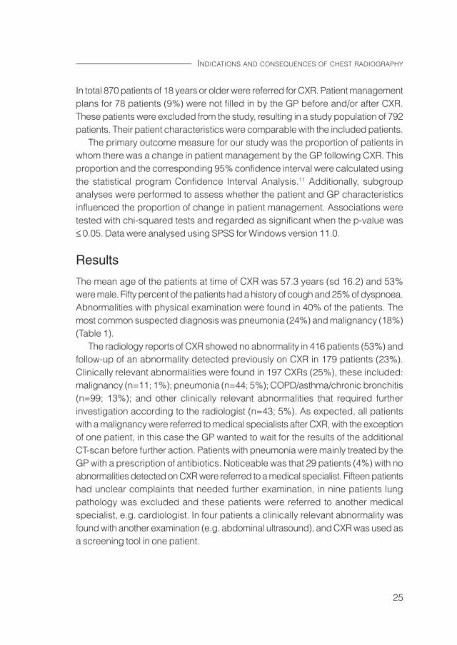
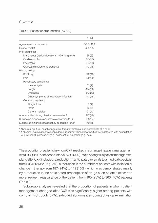
The mean age of the patients at time of CXR was 57.3 years (sd 16.2) and 53%were male. Fifty percent of the patients had a history of cough and 25% of dyspnoea.Abnormalities with physical examination were found in 40% of the patients. Themost common suspected diagnosis was pneumonia (24%) and malignancy (18%)(Table 1).
The radiology reports of CXR showed no abnormality in 416 patients (53%) andfollow-up of an abnormality detected previously on CXR in 179 patients (23%).Clinically relevant abnormalities were found in 197 CXRs (25%), these included:malignancy (n=11; 1%); pneumonia (n=44; 5%); COPD/asthma/chronic bronchitis(n=99; 13%); and other clinically relevant abnormalities that required furtherinvestigation according to the radiologist (n=43; 5%). As expected, all patientswith a malignancy were referred to medical specialists after CXR, with the exceptionof one patient, in this case the GP wanted to wait for the results of the additionalCT-scan before further action. Patients with pneumonia were mainly treated by theGP with a prescription of antibiotics. Noticeable was that 29 patients (4%) with noabnormalities detected on CXR were referred to a medical specialist. Fifteen patientshad unclear complaints that needed further examination, in nine patients lungpathology was excluded and these patients were referred to another medicalspecialist, e.g. cardiologist. In four patients a clinically relevant abnormality wasfound with another examination (e.g. abdominal ultrasound), and CXR was used asa screening tool in one patient.
CHAPTER 3
26
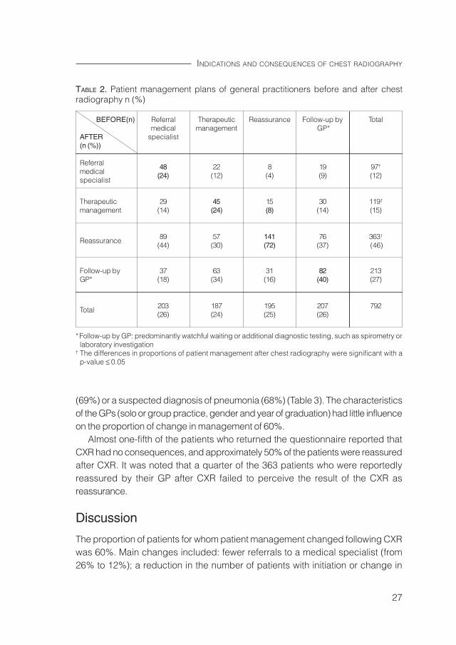
The proportion of patients in whom CXR resulted in a change in patient managementwas 60% (95% confidence interval 57%-64%). Main changes in patient managementplans after CXR included: a reduction in anticipated referrals to a medical specialistfrom 203 (26%) to 97 (12%); a reduction in the number of patients with initiation orchange in therapy from 187 (24%) to 119 (15%), which was demonstrated mainlyby a reduction in the anticipated prescription of drugs such as antibiotics; andmore frequent reassurance of the patient, from 195 (25%) to 363 (46%) patients(Table 2).
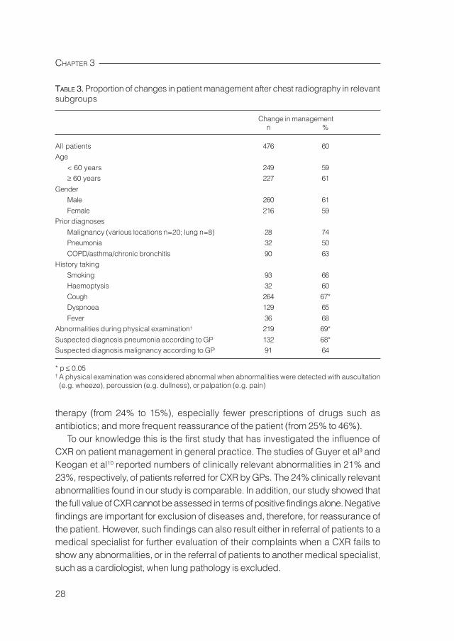
Subgroup analyses revealed that the proportion of patients in whom patientmanagement changed after CXR was significantly higher among patients withcomplaints of cough (67%), exhibited abnormalities during physical examination
TABLE 1. Patient characteristics (n=792)
n (%)
Age (mean ± sd in years) 57.3±16.2
Gender (male) 423 (53)Prior diagnoses
Malignancy (various locations n=29; lung n=9) 38 (5)Cardiovascular 95 (12)
Pneumonia 76 (10)COPD/asthma/chronic bronchitis 143 (18)
History takingSmoking 142 (18)
Pain 172 (22)Respiratory complaints
Haemoptysis 53 (7)Cough 394 (50)
Dyspnoea 99 (25)Other symptoms of respiratory infection* 117 (15)
General complaintsWeight loss 31 (4)
Fever 53 (7)General malaise 101 (13)
Abnormalities during physical examination† 317 (40)Suspected diagnosis pneumonia according to GP 193 (24)
Suspected diagnosis malignancy according to GP 142 (18)
* Abnormal sputum, nasal congestion, throat symptoms, and complaints of a cold† A physical examination was considered abnormal when abnormalities were detected with auscultation(e.g. wheeze), percussion (e.g. dullness), or palpation (e.g. pain)
27
INDICATIONS AND CONSEQUENCES OF CHEST RADIOGRAPHY
(69%) or a suspected diagnosis of pneumonia (68%) (Table 3). The characteristicsof the GPs (solo or group practice, gender and year of graduation) had little influenceon the proportion of change in management of 60%.
Almost one-fifth of the patients who returned the questionnaire reported thatCXR had no consequences, and approximately 50% of the patients were reassuredafter CXR. It was noted that a quarter of the 363 patients who were reportedlyreassured by their GP after CXR failed to perceive the result of the CXR asreassurance.
Discussion
The proportion of patients for whom patient management changed following CXRwas 60%. Main changes included: fewer referrals to a medical specialist (from26% to 12%); a reduction in the number of patients with initiation or change in
TABLE 2. Patient management plans of general practitioners before and after chestradiography n (%)
BEFORE(n) Referral Therapeutic Reassurance Follow-up by Totalmedical management GP*
AFTER specialist(n (%))
Referral 48 22 8 19 97†
medical (24) (12) (4) (9) (12)specialist
Therapeutic 29 45 15 30 119†
management (14) (24) (8) (14) (15)
Reassurance 89 57 141 76 363†
(44) (30) (72) (37) (46)
Follow-up by 37 63 31 82 213GP* (18) (34) (16) (40) (27)
Total 203 187 195 207 792(26) (24) (25) (26)
* Follow-up by GP: predominantly watchful waiting or additional diagnostic testing, such as spirometry orlaboratory investigation
† The differences in proportions of patient management after chest radiography were significant with ap-value ≤ 0.05
CHAPTER 3
28
therapy (from 24% to 15%), especially fewer prescriptions of drugs such asantibiotics; and more frequent reassurance of the patient (from 25% to 46%).
To our knowledge this is the first study that has investigated the influence ofCXR on patient management in general practice. The studies of Guyer et al9 andKeogan et al10 reported numbers of clinically relevant abnormalities in 21% and23%, respectively, of patients referred for CXR by GPs. The 24% clinically relevantabnormalities found in our study is comparable. In addition, our study showed thatthe full value of CXR cannot be assessed in terms of positive findings alone. Negativefindings are important for exclusion of diseases and, therefore, for reassurance ofthe patient. However, such findings can also result either in referral of patients to amedical specialist for further evaluation of their complaints when a CXR fails toshow any abnormalities, or in the referral of patients to another medical specialist,such as a cardiologist, when lung pathology is excluded.
TABLE 3. Proportion of changes in patient management after chest radiography in relevantsubgroups
Change in managementn %
All patients 476 60Age
< 60 years 249 59≥ 60 years 227 61
GenderMale 260 61
Female 216 59Prior diagnoses
Malignancy (various locations n=20; lung n=8) 28 74Pneumonia 32 50
COPD/asthma/chronic bronchitis 90 63History taking
Smoking 93 66Haemoptysis 32 60
Cough 264 67*Dyspnoea 129 65
Fever 36 68Abnormalities during physical examination† 219 69*
Suspected diagnosis pneumonia according to GP 132 68*Suspected diagnosis malignancy according to GP 91 64
* p ≤ 0.05† A physical examination was considered abnormal when abnormalities were detected with auscultation(e.g. wheeze), percussion (e.g. dullness), or palpation (e.g. pain)
29
INDICATIONS AND CONSEQUENCES OF CHEST RADIOGRAPHY
Subgroup analyses revealed that the proportion of patients in whom patientmanagement changed after CXR was significantly higher among patients withcomplaints of cough (67%), exhibited abnormalities during physical examination(69%) or a suspected diagnosis of pneumonia (68%). The changes in GPs’ patientmanagement plans after CXR in these patients were fewer anticipated referrals toa medical specialist, a reduction in the number of patients with initiation or changein therapy and more frequent reassurance of the patient. It is widely known thatthorough history taking and physical examination before commencement of a moreadvanced workup, such as a radiological examination, is very important. This studyshowed that even after a history and physical examination of the patient the influenceof CXR on patient management was substantial. We expected that the ability ofGPs to establish a more specific patient management plan after gaining detailedinformation of the patient with physical examination would result in a smallerproportion change in management after CXR. However the proportion of change inpatient management increased to almost 70% in patients with abnormalities detectedduring physical examination.
Almost 80% of the questionnaires were returned by the patients, which increasedthe validity of these results. Approximately 50% of patients were reassured by theirGP after CXR. Our study showed that in almost one-quarter of the patients whowere reassured by their GP after CXR, the patient did not perceive this asreassurance. Therefore, CXR did not have much value for these patients, becauseno referral or treatment followed after the radiological investigation and reassurancewas not achieved.
Before we can reach a conclusion it is important to note that this study hasseveral limitations. It was impossible to verify whether or not the GP really wouldhave conducted the anticipated patient management in accordance with the planmade on the standardized form before CXR was performed. This could result in anoverestimation of intended referrals to medical specialists. This study does notprove that the patient actually benefits from the diagnostic procedure, e.g. in termsof morbidity, mortality or quality of life. However, the study is the first to show thatthe procedure often leads to changes in patient management, which is one of theprerequisites for successfully influencing clinically relevant patient outcomes.
In conclusion, the GP’s patient management strategy was changed for 60% ofpatients following CXR. CXR substantially reduced the number of referrals to amedical specialist and initiation or change in therapy, and more patients werereassured by their GP. Thus, CXR is an important diagnostic tool for GPs andseems to reduce the burden on patients and health care.
CHAPTER 3
30
References
1. American College of Radiology. ACR Standards. ACR standard for the performanceof pediatric and adult chest radiography. United States, 2001.
2. United Nations Scientific Committee on the Effects of Atomic Radiation. Medicalradiation exposures. United Nations Scientific Committee on the Effects of AtomicRadiation UNSCEAR 2000 Report to the General Assembly, with ScientificAnnexes. Sources and effects of ionizing radiation, Volume I: Sources. New York:United Nations, 2000.
3. European Commission. Referral guidelines for imaging. Radiation protection 118.Luxembourg: Office for Official Publications of the European Communities, 2000.
4. Stolberg HO, Hynes DM, Rainbow AJ, Moran LA. Requesting diagnostic imagingexaminations: a position paper of the Canadian Association of Radiologists. CanAssoc Radiol J. 1997;48:89-91.
5. Coblentz CL, Matzinger F, Samson LM, Scherer J, Stolberg HO, Weisbrod G. CARstandards for chest radiography. Canada: Canadian Association of Radiologists,2000.
6. Health Services Utilization and Research Commission. Chest radiography: asummary of the evidence supporting selective clinical practice guidelines andrecommendations for implementation. Saskatoon: Health Services Utilization andResearch Commission, 1997.
7. Stolberg HO, Buckley N, Coblentz CL, Cornett R, Feightner K, Killian K, et al.Guidelines for chest X-rays in asymptomatic populations. Canada: The College ofPhysicians and Surgeons of Ontario, 1999.
8. World Health Organization Scientific Group. Chest and cardiovascular system.Effective choices for diagnostic imaging in clinical practice. Geneva: World HealthOrganization, 1990.
9. Guyer PB, Chalmers AG. Chest radiography for general practitioners- a low yieldinvestigation. J R Coll Gen Pract. 1983;33:477-479.
10. Keogan MT, Padhani AR, Flower CD. Chest radiography for general practitioners:scope for change? Clin Radiol. 1992;46:51-54.
11. Altman DG, Machin D, Bryant TN, Gardner MJ. Statistics with confidence:confidence intervals and statistical guidelines. London: BMJ Books, 2000.
Chest radiography in patients suspectedof pneumonia in primary care: diagnosticyield, and consequences for patientmanagement
Speets AM, Hoes AW, Van der Graaf Y, Kalmijn S, Sachs APE,Mali WPThM. Chest radiography in patients suspected ofpneumonia in primary care: diagnostic yield, and consequencesfor patient management. In revision for Eur Respir J.
4
CHAPTER 4
32
AbstractBackground. Chest radiography (CXR) is frequently performed for diagnosingpneumonia in primary care. This prospective cohort study assessed the diagnosticyield of CXR in primary care patients suspected of pneumonia.Methods. In total, 192 patients with a clinical suspicion of pneumonia aged ≥ 18years (mean age 56.8±17.6 years; 55% males) were referred by their generalpractitioner (GP) for CXR to one of the three participating hospitals in TheNetherlands. All GPs were asked to fill in a standardized form before and afterCXR. The primary outcome measures were the proportion of patients with a clearshift in the probability estimation of pneumonia, and the proportion patients with achange in patient management following CXR.Results. Pneumonia was diagnosed by GPs in 35 patients (18%), of whom 27patients (14%) had a positive CXR, and 8 patients (4%) a negative CXR, howeverwith an assumed high probability of pneumonia by the GP. CXR clearly influencedthe diagnosis of pneumonia by the GP in 53% of the patients: CXR ruled outpneumonia in 47%, and the probability of pneumonia substantially increased in 6%of the patients. Patient management changed after CXR in 69% of the patients,mainly caused by a reduction in medication prescription (from 43% to 17%); andmore frequent reassurance of the patient (from 8% to 35%).Conclusion. CXR is an important diagnostic tool in primary care patients with aclinical suspicion of pneumonia in terms of change in the estimated probability ofpneumonia, and change in patient management.
33
CHEST RADIOGRAPHY AND PNEUMONIA IN PRIMARY CARE
Introduction
Primary care physicians usually rely on patient history, and signs and symptoms todiagnose or exclude pneumonia.1 However, most signs and symptoms traditionallyassociated with pneumonia (e.g. fever and coughing) are not predictive ofpneumonia in general practice.2-4 Chest radiography (CXR) is the most frequentlyperformed diagnostic investigation requested by general practitioners (GPs) inEurope: in 22% of patients with a suspected lower respiratory tract infection CXR isrequested.5 CXR is considered the gold standard for pneumonia diagnosis. CXRcan diagnose pneumonia in case of presence of an infiltrate, and differentiatepneumonia from other conditions that may present with similar symptoms (e.g.acute bronchitis). In addition, the results may suggest specific aetiologies (e.g.lung abscess), identify coexisting conditions (e.g. bronchial obstruction), andevaluate the severity of illness.6-9
Although CXR is frequently used for diagnosing pneumonia, little is known aboutthe influence of CXR on the probability estimation of pneumonia by GPs, and onchange in patient management. Simpson et al concluded that results of CXRrequested by GPs influenced patient management in 48% of 97 patients withradiographic features of acute infection.10 However, this study was conducted onlyin patients with radiographic evidence of infection and the patient managementwas assessed with questionnaires filled in retrospectively by GPs. When assessingthe diagnostic yield of CXR, e.g. in terms of patient management, it is important tostudy the complete cohort of patients suspected of pneumonia, and not only thesubgroup of patients with a radiographic diagnosis of pneumonia.
The objective of this prospective cohort study was to assess the effect of CXRon the probability estimation of pneumonia by GPs, the influence of CXR on patientmanagement and consequences of CXR according to the patient. The studypopulation consisted of primary care patients with a clinical suspicion of pneumoniareferred for CXR by GPs.
Methods
This study is part of a large prospective cohort study conducted from April 2003 toDecember 2004 with the help of 78 GPs participating in the catchment area of oneof three general hospitals located in three main cities in The Netherlands (JeroenBosch Hospital in ‘s-Hertogenbosch; Gelre Hospitals in Apeldoorn; ’Onze LieveVrouwe Gasthuis’ in Amsterdam). In total 870 patients of 18 years and older who
CHAPTER 4
34
were referred for CXR (posteroanterior and lateral view) by their GP to one of thesehospitals were included in the cohort study. The study was approved by the MedicalEthics Review Board.
The GPs could fill in three probable diagnoses on a standard form beforerequesting a CXR. In the present study all patients who were referred for CXR witha clinical suspicion of pneumonia as one of these probable diagnoses were included(n=222). Estimated probabilities for 18 patients (8%) were not filled in by the GPbefore and/or after CXR. These patients were excluded from the study. Their patientcharacteristics were comparable with the included patients. Patients referred for afollow-up CXR for the treatment evaluation of pneumonia were also excluded (n=12),resulting in a study population of 192 patients. Additionally, all patients with incidentalpneumonia detected with CXR were included as a separate patient group (i.e.patients referred for CXR without a clinical suspicion of pneumonia).
All GPs were asked to fill in a standardized form before requesting a CXR,including information on history, physical examination, indication, probable diagnosiswith estimated prior probabilities on a visual analogue scale (range 0-100%), andanticipated patient management. The management options included: referral to amedical specialist; medication prescription; reassurance of the patient; and follow-up by the GP (watchful waiting or additional diagnostic testing). After the GPreceived the report (within 1-4 days after the CXR) he or she filled in a secondquestionnaire, again including the probable diagnosis with estimated posteriorprobabilities, and anticipated patient management plan. We considered a decreaseor increase in the estimated probability of pneumonia by the GPs after CXR of≥ 30% as a substantial change in the probability estimation.
The findings on the CXR were categorized into four groups: (1) pneumonia; (2)other clinically relevant abnormalities; (3) a known abnormality, which was detectedpreviously on CXR; (4) no abnormality. Six months after the CXR a short questionnairewas sent to all patients (response rate 84%), in order to evaluate their currentcomplaints and assess the consequences of CXR according to the patient.
The primary outcome measures for our study were the proportion of patientswith a clear shift in the probability estimation of pneumonia by the GP (≥ 30%decrease or ≥ 30% increase of the estimated probability after CXR), and theproportion of patients in whom there was a change in patient management by theGP following CXR. These proportions and corresponding 95% confidence intervalswere calculated using the statistical program Confidence Interval Analysis.11
35
CHEST RADIOGRAPHY AND PNEUMONIA IN PRIMARY CARE
Results
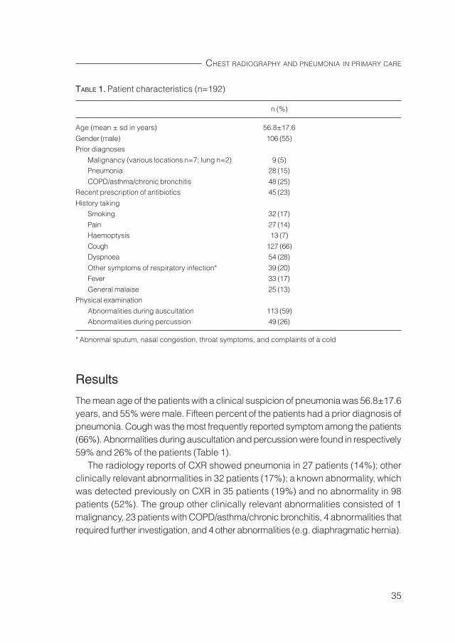
The mean age of the patients with a clinical suspicion of pneumonia was 56.8±17.6years, and 55% were male. Fifteen percent of the patients had a prior diagnosis ofpneumonia. Cough was the most frequently reported symptom among the patients(66%). Abnormalities during auscultation and percussion were found in respectively59% and 26% of the patients (Table 1).
The radiology reports of CXR showed pneumonia in 27 patients (14%); otherclinically relevant abnormalities in 32 patients (17%); a known abnormality, whichwas detected previously on CXR in 35 patients (19%) and no abnormality in 98patients (52%). The group other clinically relevant abnormalities consisted of 1malignancy, 23 patients with COPD/asthma/chronic bronchitis, 4 abnormalities thatrequired further investigation, and 4 other abnormalities (e.g. diaphragmatic hernia).
TABLE 1. Patient characteristics (n=192)
n (%)
Age (mean ± sd in years) 56.8±17.6
Gender (male) 106 (55)Prior diagnoses
Malignancy (various locations n=7; lung n=2) 9 (5)Pneumonia 28 (15)
COPD/asthma/chronic bronchitis 48 (25)Recent prescription of antibiotics 45 (23)
History takingSmoking 32 (17)
Pain 27 (14)Haemoptysis 13 (7)
Cough 127 (66)Dyspnoea 54 (28)
Other symptoms of respiratory infection* 39 (20)Fever 33 (17)
General malaise 25 (13)Physical examination
Abnormalities during auscultation 113 (59)Abnormalities during percussion 49 (26)
* Abnormal sputum, nasal congestion, throat symptoms, and complaints of a cold
CHAPTER 4
36
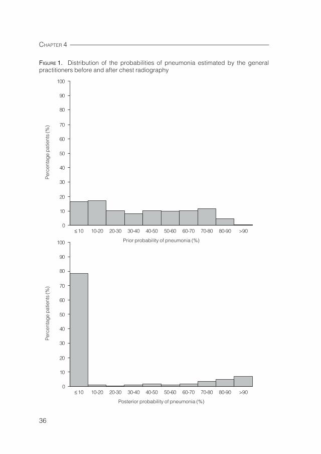
FIGURE 1. Distribution of the probabilities of pneumonia estimated by the generalpractitioners before and after chest radiography
100
90
80
70
60
50
40
30
20
10
0
Prior probability of pneumonia (%)
≤ 10 10-20 20-30 30-40 40-50 50-60 60-70 70-80 80-90 >90
Per
cent
age
pat
ient
s (%
)
100
90
80
70
60
50
40
30
20
10
0
Posterior probability of pneumonia (%)
≤ 10 10-20 20-30 30-40 40-50 50-60 60-70 70-80 80-90 >90
Per
cent
age
pat
ient
s (%
)
37
CHEST RADIOGRAPHY AND PNEUMONIA IN PRIMARY CARE
The distributions of the prior and posterior probability of pneumonia are shown inFigure 1. Noticeable were the two large groups referred for CXR with a very low orhigh prior probability of pneumonia, 64 patients (33%) and 30 patients (16%)respectively. After CXR, pneumonia was diagnosed in 4 of the 64 patients (6%)with a very low prior probability, and in only 15 of the 30 patients (50%) with a veryhigh prior probability of pneumonia. The probability estimation of pneumonia wasclearly changed by means of CXR in 53% of the patients (95% CI 46%-59%). Theestimated probability of pneumonia decreased with ≥ 30% (range 30-100%) in 89patients (47%), and increased with ≥ 30% (range 30-80%) in 12 patients (6%) afterCXR.
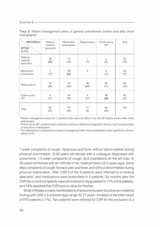
The proportion of patients for whom patient management changed followingCXR was 69% (95% CI 62%-75%). Main changes in patient management plansafter CXR included: a reduction in the number of patients with a medicationprescription from 79 (43%) to 32 (17%) patients; and more frequent reassuranceof the patient, from 15 (8%) to 64 (35%) patients (Table 2). The reduction inmedication prescription was caused mainly by a decrease in the prescription ofantibiotics from 53 patients (28%) before CXR to 26 patients (14%) after CXR.
Six months after the CXR the current complaints were diminished or disappearedin almost 80% of the patients referred for CXR by GPs with a clinical suspicion ofpneumonia. Only 15% of the patients who returned the questionnaire reported thatCXR had no value for him or her. CXR resulted in a definite diagnosis or bettertreatment according to 43% of the patients, and 44% of the patients were reassuredafter CXR.
Pneumonia was diagnosed with CXR in 27 patients (14%), with a mean age of53.8±18.8 years, and 44% were male. Abnormalities during auscultation andpercussion were found in respectively 74% and 26% of these patients. The GPsreferred 7 patients (26%) to a medical specialist, medications were prescribed in13 patients (48%), patient management was watchful waiting in 6 patients (22%),and an additional CT scan was ordered for 1 patient (4%). Six months after theCXR the current complaints were diminished or disappeared in 72% of the patients,and 8% reported that CXR had no value for him/her.
Additionally, pneumonia was diagnosed by the GP in 8 patients (4%) without apositive CXR, however with an assumed high probability of pneumonia by the GP.The GP suspected pneumonia in 4 patients, viral pneumonia in 2 patients, andmycoplasma pneumonia was shown with additional laboratory investigation in 2patients. The 4 patients suspected of pneumonia were: 1) 48-years old male with amedical history of COPD, 2 weeks complaints of cough and thoracic pain, withoutabnormalities during physical examination; 2) 52-years old female who smoked,
CHAPTER 4
38
1 week complaints of cough, dyspnoea and fever, without abnormalities duringphysical examination; 3) 62-years old female with a colleague diagnosed withpneumonia, 1.5 week complaints of cough, and crepitations on the left side; 4)20-years old female with an infiltrate in her medical history (2.5 years ago), somedays complaints of cough, thoracic pain and fever, and without abnormalities duringphysical examination. After CXR 4 of the 8 patients were referred to a medicalspecialist, and medications were prescribed in 4 patients. Six months after theCXR the current complaints were diminished or disappeared in 71% of the patients,and 14% reported that CXR had no value for him/her.
Small infiltrates or early manifestations of pneumonia were found as an incidentalfinding with CXR in 5 patients (age range 32-77 years; 3 males) of the total cohortof 870 patients (<1%). Two patients were referred for CXR for the exclusion of a
TABLE 2. Patient management plans of general practitioners before and after chestradiography*
BEFORE(n) Referral Medication Reassurance Follow-up by Totalmedical prescription GP†
AFTER specialist(n (%))
Referral 10 14 1 4 29medical (24) (18) (7) (8) (16)specialist
Medication 7 19 0 6 32‡
prescription (17) (24) (12) (17)
Reassurance 16 18 10 20 64‡
(38) (23) (67) (41) (35)
Follow-up by 9 28 4 19 60GP† (21) (35) (27) (39) (32)
Total 42 79 15 49 185(23) (43) (8) (26)
* Patient management plans for 7 patients (4%) were not filled in by the GP before and/or after chestradiography
† Follow-up by GP: predominantly watchful waiting or additional diagnostic testing, such as spirometryor laboratory investigation
‡ The differences in proportions of patient management after chest radiography were significant with a p-value ≤ 0.05
39
CHEST RADIOGRAPHY AND PNEUMONIA IN PRIMARY CARE
malignancy, 1 patient for the confirmation of COPD, and 2 patients had unclearcomplaints without any abnormalities during physical examination. After CXR 3patients were referred to a medical specialist, medications were prescribed in 1patient, and patient management was watchful waiting and an additional follow-upCXR in 1 patient.
Discussion
CXR clearly influenced the diagnosis of pneumonia by the GP in 56% of the patientsreferred for CXR with a clinical suspicion of pneumonia: CXR ruled out pneumoniain 50% of the patients, and the probability of the diagnosis pneumonia substantiallyincreased in 6% of the patients. The proportion of patients for whom patientmanagement changed following CXR was 69%, mainly caused by a decrease inthe prescription of antibiotics, and more frequent reassurance of the patient.
To our knowledge, this study is the first that assessed the effect of CXR on theprobability estimation of pneumonia by GPs. The number of patients in whom thepatient management changed (69%) is much higher than the 48% reported in thestudy of Simpson et al.10 This difference could be explained by the study designs:their study was conducted in patients with radiographic evidence of infection andthe patient management was assessed with questionnaires filled in retrospectivelyby GPs, which may have biased the results. Besides, Simpson et al did not specifywhether reassurance of the patient was considered as patient management, andhow patient management was influenced by the findings of CXR.
The distributions of the prior and posterior probability of pneumonia in Figure 1showed that the uncertain area of a diagnosis, around estimated probabilities of50%, disappeared largely as a consequence of CXR. Noticeable in our study wasthat almost half of all patients were referred for CXR with a very low or high priorprobability of pneumonia, respectively 33% and 16% of the patients. Seventy-fivepercent of the patients with a very low prior probability of pneumonia had additionaldifferential diagnoses, such as COPD or acute bronchitis, with a higher priorprobability according to the GP. After CXR, pneumonia was diagnosed in 6% of thepatients with a very low prior probability, and in only 50% of the patients with a veryhigh prior probability of pneumonia. This emphasizes the importance of referringpatients with a clinical suspicion of pneumonia for CXR, even when the priorprobability of pneumonia is very high according to the GP.
Pneumonia was diagnosed by the GP in 35 patients (18%): 27 (14%) had apositive CXR, and 8 patients (4%) a negative CXR, however with an assumed highprobability of pneumonia by the GP. Low percentages of patients diagnosed with
CHAPTER 4
40
pneumonia by a positive CXR were also found in other studies: 15% by Melbyeet al12, and 7% by Lieberman et al.13 It is noticeable that the estimated probabilitiesin the patient groups diagnosed with pneumonia with a positive and negative CXRwere high before CXR, 61% and 72% respectively. However, these percentageswere not high enough for the GPs to start treatment or refer patient to a medicalspecialist without an additional CXR. The current restrictive policy of prescribingantibiotics could encourage the GPs to order CXR in patients suspected ofpneumonia even when estimated prior probabilities are high based on medicalhistory, anamnesis and physical examination.9
The manifestations of pneumonia on CXR may vary considerably, dependingupon the degree of inflammation and the stage of the disease process. It is difficultto diagnose mild or early stage pneumonia by CXR.14,15 Besides, it is possible todetect pneumonia during physical examination without roentgengraphic evidence.14
The 8 patients with a high estimated probability of pneumonia, and a negative CXRmight have been referred too soon for CXR by their GP; mycoplasma pneumoniawas shown with additional laboratory investigation in 2 of these 8 patients.
Interestingly, no clear differences in patient characteristics, including signs andsymptoms, were observed in referred patients with or without pneumonia. Thisindicates that the GPs adequately applied their clinical skills to select those patientsfor additional imaging in whom history taking and physical examination providedinsufficient information to distinguish those with from those without pneumonia.
As expected, pneumonia was found scarcely as incidental finding with CXR. Inour study, small infiltrates or early manifestations of pneumonia were found as anincidental finding in less than 1% of the patients of the total cohort of 870 patients.
A limitation of our study is that it was impossible to verify whether or not the GPreally would have conducted the anticipated patient management in accordancewith the plan made on the standardized form before CXR was performed. Thiscould result in an overestimation of intended referrals to medical specialists.
In conclusion, CXR is a valuable diagnostic tool in primary care patients with aclinical suspicion of pneumonia referred for CXR by GPs in terms of change in theestimated probability of pneumonia by GPs, change in patient management, andaccording to the patients themselves. In particular, CXR is important for the exclusionof pneumonia in general practice.
41
CHEST RADIOGRAPHY AND PNEUMONIA IN PRIMARY CARE
References
1. Metlay JP, Fine MJ. Testing strategies in the initial management of patients withcommunity-acquired pneumonia. Ann Intern Med. 2003;138(2):109-118.
2. Hopstaken RM, Muris JW, Knottnerus JA, Kester AD, Rinkens PE, Dinant GJ.Contributions of symptoms, signs, erythrocyte sedimentation rate, and C-reactiveprotein to a diagnosis of pneumonia in acute lower respiratory tract infection. Br JGen Pract. 2003;53(490):358-364.
3. Metlay JP, Kapoor WN, Fine MJ. Does this patient have community-acquiredpneumonia? Diagnosing pneumonia by history and physical examination. JAMA.1997;278(17):1440-1445.
4. Mabie M, Wunderink RG. Use and limitations of clinical and radiologic diagnosisof pneumonia. Semin Respir Infect. 2003;18(2):72-79.
5. Woodhead M, Gialdroni Grassi G, Huchon GJ, Leophonte P, Manresa F, SchabergT. Use of investigations in lower respiratory tract infection in the community: aEuropean survey. Eur Respir J. 1996;9(8):1596-1600.
6. Niederman MS, Mandell LA, Anzueto A, et al; American Thoracic Society. Guidelinesfor the management of adults with community-acquired pneumonia. Diagnosis,assessment of severity, antimicrobial therapy, and prevention. Am J Respir CritCare Med. 2001;163(7):1730-1754.
7. British Thoracic Society Standards of Care Committee. BTS Guidelines for theManagement of Community Acquired Pneumonia in Adults. Thorax. 2001;56 Suppl4:IV1-64.
8. Mandell LA, Marrie TJ, Grossman RF, Chow AW, Hyland RH. Canadian guidelinesfor the initial management of community-acquired pneumonia: an evidence-basedupdate by the Canadian Infectious Diseases Society and the Canadian ThoracicSociety. The Canadian Community-Acquired Pneumonia Working Group. Clin InfectDis. 2000;31(2):383-421.
9. Bartlett JG, Dowell SF, Mandell LA, File Jr TM, Musher DM, Fine MJ. Practiceguidelines for the management of community-acquired pneumonia in adults.Infectious Diseases Society of America. Clin Infect Dis. 2000;31(2):347-382.
10. Simpson JCG, Hulse P, Taylor PM, Woodhead M. Do radiographic features of acuteinfection influence management of lower respiratory tract infections in thecommunity? Eur Respir J. 1998;12:1384-1387.
11. Altman DG, Machin D, Bryant TN, Gardner MJ. Statistics with confidence. 2nd
edition.London: BMJ Books, 2000.
12. Melbye H, Straume B, Aasebo U, Brox J. The diagnosis of adult pneumonia ingeneral practice. The diagnostic value of history, physical examination and someblood tests. Scand J Prim Health Care. 1988;6(2):111-117.
13. Lieberman D, Shvartzman P, Korsonsky I, Lieberman D. Diagnosis of ambulatorycommunity-acquired pneumonia. Comparison of clinical assessment versus chestX-ray. Scand J Prim Health Care. 2003;21(1):57-60.

CHAPTER 4
42
14. Fraser RG, Paré JAP. Pneumonia. Diagnosis of diseases of the chest. 2nd
edition.Philadelphia/London/Toronto: W.B. Saunders Company, 1978.
15. Melbye H. Community pneumonia—more help is needed to diagnose and assessseverity. Br J Gen Pract. 2002;52(484):886-888.
The value of abdominal ultrasound inpatients with abdominal pain referred bygeneral practitioners
5
Speets AM, Kalmijn S, Hoes AW, Van der Graaf Y, Mali WPThM.The yield of abdominal ultrasound in patients with abdominal painreferred by general practitioners. Submitted.
CHAPTER 5
44
AbstractBackground. About 8% of the patients with abdominal pain of unclear origin arereferred by general practitioners (GPs) for abdominal ultrasound (US). The objectiveof this study was to examine the prevalence and determinants of significantabnormalities on abdominal US in patients with abdominal pain referred by GPs.Methods. This cross-sectional study was conducted with data from thecomputerized hospital database of University Medical Centre Utrecht in TheNetherlands from January to December 2002, if necessary completed with datafrom an additional patient questionnaire. Findings on US were considered assignificant abnormalities when it resulted in referral to a medical specialist orhospital admission.Results. In total, 499 patients of 18 years and older with abdominal pain werereferred for abdominal US by GPs. Mean age at time of the US was 47.9 years(sd 15.2), and almost one third was male. Significant pathology was seen in 16% ofthe patients. Older age (OR 1.03 per year), colicky pain (OR 2.80) and a clinicalsuspicion of bile stones (OR 2.31) were predictors of a significant abnormal findingon abdominal US.Conclusion. A significant abnormality was found in 16% of the patients aged 18years and older with abdominal pain, who were referred for abdominal US by GPs.Older age, colicky pain, and a clinical suspicion of bile stones were predictors of asignificant abnormal finding on abdominal US.
45
ABDOMINAL ULTRASOUND IN PATIENTS WITH ABDOMINAL PAIN
Introduction
Many problems encountered by general practitioners (GPs) are of a non-acute,non-specific nature, such as abdominal complaints.1 Approximately half of allabdominal complaints consist of pain.2 The two-year cumulative incidence ofabdominal pain in The Netherlands is estimated at 3%.3 Patients with abdominalpain comprise 2% to 4% of all visits to GPs.4-6 Results of three previous studies areconsistent with the impression that abdominal pain in general practice usually is aself-limiting disease for which no definitive diagnosis is found and which can bemanaged entirely by the GP.5-7
One of the most difficult tasks confronting the GP is knowing how extensive aparticular symptom has to be investigated, taking into account that performingunnecessary radiological investigations can lead to incidental findings, extra costsand be a burden for the patient. On the other hand, investigations with negativefindings may also be of importance in daily practice, for purposes of reassurance.In the case of abdominal pain, this dilemma is stressed by the availability of a widerange of sophisticated diagnostic technology.8 In this range, ultrasound (US) is theleast expensive and most non-invasive abdominal imaging modality and a safe andeffective diagnostic procedure.9-11
Abdominal pain is one of the indications for the performance of an US examinationof the abdomen in the American College of Radiology standards.12 About 8% of thepatients with abdominal pain of unclear origin are referred by GPs for abdominalUS. 6,10 Annually, about 200,000 abdominal US are on request of GPs in TheNetherlands. It is unclear whether, and in which patients abdominal US yieldssufficiently relevant information. Abdominal US among patients with abdominalpain has received little attention in the scientific literature. The purpose of thiscross-sectional study was to examine the prevalence and determinants of significantabnormalities on abdominal US in patients with abdominal pain referred by GPs.
Methods
The study sample was taken from the Hospital Information System computerdatabase of the University Medical Centre (UMC) Utrecht in The Netherlands. Across-sectional study was conducted, covering the period from January toDecember 2002. All patients referred by their GPs for abdominal US were extractedfrom the database, in which all data of the US referral forms filled in by the GPsand US findings were available. Patients of 18 years and older were included when
CHAPTER 5
46
complaints of abdominal pain or a clinical suspicion of bile and/or renal stones bythe GP were indicated at the referral form of the GP. A total of 911 abdominal USwere performed for GPs in UMC Utrecht during this period, and 499 (55%) patientsmet the inclusion criteria. In The Netherlands no approval by the Medical EthicsReview Board and no informed consent is required when a medical specialist, inthis case the radiologist, evaluates his own patient management.
Gender, age, indications and information about the findings were collected fromthe patient files in the computer database. The indications were arranged in fourgroups: complaints of pain, other symptoms than pain (e.g. nausea), relevant medicalhistory (e.g. previous surgery), and a clinical suspicion of the GP (e.g. presence ofbile stones).
Abdominal US can result in multiple findings. These findings were initiallycategorized in four groups: no abnormality, non-relevant abnormality (steatotic liver,fatty pancreas, and other findings e.g. obstipation, simple cysts and retroflectionuterus), abnormality with unclear consequences (bile stones, benign solid tumour,renal problems, renal stones, hernia inguinalis, and pathology aorta abdominalis)and significant abnormality (malignant tumour, pathology gallbladder, and perforationliver). A short questionnaire was sent to 146 patients with an abnormality withunclear consequences, in order to assess the consequences of the US finding. Intotal, 115 (79%) questionnaires were returned. The remaining 21% of the patientswere checked in the Hospital Information System and at the general practices,resulting in complete follow-up. Findings of US were considered to be a significantabnormality when it resulted in referral to a medical specialist or hospital admission.In the logistic regression analysis the most serious finding of each patient wasused as the outcome.
Multivariate logistic regression analysis was used to investigate whether gender,age, complaints of pain, other symptoms than pain, medical history and a clinicalsuspicion of the GP were independent determinants of a significant abnormal findingon abdominal US. Statistical significance was inferred when the p-value was equalto or less than 0.05. Data were analysed using SPSS for Windows version 11.0.
Results
Mean age at time of the abdominal US was 47.9 years (sd 15.2), and almost onethird was male (30%). Table 1 gives an overview of indications for abdominal US. Alarge part of the patients had complaints of localized abdominal pain (51%) orabdominal pain e causa ignota (eci) (30%). In 10% colicky pain was the indication
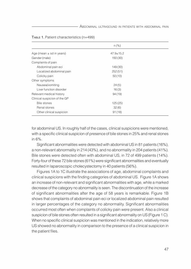
47
ABDOMINAL ULTRASOUND IN PATIENTS WITH ABDOMINAL PAIN
for abdominal US. In roughly half of the cases, clinical suspicions were mentioned,with a specific clinical suspicion of presence of bile stones in 25% and renal stonesin 6%.
Significant abnormalities were detected with abdominal US in 81 patients (16%),a non-relevant abnormality in 214 (43%), and no abnormality in 204 patients (41%).Bile stones were detected often with abdominal US, in 72 of 499 patients (14%).Forty-four of these 72 bile stones (61%) were significant abnormalities and eventuallyresulted in laparoscopic cholecystectomy in 40 patients (56%).
Figures 1A to 1C illustrate the associations of age, abdominal complaints andclinical suspicions with the finding categories of abdominal US. Figure 1A showsan increase of non-relevant and significant abnormalities with age, while a markeddecrease of the category no abnormality is seen. The discontinuation of the increaseof significant abnormalities after the age of 58 years is remarkable. Figure 1Bshows that complaints of abdominal pain eci or localized abdominal pain resultedin larger percentages of the category no abnormality. Significant abnormalitiesoccurred most often when complaints of colicky pain were present. Also a clinicalsuspicion of bile stones often resulted in a significant abnormality on US (Figure 1 C).When no specific clinical suspicion was mentioned in the indication, relatively moreUS showed no abnormality in comparison to the presence of a clinical suspicion inthe patient files.
TABLE 1. Patient characteristics (n=499)
n (%)
Age (mean ± sd in years) 47.9±15.2
Gender (male) 150 (30)Complaints of pain
Abdominal pain eci 149 (30)Localized abdominal pain 252 (51)
Colicky pain 50 (10)Other symptoms
Nausea/vomiting 24 (5)Liver function disorder 16 (3)
Relevant medical history 94 (19)Clinical suspicion of the GP
Bile stones 125 (25)Renal stones 32 (6)
Other clinical suspicion 91 (18)
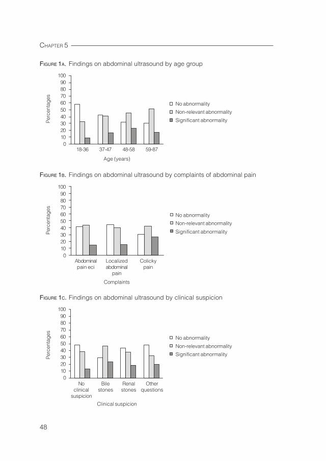
CHAPTER 5
48
FIGURE 1A. Findings on abdominal ultrasound by age group
FIGURE 1B. Findings on abdominal ultrasound by complaints of abdominal pain
FIGURE 1C. Findings on abdominal ultrasound by clinical suspicion
1009080706050403020100
Age (years)
18-36 37-47 48-58 59-87
Per
cent
ages
No abnormality
Non-relevant abnormality
Significant abnormality
1009080706050403020100
Complaints
Abdominalpain eci
Localizedabdominal
pain
Colickypain
Per
cent
ages
No abnormality
Non-relevant abnormality
Significant abnormality
Clinical suspicion
Noclinical
suspicion
Bilestones
Renalstones
Otherquestions
Per
cent
ages
No abnormality
Non-relevant abnormality
Significant abnormality
1009080706050403020100
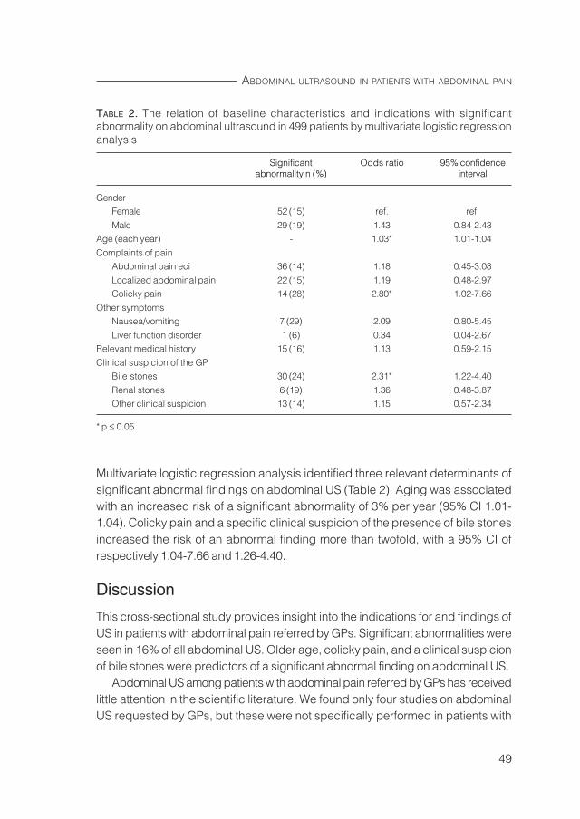
49
ABDOMINAL ULTRASOUND IN PATIENTS WITH ABDOMINAL PAIN
Multivariate logistic regression analysis identified three relevant determinants ofsignificant abnormal findings on abdominal US (Table 2). Aging was associatedwith an increased risk of a significant abnormality of 3% per year (95% CI 1.01-1.04). Colicky pain and a specific clinical suspicion of the presence of bile stonesincreased the risk of an abnormal finding more than twofold, with a 95% CI ofrespectively 1.04-7.66 and 1.26-4.40.
Discussion
This cross-sectional study provides insight into the indications for and findings ofUS in patients with abdominal pain referred by GPs. Significant abnormalities wereseen in 16% of all abdominal US. Older age, colicky pain, and a clinical suspicionof bile stones were predictors of a significant abnormal finding on abdominal US.
Abdominal US among patients with abdominal pain referred by GPs has receivedlittle attention in the scientific literature. We found only four studies on abdominalUS requested by GPs, but these were not specifically performed in patients with
TABLE 2. The relation of baseline characteristics and indications with significantabnormality on abdominal ultrasound in 499 patients by multivariate logistic regressionanalysis
Significant Odds ratio 95% confidenceabnormality n (%) interval
GenderFemale 52 (15) ref. ref.
Male 29 (19) 1.43 0.84-2.43Age (each year) - 1.03* 1.01-1.04
Complaints of painAbdominal pain eci 36 (14) 1.18 0.45-3.08
Localized abdominal pain 22 (15) 1.19 0.48-2.97Colicky pain 14 (28) 2.80* 1.02-7.66
Other symptomsNausea/vomiting 7 (29) 2.09 0.80-5.45
Liver function disorder 1 (6) 0.34 0.04-2.67Relevant medical history 15 (16) 1.13 0.59-2.15
Clinical suspicion of the GPBile stones 30 (24) 2.31* 1.22-4.40
Renal stones 6 (19) 1.36 0.48-3.87Other clinical suspicion 13 (14) 1.15 0.57-2.34
* p ≤ 0.05
CHAPTER 5
50
abdominal pain.13-16 None of these studies formulated clear definitions of significantabnormalities or examined the indications in relation to findings on abdominal US.Significant abnormalities were detected on 25% to 30% of the abdominal US.13-16
Only Charlesworth et al14 presented percentages for relevant abnormalities inpatients with left upper quadrant pain (n=27) and patients referred with lowerabdominal pain (n=100): 18% for both patient groups. This percentage iscomparable with the 16% significant abnormalities found in our study.
The present study showed three determinants of significant abnormal findingson abdominal US, i.e. age, colicky pain and a clinical suspicion of bile stones. It iswell known that morbidity increases with age. Our finding of a significant positiveassociation of age with abnormal findings on abdominal US agrees with the studyof Muris et al17, that also showed a positive association between age and organicdisease in patients with abdominal pain. The discontinuation of the increase in thenumber of significant abnormalities after the age of 58 years was remarkable. It ispossible that for example bile stones are more often found as incidental findingsduring the search for severe pathology; or GPs referred relatively older patientswith only few alarming complaints for abdominal US, leading to relatively few severeabnormalities. As expected, a significant association was found between colickypain and significant abnormalities on abdominal US. A similar association wasobserved for a clinical suspicion of bile stones. The presence of a clinical suspicionof renal stones and other suspicions also showed a positive, but non-significantassociation. These associations are in line with the statement by Muris et al6 thatthorough initial history and physical examination in abdominal pain patients beforecommencing a more advanced workup is very important.
Certain limitations of the study should be discussed. First, we used an existingdata file for collection of information about indications and findings on abdominalUS, which can lead to incomplete data and some distortion of the results. Secondly,the study was conducted in a university hospital. In The Netherlands, GPs canchoose to refer patients to a regional or university hospital. Patients suspected ofmore serious illnesses or who have been in the university hospital for other reasonsthan abdominal pain may have a larger chance of being referred to this hospital.The prevalence of pathologic findings of 16% could therefore be an overestimation,although Charlesworth et al14 presented percentages of 18% relevant abnormalitiesin patients with abdominal pain and the other percentages mentioned previouslyranged from 25 to 30%.
51
ABDOMINAL ULTRASOUND IN PATIENTS WITH ABDOMINAL PAIN
Several questions are raised by the results of this cross-sectional study: What isthe value of abdominal US in the 84% of the patients in whom no or non-relevantabnormalities were found? Do results of abdominal US change patient managementdecisions of GPs or have an effect on the outcome of illness? Can specific patientsubpopulations be indicated, in whom abdominal US is of additional value? To gainmore insight into the additional value of abdominal US in general practice we areplanning to perform a prospective study in multiple general practices and hospitals.
CHAPTER 5
52
References
1. Berkestijn van LGM, Kastein MR, Lodder A, Melker de RA, Bartelink ML. How wellare patients treated in family practice? Quality of consultations for non-acuteabdominal complaints. Int J Qual Health Care. 1998;10:221-233.
2. Lamberts H. In het huis van de huisarts. Verslag van het transitieproject. Lelystad:MEDITekst, 1994:185-206.
3. Lamberts H. Morbidity in general practice. Diagnosis related information from theMonitoring project. Utrecht: Uitgeversmaatschappij Huisartsenpers BV, 1984:72.
4. Adelman AM, Koch H. New visits for abdominal pain in the primary care setting.Fam Med. 1991;23:122-126.
5. Frear D, Tilyard MW, Gurr E. Abdominal pain in New Zealand general practice.New Zeal Med J. 1997;110:333-334.
6. Muris JW, Starmans R, Fijten GH, Crebolder HFJM, Krebber TFWA , Knottnerus JA.Abdominal pain in general practice. Fam Pract. 1993;10:387-390.
7. Adelman A. Abdominal pain in the primary care setting. J Fam Pract. 1987;25:27-32.8. Ward M. Upper abdominal pain-management issues. Aust Fam Physician.
1994;23:331-333,336.9. American College of Radiology. ACR Standards. ACR Standard for performing
and interpreting diagnostic ultrasound examinations. United States, 2000.10. Meijer JS, Groen ASM, Schadé E, Keeman JN, Baere de H, Wesdorp ICE, et al.
Bijblijven. Cumulatief Geneeskundig Nascholingssysteem. Buikpijn. Houten: BohnStafleu Van Loghum, 1991:7-24.
11. Mendelson RM, Kelsey PJ. Abdominal ultrasound. An overview for generalpractitioners. Aust Fam Physician. 1995;24:585-589,592.
12. American College of Radiology. ACR Standards. ACR Standard for the performanceof an ultrasound examination of the abdomen or retroperitoneum. United States, 2001.
13. Connor SEJ, Banerjee AK. General practitioner requests for upper abdominal ultra-sound: their effect on clinical outcome. Br J Radiol. 1998;73:1021-1025.
14. Charlesworth CH, Sampsom MA. How do general practitioners compare with theoutpatient department when requesting upper abdominal examinations? ClinRadiol. 1994;49:343-345.
15. Mills P, Joseph AEA, Adam EJ. Total abdominal and pelvic ultrasound: incidentalfindings and a comparison between outpatient and general practice referrals in1000 cases. Br J Radiol. 1989;62:974-976.
16. Colquhoun IR, Saywell WR, Dewburry KC. An analysis of referrals for primarydiagnostic abdominal ultrasound at a general X-ray department. Br J Radiol.1988;61:297-300.
17. Muris JW, Starmans R, Fijten GH, Crebolder HFJM, Schouten HJA, Knottnerus JA.Non-acute abdominal complaints in general practice: diagnostic value of signsand symptoms. Br J Gen Pract. 1995;45:313-316.
Indications and consequences of upperabdominal ultrasound in general practice
6
Speets AM, Hoes AW, Van der Graaf Y, Kalmijn S, De Wit NJ,Montauban van Swijndregt AD, Gratama JWC, Rutten MJCM,Mali WPThM. Upper abdominal ultrasound in general practice:indications, diagnostic yield, and consequences for patientmanagement. In revision for Fam Pract.
CHAPTER 6
54
AbstractBackground. Abdominal ultrasound (US) is frequently performed in Westernsocieties. There is insufficient knowledge of its diagnostic value in terms of changesin patient management decisions in primary care. The objective of this study wasto assess the influence of upper abdominal US on patient management in generalpractice.Methods. A prospective cohort study with 76 general practitioners (GPs) and threegeneral hospitals in The Netherlands. 395 patients aged ≥ 18 years referred bytheir GPs for upper abdominal US were included. The main outcome was changein anticipated patient management assessed by means of questionnaires filled inby GPs before and after abdominal US.Results. Mean age of the patients was 54.0±15.8 years, 35% were male. Clinicallyrelevant abnormalities were found in 29% of the abdominal US, mainly cholelithiasis.Anticipated patient management changed in 64% of the patients following abdominalUS. Main changes included: fewer referrals to a medical specialist (from 45% to30%); and more frequent reassurance of the patient (from 15% to 43%). However,this reassurance was not perceived as such in almost 40% of these patients. Achange in anticipated patient management occurred significantly more frequentlyin patients with a prior cholecystectomy (82%).Conclusion. Anticipated patient management by the GP changed in 64% of patientsfollowing upper abdominal US. Abdominal US substantially reduced the number ofintended referrals to a medical specialist, and more patients could be reassuredby their GP.
55
INDICATIONS AND CONSEQUENCES OF UPPER ABDOMINAL ULTRASOUND
Introduction
Abdominal ultrasound (US) is an important diagnostic method for evaluation ofmany structures in the abdomen, such as the liver, gallbladder, biliary tract, pancreasand kidneys. Indications include abdominal, flank and/or back pain, palpableabnormalities, abnormal laboratory values suggestive for abdominal pathology,follow-up of known or suspected abnormalities and search for metastatic diseaseor occult primary.1 Abdominal US is frequently performed in Western societies.Annually, about 200,000 abdominal ultrasounds are requested by GPs in TheNetherlands, usually performed in referral hospitals.
The frequency with which even relatively inexpensive and non-invasive diagnostictests are performed clearly places a burden on health care. Therefore it is importantthat their influence on patient management is assessed. Unnecessary diagnosticinvestigations may lead to incidental findings, or to additional unnecessary diagnosticprocedures or even over treatment.
Evaluations of abdominal US in patients referred by GPs have scarcely beenreported in the scientific literature. There is insufficient knowledge of its diagnosticvalue in terms of changes in patient management decisions in primary care. Weare aware of four retrospective studies that examined abdominal complaints andreferral by GPs for abdominal US.2-5 The percentages of clinically relevantabnormalities detected on abdominal US ranged from 25% to 30%. Clearly, the fullvalue of abdominal US cannot be assessed in terms of positive findings alone.Firstly, the relevance of detected abnormalities must be assessed with respect toclinical practice, because positive findings may be incidental and without anyconsequences. Positive findings are relevant only when they result in changes ofpatient management. On the other hand, negative examinations can also havepotential value when they result in changes of patient management and can bevery helpful in reassuring patients. Neither of these studies however cited bothpositive and negative findings in detail, nor assessed the value of abdominal US interms of changes in patient management. Also, the consequences of abdominalUS according to the patient were not studied before.
The objective of this study was to assess the influence of both positive andnegative findings of upper abdominal US on the change in patient management ingeneral practice and to evaluate the consequences of the abdominal US accordingto the patient.
CHAPTER 6
56
Methods
This prospective cohort study was conducted from April 2003 to December 2004.In total, 76 GPs in the catchment area of one of three participating general hospitalslocated in three main cities in The Netherlands (Jeroen Bosch hospital in ‘s-Hertogenbosch; Gelre hospitals in Apeldoorn; ’Onze Lieve Vrouwe Gasthuis’ inAmsterdam) were involved. 25 GPs (33%) worked in a solo practice, 54 (71%)were male, 32 GPs (42%) graduated between 1968-1980, 23 (30%) between 1980-1990, and 21 (28%) between 1990-1997. All patients of 18 years and older whowere referred for upper abdominal US by their GP to one of these hospitals wereincluded in the study. The patients received an exclusion form from their GP, whichthey could return to the study coordinator if we were not allowed to use their datafor this study. This study was approved by the Medical Ethics Review Board.
All GPs were asked to fill in a standardized form before requesting an upperabdominal US, including information on history, physical examination, indication,suspected diagnosis, and proposed patient management. The anticipated patientmanagement was filled in as if no abdominal US would be performed. Themanagement options included: referral to a medical specialist; initiation or changein therapy; follow-up by the GP (watchful waiting or additional diagnostic testing);and reassurance of the patient. The GP could choose only one of these managementoptions. After the GP received the report (within 1-4 days after the US) he or shefilled in a second questionnaire; again including the suspected diagnosis andanticipated patient management plan.
The reports of upper abdominal US were collected in the three hospitals todetermine the findings of the US. These findings were categorized into six groups:(1) malignancy; (2) cholelithiasis; (3) nephrolithiasis; (4) other significantabnormalities (e.g. abdominal aortic aneurysm and unclear abnormalities thatrequired further investigation according to the radiologist); (5) follow-up ofabnormalities detected previously on abdominal US; (6) no abnormality. The firstfour groups were considered clinically relevant abnormalities.
Six months after the abdominal US a short questionnaire was sent to all patients,in order to assess the value and consequences of abdominal US according to thepatient (response rate 81%). They could choose one of the following options: definitediagnosis; better treatment; reassurance; nothing; or other. With this informationwe could check whether reassurance of the patient as reported by the GP wasreally perceived as reassurance by the patient.
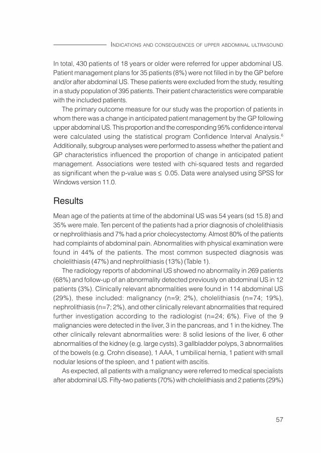
57
INDICATIONS AND CONSEQUENCES OF UPPER ABDOMINAL ULTRASOUND
In total, 430 patients of 18 years or older were referred for upper abdominal US.Patient management plans for 35 patients (8%) were not filled in by the GP beforeand/or after abdominal US. These patients were excluded from the study, resultingin a study population of 395 patients. Their patient characteristics were comparablewith the included patients.
The primary outcome measure for our study was the proportion of patients inwhom there was a change in anticipated patient management by the GP followingupper abdominal US. This proportion and the corresponding 95% confidence intervalwere calculated using the statistical program Confidence Interval Analysis.6
Additionally, subgroup analyses were performed to assess whether the patient andGP characteristics influenced the proportion of change in anticipated patientmanagement. Associations were tested with chi-squared tests and regardedas significant when the p-value was ≤ 0.05. Data were analysed using SPSS forWindows version 11.0.
Results
Mean age of the patients at time of the abdominal US was 54 years (sd 15.8) and35% were male. Ten percent of the patients had a prior diagnosis of cholelithiasisor nephrolithiasis and 7% had a prior cholecystectomy. Almost 80% of the patientshad complaints of abdominal pain. Abnormalities with physical examination werefound in 44% of the patients. The most common suspected diagnosis wascholelithiasis (47%) and nephrolithiasis (13%) (Table 1).
The radiology reports of abdominal US showed no abnormality in 269 patients(68%) and follow-up of an abnormality detected previously on abdominal US in 12patients (3%). Clinically relevant abnormalities were found in 114 abdominal US(29%), these included: malignancy (n=9; 2%), cholelithiasis (n=74; 19%),nephrolithiasis (n=7; 2%), and other clinically relevant abnormalities that requiredfurther investigation according to the radiologist (n=24; 6%). Five of the 9malignancies were detected in the liver, 3 in the pancreas, and 1 in the kidney. Theother clinically relevant abnormalities were: 8 solid lesions of the liver, 6 otherabnormalities of the kidney (e.g. large cysts), 3 gallbladder polyps, 3 abnormalitiesof the bowels (e.g. Crohn disease), 1 AAA, 1 umbilical hernia, 1 patient with smallnodular lesions of the spleen, and 1 patient with ascitis.
As expected, all patients with a malignancy were referred to medical specialistsafter abdominal US. Fifty-two patients (70%) with cholelithiasis and 2 patients (29%)
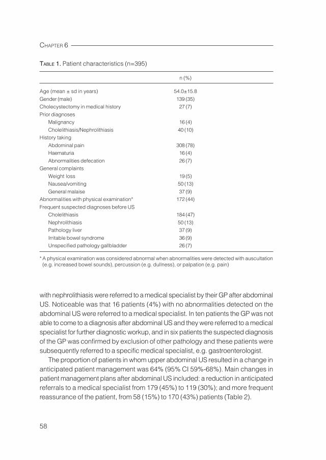
CHAPTER 6
58
with nephrolithiasis were referred to a medical specialist by their GP after abdominalUS. Noticeable was that 16 patients (4%) with no abnormalities detected on theabdominal US were referred to a medical specialist. In ten patients the GP was notable to come to a diagnosis after abdominal US and they were referred to a medicalspecialist for further diagnostic workup, and in six patients the suspected diagnosisof the GP was confirmed by exclusion of other pathology and these patients weresubsequently referred to a specific medical specialist, e.g. gastroenterologist.
The proportion of patients in whom upper abdominal US resulted in a change inanticipated patient management was 64% (95% CI 59%-68%). Main changes inpatient management plans after abdominal US included: a reduction in anticipatedreferrals to a medical specialist from 179 (45%) to 119 (30%); and more frequentreassurance of the patient, from 58 (15%) to 170 (43%) patients (Table 2).
TABLE 1. Patient characteristics (n=395)
n (%)
Age (mean ± sd in years) 54.0±15.8
Gender (male) 139 (35)Cholecystectomy in medical history 27 (7)
Prior diagnosesMalignancy 16 (4)
Cholelithiasis/Nephrolithiasis 40 (10)History taking
Abdominal pain 308 (78)Haematuria 16 (4)
Abnormalities defecation 26 (7)General complaints
Weight loss 19 (5)Nausea/vomiting 50 (13)
General malaise 37 (9)Abnormalities with physical examination* 172 (44)
Frequent suspected diagnoses before USCholelithiasis 184 (47)
Nephrolithiasis 50 (13)Pathology liver 37 (9)
Irritable bowel syndrome 36 (9)Unspecified pathology gallbladder 26 (7)
* A physical examination was considered abnormal when abnormalities were detected with auscultation(e.g. increased bowel sounds), percussion (e.g. dullness), or palpation (e.g. pain)
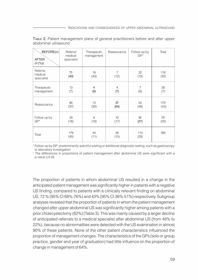
59
INDICATIONS AND CONSEQUENCES OF UPPER ABDOMINAL ULTRASOUND
The proportion of patients in whom abdominal US resulted in a change in theanticipated patient management was significantly higher in patients with a negativeUS finding, compared to patients with a clinically relevant finding on abdominalUS, 72 % (95% CI 68%-76%) and 43% (95% CI 36%-51%) respectively. Subgroupanalyses revealed that the proportion of patients in whom the patient managementchanged after upper abdominal US was significantly higher among patients with aprior cholecystectomy (82%) (Table 3). This was mainly caused by a larger declineof anticipated referrals to a medical specialist after abdominal US (from 48% to22%), because no abnormalities were detected with the US examination in almost90% of these patients. None of the other patient characteristics influenced theproportion of management changes. The characteristics of the GPs (solo or grouppractice, gender and year of graduation) had little influence on the proportion ofchange in management of 64%.
TABLE 2. Patient management plans of general practitioners before and after upperabdominal ultrasound
BEFORE(n) Referral Therapeutic Reassurance Follow-up by Totalmedical management GP*
AFTER specialist(n (%))
Referral 71 19 7 22 119†
medical (40) (43) (12) (19) (30)specialist
Therapeutic 13 4 4 7 28management (7) (9) (7) (6) (7)
Reassurance 66 13 37 54 170†
(37) (30) (64) (48) (43)
Follow-up by 29 8 10 31 78†
GP* (16) (18) (17) (27) (20)
Total 179 44 58 114 395(45) (11) (15) (29)
* Follow-up by GP: predominantly watchful waiting or additional diagnostic testing, such as gastroscopyor laboratory investigation
† The differences in proportions of patient management after abdominal US were significant with ap-value ≤ 0.05
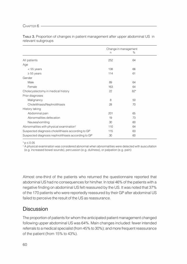
CHAPTER 6
60
Almost one-third of the patients who returned the questionnaire reported thatabdominal US had no consequences for him/her. In total 46% of the patients with anegative finding on abdominal US felt reassured by the US. It was noted that 37%of the 170 patients who were reportedly reassured by their GP after abdominal USfailed to perceive the result of the US as reassurance.
Discussion
The proportion of patients for whom the anticipated patient management changedfollowing upper abdominal US was 64%. Main changes included: fewer intendedreferrals to a medical specialist (from 45% to 30%); and more frequent reassuranceof the patient (from 15% to 43%).
TABLE 3. Proportion of changes in patient management after upper abdominal US inrelevant subgroups
Change in managementn %
All patients 252 64Age
< 55 years 138 66≥ 55 years 114 61
GenderMale 89 64
Female 163 64Cholecystectomy in medical history 22 82*
Prior diagnosesMalignancy 8 50
Cholelithiasis/Nephrolithiasis 28 70History taking
Abdominal pain 201 65Abnormalities defecation 19 73
Nausea/vomiting 30 60Abnormalities with physical examination† 110 64
Suspected diagnosis cholelithiasis according to GP 115 63Suspected diagnosis nephrolithiasis according to GP 30 60
* p ≤ 0.05† A physical examination was considered abnormal when abnormalities were detected with auscultation(e.g. increased bowel sounds), percussion (e.g. dullness), or palpation (e.g. pain)
61
INDICATIONS AND CONSEQUENCES OF UPPER ABDOMINAL ULTRASOUND
To our knowledge this is the first study that has investigated the influence of upperabdominal US on patient management in general practice. The studies ofCharlesworth et al2, Colquhoun et al3, Connor et al4, and Mills et al5 reportednumbers of clinically relevant abnormalities in 25%, 30%, 28% and 27%, respectively,of patients referred for abdominal US by GPs. The 29% clinically relevantabnormalities found in our study is comparable. In addition, our study showed thatthe full value of upper abdominal US cannot be assessed in terms of positivefindings alone. Negative findings are important for exclusion of diseases and,therefore, for reassurance of the patient. However, such findings can also resulteither in referral of patients to a medical specialist for further evaluation of theircomplaints when an abdominal US fails to show any abnormalities, or in the referralof patients to a suitable medical specialist, when specific pathology is excluded.
Subgroup analyses revealed that the proportion of patients in whom patientmanagement changed after upper abdominal US was significantly higher amongpatients with a prior cholecystectomy (82%). This was mainly caused by a largerdecline of anticipated referrals to a medical specialist after abdominal US. Obviously,in those patients hidden postcholecystectomy gallstone pathology was excludedby the absence of choledochus dilatation or intrahepatic stones.
Over 80% of the questionnaires were returned by the patients, which increasedthe validity of these results. It was noted that almost 40% of the 170 patients whowere reportedly reassured by their GP after abdominal US failed to perceive theresult of the US as reassurance. Therefore, it seems abdominal US did not havemuch value for these patients, because no referral or treatment followed after theradiological investigation and reassurance was not achieved.
Before we can reach a conclusion, it is important to note that this study hasseveral limitations. It was impossible to verify whether or not the GP really wouldhave conducted the anticipated patient management in accordance with the planmade on the standardized form before abdominal US was performed. This couldresult in an overestimation of intended referrals to medical specialists. Furthermore,this study does not prove that the patient actually benefits from the diagnosticprocedure, e.g. in terms of morbidity, mortality or quality of life. However, the studyis the first to show that the procedure often leads to changes in anticipated patientmanagement, which is one of the prerequisites for successfully influencing clinicallyrelevant patient outcomes.
In conclusion, the GP’s anticipated patient management strategy was changedfor 64% of patients following upper abdominal US. Abdominal US substantiallyreduced the number of intended referrals to a medical specialist and more patientscould be reassured by their GP.
CHAPTER 6
62
References
1. American College of Radiology. ACR Standards. ACR Standard for the performanceof an ultrasound examination of the abdomen or retroperitoneum. United States,2001.
2. Charlesworth CH, Sampsom MA. How do general practitioners compare with theoutpatient department when requesting upper abdominal examinations? ClinRadiol. 1994;49:343-345.
3. Colquhoun IR, Saywell WR, Dewburry KC. An analysis of referrals for primarydiagnostic abdominal ultrasound at a general X-ray department. Br J Radiol.1988;61:297-300.
4. Connor SEJ, Banerjee AK. General practitioner requests for upper abdominal ultra-sound: their effect on clinical outcome. Br J Radiol. 1998;73:1021-1025.
5. Mills P, Joseph AEA, Adam EJ. Total abdominal and pelvic ultrasound: incidentalfindings and a comparison between outpatient and general practice referrals in1000 cases. Br J Radiol. 1989;62:974-976.
6. Altman DG, Machin D, Bryant TN, Gardner MJ. Statistics with confidence:confidence intervals and statistical guidelines. London: BMJ Books, 2000.
Expected and unexpected gallstones inprimary care
7
Speets AM, Van der Graaf Y, Hoes AW, Kalmijn S, De Wit NJ, MaliWPThM. Expected and unexpected gallstones in primary care.Submitted.
CHAPTER 7
64
AbstractBackground. This prospective cohort study assessed the differences in signs andsymptoms of primary care patients with expected and unexpected gallstones referredfor upper abdominal ultrasound (US).Methods. 430 patients aged ≥ 18 years were referred by 76 general practitioners(GPs) for abdominal US to one of the three participating hospitals in The Netherlands.All GPs were asked to fill in a standardized questionnaire before and after abdominalUS. Patients with expected gallstones had a clinical suspicion of gallstones andcholelithiasis on abdominal US, patients with unexpected gallstones had cholelithiasiswithout a clinical suspicion.Results. Almost 50% of the patients were referred for abdominal US because of aclinical suspicion of gallstones. Cholelithiasis was detected with US in 29% of thepatients with, and 11% without a clinical suspicion of gallstones. In patients suspectedof gallstones, patients with cholelithiasis on US were significantly less often male,had no prior cholecystectomies, more often colicky pain, and were more frequentlyreferred to a medical specialist after US. Patients with unexpected gallstones weremore often male, had less complaints of upper abdominal and colicky pain, andwere less frequently referred to a medical specialist after US, compared to patientswith expected gallstones.Conclusion. Gallstones were detected with upper abdominal US in 29% of thepatients with, and 11% without a clinical suspicion of gallstones. This study showedmarked differences in signs and symptoms of patients suspected of gallstoneswith and without cholelithiasis detected on abdominal US, and in patients withexpected and unexpected gallstones.
65
EXPECTED AND UNEXPECTED GALLSTONES IN PRIMARY CARE
Introduction
The mean prevalence of gallstones reported in European populations is 10-12%.This prevalence increases with increasing age, female gender, obesity, heredityand fertility.1 Gallstones exist in three clinical stages: asymptomatic, symptomatic(e.g. biliary colic), and complicated. About 1% of the asymptomatic patients willbecome symptomatic each year.2 A general clinical guideline is that asymptomaticpatients should not undergo cholecystectomy until symptoms develop.2 Non-specificsymptoms, such as dyspepsia and intolerance of fatty food, are not indicative ofcholelithiasis, but are commonly reported in patients with gallstones.3
Abdominal ultrasound is an important diagnostic imaging method for detectinggallstones. Abdominal US provides more than 95% sensitivity and specificity forthe diagnosis of gallstones greater than 2 mm in diameter, can evaluate the conditionof the gallbladder (e.g. thickening of the gallbladder wall) and the diameter of thecholedochus duct (indicative of obstruction), and has the ability to examine adjacentorgans.2-4 Four retrospective studies demonstrated that gallstones were detectedwith abdominal US in 18-24% of the patients with a clinical suspicion of gallstones,and unexpected gallstones were found in 9% of the patients without a clinicalsuspicion of gallstones.5-8 Although abdominal US is frequently used for diagnosingcholelithiasis, evaluations of abdominal US in patients with abdominal complaintsreferred by general practitioners (GPs) have scarcely been reported in the scientificliterature.
The objective of this prospective cohort study was to assess 1) the determinantsof cholelithiasis in primary care patients referred for upper abdominal US becauseof a clinical suspicion of gallstones, and 2) the differences in signs and symptomsin patients with expected and unexpected gallstones on abdominal US.
Methods
This prospective cohort study was conducted from April 2003 to December 2004with the help of 76 GPs participating in the catchment area of one of three generalhospitals located in three main cities in The Netherlands (Jeroen Bosch Hospital in‘s-Hertogenbosch; Gelre Hospitals in Apeldoorn; ’Onze Lieve Vrouwe Gasthuis’ inAmsterdam). All patients of 18 years and older who were referred for upperabdominal US by their GP to one of these hospitals were included in the study,resulting in a study population of 430 patients. The study was approved by theMedical Ethics Review Board.
CHAPTER 7
66
All GPs were asked to fill in a standardized questionnaire before requesting anabdominal US, including information on history, physical examination, indication,clinical suspicion with estimated prior probabilities on a visual analogue scale(range 0-100%), and anticipated patient management. The management optionsincluded: referral to a medical specialist; medication prescription; reassurance ofthe patient; and follow-up by the GP (watchful waiting or additional diagnostictesting). After the GP received the report (within 1-4 days after the US) he or shefilled in a second questionnaire, again including the clinical suspicion with estimatedposterior probabilities, and anticipated patient management plan.
The findings on upper abdominal US were categorized into three groups: (1)cholelithiasis; (2) other clinically relevant abnormalities; (3) no abnormality. Sixmonths after the abdominal US a short questionnaire was sent to all patients(response rate 81%), in order to evaluate their current complaints and assess thevalue and consequences of abdominal US according to the patient.
The GPs could fill in three probable diagnoses on the standard questionnairebefore requesting an upper abdominal US. All patients who were referred forabdominal US with a clinical suspicion of cholelithiasis as one of these probablediagnoses were classified as patients with a clinical suspicion of cholelithiasis bythe GP before US (n=198). Subsequently, the patients were divided in four differentsubgroups, based on probability of cholelithiasis and the presence of cholelithiasison abdominal US: 1) patients with a clinical suspicion of gallstones and cholelithiasison abdominal US, i.e. expected gallstones; 2) patients with a clinical suspicion ofgallstones without cholelithiasis on US; 3) patients without a clinical suspicion ofgallstones and cholelithiasis on abdominal US, i.e. unexpected gallstones; 4) patientswithout a clinical suspicion of gallstones and without cholelithiasis on abdominalUS. The patient characteristics of patient group 2 and 3 were compared with group 1,and analyzed with chi-squared tests. Statistical significance was inferred when thep-value was equal to or less than 0.05. Data were analysed using SPSS for Windowsversion 11.0. In addition, the positive predictive value of a gallstone diagnosis bythe GP (percentage patients with gallstones on abdominal US in the patient groupwith a clinical suspicion of cholelithiasis), and the negative predictive value of nogallstone diagnosis by the GP (percentage patients without gallstones on abdominalUS in the patient group without a clinical suspicion of cholelithiasis) were calculated.
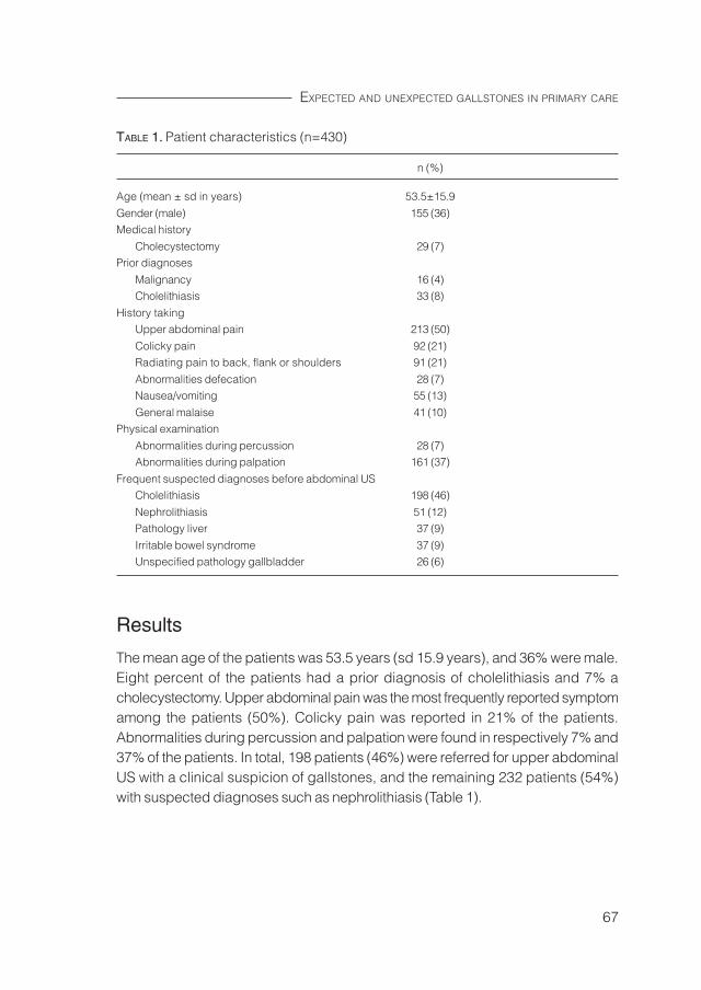
67
EXPECTED AND UNEXPECTED GALLSTONES IN PRIMARY CARE
Results
The mean age of the patients was 53.5 years (sd 15.9 years), and 36% were male.Eight percent of the patients had a prior diagnosis of cholelithiasis and 7% acholecystectomy. Upper abdominal pain was the most frequently reported symptomamong the patients (50%). Colicky pain was reported in 21% of the patients.Abnormalities during percussion and palpation were found in respectively 7% and37% of the patients. In total, 198 patients (46%) were referred for upper abdominalUS with a clinical suspicion of gallstones, and the remaining 232 patients (54%)with suspected diagnoses such as nephrolithiasis (Table 1).
TABLE 1. Patient characteristics (n=430)
n (%)
Age (mean ± sd in years) 53.5±15.9
Gender (male) 155 (36)Medical history
Cholecystectomy 29 (7)Prior diagnoses
Malignancy 16 (4)Cholelithiasis 33 (8)
History takingUpper abdominal pain 213 (50)
Colicky pain 92 (21)Radiating pain to back, flank or shoulders 91 (21)
Abnormalities defecation 28 (7)Nausea/vomiting 55 (13)
General malaise 41 (10)Physical examination
Abnormalities during percussion 28 (7)Abnormalities during palpation 161 (37)
Frequent suspected diagnoses before abdominal USCholelithiasis 198 (46)
Nephrolithiasis 51 (12)Pathology liver 37 (9)
Irritable bowel syndrome 37 (9)Unspecified pathology gallbladder 26 (6)
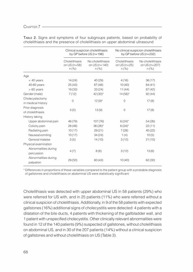
CHAPTER 7
68
Cholelithiasis was detected with upper abdominal US in 58 patients (29%) whowere referred for US with, and in 25 patients (11%) who were referred without aclinical suspicion of cholelithiasis. Additionally, in 9 of the 58 patients with expectedgallstones (16%) additional signs of cholecystitis were detected: 4 patients with adilatation of the bile ducts, 4 patients with thickening of the gallbladder wall, and1 patient with unspecified cholecystitis. Other clinically relevant abnormalities werefound in 12 of the 140 patients (9%) suspected of gallstones, without cholelithiasison abdominal US, and in 30 of the 207 patients (14%) without a clinical suspicionof gallstones and without cholelithiasis on US (Table 3).
TABLE 2. Signs and symptoms of four subgroups patients, based on probability ofcholelithiasis and the presence of cholelithiasis on upper abdominal ultrasound
Clinical suspicion cholelithiasis No clinical suspicion cholelithiasisby GP before US (n=198) by GP before US (n=232)
Cholelithiasis No cholelithiasis Cholelithiasis No cholelithiasison US (n=58) on US (n=140) on US (n=25) on US (n=207)
n (%) n (%) n (%) n (%)
Age< 40 years 14 (24) 40 (29) 4 (16) 36 (17)40-60 years 25 (43) 67 (48) 10 (40) 84 (41)
> 60 years 19 (33) 33 (24) 11 (44) 87 (42)Gender (male) 7 (12) 42 (30)* 14 (56)* 92 (44)
Cholecystectomy0 12 (9)* 0 17 (8)
in medical history
Prior diagnosis3 (5) 13 (9) 0 17 (8)
of cholelithiasis
History takingUpper abdominal pain 46 (79) 107 (76) 6 (24)* 54 (26)
Colicky pain 28 (48) 36 (26)* 6 (24)* 22 (11)Radiating pain 10 (17) 29 (21) 7 (28) 45 (22)
Nausea/vomiting 10 (17) 34 (24) 1 (4) 10 (5)General malaise 3 (5) 14 (10) 3 (12) 21 (10)
Physical examinationAbnormalities during
4 (7) 8 (6) 3 (12) 13 (6)percussionAbnormalities during
29 (50) 60 (43) 10 (40) 62 (30)palpation
* Differences in proportions of these variables compared to the patient group with a probable diagnosisof gallstones and cholelithiasis on abdominal US were statistically significant
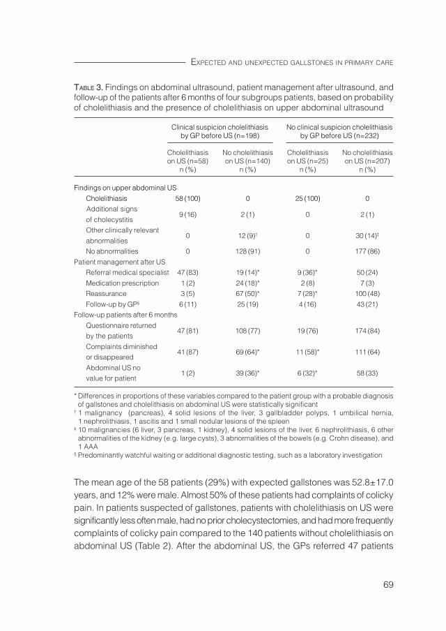
69
EXPECTED AND UNEXPECTED GALLSTONES IN PRIMARY CARE
The mean age of the 58 patients (29%) with expected gallstones was 52.8±17.0years, and 12% were male. Almost 50% of these patients had complaints of colickypain. In patients suspected of gallstones, patients with cholelithiasis on US weresignificantly less often male, had no prior cholecystectomies, and had more frequentlycomplaints of colicky pain compared to the 140 patients without cholelithiasis onabdominal US (Table 2). After the abdominal US, the GPs referred 47 patients
TABLE 3. Findings on abdominal ultrasound, patient management after ultrasound, andfollow-up of the patients after 6 months of four subgroups patients, based on probabilityof cholelithiasis and the presence of cholelithiasis on upper abdominal ultrasound
Clinical suspicion cholelithiasis No clinical suspicion cholelithiasisby GP before US (n=198) by GP before US (n=232)
Cholelithiasis No cholelithiasis Cholelithiasis No cholelithiasison US (n=58) on US (n=140) on US (n=25) on US (n=207)
n (%) n (%) n (%) n (%)
Findings on upper abdominal US
Cholelithiasis 58 (100) 0 25 (100) 0 Additional signs
9 (16) 2 (1) 0 2 (1)of cholecystitisOther clinically relevant
0 12 (9)† 0 30 (14)‡
abnormalitiesNo abnormalities 0 128 (91) 0 177 (86)
Patient management after USReferral medical specialist 47 (83) 19 (14)* 9 (36)* 50 (24)
Medication prescription 1 (2) 24 (18)* 2 (8) 7 (3)Reassurance 3 (5) 67 (50)* 7 (28)* 100 (48)
Follow-up by GP§ 6 (11) 25 (19) 4 (16) 43 (21)Follow-up patients after 6 months
Questionnaire returned47 (81) 108 (77) 19 (76) 174 (84)
by the patients
Complaints diminished41 (87) 69 (64)* 11 (58)* 111 (64)
or disappeared
Abdominal US no1 (2) 39 (36)* 6 (32)* 58 (33)
value for patient
* Differences in proportions of these variables compared to the patient group with a probable diagnosisof gallstones and cholelithiasis on abdominal US were statistically significant
† 1 malignancy (pancreas), 4 solid lesions of the liver, 3 gallbladder polyps, 1 umbilical hernia,1 nephrolithiasis, 1 ascitis and 1 small nodular lesions of the spleen
‡ 10 malignancies (6 liver, 3 pancreas, 1 kidney), 4 solid lesions of the liver, 6 nephrolithiasis, 6 otherabnormalities of the kidney (e.g. large cysts), 3 abnormalities of the bowels (e.g. Crohn disease), and1 AAA
§ Predominantly watchful waiting or additional diagnostic testing, such as a laboratory investigation
CHAPTER 7
70
(83%) to a medical specialist; 46 patients to the department of surgery, and 1 patientto the department of internal medicine. Six months after the abdominal US thecurrent complaints were diminished or disappeared in almost 90% of the patientswith expected gallstones, and only 2% reported that abdominal US had no valuefor him/her (Table 3).
The mean age of the 25 patients (11%) with unexpected gallstones was 59.4±15.8years, and 56% were male. Most unexpected gallstones were detected in thepatients aged older than 60 years. Six of the 25 patients (24%) had colicky painwith a clinical suspicion of nephrolithiasis. Compared to the patients with expectedgallstones, the percentage males was much higher and complaints of upperabdominal pain and colicky pain occurred significantly less frequent (Table 2). TheGPs referred 9 patients (36%) to a medical specialist; 3 patients to the departmentof surgery, and 6 patients to the department of internal medicine. Six months afterthe abdominal US current complaints were diminished or disappeared in almost60% of these patients. One third of the patients reported that abdominal US had novalue for him/her (Table 3).
The positive predictive value of a gallstone diagnosis by the GP was 58/198=29%.The negative predictive value of no gallstone diagnosis by the GP was 207/232=89%.
Discussion
Almost half of the primary care patients referred for upper abdominal US by theirGP had a clinical suspicion of gallstones. Cholelithiasis was detected with upperabdominal US in 29% of the patients who were referred for US with, and 11% of thepatients who were referred without a clinical suspicion of cholelithiasis. Gallstoneswere found in 18-24% of patients referred for abdominal US with a clinical suspicionof cholelithiasis in four retrospective studies.5-8 The percentage of patients withunexpected gallstones (11%) corresponds well with the 9% unexpected gallstonesfound by Colquhoun et al6 and Mills et al.8
A marked difference between these two patient groups was that the percentagemales in the group with unexpected cholelithiasis was almost 5 times as high as inthe patient group with expected gallstones. The percentage of males was alsomuch higher (2.5 times higher) in the group patients suspected of gallstones, butwithout signs of cholelithiasis on abdominal US. It is widely known that the prevalenceof gallstones is higher in females than in males. GPs may sooner suspect gallstonesin female patients with complaints suggestive of gallstones and formulate a clinicalsuspicion of cholelithiasis when referring them for upper abdominal US, resulting

71
EXPECTED AND UNEXPECTED GALLSTONES IN PRIMARY CARE
in a higher percentage of unexpected gallstones in males. In addition, the clinicalsuspicion in males with colicky pain is directed more often by GPs to kidney stonesthan to cholelithiasis. It was peculiar in our study that 6 patients with unexpectedgallstones had complaints of colicky pain, while the GP thought cholelithiasis wasunlikely; the GPs thought these complaints were caused by nephrolithiasis. Noneof these patients were referred to a medical specialist after abdominal US.
The number of prior cholecystectomies differed significantly in patients suspectedof gallstones with and without cholelithiasis on abdominal US. Twelve patients withoutcholelithiasis and none of the patients with cholelithiasis had a prior cholecystectomy.It was obvious that these 12 patients were referred for an upper abdominal US withagain a clinical suspicion of cholelithiasis (in the choledochus duct), and in none ofthem gallstones were detected with abdominal US, though choledochus or hepaticstones may have passed. It is questionable whether gallstones were the cause ofthe complaints in these patients, because after cholecystectomy they were referredonce more for an abdominal US with complaints suspected of cholelithiasis.
The percentage of patients with unexpected gallstones referred to a medicalspecialist (36%) was significantly lower than the 83% patients referred with expectedgallstones. Six months after the abdominal US the questionnaire revealed that inpatients with unexpected gallstones the abdominal complaints less often haddisappeared; patients with expected gallstones reported 6 months after the USthat the current complaints were diminished or disappeared in almost 90% of thepatients, compared to almost 60% of the patients with unexpected gallstones.Maybe a larger part of the abdominal complaints would have disappeared whenthese patients were referred to a medical specialist. Although less specific, theabdominal complaints of the patients with unexpected gallstones could have beencaused by the gallstones. On the other hand, most patients with expected gallstoneswere referred to a medical specialist, often regarded as the best possible treatmentby patients in general practice, which will have a positive effect on the perceptionof their complaints.
A limitation of our study is that part of the unexpected gallstones may have beenclassified as unexpected in our study, because the GP did not fill in a clinicalsuspicion of cholelithiasis before referral for abdominal US. Some patients withunexpected gallstones were referred for upper abdominal US with abdominalcomplaints that could be suggestive of gallstones, such as colicky pain. This couldresult in an underestimation of the differences between the patient groups withexpected and unexpected gallstones.
CHAPTER 7
72
In conclusion, gallstones were detected with upper abdominal US in 29% of thepatients with, and 11% without a clinical suspicion of gallstones. This study showedmarked differences in patients suspected of gallstones with and without cholelithiasisdetected on abdominal US, and in patients with expected and unexpectedgallstones.
73
EXPECTED AND UNEXPECTED GALLSTONES IN PRIMARY CARE
References
1. Kratzer W, Mason RA, Kachele V. Prevalence of gallstones in sonographic surveysworldwide. J Clin Ultrasound. 1999;27(1):1-7.
2. Ahmed A, Cheung RC, Keeffe EB. Management of gallstones and theircomplications. Am Fam Physician. 2000;61(6):1673-1680,1687-1688.
3. Kalloo AN, Kantsevoy SV. Gallstones and biliary disease. Prim Care.2001;28(3):591-606.
4. Bortoff GA, Chen MY, Ott DJ, Wolfman NT, Routh WD. Gallbladder stones: imagingand intervention. Radiographics. 2000;20(3):751-766.
5. Charlesworth CH, Sampsom MA. How do general practitioners compare with theoutpatient department when requesting upper abdominal examinations? ClinRadiol. 1994;49:343-345.
6. Colquhoun IR, Saywell WR, Dewburry KC. An analysis of referrals for primarydiagnostic abdominal ultrasound at a general X-ray department. Br J Radiol.1988;61:297-300.
7. Connor SEJ, Banerjee AK. General practitioner requests for upper abdominal ultra-sound: their effect on clinical outcome. Br J Radiol. 1998;73:1021-1025.
8. Mills P, Joseph AEA, Adam EJ. Total abdominal and pelvic ultrasound: incidentalfindings and a comparison between outpatient and general practice referrals in1000 cases. Br J Radiol. 1989;62:974-976.

Evaluation of routinely used diagnostictests
8

77
GENERAL DISCUSSION
In this chapter, several methodological issues pertaining to the evaluation of thevalue of diagnostic tests that are frequently applied in clinical practice for a widevariety of complaints, such as chest radiography (CXR) and abdominal ultrasound(US) will be discussed.
Phases in diagnostic research
Diagnostic research is pragmatic research with the aim to optimize the diagnosticprocess in clinical practice. The diagnostic process is a multivariable andconsecutive process of estimating the diagnostic probability of the presence of aparticular disease given combinations of test results. The evaluation of a diagnostictest can be summarized in a four phase hierarchical model, comparable with thefour phases of pharmaceutical research.1,2
Phase I consists of the test development. In diagnostic imaging studies thisphase is generally the purview of physicists, and includes parameters such assharpness, contrast and resolution.3 The diagnostic accuracy of the test, i.e. theability of a test to discriminate between diseased and non-diseased persons, isassessed in phase II. The discrimination capacity of the diagnostic test will becompared with results of a reference test, and is summarized in measures such assensitivity and specificity. Phase III estimates the clinical effectiveness of thediagnostic test. Some authors3-7 have subdivided this phase in three different levels:diagnostic impact (i.e. the influence of the test on the clinician’s confidence of adiagnosis), therapeutic impact (i.e. initiation, change, or withdrawal of plannedtherapy), and impact on the patient outcome (e.g. influence on the health status ofpatients or quality of life). Phase IV of diagnostic research goes beyond the individualrisks and benefits to a broader societal perspective, such as cost-effectivenessanalyses.
Importance of phase III diagnostic research
Diagnostic research in phase I and II often have straightforward study designs,and most new diagnostic investigations are implemented immediately after phase II.However, the results of phase II research, summarized in measures such assensitivity, specificity, and likelihood ratios have important limitations8: they do notallow for direct diagnostic interpretation, and a high sensitivity and specificity areno guarantee for diagnostic, let alone therapeutic, impact, or influence on patientoutcome. Such studies of diagnostic accuracy can neither evaluate the value of the
CHAPTER 8
78
test in the presence of other diagnostic tools, nor assess the influence on theanticipated patient management. In addition, such diagnostic studies often comparetest results in a group of patients with well-established disease (i.e. more advancedstages) with a group of healthy volunteers, whereas a diagnostic test should behelpful in recognizing disease in the domain of patients suspected of the disease ineveryday clinical practice. Therefore, phase II diagnostic research can be seen asjust a preliminary assessment of a diagnostic test. A reason often adduced to avoidphase III diagnostic research is the fast evolution of diagnostic techniques and thealleged uniqueness of the novel test. This could be a valid argument in case of anovel test capable of providing images with undisputed therapeutic consequencesin a relatively rare (suspected) disease, where no alternative techniques are available.Under these circumstances phase III and IV studies will be difficult to perform.Similar issues play a role in the less stringent way the value of orphan drugs forrare diseases is assessed. However, tests such as CXR and abdominal US arewidely used in a variety of complaints and to diagnose a range of diseases andwere developed many years ago. Phase II studies on the diagnostic accuracy ofthese tests were already performed in respectively the fifties9-11 and end of thesixties12-15. Nowadays, patients are still referred frequently for these imaginginvestigations by general practitioners (GPs), but their added clinical value in generalpractice has not been sufficiently established. Clearly, more emphasis on phase IIIstudies to determine the clinical value of these tests is required.
Study designs phase III diagnostic research
Too few studies on the clinical effectiveness of diagnostic tests have been performedin clinical practice, in particular with regard to diagnostic imaging. A cross-sectionaldesign, which uses a “gold standard” or reference test for the identification ofdisease and non-disease, and including a series of consecutive patients suspectedof a certain disease in daily clinical practice, is regarded as the optimal studydesign for phase III diagnostic research. In such studies, two multivariate modelsare often compared; one model with all information from patient history and physicalexamination, and a second model with all information from model one and theresult of the additional diagnostic test. By comparing the areas under both ROCcurves, the additional value of the diagnostic test can be then determined. Analternative study design is the randomized controlled trial (RCT). In a RCT patientsrequiring the diagnostic test under study according to the clinician will randomizefor the diagnostic test. Patient management, and sometimes patient outcome, will
79
GENERAL DISCUSSION
be compared in patients with and without the diagnostic test. Although both studydesigns are suitable for research in new diagnostic tests, difficulties arise in routinelyused tests. The disadvantages of the cross-sectional study and RCT for diagnostictests, such as CXR and abdominal US, routinely used in a wide range of patients todetermine the presence of a variety of diseases, will be outlined in next paragraphsand an alternative design, the prospective cohort study, will be discussed.
Cross-sectional study
Primary care patients referred for CXR and upper abdominal US are a veryheterogeneous patient population in which the GPs judge an additional diagnostictest necessary for further examination. The patients have a broad range of complaints,the indications for the tests vary widely, and many different diseases can be detectedwith these imaging investigations. In view of this, no “gold standard” or referencetest is available for both routinely used diagnostic tests, and this impedes the useof a cross-sectional design to examine their clinical effectiveness. It is possible touse an expert panel to identify disease and non-disease based on all availabledata, preferably without the diagnostic test under evaluation to prevent incorporationbias, but (notably because of the latter), this would result in a less valid identificationof disease. Moreover, a cross-sectional design is particularly suited when the clinicalconsequences of the, in this case multiple, diagnoses are known (such asanticoagulants in established deep vein thrombosis or surgery in certain fractures)but the consequences for many of the diagnoses established through CXR orabdominal US, such as gallbladder stones, are not unequivocal. In circumstanceswhere a valid reference test is lacking, researchers may choose a longitudinaldesign, usually a RCT or prospective cohort study, in which, typically, the effect ofthe test on patient management or patient outcome is determined.
Randomized Controlled Trial
RCTs are not often applied in diagnostic research, for reasons such as complexlogistics, the need for large sample sizes to show statistically significant differences,and high costs. In view of the multiple indications and possible diagnoses involvedwhen a CXR or abdominal US is ordered, this may pose insurmountable problems.Designing a RCT to assess the clinical effectiveness of a routinely used diagnostictest becomes even more difficult because of ethical considerations. Ethical issueswill arise when an established diagnostic test will be withheld in half of the patients.
CHAPTER 8
80
Even if Medical Ethics Review Boards approve of such a study, most clinicians,and probably patients, will be hesitant to participate when full access to the diagnostictest is denied during the (relatively long) study period. It should be emphasizedthat the primary care physician, on the basis of signs and symptoms, alreadyselected the relevant clinical domain, i.e. those patients in whom the diagnostictest is considered to provide potentially useful additional information. If, alternatively,clinicians were asked to identify patients in whom the diagnostic test can be omittedwithout serious consequences, only few patients would be included in the study,and the generalizability of the findings to the clinically relevant patient populationwould be seriously hampered.
Prospective cohort study
An alternative design for assessing the clinical effectiveness of routinely useddiagnostic tests is a prospective cohort study, sometimes described as a controlledobservational study or clinical value study.6,16 In a prospective cohort study theclinician’s confidence of a diagnosis and anticipated patient management beforeand after the clinician obtains information of the diagnostic test results can becompared, and patients in whom the diagnostic test does not lead to a change inconfidence or anticipated patient management can be identified. These studieshave a pragmatic character. Patients will be included in the study via their clinicianwho requests the diagnostic test based on the medical history and physicalexamination of a patient. All patients will undergo the diagnostic test and clinicalpractice will be as usual, which allows an evaluation of the diagnostic test in itsclinical context. In addition, this will minimize the workload of clinicians and enableslarge numbers of participating clinicians and high inclusion rates. Freedman16
emphasized almost 20 years ago that issues such as the continuous scale of testresults and the possibility of multiple diagnoses, which are difficult to accommodatein Phase II studies of diagnostic accuracy, are incorporated naturally into this studydesign.
An important limitation of this type of study is the fact that the clinicians assessthe value of the diagnostic test by filling in the patient management before andafter the test. In this way clinicians may influence the estimated value of a diagnostictest, because most clinicians will not judge their requested test as inefficient.Besides, clinicians have to fill in the anticipated patient management before thediagnostic test as if no diagnostic test would be performed. It is impossible toverify whether or not the clinician really would have conducted the anticipated
81
GENERAL DISCUSSION
patient management in accordance with the plan made before the diagnostic test.This could result in an overestimation of the prevalence of the most invasiveanticipated patient management categories, such as surgical interventions. Finally,when the outcome is change in patient management, the benefit for the patient interms of reduction in morbidity of even mortality is not measured. This would requirehuge sample sizes and long-term follow up periods, and these studies are hardlyfeasible, notably in the broad patient domain indicated for CXR or abdominal US.
Conclusion
Diagnostic research in phase I and II is essential for the development of a newdiagnostic test. These studies often have straightforward study designs, and mostnew diagnostic tests are implemented immediately after phase II. However, suchdiagnostic accuracy research can be seen as just a preliminary assessment of adiagnostic test. The next phase, the assessment of the clinical effectiveness of anew test in daily practice, is more complicated and only conducted in a smallpercentage of the new diagnostic tests. Phase III diagnostic research in routinelyused diagnostic tests is even more complicated, because the standard study design(cross-sectional study with a reference test) and a RCT are usually not feasible,and is therefore seldom performed. A prospective cohort study, in which theanticipated patient management of the clinician is compared before and after thetest, offers an alternative study design. Despite the limitations of such studies, theyprovide a pragmatic and valid way to assess the effectiveness of diagnostic testsroutinely applied in clinical practice in many patients with a wide range of complaintsand possible indications.
CHAPTER 8
82
References
1. Sackett DL, Haynes RB. The architecture of diagnostic research. BMJ.2002;324:539-541.
2. Lijmer JG, Mol BWJ, Bonsel GJ, Prins MH, Bossuyt PMM. Strategies for the evaluationof diagnostic technologies. In thesis: Evaluation of diagnostic tests: from accuracyto outcome. 2001;15-28.
3. Fryback DG, Thornbury JR. The efficacy of diagnostic imaging. Med Decis Making.1991;11(2):88-94.
4. Guyatt GH, Tugwell PX, Feeny DH, Haynes RB, Drummond M. A framework forclinical evaluation of diagnostic technologies. CMAJ. 1986;134(6):587-594.
5. Kent DL, Larson EB. Disease, level of impact, and quality of research methods.Three dimensions of clinical efficacy assessment applied to magnetic resonanceimaging. Invest Radiol. 1992;27(3):245-254.
6. Mackenzie R, Dixon AK. Measuring the effects of imaging: an evaluative framework.Clin Radiol. 1995;50(8):513-518.
7. Thornbury JR. Eugene W. Caldwell Lecture. Clinical efficacy of diagnostic imaging:love it or leave it. AJR Am J Roentgenol. 1994;162(1):1-8.
8. Moons KG, van Es GA, Deckers JW, Habbema JD, Grobbee DE. Limitations ofsensitivity, specificity, likelihood ratio, and Bayes’ theorem in assessing diagnosticprobabilities: a clinical example. Epidemiology. 1997;8(1):12-17.
9. Rigler LG. Roentgen examination of the chest; its limitations in the diagnosis ofdisease. J Am Med Assoc. 1950;142(11):773-777.
10. Feist JH, Littleton JT 3rd. The accuracy of roentgen examinations of the chest.Guthrie Clin Bull. 1956;26(2):101-114.
11. Warfield CH. Roentgen evaluation of the chest. Ill Med J. 1957;111(1):7-11.12. Kuliev ShB.The use of ultrasonics in the diagnosis of tumors and of several diseases
of organs of the abdominal cavity. Klin Med (Mosk). 1967;45(1):82-88.13. Holm HH, Mortensen T. Ultrasonic scanning in diagnosis of abdominal disease.
Acta Chir Scand. 1968;134(5):333-341.14. Richardson R, Norton LW, Eule J, Eiseman B. Accuracy of ultrasound in diagnosing
abdominal masses. Arch Surg. 1975;110(8):933-939.15. Carlsen EN. Liver, gallbladder, and spleen. Radiol Clin North Am. 1975;13(3):
543-556.16. Freedman LS. Evaluating and comparing imaging techniques: a review and
classification of study designs. Br J Radiol. 1987;60:1071-1081.
SummarySamenvattingDankwoordCurriculum Vitae
9

85
SUMMARY
Chapter 1 includes a short introduction of the diagnostic process and introducestwo of the most widely used diagnostic imaging techniques in Western societies,chest radiography (CXR) and abdominal ultrasound (US). The main objective ofthis thesis, to determine the clinical effectiveness of CXR and upper abdominal USin general practice, and the objectives of the studies presented in the subsequentchapters were described in this chapter.
In Chapter 2 the study based on data of the Health Insurance Company Agis in TheNetherlands from 1999 to 2003 was described. This study provided detailedinformation on the number of CXR and abdominal US examinations by age, gender,referring physician and ethnicity. CXRs were ordered in 130 per 1000 persons peryear, and abdominal US examinations in 39 per 1000 persons per year. Thesefrequencies did not change noticeable over the period 1999 to 2003. CXR wasperformed significantly more often in males, and abdominal US significantly moreoften in females. The frequencies of both imaging examinations were highest inpersons aged 70-79 years. General practitioners (GPs) more frequently referredyounger patients and females, especially for abdominal US, compared to medicalspecialists. Up to the age of 60 years the frequencies of both CXR and abdominalUS were higher in Turks and Moroccans compared to other nationalities.
Chest radiography
The influence of CXR on patient management in general practice was studied inChapter 3. In total, 792 patients aged ≥ 18 years referred by 78 general practitioners(GPs) for CXR to one of the three participating hospitals in The Netherlands wereincluded in this prospective cohort study. The main outcome was change inanticipated patient management assessed by means of questionnaires filled in byGPs before and after CXR. Clinically relevant abnormalities were found in 24% ofthe CXRs. Anticipated patient management changed in 60% of the patients followingCXR, and the main changes included fewer referrals to a medical specialist (from26% to 12%), a reduction in initiation or change in therapy (from 24% to 15%), andmore frequent reassurance (from 25% to 46%). A change in patient managementoccurred significant more frequently in patients with complaints of cough (67%),exhibited abnormalities during physical examination (69%), or a suspected diagnosisof pneumonia (68%).
CHAPTER 9
86
In Chapter 4 the diagnostic yield of CXR in 192 primary care patients suspected ofpneumonia was evaluated. Pneumonia was diagnosed by GPs in 35 patients (18%),of whom 27 patients (14%) had a positive CXR, and 8 patients (4%) a negativeCXR, however with an assumed high probability of pneumonia by the GP. CXRclearly influenced the diagnosis of pneumonia by the GP in 53% of the patients:CXR ruled out pneumonia in 47%, and the probability of pneumonia substantiallyincreased in 6% of the patients. Anticipated patient management changed afterCXR in 69% of the patients, mainly caused by a reduction in medication prescription(from 43% to 17%), and more frequent reassurance of the patient (from 8% to35%). Therefore, CXR is an important diagnostic tool in primary care patients witha clinical suspicion of pneumonia in terms of change in the estimated probability ofpneumonia, and change in anticipated patient management.
Abdominal ultrasound
In Chapter 5 the cross-sectional study in 499 patients aged ≥ 18 years with abdominalpain referred by their GP for abdominal US to the University Medical Centre Utrechtin The Netherlands was described. Mean age at time of the US was 47.9±15.2years, and almost one third of the patients were male. Significant pathology wasseen in 81 patients (16%). Older age (OR 1.03 per year), colicky pain (OR 2.80)and a clinical suspicion of bile stones (OR 2.31) were predictors of a significantabnormal finding on abdominal US.
The diagnostic value of upper abdominal US in terms of changes in patientmanagement decisions in primary care was determined in 395 patients referred forabdominal US in Chapter 6. The mean age of the patients was 54.0±15.8 years,and 35% were male. Clinically relevant abnormalities were found in 29% of theabdominal US, mainly cholelithiasis. Anticipated patient management changed in64% of the patients following abdominal US. Abdominal US substantially reducedthe number of intended referrals to a medical specialist (from 45% to 30%), andmore patients could be reassured by their GP (from 15% to 43%). A change inanticipated patient management occurred significantly more frequently in patientswith a prior cholecystectomy (82%).
The differences in signs and symptoms of 430 primary care patients with expectedand unexpected gallstones referred for upper abdominal US were assessed inChapter 7. 76 participating GPs were asked to fill in a standardized questionnaire
87
SUMMARY
before and after abdominal US. Almost 50% of the patients were referred forabdominal US because of a clinical suspicion of gallstones. Cholelithiasis wasdetected with US in 29% of the patients with, and 11% without a clinical suspicionof gallstones. This study showed marked differences in signs and symptoms ofpatients suspected of gallstones with and without cholelithiasis detected onabdominal US, and in patients with expected and unexpected gallstones. In patientssuspected of gallstones, patients with cholelithiasis on abdominal US weresignificantly less often male, had no prior cholecystectomies, more often colickypain, and were more frequently referred to a medical specialist after US. Patientswith unexpected gallstones were more often male, had less complaints of upperabdominal and colicky pain, and were less frequently referred to a medical specialistafter US, compared to patients with expected gallstones.
In Chapter 8 several methodological issues pertaining to the evaluation of the valueof diagnostic tests that are frequently applied in clinical practice for a wide varietyof complaints, such as the CXR and abdominal US, were be discussed. Diagnosticresearch in phase I and II is essential for the development of a new diagnostic test.These studies often have straightforward study designs, and most new diagnostictests are implemented immediately after phase II. However, such diagnosticaccuracy research can be seen as just a preliminary assessment of a diagnostictest. The next phase, the assessment of the clinical effectiveness of a new test indaily practice, is more complicated and only conducted in a small percentage ofthe new diagnostic tests. Phase III diagnostic research in routinely used diagnostictests is even more complicated, because the standard study design (cross-sectionalstudy with a reference test) and a RCT are usually not feasible, and is thereforeseldom performed. A prospective cohort study, in which the anticipated patientmanagement of the clinician is compared before and after the test, offers analternative study design. Despite the limitations of such studies, they provide apragmatic and valid way to assess the effectiveness of diagnostic tests routinelyapplied in clinical practice in many patients with a wide range of complaints andpossible indications.

SummarySamenvattingDankwoordCurriculum Vitae
9

91
SAMENVATTING
Hoofdstuk 1 bevat een korte inleiding over het diagnostische proces en er wordentwee van de meest gebruikte diagnostische beeldvormende technieken in Westerselanden geïntroduceerd, de X-thorax en buikechografie. Het doel van dit proefschriftwas om de klinische relevantie van X-thorax en buikechografie in de huisartspraktijkte bepalen. De onderwerpen van de onderzoeken beschreven in de volgendehoofdstukken van dit proefschrift worden weergegeven aan het einde van hoofdstuk 1.
In hoofdstuk 2 wordt het onderzoek beschreven dat was gebaseerd op de data vande Zorgverzekeraar Agis uit de periode 1999 t/m 2003. In dit onderzoek werdgedetailleerde informatie over het aantal X-thorax en buikechografie onderzoekengepresenteerd voor leeftijd, geslacht, verwijzende arts en etniciteit. Gemiddeldwerden 130 per 1000 personen per jaar doorverwezen voor een X-thorax en 39 per1000 personen per jaar voor een buikechografie. Deze frequenties wijzigdennauwelijks gedurende de periode van 1999 t/m 2003. De X-thorax werd significantvaker aangevraagd voor mannen, en buikechografie significant meer voor vrouwen.De frequenties voor beide beeldvormende onderzoeken waren het hoogst inpersonen met een leeftijd van 70-79 jaar. In vergelijking met specialisten verwezenhuisartsen vaker jongere mensen en vrouwen door, vooral voor buikechografie. Defrequenties van zowel de X-thorax als buikechografie waren hoger in mensen tot 60jaar met een Turkse of Marokkaanse nationaliteit in vergelijking met anderenationaliteiten.
X-thorax
De invloed van de X-thorax op het huisartsenbeleid werd onderzocht in hoofdstuk 3.In totaal werden er 792 patiënten van 18 jaar en ouder geïncludeerd in ditprospectieve cohort onderzoek. Zij werden doorverwezen voor een X-thorax naaréén van de drie participerende Nederlandse ziekenhuizen door 78 huisartsen. Deprimaire uitkomstmaat was de verandering in het voorgenomen beleid, vastgestelddoor middel van vragenlijsten ingevuld door de huisarts voor en na de X-thorax. In24% van de X-thorax werden klinische relevante afwijkingen gevonden. Het beleidvan de huisarts veranderde in 60% van de patiënten na de X-thorax. De belangrijksteveranderingen waren een afname van de voorgenomen verwijzingen naar eenmedisch specialist (van 26% naar 12%), een afname van de voorgeschreven therapie(van 24% naar 15%) en een toename van het aantal gerustgestelde patiënten (van25% naar 46%). Het beleid van de huisarts veranderde significant vaker in patiëntenmet hoestklachten (67%), afwijkingen tijdens het lichamelijk onderzoek (69%) ofeen verdenking op pneumonie (68%).
CHAPTER 9
92
In hoofdstuk 4 werd het diagnostische nut van de X-thorax geëvalueerd in 192patiënten met een klinische verdenking op pneumonie. De diagnose pneumoniewerd in 35 patiënten (18%) gesteld: 27 patiënten (14%) hadden een positieve X-thorax en 8 patiënten (4%) een negatieve X-thorax, maar met grote verdenking oppneumonie volgens de huisarts. De X-thorax beïnvloedde de diagnosestellingpneumonie door de huisarts in 53% van de patiënten: X-thorax sloot een pneumonieuit in 47% van de patiënten, en de waarschijnlijkheid van pneumonie nam substantieeltoe in 6% van de patiënten. Het beleid van de huisarts veranderde in 69% van depatiënten na de X-thorax, vooral veroorzaakt door een afname van devoorgeschreven medicatie (van 43% naar 17%), en een toename van degeruststelling van patiënten (van 8% naar 35%). Door zijn invloed op de veranderingvan de waarschijnlijkheid van pneumonie en beleidsveranderingen is de X-thoraxeen belangrijke diagnostische test voor patiënten in de huisartspraktijk met eenklinische verdenking op pneumonie.
Buikechografie
In hoofdstuk 5 wordt het cross-sectionele onderzoek beschreven dat werd uitgevoerdin het Universitair Medisch Centrum Utrecht in 499 patiënten met buikpijn van 18jaar en ouder, doorverwezen voor buikechografie vanuit de huisartspraktijk. Degemiddelde leeftijd was 47.9±15.2 jaar, en bijna een derde van de patiënten wasman. In 81 patiënten (16%) werden significante afwijkingen gevonden. Een hogereleeftijd, (OR 1.03 per jaar), koliekpijn (OR 2.80) en een klinische verdenking opgalstenen (OR 2.31) waren voorspellers van een klinisch relevante afwijking op debuikechografie.De invloed van de buikechografie op het huisartsenbeleid werd onderzocht inhoofdstuk 6. In totaal werden 395 patiënten doorverwezen voor buikechografievanuit de huisartspraktijk geïncludeerd. De gemiddelde leeftijd was 54.0±15.8 jaar,en 35% van de patiënten was man. In 29% van de buikechografieën werden klinischrelevante afwijkingen gevonden, voornamelijk galstenen. Het beleid van de huisartsveranderde in 64% van de patiënten na de buikechografie. De belangrijksteveranderingen waren een afname van de voorgenomen verwijzingen naar eenmedisch specialist (van 45% naar 30%), en een toename van het aantal patiëntendat gerustgesteld werd door de huisarts (van 15% naar 43%). Het beleid van dehuisarts veranderde significant vaker in patiënten die al eerder een cholecystectomiehadden ondergaan (82%).
93
SAMENVATTING
De verschillen in symptomen van 430 patiënten met verwachte en onverwachtegalstenen, doorverwezen voor buikechografie vanuit de huisartspraktijk werdenonderzocht in hoofdstuk 7. 76 participerende huisartsen vulden voor en na debuikechografie een gestandaardiseerde vragenlijst in. Bijna de helft van de patiëntenwerd doorverwezen voor buikechografie met een verdenking op galstenen. In totaalwerden in 29% van de patiënten met een verdenking op galstenen, en in 11% vande mensen zonder een klinische verdenking, galstenen gevonden met behulp vanbuikechografie. Dit onderzoek heeft een aantal opvallende verschillen tussen depatiëntengroepen aangetoond. Patiënten met aangetoonde galstenen op debuikechografie in de patiëntengroep verdacht van galstenen waren significant mindervaak man, hadden geen eerdere cholecystectomie ondergaan, hadden meer lastvan koliekpijn en werden vaker doorverwezen naar een specialist na de echografie.Patiënten met toevals galstenen waren vaker man, hadden minder last van buikpijnen koliekpijn en werden minder vaak doorverwezen naar een medisch specialist invergelijking met patiënten met verwachte galstenen.
In hoofdstuk 8 worden verschillende methodologische issues besproken metbetrekking tot de evaluatie van de waarde van veelvuldig aangevraagdediagnostische onderzoeken voor een breed scala van klachten, zoals de X-thoraxen buikechografie. Diagnostisch onderzoek in fase I en II is essentieel voor deontwikkeling van een nieuwe diagnostische test. Deze onderzoeken hebben vaakeen eenduidige onderzoeksopzet en de meeste nieuwe tests worden direct na fase IIgeïmplementeerd, maar de evaluatie van een diagnostische test is nog niet volledigna fase II. In de volgende fase wordt de klinische effectiviteit van een nieuwe test inde dagelijkse praktijk onderzocht. Deze fase is gecompliceerd en wordt slechts ineen klein deel van de nieuwe diagnostische tests uitgevoerd. Fase III diagnostischonderzoek in veelvuldig gebruikte diagnostische tests is nog gecompliceerder,omdat de twee basis onderzoeksopzetten (cross-sectioneel onderzoek met referentietest en RCT) niet toepasbaar zijn in deze situatie, en wordt daarom zelden uitgevoerd.Een prospectief cohort onderzoek, waarin het voorgenomen beleid van de artsvoor en na de test wordt vergeleken, biedt een alternatief. Ondanks de beperkingenvan een prospectief cohort onderzoek, is het een pragmatische en valide manierom de effectiviteit van een veelvuldig gebruikte diagnostische test in de dagelijksepraktijk te onderzoeken.

SummarySamenvattingDankwoordCurriculum Vitae
9

97
DANKWOORD
Er zijn veel mensen die ik dank verschuldigd ben bij het tot stand komen van ditproefschrift. Zonder anderen tekort te doen wil ik een aantal van hen met namenoemen.
Promotores Prof. dr. W.P.Th.M. Mali, Prof. dr. A.W. Hoes en Prof. dr. Y. van derGraaf. Co-promotor dr. S. Kalmijn. Beste Willem, Arno, Yolanda en Sandra bedanktvoor alle mogelijkheden en vrijheid die ik heb gekregen om het HORA onderzoekop te zetten, uit te voeren en op te schrijven. Ik heb de afgelopen jaren zeer veelgeleerd. Jullie verschillende inzichten en de vele discussies hebben uiteindelijk totdit proefschrift geleid.
Graag wil ik de leden van de beoordelingscommissie, Prof. dr. J-W.J. Lammers,Prof. dr. A.J.P.M. Smout, Prof. dr. Th.J.M. Verheij en Prof. dr. J.S. Laméris bedankenvoor het beoordelen van mijn manuscript.
Medewerkers HORA onderzoekRadiologen drs. M.J.C.M. Rutten (Jeroen Bosch Ziekenhuis ‘s-Hertogenbosch), dr.J.W.C. Gratama (Gelre Ziekenhuizen Apeldoorn) en dr. A.D. Montauban vanSwijndregt (Onze Lieve Vrouwe Gasthuis Amsterdam). Beste Matthieu, Jan Willemen Alexander bedankt voor jullie vertrouwen in het HORA onderzoek, de energiedie jullie hebben gestoken in het laten slagen van de dataverzameling en het co-auteurschap.Trial nurses Han de Koning (Jeroen Bosch Ziekenhuis ‘s-Hertogenbosch), IreenBrussee (Gelre Ziekenhuizen Apeldoorn) en Cecil Kressenhof (Onze Lieve VrouweGasthuis Amsterdam). Bedankt voor de logistieke ondersteuning in de ziekenhuizenen gezelligheid tijdens het bezoeken van de huisartsen.Alle deelnemende huisartsen in de regio’s ’s-Hertogenbosch, Apeldoorn enAmsterdam. Zonder jullie hulp was het onmogelijk geweest om dit onderzoek telaten slagen!Dr. A.P. Sachs en dr. N.J. de Wit. Beste Alfred en Niek, bedankt voor jullie deskundigevisie op het onderzoek, de hulp bij de codering van de data en het co-auteurschap.
CHAPTER 9
98
Afdeling RadiologieCees Haaring en Anneke Hamersma. Beste Cees en Anneke bedankt dat ik al diejaren een plekje heb mogen innemen op het trialbureau, het wegwijs maken in hetUMC Utrecht en de afdeling Radiologie, het ontcijferen van onleesbare handschriftenen natuurlijk de gezelligheid. Cees, jou wil ik in het bijzonder bedanken voor hetmaken van de HORA database, alle computer cursussen en de lay-out van ditproefschrift.De overige (buur)kamergenoten Annet Waaijer, Nicky Peters, Peter Jan van Laar,Irene van der Schaaf, Corné Biesheuvel, Auke Appelman en Saskia van de Vorst.Bedankt voor jullie inspiratie, hulp en gezelligheid.Roy Sanders bedankt voor het realiseren van het ontwerp voor de omslag.Stafsecretariaat Radiologie: Vanessa, Gabriëlle en Desirée bedankt.
Familie en vriendenLieve papa en mama bedankt voor jullie onvoorwaardelijke steun en oneindigeinteresse. Het is heerlijk om te zien hoe jullie volop van het leven genieten. LieveChiel, ik kan me geen betere grote broer wensen. Paranimfen, Annemerle en Femke,bedankt dat jullie tijdens mijn promotie letterlijk achter me willen staan (of was hetnu zitten…). Op naar jullie promoties! Beste Anouska, bedankt voor alle gezelligecarpooluren in de auto. Lieve Esther bedankt voor alles. Tot slot, lieve Hans, ik kijker naar uit om samen met ons kleine piraatje Milica nog vele zeeën te bevaren.
SummarySamenvattingDankwoordCurriculum Vitae
9

101
CURRICULUM VITAE
Anouk Speets was born on October 18th, 1978 in Harderwijk, The Netherlands.After graduating secondary school at the Regionale Scholen Gemeenschap’t Slingerbos in Harderwijk in 1997, she started the study Health Sciences (with thespecialization Movement Sciences) at the University of Maastricht. In 2001 sheperformed a research project on gait rehabilitation in spinal cord injury patients inthree Dutch rehabilitation centres (supervised by Prof. dr. H. Kuipers, dr. F.T.J.Verstappen and dr. M.W.M. Post). After graduating in August 2001 she interviewedpersons with a physical or multiple handicap for a research project of the Institutefor Rehabilitation Research (iRv) in Hoensbroek. In July 2002, she started the researchproject described in this thesis at the Department of Radiology, University MedicalCentre Utrecht, under supervision of Prof. dr. W.P.Th.M. Mali, Prof. dr. A.W. Hoes,Prof. dr. Y. van der Graaf and dr. S. Kalmijn. She obtained her Master of Sciencedegree in Clinical Epidemiology at the Netherlands Institute for Health Sciences(NIHES), Erasmus Medical Centre Rotterdam in August 2004.
Anouk Speets lives together with Hans Oordt. On February 24th, 2005 their daughterMilica is born.


HORA est...