Everlux – Photoluminescent Safety Signs, UL Listed Signs ...

ORIGINAL RESEARCH
Chemical versus physical grafting of photoluminescentamino-functional carbon dots onto transparent nematicnanocellulose gels and aerogels
Sakeena Quraishi . Sven F. Plappert . Thomas Grießer . Wolfgang Gindl-Altmutter .
Falk W. Liebner
Received: 29 December 2018 / Accepted: 26 June 2019 / Published online: 18 July 2019
� The Author(s) 2019
Abstract Transparent matrices of low refractive
index are promising carriers for photoluminescent
nanoparticles targeting true volumetric 3D display
applications. Complementation of transparency with a
highly open-porous nanomorphology renders respec-
tive hybrid gels and aerogels additionally attractive for
liquid and gas detection devices. Herein, we present
virtually fully bio-based hybrids obtained by decorat-
ing highly transparent, nematically ordered gels and
aerogels (15–20 mg cm-3) from carboxylated and
individualized cellulose nanofibers (i-CNF) with
amino-functional photoluminescent carbon dots
(CD). The latter were obtained by microwave-assisted
hydrothermolysis of lemon juice. As the way of
anchoring the CDs onto the large internal surface of
the porous i-CNF scaffolds (320 m2 g-1) has a great
impact on the final properties of the hybrid materials
including leaching of CDs and reusability of the
hybrid, this study assessed the respective pros and
cons of a physical and chemical bonding approach.
The results confirmed the superiority of covalent
grafting. Aqueous carbodiimide coupling of amino-
functionalized CDs afforded higher yields of CDs in
the final hybrid aerogels, distinctly higher specific
surface values (491 m2 g-1) and slightly enhanced
mechanical properties while the high light transmit-
tance and nanomorphology of the i-CNF precursor
alcogels is virtually not compromised. Therefore, we
conclude that the luminescent i-CNF/CD-chem hybrid
materials of this study are promising candidates for
environmentally friendly chemical sensing and volu-
metric display applications.
Sakeena Quraishi and Sven F. Plappert contributed equally to
this work.
Electronic supplementary material The online version ofthis article (https://doi.org/10.1007/s10570-019-02619-2) con-tains supplementary material, which is available to authorizedusers.
S. Quraishi � S. F. Plappert � F. W. Liebner (&)
Division of Chemistry of Renewable Resources,
University of Natural Resources and Life Sciences
Vienna, Konrad-Lorenz-Straße 24, 3430 Tulln, Austria
e-mail: [email protected]
S. F. Plappert
ICCF, SIGMA Clermont, 63000 Clermont-Ferrand,
France
T. Grießer
Chair of Chemistry of Polymeric Materials, University of
Leoben, Otto Glockel-Straße 2/IV, 8700 Leoben, Austria
W. Gindl-Altmutter
Institute of Wood Technology and Renewable, University
of Natural Resources and Life Sciences Vienna, Konrad-
Lorenz-Straße 24, 3430 Tulln, Austria
123
Cellulose (2019) 26:7781–7796
https://doi.org/10.1007/s10570-019-02619-2(0123456789().,-volV)( 0123456789().,-volV)

Graphic abstract
Keywords Nanocellulose � Cellulose nanofibers �Aerogels � Carbon dots � Photoluminescence � Hybrid
Introduction
Self-assembly (Jiang and Hsieh 2016; Kobayashi et al.
2014; Saito et al. 2011) of individualized cellulosic
nanofibers (i-CNF) in aqueous colloidal dispersion
state is an interesting phenomenon. It is currently
intensively explored since it gives access to a new
family of fully bio-based free-standing gels (Saito
et al. 2011) and aerogels (Kobayashi et al. 2014) that
feature intriguing properties. Anisotropic i-CNF aero-
gels have been demonstrated to bear a great potential
for high-performance thermal insulation (Kobayashi
et al. 2014; Plappert et al. 2017), nanoparticle filtration
(Toivonen et al. 2015), or as templates for nanostruc-
turation (Korhonen et al. 2011; Li and Huang 2016;
Olsson et al. 2010; Zhu et al. 2018). Their large
specific surface, high light transmittance and low
apparent density—the latter effectuating low scatter-
ing and refractive indexes—literally invite to use these
materials also for optical applications. This includes
(bio)-sensing (Lim et al. 2015) and true volumetric 3D
displays (Downing et al. 1996). All of these applica-
tions rely on a largely homogeneous dispersion of
photoluminescent nanoparticles (pl-NP) within the
aerogel matrix and their specific response towards
light. For sensing applications, a specific response is
triggered depending on the presence and concentration
of a certain target compound. Respective nanostruc-
tured porous i-CNF/pl-NP hybrid materials greatly
differ from their non-porous counterparts like cellu-
lose/pl-NP hybrid films (Abitbol and Gray 2008; Zeng
and Yan 2015) or cellulose/pl-NP hybrid fibers
(Kulpinski et al. 2012) in terms of properties and
target applications. Preservation of scaffold nanomor-
phology and optical properties as well as of nanoscale
size and count of accessible functional groups on the
surface of the pl-NP for inter-particulate bonding is a
major challenge along the preparation process of i-
CNF/pl-NP hybrid aerogels. However, it is essential to
all applications relying on both the large accessible
surface of the scaffold and the nanosized scale of the
functional nanoparticles.
The plethora of potential optical applications for
nanostructured porous i-CNF/pl-NP hybrid materials
requires selection of the most appropriate type of
optical active nanoparticles. Among the wide range of
pl-NP, carbonaceous materials are particularly appeal-
ing since they can respond to photon excitation in
various ways depending on their dimensionality
(Kozak et al. 2016). Beyond that, carbon dots (CDs)
as one of the probably most wide-spread type of
carbonaceous pl-NP are accessible by a multitude of
both facile bottom-up and top-down syntheses
approaches (Xu et al. 2016). While top-down
approaches rely on laser ablation, arc discharge or
123
7782 Cellulose (2019) 26:7781–7796

electrochemical synthesis, bottom-up strategies
employ methods like thermal combustion, hydrother-
mal or microwave synthesis. Precursor compounds for
the latter can be low-molecular organic compounds
(Lim et al. 2015) or natural source materials, such as
lignocellulose (Jeong et al. 2018; Rodrıguez-Padron
et al. 2018), starch (Duarah and Karak 2017) or lemon
juice. Carbon dots and semiconductor quantum dots
have been demonstrated to share many similarities
with regard to single-digit nano size or optical
properties. From the perspectives of elaborateness of
synthesis, health risk and environmental safety, CDs
have clear advantages compared to their inorganic QD
counterparts, in particular as high quantum yields of
up to 80% can be reached as well (Zhu et al. 2013).
Their better biocompatibility is another asset that
renders them more suited for biological applications
(Lim et al. 2015; Wang and Qiu 2016; Wolfbeis 2015).
Even though the principal mechanisms triggering
photoluminescence in carbon dots are still a matter of
debate (Lim et al. 2015; Zhu et al. 2015), it has been
empirically proven that the photoluminescence prop-
erties of carbon dots including brightness and can be
varied by several strategies. This includes doping of
CDs with heteroelements (Xu et al. 2016), such as
nitrogen (Ding et al. 2016; Xu et al. 2013, 2014; Yang
et al. 2014), a combination of nitrogen and phosphor
(Chandra et al. 2015) or a combination of nitrogen and
sulfur (Sun et al. 2016). Variation of the size of
conjugated p domains or introduction of surface
functional groups carrying heteroatoms (Wolfbeis
2015) are further options in this regard.
Provided a better understanding of the mechanisms
leading to photoluminescence in carbon dots, further
advances in tailoring their properties for specific
applications can be made. In general, the above-
detailed pros of carbon dots literally invite to be used
for a wide range of optical applications (Li et al. 2015;
Lim et al. 2015). This includes photo-catalysis, light
emitting diodes, optoelectrical devices or bio- and
chemo-sensing (Yang et al. 2013). However, many of
these applications require the carbon dots being
dispersed and immobilized in an appropriate transpar-
ent matrix. Aerogels are ideal materials in this regard
as their high interconnected porosity and large specific
surface allows for loading and anchoring of larger
quantities of pl-NP. Aerogels provide furthermore
good accessibility of the CDs once surface-grafted by
respective gaseous compounds. This can be used for
dry-state sensing (Aghajamali et al. 2016; Dolai et al.
2017; Takeshita et al. 2016; Wang et al. 2013b), such
as selective detection of NO2 (Wang et al. 2013b) and
volatile organic compounds (Dolai et al. 2017) or for
catalytic applications. Aerogel precursor materials,
i.e. (hydro)gels (Gogoi et al. 2015; Hu et al. 2013;
Sachdev et al. 2016; Wang et al. 2014) have been
proposed as retrievable matrices for liquid (aqueous)
applications, such as the detection and separation of
heavy metal ions (Gogoi et al. 2015). Employing such
matrices bears the advantage of a multiple reuse of
nanoparticles at the same time inhibiting their elution
into the environment.
This would be even more important for gas-phase
applications which, apart from sensing, also include
true volumetric 3D display applications. The latter is a
vividly developing field of research which developed
within short time after proof of concept, i.e. the
generation of a luminescent voxel inside a transparent
silica aerogel covalently equipped with upconverting
semiconductor CdSe/ZnS core/shell quantum dots
(Javidi et al. 2010; Sorensen et al. 2006). Recently,
other matrix materials like polyvinyl alcohol (Zhu
et al. 2014) or polydimethylsiloxane (Deng et al. 2015)
have been successfully tested, too, partly in combina-
tion with new types of pl-NP. Among them, multicolor
upconverting lanthanide-doped rare-earth metal
nanoparticles (Deng et al. 2015; Zhou et al. 2015)
(e.g. NaYF4) are particularly promising. Excited by an
appropriate setup of aligned infra-red laser beams
which are first directed into a 3D laser scanner to
generate the beam energy pattern specific for the 3D
picture to be drawn inside the transparent matrix, both
single- and multicolor NaYF4 pl-NPs doted by Yb3?,
Nd3? (sensitizers), Tm3?, Ho3? and Ce3? ions have
been demonstrated their capability of generating
pictures of great color gamut (Deng et al. 2015).
Although the above hybrids are good examples for
a promising symbiosis of two nanoscale materials
featuring different properties, they are unlike the
material we propose here: a virtually fully bio-based
hybrid aerogel that combines the unique properties of
a transparent nematic nanocellulosic scaffold with the
photoluminescence properties of carbon dots prepared
from lemon juice as a secondary renewable resource.
Main emphasis of this study was to ascertain whether
physical or chemical bonding of the amino-functional
carbon dots onto the internal surface of the carboxy-
lated i-CNF matrix would be the optimal way of
123
Cellulose (2019) 26:7781–7796 7783
modification. The proviso was that the key properties
of the hybrid material, such as transparency or
nanomorphology should not be inferior to that of their
CD-free counterparts. While physical bonding relies
on electrostatic adsorption of the nanoparticles,
chemical bonding employs aqueous carbodiimide
coupling chemistry which already has proven effi-
ciency in covalent bonding of CDs to nanocellulose
substrates (Guo et al. 2017; Junka et al. 2014).
Materials and methods
Never-dried (50% w/w H2O), bisulfite hardwood
dissolving pulp CCOA (Potthast et al. 2003)
24.3 lmol g-1, C = O, FDAM (Bohrn et al. 2006)
13.9 lmol g-1 COOH, Mw 303.7 kg mol-1, 50%
w/w) was used as cellulose starting material. Lemon
juice was prepared by squeezing halved lemons using
household lemon press. Filtration and subsequent
centrifugation (5000 rpm, 15 min) yielded a clear
extract which was stored at 4 �C until further use.
2,2,6,6-Tetramethylpiperidine-1-oxyl (TEMPO), 2,2-
(ethylenedioxy)-bis-(ethylamine) (EDEA), absolute
ethanol (abs. EtOH), NaBr, NaClO solution (available
chlorine 10–15%), NaClO2, N-(3-dimethylamino-
propyl)-N0-ethylcarbodiimide hydrochloride (EDC),
N-hydroxysuccinimide (NHS), quinine sulphate (QS)
and 2-(N-morpholino) ethanesulfonic acid (MES)
sodium salt were purchased from Sigma Aldrich
(Austria) with the highest possible purity and used
without further purification. Deionized water (DI H2O,
Millipore grade) was used for all procedures.
Synthesis of carbon dots (CDs)
Carbon dots were prepared by microwave-assisted co-
hydrothermolysis of an organic carbon and nitrogen
donor compound, similar as proposed elsewhere
(Junka et al. 2014), but using lemon extract as carbon
source (Hoan et al. 2018). In brief, 3 mL of 2,2-
(ethylenedioxy)-bis-(ethylamine) (EDEA, nitrogen
source material) was dissolved in 5 mL of lemon
extract and 2 mL PBS (10 mM). The homogeneous
solution was then transferred into a 100 mL round
bottom flask, flushed with argon and subjected while
stirring to microwave-assisted co-thermolysis at
800 W for 10 min. After cooling (30 min) the obtained
dispersion of solid thermolysis products was
centrifuged at 20,000 rpm (10 min, 4� C). The super-
natant was filtered through a 200 nm filter and
subjected to dialysis against deionized water for 3
days using an 1 kDa membrane. The resulting disper-
sion of carbon dots was then lyophilized, re-dispersed
in DI water to afford a concentration of 400 lg mL-1
(10 lg mL-1 for AFM) and stored at 4� C.
Atomic force microscopy
Dilute aqueous dispersions of the respective CDs and
CNF were cast onto flat mica plates and dried at
105� C. Atomic force microscopy (AFM) was accom-
plished using a Dimension Icon Scanning Probe
Microscope (Bruker AXS, Marne la Vallee, France)
equipped with a NanoScope V control station. An
OTESPA cantilever was used in tapping mode.
NanoScope 8.15R3 software was used for the acqui-
sition and the Gwyddion 2.47 software for image
processing.
Preparation of isolated cellulose nanofibers (i-
CNF) and i-CNF hydrogels
Dispersions of i-CNF were prepared by TEMPO-
mediated oxidation (Isogai et al. 2011) and subsequent
nanofibrillation of never-dried beech bisulfite dissolv-
ing pulp using high pressure homogenisation equip-
ment as previously described (Plappert et al. 2018). In
brief, 8 g (dry weight) of the pulp was disintegrated in
800 mL of H2O before 128 mg 2,2,6,6-tetram-
ethylpiperidine-1-oxyl (TEMPO), 800 mg NaBr and
12 mL NaClO solution were consecutively added.
Dosage of the oxidant was accomplished at a rate of
200 lL min-1 and constant stirring (1000 rpm),
maintaining the pH at 10 by adding of 0.1 M NaOH.
After granting another 20 min reaction time, the
oxidized pulp was separated by filtration and thor-
oughly washed with excess of deionized water.
Conversion of potentially formed carbonyl to carboxyl
moieties was accomplished by repeatedly (three times
for 4, 6 and 15 h, respectively) treating the pre-
oxidized pulp in 800 mL of 0.1 M sodium acetate
buffer (pH 4.8) with 2 g of NaClO2 under constant
stirring (1000 rpm).
Prior to nanofibrillation, the solid content of the
dispersion containing the TEMPO-oxidized cellulose
was reduced to 0.5% w/w and the pH was adjusted to 8
by addition of 0.1 M NaOH. Homogenization was
123
7784 Cellulose (2019) 26:7781–7796
performed using a high pressure laboratory homoge-
nizer (APV 1000, APV Manufacturing Sp. z.o.o.,
Poland). After 5 passes at 80 MPa, the dispersion was
diluted to 0.125% w/w (to prevent excessive heating
caused by increasing viscosity) and 3 more passes at
80 MPa were conducted. Eventually, the obtained
dispersions were centrifuged using a Rotina 380
(Hettich Lab Technology, Germany) equipped with a
swing-out-rotor (1754-1778 R13) for 30 min at
5000 rpm to remove any residual agglomerates and
non-fibrillated material.
The dispersions were concentrated to a solid
content of 0.91 w % at which viscosity is still low
enough to allow for homogeneous casting which was
accomplished using cylindrical PTFE molds. The cast
dispersions were submersed in 1 M HCl for 1 h to set
the ordered state of self-aligned cellulose nanofibers
by protonation of their surface carboxyl moieties.
After removing the self-standing hydrogels from the
molds, they were transferred to 50% aqueous ethanol
(v/v) for setting the gel structure by lowering of the
dielectric constant of the liquid while removing salt
and facilitating inter-nanofiber hydrogen bonding.
Ionic and chemical bonding of carbon dots
Ionic bonding: Self-standing mixed hydro-alcogels
obtained by acid-induced gelation of aqueous disper-
sions of 1 w % i-CNF (cf. above) were submersed in
1 mM HCl maintaining a solution-to-gel volumetric
ratio of 5. After a residence time of 24 h the gels were
transferred to a loading bath of identical HCl concen-
tration and volume but containing additionally
0.32 mg mL-1 of carbon dots.
Covalent grafting of CDs was accomplished by
conventional EDC/NHS carbodiimide coupling chem-
istry. The respective precursor gels were immersed for
24 h in five times the gel volume of 10 mM MES
buffer (pH 6) before EDC and NHS were added in
quantities corresponding to 25 and 100 w % of the
total i-CNF content. Then the gels were transferred
into a bath containing five times the gel volume of
10 mM phosphate buffer (pH 7.2) and 0.32 mg mL-1
of carbon dots. After a residence time of 24 h the gels
of both the physical and chemical coupling approaches
were left in excess of 50% aqueous ethanol for 24 h
before replacing aqueous ethanol by absolute ethanol
in four consecutive bathes. Both the obtained hybrid
and non-modified reference alcogels were then
subjected to supercritical CO2 drying accomplished
using SF-1 supercritical fluid extraction equipment
(Separex, Champigneulles, France). Extraction con-
ditions were as follows: 9.5 MPa, 40� C, 40 g min-1
CO2 flow rate, 4 h extraction time. Thereafter, the
autoclave was isothermally depressurized to ambient
pressure at a rate of\ 0.1 MPa min-1.
Samples were collected from the autoclave and
stored in a desiccator until further characterization.
Light transmittance measurements
Light transmittance of both CD-free and CD-grafted i-
CNF alcogels and aerogels, respectively, was studied
in the wavelength range between 1100 and 300 nm
using a Lambda 35 UV/VIS spectrometer (Perk-
inElmer, Waltham, Massachusetts, USA) and a scan-
ning speed of 480 nm min-1. The disc shaped samples
had a thickness of 3.6 mm. The spectrophotometer
was also used to record the absorbance of CDs at
350 nm for quantum yield determination.
Fluorescence spectroscopy
All measurements were performed on a Hitachi F-
7000 fluorescence spectrophotometer (Tokyo, Japan).
All measurements were carried out at 310.15 K and
700 V using a slit width of 2.5 nm for both excitation
and emission spectra. Quantum yields were deter-
mined as described elsewhere (Sahu et al. 2012) using
the following equation and quinine sulfate as a
reference compound.
U xð Þ ¼ U stð Þ � m xð Þm stð Þ �
g2 xð Þg2 stð Þ
U(x): quantum yield of the target sample; U(st):
quantum yield of QS (54%); m(x), m(st) are the slopes
of the integrated fluorescence intensities versus
absorbance which are estimated at an emission
wavelength of 350 nm for 5 different concentrations.
g2(x) and g2(st) are the squares of the refractive
indices of the sample and the standard respectively.
Respective peak areas were calculated for the quantum
yield determination, using the absorbance and the
integrated fluorescence intensities measured with UV/
Vis spectrophotometer and fluorescence spectropho-
tometer respectively.
123
Cellulose (2019) 26:7781–7796 7785

FT-IR spectroscopy
Fourier transformed infrared (FT-IR) spectra of var-
ious aerogel samples were recorded on a Perkin Elmer
FTIR Spectrometer Frontier using the attenuated total
reflection (ATR) mode (4000 cm-1 to 650 cm-1, 8
scans). The spectra were normalized to their respective
O-H stretching bands (at 3348 cm-1).
XPS spectroscopy
XPS spectra were recorded using a Thermo Scientific
instrument (K-Alpha spectrometer, Thermo Fisher
Scientific, Waltham, USA) equipped with a
monochromatic Al Ka X-ray source (1486.6 eV).
High-resolution scans were performed with a pass
energy of 50 eV and a step size of 0.1 eV. The peaks
were fitted using a Gaussian/Lorenzian mixed function
employing Shirley background correction (Software
Thermo Avantage v5.957). All analyses were per-
formed at room temperature.
Uniaxial compression of aerogels
The response of selected cellulose and cellulose-CD
hybrid aerogels towards uniaxial compressive stress
was recorded on a Z020 Materials Testing Machine
(Zwick-Roell, Ulm, Germany) using four cylindrical
specimens per variant. Strain required to achieve a
deformation speed of 4.8 mm min-1 was measured
with a 500 N load cell. Young’s modulus was
determined by regression from the linear portion of
the stress–strain curve during reversible elastic defor-
mation while yield point was defined at 0.2% plastic
deformation.
Nitrogen sorption of aerogels
Aerogel samples were degassed in a vacuum oven at
40� C for 48 h prior to the nitrogen sorption measure-
ments. Adsorption and desorption isotherms were
recorded on a TriStar II 3020 gas sorption analyzer
(Micromeritics, Norcross, USA). Specific surface
areas of the samples were evaluated by applying the
Brunauer–Emmett–Teller (Brunauer et al. 1938;
Thommes et al. 2015) method using 11 data points
of the adsorption branch of the isotherm. The area
contributed by micro pores was calculated by applying
the t-method (Lippens 1965) based on the Broekhoff–
De Boer model (Broekhoff 1968). The latter quantifies
the contributions of the isotherm points correlating to a
4–6 A thick N2 layer. The nanoporous (2–100 nm)
pore volume and correlating surface area contribution
was evaluated with the Barrett–Joyner–Halenda (BJH)
method (Barrett et al. 1951; Thommes et al. 2015). It is
based on the modified Kelvin equation which also uses
the Broekhoff–De Boer model (Broekhoff 1968) with
the Faas correction of the desorption branch of the
isotherm.
Results and discussion
Acidification of dilute aqueous dispersions of individ-
ualized cellulose I nanofibers (i-CNF) affords free-
standing hydrogels. The latter can be converted into
aerogels by consecutive solvent exchange and scCO2
drying. Individualization is typically accomplished by
i) TEMPO-mediated oxidation of cellulose affording
6-carboxyl cellulose and ii) subsequent mechanical
nanofibrillation. Carboxylate groups formed by alka-
line oxidation of accessible primary alcohol groups on
the surface of cellulose fibrils—preferably in amor-
phous domains (Shinoda et al. 2012)—impart the final
individualized nano-fibers negative surface charge.
Subsequent electrostatic inter-nanofiber repulsion
provides colloidal stability and balances attractive
van der Waals forces (Sato et al. 2017; Wagberg et al.
2008). At concentration typically above 0.4% w/v the
highly anisometric nanofibers can form anisotropic
nematic structures (Kobayashi et al. 2014; Saito et al.
2011) due to entropic effects (Onsager 1949). The
obtained degree of ordering can be preserved through-
out acid-induced gelation and conversion into aerogels
(Plappert et al. 2018). Similar materials have recently
been prepared also from 2,3-dicarboxyl cellulose (2,3-
DCC) (Plappert et al. 2017). Considering the require-
ments for matrix materials in true 3D displays,
theranostic transducers or gas sensors, i.e. high
transparency, full interconnectivity of voids, large
specific surface and appropriate surface chemistry, i-
CNF aerogels appear to be promising candidates in
this respect.
Aiming to explore in how far the large internal
surface of i-CNF aerogels can be homogeneously
decorated with pl-NP and to assess the pros and cons
of their covalent and ionic immobilization, respec-
tively, cylindrical i-CNF hydrogels were prepared as
123
7786 Cellulose (2019) 26:7781–7796

test matrices from 0.91 w % aqueous dispersions of
individualized nanofibers. The latter were obtained by
TEMPO oxidation of never-dried bisulfite hardwood
dissolving pulp and subsequent nanofibrillation. The
obtained i-CNF had an average size of
2.4 ± 0.5 nm 9 940 ± 250 nm (cf. Fig. 1b) and a
carboxyl content of 1.29 mmol g-1 (20.9% DO) as
previously reported (Plappert et al. 2018).
Carbon dots were used as model pl-NP, not least
because of their eco-friendliness and simple synthesis.
Aiming to assess the pros and cons of chemical versus
physical bonding of the CDs onto the internal surface
of i-CNF aerogels, the carbon dots were equipped with
surface amino groups which allow for both EDC/
NHS-mediated covalent coupling and physical bond-
ing through electrostatic interaction with the carboxy-
lated i-CNF surfaces. The respective amino-
functionalized CDs were prepared by microwave-
assisted co-hydrothermolysis of 2,2-(ethylenedioxy)-
bis-(ethylamine) (EDEA) and lemon juice (cf.
Fig. 2a). While EDEA is a well-known source of
nitrogen that imparts carbon dots surface amino
groups (Junka et al. 2014), lemon juice was chosen
as a second precursor material since it is bio-based and
consists of both carbon and nitrogen moieties due to its
content of fibers, sugars and proteins. Beyond that it
has recently been shown that the photoluminescence
properties of lemon juice derived carbon dots can
easily be controlled by varying thermolysis tempera-
ture (Hoan et al. 2018). Furthermore, carbon dots
prepared from similar natural source materials were
reported to feature comparatively high quantum yields
(Sahu et al. 2012; Tyagi et al. 2016).
The carbon dots of this study had an average size of
1.8 ± 0.75 nm (n = 100, cf. Fig. 1a) as determined by
atomic force microscopy and image evaluation (AFM;
Fig. 1b). This is in good agreement with complement-
ing Zetasizer particle analysis using dynamic light
scattering (1.5 ± 0.6 nm; cf. ESI Figure 1S). UV/VIS
and fluorescence spectroscopy revealed that the CDs
reach their photoluminescence maximum at an exci-
tation wavelength of kexc = 251 nm while their emis-
sion of blue light is at kem = 455 nm. The presence of
surface amino groups was not explicitly studied as
Fig. 1 AFM images of
carbon dots (a) and isolated
cellulose I nanofibers
obtained after TEMPO
oxidation of beech sulfite
pulp (b). Size distribution of
CDs as determined by AFM
image evaluation (n = 100)
(c) and pH-dependent
photoluminescence of the
used CDs at kexc = 340 nm
(d; pH steps of 7–11 are not
displayed for the sake of
clarity and similarity with
pH 6 and 12, respectively)
123
Cellulose (2019) 26:7781–7796 7787

their formation during hydrothermolysis of EDEA is
well known (Junka et al. 2014). It has been also
comprehensively documented in one of our previous
studies (Guo et al. 2017) and was indirectly confirmed
in this study by XPS spectra, FT-IR spectra (both
showing amide bonds after chemical grafting), AFM
pictures (CD decorated surface) and PL properties of i-
CNF/CD hybrid aerogels after physical and chemical
crosslinking (cf. below). As loading of the carbon dots
for the two cross-linking approaches was accom-
plished at different pH, the photoluminescence
response of respective dispersions was studied for
the range of pH 2 to 12 (Fig. 1d). It turned out that at
kexc = 340 nm the PL response of the CDs was largely
insensitive towards pH changes however below pH 3,
a significant reduction of PL intensity was observed
(Fig. 1d).
It has recently been shown that nematic i-CNF
alcogels of comparable morphology and density can
be homogeneously loaded and conformally coated
with ultrathin layers of PMMA affording hybrid
aerogels of greatly improved resistance towards
moisture-induced shrinkage (Plappert et al. 2018).
In view of their single-digit nanometer size which is
1–2 orders of magnitude smaller than the voids of i-
CNF aerogels, it was expected that the CDs prepared
in this work would penetrate and spread across the i-
CNF matrices with similar ease.
Both chemical and physical bonding of CDs was
considered to be viable grafting strategies once the
Fig. 2 Scheme of the preparation process to obtain amino-
functionalized CDs (a). Picture of three freestanding cylindrical
i-CNF alcogels (H: 10 mm, D: 10 mm), two chemically
equipped with and one without CDs (b), same alcogels viewed
under UV lamp 366 nm (c). Scheme showing inter-nanofiber
bonds formed upon gel formation and the principle of EDC/
NHS-mediated coupling of CDs onto the surface of i-CNF
scaffolds after gelation (d)
123
7788 Cellulose (2019) 26:7781–7796

nanoparticles were loaded, employing part of their
surface amino groups as anchor sites for establishing
inter-particulate cross-links to carboxyl groups on the
surface of i-CNF. While chemical grafting was
accomplished by conventional aqueous carbodiimide
coupling using EDC/NHS (Fig. 2d), physical immo-
bilization of CDs relied on pH control and electrostatic
attraction between carboxylate and ammonium ions.
The latter approach was particularly appealing con-
sidering its simplicity and the avoidance of any
undesired side products that could be difficult to
remove quantitatively from gel state. Obviously for
this reason, previous studies reporting loading of bio-
based matrices, such as agarose hydrogel films with
chitosan-derived CDs have mostly relied on physical
incorporation of the nanoparticles and not covalent
bonding (Gogoi et al. 2015). Chemical grafting of CDs
onto i-CNF in dispersion state prior gel formation was
initially considered as an alternative approach. How-
ever, owing to the formation of inhomogeneous gel
clusters, most likely by cross-linking of multiple i-
CNFs through the amino-functional CDs, this
approach was no longer pursued.
Acid induced gelation—based on reduction of
electrostatic repulsion (Sato et al. 2017) between i-
CNF upon protonation of surface carboxyl groups
effectuated by inter-nanofiber van der Waals forces
and hydrogen bonding (cf. Fig. 2d)—of nematic
aqueous 0.9 w% i-CNF dispersions afforded free-
standing, highly transparent hydrogels. Alcogels
derived thereof by sequentially replacing water by
ethanol of decreasing water content had a high light
transmittance, too. It varied from 95 to 85% for a
3.6 mm thick sample in the wavelength range of
400–800 nm (cf. Fig. 5). Volumetric shrinkage in the
sum of acid-induced gelation and the solvent exchange
steps was 4–5% only for the reference, carbon dot free
alcogels.
Loading of the amino functional carbon dots
according to the protocol for physical grafting, i.e. in
protonated state at pH 4, and the subsequent washing
and solvent exchange steps increasing the pH to about
6–7 did not translate into enhanced shrinkage. This
was different for the chemical EDC/NHS crosslinking
approach as the alcogels suffered from an additional
volumetric shrinkage of up to 20%, probably due to
formation of more rigid networks of reduced specific
volume.
The extent of shrinkage during scCO2 drying was
the same for all gels (Table 1). Correspondingly,
higher CD densities were obtained for both alcogels
and aerogels of the covalent bonding approach due to
the contraction of the respective alcogels during
loading and chemical grafting of the CDs. This is
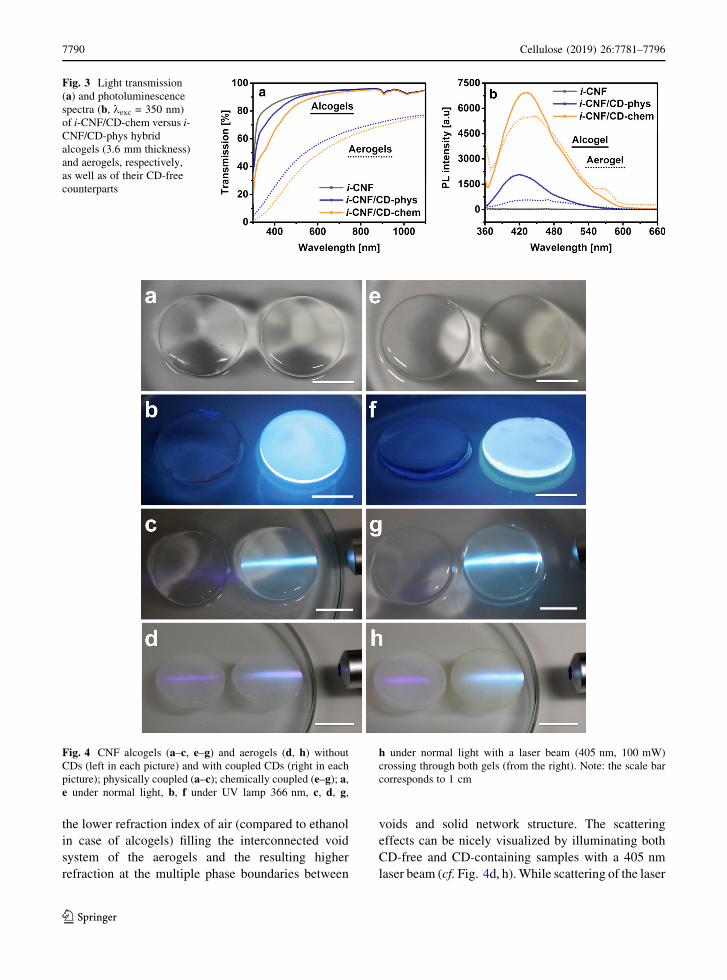
evident from the faint yellow color of the obtained
materials (cf. Fig. 4e, right) and their stronger adsorp-
tion of light in the lower wavelength range of the
visible light (300–500 nm; Fig. 3a). It is also con-
firmed by photoluminescence spectra (kexc = 350 nm)
which show higher PL intensities for the alcogels
(fourfold) and aerogels (tenfold) containing covalently
immobilized carbon dots (Fig. 3b).
However, considering their similar volumetric
contraction upon scCO2 drying, the over-proportional
increase of D PLmax (PLmax, chem/PLmax, phys) for the
aerogels of the two coupling approaches clearly
suggests that part of the CDs is lost by extraction
with the supercritical fluid.
A minor decrease in PL intensity has been also
observed during scCO2 drying of the gels of the
covalent grafting approach (Figs. 3b, 4g, h). However,
this effect is known to be caused by scattering losses in
the resulting aerogels and has been reported for
cellulose aerogels covalently equipped with semicon-
ductor quantum dots, too (Wang et al. 2013a). The
stronger scattering, in particular at lower wavelengths,
is attributed to Rayleigh scattering which occurs due to
Table 1 Composition, shrinkage and apparent density of CNF alcogels physically and chemically equipped with CDs
Initial CNF content
(w%)
Shrinkage caused by coupling
(v%)
Shrinkage caused by drying
(v%)
Bulk density
(mg cm-3)
Reference 0.91 0 35 15.4
Physical 0.91 0 37 16.3
Chemical 0.91 20 35 20.1
123
Cellulose (2019) 26:7781–7796 7789
the lower refraction index of air (compared to ethanol
in case of alcogels) filling the interconnected void
system of the aerogels and the resulting higher
refraction at the multiple phase boundaries between
voids and solid network structure. The scattering
effects can be nicely visualized by illuminating both
CD-free and CD-containing samples with a 405 nm
laser beam (cf. Fig. 4d, h). While scattering of the laser
Fig. 3 Light transmission
(a) and photoluminescence
spectra (b, kexc = 350 nm)
of i-CNF/CD-chem versus i-
CNF/CD-phys hybrid
alcogels (3.6 mm thickness)
and aerogels, respectively,
as well as of their CD-free
counterparts
Fig. 4 CNF alcogels (a–c, e–g) and aerogels (d, h) without
CDs (left in each picture) and with coupled CDs (right in each
picture); physically coupled (a–c); chemically coupled (e–g); a,
e under normal light, b, f under UV lamp 366 nm, c, d, g,
h under normal light with a laser beam (405 nm, 100 mW)
crossing through both gels (from the right). Note: the scale bar
corresponds to 1 cm
123
7790 Cellulose (2019) 26:7781–7796

beam can be nicely seen in the CD-free reference
aerogels (left samples in Fig. 4d, h), it is almost not
visible in the respective alcogels (left sample Fig. 4c,
g).
Besides from the strong photoluminescence of both
i-CNF/CD hybrid alcogels and aerogels matching the
PL properties of the implemented carbon dots (Fig. 4),
the successful decoration of the internal surface of the
i-CNF aerogels is also evident from FT-IR and X-ray
photoelectron spectroscopy (XPS) analyses (Fig. 5).
The FT-IR spectra obtained are comparable with
that of a recent study investigating improvement of the
thermal stability of TEMPO-oxidized cellulose nano-
fibers by coupling with amine-terminated polyethy-
lene glycol and employing heat-induced conversion of
initially formed ionic bonds into amide bonds
(Lavoine et al. 2016). Evaluation of the FT-IR spectral
region between 1800 cm-1 and 1500 cm-1 provided
valuable information on the bonding state of (C6)
carboxyl moieties of TEMPO-oxidized cellulose
(Fukuzumi et al. 2013; Lavoine et al. 2016). While
the band around 1610 cm-1 represent asymmetric (as)
stretching of C–O bonds, stretching of C=O bonds is
visible at 1730 cm-1 (Fig. 5a). Aerogels containing no
CDs show signals for both C–O and C=O bonds as
expected for protonated carboxyl moieties. Spectra of
aerogels containing physically coupled CDs on the
other hand virtually lack the C=O bands due to the
resonance structure of the carboxylate anion formed
by proton transfer to the amino-CDs. The contrary is
the case for chemically modified aerogels which show
only a very week C–O signal due to the formation of
amide bonds while the C=O bonds is still present
(Fig. 5a).
XPS spectra of the two types of i-CNF/CD hybrid
aerogels confirm the existence of the respective inter-
particulate bonding moieties between surface car-
boxyl(ate) groups of the cellulosic nanofibers and
surface amino groups of the carbon dots. Besides the
dominating peak at around 400 eV binding energy
(EB) which is caused by the abundance of amino
groups on the surface of the carbon dots (Guo et al.
2017; Oh et al. 2013) two smaller peaks at higher
binding energies appear in the N 1 s spectra. Based on
literature values (Beamson and Briggs 1992; Oh et al.
2013) and following the rule that increasing acidity of
the nitrogen atom shifts EB towards higher values
(Beamson and Briggs 1992), the small peaks at
401.2 eV and 402.9 eV were assigned to secondary
amide and quaternary ammonium moieties,
respectively.
Investigation of the mechanical response of the
different types of hybrid aerogels towards compres-
sive stress revealed that both stiffness and strength as
expressed by the parameters Young’s modulus (E) and
offset yield stress r0.2 % only slightly increased
compared to the CD-free reference aerogels (Table 2).
However, normalization of E for bulk density (specific
modulus Eq) suggests somewhat higher stiffness for
this type of hybrid aerogel. The Young’s modulus of
the materials obtained by chemical bonding of CDs
was almost more than twice as high as for the reference
aerogels. While this effect is owed partly to the
somewhat higher density of these hybrid aerogels, it is
evident that other factors contribute as well. The
recorded stress–strain-curves (cf. Fig. 6) reveal that
the yield point is also almost twice as high compared
to i-CNF/CD hybrid aerogels of the physical bonding
Fig. 5 FT-IR (a, section)
and XPS spectra b of i-CNF
aerogels whose internal
surface was decorated with
amino-functional carbon
dots either by chemical or
physical bonding
123
Cellulose (2019) 26:7781–7796 7791
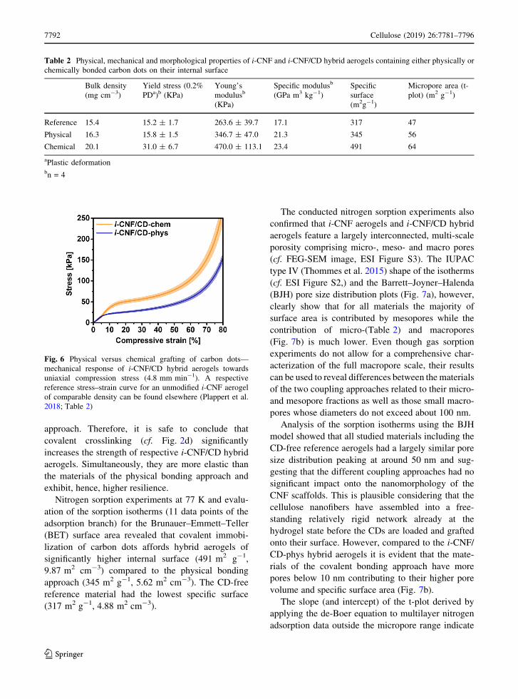
approach. Therefore, it is safe to conclude that
covalent crosslinking (cf. Fig. 2d) significantly
increases the strength of respective i-CNF/CD hybrid
aerogels. Simultaneously, they are more elastic than
the materials of the physical bonding approach and
exhibit, hence, higher resilience.
Nitrogen sorption experiments at 77 K and evalu-
ation of the sorption isotherms (11 data points of the
adsorption branch) for the Brunauer–Emmett–Teller
(BET) surface area revealed that covalent immobi-
lization of carbon dots affords hybrid aerogels of
significantly higher internal surface (491 m2 g-1,
9.87 m2 cm-3) compared to the physical bonding
approach (345 m2 g-1, 5.62 m2 cm-3). The CD-free
reference material had the lowest specific surface
(317 m2 g-1, 4.88 m2 cm-3).
The conducted nitrogen sorption experiments also
confirmed that i-CNF aerogels and i-CNF/CD hybrid
aerogels feature a largely interconnected, multi-scale
porosity comprising micro-, meso- and macro pores
(cf. FEG-SEM image, ESI Figure S3). The IUPAC
type IV (Thommes et al. 2015) shape of the isotherms
(cf. ESI Figure S2,) and the Barrett–Joyner–Halenda
(BJH) pore size distribution plots (Fig. 7a), however,
clearly show that for all materials the majority of
surface area is contributed by mesopores while the
contribution of micro-(Table 2) and macropores
(Fig. 7b) is much lower. Even though gas sorption
experiments do not allow for a comprehensive char-
acterization of the full macropore scale, their results
can be used to reveal differences between the materials
of the two coupling approaches related to their micro-
and mesopore fractions as well as those small macro-
pores whose diameters do not exceed about 100 nm.
Analysis of the sorption isotherms using the BJH
model showed that all studied materials including the
CD-free reference aerogels had a largely similar pore
size distribution peaking at around 50 nm and sug-
gesting that the different coupling approaches had no
significant impact onto the nanomorphology of the
CNF scaffolds. This is plausible considering that the
cellulose nanofibers have assembled into a free-
standing relatively rigid network already at the
hydrogel state before the CDs are loaded and grafted
onto their surface. However, compared to the i-CNF/
CD-phys hybrid aerogels it is evident that the mate-
rials of the covalent bonding approach have more
pores below 10 nm contributing to their higher pore
volume and specific surface area (Fig. 7b).
The slope (and intercept) of the t-plot derived by
applying the de-Boer equation to multilayer nitrogen
adsorption data outside the micropore range indicate
Table 2 Physical, mechanical and morphological properties of i-CNF and i-CNF/CD hybrid aerogels containing either physically or
chemically bonded carbon dots on their internal surface
Bulk density
(mg cm-3)
Yield stress (0.2%
PDa)b (KPa)
Young’s
modulusb
(KPa)
Specific modulusb
(GPa m3 kg-1)
Specific
surface
(m2g-1)
Micropore area (t-
plot) (m2 g-1)
Reference 15.4 15.2 ± 1.7 263.6 ± 39.7 17.1 317 47
Physical 16.3 15.8 ± 1.5 346.7 ± 47.0 21.3 345 56
Chemical 20.1 31.0 ± 6.7 470.0 ± 113.1 23.4 491 64
aPlastic deformationbn = 4
Fig. 6 Physical versus chemical grafting of carbon dots—
mechanical response of i-CNF/CD hybrid aerogels towards
uniaxial compression stress (4.8 mm min-1). A respective
reference stress–strain curve for an unmodified i-CNF aerogel
of comparable density can be found elsewhere (Plappert et al.
2018; Table 2)
123
7792 Cellulose (2019) 26:7781–7796

that the count of micropores and, hence, micropore
area increases with the introduction of carbon dots (cf.
Table 2). This is most pronounced for i-CNF/CD-
chem hybrid aerogels while physical immobilization
of CDs had a minor effect only. It is assumed that the
overall increase of specific surface area (SBET) with a
main contribution of small mesopores (2–10 nm) is
directly proportional to both count and accessible
surface area of the carbon dots introduced by the
respective coupling approaches. The considerable
difference in SBET gain related to the CD-free
reference material (i-CNF/CD-chem: 175 m2 g-1; i-
CNF/CD-phys: 30 m2 g-1; cf. Table 2) roughly cor-
relates with the difference in photoluminescent
response of the two types of hybrid aerogels and
emphasizes the superiority of the chemical bonding
approach. From an application perspective, this study
has been demonstrated that loading of amino-func-
tional pl-NP and their covalent immobilization on the
large internal surface of carboxyl-functional i-CNF
gels can afford aerogels that combine all of the
characteristics needed to account for a promising bio-
based matrix material for gas sensing or true volu-
metric displays. They are transparent, lightweight,
have high interconnected porosity and, hence, high
accessibility of the introduced CDs (Plappert et al.
2018; Kobayashi et al. 2014). Optionally, respective
cellulose hybrid aerogels can be conformally coated
by secondary biocompatible polymers, such as PMMA
to render them insensitive towards moisture induced
shrinkage (Plappert et al. 2018).
Conclusions
This study has demonstrated that loading of pl-NP,
specifically of amino-functional carbon dots, from
dispersion state into the virtually fully interconnected
void system of nematically ordered and largely
mesoporous gel matrix composed of self-aligned
cellulose I nanofibers (i-CNF; Plappert et al. 2018)
and their subsequent physical or chemical bonding to
surface carboxyl groups affords homogeneous
nematic nanocomposite hydrogels. The latter can be
converted into aerogels by consecutive solvent
exchange and scCO2 drying. Since the high light
transmittance and nanomorphology of i-CNF alcogels
is virtually not compromised by incorporation of pl-
NP under the tested conditions, it is safe to conclude
that the investigated approaches hold some promises
with regard to the development of optical devices.
While optical sensors rely on a sufficient density of
reporter nanoparticles dispersed across a large internal
surface which is fully accessible to soluble (lyogels) or
gaseous (aerogels) target analytes, true volumetric 3D
displays require low scattering and highly transparent
matrix materials. In case of volumetric displays, the
homogeneously distributed pl-NP should be topolog-
ically fixed and feature upconverting properties. This
is required for an appropriate setup of IR lasers,
dichroic mirror and 3D laser scanner to generate high
resolution color pictures within a respective 3D
matrix. Not least because of this aspect, covalent
immobilization of the nanoparticles represents the
superior approach as evident from the serious leaching
issue with physical pl-NP bonding, loss of PL intensity
and inferior internal surface and mechanical
properties.
Fig. 7 Pore size
distribution relating to pore
area (a) and pore volume
(b) of CD-free aerogels (i-
CNF) as well as i-CNF/CD-
phys and i-CNF/CD-chem
hybrid aerogels
123
Cellulose (2019) 26:7781–7796 7793

Acknowledgments Open access funding provided by
University of Natural Resources and Life Sciences Vienna
(BOKU). The financial support by the Austrian BMLFUW
Ministry (WoodWisdom Net? project AeroWood 2014-2017)
is gratefully acknowledged. Furthermore, the authors thank
cordially Johannes Konnerth of for his support with mechanical
testing and Tiina Nypelo for performing AFM analysis
(University of Natural Resources and Life Sciences, Vienna,
Institute of Wood Technology and Renewable Materials). The
authors also thank Alexander Bismarck and the Polymer and
Composite Engineering (PaCE) group at University of Vienna
(Department of Material Chemistry) for access to their TriStar II
3020 gas sorption analyzer.
Author contributions SQ and SFP share the first authorship of
this manuscript since both contributed equally. SQ prepared and
characterized carbon dots, performed and designed all
fluorescence measurements and contributed a significant part
of the writing of the initial manuscript. SFP designed the
coupling and loading procedures, prepared the cellulose gels
(and aerogels) and conducted and designed mechanical
characterization, gas sorption experiments, some of the UV–
Vis measurements, designed figures and contributed a
significant part of the writing of the manuscript. FWL
conceived the idea of the project, acquired funding, provided
project management, supervised and developed the individual
tasks, and contributed comprehensive revision of the initial
manuscript. TG contributed XPS analysis and evaluation. WG-
A helped significantly with planning, conducting and evaluation
of AFM and mechanical analyses.
Compliance with ethical standards
Conflict of interest The author declares that they have no
conflict of interest.
Open Access This article is distributed under the terms of the
Creative Commons Attribution 4.0 International License (http://
creativecommons.org/licenses/by/4.0/), which permits unre-
stricted use, distribution, and reproduction in any medium,
provided you give appropriate credit to the original
author(s) and the source, provide a link to the Creative Com-
mons license, and indicate if changes were made.
References
Abitbol T, Gray DG (2008) Incorporation into paper of cellulose
triacetate films containing semiconductor nanoparticles.
Cellulose 16:319–326. https://doi.org/10.1007/s10570-
008-9263-z
Aghajamali M, Iqbal M, Purkait TK, Hadidi L, Sinelnikov R,
Veinot JGC (2016) Synthesis and properties of luminescent
silicon nanocrystal/silica aerogel hybrid materials. Chem
Mater 28:3877–3886. https://doi.org/10.1021/acs.chemmater.
6b01114
Barrett EP, Joyner LG, Halenda PP (1951) The determination of
pore volume and area distributions in porous substances.
I Computations from nitrogen isotherms. J Am Chem Soc
73:373–380. https://doi.org/10.1021/ja01145a126
Beamson G, Briggs D (1992) High resolution XPS of organic
polymers: the scienta ESCA300 database. Wiley,
Chichester
Bohrn R, Potthast A, Schiehser S, Rosenau T, Sixta H, Kosma P
(2006) The FDAM method: determination of carboxyl
profiles in cellulosic materials by combining group-selec-
tive fluorescence labeling with GPC. Biomacromolecules
7:1743–1750. https://doi.org/10.1021/bm060039h
Broekhoff J (1968) Studies on pore systems in catalysts XIII. Pore
distributions from the desorption branch of a nitrogen sorp-
tion isotherm in the case of cylindrical pores B. Appl J Catal
10:377–390. https://doi.org/10.1016/0021-9517(68)90153-x
Brunauer S, Emmett PH, Teller E (1938) Adsorption of gases in
multimolecular layers. J Am Chem Soc 60:309–319.
https://doi.org/10.1021/ja01269a023
Chandra S, Laha D, Pramanik A, Ray Chowdhuri A, Karmakar
P, Sahu SK (2015) Synthesis of highly fluorescent nitrogen
and phosphorus doped carbon dots for the detection of Fe
ions in cancer cells. Luminescence. https://doi.org/10.
1002/bio.2927
Deng R, Qin F, Chen R, Huang W, Hong M, Liu X (2015)
Temporal full-colour tuning through non-steady-state
upconversion. Nat Nanotechnol 10:237–242. https://doi.
org/10.1038/nnano.2014.317
Ding H, Yu SB, Wei JS, Xiong HM (2016) Full-color light-
emitting carbon dots with a surface-state-controlled lumi-
nescence mechanism. ACS Nano 10:484–491. https://doi.
org/10.1021/acsnano.5b05406
Dolai S, Bhunia SK, Jelinek R (2017) Carbon-dot-aerogel sen-
sor for aromatic volatile organic compounds. Sens Actua-
tors B 241:607–613. https://doi.org/10.1016/j.snb.2016.10.
124
Downing E, Hesselink L, Ralston J, Macfarlane R (1996) A
three-color, solid-state, three-dimensional display. Science
273:1185–1189. https://doi.org/10.1126/science.273.5279.
1185
Duarah R, Karak N (2017) Facile and ultrafast green approach to
synthesize biobased luminescent reduced carbon nanodot: an
efficient photocatalyst. ACS Sustain Chem Eng 5:9454–9466.
https://doi.org/10.1021/acssuschemeng.7b02590
Fukuzumi H, Fujisawa S, Saito T, Isogai A (2013) Selective
permeation of hydrogen gas using cellulose nanofibril film.
Biomacromolecules 14:1705–1709. https://doi.org/10.
1021/bm400377e
Gogoi N, Barooah M, Majumdar G, Chowdhury D (2015)
Carbon dots rooted agarose hydrogel hybrid platform for
optical detection and separation of heavy metal ions. ACS
Appl Mater Interfaces 7:3058–3067. https://doi.org/10.
1021/am506558d
Guo J et al (2017) Photoluminescent hybrids of cellulose
nanocrystals and carbon quantum dots as cytocompatible
probes for in vitro bioimaging. Biomacromolecules
18:2045–2055. https://doi.org/10.1021/acs.biomac.7b00306
Hoan BT, Van Huan P, Van HN, Nguyen DH, Tam PD, Nguyen
KT, Pham VH (2018) Luminescence of lemon-derived
carbon quantum dot and its potential application in lumi-
nescent probe for detection of Mo(6?) ions. Luminescence
33:545–551. https://doi.org/10.1002/bio.3444
123
7794 Cellulose (2019) 26:7781–7796

Hu S, Zhao Q, Dong Y, Yang J, Liu J, Chang Q (2013) Carbon-
dot-loaded alginate gels as recoverable probes: fabrication
and mechanism of fluorescent detection. Langmuir
29:12615–12621. https://doi.org/10.1021/la402647t
Isogai A, Saito T, Fukuzumi H (2011) TEMPO-oxidized cel-
lulose nanofibers Nanoscale 3:71–85. https://doi.org/10.
1039/c0nr00583e
Javidi B, Marinov VR, Lima JIT, Miller R, Son J-Y, Thomas JT,
Desjardins DD (2010) Quantum dot dispersions in aero-
gels: a new material for true volumetric color displays.
Proc SPIE 7690:76900X–76900X–76907. https://doi.org/
10.1117/12.850252
Jeong Y, Moon K, Jeong S, Koh W-G, Lee K (2018) Converting
waste papers to fluorescent carbon dots in the recycling
process without loss of ionic liquids and bioimaging
applications. ACS Sustain Chem Eng 6:4510–4515. https://
doi.org/10.1021/acssuschemeng.8b00353
Jiang F, Hsieh Y-L (2016) Self-assembling of TEMPO oxidized
cellulose nanofibrils as affected by protonation of surface
carboxyls and drying methods. ACS Sustain Chem
Eng 4:1041–1049. https://doi.org/10.1021/acssuschemeng.
5b01123
Junka K, Guo J, Filpponen I, Laine J, Rojas OJ (2014) Modifi-
cation of cellulose nanofibrils with luminescent carbon
dots. Biomacromolecules 15:876–881. https://doi.org/10.
1021/bm4017176
Kobayashi Y, Saito T, Isogai A (2014) Aerogels with 3D
ordered nanofiber skeletons of liquid-crystalline nanocel-
lulose derivatives as tough and transparent insulators.
Angew Chem Int Ed Engl 53:10394–10397. https://doi.
org/10.1002/anie.201405123
Korhonen JT, Hiekkataipale P, Malm J, Karppinen M, Ikkala O,
Ras RH (2011) Inorganic hollow nanotube aerogels by
atomic layer deposition onto native nanocellulose tem-
plates. ACS Nano 5:1967–1974. https://doi.org/10.1021/
nn200108s
Kozak O, Sudolska M, Pramanik G, Cıgler P, Otyepka M, Zboril R
(2016) Photoluminescent carbon nanostructures. Chem
Mater 28:4085–4128. https://doi.org/10.1021/acs.chemmater.
6b01372
Kulpinski P, Erdman A, Namyslak M, Fidelus JD (2012) Cel-
lulose fibers modified by Eu3?-doped yttria-stabilized
zirconia nanoparticles. Cellulose 19:1259–1269. https://
doi.org/10.1007/s10570-012-9704-6
Lavoine N, Bras J, Saito T, Isogai A (2016) Improvement of the
thermal stability of TEMPO-oxidized cellulose nanofibrils
by heat-induced conversion of ionic bonds to amide bonds.
Macromol Rapid Commun 37:1033–1039
Li S, Huang J (2016) Cellulose-rich nanofiber-based functional
nanoarchitectures. Adv Mater 28:1143–1158. https://doi.
org/10.1002/adma.201501878
Li X, Rui M, Song J, Shen Z, Zeng H (2015) Carbon and gra-
phene quantum dots for optoelectronic and energy devices:
a review. Adv Funct Mater 25:4929–4947. https://doi.org/
10.1002/adfm.201501250
Lim SY, Shen W, Gao Z (2015) Carbon quantum dots and their
applications. Chem Soc Rev 44:362–381. https://doi.org/
10.1039/c4cs00269e
Lippens B (1965) Studies on pore systems in catalysts V. The t
method. J Catal 4:319–323. https://doi.org/10.1016/0021-
9517(65)90307-6
Oh D, Shin S, Lim C, Hwang D (2013) Dopamine-mediated
sclerotization of regenerated chitin in ionic liquid. Mate-
rials 6:3826–3839
Olsson RT et al (2010) Making flexible magnetic aerogels and
stiff magnetic nanopaper using cellulose nanofibrils as
templates. Nat Nanotechnol 5:584–588. https://doi.org/10.
1038/nnano.2010.155
Onsager L (1949) The effects of shape on the interaction of
colloidal particles. Ann N Y Acad Sci 51:627–659. https://
doi.org/10.1111/j.1749-6632.1949.tb27296.x
Plappert SF, Nedelec J-M, Rennhofer H, Lichtenegger HC,
Liebner FW (2017) Strain hardening and pore size har-
monization by uniaxial densification: a facile approach
toward superinsulating aerogels from nematic nanofibril-
lated 2,3-dicarboxyl cellulose. Chem Mater 29:6630–6641.
https://doi.org/10.1021/acs.chemmater.7b00787
Plappert SF, Quraishi S, Nedelec J-M, Konnerth J, Rennhofer H,
Lichtenegger HC, Liebner FW (2018) Conformal ultrathin
coating by scCO2-mediated PMMA deposition: a facile
approach to add moisture resistance to lightweight ordered
nanocellulose aerogels. Chem Mater 30:2322–2330
Potthast A, Rohrling J, Rosenau T, Borgards A, Sixta H, Kosma
P (2003) A novel method for the determination of carbonyl
groups in cellulosics by fluorescence labeling. 3. Moni-
toring oxidative processes. Biomacromolecules
4:743–749. https://doi.org/10.1021/bm025759c
Rodrıguez-Padron D et al (2018) Catalyzed microwave-assisted
preparation of carbon quantum dots from lignocellulosic
residues. ACS Sustain Chem Eng 6:7200–7205. https://doi.
org/10.1021/acssuschemeng.7b03848
Sachdev A, Matai I, Gopinath P (2016) Carbon dots incorpo-
rated polymeric hydrogels as multifunctional platform for
imaging and induction of apoptosis in lung cancer cells.
Colloids Surf B 141:242–252. https://doi.org/10.1016/j.
colsurfb.2016.01.043
Sahu S, Behera B, Maiti TK, Mohapatra S (2012) Simple one-
step synthesis of highly luminescent carbon dots from
orange juice: application as excellent bio-imaging agents.
Chem Commun (Camb) 48:8835–8837. https://doi.org/10.
1039/c2cc33796g
Saito T, Uematsu T, Kimura S, Enomae T, Isogai A (2011) Self-
aligned integration of native cellulose nanofibrils towards
producing diverse bulk materials. Soft Matter 7:8804.
https://doi.org/10.1039/c1sm06050c
Sato Y, Kusaka Y, Kobayashi M (2017) Charging and aggre-
gation behavior of cellulose nanofibers in aqueous solution.
Langmuir 33:12660–12669. https://doi.org/10.1021/acs.
langmuir.7b02742
Shinoda R, Saito T, Okita Y, Isogai A (2012) Relationship
between length and degree of polymerization of TEMPO-
oxidized cellulose nanofibrils. Biomacromolecules
13:842–849. https://doi.org/10.1021/bm2017542
Sorensen L, Strouse G, Stiegman A (2006) Fabrication of
stable low-density silica aerogels containing luminescent
ZnS capped CdSe quantum dots. Adv Mater 18:1965
Sun C et al (2016) Synthesis of nitrogen and sulfur co-doped
carbon dots from garlic for selective detection of Fe(3).
Nanoscale Res Lett 11:110. https://doi.org/10.1186/
s11671-016-1326-8
Takeshita S, Takebayashi Y, Nakamura H, Yoda S (2016) Gas-
responsive photoluminescence of YVO4:Eu3?
123
Cellulose (2019) 26:7781–7796 7795

nanoparticles dispersed in an ultralight, three-dimensional
nanofiber network. Chem Mater 28:8466–8469. https://doi.
org/10.1021/acs.chemmater.6b04160
Thommes M, Kaneko K, Neimark AV, Olivier JP, Rodriguez-
Reinoso F, Rouquerol J, Sing KSW (2015) Physisorption
of gases, with special reference to the evaluation of surface
area and pore size distribution (IUPAC Technical Report).
Pure Appl Chem 87:1051. https://doi.org/10.1515/pac-
2014-1117
Toivonen MS, Kaskela A, Rojas OJ, Kauppinen EI, Ikkala O
(2015) Ambient-dried cellulose nanofibril aerogel mem-
branes with high tensile strength and their use for aerosol
collection and templates for transparent, flexible devices.
Adv Funct Mater 25:6618–6626. https://doi.org/10.1002/
adfm.201502566
Tyagi A, Tripathi KM, Singh N, Choudhary S, Gupta RK (2016)
Green synthesis of carbon quantum dots from lemon peel
waste: applications in sensing and photocatalysis. RSC
Adv 6:72423–72432. https://doi.org/10.1039/c6ra10488f
Wagberg L, Decher G, Norgren M, Lindstrom T, Ankerfors M,
Axnas K (2008) The build-up of polyelectrolyte multilay-
ers of microfibrillated cellulose and cationic polyelec-
trolytes. Langmuir 24:784–795. https://doi.org/10.1021/
la702481v
Wang J, Qiu J (2016) A review of carbon dots in biological
applications. J Mater Sci 51:4728–4738. https://doi.org/10.
1007/s10853-016-9797-7
Wang H, Shao Z, Bacher M, Liebner F, Rosenau T (2013a)
Fluorescent cellulose aerogels containing covalently
immobilized (ZnS)x(CuInS2)1 - x/ZnS (core/shell)
quantum dots. Cellulose. https://doi.org/10.1007/s10570-
013-0035-z
Wang R, Li G, Dong Y, Chi Y, Chen G (2013b) Carbon quantum
dot-functionalized aerogels for NO2 gas sensing. Anal
Chem 85:8065–8069. https://doi.org/10.1021/ac401880h
Wang F, Xie Z, Zhang B, Liu Y, Yang W, Liu CY (2014) Down-
and up-conversion luminescent carbon dot fluid: inkjet
printing and gel glass fabrication. Nanoscale 6:3818–3823.
https://doi.org/10.1039/c3nr05869g
Wolfbeis OS (2015) An overview of nanoparticles commonly
used in fluorescent bioimaging. Chem Soc Rev
44:4743–4768. https://doi.org/10.1039/c4cs00392f
Xu Y, Wu M, Liu Y, Feng XZ, Yin XB, He XW, Zhang YK
(2013) Nitrogen-doped carbon dots: a facile and general
preparation method, photoluminescence investigation, and
imaging applications. Chemistry 19:2276–2283. https://
doi.org/10.1002/chem.201203641
Xu M et al (2014) A green heterogeneous synthesis of N-doped
carbon dots and their photoluminescence applications in
solid and aqueous states. Nanoscale 6:10307–10315.
https://doi.org/10.1039/c4nr02792b
Xu Q et al (2016) Heteroatom-doped carbon dots: synthesis,
characterization, properties, photoluminescence mecha-
nism and biological applications. J Mater Chem B
4:7204–7219. https://doi.org/10.1039/c6tb02131j
Yang Z et al (2013) Controllable synthesis of fluorescent carbon
dots and their detection application as nanoprobes. Nano
Micro Lett 5:247–259. https://doi.org/10.1007/bf03353756
Yang Z et al (2014) Nitrogen-doped, carbon-rich, highly photo-
luminescent carbon dots from ammonium citrate. Nanoscale
6:1890–1895. https://doi.org/10.1039/c3nr05380f
Zeng J, Yan L (2015) Metal-free transparent luminescent cel-
lulose films. Cellulose 22:729–736. https://doi.org/10.
1007/s10570-014-0485-y
Zhou B, Shi B, Jin D, Liu X (2015) Controlling upconversion
nanocrystals for emerging applications. Nat Nanotechnol
10:924–936. https://doi.org/10.1038/nnano.2015.251
Zhu S et al (2013) Highly photoluminescent carbon dots for
multicolor patterning, sensors, and bioimaging. Angew
Chem Int Ed Engl 52:3953–3957. https://doi.org/10.1002/
anie.201300519
Zhu M, Zhong H, Jia J, Fu W, Liu J, Zou B, Wang Y (2014) PVA
hydrogel embedded with quantum dots: a potential scalable
and healable display medium for holographic 3D applica-
tions. Adv Opt Mater 2:338–342. https://doi.org/10.1002/
adom.201300517
Zhu S, Song Y, Zhao X, Shao J, Zhang J, Yang B (2015) The
photoluminescence mechanism in carbon dots (graphene
quantum dots, carbon nanodots, and polymer dots): current
state and future perspective. Nano Res 8:355–381. https://
doi.org/10.1007/s12274-014-0644-3
Zhu L, Zong L, Wu X, Li M, Wang H, You J, Li C (2018)
Shapeable fibrous aerogels of metal-organic-frameworks
templated with nanocellulose for rapid and large-capacity
adsorption. ACS Nano 12:4462–4468. https://doi.org/10.
1021/acsnano.8b00566
Publisher’s Note Springer Nature remains neutral with
regard to jurisdictional claims in published maps and
institutional affiliations.
123
7796 Cellulose (2019) 26:7781–7796


![HFM Free Flap Versus Fat Grafting[1]](https://static.fdocuments.us/doc/165x107/54f83fc94a7959fe478b459b/hfm-free-flap-versus-fat-grafting1.jpg)