Chemical and Enzymatic Probing of Spatial Structure of the...
7
In eukaryotes 5′-untranslated regions of mRNA markedly influence the translation efficiency of the encoding part of RNA carrying these regions. The 5′- untranslated region of tobacco mosaic virus (TMV) genomic RNA contains the so-called omega sequence, a powerful enhancer of the translation of this RNA (“trans- lational enhancer”) [1]. In recombinant constructs the leader omega sequence enhances translation of foreign RNAs both in vivo and in vitro (in cell-free systems) and provides for efficient cap-independent translation initia- tion [2]. Omega leader can act as a translational enhancer in various cell types, both plant and animal, as well as in different cell-free translation systems [3-10]. The nucleotide sequence of the omega leader from RNA of TMV strain U1 is shown in Fig. 1 [11]. This sequence has an unusual primary structure. First, the omega sequence is almost completely deprived of guanylic residues (G): with the exclusion of the first residue, adjacent to the cap-structure at the 5′ end of TMV RNA, there are no other guanylic residues in the almost 70 nucleotide-long leader chain. Second, more than one-third of the omega sequence is comprised by its central part consisting only of adenylic and cytidylic residues. Third, adenylic and cytidylic residues of the central part as well as those of adjacent to 5′ and 3′ prox- ISSN 0006-2979, Biochemistry (Moscow), 2010, Vol. 75, No. 4, pp. 405-411. © Pleiades Publishing, Ltd., 2010. Original Russian Text © N. E. Shirokikh, S. Ch. Agalarov, A. S. Spirin, 2010, published in Biokhimiya, 2010, Vol. 75, No. 4, pp. 492-500. Originally published in Biochemistry (Moscow) On-Line Papers in Press, as Manuscript BM09-328, February 14, 2010. ACCELERATED PUBLICATION 405 Abbreviations: AMV, avian myeloblastosis virus; cDNA, com- plementary DNA; CXR, 5(6)-carboxy-X-rhodamine; FAM, 6- carboxy-4′,5′-dichloro-2′,7′-dimethoxyfluorescein; Hepes, 4- (2-hydroxyethyl)-1-piperazineethanesulfonic acid; TMV, tobacco mosaic virus; Tris, tris(hydroxymethyl)aminomethane. * To whom correspondence should be addressed. Chemical and Enzymatic Probing of Spatial Structure of the Omega Leader of Tobacco Mosaic Virus RNA N. E. Shirokikh, S. Ch. Agalarov, and A. S. Spirin* Laboratory of Mechanisms of Protein Biosynthesis, Institute of Protein Research, Russian Academy of Sciences, 142290 Pushchino, Moscow Region, Russia; fax: (499) 632-7871; E-mail: [email protected] Received November 12, 2009 Revision received December 3, 2009 Abstract—The 5′-untranslated sequence of tobacco mosaic virus RNA – the so-called omega leader – exhibits features of a translational enhancer of homologous and heterologous mRNAs. The absence of guanylic residues, the presence of multi- ple trinucleotide CAA repeats in its central region, and the low predictable probability of the formation of an extensive sec- ondary structure of the Watson–Crick type were reported as the peculiarities of the primary structure of the omega leader. In this work we performed chemical and enzymatic probing of the secondary structure of the omega leader. The isolated RNA comprising omega leader sequence was subjected to partial modifications with dimethyl sulfate and diethyl pyrocar- bonate and partial hydrolyses with RNase A and RNase V1. The sites and the intensities of the modifications or the cleav- ages were detected and measured by the primer extension inhibition technique. The data obtained have demonstrated that RNase A, which attacks internucleotide bonds at the 3′ side of pyrimidine nucleotides, and diethyl pyrocarbonate, which modifies N7 of adenines not involved in stacking interactions, weakly affected the core region of omega leader sequence enriched with CAA-repeats, this directly indicating the existence of a stable spatial structure. The significant stability of the core region structure to RNase A and diethyl pyrocarbonate was accompanied by its complete resistance against RNase V1, which cleaves a polyribonucleotide chain involved in Watson–Crick double helices and generally all A-form RNA helices, thus being an evidence in favor of a non-Watson–Crick structure. The latter was confirmed by the full susceptibility of all adenines and cytosines of the omega polynucleotide chain to dimethyl sulfate, which exclusively modifies N1 of adenines and N3 of cytosines not involved in Watson–Crick interactions. Thus, our data have confirmed that (1) the regular (CAA) n sequence characteristic of the core region of the omega leader does form stable secondary structure, and (2) the structure formed is not the canonical double helix of the Watson–Crick type. DOI: 10.1134/S0006297910040024 Key words: omega leader of TMV RNA, regular (САА) n polyribonucleotide, RNA triple helix, chemical modification of RNA, enzymatic cleavage of RNA, primer extension inhibition, diethyl pyrocarbonate, dimethyl sulfate, RNase A, RNase V1
Transcript of Chemical and Enzymatic Probing of Spatial Structure of the...

In eukaryotes 5′-untranslated regions of mRNA
markedly influence the translation efficiency of the
encoding part of RNA carrying these regions. The 5′-
untranslated region of tobacco mosaic virus (TMV)
genomic RNA contains the so-called omega sequence, a
powerful enhancer of the translation of this RNA (“trans-
lational enhancer”) [1]. In recombinant constructs the
leader omega sequence enhances translation of foreign
RNAs both in vivo and in vitro (in cell-free systems) and
provides for efficient cap-independent translation initia-
tion [2]. Omega leader can act as a translational enhancer
in various cell types, both plant and animal, as well as in
different cell-free translation systems [3-10].
The nucleotide sequence of the omega leader from
RNA of TMV strain U1 is shown in Fig. 1 [11]. This
sequence has an unusual primary structure. First, the
omega sequence is almost completely deprived of
guanylic residues (G): with the exclusion of the first
residue, adjacent to the cap-structure at the 5′ end of
TMV RNA, there are no other guanylic residues in the
almost 70 nucleotide-long leader chain. Second, more
than one-third of the omega sequence is comprised by its
central part consisting only of adenylic and cytidylic
residues. Third, adenylic and cytidylic residues of the
central part as well as those of adjacent to 5′ and 3′ prox-
ISSN 0006-2979, Biochemistry (Moscow), 2010, Vol. 75, No. 4, pp. 405-411. © Pleiades Publishing, Ltd., 2010.
Original Russian Text © N. E. Shirokikh, S. Ch. Agalarov, A. S. Spirin, 2010, published in Biokhimiya, 2010, Vol. 75, No. 4, pp. 492-500.
Originally published in Biochemistry (Moscow) On-Line Papers in Press, as Manuscript BM09-328, February 14, 2010.
ACCELERATED PUBLICATION
405
Abbreviations: AMV, avian myeloblastosis virus; cDNA, com-
plementary DNA; CXR, 5(6)-carboxy-X-rhodamine; FAM, 6-
carboxy-4′,5′-dichloro-2′,7′-dimethoxyfluorescein; Hepes, 4-
(2-hydroxyethyl)-1-piperazineethanesulfonic acid; TMV,
tobacco mosaic virus; Tris, tris(hydroxymethyl)aminomethane.
* To whom correspondence should be addressed.
Chemical and Enzymatic Probing of Spatial Structure
of the Omega Leader of Tobacco Mosaic Virus RNA
N. E. Shirokikh, S. Ch. Agalarov, and A. S. Spirin*
Laboratory of Mechanisms of Protein Biosynthesis, Institute of Protein Research, Russian Academy of Sciences,
142290 Pushchino, Moscow Region, Russia; fax: (499) 632-7871; E-mail: [email protected]
Received November 12, 2009
Revision received December 3, 2009
Abstract—The 5′-untranslated sequence of tobacco mosaic virus RNA – the so-called omega leader – exhibits features of a
translational enhancer of homologous and heterologous mRNAs. The absence of guanylic residues, the presence of multi-
ple trinucleotide CAA repeats in its central region, and the low predictable probability of the formation of an extensive sec-
ondary structure of the Watson–Crick type were reported as the peculiarities of the primary structure of the omega leader.
In this work we performed chemical and enzymatic probing of the secondary structure of the omega leader. The isolated
RNA comprising omega leader sequence was subjected to partial modifications with dimethyl sulfate and diethyl pyrocar-
bonate and partial hydrolyses with RNase A and RNase V1. The sites and the intensities of the modifications or the cleav-
ages were detected and measured by the primer extension inhibition technique. The data obtained have demonstrated that
RNase A, which attacks internucleotide bonds at the 3′ side of pyrimidine nucleotides, and diethyl pyrocarbonate, which
modifies N7 of adenines not involved in stacking interactions, weakly affected the core region of omega leader sequence
enriched with CAA-repeats, this directly indicating the existence of a stable spatial structure. The significant stability of the
core region structure to RNase A and diethyl pyrocarbonate was accompanied by its complete resistance against RNase V1,
which cleaves a polyribonucleotide chain involved in Watson–Crick double helices and generally all A-form RNA helices,
thus being an evidence in favor of a non-Watson–Crick structure. The latter was confirmed by the full susceptibility of all
adenines and cytosines of the omega polynucleotide chain to dimethyl sulfate, which exclusively modifies N1 of adenines
and N3 of cytosines not involved in Watson–Crick interactions. Thus, our data have confirmed that (1) the regular (CAA)n
sequence characteristic of the core region of the omega leader does form stable secondary structure, and (2) the structure
formed is not the canonical double helix of the Watson–Crick type.
DOI: 10.1134/S0006297910040024
Key words: omega leader of TMV RNA, regular (САА)n polyribonucleotide, RNA triple helix, chemical modification of RNA,
enzymatic cleavage of RNA, primer extension inhibition, diethyl pyrocarbonate, dimethyl sulfate, RNase A, RNase V1

406 SHIROKIKH et al.
BIOCHEMISTRY (Moscow) Vol. 75 No. 4 2010
imal parts of the omega sequence are mainly grouped in
CAA triplets. Thus, the central part is an almost regular
sequence consisting of CAA nucleotide triplets arranged
in succession. The CAA-containing part is restricted on
both sides by U-rich regions, a short one on the 5′-side
and a longer one on the 3′-side. On the whole, the fea-
tures exhibited by the omega leader nucleotide sequence
point to the impossibility of the formation of a stable sec-
ondary structure based on canonical Watson–Crick base
pairing of G:C and A:U type. This situation was for a long
time an argument in favor of the supposition that it is an
unstructured nature of the omega leader that provides for
its easy accessibility for ribosomes, and as a result, its
properties of a translational enhancer [12, 13]. However,
it was shown in our laboratory that unstructured
sequences, for which the absence of secondary structure
in solution was really proved, such as poly(U) [14], do not
exhibit properties of translational enhancers, at least in
translation systems of higher eukaryotes [15].
On the other hand, we have previously studied phys-
ical properties of the RNAs that were synthesized copies
of the leader omega sequence of TMV RNA and the reg-
ular polyribonucleotide (CAA)19, the latter modeling the
nucleotide sequence of the central part of the omega
leader [16]. It was shown by UV-spectroscopy and ana-
lytical centrifugation that both the complete omega
sequence and the homolog of its CAA part have a stable
and cooperatively melted structure, and their sedimenta-
tion coefficients correspond to those of compactly folded
RNAs of the same lengths. At the same time, the polynu-
cleotide of the same composition and of the same length
but with random sequence of nucleotides, poly(A2,C),
did not exhibit such properties and behaved as a disor-
dered non-compact polymer [16]. These data show that
the compact structure of the omega sequence can be
formed by the regular nucleotide sequence (CAA)n rep-
resenting the core part of the omega sequence.
Theoretical analysis of possibilities of the regular polyri-
bonucleotide (CAA)n to fold into a compact structure
due to formation of hydrogen bond pairs of non-
Watson–Crick type resulted in creation of the triple helix
model in which bases are bound to each other by hydro-
gen bond pairs into alternating triads A:C:A, C:A:A, and
A:A:C [17].
To test the type of possible structural organization of
the omega leader RNA, we used standard methods of par-
tial chemical modification of nucleotides and restricted
enzymatic degradation, which are widely used in testing
RNA spatial structures (see, for example, [18, 19]). The
data obtained in this work confirmed experimentally the
absence of double helices with Watson–Crick base pairs.
At the same time, analysis of the data has shown the pres-
ence of an extended region involved in formation of a
non-canonical secondary structure in the central part of
the omega sequence corresponding to the (CAA)-con-
taining core of the omega leader.
Fig. 2. Scheme of the use of pTZ10omegaLUC construct for the synthesis of RNA containing omega sequence of RNA from TMV strain U1
(shown in bold). The relative position of the restriction site of endonuclease XbaI used for linearization of the plasmid is indicated. Nucleotide
sequence of the 5′-untranslated RNA region (total RNA length is 136 nucleotides) containing the omega leader RNA is shown at the bottom;
the omega sequence within the 5′-untranslated RNA region is shown in bold, and the initiation codon of the luciferase gene is shown in bold
and underlined.
Fig. 1. The 5′-untranslated omega sequence of genomic RNA of TMV strain U1 [11]. The region containing successive (CAA)n repeats is
underlined. The initiation AUG codon at the beginning of the open reading frame of the North American firefly luciferase gene is also under-
lined and shown in bold.

CHEMICAL AND ENZYMATIC PROBING OF TMV OMEGA LEADER STRUCTURE 407
BIOCHEMISTRY (Moscow) Vol. 75 No. 4 2010
MATERIALS AND METHODS
Purification of plasmid containing omega sequence of
TMV RNA. Plasmid construct pTZ10omegaLUC [3]
copying omega sequence of TMV RNA (Fig. 2) was
amplified in Escherichia coli DH5α cells. Cells were lysed
and the lysate was neutralized using appropriate solutions
from the PureYield Plasmid Midiprep System kit
(Promega, USA). Cell debris, denatured proteins, and
genomic DNA were pelleted by centrifugation, and the
resulting supernatant was applied on adsorption columns
from the same reagent kit. Plasmid DNA was eluted from
the columns by deionized water and purified from protein
contaminants by phenol extraction. Sodium acetate
(pH 5.0) up to 300 mM and 2.5 volumes of 96% ethanol
were added to the solution and DNA was collected by
centrifugation at 15,000g for 15 min, dried, and dissolved
in deionized water. The integrity and purity of the
pTZ10omegaLUC plasmid were checked by electrophore-
sis in 1.5% agarose gel (stained with ethidium bromide).
Transcription of RNA using T7 RNA polymerase and
its purification. Purified plasmid pTZ10omegaLUC was
cleaved by restriction endonuclease XbaI in the region
following the luciferase gene of the American firefly
Photinus pyralis (Fig. 2). A sample of 200 µg of plasmid
pTZ10omegaLUC was incubated with 400 units of
endonuclease XbaI (Fermentas, Lithuania) at 37°C for
4 h in Tango 1X buffer (Fermentas), after which the reac-
tion mixture was twice extracted with phenol. After phe-
nol extraction, DNA was precipitated from the reaction
mixture by addition of sodium acetate (pH 5.0) to
300 mM, and 2.5 volumes of 96% ethanol and the precip-
itate was centrifuged at 15,000g for 15 min, after which
the pellet was dried and dissolved in deionized water.
Completeness of the cleavage reaction was estimated by
electrophoresis in 2% agarose gel (ethidium bromide
staining).
RNA of total length of 136 nt containing the omega
sequence was obtained by transcription of the above-
mentioned template DNA by T7 RNA polymerase (Fig.
2) under the conditions recommended in the literature
[20, 21] with slight modifications. The mixture for the
transcription reaction contained 80 mM Tris-acetate,
pH 7.5, 10 mM KCl, 22.2 mM Mg(CH3COO)2, 20 mM
dithiothreitol, 20 mM β-mercaptoethanol, 2 mM sper-
midine, 0.01% (v/v) Triton X-100, 0.2 mM EDTA, 4 mM
each nucleoside triphosphate (ATP, GTP, CTP, UTP)
(Fermentas), 1 unit/µl RNase inhibitor RiboLock
(Fermentas), 0.1 µg/µl template DNA, and 12 units/µl
T7 RNA polymerase (Fermentas) in 500 µl total volume.
The reaction mixture was incubated for 3 h at 37°C, then
extracted twice by acidic (pH 5.5) phenol, and nucleic
acids were precipitated by addition of ammonium acetate
to 2 M and 2.5 volumes of 96% ethanol. The precipitated
nucleic acids were collected by centrifugation at 15,000g
for 15 min in the cold, and the pellets were dried under
vacuum and dissolved in 250 µl of deionized water. To
purify the preparation from the initial template DNA, the
solution was brought to 10 mM Tris-acetate, pH 7.5,
2.5 mM Mg(CH3COO)2, 0.1 mM CaCl2, 2 units/µl
RNase inhibitor RiboLock (Fermentas), and 1.5 units/µl
DNase I (Boehringer Mannheim, Germany). The reac-
tion mixture was incubated for 1 h at 37°C, then it was
extracted twice with acidic phenol (pH 5.5), and nucleic
acids were precipitated by addition of 300 mM sodium
acetate (pH 5.0) and 2.5 volumes of 96% ethanol.
Precipitated nucleic acids were collected by centrifuga-
tion at 15,000g for 15 min in the cold, and pellets were
dissolved in 200 µl of deionized water. The solution was
supplemented with 68 µl of saturated LiCl solution, the
mixture was incubated for 1 h at 0°C, and the precipitate
was collected by centrifugation at 15,000g for 15 min in
the cold. The pellet was dissolved in 500 µl of deionized
water, then the solution was brought to 2 M ammonium
acetate and 2.5 volumes of 96% ethanol were added. The
resulting mixture was centrifuged at 15,000g for 15 min in
the cold, the pellet after centrifugation was dissolved in
500 µl of deionized water, and the resulting solution was
brought to 300 mM sodium acetate (pH 5.0) and 2.5 vol-
umes of 96% ethanol were added. The resulting mixture
was centrifuged at 15,000g for 15 min in the cold, the pel-
let was washed with 80% ethanol and dissolved in 150 µl
of deionized water, and the solution was frozen. The puri-
ty and homogeneity of the RNA preparation was estimat-
ed by electrophoresis in 6% denaturing polyacrylamide
gel (staining by toluidine blue).
RNA modification by dimethyl sulfate. In the case of
modification by dimethyl sulfate, 0.5 µl dimethyl sulfate
was added to 50 µl solution containing buffer A (40 mM
Hepes-KOH, pH 7.5, 50 mM KCl, 0.5 mM EDTA) and
2 µg RNA. The reaction was carried out for 30 sec at
25°C, then stopped by addition of 200 mM Tris-HCl,
10 µg total tRNA from Escherichia coli, and 2.5 volumes
of 96% ethanol, and the mixture was cooled in liquid
nitrogen.
RNA modification by diethyl pyrocarbonate. For
modification by diethyl pyrocarbonate, 4 µl diethyl pyro-
carbonate was added to 20 µl solution containing buffer A
and 2 µg RNA. The reaction was run for 5 min at 25°C
with continuous mixing, after which it was stopped by
addition of 10 µg total tRNA of Escherichia coli and 2.5
volumes of 96% ethanol, and then cooled in liquid nitro-
gen.
RNA cleavage by RNase V1. For partial cleavage by
RNase V1, reaction mixture (50 µl) containing buffer A,
10 mM Mg(OAc)2, 4 µg RNA, and 0.075 ng/µl RNase
was kept for 20 min at 25°C, then the solution was twice
extracted with phenol. Acidic (pH 5.0) 300 mM sodium
acetate and 2.5 volumes of 96% ethanol were added to the
aqueous phase, and the mixture was cooled to –20°C.
RNA cleavage by RNase A. In the case of partial
cleavage by RNase A, reaction mixture (50 µl) containing
408 SHIROKIKH et al.
BIOCHEMISTRY (Moscow) Vol. 75 No. 4 2010
buffer A, 4 µg RNA, and 10–6 unit/µl RNase was incubat-
ed for 5 min at 25°C, and then the solution was twice
extracted by phenol. Acidic sodium acetate (pH 5.0)
(final concentration 300 mM) and 2.5 volumes of 96%
ethanol were added to the aqueous phase, and then the
mixture was cooled to –20°C.
RNA purification after modification or cleavage. After
precipitation with ethanol, RNA in all cases was collect-
ed by centrifugation at 14,000g in the cold for 15 min, and
the pellet was washed with 80% ethanol, dried in vacuum,
and dissolved in 10 µl deionized water.
Reverse transcription. The reaction of reverse tran-
scription was carried out mainly as described previously
[22, 23]. Reaction mixture for reverse transcription of the
total volume 20 µl contained 1 µg of untreated RNA
(control) or RNA after treatment (as described previous-
ly), 100 pmol of fluorescence-labeled DNA primer (5′-
FAM-GGGCCTTTCTTTATG), 1× buffer for reverse
transcriptase of avian myeloblastosis virus (AMV) recom-
mended by Promega (USA) (50 mM Tris-HCl, pH 8.3 at
25°C, 50 mM KCl, 10 mM MgCl2, 0.5 mM spermidine,
10 mM dithiothreitol), 0.5 mM of each deoxynucleotide
triphosphate (dATP, dGTP, dTTP, dCTP), 1 unit/µl
RNase inhibitor RiboLock (Fermentas), and 0.33 unit/µl
AMV reverse transcriptase from Promega. The mixture
was incubated for 20 min at 37°C, then 130 µl solution for
reaction stopping (1% SDS, 10 mM EDTA) was added,
and the mixture was extracted by an equal volume of phe-
nol. The nucleic acid fraction was precipitated from the
aqueous phase by 300 mM acidic (pH 5.0) sodium acetate
and 2.5 volumes of 96% ethanol and collected by cen-
trifugation at 14,000g for 15 min in the cold; 20-30 µg of
total purified tRNA of Escherichia coli from Serva (USA)
was used as co-precipitator. The nucleic acid precipitates
were dried at 45°C for 15 min and dissolved in 50 µl 90%
formamide with 1× TBE buffer (89 mM Tris-base, 89 mM
boric acid, and 2 mM EDTA).
Capillary electrophoresis, fluorescence detection, and
analysis of signal from separated cDNA transcripts.
Immediately before separation the samples (see above
“Reverse transcription”) were additionally diluted 2 to 10
times (depending on the amount of cDNA correspon-
ding to the maximal fluorescence peak) by 90% form-
amide containing 1× TBE buffer and supplemented with
Fluorescent Ladder 60-400 bases (CXR) fluorescent
DNA length markers (Promega) (0.3-0.6 µl marker per
15 µl sample). Then samples were heated at 99°C for
2 min. The total volume of each sample introduced in one
well of a standard 96-well plate was 15 µl. Fluorescence-
labeled cDNA in samples was separated and analyzed on
ABI PRISM 3100-Avant Genetic Analyzer apparatus for
capillary electrophoresis from Applied Biosystems
(USA), mainly according to recommendations of the
manufacturer, as described in [22, 23]. The data were
processed using GeneMarker 1.5 software from
SoftGenetics (USA). The fluorescence distribution pro-
files of separated cDNA were aligned with initial RNA
sequence using calibration of separation by the lengths of
marker DNA.
RESULTS AND DISCUSSION
Experiments were performed according to the fol-
lowing scheme. The RNA containing nucleotide
sequence of the omega leader (omega RNA) was treated
with modifying or cleaving agents at room temperature
(25°C) in buffer conditions identical to those described in
the literature [16] (40 mM Hepes-KOH, pH 7.5, 50 mM
KCl, 0.5 mM EDTA) except for reactions with RNase V1.
Since RNase V1 is active only in the presence of magne-
sium ions, 10 mM magnesium acetate was additionally
introduced into the corresponding reaction buffer.
Conditions for all reactions were adjusted to provide for
modification or hydrolysis of less than half of the RNA
molecules in each reaction mixture. Positions of modifi-
cation or cleavage sites were determined using analysis of
lengths of the cDNA-products of reverse transcription
(method of inhibition of primer extension) synthesized
on the modified or partially cleaved omega RNA tem-
plate. To achieve this, the DNA primer carrying a fluores-
cent group at the 5′ terminus was added to the reaction
mixture containing all four deoxyribonucleotide triphos-
phates and AMV reverse transcriptase. The primer
annealing site was located within the coding region of the
luciferase gene at the 3′ end of the RNA molecule and was
designed to provide efficient resolution of reverse tran-
scription products. Extension of the DNA primer on the
control RNA chain led to reading out of the whole
remaining RNA sequence up to its 5′ end. In the case of
partially cleaved or modified omega RNA, additional
stops of reverse transcriptase occurred at the sites of chain
breaks or near the sites of nucleotide modifications. In
such cases shortened DNA copies of omega RNA were
formed. Lengths of synthesized shortened cDNAs direct-
ly indicated sites of cleavage or modification in the initial
RNA, while the amount of shortened cDNA molecules
showed intensity of the cleavage or modification at the
corresponding sites. Capillary electrophoresis with detec-
tion of fluorescent signal of reverse transcription cDNA
products in gel was used for analysis of lengths and
amounts of cDNA generated during reverse transcription
[22-26]. The distribution profiles of fluorescence of sepa-
rated cDNAs were aligned with initial RNA sequence
using calibration of separation by the DNA markers of
different lengths. Figure 3 (a-e) shows the dependence of
the fluorescence intensities of the cDNAs of particular
lengths on the initial RNA nucleotide position after the
data processing.
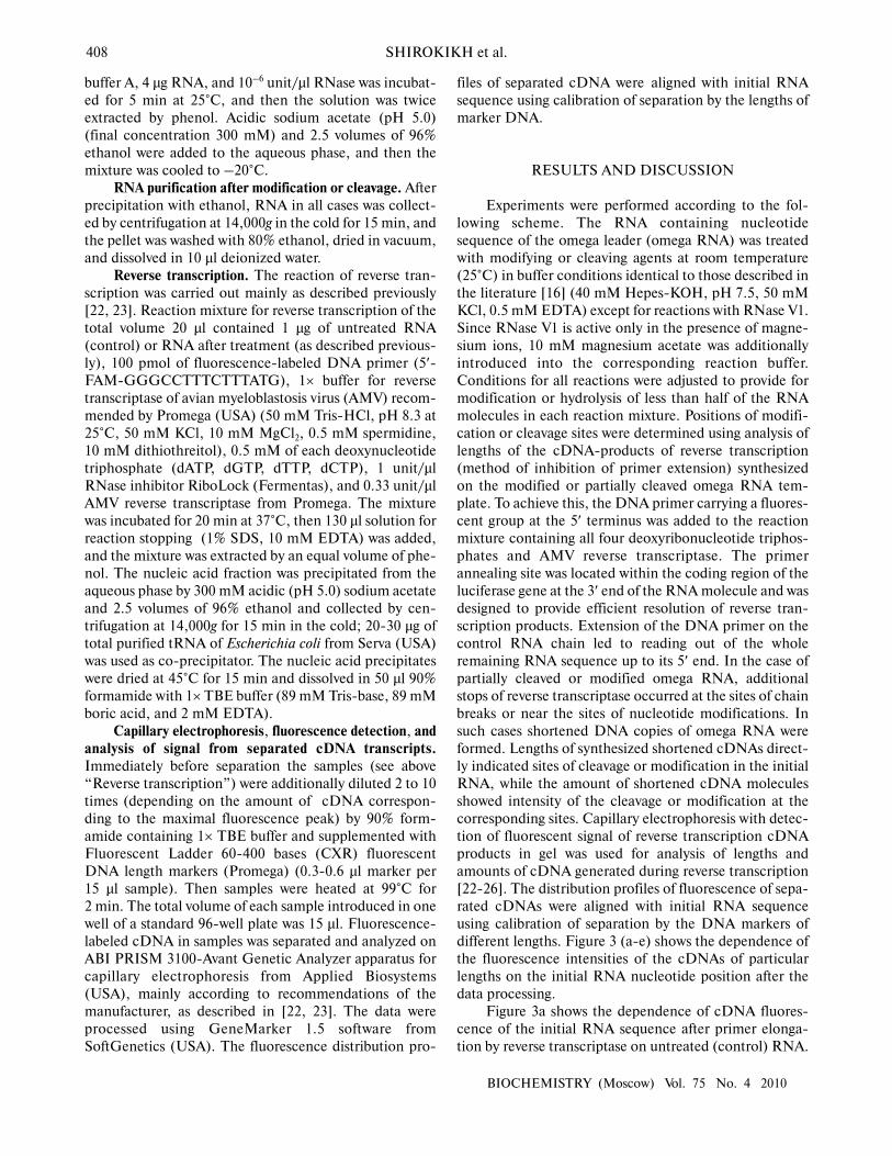
Figure 3a shows the dependence of cDNA fluores-
cence of the initial RNA sequence after primer elonga-
tion by reverse transcriptase on untreated (control) RNA.

CHEMICAL AND ENZYMATIC PROBING OF TMV OMEGA LEADER STRUCTURE 409
BIOCHEMISTRY (Moscow) Vol. 75 No. 4 2010
In this experiment a group of peaks corresponding to for-
mation of the full-size cDNA transcript is prevalent.
There are no additional sites of polymerase stops along
the RNA chain.
Dimethyl sulfate can attack the nitrogen atom in the
first position of adenine or third position of cytosine.
These atoms are necessary for hydrogen bond formation
upon canonical (Watson–Crick) base pairing. Thus,
dimethyl sulfate has to modify RNA only in sites free of
canonical base pairing [27-29]. Numerous additional
sites of reverse transcriptase stops are generated on RNA
in the case of omega RNA modification by dimethyl sul-
fate (Fig. 3b). Modifications are distributed here along
the whole RNA strand and practically all adenines and
cytosines are modified. Thus, there are no extended
regions with Watson–Crick base interactions in the
omega RNA structure.
Diethyl pyrocarbonate can modify the adenine
nitrogen atom in the seventh position if the base is not
involved in stacking interactions, for example, in the case
Fig. 3. Results of chemical modifications and enzymatic cleavages of omega RNA. Control unmodified omega RNA (a) or omega RNA after
modification (b-e; see text) were used for reverse transcription, where fluorescence-labeled DNA primer (FAM) was used. The fluorescence-
labeled cDNA was separated by gel electrophoresis in capillary tubes. Fluorescence profiles of separated cDNAs were read from the gel and
compared with the initial RNA sequence using fluorescent length markers (CXR) added to the samples. The figure shows the cDNA fluores-
cence plotted against the corresponding nucleotide positions of the initial omega RNA. All plots are normalized to total FAM fluorescence in
the gel. The scale on ordinate axis is chosen so that the shortened cDNA formed as a result of reverse transcriptase stopping at the modifica-
tion or cleavage sites were well visualized. The fluorescence peak corresponding to the full-size cDNA (at 5′-end of omega RNA sequence) is
shown incompletely to meet scaling requirements. Numbers near the peak of the full-size cDNA show the amount of full-size transcripts of
omega RNA (free of hydrolysis or modifications) as % of total amount of transcripts from this RNA. The gray area shows the region of omega
RNA containing successive (CAA)n repeats. a) Unmodified (control) omega RNA; b) omega RNA modified by dimethyl sulfate; c) omega
RNA modified by diethyl pyrocarbonate; d) partial cleavage of omega RNA by RNase V1; e) partial cleavage of omega RNA by RNase A.
97%
a
b
c
d
e
Flu
ore
sc
en
ce
, a
rbit
rary
un
its
70%
65%
80%
50%

410 SHIROKIKH et al.
BIOCHEMISTRY (Moscow) Vol. 75 No. 4 2010
of the absence of A-form helical structure of the RNA
[27-32]. The distribution of modification sites along the
RNA after its treatment by diethyl pyrocarbonate is
shown in Fig. 3c. In this case mainly the 3′-proximal part
of the omega RNA is modified. The 5′ region of omega
RNA is relatively weakly modified. This implies that in
the 5′ region the stacking of bases is possible on an
extended part. On the other hand, the region of relatively
low modification includes almost the entire regular
omega RNA part with CAA repeats.
RNase V1 is able to catalyze cleavage of the phos-
phoester bond of any nucleotide residues incorporated in
A-form helices [28, 29, 33]. One can see that upon RNase
V1 cleavage of omega RNA (Fig. 3d), hydrolysis of the
sugar-phosphate chain near the 5′-terminal end of the
omega sequence occurs, and a group of isolated peaks is
formed, which are indicative of chain cleavage in the 3′
part of the molecule. The complete absence of cleavage
sites along the whole length of the central part, contain-
ing regular CAA repeats also confirms the absence of an
A-form RNA helix in this region. At the same time, for-
mation of some short A-form helices, separated by non-
helical RNA regions, is probable in the 3′-proximal part
of the omega sequence.
RNase A mainly cleaves single-stranded RNA in the
sites following pyrimidine nucleotide residues, which are
not incorporated into helices and form no tertiary inter-
actions [34, 35]. The result of omega RNA cleavage by
RNase A is shown in Fig. 3e. The most intensive omega
RNA cleavage occurs in the 3′-proximal third of the
chain rich in uridylic residues. In the 5′-proximal and
central parts of the molecule the amount of RNase A
cleavage sites is relatively low. This part of omega RNA
includes the regular region of the molecule with CAA
repeats. The resistance of 5′-proximal and central parts of
omega RNA to RNase A directly points to existence of
some stable secondary structure in this region, whereas
the 3′-proximal part is evidently single-stranded and
available for the cleavage. It should be noted that diethyl
pyrocarbonate and RNase A affect approximately the
same, 3′-proximal, part of omega RNA polyribonu-
cleotide chain. The 5′-proximal half and central part of
the omega sequence, including the ordered regular region
with CAA repeats, are weakly susceptible to their attack.
Relative structural stability of the central part against
these agents is also accompanied by its complete resist-
ance to RNase V1, cleaving polynucleotide strands with-
in Watson–Crick double helices.
Hence, the experimental data shown in this paper
well agree with the recently proposed model of structural
organization of polyribonucleotides containing consecu-
tive CAA repeats [17]. They confirm that, first, regular
sequence (CAA)n characteristic of the omega leader cen-
tral part is able to generate a stable secondary structure,
and second, the structure formed is not a canonical dou-
ble helix of a Watson–Crick type or anything similar in
the character of base interactions. The model suggests the
folding of RNA regions containing successive (CAA)n
repeats into a relatively stable and compact triple helix.
Data of this work do not contradict the proposed model.
The high sedimentation coefficient of the omega leader
and a higher thermodynamic stability of this RNA, as
compared to the polyribonucleotide consisting of only
regular (CAA)n repeats [16], agree with the previously
published data on enzymatic testing showing a higher,
compared to polyribonucleotide (CAA)n, resistance of
omega RNA to nucleases cleaving single-stranded RNA
[9]. Additional structural stabilization of the omega RNA
region with CAA repeats can be attained by the interac-
tions with 3′-terminal U-rich region of omega RNA. On
the other hand, the described type of triple helices have a
relatively short length [17], which should result in
inevitable structural fluctuations making nitrogen atoms
(N1 of adenine and N3 of cytosine) in the omega RNA
accessible for dimethyl sulfate attack.
The authors are grateful to A. B. Poltaraus for
methodical help, to M. V. Kryuchkov for assistance in
some experiments, and to A. V. Efimov for discussion and
advice during experiments and manuscript preparation.
This work was supported by the Russian Foundation
for Basic Research (grants 06-04-48964-a, 09-04-01729-
a, 09-04-00537-a, and 09-04-01726-a), by the Russian
Federation State Program for Support of Leading
Research Schools NSh-4610.2008.4, and by a grant from
the Cell and Molecular Biology Program of the
Presidium of the Russian Academy of Sciences.
REFERENCES
1. Sleat, D. E., Gallie, D. R., Jefferson, R. A., Bevan, M.
W., Turner, P. C., and Wilson, T. M. (1987) Gene, 60, 217-
225.
2. Gallie, D. R. (2002) Nucleic Acids Res., 30, 3401-3411.
3. Alekhina, O. M., Vassilenko, K. S., and Spirin, A. S. (2007)
Nucleic Acids Res., 35, 6547-6559.
4. Zeyenko, V. V., Ryabova, L. A., Gallie, D. R., and Spirin,
A. S. (1994) FEBS Lett., 354, 271-273.
5. Saejung, W., Fujiyama, K., Takasaki, T., Ito, M., Hori, K.,
Malasit, P., Watanabe, Y., Kurane, I., and Seki, T. (2007)
Vaccine, 25, 6646-6654.
6. Schmitz, J., Prufer, D., Rohde, W., and Tacke, E. (1996)
Nucleic Acids Res., 24, 257-263.
7. Gallie, D. R., Walbot, V., and Hershey, J. W. (1988) Nucleic
Acids Res., 16, 8675-8694.
8. Gallie, D. R., Sleat, D. E., Watts, J. W., Turner, P. C., and
Wilson, T. M. (1988) Nucleic Acids Res., 16, 883-893.
9. Tzareva, N. V., Makhno, V. I., and Boni, I. V. (1994) FEBS
Lett., 337, 189-194.
10. Kopeina, G. S., Afonina, Z. A., Gromova, K. V., Shirokov,
V. A., Vasiliev, V. D., and Spirin, A. S. (2008) Nucleic Acids
Res., 36, 2476-2488.
11. Kukla, B. A., Guilley, H. A., Jonard, G. X., Richards, K.
E., and Mundry, K. W. (1979) Eur. J. Biochem., 98, 61-66.

CHEMICAL AND ENZYMATIC PROBING OF TMV OMEGA LEADER STRUCTURE 411
BIOCHEMISTRY (Moscow) Vol. 75 No. 4 2010
12. Gallie, D. R., and Walbot, V. (1992) Nucleic Acids Res., 20,
4631-4638.
13. Mundry, K. W., Watkins, P. A., Ashfield, T., Plaskitt, K. A.,
Eisele-Walter, S., and Wilson, T. M. (1991) J. Gen. Virol., 72
(Pt. 4), 769-777.
14. Saenger, W. (1984) in Principles of Nucleic Acid Structure,
Springer, New York.
15. Gudkov, A. T., Ozerova, M. V., Shiryaev, V. M., and Spirin,
A. S. (2005) Biotechnol. Bioeng., 91, 468-473.
16. Kovtun, A. A., Shirokikh, N. E., Gudkov, A. T., and Spirin,
A. S. (2007) Biochem. Biophys. Res. Commun., 358, 368-372.
17. Efimov, A. V., and Spirin, A. S. (2009) Biochem. Biophys.
Res. Commun., 388, 127-130.
18. Huntzinger, E., Possedko, M., Winter, F., Moine, H.,
Ehresmann, C., Romby, P., (2008) in Handbook of RNA
Biochemistry (Hartmann, R. K., Bindereif, A., Schon, A.,
and Westhof, E., eds.) Wiley-VCH Verlag, Weinheim, pp.
151-171.
19. Merryman, C., and Noller, H. F. (1998) in RNA:Protein
Interactions, A Practical Approach (Smith, C. W. J., ed.)
Oxford University Press, New York, pp. 237-253.
20. Gurevich, V. V. (1996) Meth. Enzymol., 275, 382-397.
21. Gurevich, V. V., Pokrovskaya, I. D., Obukhova, T. A., and
Zozulya, S. A. (1991) Anal. Biochem., 195, 207-213.
22. Shirokikh, N. E., and Spirin, A. S. (2008) Proc. Natl. Acad.
Sci. USA, 105, 10738-10743.
23. Shirokikh, N. E., Alkalaeva, E. Z., Vassilenko, K. S.,
Afonina, Z. A., Alekhina, O. M., Kisselev, L. L., and
Spirin, A. S. (2009) Nucleic Acids Res., doi:
10.1093/nar/gkp1025.
24. Fekete, R. A., Miller, M. J., and Chattoraj, D. K. (2003)
Biotechniques, 35, 90-98.
25. Yindeeyoungyeon, W., and Schell, M. A. (2000)
Biotechniques, 29, 1034-1041.
26. Gould, P. S., Bird, H., and Easton, A. J. (2005)
Biotechniques, 38, 397-400.
27. Peattie, D. A., and Gilbert, W. (1980) Proc. Natl. Acad. Sci.
USA, 77, 4679-4682.
28. Mandiyan, V., and Boublik, M. (1990) Nucleic Acids Res.,
18, 7055-7062.
29. Mougel, M., Eyermann, F., Westhof, E., Romby, P.,
Expert-Bezancon, A., Ebel, J. P., Ehresmann, B., and
Ehresmann, C. (1987) J. Mol. Biol., 198, 91-107.
30. Maxam, A. M., and Gilbert, W. (1977) Proc. Natl. Acad.
Sci. USA, 74, 560-564.
31. Leonard, N. J., McDonald, J. J., Henderson, R. E., and
Reichmann, M. E. (1971) Biochemistry, 10, 3335-3342.
32. Weeks, K. M., and Crothers, D. M. (1993) Science, 261,
1574-1577.
33. Lockard, R. E., and Kumar, A. (1981) Nucleic Acids Res., 9,
5125-5140.
34. Kop, J., Kopylov, A. M., Magrum, L., Siegel, R., Gupta,
R., Woese, C. R., and Noller, H. F. (1984) J. Biol. Chem.,
259, 15287-15293.
35. Hartshorne, T., and Agabian, N. (1994) Nucleic Acids Res.,
22, 3354-3364.