CHEMICAL ACTINOMETRY - International Union of … Fields of application of chemical actinometry 1.4...
42
2105 Pure Appl. Chem., Vol. 76, No. 12, pp. 2105–2146, 2004. © 2004 IUPAC INTERNATIONAL UNION OF PURE AND APPLIED CHEMISTRY ORGANIC AND BIOMOLECULAR CHEMISTRY DIVISION * SUBCOMMITTEE ON PHOTOCHEMISTRY** CHEMICAL ACTINOMETRY (IUPAC Technical Report) Prepared for publication by H. J. KUHN 1 , S. E. BRASLAVSKY 1,‡ , AND R. SCHMIDT 2 1 Max-Planck-Institut für Bioanorganische Chemie (formerly Strahlenchemie), D−45413 Mülheim a.d. Ruhr, Germany; 2 Institut für Physikalische und Theoretische Chemie, Universität Frankfurt, D−60054 Frankfurt/Main, Germany *Membership of the Organic and Biomolecular Chemistry Division during the final preparation of this report (2002–2003) was as follows: President: T. T. Tidwell (1998–2003), M. Isobe (2002–2005); Vice President: D. StC. Black (1996–2003), V. T. Ivanov (1996–2005); Secretary: G. M. Blackburn (2002–2005); Past President: T. Norin (1996–2003), T. T. Tidwell (1998–2005) (initial date indicates the first time elected as Division member). The list of the other members of the Division can be found in <http://www.iupac.org/divisions/III/members.html>. In addition to the members of the IUPAC Photochemistry Commission (1985–1988), see first version in Pure Appl. Chem. 61, 187–210 (1989), and Subcommittee (2001–2003), the following colleagues kindly supplied literature, special information, helpful comments, and discussions which are gratefully acknowledged: O. M. Alfano, D. Brämert, A. M. Braun, N. J. Bunce, D. De Keukeleire, H. Deinert, J. N. Demas, D. F. Eaton, G. Gauglitz, H. Görner, G. G. Gurzadyan, G. Guyot, H. G. Heller, W. E. Klotzbücher, G. Koç-Weier, D. Kreft, J. J. McCullough, E. Oliveros, P. Potzinger, W. H. Powell, A. Rizzi, R. Schrader, H.-P. Schuchmann, C. von Sonntag, R. Srinivasan, J. Theurich, L. Vincze, J. Wirz, H. E. Zimmerman. Members of the Photochemistry Commission (1998–2001) were: Chairmen: J. R. Bolton (Canada, 1996–1999), R. G. Weiss (USA, 2000–2001); Secretaries: R. G. Weiss (USA, 1998–1999), J. Wirz (Switzerland, 2000–2001); Titular Members: J. R. Bolton (Canada), S. E. Braslavsky (Germany), H. Bouas-Laurent (France), R. G. Weiss (USA), J. Wirz (Switzerland); Associate Members: A. U. Acuña–Fernández (Spain), H. Masuhara (Japan); National Representatives: F. C. De Schryver (Belgium), Y. Eichen (Israel), M. G. Neumann (Brazil), T. Bérczes (Hungary), S. J. Formosinho (Portugal), A. Horvath (Hungary), S. Içli (Turkey), B. S. Martineigh (South Africa), G. Pandey (India), F. H. Quina (Brazil), S. C. Shim (Korea), P. Hrdlovic (Slovakia), C.-H. Tung (China), C. Wentrup (Australia), I. Willner (Israel), Y. Yagci (Turkey). **Members of the Subcommittee on Photochemistry (2001–2003): S. E. Braslavsky (Germany, Chairperson), A. U. Acuña–Fernández (Spain), G. A. Argüello (Argentina), T. D. Z. Atvars (Brazil), R. Bonneau (France), A. M. Braun (Germany), A. Chibisov (Russia), K. Ghiggino (Australia), H. Lemmetyinen (Finland), H. Miyasaka (Japan), D. C. Neckers (USA), J. Niytrai (Hungary), M. Olivucci (Italy), D. Phillips (UK), R. O. Rahn (USA), N. Serpone (Canada), R. Schmehl (USA), M. Terazima (Japan), S. Wu (China). ‡ Corresponding author: E-mail: [email protected] Republication or reproduction of this report or its storage and/or dissemination by electronic means is permitted without the need for formal IUPAC permission on condition that an acknowledgment, with full reference to the source, along with use of the copyright symbol ©, the name IUPAC, and the year of publication, are prominently visible. Publication of a translation into another language is subject to the additional condition of prior approval from the relevant IUPAC National Adhering Organization.
Transcript of CHEMICAL ACTINOMETRY - International Union of … Fields of application of chemical actinometry 1.4...

2105
Pure Appl. Chem., Vol. 76, No. 12, pp. 2105–2146, 2004.© 2004 IUPAC
INTERNATIONAL UNION OF PURE AND APPLIED CHEMISTRYORGANIC AND BIOMOLECULAR CHEMISTRY DIVISION *
SUBCOMMITTEE ON PHOTOCHEMISTRY**
CHEMICAL ACTINOMETRY
(IUPAC Technical Report)
Prepared for publication byH. J. KUHN1, S. E. BRASLAVSKY1,‡, AND R. SCHMIDT2
1Max-Planck-Institut für Bioanorganische Chemie (formerly Strahlenchemie), D−45413 Mülheim a.d.Ruhr, Germany; 2Institut für Physikalische und Theoretische Chemie, Universität Frankfurt, D−60054
Frankfurt/Main, Germany
*Membership of the Organic and Biomolecular Chemistry Division during the final preparation of this report(2002–2003) was as follows:
President: T. T. Tidwell (1998–2003), M. Isobe (2002–2005); Vice President: D. StC. Black (1996–2003), V. T.Ivanov (1996–2005); Secretary: G. M. Blackburn (2002–2005); Past President: T. Norin (1996–2003),T. T. Tidwell (1998–2005) (initial date indicates the first time elected as Division member). The list of the othermembers of the Division can be found in <http://www.iupac.org/divisions/III/members.html>.
In addition to the members of the IUPAC Photochemistry Commission (1985–1988), see first version in Pure Appl.Chem. 61, 187–210 (1989), and Subcommittee (2001–2003), the following colleagues kindly supplied literature,special information, helpful comments, and discussions which are gratefully acknowledged: O. M. Alfano, D.Brämert, A. M. Braun, N. J. Bunce, D. De Keukeleire, H. Deinert, J. N. Demas, D. F. Eaton, G. Gauglitz, H. Görner,G. G. Gurzadyan, G. Guyot, H. G. Heller, W. E. Klotzbücher, G. Koç-Weier, D. Kreft, J. J. McCullough, E.Oliveros, P. Potzinger, W. H. Powell, A. Rizzi, R. Schrader, H.-P. Schuchmann, C. von Sonntag, R. Srinivasan, J.Theurich, L. Vincze, J. Wirz, H. E. Zimmerman.
Members of the Photochemistry Commission (1998–2001) were: Chairmen: J. R. Bolton (Canada, 1996–1999),R. G. Weiss (USA, 2000–2001); Secretaries: R. G. Weiss (USA, 1998–1999), J. Wirz (Switzerland, 2000–2001);Titular Members: J. R. Bolton (Canada), S. E. Braslavsky (Germany), H. Bouas-Laurent (France), R. G. Weiss(USA), J. Wirz (Switzerland); Associate Members: A. U. Acuña–Fernández (Spain), H. Masuhara (Japan);National Representatives: F. C. De Schryver (Belgium), Y. Eichen (Israel), M. G. Neumann (Brazil), T. Bérczes(Hungary), S. J. Formosinho (Portugal), A. Horvath (Hungary), S. Içli (Turkey), B. S. Martineigh (South Africa),G. Pandey (India), F. H. Quina (Brazil), S. C. Shim (Korea), P. Hrdlovic (Slovakia), C.-H. Tung (China), C.Wentrup (Australia), I. Willner (Israel), Y. Yagci (Turkey).
**Members of the Subcommittee on Photochemistry (2001–2003): S. E. Braslavsky (Germany, Chairperson), A.U. Acuña–Fernández (Spain), G. A. Argüello (Argentina), T. D. Z. Atvars (Brazil), R. Bonneau (France), A. M.Braun (Germany), A. Chibisov (Russia), K. Ghiggino (Australia), H. Lemmetyinen (Finland), H. Miyasaka(Japan), D. C. Neckers (USA), J. Niytrai (Hungary), M. Olivucci (Italy), D. Phillips (UK), R. O. Rahn (USA), N.Serpone (Canada), R. Schmehl (USA), M. Terazima (Japan), S. Wu (China).
‡Corresponding author: E-mail: [email protected]
Republication or reproduction of this report or its storage and/or dissemination by electronic means is permitted without theneed for formal IUPAC permission on condition that an acknowledgment, with full reference to the source, along with use of thecopyright symbol ©, the name IUPAC, and the year of publication, are prominently visible. Publication of a translation intoanother language is subject to the additional condition of prior approval from the relevant IUPAC National AdheringOrganization.

Chemical actinometry
(IUPAC Technical Report)
Abstract: This document updates the first version of the IUPAC technical report on“Chemical actinometers” published in Pure Appl. Chem. 61, 187–210 (1989).Since then, some methods have been improved, procedures have been modified,and new substances have been proposed as chemical actinometers. An actinometeris a chemical system or a physical device by which the number of photons in abeam absorbed into the defined space of a chemical reactor can be determined in-tegrally or per time. This compilation includes chemical actinometers for the gas,solid, microheterogeneous, and liquid phases, as well as for the use with pulsedlasers for the measurement of transient absorbances, including the quantum yieldof phototransformation, as well as the literature for each of the actinometers. Theactinometers listed are for the use in the wavelength range from the UV to the redregion of the spectrum. A set of recommended standard procedures is also given.Advantages and disadvantages are discussed regarding the use of chemical actino-meters vs. electronic devices for the measurement of the number of photons ab-sorbed. Procedures for the absolute measurement of incident photon flux by meansof photodiodes are also discussed.
CONTENTS
ABBREVIATIONSQUANTITIES, SYMBOLS, AND UNITS PREFACE 1. GENERAL CONSIDERATIONS ON CHEMICAL ACTINOMETRY
1.1 Pros and cons of chemical actinometry1.2 Quality marks of a chemical actinometer1.3 Fields of application of chemical actinometry1.4 Potential errors in chemical actinometry
1.4.1 Refractive index1.4.2 Temperature1.4.3 Absorption by photoproducts1.4.4 Degree of absorption by the chemical actinometer1.4.5 Polychromatic quantum counters
2. LIST OF ACTINOMETERS2.1 Chemical systems
2.1.1 Solid- and microheterogeneous-phase chemical actinometers2.1.2 Gas-phase chemical actinometers2.1.3 Liquid-phase chemical actinometers
2.2 Electronic devices2.3 Absolute measurement of incident photon flux by means of photodiodes
3. STANDARD LIQUID-PHASE ACTINOMETRIC PROCEDURESAPPENDIX
H. J. KUHN et al.
© 2004 IUPAC, Pure and Applied Chemistry 76, 2105–2146
2106

ABBREVIATIONS
AM analytical method CA chemical actinometer E electronic deviceG gas phase GC gas chromatographyHPLC high-pressure liquid chromatographyIR infraredL liquid phaseLC liquid chromatographyPP procedure, precautions, commentsS solid phaseWR wavelength range
QUANTITIES, SYMBOLS, AND UNITS
Name Symbol Definition SI units Notes
absorbance (decadic) A, A10 A = lg(P0/P) = –lg T 1 1absorption coefficientlinear decadic a a = A10/l m–1 2molar decadic ε ε = a/c = A10/cl m2 mol–1 3absorption cross-section σ σ = (ε/NA)ln 10 m2 4actinometric factor See sensitivityamount of photons np mol 5amount concentration c c = n/V mol m–3 6area S m2
Avogadro constant NA NA = 6.022 141 5 × 10²³ mol–1 mol–1 7electric current i Afluence H ' H' = ∫E' dt J m–2 8frequency (linear) ν ν = c/λ Hzirradiance, intensity E E = dP/dS W m–2
(radiant flux received)lifetime τ c(t = τ) = c(t = 0)/e soptical pathlength l mphoton fluence Hp' Hp' = dNp/dS m–2 9photon fluence rate Ep' Ep' = dHp/dt m–2 s–1 9photon flux, amount basis qn,p qn,p = qp/NA mol s–1 10
photon flux, number basis qp dNp/dt s–1 9, 11photon irradiance Ep Ep = dqp/dS m–2 s–1 9, 12photon number Np Np = np NA 1pressure p Paquantum yield Φ Φ = (number of events)/ 1
(number of photons absorbed)of charge carrier formation Φcin a photodiode radiant energy Q ∫λ Qλ dλ Jradiant exposure See fluenceradiant intensity I I = dP/dΩ W sr–1
radiant power, radiant energy P P = dQ/dt W 13per time
© 2004 IUPAC, Pure and Applied Chemistry 76, 2105–2146
Chemical actinometry 2107
(continues on next page)

radiative lifetime τ0 τ0 = 1/kr srate constant for radiative step kr s–1
refractive index n n = c0/c 1sensitivityof an actinometer Sac Sac(λ) = Φ (λ) ε(λobs) m2 mol–1 14
or Sac(λ) = Φ (λ) ∆ε (λobs)of a photodiode Spd Spd = Ipd /P A W–1 15
= V–1
solid angle Ω Ω = S/r2 sr, 1 16spectral fluence (in terms H'λ H'λ = dH'/dλ J m–3 17of wavelength) speed of lightin vacuum c0 c0 = 299 792 458 m s–1 m s–1
in a medium c c = c0/n m s–1
temperature, Celsius θ θ/°C = T /K – 273.15 °Ctemperature, thermodynamic T Ktime t stransmittance T T = P/P0 1 1volume V m3
wavelength λ mwavenumber ν∼ ν∼ = ν/c0 = 1/n λ m–1
Notes
1. If losses from reflection, scattering, and luminescence are neglected, T = P/P0 = I/I0, where superscript 0 in-dicates incident radiant energy and no superscript transmitted radiant energy.
2. In spectroscopy, usually defined in terms of the spectral radiant power, Pλ = dP(λ)/dλ.3. Numerical values are often quoted in mol–1 dm3 cm–1. Note the lack of compactness in using two submul-
tiples of length. 4. In spectroscopy, the net cross-section resulting from the sum of effects due to absorption and induced emis-
sion. A conversion equation in common units is σ/cm2 = (3.823 6 × 10–21/mol) × (ε/dm3 cm–1 mol–1).5. Amount of photons is often given in the non-SI unit einstein = mol.6. The usual units are mol dm–3 or mol l–1 or submultiples. Commonly, the non-SI unit M (small cap) is used
as an abbreviation for mol dm–3.7. 2002 value [1].8. For a parallel and normally incident beam, a synonym is radiant exposure. When applied to the total radi-
ant energy incident from all directions, the symbol H′ is used.9. These quantities, defined on a number basis, can be expressed on a chemical amount basis by dividing by
the Avogadro constant, e.g., photon flux (chemical amount basis) = qp/NA. If distinction needs to be madebetween quantities based on chemical amount and number, then symbols such as qn,p and qp can be used.
10. A common unit is einstein s–1. A term not in accordance with the usual definition of flux [2]. 11. As defined in [4] (see also [5]); called photon flux in [3], a term not in accordance with the usual definition
of flux [2]. 12. Equivalent to photon fluence rate for a parallel beam not scattered or reflected.13. Definition from [2]; synonymous with radiant energy power, radiant energy flux defined in [3].14. λ is the excitation wavelength, and λobs is the observation wavelength which may be the same as or differ
from the former. The first definition corresponds to the case in which the actinometer does not absorb at λobs,whereas the second definition corresponds to the case in which the actinometer absorbs at λobs. Commonunits are dm3 mol–1 cm–1.
15. Related to qn,p by qn,p = [λ/(NA h c Spd)] ipd with ipd the electric current of the photodiode.16. The stearadian is an SI supplementary unit, but is dimensionless, so has SI unit 1.
H. J. KUHN et al.
© 2004 IUPAC, Pure and Applied Chemistry 76, 2105–2146
2108
Table (Continued).
Name Symbol Definition SI units Notes

17. Other physical quantities X such as irradiance, photon flux, photon fluence, photon fluence rate, and radiantintensity may be used to derive the corresponding “spectral” quantity (relative to wavelength) by Xλ = dX/dλ.Analogous quantities relative to frequency or to wavenumber may also be defined.
Entries in the table are consistent with terminology, symbols, and units given in [2,3], and are slightly modifiedfrom those in [4], terms from which are included in [5].
References for Table 1:
1. <http://physics.nist.gov/constants>2. IUPAC. Quantities, Units and Symbols in Physical Chemistry (the ‘Green Book’), 2nd ed., pre-
pared for publication by I. Mills, T. Cvitaš, K. Homann, N. Kallay, K. Kuchitsu, BlackwellScience, Oxford, UK (1993).
3. ISO 31–6:1992(E), Handbook on Quantities and Units, Part 6: Light and related electromagneticradiations, ISO, Geneva, Switzerland (1992).
4. IUPAC Organic Chemistry Division, Commission on Photochemistry. Glossary of Terms Used inPhotochemistry (2nd ed.), prepared for publication by S. E. Braslavsky, K. N. Houk (1st ed.), J. W.Verhoeven (2nd ed.). Pure Appl. Chem. 60, 1055 (1996).
5. IUPAC. Compendium of Chemical Terminology, IUPAC Recommendations, 2nd ed., compiled byA. D. McNaught and A. Wilkinson, Blackwell Science, Oxford, UK (1997).
PREFACE
The first version of this document appeared in Pure Appl. Chem. 61, 187–210 (1989). Since then, somemethods have been improved, and some procedures have been modified. The PhotochemistryCommission (1998–2001) and subsequently the Subcommittee on Photochemistry (from 2001) decidedto update and expand the document. In particular, the sections on gas-phase and solid-state actinometershave been expanded. The terms related to radiation quantities have been made consistent with theInternational Organization for Standardization (ISO) recommendations, and a list of terms used is in-cluded. Some actinometers have been added, whereas others have been deleted from the list with recom-mended procedures for their use. The latter has been due to various reasons. In some cases, the actinom-eter reagent is no longer commercially available, and in other cases, the complexity of the procedure forthe use or disposal of the waste does not justify the inclusion of the actinometer in a recommended list.
According to the “Glossary of terms used in photochemistry” (IUPAC Recommendations 1996,see below), an actinometer is a chemical system or a physical device by which the number of photons ina beam absorbed into the defined space of a chemical reactor can be determined integrally or per time.
A chemical actinometer or dosimeter is a chemical system (fluid, gas, solid, or in a micro-heterogeneous environment) that undergoes a light-induced reaction (at a certain wavelength, λ) forwhich the quantum yield, Φ(λ), is accurately known. Measuring the reaction rate allows the calculationof the absorbed photon flux. The incident photon flux qp
o is calculated from the relationqp(abs, λ) = qp
o(λ) (1–10–A(λ)), provided that the decadic absorbance A(λ) is constant ±10 % duringthe irradiation time. Should this not be the case, integration of the differential absorbance over timewould be necessary. The easiest case is for qp(abs, λ) = qo(λ) for total absorption during the whole ir-radiation period. Determination of conversion to the products affords the total number of photons ab-sorbed by the liquid or gas volume or solid surface, which may have any form or geometry.
The quantum yield of a photochemical reaction is defined as Φ(λ) = the number of events, e.g.,molecules changed, formed, or destroyed, divided by the number of absorbed photons of that particu-lar wavelength in the same period of time.
Calibration of an actinometer is done by applying a calibration lamp or by absolute measurementof irradiance (using, e.g., a calibrated radiometer, a calorimeter, or a photodiode). Photothermal meth-ods are often used to calibrate actinometers in absolute terms.
© 2004 IUPAC, Pure and Applied Chemistry 76, 2105–2146
Chemical actinometry 2109

Absolute measurement of incident irradiance, E/W m–2 or photon irradiance, Ep/s–1 m–2 or
Εn,p/einstein s–1 m–2 or photon flux, qn,p/einstein s
–1, using precalibrated photodiodes is described inSection 2.3 of this document.
For details and references concerning radiometry and actinometry, principles of photochemistry,light sources, and reactors, as well as possible sources of error when performing actinometric meas-urements, read Section 1 of this document and consult, e.g.,
• A. M. Braun, M.-T. Maurette, E. Oliveros. Photochemical Technology, John Wiley, New York(1991); idem, Technologie Photochimique, Presses Polytechniques Romandes, Lausanne (1986);CRC Handbook of Organic Photochemistry, J. C. Scaiano (Ed.), 2 vols., CRC Press, Boca Raton(1989).
• S. L. Murov, I. Carmichael, G. L. Hug. Handbook of Photochemistry, 2nd ed., Marcel Dekker,New York (1993).
For the most recent version of “Glossary of terms and definitions used in photochemistry”, refer to:
• J. W. Verhoeven. Pure Appl. Chem. 68, 2223–2286 (1996); available at <http://www.unibas.ch/epa/>. A new version is in preparation.
1. GENERAL CONSIDERATIONS ON CHEMICAL ACTINOMETRY
1.1 Pros and cons of chemical actinometry
In a chemical actinometer (CA), photochemical conversion is directly related to the number of photonsabsorbed because the chemical action of light means reversible or irreversible chemical change, i.e., de-struction or buildup of molecules and, consequently, of their properties such as spectra. Chemical acti-nometry has been employed for about 70 years in photochemistry as a relatively simple and accuratemethod for radiation measurement [2].
Owing to the recent progress in the development of radiation detectors, semiconductors, and elec-tronic equipment, physical devices furnished with a direct readout become more and more popularamong photochemists for the measurement of radiation. Physical devices are often preferred to CAs forthe case of simple irradiation geometries because of their easy, fast, and precise performance.
However, these outstanding properties are inherent in only a small number of electrically cali-brated radiometers (ECRs) available in a few highly equipped laboratories. ECRs are special ther-mopiles [3] or piezoelectric radiometers [4,5], which can be calibrated in an absolute manner by elec-trical substitution without the need of any standard. The majority of physical detectors, like usualthermopiles, piezoelectric joulemeters, or photodiodes are only secondary standards, the response ofwhich can be subject to changes.
The sensitivity of a joulemeter may decrease with use due to surface damage by high-power ra-diation. The same is valid for thermopiles. The spectral sensitivity of the widespread silicon photodi-odes is even altered without use, just by aging. The extent of this effect depends on the wavelengthrange in which the detector will be used.
An 18 % decrease in sensitivity at 300 nm in one year was reported to be a typical value [6].Visually unnoticeable damage of photodiodes occurs during exposure to high irradiation levels (gener-ally more than 10 mW cm–2 for silicon photodiodes in continuous wave experiments) resulting in an ir-reversible decrease of sensitivity and severe inhomogeneities in the surface. Consequently, occasionalrecalibration of radiation detectors against a standard is strongly recommended.
In contrast to the physical detectors, well-established CAs lead to accurate absolute radiationmeasurements, provided that they are employed according to the recommended procedures. These CAshave been proven reproducible and do not demand any recalibration.
H. J. KUHN et al.
© 2004 IUPAC, Pure and Applied Chemistry 76, 2105–2146
2110

1.2 Quality marks of a chemical actinometer
An established CA should meet the following requirements:
• The photochemical system should be simple and well studied. The photoreaction must be repro-ducible under well-defined and easily controllable experimental conditions. Quantum yieldsshould be accurately known for a large number of wavelengths. A wide usable spectral range andwavelength-independent quantum yields are desired.
• The chemical components should be thermally stable to exclude complications due to dark reac-tions.
• The analytical methods should be simple. Direct spectrophotometric analysis is preferred.• The system should display large sensitivity.• The handling of the photochemical system and the evaluation of the number of photons absorbed
should be simple and straightforward.• The actinometric material should be easy to synthesize and purify. Preferably, it should be com-
mercially available. Disposal of the waste should be straightforward. • The CAs offered in the present list meet the requirements mentioned above in various degrees.
Each system suffers also from disadvantages, and a careful selection among the CAs is appropri-ate, depending on the intended experiment. Reading the original literature on each actinome-ter prior to its use is highly recommended.
1.3 Fields of application of chemical actinometry
It is important to mention that chemical actinometry covers only the wavelength range up to 795 nm.In photochemical experiments involving a complex irradiation geometry (e.g., photoreactors of
the merry-go-round type), CAs serve best for the purpose of absolute radiation measurement and areunrivalled by physical devices. In any case, in photobiological and photochemical laboratories with lesssophisticated equipment but where workers are experienced in chemical techniques, chemical actino-metry is the standard procedure for radiation measurement.
Photochemists using physical equipment for radiation measurements need standards for the oc-casional recalibration of their detectors. CAs are the first choice for this procedure. For detectors withwavelength-independent response like thermopiles and joulemeters, the calibration by CAs is particu-larly easy since calibration at only one wavelength is sufficient.
Measurements of laser pulse energies can conveniently be done by joulemeters. At high laserpowers, most CAs will probably lose accuracy and sensitivity due to multiple photon processes occur-ring at high photon densities. However, if linearity of the joulemeter readout is guaranteed, a periodi-cally repeated calibration by CAs at reduced laser power is an easy way of controlling the accuracy ofthe power meter. Those CAs that have been investigated especially at high photon fluxes using laser ex-citation can be used as standards for this purpose [7].
1.4 Potential errors in chemical actinometry
The detailed description of the chemical system and the limitations and possible sources of errors foreach CA should be made explicit in every publication. In the following section, only general aspects arediscussed.
1.4.1 Refractive indexIn this paragraph, the discussion will be restricted to the case of monochromatic irradiation. Only in thiscase, a CA yields the radiant power P entering the sample cell, since a CA yields a photon flux, num-ber basis, qp/s
–1, or photon flux, amount basis, qn,p/einstein s–1, entering the sample cell at a given
wavelength. In the case of polychromatic irradiation, conversion of photon flux to radiant power needs
© 2004 IUPAC, Pure and Applied Chemistry 76, 2105–2146
Chemical actinometry 2111
not only the knowledge of quantum yields and absorbances of the CA in the wavelength range consid-ered, but also the spectral distribution of the irradiation source.
A correction for the reflectance R should be performed in order to obtain the radiant power of theirradiation beam Pb, i.e., Pb = P/(1 – R).
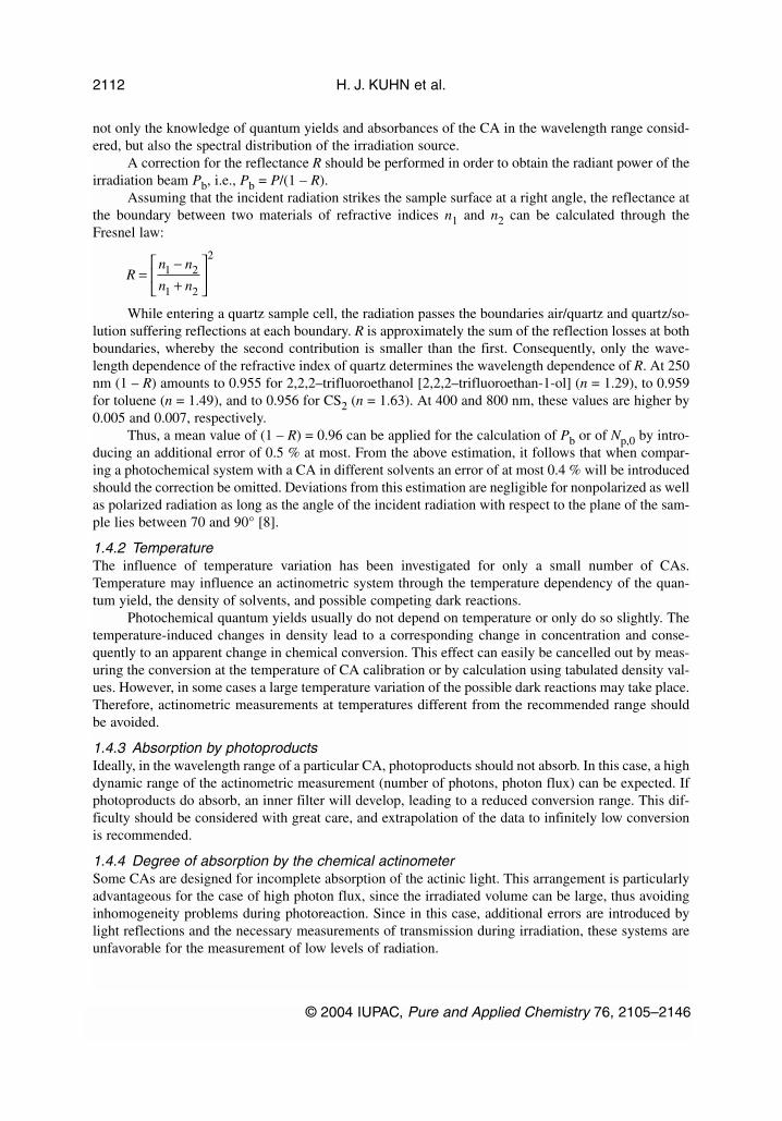
Assuming that the incident radiation strikes the sample surface at a right angle, the reflectance atthe boundary between two materials of refractive indices n1 and n2 can be calculated through theFresnel law:
While entering a quartz sample cell, the radiation passes the boundaries air/quartz and quartz/so-lution suffering reflections at each boundary. R is approximately the sum of the reflection losses at bothboundaries, whereby the second contribution is smaller than the first. Consequently, only the wave-length dependence of the refractive index of quartz determines the wavelength dependence of R. At 250nm (1 – R) amounts to 0.955 for 2,2,2–trifluoroethanol [2,2,2–trifluoroethan-1-ol] (n = 1.29), to 0.959for toluene (n = 1.49), and to 0.956 for CS2 (n = 1.63). At 400 and 800 nm, these values are higher by0.005 and 0.007, respectively.
Thus, a mean value of (1 – R) = 0.96 can be applied for the calculation of Pb or of Np,0 by intro-ducing an additional error of 0.5 % at most. From the above estimation, it follows that when compar-ing a photochemical system with a CA in different solvents an error of at most 0.4 % will be introducedshould the correction be omitted. Deviations from this estimation are negligible for nonpolarized as wellas polarized radiation as long as the angle of the incident radiation with respect to the plane of the sam-ple lies between 70 and 90° [8].
1.4.2 Temperature The influence of temperature variation has been investigated for only a small number of CAs.Temperature may influence an actinometric system through the temperature dependency of the quan-tum yield, the density of solvents, and possible competing dark reactions.
Photochemical quantum yields usually do not depend on temperature or only do so slightly. Thetemperature-induced changes in density lead to a corresponding change in concentration and conse-quently to an apparent change in chemical conversion. This effect can easily be cancelled out by meas-uring the conversion at the temperature of CA calibration or by calculation using tabulated density val-ues. However, in some cases a large temperature variation of the possible dark reactions may take place.Therefore, actinometric measurements at temperatures different from the recommended range shouldbe avoided.
1.4.3 Absorption by photoproductsIdeally, in the wavelength range of a particular CA, photoproducts should not absorb. In this case, a highdynamic range of the actinometric measurement (number of photons, photon flux) can be expected. Ifphotoproducts do absorb, an inner filter will develop, leading to a reduced conversion range. This dif-ficulty should be considered with great care, and extrapolation of the data to infinitely low conversionis recommended.
1.4.4 Degree of absorption by the chemical actinometerSome CAs are designed for incomplete absorption of the actinic light. This arrangement is particularlyadvantageous for the case of high photon flux, since the irradiated volume can be large, thus avoidinginhomogeneity problems during photoreaction. Since in this case, additional errors are introduced bylight reflections and the necessary measurements of transmission during irradiation, these systems areunfavorable for the measurement of low levels of radiation.
H. J. KUHN et al.
© 2004 IUPAC, Pure and Applied Chemistry 76, 2105–2146
2112
Rn n
n n=
−+
1 2
1 2
2

CAs exhibiting complete absorption of the radiation allow convenient measurement and easyevaluation and are thus preferred. At high absorbance, the penetration depth of irradiation is small.Therefore, at the high photon flux met (e.g., in laser beams), severe inhomogeneity problems arise,which cannot be overcome even by effective stirring. This possible source of error is insufficiently dis-cussed in the literature. Since at high absorbances the photoreaction occurs only in a very small reac-tion volume, multiple photonic processes can be expected at high photon flux as often reported [7].
1.4.5 Polychromatic quantum counters CAs with wavelength-independent quantum yields allow polychromatic quantum counting in the spec-ified wavelength range provided complete absorption is maintained. In this case, each photon enteringthe actinometric solution causes chemical conversion with the same probability regardless of its energy.Polychromatic quantum counters are particularly accurate due to their wavelength-independent conver-sion yield.
References to Section 1:1. J. W. Verhoeven. Pure Appl. Chem. 68, 2223–2286 (1996).2. W. G. Leighton and G. S. Forbes. J. Am. Chem. Soc. 52, 3139–3152 (1930).3. K. Bischoff. Optik 28, 183–189 (1968/69).4. R. J. Phelan, Jr. and A. R. Cook. Appl. Opt. 12, 2494–2500 (1973). 5. J. Geist and W. R. Blevin. Appl. Opt. 12, 2532–2535 (1973). 6. J. L. Gardner and F. J. Wilkinson. Appl. Opt. 24, 1531–1534 (1985).7. J. N. Demas, W. D. Bowman, E. F. Zalewski, R. A. Velapoldi. J. Phys. Chem. 85, 2766–2771
(1981).8. B. M. Jaworski and A. A. Detlaf. Physik griffbereit, p. 567, Vieweg und Sohn, Braunschweig
(1972).
2. LIST OF ACTINOMETERS
The present compilation lists chemical systems that have been shown suitable for the integration of in-cident light by chemical conversion. Emphasis is mainly on gas- and liquid-phase systems which photo-chemists are most frequently dealing with. In addition, some solid-phase actinometers and convenientelectronic devices are mentioned. Actinometric systems or procedures marked by an asterisk (* = wellestablished) in the following list have been used over years by different authors in several laboratoriesand can thus be recommended (not necessarily, however, without precautions). All other systems ap-pear to be not yet widely used and may be labelled under discussion. Systems that have fallen into dis-credit are given at the bottom of this list under the heading Disproved.
Note carefully that names of chemicals are given usually as common names that appear inthe relevant publications. Systematic IUPAC names are given in square brackets where applica-ble.
Readers are encouraged to send complementary information, corrections, and comments.
2.1 Chemical systems
Several physical methods for the calibration of light sources (see Sections 1, 2.2, and 2.3) are known.With chemical systems, however, it is in general easier to mimic the experimental situation of the sam-ple. By choosing strictly identical experimental set-ups for standard and sample, experimental errorsdue to differences in shape, surface, and spatial arrangement of the reaction vessel, filters, lenses, etc.can be avoided without much effort. The same solvent and equal absorbances should be chosen foractinometry and reaction under study, whenever possible. Corrections due to reflection losses at sur-faces should be taken into account (see Section 1.4.1). A recommended alternative, especially for low
© 2004 IUPAC, Pure and Applied Chemistry 76, 2105–2146
Chemical actinometry 2113
photon densities at which multiphoton processes do not play a role, is to use a totally absorbing acti-nometer solution (see Section 1.4.4).
Order in the following lists is with increasing wavelength range. See the Abbreviations at the be-ginning of the document.
2.1.1 Solid- and microheterogeneous-phase chemical actinometers For solid-state quantum yield determination procedures cf., e.g.: Y. Ito and T. Matsuura. J. Photochem.Photobiol. A 50, 141–145 (1989); Tetrahedron Lett. 29, 3087–3090, 3091–3094 (1988); H. E.Zimmerman and M. J. Zuraw. J. Am. Chem. Soc. 111, 7974–7989 (1989).
For quantitative measurements in sol-gel materials, see: D. Levy. Chem. Mater. 9, 2666–2670(1997).
The references given below in the area of solid-state chemical and biological dosimetry are notmeant to cover this vast field as a whole but to give just some selected entry keys.
S01: Uracil photodimerization in polycrystalline thin layerBiological UV dosimeterWR: >250 nmAM: absorption spectrum 250–400 nmPhage T7 used as a calibration system1. G. J. Fisher and H. Johns. In Photochemistry and Photobiology of Nucleic Acids, Vol. 1, S. Y.
Wang (Ed.), Chap. 5, pp. 225–294, Academic Press, New York (1976).2. P. Grof, S. Gaspar, G. Rontó. Photochem. Photobiol. 64, 800–806 (1996).
S02: DNA photodamaging, e.g., on nylon membrane sealed in a polyethylene envelopeBiological solar UV dosimetersWR: 254–330 nmAM: immunostaining with damage-specific monoclonal antibody [1]1. Y. Ishigaki, A. Takayama, S. Yamashita, O. Nikaido. J. Photochem. Photobiol., B 50, 184–188
(1999), and refs. therein; some related “living dosimeters”:2. Biofilm of Bacilus subtilis spores, L. E. Quintern, G. Horneck, U. Eschweiler, H. Bücker.
Photochem. Photobiol. 55, 389–395 (1992).3. Special Escherichia coli bacteria strain K12 AB2480 (uvrA– recA–) with photodamage repair de-
ficiency (193, 254 nm), G. G. Gurzadyan, H. Görner, D. Schulte–Frohlinde. Rad. Research 141,244–251 (1995) and refs. therein, especially by P. Howard-Flanders et al.
4. cf., e.g., Special issue on biological dosimetry of UV radiation, A. Fekete and G. Rontó (Eds.). J.Photochem. Photobiol., B 53 (1999).
S03: 1-Ethyl-7-methyl-4-oxo-1,4-dihydro-1,8-naphthyridine-3-carboxylic acid (nalidixic acid)UV-A polyvinyl chloride film dosimeterWR: 280–360 nm AM: absorbance change at 330 nm PP: very preliminary batch-dependent data1. T. J. Tate, B. L. Diffey, A. Davis. Photochem. Photobiol. 31, 27–30 (1980). 2. N. K. Gibbs and A. R. Young. Photochem. Photobiol. 37, 345–348 (1983).
H. J. KUHN et al.
© 2004 IUPAC, Pure and Applied Chemistry 76, 2105–2146
2114

S04: Thymine dimerizationUV-B radiation dosimeter in SDS microemulsionsWR: 290–320 nm; Φ = 10–3–10–1 depending on [Thymine] and [SDS]/[H2O] ratioA TiO2-sol-based actinometer used to determine number of photons absorbedAM: decrease of thymine absorption at 265 nm1. Y. S. Che, J. S. Huang, W. Barnard, Y. H. Li. Analyt. Chim. Acta 318, 103–112 (1995).
S05: Azoxybenzene → 2-hydroxyazobenzene, cf. L22WR: 300–400 nm; poly(methyl methacrylate) block; Φ ~ 10–3
AM: absorbance at 420 nm 1. N. J. Bunce and J. J. Smith. J. Photochem. 23, 219–231 (1983).2. N. J. Bunce, G. G. Debrabandere, K. B. Jacobs, M. E. Lemke, C. R. Montgomery, J. S. Nakai,
E. J. Stewart. J. Photochem. 34, 105–115 (1986).
S06: o-Nitrobenzaldehyde → o-nitrosobenzoic acid photoisomerization WR: 310–400 nm; KBr pellet; Φ = 0.5 (same in all phases)WR: 280–410 nm; poly(methyl methacrylate); Φ = 0.5 ± 0.06AM: IR 1530 cm–1 (NO2 band disappearance)
1. J. N. Pitts, Jr., J. K. S. Wan, E. A. Schuck. J. Am. Chem. Soc. 86, 3606–3610 (1964).2. J. N. Pitts, Jr., L. D. Hess, E. J. Baum, E. A. Schuck, J. K. S. Wan, P. A. Leermakers, G. Vesley.
Photochem. Photobiol. 4, 305–321 (1965). 3. G. W. Cowell and J. N. Pitts, Jr. J. Am. Chem. Soc. 90, 1106–1110 (1968).4. P. Leighton and F. A. Lucy. J. Chem. Phys. 2, 756–759 (1934).5. C. B. Roy and S. C. Das. Ind. J. Chem. 14A, 653–655 (1976).6. E. M. Fleischmann. Limnol. Oceanogr. 34, 1623–1629 (1989); for the measurement and penetra-
tion of UV radiation into marine water (see also L35, [5]).7. Also applicable in gas and liquid phase, cf. e.g., J. M. Allen, S. A. Allen, J. Dreiman, S. W.
Baertschi. Photochem. Photobiol. 69, 17S–18S (1999).
S07: Polysulfone UV-B film dosimeter WR: ≤315 nm AM: absorbance change at 300 nm 1. A. Davis, B. L. Diffey, T. K. Tate. Photochem. Photobiol. 34, 283–286 (1981); ∆A(300 nm) math-
ematically correlates with the UV (300 nm) radiant exposure H/J m–2. Irradiance was measuredusing a vacuum thermopile.
2. B. L. Diffey. Photochem. Photobiol. 46, 55–60 (1987); Quantitative erythemal effectiveness givenvs. λ; comparison of radiometric with actinometric measurements.
3. Some related UV-A and B dosimeter systems: polycarbonate plastic, C. F. Wong, R. Fleming, S.J. Carter. Photochem. Photobiol. 50, 611–615 (1989).
4. Fuchsin and sandolan yellow dyes in polyvinyl alcohol polymer, F. A. Rehim, A. S. A. Gawad,A. A. A. Fattah. J. Photochem. Photobiol., A 64, 123–131 (1992).
S08: Diels–Alder adduct of 2,5-dimethylbenzoquinone and cyclopentadiene [4a,7-dimethyl-1,4,4a,8a-tetrahydro-1,4-methanonaphthalene-5,8-dione] photoisomerization in a dry silica matrix Irradiation apparatus described for adsorbed substances WR: 362 ± 5 nm; Φ = 1.0 (vs. ferrioxalate, L31*; starting material disappearance and product appear-ance; also in solution) AM: absorbance, GC 1. S. Lazare, P. de Mayo, W. R. Ware. Photochem. Photobiol. 34, 187–190 (1981).
© 2004 IUPAC, Pure and Applied Chemistry 76, 2105–2146
Chemical actinometry 2115

S09: Quinonaphthalone [2-(2-quinolyl)indane-1,3-dione] photofading Plastic plates WR: 366–436 nm; Φ = 2 × 10⁻⁵
AM: absorbance decrease at 420 nm 1. H. Okabe. Appl. Opt. 20, 4054–4058 (1981).
2.1.2 Gas-phase chemical actinometersFor reviews on UV and vacuum UV actinometers, cf. A. M. Pravilov. “Gas-phase actinometry for UVand vacuum UV spectral regions”, High Energy Chem. 21, 243–255 (1987) [Russian: Khim. Vys. Energ.21, 291–304 (1987)] and J. Bercowitz. “The quantum yield of ionization”, Physics Essays 13, 248–255(2000).
G01: Rare gases photoionization WR: 4.4–102.2 nm, Φ(ion) = 1 at infinitely low pressureAM: He or Ne ion current measurement, extrapolation to zero pressure to correct for secondary ioniza-tion [1]1. J. A. R. Samson and G. N. Haddad. J. Opt. Soc. Am. 64, 47–54 (1974). 2. T. Saito and H. Onuki. Metrologia 32, 525–529 (1996).3. J. Bercowitz. Physics Essays 13, 248–255 (2000).
G02: Nitrogen monoxide (NO) photoionization WR: ≤134 nm, Φ(λ, ion) vary with λ, from 0.66 to 0.98 in the 58–75 nm region and are 0.3–0.6 in the75–105 nm region [1]AM: ion yield by photocurrent measurement, absolute photon fluences measured with a calibratedthermocouple1. K. Watanabe, F. M. Matsunaga, H. Sakai. Appl. Opt. 5, 391–396 (1967). 2. J. Bercowitz. Physics Essays 13, 248–255 (2000).
PP: The use of the traditional name nitric oxide is not recommended by IUPAC.
G03*: Dinitrogen oxide (N2O) photolysisWR: 147–185 (138–210) nm; Φ(∆n) = Φ(Ν2) + Φ(Ο2) + Φ(NO) – Φ(Ν2Ο) = 1.00 ± 0.05; Φ(Ν2) = 1.44± 0.1 at moderate pressure near room temperature; Φ(Ν2) = 1.18 at 123.6 nm [3]; Φ(∆n) ~ 0.8 at105–120 nm [3]AM: N2 analysis, pressure measurement PP: low absorbance at 150–170 nm (ε ≤ 30 dm3 mol–1 cm–1); separation of N2 from NO and O2 or veryaccurate pressure measurement The use of the traditional name nitrous oxide is not recommended by IUPAC.1. cf. J. G. Calvert and J. N. Pitts, Jr. Photochemistry, p. 782, John Wiley, New York (1966). 2. M. Zelikoff and L. M. Aschenbrand. J. Chem. Phys. 22, 1685–1687 (1954). 3. W. E. Groth and H. Schierholz. Planet. Space Sci. 1, 333–336 (1959). 4. N. R. Greiner. J. Chem. Phys. 47, 4373–4377 (1967).5. A. M. Pravilov and I. O. Shul’pyakov. High Energy Chem. 19, 351–354 (1985). 6. M. C. Dodge and J. Heicklen. Int. J. Chem. Kinet. 3, 269–282 (1971).
G04: Oxygen photolysis WR: 130–190 nm; Φ(O3) = 2.0 at low conversions and fast flow systemsAM: absorbance at 353 nm of iodine–iodide complex (formed from the produced ozone bubbledthrough a KI solution)1. cf. J. G. Calvert and J. N. Pitts, Jr. Photochemistry, p. 782, John Wiley, New York (1966).2. J. N. Driscoll and P. Warneck. Photochem. Photobiol. 13, 283–287 (1971).
H. J. KUHN et al.
© 2004 IUPAC, Pure and Applied Chemistry 76, 2105–2146
2116

3. G. R. Powell and D. S. Sethi. Int. J. Chem. Kinet. 10, 1161–1166 (1978). 4. A. K. Davies, K. A. Khan, J. F. McKellar, G. O. Phillips. Mol. Photochem. 7, 389–398 (1976).
G05: Hexafluoroacetone photolysis WR: 147 nm; Φ(CO) = 0.97 ± 0.05 AM: CO 1. J. J. Magenheimer and R. B. Timmons. J. Chem. Phys. 52, 2790–2791 (1970).2. G. R. Powell and D. S. Sethi. Int. J. Chem. Kinet. 10, 1161–1166 (1978).
G06: Ethene photolysis WR: 147, 163, 185 nm; Φ(H2) = 0.4, independent of pressure, wavelength, and temperatureAM: H2 analysis, pressure measurement PP: care must be taken for λ > 180 nm [1] 1. L. C. Glasgow and P. Potzinger. J. Phys. Chem. 76, 138–140 (1972).2. P. Potzinger, L. C. Glasgow, G. von Bünau. Z. Naturforsch. 27a, 628–638 (1972).3. H. Okabe. Photochemistry of Small Molecules, p. 126, Wiley-Interscience, New York (1978).
G07: Trimethylamine photoionization WR: 147 nm; Φ(ion) = 0.38 AM: ion saturation current, very precise 1. D. Salomon and A. A. Scala. J. Chem. Phys. 62, 1469–1472 (1975).
G08: Carbon dioxide photolysis WR: below 170 nm; Φ(CO) = 0.5–1.06 variable with λAM: CO analysis, oxygen formed must be separatedPP: complex system with interferences by O3 and heterogeneous reactions on the walls1. cf. J. G. Calvert and J. N. Pitts, Jr. Photochemistry, p. 782, John Wiley, New York (1966).2. P. Warneck. J. Opt. Soc. Am. 56, 408–409 (1966). 3. H. Okabe. Photochemistry of Small Molecules, p. 127, Wiley-Interscience, New York (1978).
G09*: Hydrogen bromide photolysisWR: 170–255 nm; Φ = 1 (H2 or Br2) at 100 mm, 25 °C, at low conversion (<1 %) due to interferenceby product Br2AM: H2 analysis, Hg interferes; Br2 absorbance at 450 nm [3]
1. cf. J. G. Calvert and J. N. Pitts, Jr. Photochemistry, p. 782, John Wiley, New York (1966).2. R. M. Martin and J. E. Willard. J. Chem. Phys. 40, 2999–3007 (1964).3. R. P. Roland, M. Bolle, R. W. Anderson. J. Phys. D: Appl. Phys. 31, 1336–1342 (1998).
G10: Phosgene photolysisWR: 200–280 nm; Φ(CO) = 1.0 ± 0.1 at 254 nm, 1 at 185 nmAM: CO measurement at liquid nitrogen temperature1. H. Okabe. Photochemistry of Small Molecules, p. 127, Wiley-Interscience, New York (1978).2. S. Glicker and H. Okabe. J. Phys. Chem. 91, 437–440 (1987).
© 2004 IUPAC, Pure and Applied Chemistry 76, 2105–2146
Chemical actinometry 2117

G11: Nitrosyl chloride photolysis WR: 230–630 nm; Φ(NO) ~ 2.0 AM: NO analysis 1. cf. J. G. Calvert and J. N. Pitts, Jr. Photochemistry, p. 782, John Wiley, New York (1966).2. F. I. Vilesov, L. G. Karpov, A. S. Kozlov, A. M. Pravilov, L. G. Smirnova. High Energy Chem.
12, 468–469 (1978).3. A. M. Pravilov and S. E. Ryabov. High Energy Chem. 16, 331–333 (1982).
G12: Perfluoroglutaryl dichloride [4-chloro-2,2,3,3,4,4-hexafluorobutanoyl chloride] (stable intermedi-ate product) photolysisWR: 232–270 nm or 240–270 nm; Φ(CO) = 1.02 ± 0.06 and 1.03 ± 0.06, resp.AM: GC for CO, Cl(CF2)3COCl, or Cl(CF2)3Cl PP: independent of total pressure, photon fluence, and λ1. D. E. Weibel, E. R. de Staricco, E. H. Staricco. J. Photochem. Photobiol. 54, 181–186 (1990).
G13*: Acetone photolysisWR: 250–320 nm; Φ(CO) = 1.0 at ≥125 °C and ≤50 mm Hg AM: pressure measurement and GC (CO and CH4) after freezing down at –196 °C
1. cf. J. G. Calvert and J. N. Pitts, Jr. Photochemistry, p. 782, John Wiley, New York (1966).
G14: Acetone/hydrogen chloride photolysisWR: 285, 300, 313 nm; Φ(CH4)/Φ(CO) = 2 at p(acetone) = 5–60 mm Hg, p(HCl) = 1–8 mm Hg, 130 °C AM: GC or MS for CH4 and CO
1. A. Horowitz. J. Photochem. 37, 241–246 (1987).
G15*: Pentan-3-one photolysis As G13, simple pressure measurement for CO after freezing down at –196 °C
G16: But-2-ene, Hg-sensitized photoisomerization WR: 254 nm; Φ(cis → trans) = Φ(trans → cis) = 0.50 ± 0.02 at p ≥ 4 kPaAM: GC 1. R. B. Cundall and T. F. Palmer. Trans. Faraday Soc. 56, 1211–1224 (1960). 2. R. B. Cundall. Prog. React. Kinet. 2, 165–215 (1964). 3. M. Termonia and G. R. De Maré. Chem. Phys. Lett. 25, 402–404 (1974).
G17: Propane, Hg-sensitized photolysis WR: 254 nm; Φ(H2) = 0.581 at p = 40 kPaAM: pressure measurement 1. Y. Rousseau. Dissertation, University of Alberta (1963). 2. T. L. Pollock. Dissertation, University of Alberta (1971).
G18: Perfluoroacetic anhydride WR: 254, 265 nm; Φ(CO) = 0.29 ± 0.02 at 25 °C, 0.34 at 20 °C AM: CO measurement, Fourier transform infrared (FTIR) spectroscopy1. G. A. Chamberlain and E. Whittle. J. Chem. Soc., Faraday Trans. 1 71, 1978–1990 (1975).2. K. C. Clemitshaw and J. R. Sodeau. J. Photochem. Photobiol., A 86, 9–14 (1995).
G19: Perfluoropropionic anhydride WR: 254, 265 nm; Φ(CO) = 0.24 at 26 °C, 0.29 at 200 °C AM: CO measurement
H. J. KUHN et al.
© 2004 IUPAC, Pure and Applied Chemistry 76, 2105–2146
2118
1. G. A. Chamberlain and E. Whittle. J. Chem. Soc., Faraday Trans. 1 71, 1978–1990 (1975).2. Study of perfluorosuccinic anhydride photolysis: D. E. Weibel, E. R. de Staricco, E. H. Staricco.
J. Photochem. Photobiol., A 49, 279–285 (1989).
G20: Azomethane WR: 270–410 nm: Φ(N2) = 1, independent of temperature (24–164 °C), fluence, and pressureAM: N2 must be isolated from the N2 + CH4 mixture
1. H. Okabe. Photochemistry of Small Molecules, p. 128, Wiley-Interscience, New York (1978).
G21: Chlorine/hydrogen chain photoinitiation WR: 280–380 nm; for Cl2:O2 = 1:1, Φ ~ 30–36 (chain length between 15 and 18; absolute measure-ment of radiant power with thermoelements) AM: Cl2 consumption
1. E. Cremer and H. Margreiter. Z. Phys. Chem. (Leipzig) 199, 90–99 (1952).2. E. Cremer and H. Margreiter. Angew. Chem. 64, 427 (1952).
G22: Chlorine monofluoride WR: vacuum UV up to 320 nm; Φ(ClF3) = 1 at room temperature, independent of initial ClF pressureAM: Absorption at 210 nm of produced ClF3
1. N. F. Chebotarev. Russ. J. Phys. Chem. 60, 1105–1106 (1986); Chem. Abstr. 105, 87729c (1986).
2.1.3 Liquid-phase chemical actinometers For additional literature, cf. Appendix.
L01: Water photohomolysis (OH radical production) in methanol (0.2–0.3 M) solutionWR: 172 nm Xe-excimer source; Φ = 0.42 ± 0.04AM: GC analysis of methanol degradation and products, ethylene glycol, formaldehyde (2,4-DNPH[2,4-(dinitrophenyl)hydrazine], HPLC), formic acid; DOC = dissolved organic carbon; calibration bycyclooctene actinometer, cf. L02
1. G. Heit, A. Neuner, P.-Y. Saugy, A. M. Braun. J. Phys. Chem. A 102, 5551–5561 (1998).
L02*: cis-Cyclooctene cis–trans photoisomerization WR: 185 nm: Φ(cis → trans) = 0.34 ± 0.02 in n-pentane (0.02 M); WR: 172 nm (Xe-excimer source): Φ(cis → trans) = 0.32, Φ (trans → cis) = 0.44 [5]AM: trans-cyclooctene analysis (GC, Ag complex) PP: The 254 nm Hg line is ineffective. 1. R. Srinivasan and J. A. Ors. J. Am. Chem. Soc. 100, 7089–7091 (1978). 2. H.-P. Schuchmann, C. von Sonntag, R. Srinivasan. J. Photochem. 15, 159–162 (1981). 3. F. Weeke, E. Bastian, G. Schomburg. Chromatographia 7, 163–170 (1974).4. W. Adam and T. Oppenländer. Photochem. Photobiol. 39, 719–723 (1984).5. G. Heit, A. Neuner, P.-Y. Saugy, A. M. Braun. J. Phys. Chem. A 102, 5551–5561 (1998).
L03*: Ethanol photolysis (Farkas actinometer) WR: 185 nm; Φ(H2) = 0.4 in 5 M aqueous solution AM: H2 analysis (GC) PP: Solution must be free of H atom scavengers like O2 and acetaldehyde which implies low conver-sions (≤0.1 %); Φ depends on the ethanol concentration; the 254 nm Hg line is ineffective; no temper-ature dependence in the 15–40 °C range.
© 2004 IUPAC, Pure and Applied Chemistry 76, 2105–2146
Chemical actinometry 2119

1. C. von Sonntag and H.-P. Schuchmann. Adv. Photochem. 10, 59–145 (1977), ibid. 81 and refs.therein.
2. C. von Sonntag. Z. Phys. Chem. N. F. 69, 292–304 (1970). 3. H.-P. Schuchmann and C. von Sonntag. J. Photochem. 16, 289–295 (1981).4. F. Weeke, E. Bastian, G. Schomburg. Chromatographia 7, 163–170 (1974).5. A. K. Davies, K. A. Khan, J. F. McKellar, G. O. Phillips. Mol. Photochem. 7, 389–398 (1976).
L04: Hydrogen azide (hydrazoic acid) photolysis WR: 200–260 nm; Φ = 1.00 ± 0.05 in water AM: absorbance (HN3), N2, NH2OH PP: HN3 (CAUTION, EXPLOSIVE) was prepared in solution from NaN3 and HClO4 immediately prior toirradiation, and no thermal decomposition was observed under the conditions applied. 1. D. Shapira and A. Treinin. J. Phys. Chem. 77, 1195–1198 (1973).
L05: Glucose photolysis WR: 200–300 nm; Φ ~ 0.33 in 5 % aqueous solution AM: absorbance of malonaldehyde/2-methylindole complex at 555 nm 1. R. K. Datta and K. N. Rao. Ind. J. Chem. 14A, 122–123 (1976).
L06*: Uranyl oxalate [dioxouranium(VI) oxalate] photolysis
WR: 200–500 nm: Φ ~ 0.5–0.6 in acidic aqueous solution AM: KMnO4 titration (rather insensitive at low conversion [9]) or absorbance at 320 nm of Ce(IV) afterincubation (80 °C, 10 min) of photolyzed solution with cerium(IV) sulfate in H2SO4 (vs. a nonirradi-ated blank), or GC (pH dependency [7,10]) for CO, CO2PP: Use of commercial Ce(IV) sulfate for titration of nonphotolyzed C2O4
2– is not recommended be-cause ammonium nitrate impurities may limit the stability of the standardized solution. For the prepa-ration of Ce(IV) cf. [12]; method depends critically on Ce(IV) concentration which should not be lessthan that of, and not higher than twice, the C2O4
2– concentration of the blank. 1. W. G. Leighton and G. S. Forbes. J. Am. Chem. Soc. 52, 3139–3152 (1930).2. F. P. Bracket and G. S. Forbes. J. Am. Chem. Soc. 55, 4459–4466 (1933).3. C. A. Parker. Proc. R. Soc. (London) A220, 104–116 (1953). 4. C. G. Hatchard and C. A. Parker. Proc. R. Soc. (London), A235, 518–536 (1956). 5. J. N. Pitts, Jr., J. D. Margerum, R. P. Taylor, W. Brim. J. Am. Chem. Soc. 77, 5499–5501 (1955).6. C. A. Discher, P. F. Smith, I. Lippman, R. Turse. J. Phys. Chem. 67, 2501–2503 (1963).7. D. H. Volman and J. R. Seed. J. Am. Chem. Soc. 86, 5095–5098 (1964).8. J. G. Calvert and J. N. Pitts, Jr. Photochemistry, p. 787, John Wiley, New York (1966).9. S. L. Murov. Handbook of Photochemistry, 2nd ed., Sect. 13, p. 305, Marcel Dekker, New York
(1993).10. L. J. Heidt, G. W. Tregay, F. A. Middleton, Jr. J. Phys. Chem. 74, 1876–1882 (1970); K. Porter
and D. H. Volman. J. Am. Chem. Soc. 84, 2011–2012 (1962).11. H. A. Taylor. In Analytical Photochemistry and Photochemical Analysis, J. M. Fitzgerald (Ed.),
p. 91, Marcel Dekker, New York (1971).12. D. F. Eaton. Ph.D. thesis, p. 120, California Institute of Technology (1972). 13. P. de Mayo and H. Shizuka. Creation and Detection of the Excited State, W. R. Ware (Ed.) 4, 140,
Marcel Dekker, New York (1976).14. G. F. Smith and W. H. Fly. Anal. Chem. 21, 1233–1237 (1949).15. F. J. Benítez, J. Beltrán–Heredia, T. González, J. L. Acero. Water Res. 28, 2095–2100 (1994).
H. J. KUHN et al.
© 2004 IUPAC, Pure and Applied Chemistry 76, 2105–2146
2120
H C O H O + CO + CO2 4 2 22hν →
16. G. H. Rossetti, E. D. Albizzati, O. M. Alfano. Ind. Eng. Chem. Res. 37, 3592–3601 (1998).17. A. Sánchez Mirón, E. Molina Grima, J. M. Fernández Sevilla, Y. Chisti, F. García Camacho. J.
Appl. Phycol. 12, 385–394 (2000).PP: Take care to dispose of the uranium waste in accordance with the legal regulations.
L07: Malachite green leucocyanide (4-dimethylamino)-α-[4-(dimethylamino)phenyl]-α-phenylben-zeneacetonitrile [2,2-bis(4-dimethylamino)phenyl]-2-phenylacetonitrile] photoionization in slightlyacidified ethanol WR: 225–289 nm (especially useful for very low fluences); Φ = 0.91 ± 0.01 (determined by using athermopile for absolute measurement of fluences)AM: absorbance at 620 nm 1. J. G. Calvert and H. J. L. Rechen. J. Am. Chem. Soc. 74, 2101–2103 (1952).2. G. J. Fisher, J. C. LeBlanc, H. E. Johns. Photochem. Photobiol. 6, 757–767 (1967).3. H. A. Taylor. In Analytical Photochemistry and Photochemical Analysis, J. M. Fitzgerald (Ed.),
pp. 91–115, Marcel Dekker, New York (1971). PP: Care should be taken to avoid product accumulation.1. On paper: L. Chalkley. J. Opt. Soc. Am. 42, 387–392 (1952).2. In thin films: H. Nakazumi, K. Makita, R. Nagashiro. J. Sol.-Gel. Sci. Technol. 8, 901–909 (1997).
L08: Chloroacetic acid photohydrolysisWR: ≤270 nm; Φ(λ) = 0.31 at 254 nm (vs. other actinometers), 25 °C, temperature coefficient+ 0.009/°C in aqueous solutionAM: product formation by potentiometry, polarography, turbidimetry, Cl– ion-specific electrode
1. R. N. Smith, P. A. Leighton, W. G. Leighton. J. Am. Chem. Soc. 61, 2299–2301 (1939). 2. L. B. Thomas. J. Am. Chem. Soc. 62, 1879–1880 (1940).3. W. Kemula and A. Grabowska. Roczn. Chemii 29, 834–838 (1955).4. H. A. Taylor. In Analytical Photochemistry and Photochemical Analysis, J. M. Fitzgerald (Ed.),
pp. 91–115, Marcel Dekker, New York (1971). 5. M. Neumann-Spallart and N. Getoff. Radiat. Phys. Chem. 13, 101–105 (1979).
L09*: Uridine (1-β-D-ribofuranosyluracil) photohydration in H2OWR: 216–280 nm; Φ(λ) = 0.017 (238 nm), 0.019 (248 nm), 0.017 (265 nm), 0.016 (280 nm);Φ(λ) = 0.034 (doubtful, conditions not given, applied to 222-nm excimer lamp [9]); Φ(λ) = 0.019(254 nm, [4,6]); Φ(λ) = 0.018 (254 nm [8], fluence measured with a calibrated joulemeter); Φ(λ) =0.018 (216-nm laser [8]); Φ(λ) = 0.027, 0.032 (193 nm laser, Ar [5]); Φ(λ) = 0.044 (193 nm, Ar [8])AM: absorbance of uridine at 262 nm (chromophore loss), HPLC PP: (1–1.5) × 10–4–10–5 M neutral aqueous solution; no effect of oxygen at 254 nm.Φ(λ) ca. 0.019 [1,5,6] refer to chromophore loss due to photohydration; higher values [5,8] at shorterλ (193 nm) are ascribed to additional ionization pathways and to dimerization reactions at higher con-centrations.1. P. A. Swenson and R. B. Setlow. Photochem. Photobiol. 2, 419–434 (1963).2. G. J. Fisher and H. Johns. In Photochemistry and Photobiology of Nucleic Acids, Vol. 1, S. Y.
Wang (Ed.), Chap. 4, pp. 169–224, Academic Press, New York (1976).3. E. V. Khoroshilova, D. N. Nikogosyan. J. Photochem. Photobiol., B 5, 413–427 (1990).4. H. Görner. J. Photochem. Photobiol., B 10, 91–110 (1991).5. G. G. Gurzadyan and H. Görner. Photochem. Photobiol. 60, 323–332 (1994).6. C. von Sonntag and H.-P. Schuchmann. J. Water SRT–Aqua 41, 67–74 (1992).7. R. S. Nohr, J. G. MacDonald, U. Kogelschatz, G. Mark, H.-P. Schuchmann, C. von Sonntag. J.
Photochem. Photobiol., A 79, 141–149 (1994).8. G. G. Gurzadyan and H. Görner. Photochem. Photobiol. 63, 143–153 (1996).
© 2004 IUPAC, Pure and Applied Chemistry 76, 2105–2146
Chemical actinometry 2121

9. J.-Y. Zhang, I. W. Boyd, H. Esrom. Appl. Surf. Sci. 109/110, 482–486 (1997); Chem. Abstr.126–256881/19.
10. K. G. Linden and J. L. Darby. J. Environ. Eng. 123, 1142–1149 (1997).
L10: 1,3-Dimethyluracil photohydration in aqueous solutionWR: 240–280 nm; Φ(λ) = 0.0130 ± 0.0007 in water (254 nm); 0.0140 (240 nm), 0.0111 (248 nm),0.0138 (265 nm), 0.0147 (280 nm) at 0.9 × 10⁻³ M in 10–2 M phosphate buffer, Φ(average) = 0.0134 ±0.0016; at 6 × 10⁻⁵ M, Φ(average) = 0.0139 ± 0.0005AM: absorbance at 266 nm 1. G. J. Fisher and H. Johns. In Photochemistry and Photobiology of Nucleic Acids, Vol. 1, S. Y.
Wang (Ed.), Chap. 4, pp. 169–224, Academic Press, New York (1976).2. N. Numao, T. Hamada, O. Yonemitsu. Tetrahedron Lett. 1661–1164 (1977).3. R. O. Rahn and H. G. Sellin. Photochem. Photobiol. 30, 317–318 (1979).4. H. Görner. J. Photochem. Photobiol., B 10, 91–110 (1991).
L11*: Azobenzene Actinochrome 2R (245/440) [diazenediyldibenzene] photoisomerization(reusable)WR: 230–460 nm; Φ(cis → trans) = 0.4–0.5; Φ(trans → cis) = 0.1–0.2 in methanol or isooctane [2,2,4-trimethylpentane] different for the two bands; Sac about 10
3 dm3 mol–1 cm–1
AM: absorbance at 358 nm; HPLC PP: 6.4 × 10⁻⁴ M in CH3OH; Φ(trans → cis) = 0.14 in CH3OH, 0.24 in isooctane; Φ(cis → trans) =0.48 in CH3OH, 0.55 in isooctane; commercial p.A. quality is sufficient, no side products, conversionlimit 20 % (total absorption); no wavelength dependence of Φ for the trans → cis reaction; no tem-perature dependence; thermal cis → trans regeneration at 60 °C possible; reproducibility better than2 %; also applicable for lasers1. G. Zimmerman, L.-Y. Chow, U.-J. Paik. J. Am. Chem. Soc. 80, 3528–3531 (1958).2. G. Gauglitz. J. Photochem. 5, 41–47 (1976). 3. G. Gauglitz and S. Hubig. J. Photochem. 15, 255–257 (1981).4. G. Gauglitz and S. Hubig. J. Photochem. 30, 121–125 (1985). 5. J. Drabek, I. Cepciansky, J. Poskocil. Chem. Listy 78, 94–98 (1984).6. G. Gauglitz and S. Hubig. Z. Phys. Chem. N. F. 139, 237–246 (1984). 7. G. Persy and J. Wirz. EPA Newslett. 29, 45–46 (1987).
PP: Quantum yields for the cis → trans reaction at 313 nm in various solvents were redetermined:Φ = (0.35 ± 0.030) in acetonitrile, (0.37 ± 0.025) in methanol, (0.40 ± 0.035) in THF or 20 %ethanol/water, (0.40 ± 0.030) in cyclohexane, (0.44 ± 0.035) in n-hexane: 8. N. Siampiringue, G. Guyot, S. Monti, P. Bortolus. J. Photochem. 37, 185–188 (1987).9. Application to polychromatic radiation (medium pressure Hg-arc plus filter solutions): A. Gahr,
L. Weil, R. Nießner. Water Res. 29, 2125–2137 (1995).
L12: 1-Deazapurine N(3)-oxide [imidazo[4,5-b]pyridine 4-oxide] or 1-methyl-1-deazapurineN(3)-oxide [6-methylimidazo[4,5-b]pyridine 4-oxide] fluorogenic photorearrangementWR: 250–310 nm; 1 mM solutions in 10 mM phosphate buffer, pH 7; Φ = 0.13–0.15 (±0.01–0.02) at250, 254, 265, 280, 295, and 310 nmAM: fluorometric estimation (excitation at 340 nm, emission at 375 nm) of product 1-deazapurin-2-oneor 1-methyl analog1. R. Blaney, T. Al-Nakib, R. J. H. Davies. Photochem. Photobiol. 57, 380–382 (1993).
H. J. KUHN et al.
© 2004 IUPAC, Pure and Applied Chemistry 76, 2105–2146
2122

L13*: Potassium peroxodisulfate/tert-butyl alcohol [2-methylpropan-2-ol] in oxygen-saturated aqueoussolutionWR: 254 nm, Φ = 1.8 ± 0.2 at 20 °C (H+ production); at other temperatures (θ): Φ = 1.5 + 0.015θ; stan-dard deviation < ± 5 %; independent of photon irradiance En,p between 5 × 10⁻⁶ – 2 × 10⁻⁵ einstein m–2
s–1
AM: pH measurement and/or titration; or irradiation under pH monitoring up to the equivalence point(consumption of preadded base sodium borate)PP: freshly prepared oxygenated actinometer solution: 10 × 10–3 M K2S2O8 and 0.1 M tert-butanol; sys-tem developed for use in waterworks; only basic laboratory equipment required. However, exact cali-bration of pH instrument necessary in the 2–5 range; tert-butanol is essential (no other alcohol); lack ofoxygen leads to lower Φ values; working range 10–25 °C, above 25 °C apparently higher Φ values dueto thermal decomposition of the peroxo salt. 1. G. Mark, M. N. Schuchmann, H.-P. Schuchmann, C. von Sonntag. J. Photochem. Photobiol., A
55, 157–168 (1990).2. G. Mark, M. N. Schuchmann, H.-P. Schuchmann, C. von Sonntag. J. Water SRT–Aqua 39,
309–313 (1990).3. O. Hoyer, R. Kryschi, I. Piecha, G. Mark, M. N. Schuchmann, H.-P. Schuchmann, C. von
Sonntag. J. Water SRT–Aqua 41, 75–81 (1992).
L14: Potassium iodide in dinitrogen oxide (N2O)-saturated aqueous solutionWR: 254 nm, Φ = 0.235 at 25 °C in aqueous solution, temperature (θ) dependence = +0.004 (θ – 25)[°C]AM: absorbance at 352 nm of triiodidePP: 0.15 M KI in 0.1 M borate buffer pH 4, N2O bubbling (electron scavenger); gradual decrease (1–2 %per hour) of triiodide. 1. R. O. Rahn. Photochem. Photobiol. 58, 874–880 (1993).
PP: The use of the traditional name nitrous oxide is not recommended by IUPAC.
L15: Potassium iodide/potassium iodate in aqueous solutionWR: 254 nm, Φ = 0.73 ± 0.02 (determined against a calibrated radiometer [3]) at 20.7 °CAM: absorbance at 352 nm of triiodide ion, of iodide at 300 nmPP: 0.6 M iodide and 0.1 M potassium iodate (electron scavenger) in 0.01 M borate buffer at pH 9.25;no bubbling necessary, but note concentration and temperature dependence; Φ(λ) increases atλ < 254 nm and decreases at λ > 254 nm [3].
1. R. O. Rahn. Photochem. Photobiol. 66, 450–455 (1997); erratum: ibid 66, 885 (1997).2. R. O. Rahn, P. Xu, S. L. Miller. Photochem. Photobiol. 70, 314–318 (1999).3. R. O. Rahn, M. I. Stephan, J. R. Bolton, E. Goren, P.-S. Shaw, K. R. Lykke. Photochem.
Photobiol. 78, 146–152 (2003).
L16: 1,1–Diphenylsilacyclobutane photolysisWR: 254 nm, Φ = 0.21 ± 0.03 in deoxygenated methanolic hexaneAM: GC analysis of diphenylmethoxymethylsilane (trapping of primary product 1.1-diphenylsilene, be-sides ethene)1. W. J. Leigh, C. J. Bradaric, C. Kerst, J. H. Banisch. Organometallics 15, 2246–2253 (1996).2. W. J. Leigh, R. Boukherroub, C. J. Bradaric, C. C. Cserti, J. M. Schmeisser. Can. J. Chem. 77,
1136–1147 (1999).
© 2004 IUPAC, Pure and Applied Chemistry 76, 2105–2146
Chemical actinometry 2123
L17: Cyclohepta-1,3-diene photoisomerizationWR: 254 nm; Φ = 0.48 ± 0.01 in ethanol AM: absorbance at 246 nm 1. N. Numao, T. Hamada, O.Yonemitsu. Tetrahedron Lett. 1661–1664 (1977).
L18: Iodomethane photoexchange with 132I2WR: 254 nm; Φ = 1.0 AM: radiochemical 1. G. M. Harris and J. E. Willard. J. Am. Chem. Soc. 76, 4678–4687 (1954).2. J. R. Majer and J. P. Simons. Adv. Photochem. 2, 137–181 (1964).
L19: 1,2,3,4-Tetraphenylcyclobutane (TPCB) photocycloreversion WR: 250–270 nm, continuous irradiation or 266 nm laser flashes (<1017 photons per pulse); Φ = 0.29± 0.01 in 1-chlorobutane or methylcyclohexane (MCH)AM: absorbance at 295 nm (trans-stilbene) PP: no effect of temperature (270–310 K, MCH) or dissolved oxygen (MCH)1. S. Takamuku, G. Beck, W. Schnabel. J. Photochem. 11, 49–52 (1979). 2. K. Murata, Y. Yamaguchi, H. Shizuka, S. Takamuku. J. Photochem. Photobiol., A 60, 207–214
(1991).
L20: 2,4-Dimethoxy-6-phenoxy-s-triazine (DMPT) photo-Fries rearrangementWR: 250–270 nm, continuous irradiation or 266 nm laser flashes (<1017 photons per pulse); Φ = 0.12± 0.01 in ethanol and 0.15 ± 0.01 in methylcyclohexane, no effect of oxygenAM: absorbance at 331 nm of product 2.4-dimethoxy-6-(2-hydroxyphenyl)-s-triazine (besides 4-OHisomer) 1. K. Murata, Y. Yamaguchi, H. Shizuka, S. J. Takamuku. J. Photochem. Photobiol., A 60, 207–214
(1991).
L21* Heterocoerdianthrone endoperoxide Actinochrome 1R (248/334) [4b,12b-Epidioxy-dibenzo[a,j]perylene-8,16-dione] photoreversible photodissociationWR: 248–334 nm for the dissociation; Φ = 0.27 ± 0.01 in dichloromethane (2 × 10⁻³ M) independentof λ between 253 and 302 nm; Sac = (7770 ± 200) dm3 mol–1 cm–1 (253–302 nm), Sac(λ) = 7050, 6740,and 4630 dm3 mol–1 cm–1 at 248, 313, and 334 nm, respectively, all at 23 ± 2 °C AM: absorbance at 572 nm PP: temperature coefficient + 0.2 %/°C; conversion limit ≤ 1 %; suitable as polychromatic quantumcounter; solutions recover on exposure to sunlight behind a 455 nm cut-off filter and may be reused~100 times without loss in accuracy. Actinometric use of the reverse reaction [3] is no longer recom-mended by the authors, since there are more convenient systems available for this wavelength region.The compound is no longer commercially available.1. H.-D. Brauer and R. Schmidt. Photochem. Photobiol. 37, 587–591 (1983).2. R. Schmidt and H.-D. Brauer. J. Photochem. 25, 489–499 (1984).3. H.-D. Brauer, W. Drews, R. Schmidt, G. Gauglitz, S. Hubig. J. Photochem. 20, 335–340 (1982).4. K. Jesse, F. J. Comes, R. Schmidt, H.-D. Brauer. Chem. Phys. Lett. 160, 8–12 (1989).
L22: Azoxybenzene [diphenyldiazene oxide] → 2-hydroxyazobenzene [2-(phenyldiazenyl)phenol]photorearrangement In solid phase, cf. S05WR: 250–350 nm: Φ ~ 0.02 in ethanol, moderately dependent on λAM: absorbance at 458 nm in ethanolic KOH PP: no concentration dependence; no temperature dependence up to at least 45 °C
H. J. KUHN et al.
© 2004 IUPAC, Pure and Applied Chemistry 76, 2105–2146
2124

1. N. J. Bunce, J. LaMarre, S. P. Vaish. Photochem. Photobiol. 39, 531–533 (1984).2. cf. H. Mauser, G. Gauglitz, F. Stier. Liebigs Ann. Chem. 739, 84–94 (1970).
L23: 3-(2-Hydroxyphenyl)-2-propenoic acid [prop-2-enoic acid] (o-coumaric acid dianion) photo-isomerization in aqueous solutioncis → trans reaction WR: 254–300 nm: Φ(cis → trans) = 0.52 ± 0.05; 313–400 nm: Φ(cis → trans) = 0.75 ± 0.05trans → cis reaction WR: 254–300 and 350–400 nm: Φ(trans → cis) = 0.10 ± 0.01 AM: absorbance, fluorescence at 492 nm PP: Not recommended as a reliable actinometer; strong dependence on pH, O2, and counterion (Cl
–)concentration1. G. Perbet, L.-M. Coulangeon, P. Boule, J. Lemaire. J. Chim. Phys. 75, 1096–1104 (1978).2. G. Guyot, C. Pizzocaro, J. Lemaire. J. Photochem. 36, 11–26 (1987).
L24: Adenine photolysisWR: 254–300 nm; Φ = 0.0027–0.0029 (deoxygenated), 0.0081–0.0085 (aerated); Φ (chromophoreloss) ≈ 0.1 × 10⁻³ (254 nm, Ar or O2, [2])AM: absorbance at 260 nmPP: 4 × 10⁻⁵ M aqueous solution; student experiment [1]; 2 × 10⁻⁵ M [2]Not recommended as a reliable actinometer, general dependence on concentration, oxygen, wavelength,and pH [2].1. M. Rivera. J. Chem. Educ. 66, 1049–1051 (1989).2. 193 nm laser and 254 nm continuous irradiation: G. G. Gurzadyan and H. Görner. Photochem.
Photobiol. 60, 323–332 (1994).
L25: 2-Hydroxy-4′-(dimethylamino)chalcone [1-[4-(dimethylamino)phenyl]-3-(2-hydroxyphenyl)-prop-2-en-1-one] to 4′-(dimethylamino)flavylium ion photoconversionWR: 254–366 nm; Φ(λ) = 0.23 ± 0.01 (366 nm), 0.21 (334 nm), 0.23 (313 nm), 0.16 (254 nm) at 20 °C,in acidic THF (99.5 % purity) solutionAM: absorbance near 450 nmPP: (0.075–10) × 10 –3 M chalcone, 5 × 10⁻³ M p-toluenesulfonic acid [4-methylbenzenesulfonic acid](necessary for ring closure), water content of THF well below 0.4 %; water decreases quantum yield;strong effects of solvent and acidity; thermal bleaching of photoproduct at 50 °C; no photoreversion,negligible effect of oxygen1. R. Matsushima, N. Suzuki, T. Muarakami, M. Morioka. J. Photochem. Photobiol., A 109, 91–94
(1997).2. cf. R: Matsushima, H. Mizuno, A. Kajiura. Bull. Chem. Soc. Jpn. 67, 1762–1764 (1994).
L26: Stilbenes cis → trans photoisomerizationWR: 254–366 nm; quantum yields depend on stilbene substitution and solvent AM: absorbance, GC 1. J. Saltiel, A. Marinari, D. W.-L. Chang, J. C. Mitchener, E. D. Megarity. J. Am. Chem. Soc. 101,
2982–2996 (1979).2. J. Saltiel and J.-L. Charlton. In Rearrangements in Ground and Excited States, P. de Mayo (Ed.)
3, pp. 25–90, Academic Press, New York (1980).3. H. Görner. Ber. Bunsen-Ges. Phys. Chem. 88, 1199–1208 (1984).4. T.-I. Ho, T.-M. Su, T.-C. Hwang. J. Photochem. Photobiol., A 41, 293–298 (1988).5. For quantum yields, cf. H. Görner and H. J. Kuhn. Adv. Photochem. D. C. Neckers, D. H. Volman,
C. von Bünau (Eds.), 19, 1–117 (1995).
© 2004 IUPAC, Pure and Applied Chemistry 76, 2105–2146
Chemical actinometry 2125

L27: 1,2-Dimethoxy-4-nitrobenzene → 2-methoxy-5-nitrophenolate, photohydrolysisWR: 254–365 nm (applied to 308 nm XeCl-excimer lamp, [2]); Φ = 0.116 ± 0.002 (0.136 mM in 0.5 Maqueous KOH)AM: absorbance of product 2-methoxy-5-nitrophenolate1. L. Pavlickova, P. Kuzmic, M. Soucek. Collect. Czech. Chem. Commun. 51, 368–374 (1986).2. J.-Y. Zhang, H. Esrom, I. W. Boyd. Appl. Surf. Sci. 138–139, 315–319 (1999); Chem. Abstr. 130,
259437.
L28: p-Benzoquinone photoreduction in aqueous solutionWR: 260–380 nm, Φ = 0.47 ± 0.04, pH 7, (0.3–1) 10–3 M in 5 × 10⁻³ M phosphate bufferAM: transient absorption at 430 nm (flash photolysis) of p-benzosemiquinone radical anion; EPR
1. A. I. Ononye and J. R. Bolton. J. Phys. Chem. 90, 6270–6274 (1986).2. T. L. Ho, J. R. Bolton, E. Lipczynska-Kochany. J. Adv. Oxid. Technol. 1, 170–178 (1996).
PP: See, however, J. von Sonntag, E. Mvula, K. Hildenbrand, C. von Sonntag. Chem. Eur. J. 10,440–451 (2004).
L29: Phenylglyoxylic acid photodecarboxylation WR: 254–405 nm; Φ = 0.6–0.8 in acidic acetonitrile–water (3:1 v/v) solutionAM: absorbance at 360–390 nm, potentiometry, HPLC, NMR PP: moderately dependent on λ; strongly dependent on pH and water content. Reproducibility betterthan ±0.05; correction for product benzaldehyde content at λobs ≤ 370 nm is necessary; not to be usedabove 45 °C; temperature (θ) coefficient ∆Φ/∆θ = 0.0029 ± 0.0007/°C; conversion limit about 20 %;no significant dependence on air, concentration or radiant power; freshly recrystallized acid should beused.1. A. Defoin, R. Defoin-Straatmann, K. Hildenbrand, E. Bittersmann, D. Kreft, H. J. Kuhn. J.
Photochem. 33, 237–255 (1986). 2. A. Defoin and H. J. Kuhn. EPA Newslett. 26, 23–25 (1986).3. H. J. Kuhn and H. Görner. J. Phys. Chem. 92, 6208–6219 (1988).4. Photorheological effects in micellar solution: T. Wolff, F. Schmidt, G. von Bünau. J. Photochem.
Photobiol., A 48, 435–446 (1989).
L30: Hydrogen peroxide photolysis sensitized by uranyl sulfate [dioxouranium(VI) sulfate] WR: 250–450 nm; Φ = 1.30 ± 0.03 (20 °C) in 3 M H2SO4; temperature coefficient +0.0095/°C AM: O2 volumetric, H2O2 consumption1. G. O. Schenck and W. Haubold. Referred to in: G. O. Schenck. Angew. Chem. 69, 579–599
(1957).2. G. Folcher, J. Paris, E. Saito. Nouv. J. Chim. 7, 703–709 (1983). 3. cf. I. Nicole, J. DeLaat, M. Dore, J. P. Duguet, C. Bonnel. Water Res. 24, 157–168 (1990).4. For primary Φ of Η2Ο2 decomposition: cf. J. L. Weeks and M. S. Matheson. J. Am. Chem. Soc.
78, 1273–1278 (1956); F. S. Dainton. J. Am. Chem. Soc. 78, 1278–1279 (1956); T. L. Ho, J. R.Bolton, E. Lipczynska-Kochany. J. Adv. Oxid. Technol. 1, 170–178 (1996).
PP: Take care to dispose of the uranium waste in accordance with current legal regulations.
L31*: Potassium ferrioxalate, K3[Fe(C2O4)3]3H2O [potassium tris(oxalato)ferrate(III) trihydrate]photoreduction (Hatchard–Parker actinometer).The most widely accepted standard actinometer, commonly called ferrioxalate actinometer.WR: 250–500 nm; Φ = 1.25–0.9 AM: absorbance at 510 nm of Fe(II)-1,10-phenanthroline complex [tris(1,10-phenanthroline)iron(II)]in buffered acidic solution
H. J. KUHN et al.
© 2004 IUPAC, Pure and Applied Chemistry 76, 2105–2146
2126

PP: See [36] for possible wavelength dependence in the 240–280 nm range.PP: All work must be done under dark red light; pure solid green K3[Fe(C2O4)3]3H2O, recrystallizedfrom water is stable in the dark; (0.006–0.20) M solutions have been used; only freshly prepared andmixed solutions should be used; make sure that there is total absorption; sufficient stirring is essential;complexation should be done immediately after irradiation; add irradiated solution to premixedbuffer–phenanthroline solution; complete complexation takes about an hour; phenanthroline solution issensitive to UV (fluorescent room lights) and should be stored in the dark; in case of high conversion,there may not be sufficient phenanthroline to complex all Fe(II); test for the linearity of the results withirradiation time; for use with lasers [19,22,33]; no problems at 60 °C without deoxygenation. PP: In view of the numerous different and often contradictory observations and recommendations givenin the literature concerning the adequate use of this actinometer, it is suggested that publications con-tain a short but unequivocal indication as to which experimental conditions have been applied.
1. C. G. Hatchard and C. A. Parker. Proc. R. Soc. London A235, 518–536 (1956); part I: C. A.Parker. Proc. R. Soc. London A220, 104–116 (1953).
2. J. H. Baxendale and N. K. Bridge. J. Phys. Chem. 59, 783–788 (1955).3. C. E. Bricker and S. S. Schonberg. Anal. Chem. 30, 922–928 (1958).4. J. Lee and H. H. Seliger. J. Chem. Phys. 40, 519–523 (1964).5. J. Calvert and J. N. Pitts, Jr. Photochemistry, p. 783, John Wiley, New York (1966). 6. E. E. Wegner and A. W. Adamson. J. Am. Chem. Soc. 88, 394–404 (1966).7. H. P. Wolf, J. J. Bohning, P. A. Schnieper, K. Weiss. Photochem. Photobiol. 6, 321–329 (1967). 8. C. A. Parker. Photoluminescence of Solutions, pp. 208–214, Elsevier, Amsterdam (1968).9. H. A. Taylor. “Analytical methods and techniques for actinometry”, in Analytical Photochemistry
and Photochemical Analysis, J. M. Fitzgerald (Ed.), pp. 91–115, Marcel Dekker, New York(1971).
10. K. C. Kurien. J. Chem. Soc. B, 2081–2082 (1971).11. M. S. Wrighton and S. Witz. Mol. Photochem. 3, 387–394 (1972).12. S. L. Murov. Handbook of Photochemistry, 2nd ed., Sect. 13, pp. 299–313, Marcel Dekker, New
York (1993).13. P. de Mayo and H. Shizuka. “Measurement of reaction quantum yields”, Creation and Detection
of the Excited State, W. R. Ware (Ed.) 4, 139–216 (1976). 14. W. D. Bowman and J. N. Demas. J. Phys. Chem. 80, 2434–2435 (1976).15. D. E. Nicodem, M. L. P. F. Cabral, J. C. N. Ferreira. Mol. Photochem. 8, 213–238 (1977). 16. P. Thomas, M. Benedix, H. Hennig. Z. Chem. 17, 114–115 (1977).17. E. Fernández, J. M. Figuera, A. Tobar. J. Photochem. 11, 69–71 (1979).18. A. D. Baker, A. Casadavell, H. D. Gafney, M. Gellender. J. Chem. Educ. 57, 314–315 (1980).19. H. Gruter. J. Appl. Phys. 51, 5204–5206 (1980).20. J. S. Connolly and T. H. Meyer. Photochem. Photobiol. 34, 145–146 (1981). 21. E. W. Vitz and E. Boschmann. J. Chem. Educ. 58, 655 (1981).22. J. N. Demas, W. D. Bowman, E. F. Zalewski, R. A. Velapoldi. J. Phys. Chem. 85, 2766–2771
(1981).23. C. H. Langford and C. A. Holubov. Inorgan. Chim. Acta 53, L59–L60 (1981).24. E. A. Wolfenden, A. D. Q. Agnew, D. R. Causton. Acta Oecolog. Oecol. Plant. 3, 101–111
(1982). 25. D. E. Nicodem and O. M. V. Aquilera. J. Photochem. 21, 189–193 (1983).26. A. D. Kirk and C. Namasivayam. Anal. Chem. 55, 2428–2429 (1983).27. G. Gauglitz. EPA Newslett. 19, 49–53 (1983).28. E. Fischer. EPA Newslett. 21, 33–34 (1984).29. A. M. Braun, M.-T. Maurette, E. Oliveros. Technologie Photochimique, pp. 43–96, Presses
Polytechniques Romandes, Lausanne (1986), idem, Photochemical Technology, pp. 77–81, JohnWiley, Chichester (1991).
© 2004 IUPAC, Pure and Applied Chemistry 76, 2105–2146
Chemical actinometry 2127

30. L. Vincze and S. Papp. J. Photochem. 36, 289–296 (1987).31. G. D. Harris, V. D. Adams, W. M. Moore, D. L. Sorensen. J. Environ. Eng. 113, 612–627 (1987).32. H.-J. Timpe, S. Ulrich, S. Ali. J. Photochem. Photobiol. A: Chem. 61, 77–89 (1991) (actinometry
in an EPR tube).33. Y. Izumi. Reza Kenkyu 19, 247–253 (1991); Chem. Abstr. 115 193378/18.34. L. Sun and J. R. Bolton. J. Phys. Chem. 100, 4127–4134 (1996) (EPR flat cell).35. L. Vincze, T. J. Kemp, P. R. Unwin. J. Photochem. Photobiol., A 123, 7–13 (1999) (flow actin-
ometer). 36. R. O. Rahn, M. I. Stefan, J. R. Bolton, E. Goren, P.-S. Shaw, K. R. Lykke. Photochem. Photobiol.
78, 146–152 (2003).37. S. M. Fonseca, S. Ahmed, T. J. Kemp, P. R. Unwin. Photochem. Photobiol Sci. 2 (2), 98–103
(2003) (microelectrochemical actinometer).
L32*: Laser pulse actinometry via standard transients WR: 265–532 nm in various solvents (depending on the substance used)AM: transient absorbance1. 265 nm (anthracene in hexane, naphthalene in cyclohexane): R. Bensasson, C. R. Goldschmidt,
E. J. Land, T. G. Truscott. Photochem. Photobiol. 28, 277–281 (1978).2. 308 nm: (benzophenone-4-carboxylate in aqueous solution): J. von Sonntag. J. Photochem.
Photobiol., A 126, 1–5 (1999).3. 308 nm, 355 nm (benzophenone, benzoins): C. García, G. A. Smith, W. G. McGimpsey, I. E.
Kochevar, R. W. Redmond. J. Am. Chem. Soc. 117, 10871–10878 (1995); N. K. Shestra, E. J.Yagi, Y. Takatori, A. Kawai, Y. Kajii, K. Shibuya, K. Obi. J. Photochem. Photobiol., A 116,179–185 (1998).
4. 337 nm (benzophenone-4-carboxylate in aqueous solution): B. Marciniak, E. Andrzejewska, G.L. Hug. J. Photochem. Photobiol., A 112, 21–28 (1998).
5. 347 nm (benzophenone, ZnTPP in toluene and other solvents): L. V. Romashov, Yu. I. Kiryukhin,K. S. Bagdasar’yan. High Energy Chem. 12, 132–134 (1978); I. Carmichael and G. L. Hug. J.Phys. Chem. Ref. Data 15, 1–250 (1986); J. K. Hurley, N. Sinai, H. Linschitz. Photochem.Photobiol. 38, 9–14 (1983).
6. 347 nm (benzophenone-4-carboxylate in aqueous solution): J. K. Hurley, H. Linschitz, A. Treinin.J. Phys. Chem. 92, 5151–5159 (1988).
7. 353 nm (all-trans-retinal in hexane): R. Bensasson, C. R. Goldschmidt, E. J. Land, T. G. Truscott.Photochem. Photobiol. 28, 277–281 (1978).
8. 355 nm, 532 nm [Ru(bpy)32+ in aqueous solutions and polar organic solvents]: A. Yoshimura,
M. Z. Hoffman, H. Sun. J. Photochem. Photobiol., A 70, 29–33 (1993).9. cf. R. Bonneau, I. Carmichael, G. L. Hug. Pure Appl. Chem. 63, 289–299 (1991).
10. cf. R. Bonneau, J. Wirz, A. D. Zuberbühler. Pure Appl. Chem. 69, 979–992 (1997). See:<http://www.rcdc.nd.edu/browse_compil.html#browse_access>.
L33: 2,3-Dimethylbut-2-ene, sensitized photooxygenation Especially designed for high-power pulse and cw lasersWR: 280–560 nm; Φ = 0.76 ± 0.03 with methanolic Ru(bpy)3
2+ chloride and 2,3-dimethyl-2-butene asacceptor, in the presence of O2 in a sealed cuvette AM: O2 consumption, volumetric PP: slight dependence of Φobs on 2,3-dimethyl-2-butene concentration and oxygen pressure
1. J. N. Demas, E. W. Harris, R. P. McBride. In Lasers in Physics, Chemistry, and Biophysics, Proc.Int. Meet. Soc. Chim. Phys. 27, J. Joussot-Dubien (Ed.), pp. 477–484, Elsevier, Amsterdam (1975).
2. J. N. Demas, R. P. McBride, E. W. Harris. J. Phys. Chem. 80, 2248–2253 (1976).
H. J. KUHN et al.
© 2004 IUPAC, Pure and Applied Chemistry 76, 2105–2146
2128

L34: Iodide photooxidation in aerated aqueous buffered solutionWR: Rose Bengal sensitization AM: amperometric detection of I2 or redox titration of iodinePP: Efficiency of I2 formation depends on pH and temperature.Method applied for the analytical determination of I2 reducing substances
1. C. Sánchez–Pedreño, T. Pérez-Ruiz, C. Martínez-Lozano, M. Hernández-Córdoba. Anal. Chim.Acta 104, 397–402 (1979).
L35: o-Nitrobenzaldehyde → o-nitrosobenzoic acid photoisomerizationWR: 300–410 nm; Φ = 0.5 in acetone and CH2Cl2, independent of temperature. Same Φ in all phases(cf. S06)AM: GC; LC; IR NO2 band disappearance
1. P. Leighton and F. A. Lucy. J. Chem. Phys. 2, 756–759 (1934).2. J. N. Pitts, Jr., J. M. Vernon, J. K. S. Wan. Int. J. Air Water Pollut. 9, 595–600 (1965). 3. K. L. Willett and R. A. Hites. J. Chem. Ed. 77, 900–902 (2000).4. J. M. Allen, S. A. Allen, J. Dreiman, S. W. Baertschi. Photochem. Photobiol. 69, 17S–18S (1999);
when the reaction is carried out in the presence of NaOH, the pH of the solution serves as a meas-ure of the degree of photoisomerization.
5. R. G. E. Morales, G. P. Jara, S. Cabrera. Limnol. Oceanogr. 38, 703–705 (1993); for the determi-nation of solar UV radiation and penetration in waters.
6. J. von Sonntag and W. Knolle. J. Photochem. Photobiol., A 136, 133–139 (2000); for the deter-mination of quantum yields by conductimetry; Φ(λ) = 0.5 at 308 nm, confirmed against ferri-oxalate actinometer (L31*).
L36: Sodium nitrate/benzoic acid in aqueous solutionWR: 305–320 nm; Φ = 0.00152 ± 0.00004 (scavenging product salicylic acid), Φ = 0.00105 ± 0.00003(formation of p-hydroxybenzoic acid)AM: HPLC analysis (with absorbance and fluorescence detection) of salicylic acid and m/p-hydroxy-benzoic acid or batch fluorescence (λexc = 305 nm, λem = 410 nm) of salicylic acid (without HPLC)PP: 10–2 M sodium nitrate and 10–3 M benzoic acid (OH radical trap) in air-saturated 2.5 × 10⁻³ M
sodium bicarbonate solution at pH 7.2PP: strong wavelength and temperature dependence, insensitive to room lights, extremely sensitive(10–9 einstein still detectable)1. 290–410 nm: J. J. Jankowski, D. J. Kleber, K. Mopper. Photochem. Photobiol. 70, 319–328
(1999).2. cf. also: G. Mark, H.-G. Korth, H.-P. Schuchmann, C. von Sonntag. J. Photochem. Photobiol., A
101, 89–103 (1996). 3. 172 nm: cf.: M. C. González and A. M. Braun. Res. Chem. Intermed. 21, 837–859 (1995).
L37: Benzophenone/benzhydrol [diphenylmethanol] photoreduction in benzene WR: 300–390 nm; relative method, no predetermined ΦAM: differential absorbance1. W. M. Moore, G. S. Hammond, R. P. Foss. J. Am. Chem. Soc. 83, 2789–2794 (1961). 2. W. M. Moore and M. Ketchum. J. Am. Chem. Soc. 84, 1368–1371 (1962). 3. S. L. Murov. Handbook of Photochemistry, 2nd ed., Sect. 13, p. 307, Marcel Dekker, New York
(1993).4. B. Borderie, D. Lavabre, G. Levy, J. P. Laplante, J. C. Micheau. J. Photochem. Photobiol., A 56,
13–23 (1991).
© 2004 IUPAC, Pure and Applied Chemistry 76, 2105–2146
Chemical actinometry 2129

L38: Hexan-2-one photolysis WR: 313 nm; Φ = 0.22 (acetone), 0.25 (propene), 0.075 (cyclobutanol), 0.33 (2-hexanone consumed)in pentane solution AM: GC 1. D. R. Coulson and N. C. Yang. J. Am. Chem. Soc. 88, 4511–4513 (1966). 2. P. J. Wagner. Tetrahedron Lett. 5385–5388 (1968). 3. S. L. Murov. Handbook of Photochemistry, 2nd ed., Sect. 13, p. 309, Marcel Dekker, New York
(1993).
L39: Butyrophenone [1-phenylbutan-1-one] photoelimination WR: 313 nm; Φ = 0.24 (in benzene, ferrioxalate, L31*, used as actinometer)AM: GC, products acetophenone and ethene1. J. N. Pitts, Jr., L. D. Hess, E. J. Baum, E. A. Schuck, J. K. S. Wan, P. A. Leermakers, G. Vesley.
Photochem. Photobiol. 4, 305–321 (1965).
L40: Valerophenone [1-phenylpentan-1-one] photolysis WR: 313 nm, Φ = 0.30–0.35 in benzene; 290–330 nm: in aqueous solution Φ ~ 0.98 ± 0.04 (cleavageplus cyclization), Φ = 0.65 ± 0.03 (only acetophenone formation)AM: GC, HPLC; products: acetophenone and propene, besides cyclobutanols and 1-phenylcyclo-pentanolPP: no effect of pH between 4 and 71. P. J. Wagner and A. E. Kemppainen. J. Am. Chem. Soc. 94, 7495–7499 (1972); cf. 7489–7494.2. W. Amrein, J. Gloor, K. Schaffner. Chimia 28, 185–188 (1974). 3. V. Wintgens, L. J. Johnston, J. C. Scaiano. J. Am. Chem. Soc. 110, 511–517 (1988).4. R. G. Zepp, M. M. Gumz, W. L. Miller, H. Gao. J. Phys. Chem. A 102, 5716–5723 (1998).
L41: 2-Methyl-2-phenylpropanal photolysis. WR: 313 nm; Φ = 0.83 in isooctaneAM: GC; products isopropylbenzene, CO, besides cyclobutanols1. W. Amrein, J. Gloor, K. Schaffner. Chimia 28, 185–188 (1974). 2. H. Küntzel, K. Wolf, K. Schaffner. Helv. Chim. Acta 54, 868–897 (1971).
L42: trans-2-Nitrocinnamaldehyde [trans-3-(2-nitrophenyl)prop-2-en-al] photoisomerizationWR: 313 nm, 365 nm; Φ = 0.15AM: absorbance at 440 nm, HPLCPP: 0.5 % methanolic solution (A > 2); no wavelength or temperature effect (25–40 °C); secondaryproducts besides the cis-isomer upon prolonged irradiation1. E. Bovina et al., oral commun., 2nd Intern. Meet. Photostability of Drugs, 14–16 Sept. 1997,
Pavia, Italy; A. Albini, EPA Newslett. 61, 85–86 (1997). 2. E. Bovina, P. De Filippis, V. Cavrini, R. Ballardini. Spec. Publ.–R. Soc. Chem. 225 (Drugs:
Photochemistry and Photostability) 305–316 (1998); Chem. Abstr. 130, 43207.
L43: Penta-1,3-diene photoisomerization sensitized by benzophenone WR: 313, 366 nm; Φ(cis → trans) = 0.55; Φ(trans → cis) = 0.44 AM: GC 1. S. L. Murov. Handbook of Photochemistry, 2nd ed., Sect. 13, p. 310, Marcel Dekker, New York
(1993)2. G. F. Vesley. Mol. Photochem. 3, 193–200 (1971).
H. J. KUHN et al.
© 2004 IUPAC, Pure and Applied Chemistry 76, 2105–2146
2130
L44: Cyclohexa-1,3-diene, sensitized photodimerizationWR: 313, 366 nm: Φ = 0.97 in benzene AM: GC 1. G. F. Vesley and G. S. Hammond. Mol. Photochem. 5, 367–369 (1973).
L45*: Fulgide reversible photocyclization: Aberchrome 540 [(E)-[1-(2,5-dimethyl-3-furyl)ethylidene](isopropylidene)succinic anhydride] Aberchrome 540P [7,7a-dihydro-2,4,7,7,7a-pentamethylbenzo[b]furan-5,6-dicarboxylic anhydride] WR: 310–375 nm (reversible with white light); Φ = 0.20 in toluene solution (5 × 10⁻³–10–2 M);Sac(λ) = 1640 dm3 mol–1 cm–1 at 494 nm, 1214 dm3 mol–1 cm–1 at 343 nm; Φ = 0.18 (365 nm, intoluene or ethylacetate [10e]), Φ = 0.20 (365 nm, n-hexane [10e])WR: back reaction 435–535 nm; Φ(λ) = 0.073 (436 nm), 0.0594 (494 nm), 0.0470 (546 nm), linearlydependent on λ; Sac(λ) = 487 dm3 mol–1 cm–1 at 494 nm; Φ = 0.09 (436 nm, in toluene [10e]), Φ = 0.06(436 nm, in ethylacetate [10e]), Φ = 0.11 (436 nm, n-hexane [10e])AM: absorbance at 494 nm [ε (494 nm) = 8200 dm3 mol–1 cm–1] or at 343 nm [ε (343 nm) = 6077 dm3
mol–1 cm–1] PP: reproducibility ±1 %; precision ±3 %; other solvents possible; degassing desirable while not es-sential; no conversion limit, but only small A changes necessary PP: Aberchrome 540 is a widely used actinometer. However, there is a controversy about decreasedquantum yields after repeated use [10], probably caused by side processes (e.g., the E → Z photo-isomerization, cf. [6,10]); therefore, reuse of the actinometer solution after photoreversion isstrongly discouraged; other authors [10e] see only negligible amounts of Z isomer and suggest othercauses (ring opening), at the same time recommending a stringent recalibration. PP: Aberchrome 540 is no longer commercially available.1. H. G. Heller. Chem. Ind. 193 (1978); Brit. Patent 7/1464603, U.S. Patent 9/719,254. 2. H. G. Heller and J. R. Langan. J. Chem. Soc., Perkin Trans. 1 341–343 (1981).3. H. G. Heller and J. R. Langan. EPA Newslett. Oct. 71–73 (1981).4. Use in one- and two-laser experiments: V. Wintgens, L. J. Johnston, J. C. Scaiano. J. Am. Chem.
Soc. 110, 511–517 (1988).5. J. Whittal. In Photochromism, Molecules and Systems, H. Dürr and H. Bouas-Laurent (Eds.),
Chap. 9, pp. 467–492, Elsevier, Amsterdam (1990).6. Quantum yields of E → Z and Z → E isomerization and E–cyclization and back reaction in vari-
ous solvents and polymers: Y. Yokoyama, H. Hayata, H. Ito, Y. Kurita. Bull. Chem. Soc. Jpn. 63,1607–1610 (1990); V. Deblauwe and G. Smets. Makromol. Chem. 189, 2503–2512 (1988).
7. Details concerning bleaching reaction: cf. A. P. Glaze, H. G. Heller, J. Whittal. J. Chem. Soc.Perkin Trans. 2 591–594 (1992); solvent effects on bleaching: M. Rappon and R. T. Syvitski. J.Photochem. Photobiol., A 94, 243–247 (1996).
8. Kinetics of photodecolouration in polymer films, polystyrene, PMMA, PVA, and others: M.Rappon, A. Chuenarm, A. J. Duggal, H. Gill, O. Bhaovibul, R. T. Syvitski. Eur. Polym. J. 27,365–370 (1991); M. Rappon, R. T. Syvitski, A. Chuenarm. Eur. Polym. J. 28, 399–403 (1992).
9. Electronic substituent effects on quantum yields: A. Tomoda, A. Kaneko, H. Tsuboi, R.Matsushima. Bull. Chem. Soc. Jpn. 66, 330–333 (1993).
Critical discussion: 10a. P. Boule and J. F. Pilichowski. EPA Newslett. 47, 42–43 (1993).10b. Reply: H. G. Heller. EPA Newslett. 47, 44 (1993).10c. P. Boule and J. F. Pilichowski. J. Photochem. Photobiol., A 71, 51–53 (1993).10d.Photokinetics: Z. Guo, G. Wang, Y. Tang, X. Song. J. Photochem. Photobiol., A 88, 31–34 (1995).10e. New quantum yields: E. Uhlmann and G. Gauglitz. J. Photochem. Photobiol., A 98, 45–49 (1996).
© 2004 IUPAC, Pure and Applied Chemistry 76, 2105–2146
Chemical actinometry 2131
11. Laser actinometry, e.g.: J. C. Scaiano, B. R. Arnold, W. G. McGimpsey. J. Phys. Chem. 98,5431–5434 (1994); 12. C. García, G. A. Smith, W. G. McGimpsey, I. E. Kochevar, R. W.Redmond. J. Am. Chem. Soc. 117, 10871–10878 (1995).
L46: Potassium reineckate, K[Cr(NH3)2(CNS)4] [potassium diaminetetrakis(thiocyanato-N) chromate]photoaquation in dilute acidic solution. Commonly called Reinecke’s salt actinometer.WR: 316–750 nm: Φ(λ) ~ 0.3 dependent on λ; only slightly dependent on temperature; no significantdependence on fluence; Sac(λ) = 30.5 dm3 mol–1 cm–1 at 520 nm; ε(520 nm) = (110.7 ± 2.3) dm3 mol–1
cm–1; ε(542 nm) = (36.2 ± 1.7) dm3 mol–1 cm–1, ε(392 nm) = (96.3 ± 2.5) dm3 mol–1 cm–1 [4]AM: absorbance at 450 nm of iron(III) thiocyanate complex ε = (3400 ± 200) dm3 mol–1 cm–1[7],note: diff. from [1]PP: Φ(λ) = 0.291 (316 nm), 0.388 (350 nm), 0.335 (366 nm), 0.316 (392 nm), 0.310 (416 nm), 0.311(452 nm), 0.299 (504 nm), 0.286 (520 nm), 0.282 (545 nm), 0.270 (577 and 585 nm), 0.276 (600 nm),0.271 (676 nm), 0.284 (713 nm), 0.302 (735 nm), 0.273 (750 nm) [4]; thermal dark reaction; only weakred light ≥ 750 nm is admitted; the commercially available salt must be carefully recrystallized in thedark from water below 80 °C to limit thermal aquation; in basic solution, thermal aquation (substitu-tion) becomes a major problem; temperature dependence is related to pH; conversion should not exceed10 %; reproducibility ±2 %; precision ±5 %.PP: This actinometer is not recommended due to its large error and cumbersome handling.1. E. E. Wegner and A. W. Adamson. J. Am. Chem. Soc. 88, 394–404 (1966); cf. G. H. Searle, G. S.
Bull, D. A. House. J. Chem. Educ. 66, 605–608 (1989). 2. P. de Mayo and P. H. Shizuka. In Creation and Detection of the Excited State, W. R. Ware (Ed.),
4, pp. 139–216, Marcel Dekker, New York (1976).3. J. N. Demas. In Creation and Detection of the Excited State, W. R. Ware (Ed.) 4, pp. 1–62, Marcel
Dekker, New York (1976). 4. S. Hubig. Diplomarbeit (G. Gauglitz) Tübingen (1980).5. J. Szychlinski, P. Bilski, K. Martuszewski, J. Blazejowski. Analyst 114, 739–741 (1989). 6. E. Görgens, R. Hänsel, H. Böttcher. Exp. Techn. Physik 35, 201–212 (1987). 7. Improved preparation of Reinecke’s salt, application to biophotoreactor: J.-F. Cornet, A. Marty,
J.-B. Gros. Biotechnol. Prog. 13, 408–415 (1997).
L47*: 9,10-Dimethylanthracene self-sensitized photooxygenation WR: 334–395 nm; Φ ~ 0.6 (cf. table below for the exact Φ(λ) values, depending on the excitation wave-length λ) 1.7 × 10⁻³ M in air-saturated Freon 113 Uvasol Merck AM: absorbance at 324 nm
λ/nm Sac(λ) = Standard Φ(λ) StandardΦ(λ) ε(324 nm)/ deviation deviationdm3 mol–1 cm–1 ± ±
334 627 8 0.581 0.016350 642 9 0.595 0.014365 611 10 0.566 0.015380 651 10 0.603 0.017395 616 9 0.571 0.015
PP: conversion limit ~20 %; negligible dark reaction; independent of photon flux 10–7–10–9 einsteins–1; suitable as polychromatic actinometer
1. H.-J. Adick, R. Schmidt, H.-D. Brauer. J. Photochem. Photobiol., A 45, 89–96 (1988).
H. J. KUHN et al.
© 2004 IUPAC, Pure and Applied Chemistry 76, 2105–2146
2132

L48: Azo dye ABD [6-4-[(4-octylphenyl)diazenyl]phenoxyhexanoic acid] trans → cis photo-isomerization in an assembled monolayer film (Langmuir–Blodgett technique) coated on electrodeWR: 334–405 nm; Φ(λ) = 0.12 ± 0.01 (334 nm), 0.11 ± 0.01 (340 nm), 0.13 ± 0.02 (365 nm), 0.1 ± 0.05(405 nm)AM: in situ selective electrochemical reduction of cis-ABD; direct reading of the Faradaic charge(Coulomb meter) or the cathodic saturation current gives irradiance (after calibration against ferri-oxalate, L31*)PP: 2 × 10⁻⁴ M CdCl2 subphase (1.5–2.5) × 10–3 M ABD, commercially available SnO2 glass (10 Ω lat-eral resistance) pretreated with 50 % v/v hot sulfuric acid. System several times reusable; repro-ducibility problems (< ± 1.7 %) with different glass substrates. Suitable for < 10–8 einstein cm–2 s–1;irradiation times of only few seconds.1. Z.-F. Liu, K. Morigaki, K. Hashimoto, A. Fujishima. Anal. Chem. 64, 134–137 (1992); Mod.
Methodol. Org. Synth., Proc. Int. Symp. Org. React. Kyoto 1991, T. Shono (Ed.), p. 425,Kodansha, Tokyo (1992); Chem. Abstr. 123, 097468/08.
2. K. Morigaki, Z.-F. Liu, K. Hashimoto, A. Fujishima. Sens. Actuators, B 13–14, 226–229 (1993). 3. K. Morigaki, Z.-F. Liu, K. Hashimoto, A. Fujishima. Ber. Bunsen-Ges. Phys. Chem. 97, 860–864
(1993).4. K. Morigaki, Z.-F. Liu, K. Hashimoto, A. Fujishima. J. Photochem. Photobiol., A 65, 285–292
(1992).
L49: 3,3,8,8-Tetramethyl-1,2,6,7-tetraazaspiro[4.4]nona-1,6-diene photolysis in hexane For actinometry with circularly polarized light. For the determination of molar ellipticity or the degreeof circularly polarized light.WR: 345 nm AM: CD 1. R. Blume, H. Rau, O. Schuster. J. Am. Chem. Soc. 98, 6583–6586 (1976).
L50: 1,1'-Azoxynaphthalene [di-(1-naphthyl)diazene oxide, 1,1'-(diazene-1,2-diyl)dinaphthaleneoxide] WR: 350 nm; Φ = 0.022 at 350 nm in 95 % ethanol AM: absorbance at ≥500 nm PP: more pronounced spectral shift from reactant to product than with azoxybenzene (L22) but moredifficult to prepare 1. N. J. Bunce, G. G. Debrabandere, K. B. Jacobs, M. E. Lemke, C. R. Montgomery, J. S. Nakai,
E. J. Stewart. J. Photochem. 34, 105–115 (1986).
L51: 2,2',4,4'-Tetraisopropylazobenzene [bis-(2,4-isopropylphenyl)diazene, 1,1'-(diazene-1,2-diyl)bis[2,4-bis(2-methylpropyl)benzene] WR: 350–390 nm; Φ(trans → cis) = 0.112 ± 0.04 in heptane AM: absorbance at 365 nm 1. R. Frank and G. Gauglitz. J. Photochem. 7, 355–357 (1977).
L52: Riboflavin irreversible reductive quenching by Na2EDTA in aqueous medium in a photoelectro-chemical cellWR: 350–450 nm; photocurrent depends on quencher concentrationAM: reading of photocurrentPP: calibration against ferrioxalate, L31*, at 366, 405, 436 nm
1. S. Lingamurthy, V. Bhanumathi, B. Sethuram. J. Photochem. Photobiol., A 68, 395–399 (1992).
© 2004 IUPAC, Pure and Applied Chemistry 76, 2105–2146
Chemical actinometry 2133

L53: Sodium nitrite/benzoic acid in aqueous solutionWR: 360–380 nm; Φ = 0.001 87 ± 0.000 04 (scavenging product salicylic acid [p-hydroxybenzoicacid]), Φ = 0.001 16 ± 0.000 05AM: HPLC analysis (with absorbance and fluorescence detection) of salicylic acid and m/p-hydroxy-benzoic acid or batch fluorescence (λexc = 305, λem = 410 nm) of salicylic acid (without HPLC)PP: 10–3 M sodium nitrite and 10–3 M benzoic acid (OH radical trap) in air-saturated 2.5 × 10⁻³ M
sodium hydrogencarbonate solution at pH 7.2 PP: strong λ and temperature dependence; limited scavenging; insensitive to room lights, but extremelysensitive (10–9 einstein still detectable)
1. J. J. Jankowski, D. J. Kleber, K. Mopper. Photochem. Photobiol. 70, 319–328 (1999).
L54: Tris(2,2'–bipyridine)ruthenium(II) irreversible electron transfer quenching in an electrochemicalcell. WR: 360–580 nm; photocurrent depends on quencher and its concentrationAM: reading of photocurrent 1. W. J. Dressick, T. J. Meyer, B. Durham. Isr. J. Chem. 22, 153–157 (1982).
L55: Tris(2,2'-bipyridine)ruthenium(II) oxidation upon photolysis in the presence of peroxodisulfate WR: 366, 405, 436 nm; Φ = 1.3 in argon-saturated aqueous solution (1.4 × 10⁻⁴ M Ru(bpy)3
2+ and2 × 10–3 M potassium peroxydisulfate [µ-peroxo-bis(trioxysulfate)(2-)], pH ~ 5); Sac = 16 600 dm3
mol–1 cm–1
AM: absorbance at 450 nm PP: Φ depends on pH and peroxodisulfate concentration; there is a slow dark reaction. Therefore, mix-tures should not be stored. Absorption must be measured immediately after irradiation because of a re-verse dark reaction. 1. F. Bolletta, A. Juris, M. Maestri, D. Sandrini. Inorg. Chim. Acta 44, L175–L176 (1980). 2. H. Görner, H. J. Kuhn, D. Schulte-Frohlinde. EPA Newslett. 31, 13–33 (1987).
L56: Degradation of 2,2,6,6-tetramethyl-4-oxo-1-piperidinyloxyl (TAN) by photoproduced hematopor-phyrin anion radicals in buffered aqueous solutions. Actinometry in an EPR cavity. WR: 366, 405, 546 nm, Φ = (4.1 ± 0.2) 10–3 in 0.1 M ionic strength amine and borax buffer (pH 9.2),independent of λAM: EPR measurement of radical production 1. J. Moan, B. Hovik, E. Wold. Photochem. Photobiol. 30, 623–624 (1979).
L57*: 5,12–Diphenylnaphthacene self-sensitized photooxygenation WR: 405–500 nm; Φ ~ 0.75 (cf. table below for the Φ(λ) values depending on the excitation wavelengthλ); in air-saturated Freon 113 Uvasol Merck (1.6 × 10⁻³ M) AM: absorbance at 383 nm
λ/nm Sac(λ) = Standard Φ(λ) StandardΦ(λ) ε(383 nm)/ deviation deviationdm3 mol–1 cm–1 ± ±
405 666 12 0.763 0.020436 662 11 0.759 0.018450 670 12 0.767 0.018475 671 12 0.769 0.016490 657 13 0.753 0.018500 644 13 0.738 0.018
H. J. KUHN et al.
© 2004 IUPAC, Pure and Applied Chemistry 76, 2105–2146
2134

PP: conversion limit ~30 %; negligible dark reaction; suitable as polychromatic quantum counter 1. H.-J. Adick, R. Schmidt, H.-D. Brauer. J. Photochem. Photobiol., A 45, 89–96 (1988).
L58: p-(Diethylamino)benzenediazonium tetrafluoroborate (DEAD) photolysis WR: 405 nm; Φ = 0.48 in aqueous solution AM: absorbance at about 376 nm 1. A. Cox, T. J. Kemp, D. R. Payne, P. Pinot de Moira. J. Photogr. Sci. 25, 208–214 (1977).2. H. Baumann, K. Behrmann, H. Jahnke, W. Ortmann, G. Waldmann. J. Signalaufz.-Mater. 11,
385–394 (1983).
L59: Thiourea photooxidation WR: 420–660 nm; Φ ~ 0.98 with ethyl chlorophyllide [chlorophyllide ethyl ester], Φ = 0.74 with pheo-phytin or pheophorbide as sensitizers in pyridine AM: O2 consumption, manometric 1. O. Warburg and V. Schocken. Arch. Biochem. 21, 363–369 (1949). 2. M. Schwartz. Biochim. Biophys. Acta 22, 175–182 (1956).
L60: 4,7,7,7a-tetramethyl-2-phenyl-7,7a-dihydro-benzo[b]thiophene-5,6-dicarboxylic anhydride(Aberchrome 999P) reversed photocyclizationWR: 435–640 nm; linear dependence of Φ on λ. Φ(λ) = 0.0351 (436 nm), 0.0240 (546 nm), 0.0210(575) nm in toluene. Sac(λ) = 288 dm3 mol–1 cm–1 at 546 nm AM: absorbance at 546 nm [ε(546 nm) = 12 000 dm3 mol–1 cm–1] PP: reproducibility ±0.5 %; no conversion limit; reversible (photocyclization) on exposure to 366 nm;reuse of the solution not recommended because some degradation takes place during recoloring
1. Private communication by H. G. Heller.2. For similar compounds, cf. A. P. Glaze, S. A. Harris, H. G. Heller, W. Johncock, S. N. Oliver, P. J.
Strydom, J. Whittal. J. Chem. Soc., Perkin Trans. 1 957–961 (1985). PP: This actinometer is no longer commercially available.
L61: Hexakis(urea)chromium(III) chloride, [Cr(urea)6]Cl3 photoaquation WR: 452–735 nm; Φ ~ 0.09–0.10 AM: precipitation of the unreacted complex as the perchlorate salt and determination of chromium inthe supernatant1. E. E. Wegner and A. W. Adamson. J. Am. Chem. Soc. 88, 394–404 (1966).
L62: 4-Dicyanomethylene-2-methyl-6-[p-(dimethylamino)styryl]-4H-pyran (DCM) [2-(2-2-[4-(di-methylamino)phenyl]ethen-1-yl-6-methyl-4H-pyran-4-ylidine)propanedinitrile] trans → cis photo-isomerizationWR: 462 nm at continuous and 532 nm at ns laser photolysis, Φ = 0.28 ± 0.02 (in CHCl3), Φ = (2.22 ±0.05) × 10–2 (in methanol)AM: HPLC of cis-isomer (401 nm)PP: strong dependence on solvent polarity; beware of light absorption by cis-isomer traces below 410nm; independent of concentration; isomer equilibrium stable in the dark; no thermal reversion; insensi-tive to oxygen1. J. C. Mialocq, X. Armand, S. Marguet. J. Photochem. Photobiol., A 69, 351–356 (1993).
© 2004 IUPAC, Pure and Applied Chemistry 76, 2105–2146
Chemical actinometry 2135

L63*: 7,16-Diphenyldibenzo[a,o]perylene [meso-diphenylhelianthrene] Actinochrome N (475/610)self-sensitized photooxygenationWR: 475–610 nm; Φ = 0.224 ± 0.004 in air-saturated toluene (10–3 M) independent of λ; Sac = (4080 ±90) dm3 mol–1 cm–1 at sea level (Sac= 4030, 4000, 3970 dm
3 mol–1 cm–1 at 1000, 2000, and 3000 maltitude, respectively) AM: absorbance at 429 nm PP: conversion limit ~10 %; reproducibility better than ±1 %; irreversible; no side products; suitable aspolychromatic quantum counter; no dependence on λ; solutions stable at –15 °C for at most 3 months
1. G. Sauvage. Ann. Chim. 2, 844–873 (1947). 2. H.-D. Brauer, R. Schmidt, G. Gauglitz, S. Hubig. Photochem. Photobiol. 37, 595–598 (1983).3. A. Acs, R. Schmidt, H.-D. Brauer. Photochem. Photobiol. 38, 527–531 (1983).4. W. Drews. Dissertation Frankfurt (Main) (1980); S. Hubig. Dissertation Tübingen (1984).5. R. Schmidt and H.-D. Brauer. J. Photochem. 25, 489–499 (1984).6. R. Schmidt. Ber. Bunsen-Ges. Phys. Chem. 90, 813–819 (1986). 7. H.-J. Adick, R. Schmidt, H.-D. Brauer. J. Photochem. Photobiol., A 45, 89–96 (1988).
PP: Actinochrome is no longer commercially available.
L64: Cobalt(III) amines photolysis in aqueous solutionsWR: 488 nm; Φ ~ 10–4 – 5 × 10⁻³ at 25 °C, pH-dependentAM: NH3 release and halide aquation; absorbance at 625 nm
1. R. A. Pribush, C. K. Poon, C. M. Bruce, A. W. Adamson. J. Am. Chem. Soc. 96, 3027–3032(1974).
L65: 7,16-Diphenyldibenzo[a,o]perylene (meso-diphenylhelianthrene, MDH) photooxygenation sensi-tized by methylene blue (MB)WR: 610–670 nm; A > 3; Φ ~ 0.1 in air-saturated CHCl3 solution, 10
–4 M MDH, 8 × 10⁻⁵ M MB; cf.table below for the Φ(λ) values as a function of the excitation wavelength λ (halfwidth of mono-chromatic light: 6 nm), at 22 ± 2 °C; suitable for polychromatic radiationAM: change in absorbance at 405 nm of endoperoxide, ∆ε(405 nm) = ε(405 nm, MDH) – ε(405 nm,MDHPO)
λ/nm S(λ) = Standard Φ(λ) StandardΦ(λ) ∆ε(405 nm)/ deviation deviationdm3 mol–1 cm–1 ± ±
610 2062 39 0.114 0.003620 1972 38 0.109 0.003633* 1918 38 0.106 0.003654 1918 36 0.106 0.003670 1919 35 0.106 0.003
*(He–Ne laser)
PP: contribution to initial absorbance at 405 nm by MB (0.035) and MDH (0.165); Φ is independent ofconcentration and photon flux = 1.5 × 10⁻¹¹–10–9 einstein s–1; variation of oxygen concentration hasno influence; bleaching of MB is negligible 1. H.-J. Adick, R. Schmidt, H.-D. Brauer. J. Photochem. Photobiol., A 49, 311–316 (1989).
L66: 7,16-Diphenyldibenzo[a,o]perylene (meso-diphenylhelianthrene, MDH) photooxygenation, sensi-tized by 1,1',3,3,3',3'-hexamethylindotricarbocyanine iodide [1,7-bis-(1,3,3-trimethylindoline-2-yli-dine)heptadienylium iodide] (HITC)
H. J. KUHN et al.
© 2004 IUPAC, Pure and Applied Chemistry 76, 2105–2146
2136

WR: 670–795 nm; Φ ~ 0.04 , 1.6 × 10⁻⁴ M MDH, 7.2 × 10⁻⁵ M HITC in air-saturated CHCl3 solution;cf. table below for the Φ(λ) values as a function of the excitation wavelength λ (half-width of mono-chromatic light 6 nm); suitable for polychromatic radiationAM: change in absorbance at 405 nm of endoperoxide, ∆ε(405) = ε(405, MDH) – ε(405, MDHPO)
λ/nm S(λ) = Standard Φ(λ) StandardΦ(λ) ∆ε(405 nm)/ deviation deviationdm3 mol–1 cm–1 ± ±
670 818 19 0.0452 0.0017680 780 18 0.0431 0.0017685 769 18 0.0425 0.0019695 772 18 0.0427 0.0018700 780 17 0.0431 0.0018705 796 17 0.0440 0.0016710 809 17 0.0447 0.0016720 810 17 0.0448 0.0016730 781 16 0.0432 0.0018745 738 15 0.0408 0.0016755 716 15 0.0396 0.0016770 680 15 0.0376 0.0014780 678 14 0.0375 0.0014795 678 14 0.0375 0.0014
PP: contribution to initial absorbance at 405 nm by MB (0.3) and HITC (0.25); Φ independent of pho-ton flux in the range 5 × 10–11 – 5 × 10⁻¹⁰ einstein s–1; decrease of oxygen concentration of 20 % de-creases quantum yields by only 2 %; no significant dark reaction in the cold 1. H.-J. Adick, R. Schmidt, H.-D. Brauer. J. Photochem. Photobiol., A 54, 27–30 (1990).
Disproved:
L67: Decafluorobenzophenone photoreduction in propan-2-ol WR: 290–370 nm; Φ = 0.60 ± 0.011. N. Filipescu, J. P. Pinion, F. L. Minn. J. Chem. Soc., Chem. Commun. 1413 (1970).
Two independent studies advised against the use of this system because of complex stoichiometry, λ-de-pendent Φ, and dependence on fluence: 2. P. Margaretha, J. Gloor, K. Schaffner. J. Chem. Soc., Chem. Commun. 565 (1974). 3. G. Gauglitz and U. Kölle. J. Photochem. 4, 309–312 (1975).
2.2 Electronic devices
Radiant power measurements before and after, or during, a photochemical reaction (the quantum yieldof which is of interest) are rather cumbersome to perform with traditional methods of chemical actin-ometry. Integrating electronic actinometers of the “Quantacount” type [E06, E07, E10] are designed tomeasure and integrate the amount of light absorbed by the sample during irradiation, largely independ-ent of the absorbance. For that purpose, the entering monochromatic light is split into a sample beamand a reference beam. By means of a chemical quantum counter (e.g., Rhodamine B) and photodiodes(or photoelements or photomultipliers) the two light beams produce voltages that are electricallymatched. During irradiation the difference in radiant power before and behind the sample is continu-ously computed and converted into a frequency [E06], which is integrated, counted, and displayed. Theinstruments allow the application of a preset amount of quanta to the sample and account for changes
© 2004 IUPAC, Pure and Applied Chemistry 76, 2105–2146
Chemical actinometry 2137

of the power of the light source and of the absorbance of the sample. Registration of the absorbance[E06] or taking a complete absorption spectrum [E10] are comfortable additional options allowing themonitoring of the photochemical conversion. The complete optical and electronic set-up has to be cal-ibrated by chemical or physical means (see Preface) determining the einstein per count relationship forthe desired wavelengths.
E01: UV light integrator as photochemical monitor 1. R. W. Yip and D. R. Dickinson. J. Sci. Instrum. 43, 758–759 (1966).
E02: Calibrated thermopile for continuous measurement of light absorption during irradiation 1. H. P. Wolf, J. J. Bohning, P. A. Schnieper, K. Weiss. Photochem. Photobiol. 6, 321–329 (1967).
E03: Photon irradiance monitor consisting of a Rhodamin B solution and a photomultiplier 1. J. Yguerabide. Rev. Sci. Instrum. 39, 1048–1052 (1968).2. B. Schaarschmidt. Z. Naturforsch. 25b, 330–331 (1970).
E04: Physical light integrator 1. H. Schultze and H.-R. Vogel. Mol. Photochem. 5, 223–229 (1973).
E05: Digital quantum dose integrator with memory 1. J. N. Demas. Chem. Instrum. 5, 283–290 (1973/74).
E06*: Electronically integrating actinometer WR: 254–578 nm (Rhodamine B as quantum counter; photoelements)1. W. Amrein, J. Gloor, K. Schaffner. Chimia 28, 185–188 (1974). 2. H. J. Kuhn. In Instruments of Science, An Historical Encyclopedia, R. Bud and D. Warner (Eds.),
Vol. 2, p. 15, Garland Encyclopedias in the History of Science, Garland Publ. Inc., New York(1998).
E07*: Electronic actinometer with photomultipliers 1. H. E. Zimmerman, T. P. Cutler, V. R. Fitzgerald, T. J. Weigt. Mol. Photochem. 8, 379–389 (1977).
E08: Luminescent quantum counters 1. D. G. Taylor and J. N. Demas. Anal. Chem. 51, 712–717 (1979); ibid. 717–722.
E09: MgWO4 phosphor photoelectronic UV-B dosimeter for microcomputer read-out WR: UV-B; the emission at 420–560 nm is detected by a photodiode 1. G. Davidson, R. B. Sluder, I. L. Kofsky, G. Goldsmith. Photochem. Photobiol. 34, 287–296
(1981).
E10*: Integrating electronic actinometer with automatic UV/VIS monitoring WR: 365–578 nm (Rhodamine B as fluorescent converter; photodiodes)1. H. Baumann, K. Behrmann, H. Jahnke, W. Ortmann, G. Waldmann. J. Signalaufz.-Mater. 11,
385–394 (1983).2. K. Behrmann, E. Fanghänel, J. Kyziol. Z. Chem. 24, 20–21(1984).
2.3 Absolute measurement of incident photon flux by means of photodiodes
Photodiodes are used for the detection of electromagnetic radiation in the ultraviolet, visible, and in-frared range. Their basic principle rests on the photoelectric effect. Each photon which is absorbed in
H. J. KUHN et al.
© 2004 IUPAC, Pure and Applied Chemistry 76, 2105–2146
2138

the semiconductor material leads to excitation into the conduction band and produces one electron-hole-pair, which rapidly recombines in the absence of an electric field. However, if the absorption takes placein the depletion region of the p–n junction of the diode, electrons and holes are very effectively sepa-rated by the strong local electric field. When an outside electrical connection between both sides of thejunction is made, the flowing photoinduced current can be measured with high precision using an op-erational amplifier. Hereby, the output voltage U equals the product of photocurrent ipd, and the resist-ance Rf of the feedback resistor: U = ipd Rf.
Owing to the wavelength dependence of the absorption coefficient of the semiconductor mate-rial, absorption of radiation can also occur in the layers where the separation of electrons and holes isless effective, i.e., before (at shorter wavelengths than the optimum one) and behind (at longer wave-lengths) the p–n diffusion layer. The sensitivity of the semiconductor at a given wavelength λ is givenin photocurrent (A) per watt of incident radiation, i.e., Spd(λ)/A W–1. In actinometry, however, it is im-portant to have diode sensitivities related to photon flux (qn,p) rather than to radiant power. WithE(λ) = NA h c/λ, one einstein has the energy E(λ)/J = 0.119 586 m/λ. The needed diode sensitivity isSn(λ)/(einstein s–1 A–1) = [Spd(λ) E(λ)]–1. By multiplying Sn(λ) by the measured photocurrent ipd thedesired photon flux/einstein s–1 at wavelength λ is readily obtained.
After proper calibration with an external standard, a photodiode may be used as a detector for theabsolute determination of the photon flux of monochromatic radiation. Such measurements have beenperformed in photochemical investigations [1] and in the calibration of several chemical actinometers[2].
Precise investigations of Si photodiodes have shown that the quantum yield Φc of electron-hole-pair formation is actually unity for radiation of wavelengths for which absorption takes place in thep–n junction [3]. Φc = 1 means that the charge of 96 485 Å is produced by the energy E(λ)/J =0.119 586 m/λ of one einstein of absorbed photons, leading to the maximum possible sensitivitySpd(λ)/A W–1 = 8.066 × 10⁵ λ/m. The main process reducing the sensitivity of Si photodiodes in thevisible range is the reflection of the incident light at the planar surface, which amounts to about 30 %for uncoated Si. For the wavelength region where direct proportionality of Spd(λ) with λ holds true, thequantum yield of charge carrier formation Φc of the diode is constant. Photodiodes can be used as poly-chromatic quantum counters in this wavelength range. The determination of the quantum yield of the1940 nm b → a emission of singlet molecular oxygen was performed with an InAs photodiode, with aconstant Φc from 1100 to 2700 nm according to the data sheets of the manufacturer [4].
Some mass-produced Si photodiodes show direct proportionality of Spd(λ) with λ in a broadrange of the visible spectrum, indicating a constant value of Φc (e.g., Hamamatsu S2387). The rela-tive Spd(λ) data plotted vs. λ in Fig. 1 have been extracted from the S2387 data sheet(<http://www.hamamatsu.de/assets/pdf/parts_S/S2387-1010R.pdf>). Linear least-squares fitting be-tween 290 and 895 nm with fixed zero intercept results in the straight line in Fig. 1. The deviation ofthe linear fit from the data is smaller than 12 % between 430 and 1000 nm, and even <7 % between450 and 1000 nm. Thus, Φc is in good approximation constant in this range as indicated in Fig. 1. Thefit follows the relation S′pd(λ)/ A W–1 = 5.82 × 10⁵ λ/m. As shown above, Spd(λ)/A W–1 = 8.066 ×10⁵ λ/m holds true in the absence of reflection loss for Φc = 1. Therefore, the mean apparent quantumyield of charge carrier formation of the S2387 diode including reflection loss amounts to Φc = 0.72 ±0.05 between 450 and 1000 nm. Thus, the charge of 72 180 A s is produced by the incidence of oneeinstein onto the surface of the S2387 photodiode in this wavelength range resulting in the constantsensitivity Sn(λ)/einstein s–1A–1 = 1.44 × 10⁻⁵ between 450 and 1000 nm.
Therefore, this cheap Si-photodiode may be used as a polychromatic quantum counter of constantsensitivity Sn(λ)/einstein s–1A–1 = 1.44 × 10⁻⁵ in practically the whole visible wavelength range andeven in the NIR, when only moderate accuracy is required. However, it is strongly recommended to oc-casionally calibrate the photodiode vs. a chemical actinometer or vs. other photodiodes of the sametype, which have been kept in the dark for reference purposes.
© 2004 IUPAC, Pure and Applied Chemistry 76, 2105–2146
Chemical actinometry 2139

Other detectors (Si, Ge, InAs, CdHgTe) may also be used for absolute radiation measurements inthe near and middle IR range, since the condition of direct proportionality of Spd(λ) with λ is fulfilledfor several semiconductor devices [5].
References to Section 2.3:1. Photochemical set-up: W. Drews, R. Schmidt, H.-D. Brauer. J. Photochem. 6, 391–403 (1976/77).2. Calibration of chemical actinometers: H.-D. Brauer and R. Schmidt. Photochem. Photobiol. 37,
587–591 (1983); A. Acs, R. Schmidt, H.-D. Brauer. Photochem. Photobiol. 38, 527–531 (1983). 3. Quantum yield of electron-hole-pair formation of a Si photodiode: J. Geis, E. F. Zalewski, A. R.
Schaefer. Appl. Opt. 19, 3795–3799 (1980).4. Determination of the quantum yield of the 1940 nm b → a emission of singlet oxygen: R. Schmidt
and M. Bodesheim. J. Phys. Chem. 99, 15919–15924 (1995).5. Spectral sensitivity of other semiconductor devices (Si, Ge, InAs, HgCdTe): Infrared Detector
1998, EG & G Optoelectronics, Judson, Laser Components GmbH, URL: <http://www.laser-components.de>.
3. STANDARD LIQUID-PHASE ACTINOMETRIC PROCEDURES
(For references and more data, cf. Section 2.1.3, above.)
L02*: cis-Cyclooctene cis → trans photoisomerization (submitted by R. Srinivasan, C. von Sonntag, and H.-P. Schuchmann) WR: 185 nm; Φ(cis → trans) = 0.34; Φ(trans → cis) = 0.44
A volume of 2–4 cm3 of a solution of cis-cyclooctene in pentane (2 × 10⁻² M, total absorption)containing nonane as a GC standard is placed in a suprasil cell and deaerated by purging with a gentlestream of oxygen-free argon while the sample is kept cold to minimize solvent evaporation. A gas-tightcell assembly is necessary. Even though oxygen is not expected to interfere with the isomerization re-action, it may lead to relatively longer wavelengths charge-transfer absorption involving the solvent.The irradiation is performed at room temperature. It is recommended to stir the solution during purg-ing and irradiation by a Teflon-coated bar magnet. The conversion after 1 min of irradiation is easilymeasurable by gas chromatography at a photon flux of about 1.6 × 10¹⁶ s–1 (cross-section 2–4 cm2) at185 nm.
This actinometer involves a system where the product, trans-cyclooctene, is photoisomerizedback to the cis compound (side reactions are very minor). The number of photons Np absorbed per sam-ple volume V and unit time t, is calculated from
H. J. KUHN et al.
© 2004 IUPAC, Pure and Applied Chemistry 76, 2105–2146
2140
Fig. 1

where f([cis]) is given by the expression
[cis] is the time-dependent cis-cyclooctene concentration. Subscript o indicates zero conversion and ∞refers to the stationary state ([trans]∞/[cis]∞ ~ 1). [cis]∞/[cis]o is close to 0.5, ε (cis) = 8500 dm3 mol–1
cm–1, ε (trans) = 6000 dm3 mol–1 cm–1, NA = Avogadro constant, t = irradiation time. For details andan alternative graphical method of evaluation, cf. references in L02, Section 2.1.3.
L03*: Ethanol photolysis (Farkas actinometer) (submitted by C. von Sonntag and H.-P. Schuchmann) WR: 185 nm; Φ(H2) = 0.4
2–4 cm3 of a 5 M aqueous solution of pure ethanol (free of acetaldehyde and oxygen) are placedin a suprasil cell and purged with a gentle stream of oxygen-free argon while the sample is kept cold inorder to minimize evaporation. A gas-tight cell assembly is necessary. Enough hydrogen is producedafter 1 min excitation with a photon flux (number basis) of about 2 × 10¹⁶ s–1 (cross-section 2–4 cm2)at 185 nm to be easily measurable by gas chromatography. Hydrogen is stripped from the sample di-rectly into the gas chromatograph by the carrier gas. The product-collecting procedure is the same asthe purging procedure. It is recommended to stir the solution during purging, irradiation, and strippingby a Teflon-coated bar magnet. As usual, the GC determination requires calibration. The hydrogenquantum yield depends on the concentration of ethanol; there is total absorption at concentrations≥0.5 M.
L06*: Uranyl oxalate [dioxouranium(VI) oxalate] photolysis (submitted by D. F. Eaton and O. Alfano) WR: 200–500 nm; Φ = 0.5–0.6
The chemical reaction used is the photodecomposition of oxalic acid, H2C2O4, from a solution ofuranyl oxalate.
After irradiation, the nonreacted C2O4–2 is titrated with a solution of Ce(IV) sulfate, following the re-
action:
2Ce(IV) + C2O4–2 → 2 Ce(III) + 2CO2
Preparation of solutions: 0.42478 g uranyl sulfate (UO2SO4) and 0.672 62 g disodium oxalate(Na2C2O4, primary standard grade) are dissolved in 0.1 M H2SO4 and diluted to 1.0 dm
3 in the dark.The solution is 5.004 × 10⁻³ M in oxalate dianion and 1.011 × 10⁻³ M in uranyl ion. Store in the dark.Ce(IV) sulfate is prepared from ammonium hexanitratocerate according to the procedure by Smith andFly (L06* [14]), as follows. Approximately 5.5 g (NH4)2Ce(NO3)6 are dissolved in 15 cm
3 water andtreated with 100 cm3 15 M NH4OH. The resulting precipitate is washed with six portions of water(25 cm3 each) until neutral to pH paper. The precipitate is then dissolved in hot 1 M sulfuric acid(125 cm3) and washed into a 1.0 dm3 volumetric flask 1. The solution is cooled and brought to ap-proximately 950 cm3. The Ce(IV) concentration is then determined spectrophotometrically by dilution
© 2004 IUPAC, Pure and Applied Chemistry 76, 2105–2146
Chemical actinometry 2141
f
V tp([ ])
= –[ ]
[ ]ocis
t
N cis
N cistran
A ∞Φ( ss cis→ )
([ ]) =–
f ciscis transε ε( ) ( )
εεε
( )
(
transcis cis
cisc
([ ] – [ ] ) +
+ ( [ ] +
o
oiis trans
transcis
cis) ( )
( )
–[ ] ) ln
[ ]εε ∞
– [ ]
[ ] – [ ]o
cis
cis cis∞
∞
H C O H O + CO + CO2 4 2 22hν →

of an aliquot and measurement of the absorbance at 320.0 nm vs. 0.1 M H2SO4 in a 1.0-cm cell. Thedecadic molar absorption coefficient of Ce(IV) at 320 nm is ε(320 nm) = 5.41 × 10³ dm3 mol–1 cm–1.The concentration of Ce(IV) is then adjusted until it is slightly larger than twice the (molar) concentra-tion of oxalate in the actinometer solution.
PP: The use of commercial Ce(IV) sulfate is not recommended, since it is contaminated by am-monium nitrate which limits the stability of standardized cerium solutions.
Procedure: After irradiation of two 3.0-cm3 aliquots of actinometer solution, a 1.0-cm3 aliquot ispipetted into a 10.0-cm3 volumetric flask 2. 1.0 cm3 of the standard Ce(IV) solution and 1.0 cm3 H2SO4(1 M) are added, and the solution is brought to the mark with water. Both irradiated samples and non-irradiated blanks are treated in this way. The solutions are then incubated at 80 °C for 10 min and al-lowed to cool in the dark.
Calculations: The photon flux, amount basis, qn,p/einstein s–1 entering the sample cell is given
by:
with Virrad = volume of irradiated actinometer solution (recommended 3 cm3); Vflask2 = volume of thereaction flask 2 (recommended 0.01 dm3), Valiq = volume of the measured aliquot (recommended1 cm3), t = irradiation time; Φ = quantum yield of oxalate loss; f = fraction of incident actinometric lightabsorbed by actinometer solution (0.999; this factor depends on irradiation wavelength and pathlength);ε(320 nm) = molar absorption coefficient of Ce(IV). The factor 2 in the denominator results from thestoichiometric factor in the titration reaction. ∆Α(320 nm) is the absorbance difference between sampleand nonirradiated blank, and l/cm is the pathlength.
The oxalate photodecomposition quantum yields [Φ(λ)] are:
λ/nm Φ(λ) λ/nm Φ(λ) λ/nm Φ(λ)
208 0.48 254 0.60 335 0.53237 0.58# 265 0.58 366 0.49243 0.60# 278 0.58 405 0.56245 0.61 302 0.57 436 0.58248 0.61# 313 0.56
The marked (#) values in the table are interpolations from the other data (after Leighton andForbes [1], and Brackett and Forbes [2], L06*).
Comments: The procedure discussed here is an improved version of one due to Pitts. The sensi-tivity of the method is only half of that of the conventional ferrioxalate procedure [L31*, largely be-cause of Ce(IV) at the analyzing wavelength is only half that of the phenanthroline-Fe(II) complex usedin the analysis during the ferrioxalate procedure]. The success of this method also depends critically onthe concentration of Ce(IV): if the concentration is less than that required to oxidize all the oxalate inthe nonirradiated blank, the results will be meaningless; if the concentration is significantly greater thantwice the oxalate concentration of the blank then the absorbances measured for the blank and irradiatedsamples will be so large as to render invalid differential absorption techniques. Considerable care mustbe exercised when using the uranyl oxalate actinometer, and one should always check the integrity offreshly prepared solutions by comparing photon flux measured by uranyl oxalate actinometry to thatobtained using ferrioxalate actinometry at a wavelength at which quantum yields are known for bothsystems.
H. J. KUHN et al.
© 2004 IUPAC, Pure and Applied Chemistry 76, 2105–2146
2142
=pqV
( ),irrad flask2
n
V A∆
2 Φ λ nm aliqf V l tε( )320

The uranyl may be recovered as sulfate. For this purpose, the used actinometric solutions are col-lected and photolyzed until complete oxidation of the oxalate. The photolyzed solution is then concen-trated under vacuum and the sulfate may be crystallized and recrystallized after adding sulfuric acid.PP: Take care to dispose of the uranium waste in accordance with the current legal regulations.
L11*: Azobenzene Actinochrome 2R (245/440) photoisomerization (reusable) (submitted by G. Gauglitz) WR: 230–460 nm; Φ(trans → cis) = 0.14; Φ(cis → trans) = 0.48; Sac = 130–3900 mol–1 dm3 cm–1
Wavelength range 245–266 nm: The solution is preirradiated at 313 nm. The actinometry (asabove) then starts at the photostationary state [A(358 nm) ~ 0.30] and the final value should beA(358 nm) < 0.45.
Wavelength range 270–340 nm: The absorbance A of a 6.4 × 10⁻⁴ M methanol solution of azoben-zene, stored in the dark (see below), is measured at 358 nm. A value of A(358 nm) ca. 1 is recom-mended. Each irradiation period should cause a change in A(358 nm) of about 0.02 and the final valueshould be kept between 0.85 and 0.9. Except during irradiation, only radiation ≥ 500 nm is to be ad-mitted to the sample. For regeneration, a sealed cell should be stored in the dark or irradiated at 254 nm.
Wavelength range 350–440 nm: There is no total absorption in this range. Irradiation and obser-vation wavelengths should be the same. For the range 375–440 nm, a preirradiated solution is used andthe procedure starts at the photostationary state. Efficient stirring of the solution is essential in all cases.
Evaluation: For the range 245–340 nm, the following formula is used, where ∆A(358 nm) is thechange in absorbance at 358 nm and t/s = irradiation time. The factors F(λ) are given in the tablebelow. Thus, the photon irradiance (in amount basis) entering the sample cell is EP/einstein cm
–2 s–1 =F(λ) ∆A(358 nm)/t.
λ/nm F(λ)/einstein cm–2
245 2.3010–6
280 4.6010–6
302 4.6310–6
313 5.3010–6
334 3.6010–6
The ratios of the irradiated volumes as well as of the front areas of actinometric and reaction cellhave to be taken into consideration in case the photon flux (qn,p/einstein s
–1) is desired. Ideally, thephotoreaction should be made in a cuvette of the same dimensions as that used for the actinometer.
For the range 350–440 nm, graphical or arithmetical kinetic evaluation methods have to be used(cf. L11* [2,3]). The latter is done with the aid of a computer. The graphical method uses the expres-sion EP = τ—
C t , with C = [Φ(trans → cis) ε(λ,trans) + Φ(cis → trans) ε(λ,cis)] 1000, the pseudo totalquantum yield and ε at the irradiation wavelength. C values are independent of time and concentration.They are listed in the literature, but should be determined for each lamp/filter combination at the irra-diation wavelengths. The lifetime τ is taken from calibration graphs of the time-dependent absorbancevs. t.
L31*: Iron(III) potassium oxalate trihydrate [potassium tris(oxalato)ferrate(III) trihydrate]K3[Fe(C2O4)3]3H2O), photoreduction (Hatchard–Parker actinometer, commonly called ferrioxalateactinometer)(submitted by G. Gauglitz and S. Hubig)WR: 250–500 nm; Φ = 1.25 – 0.9; Sac = 13 900 – 9990 dm
3 mol–1 cm–1
© 2004 IUPAC, Pure and Applied Chemistry 76, 2105–2146
Chemical actinometry 2143

All procedures should be done under red safe-light. Green crystals of K3[Fe(C2O4)3]3H2O areprepared by mixing 3 vol 1.5 M K2C2O4 with 1 vol 1 M FeCl3 solution, recrystallized three times fromwarm water, dried at 45 °C and kept in the dark. 0.006, 0.012, or 0.15 M (≥405 nm) solutions can beused for actinometry. A 0.006 M solution is prepared by dissolving 2.947 g of the crystals in 100 cm3
H2SO4 (0.5 M) and dilution with distilled water to 1 dm3. An absorption spectrum of the solution istaken. Total absorption at the desired irradiation λ and optical pathlength is recommended. For exam-ple, 3 cm3 (V1) of the solution are irradiated under efficient stirring. 1 cm
3 (V2) of the irradiated solu-tion is given into a 10 cm3 (V3) volumetric flask containing a mixture of 4 cm3 0.1 % 1.10-phen-anthroline solution (store in the dark!) and 0.5 cm3 buffer (stock solution: 82 g NaC2H5CO2, 10 cm
3
conc. H2SO4, diluted to 1 dm3 with distilled water) which is then diluted to the mark with distilled water.
It is essential that the irradiation time measurement (electronic shutter) as well as the pipetting is donevery precisely. A reference is prepared in the same way except that it has not been irradiated. Both solu-tions are kept in the dark (about an hour) until full color development is achieved, and the absorbancedifference between the two samples is measured at 510 nm [optical pathlength l = 1 cm, ε(510 nm) = 11100 dm3 mol–1 cm–1]. A(510 nm) should be within the range 0.4–1.8. Conversion should not exceed 5%. The photon flux, amount basis, qn,p/einstein s
–1 , entering the sample cell is given by (in coherent SIunits):
with t = irradiation time. At room temperature Φ(λ) = 1.20–1.26 (0.006 M) for the wavelength range254–366 nm, Φ(λ) = 1.14 at 405 nm, 1.11 (0.006 M) at 436 nm, 1.12 (0.01 M) at 458 nm, 1.08 (0.08 M)at 488 nm, and 0.93 (0.20 M) at 514 nm. There is no total absorption above 450 nm even in 0.15 M so-lution. In this case, the value of the photon flux must be divided by the fraction of absorbed light at theirradiation wavelength (1–10–A). The procedure described in Murov’s Handbook of Photochemistry(L31*[12]) is also recommended, but the precautions listed above should be obeyed.PP: Owing to its high Sac-value, L31* is not really an integrative system, i.e., periodical fluctuations inradiation power may not be “seen” during the rather short irradiation periods.
L45*: Fulgide reversible photocyclization: Aberchrome 540 [(E)-[1-(2,5-dimethyl-3-furyl)ethyl-idene](isopropylidene)succinic anhydride] →← Aberchrome 540P [7,7a-dihydro-2,4,7,7,7a-pentamethyl-benzo[b]furan-5,6-dicarboxylic anhydride] (submitted by H. G. Heller)WR: 310–375 nm; Φ = 0.2; Sac = 1640 dm
3 mol–1 cm–1
A ca. 5 × 10⁻³ M solution is prepared by dissolving 25 mg Aberchrome 540 in 20 cm3 toluene.An accurately measured volume (e.g., 3 cm3) of this solution is pipetted into a cuvette and the ab-sorbance (if any) at 494 nm is noted. The stirred solution is then irradiated for a known period. A mag-netic stirrer is recommended. Care should be taken to prevent the magnetic stirrer from entering thelight beam. It is essential that the solution absorbs all radiation falling upon it. After irradiation in al = 1 cm cell and mixing, the absorbance at 494 nm is measured. The increase in ∆Α(494 nm) enablesthe calculation of the photon flux, amount basis, qn,p/einstein s
–1, entering the sample cell with
where V = irradiated volume, Φ = 0.20 from 310–375 nm, ε(494 nm) = 8200 dm3 mol–1 cm–1, t = time.For recent quantum yields cf. [10e]. The colored form is reversed with white light.PP: Reuse of the actinometer solution after photoreversion is strongly discouraged. Aberchrome 540 is no longer commercially available.
H. J. KUHN et al.
© 2004 IUPAC, Pure and Applied Chemistry 76, 2105–2146
2144
=( ),p
1 3qA V V
n
∆Φ λ εε(510 nm) 2V l t
=( )
,pqA V
n∆ 494 nm
Φ ( )ε 494 nm l t

L47*: 9,10-Dimethylanthracene self-sensitized photooxygenation (submitted by R. Schmidt) WR: 334–395 nm; Φ = 0.583 ± 0.15; Sac = (629 ± 17) dm3 mol–1 cm–1
A defined volume of a 1.7 × 10⁻³ M solution of 9,10-dimethylanthracene in air-saturated Freon113 (molar absorption coefficient ε (324 nm) = 1080 mol–1 dm3 cm–1) of initial absorbanceA(324 nm) = 1.8 (optical pathlength l = 1 cm) is filled into a spectrophotometric cell equipped with amagnetic stirring bar. Radiation of λ < 435 nm should be completely avoided during preparation andhandling of the solution. The solution completely absorbs the incident radiation in the wavelength range334–395 nm. During irradiation, stirring is essential. The photon flux, amount basis, qn,p/einstein s
–1,entering the sample cell is calculated by using the absolute value of the absorbance decrease∆A(324 nm) and the following equation
with t = irradiation time, V = volume of the solution (density = 1.575 g cm–3). The conversion shouldnot exceed 20 %. The system is suited as a polychromatic quantum counter independent of wavelengthwith a mean value Φ(λ) ε (324 nm) = (629 ± 17) dm3 mol–1 cm–1 and Φ(λ) = 0.583 ± 0.015. Values ofSac = Φ(λ) ε (324 nm) at particular irradiation wavelengths are given under L47* in Section 2.1.3.
L57*: 5,12-Diphenylnaphthacene self-sensitized photooxygenation (submitted by R. Schmidt) WR: 405–500 nm; Φ = 0.758 ± 0.16; Sac = (662 ± 13) dm3 mol–1 cm–1
A defined volume of a 1.6 × 10⁻³ M solution of 5,12-diphenylnaphthacene in air-saturated Freon113 (molar absorption coefficient ε (383 nm) = 873 dm3 mol–1 cm–1) of initial absorbance A(383 nm) =1.4 (optical pathlength l = 1 cm) is filled into a spectrophotometric cell equipped with a magnetic stir-ring bar. Only yellow or red light ≥ 560 nm may be used during preparation and handling of the solu-tion. The solution completely absorbs the incident radiation in the useful wavelength range 405–520nm. During irradiation, stirring is essential. The photon flux, amount basis, qn,p/einstein s
–1, enteringthe sample cell is calculated by using the absolute value of the absorbance decrease ∆A(383 nm) uponirradiation and the following equation
with t = irradiation time, V = volume of the solution (density = 1.575 g cm–3). The conversion shouldnot exceed 30 %. The system is suited as a polychromatic quantum counter independent of wavelengthwith a mean value Sac = Φ(λ) ε(383 nm) = (662 ± 13) dm3 mol–1 cm–1 and Φ(λ) = 0.758 ± 0.016. Valuesof Sac = Φ(λ) ε (383 nm) at particular irradiation wavelengths are given under L57* in Section 2.1.3.
APPENDIX
Additional references concerning practical problems as well as kinetic and theoretical aspects of quan-tum yield calculation:1. O. Kling, E. Nikolaiski, H. L. Schläfer. Ber. Bunsen-Ges. Phys. Chem. 67, 883–892 (1963).2. H. E. Zimmerman, R. C. Hahn, H. Morrison, M. C. Wani. J. Am. Chem. Soc. 87, 1138–1140
(1965).3. H. E. Johns. Photochem. Photobiol. 8, 547–563 (1968).4. E. Fischer. J. Phys. Chem. 71, 3704–3706 (1967). 5. S. H. Jones and K. Salisbury. Photochem. Photobiol. 16, 435–445 (1972). 6. H. Mauser. Formale Kinetik, Bertelsmann Universitätsverlag, Düsseldorf (1974).
© 2004 IUPAC, Pure and Applied Chemistry 76, 2105–2146
Chemical actinometry 2145
q =A V
( ) t ln,p(324 nm)
(324 nm)
∆Φ λ ε
qV
( l tn,pnm
nm= ∆A( )
) ( )
383
383Φ λ ε

7. G. Mark and F. Mark. Z. Naturforsch. 29a, 610–613 (1974).8. H. Mauser. Z. Naturforsch. 30c, 157–160 (1975).9. G. Gauglitz. J. Photochem. 5, 41–47 (1976).
10. R. Mitzner, H. Dorst, D. Frosch. Z. Phys. Chem. (Leipzig) 258, 857–863 (1977). 11. N. J. Bunce. J. Photochem. 15, 1–11 (1981); G. Gauglitz and S. Hubig. Z. Phys. Chem. N.F. 139,
237–246 (1984). 12. H. Rau. EPA Newslett. 21, 31–46 (1984). 13. R. L. Jackson and D. G. Lishan. J. Phys. Chem. 88, 5986–5990 (1984).14. N. J. Bunce. J. Photochem. 38, 99–108 (1987). 15. E. Görgens, R. Hänsel, H. Böttcher. Exp. Techn. Physik 35, 201–212 (1987). 16. N. J. Bunce. “Actinometry”, in CRC Handbook of Organic Photochemistry, J. C. Scaiano (Ed.),
Chap. 9, Vol. I, pp. 241–259, CRC Press, Boca Raton, FL (1989).17. J. Malac. Polym. Degrad. Stab. 26, 57–63 (1989). 18. N. E. Stacy and D. R. McMillin. J. Photochem. Photobiol., A 47, 83–89 (1989).19. H. Angermund, A. K. Bandyopadhyay, F.-W. Grevels, F. Mark. J. Amer. Chem. Soc. 111,
4656–4661 (1989).20. A. K. Dioumaev, V. V. Savransky, N. V. Tkachenko, V. I. Chukharev. J. Photochem. Photobiol.,
B 3, 385–395 (1989).21. H. Rau, G. Greiner, G. Gauglitz, H. Meier. J. Phys. Chem. 94, 6523–6524 (1990).22. G. Gauglitz. In Photochromism, Molecules and Systems, H. Dürr and H. Bouas-Laurent (Eds.),
Chap. 25, pp. 883–902, Elsevier, Amsterdam (1990).23. W. J. Masschelein, M. Denis, G. Minon. Ozone: Sci. Eng. 13, 221–238 (1991); Chem. Abstr. 115
098881/10.24. R. Hrdina, I. Cepciansky, H. Bittova. Coll. Czech. Chem. Commun. 56, 1173–1179 (1991).25. G. Gauglitz and E. Scheerer. J. Photochem. Photobiol., A 71, 205–212 (1993).26. A. Gahr, L. Weil, R. Nießner. Water Res. 29, 2125–2137(1995).27. N. Serpone. J. Photochem. Photobiol., A 104, 1–3 (1997); EPA Newslett. 59, 54 (1997). 28. R. Gade and Th. Porada. J. Photochem. Photobiol., A 107, 27–34 (1997).
H. J. KUHN et al.
© 2004 IUPAC, Pure and Applied Chemistry 76, 2105–2146
2146