
Chem Revs 2009
40
Application of Metal Coordination Chemistry To Explore and Manipulate Cell Biology Kathryn L. Haas and Katherine J. Franz* Department of Chemistry, Duke University, 124 Science Drive, Durham, North Carolina 27708-0346 Received April 3, 2009 Contents 1. Introduction 4921 2. Principles of Metal-Ligand Coordination Chemistry 4924 2.1. Donor Atom Preference 4924 2.2. Chelate Rings, Steric Strain, and Preorganization 4924 2.3. Complex Geometry 4924 3. Commonly used Chelators To Alter Bioavailability of Metal Ions in Cell Biology 4925 3.1. Iron Chelators 4925 3.2. Copper Chelators 4927 3.3. Zinc Chelators 4927 4. Selectivity by Reactivity or Localization 4927 5. Complexes That Release Metals 4928 6. Metal Complexes that Bind/Release Small Molecules 4929 6.1. NO and CO 4929 6.2. Metal-Based NO Sensors 4930 6.3. Metal Complexes as NO-Releasing Compounds 4930 6.3.1. NO Donors Sensitive to Visible and Near-IR Light 4931 6.3.2. Trackable NO Donors 4932 6.3.3. NO Donors Sensitive to Two-Photon Excitation 4932 6.3.4. Other NO Donors 4933 6.4. Metal Complexes as CO-Releasing Compounds 4933 6.5. Metal Complexes That Bind Phospho Anions 4934 6.6. Metal Complexes for Phosphoprotein Detection 4936 6.6.1. Phosphoprotein Detection by Zinc Complexes 4936 6.6.2. Phosphoprotein Detection by Calcium and Magnesium Complexes 4938 6.6.3. Phosphoprotein Detection by Lanthanide Complexes 4938 6.7. Metal Complexes That Release Bioactive Small Molecules 4938 6.7.1. Photorelease of Bioactive Molecules 4938 6.7.2. Reductive Activation and Ligand Exchange 4939 6.7.3. Catalytic Release of Bioactive Molecules 4940 7. Metal Complexes as Enzyme Inhibitors 4940 7.1. Inhibitors with Metal-Binding Headgroups for Targeting Metalloproteins 4940 7.2. Inhibitors That Recruit Endogenous Metals To Increase Potency 4941 7.3. Inert Metal Complexes as Enzyme Inhibitors 4942 7.4. Metal Complexes as Catalytic Protein Inactivators 4942 8. Metal Complexes for Probing DNA 4943 8.1. Sequence-Specific DNA Probes 4944 8.2. Metal Complexes That Recognize Mismatched DNA 4945 8.3. Metal Complexes That Recognize G-Quadruplex DNA 4945 8.4. Metal Complexes That Recognize Single-Double Strand Junctions 4946 9. Metal-Responsive MRI Agents 4946 9.1. MRI Agents for Calcium 4946 9.2. MRI Agents for Zinc and Copper 4947 10. Luminescent Metal Complexes for Cellular Imaging 4948 10.1. Luminescent Transition Metal Complexes 4949 10.2. Luminescent Lanthanide Complexes 4950 11. Using Coordination Chemistry To Label Proteins 4951 11.1. Polycysteine Tags 4951 11.2. Polyhistidine Tags 4953 11.3. Polyaspartate Tags 4953 11.4. Covalent Protein Labeling Facilitated by Metal Chelation 4953 11.5. Lanthanide Binding Tags 4954 11.6. Unnatural Metal-Binding Amino Acids 4954 12. Conclusions 4955 13. Abbreviations 4955 14. Acknowledgments 4956 15. References 4956 1. Introduction Our desire to understand how the individual molecules that make up cells organize, interact, and communicate to form living systems has led to the burgeoning field of chemical biology, an interfacial area of science that combines aspects of chemistry (the study of matter and its transforma- tions) and biology (the study of living things and their interactions with the environment). The defining feature of chemical biology is the use of chemical approaches and small molecules to interrogate or manipulate biology. 1,2 These small molecules are synthetic or naturally occurring ones that, for example, bind to DNA to affect protein expression levels, bind to proteins to inhibit their function, interact with lipids to alter membrane integrity, or become fluorescent in response to a metabolic event. Because small molecules can affect biochemical function, there is a clear link between chemical biology and pharmacology and medicine. 3 While small molecules are usually implied as being organic compounds, 4 inorganic small molecules also have a long Chem. Rev. 2009, 109, 4921–4960 4921 10.1021/cr900134a CCC: $71.50 © 2009 American Chemical Society Published on Web 08/28/2009
-
Upload
pasargadaem -
Category
Documents
-
view
175 -
download
1
Transcript of Chem Revs 2009
Application of Metal Coordination Chemistry To Explore and Manipulate CellBiology
Kathryn L. Haas and Katherine J. Franz*
Department of Chemistry, Duke University, 124 Science Drive, Durham, North Carolina 27708-0346
Received April 3, 2009
Contents
1. Introduction 49212. Principles of Metal-Ligand Coordination
Chemistry4924
2.1. Donor Atom Preference 49242.2. Chelate Rings, Steric Strain, and
Preorganization4924
2.3. Complex Geometry 49243. Commonly used Chelators To Alter Bioavailability
of Metal Ions in Cell Biology4925
3.1. Iron Chelators 49253.2. Copper Chelators 49273.3. Zinc Chelators 4927
4. Selectivity by Reactivity or Localization 49275. Complexes That Release Metals 49286. Metal Complexes that Bind/Release Small
Molecules4929
6.1. NO and CO 49296.2. Metal-Based NO Sensors 49306.3. Metal Complexes as NO-Releasing
Compounds4930
6.3.1. NO Donors Sensitive to Visible andNear-IR Light
4931
6.3.2. Trackable NO Donors 49326.3.3. NO Donors Sensitive to Two-Photon
Excitation4932
6.3.4. Other NO Donors 49336.4. Metal Complexes as CO-Releasing
Compounds4933
6.5. Metal Complexes That Bind Phospho Anions 49346.6. Metal Complexes for Phosphoprotein Detection 4936
6.6.1. Phosphoprotein Detection by ZincComplexes
4936
6.6.2. Phosphoprotein Detection by Calcium andMagnesium Complexes
4938
6.6.3. Phosphoprotein Detection by LanthanideComplexes
4938
6.7. Metal Complexes That Release BioactiveSmall Molecules
4938
6.7.1. Photorelease of Bioactive Molecules 49386.7.2. Reductive Activation and Ligand Exchange 49396.7.3. Catalytic Release of Bioactive Molecules 4940
7. Metal Complexes as Enzyme Inhibitors 49407.1. Inhibitors with Metal-Binding Headgroups for
Targeting Metalloproteins4940
7.2. Inhibitors That Recruit Endogenous Metals ToIncrease Potency
4941
7.3. Inert Metal Complexes as Enzyme Inhibitors 4942
7.4. Metal Complexes as Catalytic ProteinInactivators
4942
8. Metal Complexes for Probing DNA 49438.1. Sequence-Specific DNA Probes 49448.2. Metal Complexes That Recognize Mismatched
DNA4945
8.3. Metal Complexes That RecognizeG-Quadruplex DNA
4945
8.4. Metal Complexes That RecognizeSingle-Double Strand Junctions
4946
9. Metal-Responsive MRI Agents 49469.1. MRI Agents for Calcium 49469.2. MRI Agents for Zinc and Copper 4947
10. Luminescent Metal Complexes for Cellular Imaging 494810.1. Luminescent Transition Metal Complexes 494910.2. Luminescent Lanthanide Complexes 4950
11. Using Coordination Chemistry To Label Proteins 495111.1. Polycysteine Tags 495111.2. Polyhistidine Tags 495311.3. Polyaspartate Tags 495311.4. Covalent Protein Labeling Facilitated by Metal
Chelation4953
11.5. Lanthanide Binding Tags 495411.6. Unnatural Metal-Binding Amino Acids 4954
12. Conclusions 495513. Abbreviations 495514. Acknowledgments 495615. References 4956
1. IntroductionOur desire to understand how the individual molecules
that make up cells organize, interact, and communicate toform living systems has led to the burgeoning field ofchemical biology, an interfacial area of science that combinesaspects of chemistry (the study of matter and its transforma-tions) and biology (the study of living things and theirinteractions with the environment). The defining feature ofchemical biology is the use of chemical approaches and smallmolecules to interrogate or manipulate biology.1,2 These smallmolecules are synthetic or naturally occurring ones that, forexample, bind to DNA to affect protein expression levels,bind to proteins to inhibit their function, interact with lipidsto alter membrane integrity, or become fluorescent inresponse to a metabolic event. Because small molecules canaffect biochemical function, there is a clear link betweenchemical biology and pharmacology and medicine.3 Whilesmall molecules are usually implied as being organiccompounds,4 inorganic small molecules also have a long
Chem. Rev. 2009, 109, 4921–4960 4921
10.1021/cr900134a CCC: $71.50 © 2009 American Chemical SocietyPublished on Web 08/28/2009
history in both biology and medicine. Ancient civilizationsused gold and copper for healing purposes, and the modernera of drug discovery was ushered in when arsenic-containingsalvarsan was discovered as an antisyphilis agent to becomethe world’s first blockbuster drug.5 Inorganic compoundsshould therefore not be overlooked in the realm of chemicalbiology, since their distinctive electronic, chemical, andphotophysical properties render them particularly useful fora variety of applications.6-8
What are the properties of metal ions that impart utilityto biology? Because inorganic elements comprise the bulkof the periodic table, the diversity of these properties is
likewise broad and has been thoroughly covered by severalbooks in the field of bioinorganic chemistry.9-11 A briefsummary of the general chemical properties of metals isgiven below.
1. Charge. Metal ions are positively charged in aqueoussolution, but that charge can be manipulated dependingon the coordination environment so that a metalcomplexed by ligands can be cationic, anionic, orneutral.
2. Interactions with ligands. Metal ions bind to ligandsvia interactions that are often strong and selective.The ligands impart their own functionality and cantune properties of the overall complex that are uniquefrom those of the individual ligand or metal. Thethermodynamic and kinetic properties of metal-ligandinteractions influence ligand exchange reactions.
3. Structure and bonding. Metal-ligand complexes spana range of coordination geometries that give themunique shapes compared with organic molecules. Thebond lengths, bond angles, and number of coordinationsites can vary depending on the metal and its oxidationstate.
4. Lewis acid character. Metal ions with high electronaffinity can significantly polarize groups that arecoordinated to them, facilitating hydrolysis reactions.
5. Partially filled d-shell. For the transition metals, thevariable number of electrons in the d-shell orbitals (orf-shell for lanthanides) imparts interesting electronicand magnetic properties to transition metal complexes.
6. Redox activity. Coupled with the variability of elec-trons in the d-shell is the ability for many transitionmetals to undergo one-electron oxidation and reductionreactions.
Biology has taken advantage of these chemical propertiesof metals to perform several functional roles, which aresummarized in Table 1. This is by no means an exhaustivelist but rather a primer to highlight important themes. Somemetal ions, particularly the alkali and alkaline earth metals,are stable in aqueous solution as cations, making Na+, K+,and Ca2+ ideal for maintaining charge balance and electricalconductivity.10 On the other hand, the distinct architecturesaccessible via metal-ligand bonding interactions impartimportant structural roles to metal ions that encompass bothmacroscopic structural stabilization, as in biomineralizedtissues,12 and molecular structural stabilization, as in proteinsand nucleic acids that are stabilized in a preferred fold bymetal ions.13-16 Metal-ligand bonding is also significant inits reversibility. For example, Nature takes advantage ofreversible binding of metal ions like Ca2+ and Zn2+ toproteins or other storage repositories in order to propagatevarious biochemical signals.13,17 Metal ions themselves canbe their own signal to adjust DNA transcription, as in thecase of metalloregulatory proteins.18,19 Reversible metal-ligandcoordination is also exploited to bind and release moleculesto and from a metal center, a prime example being O2 bindingand release from hemoglobin.
The reactivity of metallic centers in biology rests mostlyin their Lewis acid or redox-active characters. Metal centersthat are strong Lewis acids can activate coordinated ligandsfor reactivity, so for example, a water molecule coordinatedto a Zn(II) center becomes a potent nucleophile for amidebond hydrolysis of a protein substrate.20 In terms of redoxactivity, a wide variety of transition metals that can accessvariable oxidation states are found incorporated as enzyme
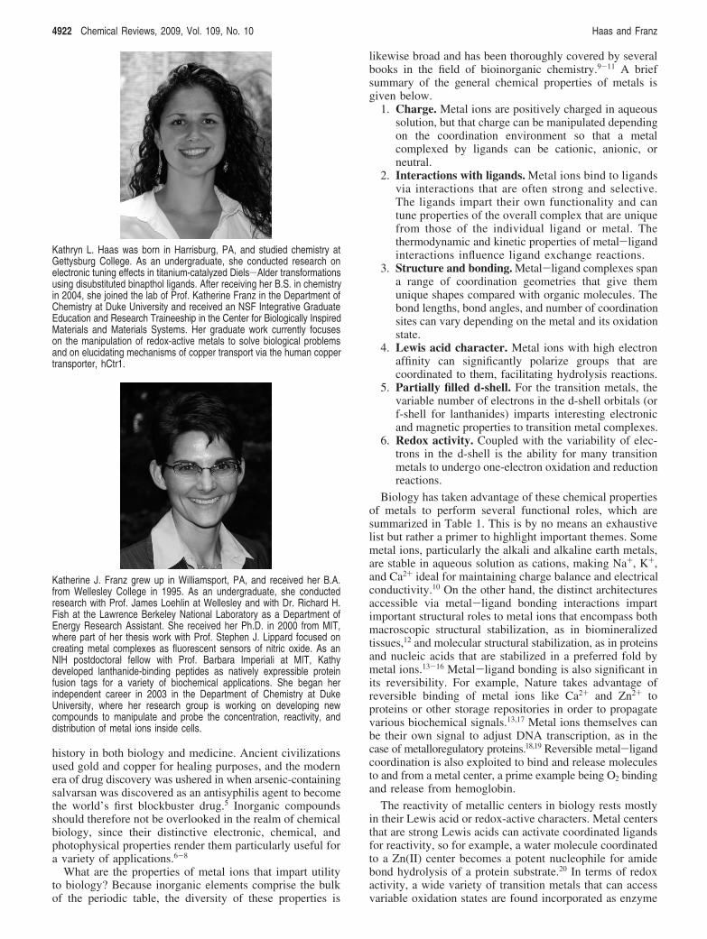
Kathryn L. Haas was born in Harrisburg, PA, and studied chemistry atGettysburg College. As an undergraduate, she conducted research onelectronic tuning effects in titanium-catalyzed Diels-Alder transformationsusing disubstituted binapthol ligands. After receiving her B.S. in chemistryin 2004, she joined the lab of Prof. Katherine Franz in the Department ofChemistry at Duke University and received an NSF Integrative GraduateEducation and Research Traineeship in the Center for Biologically InspiredMaterials and Materials Systems. Her graduate work currently focuseson the manipulation of redox-active metals to solve biological problemsand on elucidating mechanisms of copper transport via the human coppertransporter, hCtr1.
Katherine J. Franz grew up in Williamsport, PA, and received her B.A.from Wellesley College in 1995. As an undergraduate, she conductedresearch with Prof. James Loehlin at Wellesley and with Dr. Richard H.Fish at the Lawrence Berkeley National Laboratory as a Department ofEnergy Research Assistant. She received her Ph.D. in 2000 from MIT,where part of her thesis work with Prof. Stephen J. Lippard focused oncreating metal complexes as fluorescent sensors of nitric oxide. As anNIH postdoctoral fellow with Prof. Barbara Imperiali at MIT, Kathydeveloped lanthanide-binding peptides as natively expressible proteinfusion tags for a variety of biochemical applications. She began herindependent career in 2003 in the Department of Chemistry at DukeUniversity, where her research group is working on developing newcompounds to manipulate and probe the concentration, reactivity, anddistribution of metal ions inside cells.
4922 Chemical Reviews, 2009, Vol. 109, No. 10 Haas and Franz
cofactors to carry out oxidation/reduction chemistry. Electrontransfer units like cytochromes, iron-sulfur clusters, and bluecopper proteins shuttle electrons to other proteins that requireredox chemistry for their function, while other redox proteinscatalyze multielectron oxidation/reduction reactions directlyon a substrate. Examples here involve oxygen metabolism,including the reduction of dioxygen to water by cytochromec oxidase and hydrocarbon oxidation catalyzed by cyto-chrome P-450 enzymes, to name just a few.
When it comes to applying inorganic compounds tobiology, chemists are not restricted to the naturally bioavail-able set of metals and can take advantage of the propertiesof biologically exotic elements, including second and thirdrow transition elements and the lanthanide (Ln) elements.This expansion leads to the list of functional roles ofinorganic elements applied to biology shown in Table 2.Many of the functions listed in Table 2 mirror those of Table1, but applied in novel ways. For example, the structures ofkinetically inert metal complexes are found to interact withproteins and nucleic acids in unique ways, and the acid-baseand redox activity of native and non-native metals can beharnessed for artificial reactivity. Metal complexes can alsoimpart additional functionality not found naturally. The most
striking addition to the list is in visualization, where thephotophysical, magnetic, and radioactive properties of metalsmake possible studies based on luminescence, magneticresonance, PET, and SPECT imaging modalities.
This review will explore how the properties of inorganiccoordination complexes are applied in the context ofinorganic chemical biology, with a particular focus onapplications related to cellular trafficking and regulation. Wewill delve into the structure, bonding, spectroscopy, andreactivity of transition metal coordination compounds andexplore how their unique properties can be used as probesand tools to understand or control biological processes. Ourdiscussion will expand on the functions and examples listedin Table 2, which is really only a partial list, because theexamples of inorganic and organometallic complexes appliedto biology continue to grow. Our focus is on compoundsused in cells to understand the trafficking or regulation ofsomething, be it the metal itself or some other molecule orprocess that is enabled or visualized by a metal complex.Many of the compounds that are discussed have potentialapplications in medicine, but the reader is referred to otherexcellent sources for implicit coverage of medicinal anddiagnostic uses of metal complexes.24-33 There have been
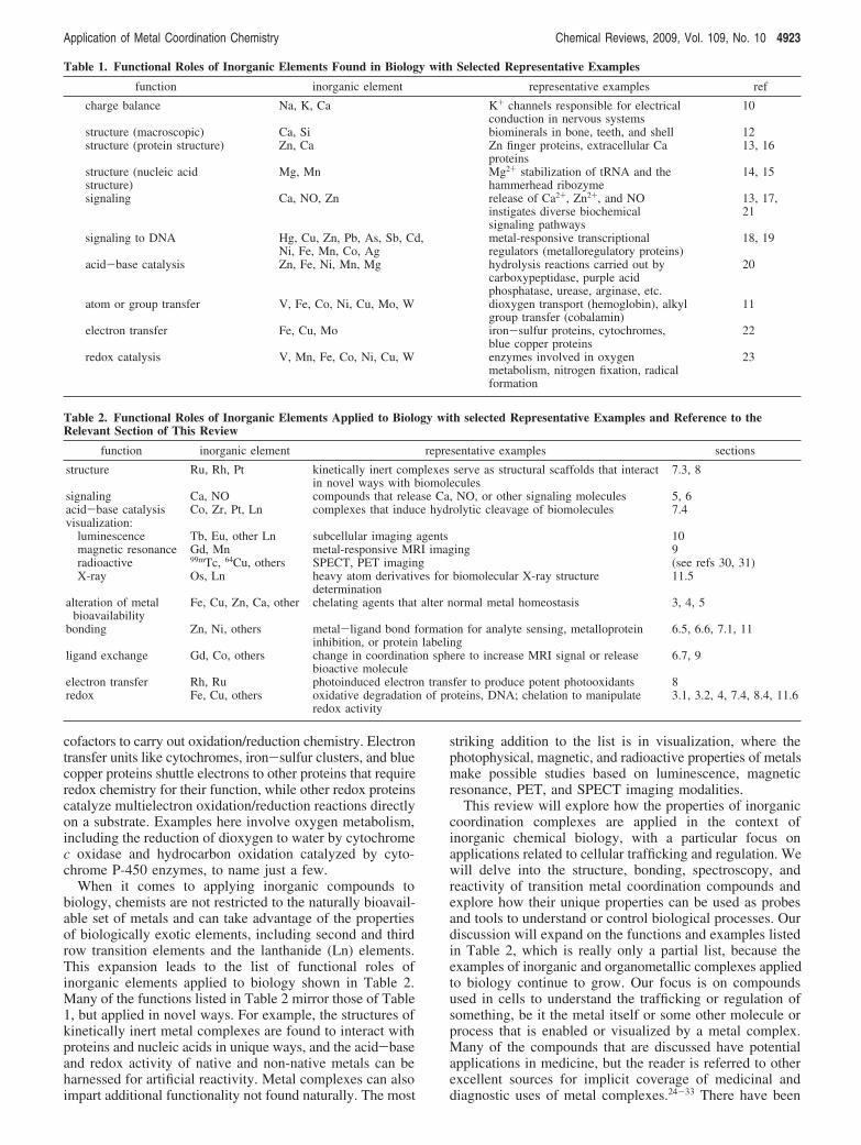
Table 1. Functional Roles of Inorganic Elements Found in Biology with Selected Representative Examples
function inorganic element representative examples ref
charge balance Na, K, Ca K+ channels responsible for electricalconduction in nervous systems
10
structure (macroscopic) Ca, Si biominerals in bone, teeth, and shell 12structure (protein structure) Zn, Ca Zn finger proteins, extracellular Ca
proteins13, 16
structure (nucleic acidstructure)
Mg, Mn Mg2+ stabilization of tRNA and thehammerhead ribozyme
14, 15
signaling Ca, NO, Zn release of Ca2+, Zn2+, and NOinstigates diverse biochemicalsignaling pathways
13, 17,21
signaling to DNA Hg, Cu, Zn, Pb, As, Sb, Cd,Ni, Fe, Mn, Co, Ag
metal-responsive transcriptionalregulators (metalloregulatory proteins)
18, 19
acid-base catalysis Zn, Fe, Ni, Mn, Mg hydrolysis reactions carried out bycarboxypeptidase, purple acidphosphatase, urease, arginase, etc.
20
atom or group transfer V, Fe, Co, Ni, Cu, Mo, W dioxygen transport (hemoglobin), alkylgroup transfer (cobalamin)
11
electron transfer Fe, Cu, Mo iron-sulfur proteins, cytochromes,blue copper proteins
22
redox catalysis V, Mn, Fe, Co, Ni, Cu, W enzymes involved in oxygenmetabolism, nitrogen fixation, radicalformation
23
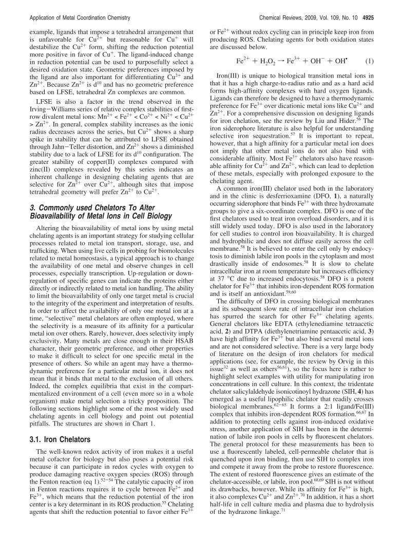
Table 2. Functional Roles of Inorganic Elements Applied to Biology with selected Representative Examples and Reference to theRelevant Section of This Review
function inorganic element representative examples sections
structure Ru, Rh, Pt kinetically inert complexes serve as structural scaffolds that interactin novel ways with biomolecules
7.3, 8
signaling Ca, NO compounds that release Ca, NO, or other signaling molecules 5, 6acid-base catalysis Co, Zr, Pt, Ln complexes that induce hydrolytic cleavage of biomolecules 7.4visualization:
luminescence Tb, Eu, other Ln subcellular imaging agents 10magnetic resonance Gd, Mn metal-responsive MRI imaging 9radioactive 99mTc, 64Cu, others SPECT, PET imaging (see refs 30, 31)X-ray Os, Ln heavy atom derivatives for biomolecular X-ray structure
determination11.5
alteration of metalbioavailability
Fe, Cu, Zn, Ca, other chelating agents that alter normal metal homeostasis 3, 4, 5
bonding Zn, Ni, others metal-ligand bond formation for analyte sensing, metalloproteininhibition, or protein labeling
6.5, 6.6, 7.1, 11
ligand exchange Gd, Co, others change in coordination sphere to increase MRI signal or releasebioactive molecule
6.7, 9
electron transfer Rh, Ru photoinduced electron transfer to produce potent photooxidants 8redox Fe, Cu, others oxidative degradation of proteins, DNA; chelation to manipulate
redox activity3.1, 3.2, 4, 7.4, 8.4, 11.6
Application of Metal Coordination Chemistry Chemical Reviews, 2009, Vol. 109, No. 10 4923

significant advances in the development of fluorescent probesfor monitoring cellular metals. Such molecules are clearlyimportant tools in inorganic chemical biology but will notbe covered here, because there are several excellent reviewsavailable.34-39 We define a metal complex that is a “probefor biological systems” as one that can be used in ways toteach us about the chemical biology of living cells. It maybe used in vivo or in vitro with the aim of understandinghow cells operate. With this definition in mind, we willdiscuss metal chelators and metal complexes that are beingused or have potential to be used to this end, with anemphasis on those that are applied in cellular studies.
2. Principles of Metal-Ligand CoordinationChemistry
The principles governing metal-ligand complex stabilityand specificity depend on the properties of both the metalion and the chelating agent, as summarized briefly in thefollowing sections. More comprehensive reviews on liganddesign for selective complexation of metal ions in aqueoussolution are available.40-45 This discussion sets the stage forunderstanding the properties of the compounds presentedthroughout this review.
2.1. Donor Atom PreferenceThe principle of hard and soft acids and bases (HSAB)
was developed in 1965 by R.G. Pearson following criteriaintroduced by Irving, Williams, Arhland, Chatt, and Davies.46
The classification is based on an atom’s polarizability, wherenonpolarizable acids or bases are small with high chargedensity and are classified as “hard”. Polarizable acids andbases are usually large with low charge density and areclassified as “soft”. Acids and bases that have intermediatehard/soft character are classified as “borderline”. The HSABprinciple predicts that hard acids prefer hard bases, soft acidsprefer soft bases, and borderline acids prefer borderline bases.Pearson’s classifications of metal ions (Lewis acids) and theirligands (Lewis bases) are shown in Table 3, which servesas a useful starting point for predicting the preference ofmetal ions for ligands with various donor groups. Forexample, soft donor groups such as thioethers (R2S) andthiolates (RS-) prefer soft metal ions, like Cu+, whereas hardoxygen donors like carboxylates and phenolates are ap-propriate for hard metal ions, like Fe3+.46
In fitting with the Lewis acid-Lewis base description ofmetal-ligand coordination, it would seem apparent thatincreasing the Lewis basicity of the donor would enhancemetal-ligand bonding. While this principle can be used totune metal-ligand affinity, other factors must also beconsidered. For example, increasing the basicity of aphenolate also increases its pKa. Since metal ions competewith protons for ligand binding in aqueous solution, such
an adjustment might actually decrease the effective metalbinding at a desired pH. Because of proton competition,overall stability constants (!) do not reflect the actual affinityof a ligand for a metal under biologically relevant solutionconditions. A pH-dependent conditional binding constant canbe calculated from known ! and pKa values.47-49 Alterna-tively, an apparent binding constant (Kapp, also called Keff
for effective binding constant) can be measured directly asthe equilibrium constant under the specified solution condi-tions of pH and buffer components. For convenience, bindingconstants are often inverted and discussed as dissociationconstants (Kd).
2.2. Chelate Rings, Steric Strain, andPreorganization
Polydentate ligands that present multiple donor atoms formetal binding provide greater complex stability comparedwith monodentate analogs due to the chelate effect. Thiseffect can be maximized if the number and size of the chelaterings are optimized for the size of the cation in a way thatminimizes steric strain upon metal binding.41,42 The chelaterings formed when two donor groups from the same ligandbind a metal center are most favorable for five- and six-membered rings. Adjacent six-membered rings formed frompolydentate ligands, however, can induce unfavorable stericstrain that is relieved in ligands containing adjacent five- andsix-membered rings.41,42 In general, ligands that minimizesteric strain in the complex on coordination of the ligand tothe metal ion or that preorganize their donor atoms spatiallyas required for complexation or both are preferred for high-affinity binding.43 Macrocycles that incur minimal strain uponmetal complexation can therefore retain their metal ion bytight complexation.
2.3. Complex GeometryOn the basis of its number of valence d electrons, a metal
ion may prefer certain binding geometries over others. Thispreference is based on ligand field stabilization energy(LFSE), a full description of which can be found in standardinorganic chemistry text books.50,51 Comparing the geometricpreferences of iron, copper, and zinc illustrates the point.The common biologically relevant oxidation states of ironare Fe2+ and Fe3+, which prefer octahedral and distortedoctahedral geometries. Copper, on the other hand, existsprimarily as Cu+ and Cu2+, with Cu2+ favoring square planar,square pyramidal, or axially distorted octahedral geometriesdue to Jahn-Teller distortions of its d9 electron configuration.Its reduced Cu+ form has a filled d10 configuration with nopreference for geometry based on LFSE and can thereforebe found in a range of coordination geometries includingtwo-, three-, and four-coordinate sites. The geometry of theligand field can influence the redox state of copper. For
Table 3. Classification of Select Metal Ions and Donor Atoms According to Pearson’s HSAB Principle
hard Lewis acids borderline acids soft acids
H+, Li+, Na+, K+, Be2+, Mg2+, Ca2+,Sr2+, Sc3+, Ti4+, Zr4+, Cr3+, Al3+, Ga3+,La3+, Gd3+, Co3+, Fe3+
Fe2+, Co2+, Ni2+, Cu2+, Zn2+, Pb2+, Bi3+,Rh3+, Ir3+
Cu+, Au+, Ag+, Tl+, Hg+, Pd2+, Cd2+,Pt2+, Hg2+
hard Lewis bases borderline bases soft bases
F-, OH-, H2O, ROH, Cl-, RO-, R2O,CH3CO2
-, NH3, RNH2, NH2NH2, CO32-,
NO3-, O2
-, SO42-, PO4
3-, ClO4-
NO2-, Br-, N3
-, N2, C6H5NH2, pyridine,imidazole
RSH, RS-, R2S, S2-, CN-, RNC, CO,
I-, R3As, R3P, C6H5, C2H4, H2S, HS-,H-, R-
4924 Chemical Reviews, 2009, Vol. 109, No. 10 Haas and Franz
example, ligands that impose a tetrahedral arrangement thatis unfavorable for Cu2+ but reasonable for Cu+ willdestabilize the Cu2+ form, shifting the reduction potentialmore positive in favor of Cu+. The ligand-induced changein reduction potential can be used to purposefully select adesired oxidation state. Geometric preferences imposed bythe ligand are also important for differentiating Cu2+ andZn2+. Because Zn2+ is d10 and has no geometric preferencebased on LFSE, tetrahedral Zn complexes are common.
LFSE is also a factor in the trend observed in theIrving-Williams series of relative complex stabilities of first-row divalent metal ions: Mn2+ < Fe2+ < Co2+ < Ni2+ < Cu2+
> Zn2+. In general, complex stability increases as the ionicradius decreases across the series, but Cu2+ shows a sharpspike in stability that can be attributed to LFSE obtainedthrough Jahn-Teller distortion, and Zn2+ shows a diminishedstability due to a lack of LFSE for its d10 configuration. Thegreater stability of copper(II) complexes compared withzinc(II) complexes revealed by this series indicates aninherent challenge in designing chelating agents that areselective for Zn2+ over Cu2+, although sites that imposetetrahedral geometry will prefer Zn2+ to Cu2+.
3. Commonly used Chelators To AlterBioavailability of Metal Ions in Cell Biology
Altering the bioavailability of metal ions by using metalchelating agents is an important strategy for studying cellularprocesses related to metal ion transport, storage, use, andtrafficking. When using live cells in probing for biomoleculesrelated to metal homeostasis, a typical approach is to changethe availability of one metal and observe changes in cellprocesses, especially transcription. Up-regulation or down-regulation of specific genes can indicate the proteins eitherdirectly or indirectly related to metal ion handling. The abilityto limit the bioavailability of only one target metal is crucialto the integrity of the experiment and interpretation of results.In order to affect the availability of only one metal ion at atime, “selective” metal chelators are often employed, wherethe selectivity is a measure of its affinity for a particularmetal ion over others. Rarely, however, does selectivity implyexclusivity. Many metals are close enough in their HSABcharacter, their geometric preference, and other propertiesto make it difficult to select for one specific metal in thepresence of others. So while an agent may have a thermo-dynamic preference for a particular metal ion, it does notmean that it binds that metal to the exclusion of all others.Indeed, the complex equilibria that exist in the compart-mentalized environment of a cell (even more so in a wholeorganism) make metal selection a tricky proposition. Thefollowing sections highlight some of the most widely usedchelating agents in cell biology and point out potentialpitfalls. The structures are shown in Chart 1.
3.1. Iron ChelatorsThe well-known redox activity of iron makes it a useful
metal cofactor for biology but also poses a potential riskbecause it can participate in redox cycles with oxygen toproduce damaging reactive oxygen species (ROS) throughthe Fenton reaction (eq 1).52-54 The catalytic capacity of ironin Fenton reactions requires it to cycle between Fe2+ andFe3+, which means that the reduction potential of the ironcenter is a key determinant in its ROS production.55 Chelatingagents that shift the reduction potential to favor either Fe3+
or Fe2+ without redox cycling can in principle keep iron fromproducing ROS. Chelating agents for both oxidation statesare discussed below.
Iron(III) is unique to biological transition metal ions inthat it has a high charge-to-radius ratio and as a hard acidforms high-affinity complexes with hard oxygen ligands.Ligands can therefore be designed to have a thermodynamicpreference for Fe3+ over dicationic metal ions like Cu2+ andZn2+. For a comprehensive discussion on designing ligandsfor iron chelation, see the review by Liu and Hider.56 Theiron siderophore literature is also helpful for understandingselective iron sequestration.57 It is important to repeat,however, that a high affinity for a particular metal ion doesnot imply that other metal ions do not also bind withconsiderable affinity. Most Fe3+ chelators also have reason-able affinity for Cu2+ and Zn2+, which can lead to depletionof these metals, especially with prolonged exposure to thechelating agent.
A common iron(III) chelator used both in the laboratoryand in the clinic is desferrioxamine (DFO, 1), a naturallyoccurring siderophore that binds Fe3+ with three hydroxamategroups to give a six-coordinate complex. DFO is one of thefirst chelators used to treat iron overload disorders, and it isstill widely used today. DFO is also used in the laboratoryfor cell studies to control iron bioavailability. It is chargedand hydrophilic and does not diffuse easily across the cellmembrane.58 It is believed to enter the cell only by endocy-tosis to diminish labile iron pools in the cytoplasm and mostdrastically inside of endosomes.58 It is slow to chelateintracellular iron at room temperature but increases efficiencyat 37 °C due to increased endocytosis.58 DFO is a potentchelator for Fe3+ that inhibits iron-dependent ROS formationand is itself an antioxidant.59,60
The difficulty of DFO in crossing biological membranesand its subsequent slow rate of intracellular iron chelationhas spurred the search for other Fe3+ chelating agents.General chelators like EDTA (ethylenediamine tetraaceticacid, 2) and DTPA (diethylenetriamine pentaacetic acid, 3)have high affinity for Fe3+ but also bind several metal ionsand are not considered selective. There is a very large bodyof literature on the design of iron chelators for medicalapplications (see, for example, the review by Orvig in thisissue32 as well as others56,61), so the focus here is rather tohighlight select examples with utility for manipulating ironconcentrations in cell culture. In this context, the tridentatechelator salicylaldehyde isonicotinoyl hydrazone (SIH, 4) hasemerged as a useful lipophilic chelator that readily crossesbiological membranes.62-65 It forms a 2:1 ligand/Fe(III)complex that inhibits iron-dependent ROS formation.66,67 Inaddition to protecting cells against iron-induced oxidativestress, another application of SIH has been in the determi-nation of labile iron pools in cells by fluorescent chelators.The general protocol for these measurements has been touse a fluorescently labeled, cell-permeable chelator that isquenched upon iron binding, then use SIH to complex ironand compete it away from the probe to restore fluorescence.The extent of restored fluorescence gives an estimate of thechelator-accessible, or labile, iron pool.68,69 SIH is not withoutits drawbacks, however. While its affinity for Fe3+ is high,it also complexes Cu2+ and Zn2+.70 In addition, it has a shorthalf-life in cell culture media and plasma due to hydrolysisof the hydrazone linkage.71
Fe2+ + H2O2 f Fe3+ + OH- + OH• (1)
Application of Metal Coordination Chemistry Chemical Reviews, 2009, Vol. 109, No. 10 4925
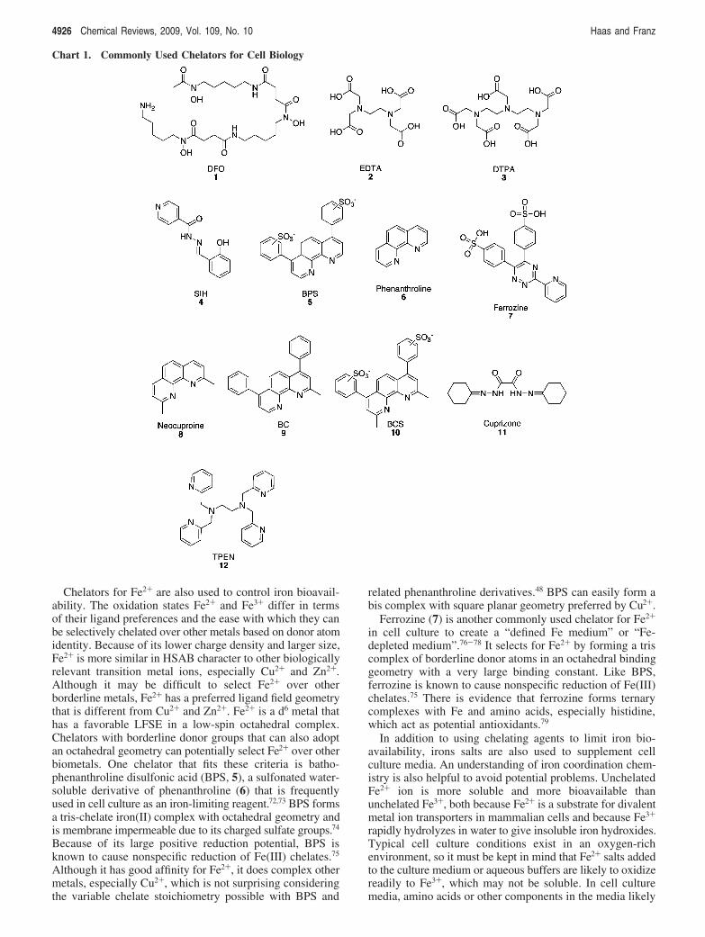
Chelators for Fe2+ are also used to control iron bioavail-ability. The oxidation states Fe2+ and Fe3+ differ in termsof their ligand preferences and the ease with which they canbe selectively chelated over other metals based on donor atomidentity. Because of its lower charge density and larger size,Fe2+ is more similar in HSAB character to other biologicallyrelevant transition metal ions, especially Cu2+ and Zn2+.Although it may be difficult to select Fe2+ over otherborderline metals, Fe2+ has a preferred ligand field geometrythat is different from Cu2+ and Zn2+. Fe2+ is a d6 metal thathas a favorable LFSE in a low-spin octahedral complex.Chelators with borderline donor groups that can also adoptan octahedral geometry can potentially select Fe2+ over otherbiometals. One chelator that fits these criteria is batho-phenanthroline disulfonic acid (BPS, 5), a sulfonated water-soluble derivative of phenanthroline (6) that is frequentlyused in cell culture as an iron-limiting reagent.72,73 BPS formsa tris-chelate iron(II) complex with octahedral geometry andis membrane impermeable due to its charged sulfate groups.74
Because of its large positive reduction potential, BPS isknown to cause nonspecific reduction of Fe(III) chelates.75
Although it has good affinity for Fe2+, it does complex othermetals, especially Cu2+, which is not surprising consideringthe variable chelate stoichiometry possible with BPS and
related phenanthroline derivatives.48 BPS can easily form abis complex with square planar geometry preferred by Cu2+.
Ferrozine (7) is another commonly used chelator for Fe2+
in cell culture to create a “defined Fe medium” or “Fe-depleted medium”.76-78 It selects for Fe2+ by forming a triscomplex of borderline donor atoms in an octahedral bindinggeometry with a very large binding constant. Like BPS,ferrozine is known to cause nonspecific reduction of Fe(III)chelates.75 There is evidence that ferrozine forms ternarycomplexes with Fe and amino acids, especially histidine,which act as potential antioxidants.79
In addition to using chelating agents to limit iron bio-availability, irons salts are also used to supplement cellculture media. An understanding of iron coordination chem-istry is also helpful to avoid potential problems. UnchelatedFe2+ ion is more soluble and more bioavailable thanunchelated Fe3+, both because Fe2+ is a substrate for divalentmetal ion transporters in mammalian cells and because Fe3+
rapidly hydrolyzes in water to give insoluble iron hydroxides.Typical cell culture conditions exist in an oxygen-richenvironment, so it must be kept in mind that Fe2+ salts addedto the culture medium or aqueous buffers are likely to oxidizereadily to Fe3+, which may not be soluble. In cell culturemedia, amino acids or other components in the media likely
Chart 1. Commonly Used Chelators for Cell Biology
4926 Chemical Reviews, 2009, Vol. 109, No. 10 Haas and Franz
keep iron in solution, but addition of simple iron salts tostandard laboratory buffers, especially phosphate buffers, willresult in insoluble and bio-unavailable iron.
3.2. Copper ChelatorsLike iron, the redox chemistry of Cu2+/+ makes it essential
to biological processes but also potentially dangerous if it isnot handled properly by the cell and becomes available forFenton-like chemistry to produce ROS. Typically under theoxidizing extracellular environment, copper exists as Cu2+,but in the reducing conditions inside the cell, it likely existsin the reduced Cu+ oxidation state. Soft character makes Cu+
unique among the biological metal ions, so it has potentialto be selected based on ligand donor groups. In addition,Cu+ is a d10 ion, giving it flexibility in geometric arrange-ments. This means Cu+ can adopt tetrahedral, trigonal, oreven linear geometries that are disfavored by other metals.Zn2+ is also a d10 ion but is harder in character than Cu+, socan be minimized as an interfering species based on liganddonor choice. Zn2+ is also a smaller metal ion, so chelatering size can be a determining factor.
2,9-Dimethyl-substituted phenanthroline ligands are well-known to select for Cu+. Neocuproine (8), bathocuproine(BC, 9), and bathocuproine disulfonate (BCS, 10) are thethree commonly used 2,9-dimethyl-substituted phenanthro-line chelates used in cell culture.80-86 Unlike phenanthroline,these ligands disfavor octahedral tris-chelate or square-planarbis-chelate coordination modes because of steric interferenceof the methyl substituents. Instead, when binding to a metalin a bis complex, the metal is forced into a tetrahedral bindinggeometry with the two chelate ligands nearly perpendicularto each other. This tetrahedral geometry combined with thelarge bite size of the five-membered chelate ring effectivelybinds Cu+ over other metals. However, these ligands do havesignificant interaction with Cu2+ and are known to bind toCu2+, forcing it into a tetrahedral geometry and inducing itsreduction to Cu+. BC-bound Cu(II) is a stronger oxidizingagent by 0.5 V compared with uncomplexed Cu2+, whichhighlights the fact that these “Cu(I)-selective” chelators arenot innocent chelators of Cu+.87-89 Although they may beable to promote reduction of Cu2+ to Cu+, once the reducedstate is reached, it is stabilized and does not participate inredox cycling.90 In fact BCS has been shown to inhibit Cu-dependent redox cycles.86
Bathocuproine (9) is not very water-soluble, so a sul-fonated version (BCS) was developed to give a more solublederivative.74 Because BCS is charged and not membranepermeable, it is commonly used in cell studies as anextracellular Cu-limiting agent. Because Cu2+ is the dominantextracellular form of copper, it should be considered thatBCS is a potential promoter of Cu2+ reduction. Neocuproine(8) is more hydrophobic and is used for intracellular andextracellular Cu chelation since it can diffuse over the cellmembrane.
Cuprizone (oxalic acid bis(cyclohexylidene)hydrazide, 11)is another chelator used to selectively bind Cu2+ in cellstudies;82,83 however, the actual nature of its Cu complexhas long been under debate. There is strong evidence thatcuprizone stabilizes a Cu3+ oxidation state in a square planard8 complex.91-93 The cytotoxic and neurotoxic effects ofcuprizone may be related to Cu2+-Cu3+ redox cyclinginduced by this chelator under biological conditions.91,94
3.3. Zinc ChelatorsThe only relevant oxidation state for zinc is Zn2+. It has
borderline HSAB character, and it is d10 with no realpreference for ligand field geometry. Zn2+ is very close toCu2+ in size and charge density and can be difficult to selectfor over Cu2+. TPEN (N,N,N′,N′-tetrakis-(2-pyridylmethyl)-ethylenediamine, 12) is the most common zinc chelator usedin the literature;85,95-101 however there is an apparentmisconception that TPEN is Zn2+-specific. In fact, the affinityof TPEN for Fe2+ (log Ka 14.6) is relatively significant, whileits affinity for Cu2+ (log Ka 20.6) is higher than that for Zn2+
(log Ka 18.0).102,103 There are several studies that show TPENaffects cellular concentrations of Zn and Cu and thatreplenishing either metal into TPEN-treated cells will rescuethe cells from TPEN-induced apoptosis.100 Another studyshows that levels of Fe are affected in addition to Cu andZn when TPEN is added to cell culture.104 Part of the reasonthat TPEN can have such significant affinity for Zn2+, Cu2+,and Fe2+ is its potential variability in chelate bindinggeometry.105 TPEN has six possible donor groups, and forZn and Fe, all six are used to chelate the ion in an octahedralgeometry. Cu2+, which is destabilized by octahedral geom-etry, is chelated by only five of the six possible donor groupsand forms a distorted square pyramid with high stability.105
It is necessary to consider the Cu, Fe, and Zn bindingproperties of TPEN before interpreting the results of anexperiment where TPEN is used as a “selective” chelator.
4. Selectivity by Reactivity or LocalizationThe challenges associated with selective metal chelation
have led to the development of new generation chelatingagents that achieve selectivity based on reactivity or localiza-tion. Examples are shown in Chart 2. Our own lab hasintroduced a prochelator strategy that uses the reactivity ofoxidative stress to generate metal-binding agents to inhibitfurther oxidative damage.106-108 The concept is based on thehypothesis that oxidative stress is exacerbated by Fentonreactions wherein labile metal ions, particularly iron andcopper, react with H2O2 to produce damaging hydroxylradicals (see eq 1). As described in section 3.1, appropriatelydesigned chelating agents can prevent this reactivity, but itis difficult to design ligands that can select for the metal ioncausing the damage without altering normal, healthy metalion distribution. In our prochelator strategy, a metal-bindingligand is masked with a H2O2-sensitive protecting group toprevent metal coordination under normal conditions. Reactionwith H2O2 converts the prochelator to the chelator, therebytriggering metal sequestration. A boronic ester was selectedas the H2O2-sensitive masking group in our first-generationprochelator, BSIH (13, isonicotinic acid [2-(4,4,5,5-tetram-ethyl-[1,3,2]dioxaborolan-2-yl)-benzylidene]-hydrazide), whichconverts to SIH (4) in the presence of H2O2 and forms the[Fe(SIH)2]+ complex that prevents iron from Fenton reactiv-ity.106 BSIH itself has only weak interactions with metal ions,whereas SIH strongly interacts with most divalent andtrivalent metals. The BSIH to SIH conversion also inhibitscopper-catalyzed Fenton reactions,109 demonstrating that thestrategy is not metal specific per se, but rather reactivityspecific.108
BSIH was shown to protect cultured retinal pigmentepithelial cells against cell death induced by hydrogenperoxide.107 Significantly, cells that were not stressed withH2O2 remained viable even after repetitive, prolonged
Application of Metal Coordination Chemistry Chemical Reviews, 2009, Vol. 109, No. 10 4927
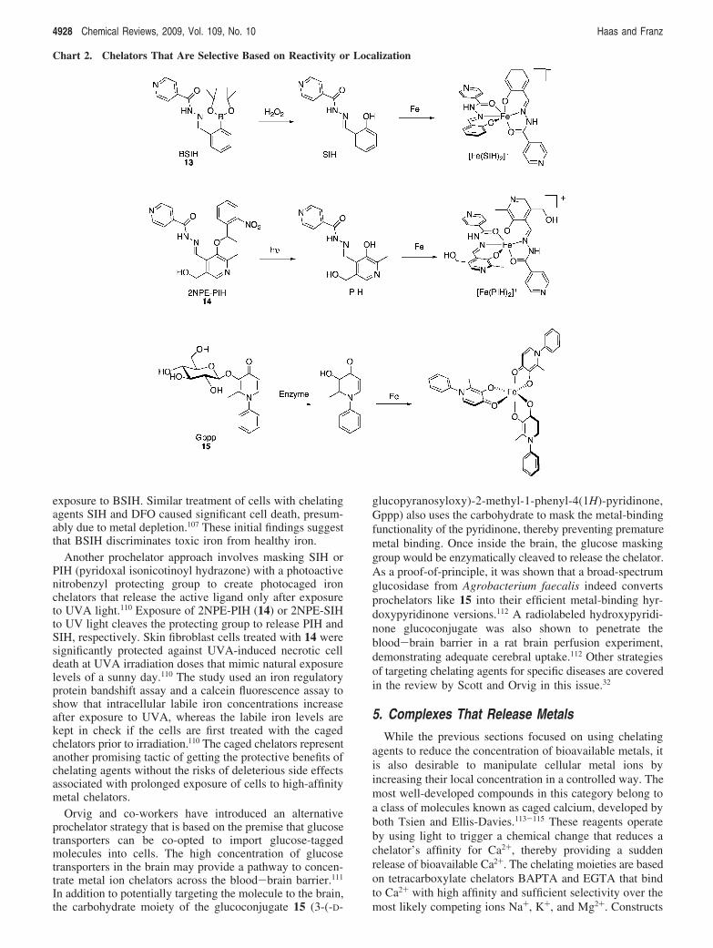
exposure to BSIH. Similar treatment of cells with chelatingagents SIH and DFO caused significant cell death, presum-ably due to metal depletion.107 These initial findings suggestthat BSIH discriminates toxic iron from healthy iron.
Another prochelator approach involves masking SIH orPIH (pyridoxal isonicotinoyl hydrazone) with a photoactivenitrobenzyl protecting group to create photocaged ironchelators that release the active ligand only after exposureto UVA light.110 Exposure of 2NPE-PIH (14) or 2NPE-SIHto UV light cleaves the protecting group to release PIH andSIH, respectively. Skin fibroblast cells treated with 14 weresignificantly protected against UVA-induced necrotic celldeath at UVA irradiation doses that mimic natural exposurelevels of a sunny day.110 The study used an iron regulatoryprotein bandshift assay and a calcein fluorescence assay toshow that intracellular labile iron concentrations increaseafter exposure to UVA, whereas the labile iron levels arekept in check if the cells are first treated with the cagedchelators prior to irradiation.110 The caged chelators representanother promising tactic of getting the protective benefits ofchelating agents without the risks of deleterious side effectsassociated with prolonged exposure of cells to high-affinitymetal chelators.
Orvig and co-workers have introduced an alternativeprochelator strategy that is based on the premise that glucosetransporters can be co-opted to import glucose-taggedmolecules into cells. The high concentration of glucosetransporters in the brain may provide a pathway to concen-trate metal ion chelators across the blood-brain barrier.111
In addition to potentially targeting the molecule to the brain,the carbohydrate moiety of the glucoconjugate 15 (3-(-D-
glucopyranosyloxy)-2-methyl-1-phenyl-4(1H)-pyridinone,Gppp) also uses the carbohydrate to mask the metal-bindingfunctionality of the pyridinone, thereby preventing prematuremetal binding. Once inside the brain, the glucose maskinggroup would be enzymatically cleaved to release the chelator.As a proof-of-principle, it was shown that a broad-spectrumglucosidase from Agrobacterium faecalis indeed convertsprochelators like 15 into their efficient metal-binding hyr-doxypyridinone versions.112 A radiolabeled hydroxypyridi-none glucoconjugate was also shown to penetrate theblood-brain barrier in a rat brain perfusion experiment,demonstrating adequate cerebral uptake.112 Other strategiesof targeting chelating agents for specific diseases are coveredin the review by Scott and Orvig in this issue.32
5. Complexes That Release MetalsWhile the previous sections focused on using chelating
agents to reduce the concentration of bioavailable metals, itis also desirable to manipulate cellular metal ions byincreasing their local concentration in a controlled way. Themost well-developed compounds in this category belong toa class of molecules known as caged calcium, developed byboth Tsien and Ellis-Davies.113-115 These reagents operateby using light to trigger a chemical change that reduces achelator’s affinity for Ca2+, thereby providing a suddenrelease of bioavailable Ca2+. The chelating moieties are basedon tetracarboxylate chelators BAPTA and EGTA that bindto Ca2+ with high affinity and sufficient selectivity over themost likely competing ions Na+, K+, and Mg2+. Constructs
Chart 2. Chelators That Are Selective Based on Reactivity or Localization
4928 Chemical Reviews, 2009, Vol. 109, No. 10 Haas and Franz
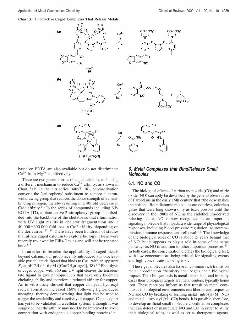
based on EDTA are also available but do not discriminateCa2+ from Mg2+ as effectively.
There are two general series of caged calcium, each usinga different mechanism to reduce Ca2+ affinity, as shown inChart 3a,b. In the nitr series (nitr-7, 16), photoactivationconverts the 2-nitrophenyl substituent to a more electron-withdrawing group that reduces the donor strength of a metal-binding nitrogen, thereby resulting in a 40-fold decrease inCa2+ affinity.116 In the series of compounds including NP-EGTA (17), a photoactive 2-nitrophenyl group is embed-ded into the backbone of the chelator so that illuminationwith UV light results in chelator fragmentation and a40 000-600 000-fold loss in Ca2+ affinity, depending onthe derivative.117,118 There have been hundreds of studiesthat utilize caged calcium to explore biology. These wererecently reviewed by Ellis-Davies and will not be repeatedhere.115
In an effort to broaden the applicability of caged metalsbeyond calcium, our group recently introduced a photocleav-able pyridal amide ligand that binds to Cu2+ with an apparentKd at pH 7.4 of 16 pM ([Cu(OH2)(cage)], 18).119 Photolysisof caged copper with 360 nm UV light cleaves the tetraden-tate ligand to give photoproducts that have only bidentatechelating ability and therefore a reduced affinity for copper.An in vitro assay showed that copper-catalyzed hydroxylradical formation increased 160% following light-induceduncaging, thereby demonstrating that light can be used totrigger the availability and reactivity of copper. Caged copperhas yet to be validated in a cellular system, although it wassuggested that the affinity may need to be improved to avoidcompetition with endogenous copper-binding proteins.119
6. Metal Complexes that Bind/Release SmallMolecules
6.1. NO and COThe biological effects of carbon monoxide (CO) and nitric
oxide (NO) can aptly be described by the general observationof Paracelsus in the early 16th century that “the dose makesthe poison”. Both diatomic molecules are odorless, colorlessgases that were long known only as toxic poisons until thediscovery in the 1980s of NO as the endothelium-derivedrelaxing factor. NO is now recognized as an importantsignaling molecule that impacts a wide range of physiologicalresponses, including blood pressure regulation, neurotrans-mission, immune response, and cell death.120 The knowledgeof the biological roles of CO is about 15 years behind thatof NO, but it appears to play a role in some of the samepathways as NO in addition to other important processes.121
In both cases, the concentration dictates the biological effect,with low concentrations being critical for signaling eventsand high concentrations being toxic.
These gas molecules also have in common rich transitionmetal coordination chemistry that begets their biologicalimpact. Their biosynthesis is metal-dependent, and in manycases their biological targets are metal centers, typically hemeiron. These reactions inform us that transition metal com-plexes in biological environments can liberate and sequesterNO and CO by breaking or forming metal-nitrosyl (M-NO)and metal-carbonyl (M-CO) bonds. It is possible, therefore,to develop artificial small molecule coordination complexesthat can detect or manipulate NO and CO in order to studytheir biological roles, as well as act as therapeutic agents.
Chart 3. Photoactive Caged Complexes That Release Metals
Application of Metal Coordination Chemistry Chemical Reviews, 2009, Vol. 109, No. 10 4929
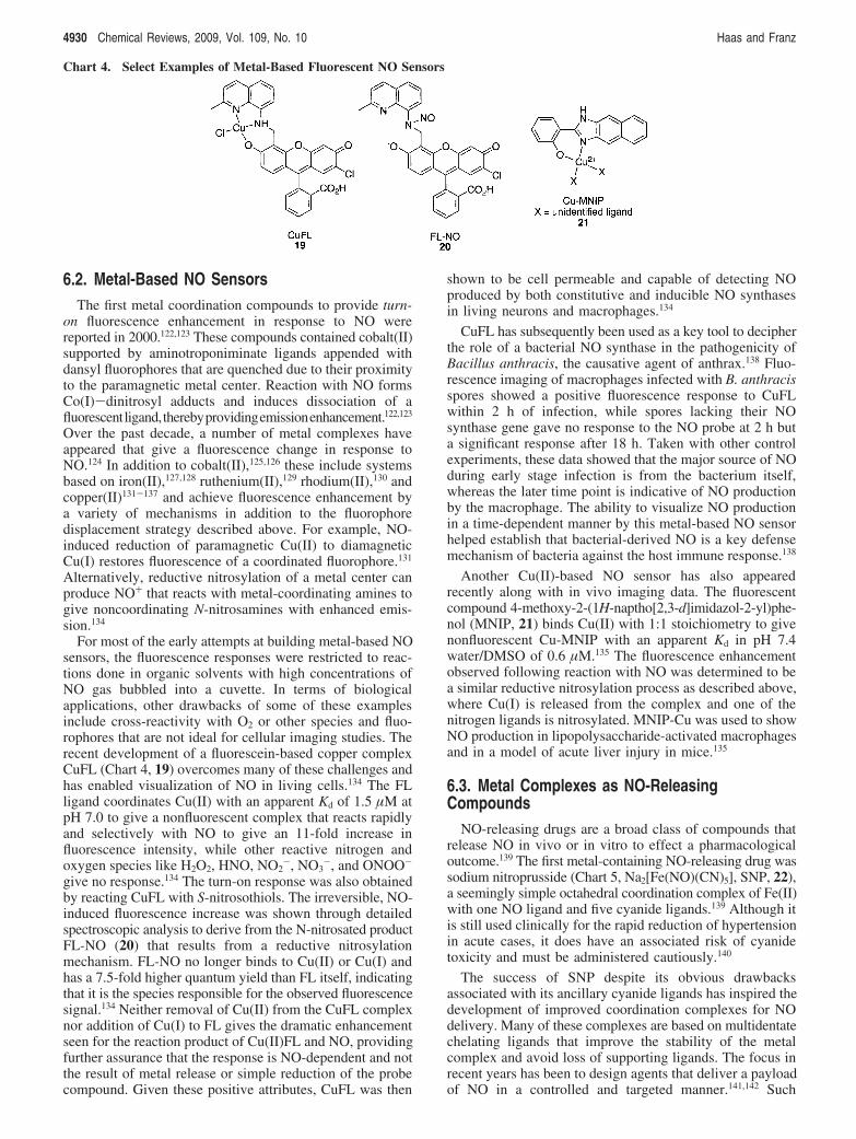
6.2. Metal-Based NO SensorsThe first metal coordination compounds to provide turn-
on fluorescence enhancement in response to NO werereported in 2000.122,123 These compounds contained cobalt(II)supported by aminotroponiminate ligands appended withdansyl fluorophores that are quenched due to their proximityto the paramagnetic metal center. Reaction with NO formsCo(I)-dinitrosyl adducts and induces dissociation of afluorescentligand,therebyprovidingemissionenhancement.122,123
Over the past decade, a number of metal complexes haveappeared that give a fluorescence change in response toNO.124 In addition to cobalt(II),125,126 these include systemsbased on iron(II),127,128 ruthenium(II),129 rhodium(II),130 andcopper(II)131-137 and achieve fluorescence enhancement bya variety of mechanisms in addition to the fluorophoredisplacement strategy described above. For example, NO-induced reduction of paramagnetic Cu(II) to diamagneticCu(I) restores fluorescence of a coordinated fluorophore.131
Alternatively, reductive nitrosylation of a metal center canproduce NO+ that reacts with metal-coordinating amines togive noncoordinating N-nitrosamines with enhanced emis-sion.134
For most of the early attempts at building metal-based NOsensors, the fluorescence responses were restricted to reac-tions done in organic solvents with high concentrations ofNO gas bubbled into a cuvette. In terms of biologicalapplications, other drawbacks of some of these examplesinclude cross-reactivity with O2 or other species and fluo-rophores that are not ideal for cellular imaging studies. Therecent development of a fluorescein-based copper complexCuFL (Chart 4, 19) overcomes many of these challenges andhas enabled visualization of NO in living cells.134 The FLligand coordinates Cu(II) with an apparent Kd of 1.5 µM atpH 7.0 to give a nonfluorescent complex that reacts rapidlyand selectively with NO to give an 11-fold increase influorescence intensity, while other reactive nitrogen andoxygen species like H2O2, HNO, NO2
-, NO3-, and ONOO-
give no response.134 The turn-on response was also obtainedby reacting CuFL with S-nitrosothiols. The irreversible, NO-induced fluorescence increase was shown through detailedspectroscopic analysis to derive from the N-nitrosated productFL-NO (20) that results from a reductive nitrosylationmechanism. FL-NO no longer binds to Cu(II) or Cu(I) andhas a 7.5-fold higher quantum yield than FL itself, indicatingthat it is the species responsible for the observed fluorescencesignal.134 Neither removal of Cu(II) from the CuFL complexnor addition of Cu(I) to FL gives the dramatic enhancementseen for the reaction product of Cu(II)FL and NO, providingfurther assurance that the response is NO-dependent and notthe result of metal release or simple reduction of the probecompound. Given these positive attributes, CuFL was then
shown to be cell permeable and capable of detecting NOproduced by both constitutive and inducible NO synthasesin living neurons and macrophages.134
CuFL has subsequently been used as a key tool to decipherthe role of a bacterial NO synthase in the pathogenicity ofBacillus anthracis, the causative agent of anthrax.138 Fluo-rescence imaging of macrophages infected with B. anthracisspores showed a positive fluorescence response to CuFLwithin 2 h of infection, while spores lacking their NOsynthase gene gave no response to the NO probe at 2 h buta significant response after 18 h. Taken with other controlexperiments, these data showed that the major source of NOduring early stage infection is from the bacterium itself,whereas the later time point is indicative of NO productionby the macrophage. The ability to visualize NO productionin a time-dependent manner by this metal-based NO sensorhelped establish that bacterial-derived NO is a key defensemechanism of bacteria against the host immune response.138
Another Cu(II)-based NO sensor has also appearedrecently along with in vivo imaging data. The fluorescentcompound 4-methoxy-2-(1H-naptho[2,3-d]imidazol-2-yl)phe-nol (MNIP, 21) binds Cu(II) with 1:1 stoichiometry to givenonfluorescent Cu-MNIP with an apparent Kd in pH 7.4water/DMSO of 0.6 µM.135 The fluorescence enhancementobserved following reaction with NO was determined to bea similar reductive nitrosylation process as described above,where Cu(I) is released from the complex and one of thenitrogen ligands is nitrosylated. MNIP-Cu was used to showNO production in lipopolysaccharide-activated macrophagesand in a model of acute liver injury in mice.135
6.3. Metal Complexes as NO-ReleasingCompounds
NO-releasing drugs are a broad class of compounds thatrelease NO in vivo or in vitro to effect a pharmacologicaloutcome.139 The first metal-containing NO-releasing drug wassodium nitroprusside (Chart 5, Na2[Fe(NO)(CN)5], SNP, 22),a seemingly simple octahedral coordination complex of Fe(II)with one NO ligand and five cyanide ligands.139 Although itis still used clinically for the rapid reduction of hypertensionin acute cases, it does have an associated risk of cyanidetoxicity and must be administered cautiously.140
The success of SNP despite its obvious drawbacksassociated with its ancillary cyanide ligands has inspired thedevelopment of improved coordination complexes for NOdelivery. Many of these complexes are based on multidentatechelating ligands that improve the stability of the metalcomplex and avoid loss of supporting ligands. The focus inrecent years has been to design agents that deliver a payloadof NO in a controlled and targeted manner.141,142 Such
Chart 4. Select Examples of Metal-Based Fluorescent NO Sensors
4930 Chemical Reviews, 2009, Vol. 109, No. 10 Haas and Franz
directed release of NO could have applications as anti-infectious agents143 or in cancer therapy as sensitizers for"-radiation or as agents for photodynamic therapy. Theknown photosensitivity of many metal-nitrosyl compoundsmakes light an attractive stimulus to release NO from a metalcenter.
Several photosensitive metal compounds including SNP,Fe-S-NO clusters known as Roussin’s salts, and simpleRu complexes like [Ru(NO)(Cl)5]2-, have been used ascontrolled sources of NO to elucidate the biological andnotably neurophysiological roles of NO in cells and tissues;however, these complexes all suffer from undesirable sidereactions.144,145 In addition to release of toxic side productslike cyanide, the NO is lost prior to photolysis eitherthermally or by conversion to other NOx products in the caseof the Fe-NO compounds, or the metal itself reacts withbiomolecules as in the case of Ru forming DNA adducts.144,145
Metalloporphyrins that were studied for their photolytic
properties by Ford in the early 1990s have the advantage ofbeing very stable macrocyclic metal complexes with intenselong-wavelength absorptions but suffer thermal instabilityand oxygen sensitivity in the case of the Fe-NO adductsand complicated back and side reactions in the case ofRu-NO porphyrins.141,146 Considerable effort has thereforegone into designing alternative multidentate ligands thatsupport photoactive Mn and Ru nitrosyl adducts,142,145 as wellas water-soluble Fe complexes,147,148 particularly those thatcan be activated by two-photon excitation.141 These com-pounds are shown in Chart 5 and described in more detailin the following sections.
6.3.1. NO Donors Sensitive to Visible and Near-IR Light
The Mascharak group introduced the pentadentate ligandPaPy3H (N,N-bis(2-pyridylmethyl)amine-N-ethyl-2-pyridine-2-carboxamide), which supports octahedral M-NO com-
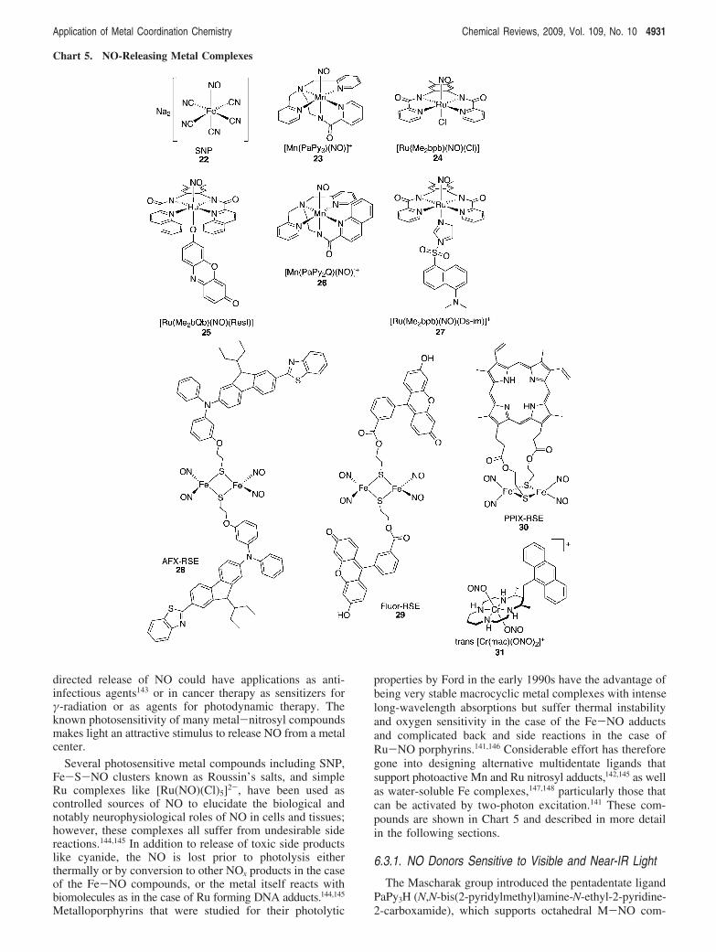
Chart 5. NO-Releasing Metal Complexes
Application of Metal Coordination Chemistry Chemical Reviews, 2009, Vol. 109, No. 10 4931
plexes by providing four nitrogen atoms around the equatorialplane of the metal with a carboxamide group trans to NO;the Mn complex [Mn(PaPy3)(NO)]+ (23) is an example. Thestrong #-donor character of the negatively charged carboxa-mide being trans to NO has proven to be a key feature inthe photolability of these complexes, with the nature of themetal center dictating the wavelength of light required toachieve photorelease. The iron complex [Fe(PaPy3)(NO)]2+
can be activated by visible light in the 500-600 nm range,but the complex is not stable in aqueous solutions andtransfers its NO to thiol-containing compounds even in theabsence of light.149 Replacing Fe with Ru improves thesolubility and stability of the complex, because[Ru(PaPy3)(NO)]2+ is soluble in water and stable betweenpH 5 and 9.150 Photolysis only occurs with UV light, whichmay limit future in vivo use, but the complex is a usefultool for studying fast reactions of NO with heme proteins.151
The Mn analog [Mn(PaPy3)(NO)]+ (23) has proven to bethe most interesting among this series, because it is solubleand stable in aqueous buffers and is activated by visible light(500-650 nm) to release NO and form the Mn(II) aquaspecies [Mn(PaPy3)(H2O)]+.152 Both the Mn and the Rucompounds activate soluble guanylate cyclase activity in vitroin a light- and concentration-dependent manner.153 They alsoelicit a concentration-dependent increase in cGMP in vascularsmooth muscle cells, demonstrating that the complexesrelease NO intracellularly under the control of light. Fur-thermore, the compounds showed light-dependent vasore-laxant activity in a rat thoracic aortic ring.153
The photoactivity of metal nitrosyls requires promotionof an electron from a metal-based molecular orbital to a$*(NO) antibonding orbital; therefore the sensitivity to lightdepends on the energy of the M f $*(NO) electronictransition.154-156 To push the excitation wavelength into thevisible and near-IR, the M-NO photoband needs to be inthis longer wavelength region. As illustrated in the PaPy3
examples described above, one way to do this is to changethe metal. Two other strategies are to change the fieldstrength of the ligand or to attach light-harvesting chro-mophores to the complex. Both strategies are further exploredbelow.
The ligand H2bpb (N,N′-bis(bipyridine-2-carboxamido)-1,2-diaminobenzene) nicely demonstrates how clever alter-ations to the coordination chemistry of the complex can tuneits photophysical properties. Several variations of this ligandexist; one example is the dimethyl derivative[Ru(Me2bpb)(NO)(Cl)] shown in 24, which is only sensitiveto low-intensity UV light. However, replacing the pyridalarms with quinolines red shifts the Ru-NO photoband from380 to 455 nm, thereby accessing visible light photoactiva-tion.157 Furthermore, the four-coordinate H2bpb frameworkallows an additional donor ligand to be installed trans to NO.By using this open coordination site to attach a light-harvesting dye directly to the metal, additional sensitizationis achieved. Resorufin (Resf) is a dye with intense absorptionin the visible and a phenolate moiety that enables directattachment to the Ru center to give Ru-NO complexes withstrong absorption bands around 500 nm.158,159 The combina-tion of the quinoline arms on the ligand and the dye attachedto the metal to give [Ru(OMe2bQb)(NO)(Resf)] (25) dem-onstrates that these effects are additive. Adjustment of theligand frame effectively merges the Ru-NO photoband withthe intense absorption of the coordinated dye, thereby
increasing the extinction coefficient and sensitizing thecompound to visible light photoactivation.158
Because light penetration through mammalian tissue ismostly restricted to the 700-1100 nm region, it is verydesirable to have NO donors that are photoresponsive in thisregion if they are to be used in applications like photody-namic therapy.156 Again by clever alterations of the ligand,Mascharak reported that replacing one of the pyridal armsof HpaPy3 with a more conjugated quinoline arm to giveHpaPy2Q provides a framework that supports Mn-NOcomplex 26 with near-IR sensitization.156 To demonstrate NOtransfer to biological targets, 26 has been immobilized intoa biocompatible, polyurethane-coated sol-gel matrix thatdelivers NO to myoglobin under near-IR light at 780 nm.156
6.3.2. Trackable NO Donors
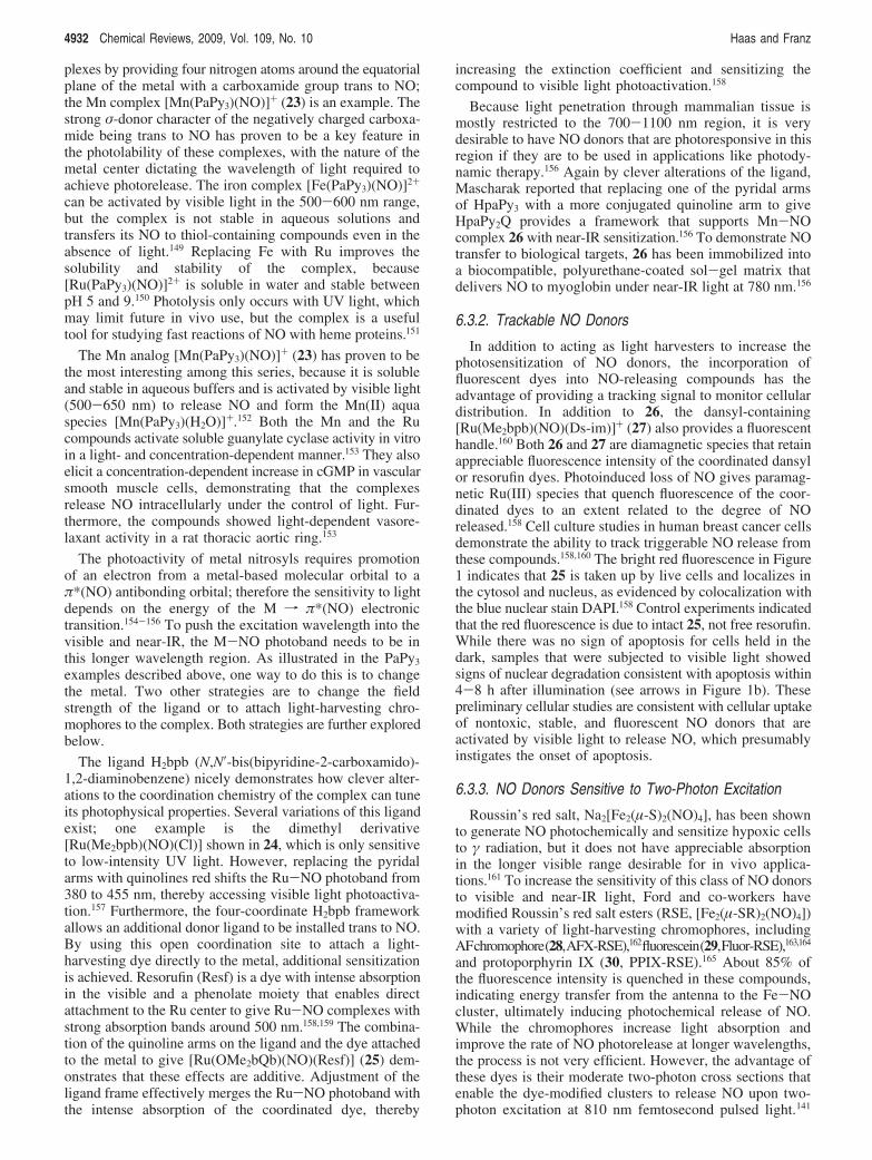
In addition to acting as light harvesters to increase thephotosensitization of NO donors, the incorporation offluorescent dyes into NO-releasing compounds has theadvantage of providing a tracking signal to monitor cellulardistribution. In addition to 26, the dansyl-containing[Ru(Me2bpb)(NO)(Ds-im)]+ (27) also provides a fluorescenthandle.160 Both 26 and 27 are diamagnetic species that retainappreciable fluorescence intensity of the coordinated dansylor resorufin dyes. Photoinduced loss of NO gives paramag-netic Ru(III) species that quench fluorescence of the coor-dinated dyes to an extent related to the degree of NOreleased.158 Cell culture studies in human breast cancer cellsdemonstrate the ability to track triggerable NO release fromthese compounds.158,160 The bright red fluorescence in Figure1 indicates that 25 is taken up by live cells and localizes inthe cytosol and nucleus, as evidenced by colocalization withthe blue nuclear stain DAPI.158 Control experiments indicatedthat the red fluorescence is due to intact 25, not free resorufin.While there was no sign of apoptosis for cells held in thedark, samples that were subjected to visible light showedsigns of nuclear degradation consistent with apoptosis within4-8 h after illumination (see arrows in Figure 1b). Thesepreliminary cellular studies are consistent with cellular uptakeof nontoxic, stable, and fluorescent NO donors that areactivated by visible light to release NO, which presumablyinstigates the onset of apoptosis.
6.3.3. NO Donors Sensitive to Two-Photon Excitation
Roussin’s red salt, Na2[Fe2(µ-S)2(NO)4], has been shownto generate NO photochemically and sensitize hypoxic cellsto " radiation, but it does not have appreciable absorptionin the longer visible range desirable for in vivo applica-tions.161 To increase the sensitivity of this class of NO donorsto visible and near-IR light, Ford and co-workers havemodified Roussin’s red salt esters (RSE, [Fe2(µ-SR)2(NO)4])with a variety of light-harvesting chromophores, includingAFchromophore(28,AFX-RSE),162fluorescein(29,Fluor-RSE),163,164
and protoporphyrin IX (30, PPIX-RSE).165 About 85% ofthe fluorescence intensity is quenched in these compounds,indicating energy transfer from the antenna to the Fe-NOcluster, ultimately inducing photochemical release of NO.While the chromophores increase light absorption andimprove the rate of NO photorelease at longer wavelengths,the process is not very efficient. However, the advantage ofthese dyes is their moderate two-photon cross sections thatenable the dye-modified clusters to release NO upon two-photon excitation at 810 nm femtosecond pulsed light.141
4932 Chemical Reviews, 2009, Vol. 109, No. 10 Haas and Franz
While PPIX-RSE is only soluble in organic solvents, Fluor-RSE has modest solubility in water and therefore haspotential as a biological NO donor via two-photon excitation.Poly(ethylene glycol) units have also been incorporated ontoa RSE cluster modified with a two-photon chromophore toinstill water solubility. The complex was shown to havemoderate cancer cell killing ability following two-photonexcitation.166
6.3.4. Other NO Donors
In addition to metal nitrosyl complexes, metal nitrito(M-NO2) complexes can also act as precursors for NOrelease. Examples in this category include Ru supported bybipyridine (bpy) and pyridine (py) spectator ligands, cis-[Ru(NO2)(py)(bpy)2]+,167 and Cr(III) supported by the tet-raaza macrocycle cyclam to give the water-soluble trans-[Cr(cyclam)(ONO)2]+, which release NO upon visible lightexcitation.168 To increase the absorption cross-section, pen-dant chromophores such as anthracene shown in 31 havebeen attached to the cyclam ring;169 alternatively, CdSe/ZnScore/shell quantum dots have also served as antennas tosensitize photorelease of NO from [Cr(cyclam)(ONO)2]+cations that are electrostatically embedded in the nanoparticleassembly.170,171
6.4. Metal Complexes as CO-ReleasingCompounds
CO is produced endogenously in mammalian cells as abyproduct of heme degradation by heme oxygenases (HO),which also release biliverdin and iron as coproducts. Longthought of as a potentially toxic waste product, within thepast decade this diatomic gaseous molecule has emerged asan important signaling molecule that influences numerousphysiological processes.172 The basal level of CO in healthyhumans is approximately 20 µmol/h, but diseases includingasthma, cystic fibrosis, and diabetes can significantly elevatethis capacity.173 In addition to a constitutively expressedHO-2 isozyme, there is also inducible HO-1, which isupregulated in response to both chemical and physicalstress.172 Several protective roles for CO have been identifiedby increasing the activity of HO-1 or by administering lowconcentrations of CO gas in cell culture or animal models.These roles include vasorelaxtion, inhibition of smooth
muscle cell proliferation, protection against organ transplantrejection, inhibition of platelet aggregation, antiapoptosis,anti-inflammatory, neurotransmission, and protection againstischemic tissue injury.172-174
The expanding possibilities for how heme oxygenaseregulates physiological response via CO production alongwith the possibility that CO could be used therapeutically ifappropriately administered has led to a search for smallmoleculesthatcandeliverCOinacontrolledfashion.121,173,175-177
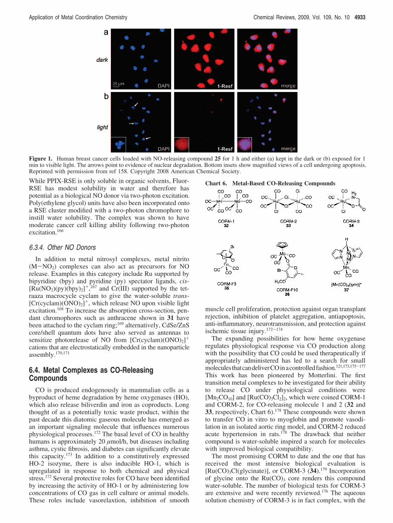
This work has been pioneered by Motterlini. The firsttransition metal complexes to be investigated for their abilityto release CO under physiological conditions were[Mn2CO10] and [Ru(CO)3Cl2]2, which were coined CORM-1and CORM-2, for CO-releasing molecule 1 and 2 (32 and33, respectively, Chart 6).178 These compounds were shownto transfer CO in vitro to myoglobin and promote vasodi-lation in an isolated aortic ring model, and CORM-2 reducedacute hypertension in rats.178 The drawback that neithercompound is water-soluble inspired a search for moleculeswith improved biological compatibility.
The most promising CORM to date and the one that hasreceived the most intensive biological evaluation is[Ru(CO)3Cl(glycinate)], or CORM-3 (34).179 Incorporationof glycine onto the Ru(CO)3 core renders this compoundwater-soluble. The number of biological tests for CORM-3are extensive and were recently reviewed.176 The aqueoussolution chemistry of CORM-3 is in fact complex, with the
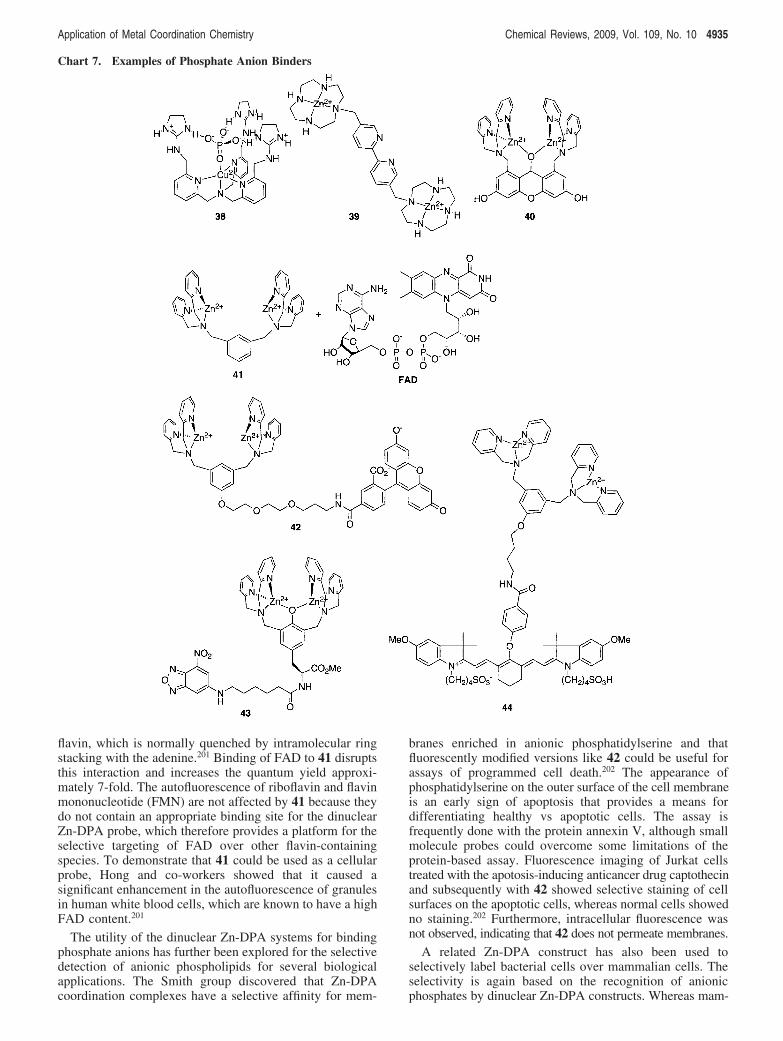
Figure 1. Human breast cancer cells loaded with NO-releasing compound 25 for 1 h and either (a) kept in the dark or (b) exposed for 1min to visible light. The arrows point to evidence of nuclear degradation. Bottom insets show magnified views of a cell undergoing apoptosis.Reprinted with permission from ref 158. Copyright 2008 American Chemical Society.
Chart 6. Metal-Based CO-Releasing Compounds
Application of Metal Coordination Chemistry Chemical Reviews, 2009, Vol. 109, No. 10 4933
major species present at physiological pH 7.4 conditionsexpected to be [Ru(CO)2(CO2)Cl(glycinate)]2- or[Ru(CO)2(CO2H)(OH)(glycinate)]-, which result from attackof hydroxide onto a coordinated carbonyl.180 The half-lifeof CO loss from CORM-3 varies dramatically depending onthe solution conditions: in distilled water, it is 98 h, whereasit shortens to under 4 min in human plasma.177 The differencein rates of CO liberation is likely a consequence of themechanism of CORM-3 CO loss, which has been suggestedto involve replacement of the labile glycinate and chlorideligands by other components in solution.121,177 In plasma orcellular environments, competing ligands like cysteine orglutathione could act as strong trans-labilizing ligands thatinduce CO loss. The fact that the rate of CO release can betuned depending on the trans ligand portends that furtheroptimization of this system could result in compounds whereexchange of the trans ligand itself could be manipulatedunder certain conditions to instigate CO loss.
In addition to Mn and Ru, Fe carbonyl complexes are alsobeing investigated for their CO-releasing properties. Thehomoleptic compound [Fe(CO)5] was dismissed early on asa biologically viable candidate because photolysis to releaseCO caused undesirable precipitation.178 The water-soluble[CpFe(CO)3]Cl also suffered from precipitation followingCO loss, but substitution of the cyclopentadienyl ring resultsin water-soluble compounds of the type[(C5H4CO2Me)Fe(CO)3]+ with promising biological activ-ity.181 A series of CORMs containing 2-pyrone have alsobeen prepared in the hope that the pyrone moiety mightfacilitate cell membrane transport and intracellulardistribution.182,183 Examples include CORM-F3 (35) and theMo complex CORM-F10 (36). In these compounds, the rateand extent of CO release could be tuned by varying thehalogen directly attached to the 2-pyrone ring. With a COrelease rate of 3.4 µM/min in a DMSO/phosphate buffer,CORM-F10 is one of the fastest CO releasers to date.182
As with the case of photoactive NO-releasing molecules,photoactive CO-releasing molecules are attractive for directedand triggerable CO release in high concentration at alocalized site. Toward this goal, photoreactive Mn(CO)3 unitsstabilized by a tris(pyrazolyl)methane ligand to give[Mn(CO)3(tpm)]+ (37) have been introduced.184 This com-pound was shown to release two equivalents of CO tomyoglobin in solution following irradiation at 365 nm. Itwas also shown that the compound was taken up via passivediffusion by human colon cancer cells and induced photo-initiated cytotoxicity.184 Importantly, the compound was notcytotoxic in the dark.
6.5. Metal Complexes That Bind Phospho AnionsThe charge and Lewis acid character of metal ions make
them attractive components of sophisticated anion receptorswherein a metal ion is anchored by a ligand that allows opencoordination sites for anion binding. The ligand scaffold alsoprovides added molecular recognition for anion selectivityorreportergroupsforcolorimetricorfluorescencesensing.185-187
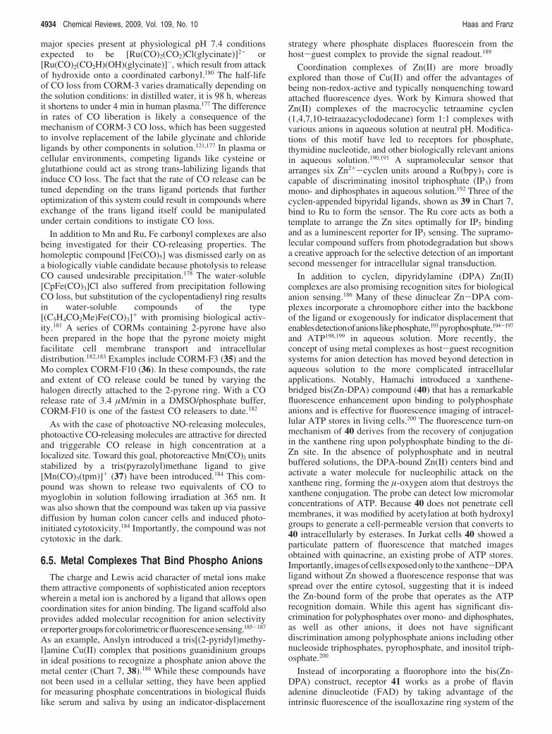
As an example, Anslyn introduced a tris[(2-pyridyl)methy-l]amine Cu(II) complex that positions guanidinium groupsin ideal positions to recognize a phosphate anion above themetal center (Chart 7, 38).188 While these compounds havenot been used in a cellular setting, they have been appliedfor measuring phosphate concentrations in biological fluidslike serum and saliva by using an indicator-displacement
strategy where phosphate displaces fluorescein from thehost-guest complex to provide the signal readout.189
Coordination complexes of Zn(II) are more broadlyexplored than those of Cu(II) and offer the advantages ofbeing non-redox-active and typically nonquenching towardattached fluorescence dyes. Work by Kimura showed thatZn(II) complexes of the macrocyclic tetraamine cyclen(1,4,7,10-tetraazacyclododecane) form 1:1 complexes withvarious anions in aqueous solution at neutral pH. Modifica-tions of this motif have led to receptors for phosphate,thymidine nucleotide, and other biologically relevant anionsin aqueous solution.190,191 A supramolecular sensor thatarranges six Zn2+-cyclen units around a Ru(bpy)3 core iscapable of discriminating inositol triphosphate (IP3) frommono- and diphosphates in aqueous solution.192 Three of thecyclen-appended bipyridal ligands, shown as 39 in Chart 7,bind to Ru to form the sensor. The Ru core acts as both atemplate to arrange the Zn sites optimally for IP3 bindingand as a luminescent reporter for IP3 sensing. The supramo-lecular compound suffers from photodegradation but showsa creative approach for the selective detection of an importantsecond messenger for intracellular signal transduction.
In addition to cyclen, dipyridylamine (DPA) Zn(II)complexes are also promising recognition sites for biologicalanion sensing.186 Many of these dinuclear Zn-DPA com-plexes incorporate a chromophore either into the backboneof the ligand or exogenously for indicator displacement thatenablesdetectionofanionslikephosphate,193pyrophosphate,194-197
and ATP198,199 in aqueous solution. More recently, theconcept of using metal complexes as host-guest recognitionsystems for anion detection has moved beyond detection inaqueous solution to the more complicated intracellularapplications. Notably, Hamachi introduced a xanthene-bridged bis(Zn-DPA) compound (40) that has a remarkablefluorescence enhancement upon binding to polyphosphateanions and is effective for fluorescence imaging of intracel-lular ATP stores in living cells.200 The fluorescence turn-onmechanism of 40 derives from the recovery of conjugationin the xanthene ring upon polyphosphate binding to the di-Zn site. In the absence of polyphosphate and in neutralbuffered solutions, the DPA-bound Zn(II) centers bind andactivate a water molecule for nucleophilic attack on thexanthene ring, forming the µ-oxygen atom that destroys thexanthene conjugation. The probe can detect low micromolarconcentrations of ATP. Because 40 does not penetrate cellmembranes, it was modified by acetylation at both hydroxylgroups to generate a cell-permeable version that converts to40 intracellularly by esterases. In Jurkat cells 40 showed aparticulate pattern of fluorescence that matched imagesobtained with quinacrine, an existing probe of ATP stores.Importantly, imagesofcellsexposedonlyto thexanthene-DPAligand without Zn showed a fluorescence response that wasspread over the entire cytosol, suggesting that it is indeedthe Zn-bound form of the probe that operates as the ATPrecognition domain. While this agent has significant dis-crimination for polyphosphates over mono- and diphosphates,as well as other anions, it does not have significantdiscrimination among polyphosphate anions including othernucleoside triphosphates, pyrophosphate, and inositol triph-osphate.200
Instead of incorporating a fluorophore into the bis(Zn-DPA) construct, receptor 41 works as a probe of flavinadenine dinucleotide (FAD) by taking advantage of theintrinsic fluorescence of the isoalloxazine ring system of the
4934 Chemical Reviews, 2009, Vol. 109, No. 10 Haas and Franz
flavin, which is normally quenched by intramolecular ringstacking with the adenine.201 Binding of FAD to 41 disruptsthis interaction and increases the quantum yield approxi-mately 7-fold. The autofluorescence of riboflavin and flavinmononucleotide (FMN) are not affected by 41 because theydo not contain an appropriate binding site for the dinuclearZn-DPA probe, which therefore provides a platform for theselective targeting of FAD over other flavin-containingspecies. To demonstrate that 41 could be used as a cellularprobe, Hong and co-workers showed that it caused asignificant enhancement in the autofluorescence of granulesin human white blood cells, which are known to have a highFAD content.201
The utility of the dinuclear Zn-DPA systems for bindingphosphate anions has further been explored for the selectivedetection of anionic phospholipids for several biologicalapplications. The Smith group discovered that Zn-DPAcoordination complexes have a selective affinity for mem-
branes enriched in anionic phosphatidylserine and thatfluorescently modified versions like 42 could be useful forassays of programmed cell death.202 The appearance ofphosphatidylserine on the outer surface of the cell membraneis an early sign of apoptosis that provides a means fordifferentiating healthy vs apoptotic cells. The assay isfrequently done with the protein annexin V, although smallmolecule probes could overcome some limitations of theprotein-based assay. Fluorescence imaging of Jurkat cellstreated with the apotosis-inducing anticancer drug captothecinand subsequently with 42 showed selective staining of cellsurfaces on the apoptotic cells, whereas normal cells showedno staining.202 Furthermore, intracellular fluorescence wasnot observed, indicating that 42 does not permeate membranes.
A related Zn-DPA construct has also been used toselectively label bacterial cells over mammalian cells. Theselectivity is again based on the recognition of anionicphosphates by dinuclear Zn-DPA constructs. Whereas mam-
Chart 7. Examples of Phosphate Anion Binders
Application of Metal Coordination Chemistry Chemical Reviews, 2009, Vol. 109, No. 10 4935
malian cell surfaces are composed mostly of zwitterionicphospholipids, the surfaces of bacterial cells contain a highpercentage of anionic phospholipids. The Smith grouptherefore explored the differential interactions of a familyof complexes based on the 2,6-bis(zinc(II)-dipicolylami-ne)phenoxide construct.203 An NBD-tagged version is shownin 43, but other derivatives have been made in which thealkyl substituent trans to the phenoxide is varied. Whereaslipophilic versions partition into zwitterionic vesicles and aremoderately toxic to mammalian cells, a hydrophilic versionthat contains just a methyl substituent off the phenoxide isnot toxic to mammalian cells but is very effective at killingthe pathogenic bacterium Staphylococcus aureus. The anti-biotic action appears to be caused by depolarization of thebacterial cell membrane, which is proposed to occur as aresult of large fluctuations in local charge following thebinding of cationic Zn-DPA complexes to anionic phospho-lipids head groups.203 Fluorescence imaging studies of thefluorophore-labeled version 43 show that staining of both S.aureus and E. coli is localized to the cell walls, whichsuggests that these complexes might be useful for detectingand imaging bacteria.203
Indeed, incorporating a carbocyanine dye that is sensitiveto tissue-penetrating near-IR light onto a Zn-DPA constructenabled in vivo imaging of bacterial infection in a livinganimal.204 Nude mice were infected with S. aureus byinjection of bacteria into the thigh muscle. The NIRfluorescent probe 44 was introduced 6 h postinfection intothe bloodstream via a tail-vein injection, and anesthetizedanimals were irradiated with 720 nm light for imaging. Thefluorescent probe accumulated at the site of infection, withthe maximum signal contrast reached after 18 h (Figure 2).204
6.6. Metal Complexes for PhosphoproteinDetection
The interest in developing sensors that can selectivelydetect protein phosphorylation stems from the central rolethat this post-translational modification plays in signaltransduction pathways and a host of other biologicalprocesses.205,206 Phosphate groups are installed onto aprotein’s serine, threonine, or tyrosine amino acid side chainsby protein kinases and removed by phosphatases. Thedifference between the negatively charged, moderatelysterically demanding phosphomonoester compared with theneutral hydroxyl group of the unmodified amino acidprovides a means of using chemistry to differentiate phos-phorylated peptide or protein sequences from their unphos-
phorylated analogs. Metal complexes in particular areattractive components of kinase/phosphatase sensors becausethey can couple a strong metal-phosphate interaction (asdemonstrated in the previous section) to a change influorescence as a spectroscopic readout of phosphorylationstate. Chart 8 shows examples of metal-dependent sensorsfor protein phosphorylation that are based on Zn(II), Ca(II),Mg(II), and Ln(III) complexes. The goal is to create probesthat can selectively recognize a phosphoprotein of interestand thereby visualize the activity of specific protein kinasesand phosphatases in real time in living cells, tissues, andeven whole organisms.206,207 While this ultimate goal has yetto be realized, the probes described below have achievedsome success in biological applications.
6.6.1. Phosphoprotein Detection by Zinc Complexes
In furthering the anion receptor capability of the Zn-DPAunit, Hamachi has developed a family of probes thatrecognize and sense phosphopeptides.208 The first probescontained two Zn-DPA units spanned by an anthracenebridge, one example is shown in 45. Given the similarity of45 to the ATP sensors described above, it is not surprisingthat 45 also has a high affinity for ATP and ADP (Kapp >107 M-1) in addition to phosphate monoesters but weakaffinity for other anions including carbonate, sulfonate,nitrate, acetate, and chloride.209,210 The Kapp of 45 forNaH2PO4 and phosphotyrosine is !105 M-1, whereas itsaffinity for select phosphopeptides increases 1 or 2 ordersof magnitude depending on the peptide sequence. A com-parison of a series of short peptides with sequences basedon various kinase recognition domains revealed that receptor45 has the highest affinity for peptides with the greatestoverall net negative charge. For example, 45 has a Kapp of 9" 106 M-1 for the phosphotryosine (pTyr)-containingsequence Glu-Glu-Glu-Ile-pTyr-Glu-Glu-Phe-Asp, the con-sensus sequence of the protein kinase, v-Src. Peptides withfewer negative charges or overall positive charge elicitweaker fluorescent changes by the Zn-DPA probes. Thiscorrelation between overall net charge of the phosphopeptideand its affinity for the probe presumably arises from afavorable electrostatic attraction between the tetracationicdinuclear Zn-DPA receptor and negatively charged peptides.The unphosphorylated version of the v-Src peptide does notinduce a fluorescence change of 45, proving that thephosphate functionality is required for molecular recognitionby the probe.
The dinuclear design of these receptors is critical to theirfunction, since mononuclear versions show very little affinityfor phospho species. An X-ray crystal structure of 45 boundto phenyl phosphate shows the phosphate unit bridging thetwo Zn(II) centers by providing a separate O ligand to eachmetal210 (as indicated by the binding mode of p-Tyr to 45 inChart 8), thus providing a structural basis for the cooperat-ivity of the dinuclear site in recognizing phosphomonoesterfunctional groups.
The cooperativity of the dinuclear site is also critical forthe fluorescence sensing mechanism of 45. Binding of thed10 ion Zn2+ to the receptor increases fluorescence bycanceling the photoinduced electron transfer of the benzylicamine that otherwise quenches the appended anthracenefluorescence. The receptor is not maximally fluorescentunless both sites are saturated with Zn2+, a situation that isfacilitated by the presence of phosphate derivatives.210
Figure 2. Optical image of a mouse with a S. aureus infection inthe left rear thigh muscle 18 h after tail-vein injection of carbocya-nine-modified bis(ZnDPA) 44, a probe designed to detect anionicphospholipids found in bacterial cell walls. Scale represents relativefluorescence in arbitrary units. Reprinted with permission from ref204. Copyright 2006 American Chemical Society.
4936 Chemical Reviews, 2009, Vol. 109, No. 10 Haas and Franz
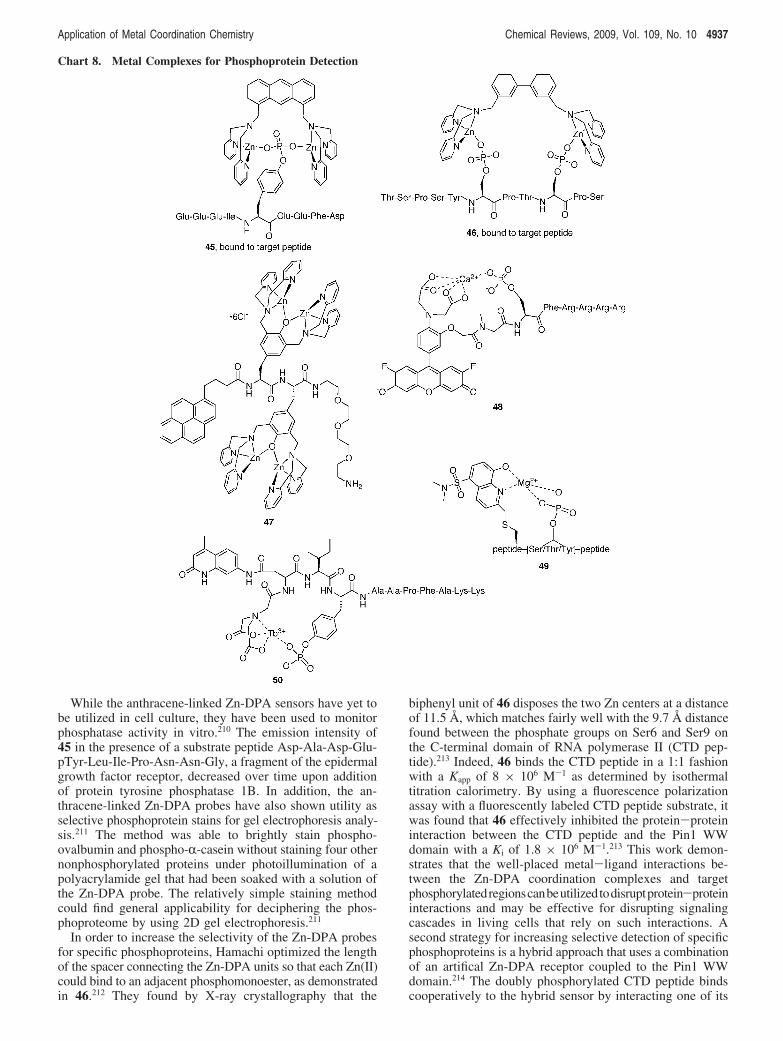
While the anthracene-linked Zn-DPA sensors have yet tobe utilized in cell culture, they have been used to monitorphosphatase activity in vitro.210 The emission intensity of45 in the presence of a substrate peptide Asp-Ala-Asp-Glu-pTyr-Leu-Ile-Pro-Asn-Asn-Gly, a fragment of the epidermalgrowth factor receptor, decreased over time upon additionof protein tyrosine phosphatase 1B. In addition, the an-thracene-linked Zn-DPA probes have also shown utility asselective phosphoprotein stains for gel electrophoresis analy-sis.211 The method was able to brightly stain phospho-ovalbumin and phospho-R-casein without staining four othernonphosphorylated proteins under photoillumination of apolyacrylamide gel that had been soaked with a solution ofthe Zn-DPA probe. The relatively simple staining methodcould find general applicability for deciphering the phos-phoproteome by using 2D gel electrophoresis.211
In order to increase the selectivity of the Zn-DPA probesfor specific phosphoproteins, Hamachi optimized the lengthof the spacer connecting the Zn-DPA units so that each Zn(II)could bind to an adjacent phosphomonoester, as demonstratedin 46.212 They found by X-ray crystallography that the
biphenyl unit of 46 disposes the two Zn centers at a distanceof 11.5 Å, which matches fairly well with the 9.7 Å distancefound between the phosphate groups on Ser6 and Ser9 onthe C-terminal domain of RNA polymerase II (CTD pep-tide).213 Indeed, 46 binds the CTD peptide in a 1:1 fashionwith a Kapp of 8 " 106 M-1 as determined by isothermaltitration calorimetry. By using a fluorescence polarizationassay with a fluorescently labeled CTD peptide substrate, itwas found that 46 effectively inhibited the protein-proteininteraction between the CTD peptide and the Pin1 WWdomain with a Ki of 1.8 " 106 M-1.213 This work demon-strates that the well-placed metal-ligand interactions be-tween the Zn-DPA coordination complexes and targetphosphorylatedregionscanbeutilizedtodisruptprotein-proteininteractions and may be effective for disrupting signalingcascades in living cells that rely on such interactions. Asecond strategy for increasing selective detection of specificphosphoproteins is a hybrid approach that uses a combinationof an artifical Zn-DPA receptor coupled to the Pin1 WWdomain.214 The doubly phosphorylated CTD peptide bindscooperatively to the hybrid sensor by interacting one of its
Chart 8. Metal Complexes for Phosphoprotein Detection
Application of Metal Coordination Chemistry Chemical Reviews, 2009, Vol. 109, No. 10 4937
pSer residues with the WW domain and the other with theZn-DPA unit.
In a slightly different application from phosphoproteindetection, Zn-DPA complexes have also shown an abilityto deliVer phosphopeptides and other phosphate derivativesintracellularly. The highly cationic tetranuclear complex 47with sufficient lipophilic character was found as the optimalcarrier vector to shuttle anionic phosphorylated peptides intocells via endocytosis.215 The intracellular fluorescence offluorescently tagged phosphopeptides was found to increase4-6-fold in the presence of 47 compared with the absenceof the metalloreceptor.
6.6.2. Phosphoprotein Detection by Calcium andMagnesium Complexes
In addition to Zn2+, Ca2+ and Mg2+ are also effective atenhancing the fluorescence signal of designed probes viaphosphorylation-dependent chelation. The design of 48 isbased on Ca2+ indicators developed by Tsien that rely onCa2+ coordination to turn on fluorescence.216 To convert thisidea into a kinase sensor, Lawrence and co-workers replacedone of the iminodiacetate arms with a serine-containingpeptide that is a substrate for protein kinase C.217 Theunphosphorylated probe has low affinity for Ca2+ and istherefore weakly fluorescent, but phosphorylation increasesthe binding affinity of the receptor and turns on the signal.Optimization of the linker connecting the fluorophore andthe peptide provided probe 48 with a 23-fold increase influorescence after phosphorylation.
Imperiali and co-workers have developed a family ofkinase probes that incorporate a sulfonamido-oxine aminoacid (Sox) strategically embedded in kinase recognitiondomains such that phosphorylation improves the chelationof Mg2+ to enhance Sox fluorescence 4-12-fold.207 Theseprobes have been validated for the measurement of kinaseactivities in crude mammalian cell lysates.218 The originaldesigns required a conformationally constrained peptide!-turn to position Sox appropriately for cooperative bindingof Mg2+ with the phosphorylated amino acid.219 Morerecently, they have greatly expanded the scope of Sox-basedkinase sensors by developing a Sox derivative called C-Soxthat can alkylate a cysteine residue with the chromophore(49 in Chart 8).220 This strategy has allowed the probe to beincorporated into a larger number of kinase recognitiondomains with improved specificity. The probes are amenablefor high-throughput screening for kinase inhibitors and,because they are sensitive under conditions of physiologicalMg2+ levels, may be amenable to future in vivo applica-tions.220
6.6.3. Phosphoprotein Detection by LanthanideComplexes
Long-lived emission lifetimes and sharp emission bandsare some of the photophysical properties that make lanthanide(Ln) ions attractive for incorporation into biological probes.The trivalent Ln3+ is the most common oxidation state acrossthe series. Being hard acids, they have a strong preferencefor negatively charged, oxygen-rich coordination environ-ments. Not surprisingly, they interact strongly with phosphate-containing groups. Our lab has shown that the phosphory-lation state of a peptide can dramatically increase its affinityfor metal ions.221,222 As a consequence, Tb3+ can be aneffective reporter of phosphorylation status.221,223 In these
cases, the peptides are short sequences resembling calcium-binding loops where the phosphorylated amino acid eitheris embedded within the peptide, as in DPDNEA(pY)EMP-SEEG,221 or is the anchoring metal-ligating residue near theend of the peptide, as in DKNADGWIDRA(pS)LA,223 wherepY and pS are phosphotyrosine and phosphoserine, respec-tively. Since lanthanides are poor absorbers, sensitizingchromophores are required to achieve luminescence. In thesepeptide examples, the pY itself or a tryptophan (W) residueprovides sufficient sensitization to observe luminescence.
To increase the sensitization and the metal affinity, Samesincorporated a carbostyril chromophore and an iminodiacetatechelating motif onto peptides that contain a tyrosine residuewithin a recognition domain for src and abl protein tyrosinekinases (50).224 The peptide-based probe can detect bothkinase (phosphorylation) and phosphatase (dephosphoryla-tion) activity by diluting aliquots of the enzyme assay intobuffers containing either Tb3+ or Eu3+. The probe may beuseful for in vitro evaluation of kinase and phosphataseinhibitors.224
6.7. Metal Complexes That Release BioactiveSmall Molecules6.7.1. Photorelease of Bioactive Molecules
Coordinating a bioactive molecule to a metal center is anattractive way to mask the activity of a molecule of interest.Subsequent rupture of that metal-ligand bond by a specifictrigger could in principle release the desired molecule in acontrolled way. Etchenique and colleagues have applied thisgeneral principle in their development of ruthenium poly-pyridine complexes as inorganic photolabile caging groupsfor several amine-containing neurotransmitters. In the ex-ample 51 shown in Chart 9, two molecules of "-aminobutyricacid (GABA) coordinate to a Ru(II) center supported by twobipyridine ligands to form the caged complex[Ru(bpy)2(L)2]2+, where L is GABA or one of several aminecompounds tested, including serotonin, tryptamine, buty-lamine, and tyramine.225 Amino acids like glutamate can alsobe caged by replacing one of the bpy ligands with a tridentatetris(pyrazolylmethane) ligand to give a Ru complex with onlyone available open coordination site. This strategy forces theamino acid ligand to bind through its amine functional groupand avoid a bidentate mode that would be recalcitrant tophotorelease.226 Excitation of these molecules with 450 nmlight into a metal-ligand charge transfer band of the Ru(bpy)core induces heterolytic bond cleavage of one of themonodentate ligands, thereby releasing one equivalent of thefree amine. As monitored by NMR, the photoreaction isclean, providing only the monoaquo complex[Ru(bpy)2(L)(H2O)]2+ (52) as the other photoproduct. Thequantum efficiency of the uncaging photoreaction wasdetermined to be 0.03.
Incorporation of triphenylphosphine (PPh3) into the co-ordination sphere to form [Ru(bpy)2(PPh3)(GABA)]2+ withonly one amine ligand coordinated increases the quantumyield to 0.21, which is slightly higher than that of the moreconventional nitrobenzyl ester photolabile protecting groupsthat have been used to cage GABA and other otherbiomolecules.227 Conventional, organic photocaging groupsrequire UV excitation that could cause cellular photodamage.An advantage of the Ru-based caging groups is their longerwavelength photoreactivity with visible light. In addition, it
4938 Chemical Reviews, 2009, Vol. 109, No. 10 Haas and Franz

has been shown that the Ru-based caged compounds canundergo two-photon excitation at 800 nm.228
To date the Ru-based cages have been applied in twobiological settings. Current flow was measured by voltage-clamp recordings of frog oocytes expressing the GABAc
receptor, a ligand-gated ion channel that opens for Cl- flowonly upon activation by GABA. When applied in the dark,the Ru-caged GABA complex [Ru(bpy)2(PPh3)(GABA)]2+
caused no change in Cl- currents. Pulses of visible light,however, resulted in measurable increases in current flow,consistent with release of GABA, which is then free toactivate the GABAc receptor.227 In another example, actionpotentials of a leech neuron were measured by a micropipetelectrode inserted into isolated neurons that were exposedto [Ru(bpy)2(4AP)2]2+, where 4AP is 4-aminopyridine, acompound known to increase neuronal activity by blockingcertain K+ channels. Action potentials increased when theneurons were exposed to both the Ru compound and flashesof visible light, consistent with photorelease of 4AP tostimulate neuronal response.229
6.7.2. Reductive Activation and Ligand Exchange
Reductive activation or ligand exchange is an alternativestrategy of releasing a bioactive compound that is deactivatedby coordination to a metal center. In principle, kineticallyinert metal ions like Co3+ could be used as chaperones todeliver bioactive molecules intracellularly by releasing themupon reduction to the more labile Co2+ oxidation state.230
This strategy has been explored with a variety of Co(III)coordination complexes with amine-containing mustardcompounds that are known alkylating agents.231,232 Some ofthis family of compounds show hypoxia-selective cytotox-icity, suggesting that the more reducing environment of
hypoxic tumors favors reduction of the metal complex andrelease of the cytotoxic agent. Details of this mechanism arestill not clear, and more recent work suggests that ligandexchange is the more likely mechanism.230 More recently,Hambley has shown that the matrix metalloproteinaseinhibitor marimastat can be incorporated into a Co(III)coordination complex (53) and used as a prodrug to inhibittumor growth.233 Unfortunately, both free marimisat and themetal-bound prodrug also potentiated tumor metastasisrelative to controls.
Given the promise of using cobalt as a drug chaperoneand the clear need to understand its mechanisms of cellulardistribution and prodrug activation in order to rationallydesign effective agents, complexes with fluorescent ligandssuch as 54 were developed.234 In the example shown, ahydroxamic acid derivative of coumarin-343 mimics thehydroxamic acid motif of marimistat. In the Co(III) form,the fluorescence of the metal-bound coumarin is quenchedbut is restored upon ligand release, be it by reduction orligand exchange. Fluorescence microscopy studies showedthat the subcellular distribution of the fluorophore was alteredby coordination to the metal complex. Because the intactmetal-fluorophore emmission is weak, the intensity of theimages suggests that the fluorophore is released from themetal, while the different staining patterns of the metalcomplexes vs the free coumarin suggest that metal com-plexation influences its cellular distribution. These studiesimportantly demonstrate that metal complexes can effectivelychaperone bioactive molecules and traffic them in differentways compared with the uncomplexed agents, which mayhave important ramifications for altering the pharmacologyof small molecules. Furthermore, these studies provide astrategy for monitoring the cellular trafficking and reactivity
Chart 9. Metal Complexes That Release Bioactive Amine-Containing Small Molecules
Application of Metal Coordination Chemistry Chemical Reviews, 2009, Vol. 109, No. 10 4939
of cobalt coordination complexes. Further studies aimed atunderstanding how to control the localization and release ofactive agents from these kinds of complexes remain animportant goal.
6.7.3. Catalytic Release of Bioactive Molecules
Meggers and co-workers have developed a differentstrategy for releasing amine-containing molecules insidecells. Rather than relying on breaking a metal-ligand bondto release the small molecule, they are developing organo-metallic complexes as biocompatible catalysts to carry outintracellular organic transformations. Although the aqueous,aerobic environment of a cell presents significant challengesfor most organometallic complexes, [Cp*Ru(cod)Cl] (Cp*) pentamethylcyclopentadienyl, cod ) 1,5-cyclooctadiene)(55) was found to be tolerant of such conditions and able tocatalyze the cleavage of allylcarbamates in the presence ofexcess thiophenol to give amines.235 To show that such atransformation could be conducted in a mammalian cell, abis-allyloxycarbonyl-protected rhodamine (56) was incubatedwith cultured HeLa cells. The protected rhodamine derivativeis not fluorescent, but treatment with the Ru catalyst andthiols deprotects it to give highly fluorescent rhodamine 57,as shown in Chart 9. Treatment of the cells with the Rucatalyst caused a 3.5-fold increase in fluorescence within thecytoplasm, as observed by confocal fluorescence microscopy.Addition of thiophenol to the cell medium increased thefluorescence 10-fold. Cell viability was not affected by thetreatment with the Ru compound, thiophenol, or the cagedrhodamine.235 These encouraging results suggest that thereis significant scope for further development of organometalliccomplexes used in biology. Indeed, the growing field ofbioorganometallic chemistry offers exciting opportunities touncover compounds with unique biological properties.33,236,237
7. Metal Complexes as Enzyme InhibitorsSmall molecules that are capable of selectively inhibiting
a particular enzyme are powerful tools for deciphering anenzyme’s biological role. If the enzyme is involved indisease, its inhibitors become important drug candidates.While most inhibitors are organic compounds, inorganiccoordination chemistry provides attractive, and perhaps toooften overlooked, strategies for enzyme and protein inhibi-tion. Metal-binding compounds and metal complexes them-selves both offer unique properties that contribute to enzymeinhibition and are not found in conventional organic mol-ecules. Metalloproteins, which comprise one-third of allproteins, are obvious targets for inhibition based on coor-dination chemistry, but metal complexes can also targetnonmetalloproteins.
Because the metal cofactor of a metalloprotein is criticalfor function, removing or replacing the metal are veryeffective means of abrogating activity. Chelators that stripmetals from their active sites, however, are usually nonspe-cific and provide little advantage for developing inhibitorstargeted for a specific enzyme. Likewise, substituting a nativemetal ion with an inactive, non-native one can help instudying an isolated metalloenzyme in vitro, but is a difficultstrategy to implement in the complex environment of a cellor whole organism. In contrast, compounds that bind directlyto an active site metal ion can inhibit enzyme activity byblocking access of the substrate to the catalytic site.Additional interactions like hydrogen bonding and hydro-
phobic contacts between the inhibitor and the protein canincrease specificity of the inhibitor for the protein pocket.For proteins that do not contain a metal site, intact metalcomplexes can take advantage of their shape and organicframework to bind to proteins with high specificity throughouter-sphere interactions. The inhibitor-protein interactioncan be further strengthened if an amino-acid side chain fromthe protein binds directly in the inner sphere of the metalcomplex to anchor the inhibitor in place. In some cases, themetal ion may not be part of the administered compoundbut is instead picked up endogenously from the biologicalmilieu. Finally, the reactivity of metal complexes can beharnessed to develop catalytic inhibitors that incorporateaspects of molecular recognition for specificity along withhydrolytic or oxidative reactivity to inactivate proteinsthrough chemical modification.
The use of metal complexes as enzyme inhibitors wasreviewed by Louie and Meade in 1999.238 Since that time,the sophistication in terms of the design and understandingof metal complexes as enzyme inhibitors has increasedsubstantially for a handful of examples, as emphasized in arecent perspective on the topic.239 These examples, however,still represent only a small minority of compounds comparedwith conventional, organic inhibitors. Rather than provideexhaustive coverage of all chelator-inhibited enzymes, thefollowing discussion provides select examples to highlightfour strategies for using inorganic and organometallicchemistry for targeted enzyme inhibition: (1) metalloproteininhibitors that use metal binding interactions as a significantcomponent of their molecular recognition, (2) compoundsthat recruit endogenous metal ions to increase their affinityfor protein binding and inhibition, (3) inert metal complexesthat provide novel scaffolds for binding protein active sites,and (4) metal complexes that catalytically inactivate proteins.The structures of compounds discussed below are shown inChart 10.
7.1. Inhibitors with Metal-Binding Headgroups forTargeting Metalloproteins
Carbonic anhydrase, matrix metalloproteinases (MMP),histone deacetylases (HDAC), and anthrax lethal factor aresome of the examples of zinc(II)-dependent hydrolyticenzymes that are attractive drug targets for a wide range ofdiseases.240 Inhibitors of these mononuclear Zn(II) enzymestypically contain two components: an organic backbone thatforms noncovalent, hydrogen bonding, hydrophobic, andelectrostatic interactions with the protein surface, and a metal-binding headgroup that coordinates the active-site Zn(II). Asignificant amount of work has gone into optimizing thebackbone fragments to impart specificity for a particularenzyme isoform, while less effort has focused on the zinc-binding group, which is typically a hydroxamic acid moiety,as shown in 58.241 In recent years, Cohen and co-workershave been reversing that trend by taking a bioinorganicapproach to identify alternative zinc-binding groups. Crystalstructures of small chelators coordinated to a Zn(II) modelcomplex that mimics the three histidine ligands in the activesite of MMPs were used in conjunction with computationalmodeling to identify promising zinc-binding groups thatcould be extended into full-length MMP inhibitors.242 Arange of chelating moieties have been investigated, includingpyrones, thiopyrones, hydroxypridinones, hydroxypyridi-nethiones, dipyridylamine, and aza-macrocycles.243
4940 Chemical Reviews, 2009, Vol. 109, No. 10 Haas and Franz
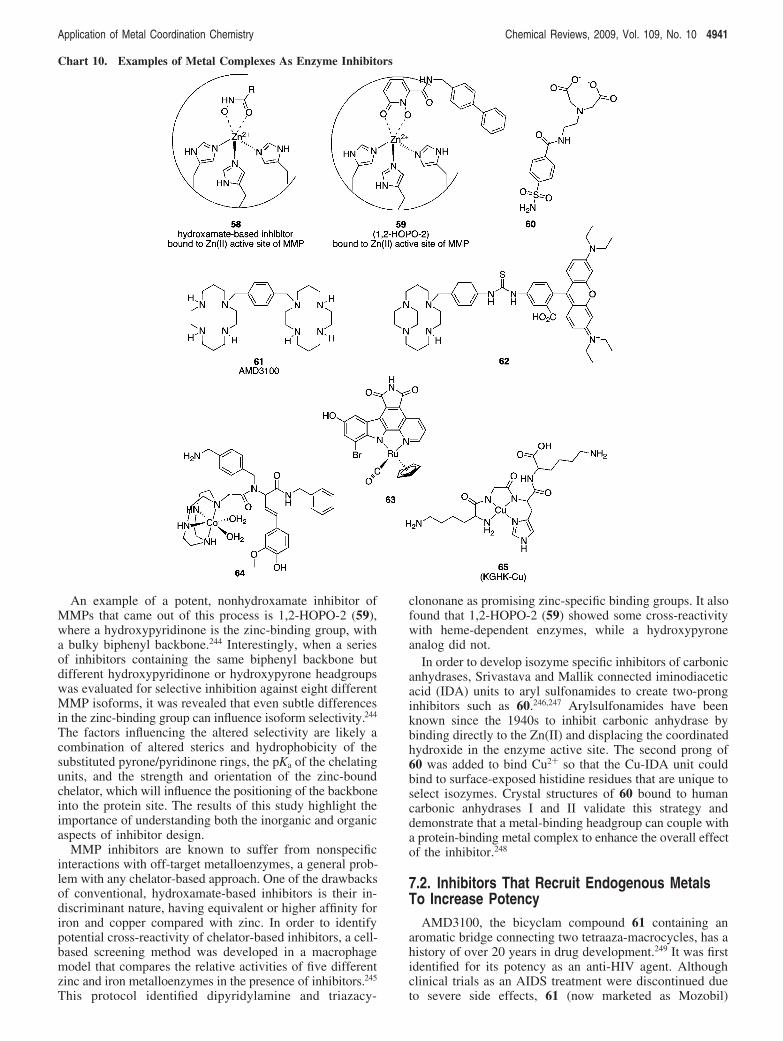
An example of a potent, nonhydroxamate inhibitor ofMMPs that came out of this process is 1,2-HOPO-2 (59),where a hydroxypyridinone is the zinc-binding group, witha bulky biphenyl backbone.244 Interestingly, when a seriesof inhibitors containing the same biphenyl backbone butdifferent hydroxypyridinone or hydroxypyrone headgroupswas evaluated for selective inhibition against eight differentMMP isoforms, it was revealed that even subtle differencesin the zinc-binding group can influence isoform selectivity.244
The factors influencing the altered selectivity are likely acombination of altered sterics and hydrophobicity of thesubstituted pyrone/pyridinone rings, the pKa of the chelatingunits, and the strength and orientation of the zinc-boundchelator, which will influence the positioning of the backboneinto the protein site. The results of this study highlight theimportance of understanding both the inorganic and organicaspects of inhibitor design.
MMP inhibitors are known to suffer from nonspecificinteractions with off-target metalloenzymes, a general prob-lem with any chelator-based approach. One of the drawbacksof conventional, hydroxamate-based inhibitors is their in-discriminant nature, having equivalent or higher affinity foriron and copper compared with zinc. In order to identifypotential cross-reactivity of chelator-based inhibitors, a cell-based screening method was developed in a macrophagemodel that compares the relative activities of five differentzinc and iron metalloenzymes in the presence of inhibitors.245
This protocol identified dipyridylamine and triazacy-
clononane as promising zinc-specific binding groups. It alsofound that 1,2-HOPO-2 (59) showed some cross-reactivitywith heme-dependent enzymes, while a hydroxypyroneanalog did not.
In order to develop isozyme specific inhibitors of carbonicanhydrases, Srivastava and Mallik connected iminodiaceticacid (IDA) units to aryl sulfonamides to create two-pronginhibitors such as 60.246,247 Arylsulfonamides have beenknown since the 1940s to inhibit carbonic anhydrase bybinding directly to the Zn(II) and displacing the coordinatedhydroxide in the enzyme active site. The second prong of60 was added to bind Cu2+ so that the Cu-IDA unit couldbind to surface-exposed histidine residues that are unique toselect isozymes. Crystal structures of 60 bound to humancarbonic anhydrases I and II validate this strategy anddemonstrate that a metal-binding headgroup can couple witha protein-binding metal complex to enhance the overall effectof the inhibitor.248
7.2. Inhibitors That Recruit Endogenous MetalsTo Increase Potency
AMD3100, the bicyclam compound 61 containing anaromatic bridge connecting two tetraaza-macrocycles, has ahistory of over 20 years in drug development.249 It was firstidentified for its potency as an anti-HIV agent. Althoughclinical trials as an AIDS treatment were discontinued dueto severe side effects, 61 (now marketed as Mozobil)
Chart 10. Examples of Metal Complexes As Enzyme Inhibitors
Application of Metal Coordination Chemistry Chemical Reviews, 2009, Vol. 109, No. 10 4941
continues to show promise for other diseases, in particularfor hematopoietic stem cell transplantation for treatment ofnon-Hodgkin’s lymphoma and multiple myeloma. Compound61 has been shown to be a highly specific antagonist for theCXCR4 chemokine receptor, a transmembrane protein in theG-protein-coupled receptor family that assists entry of HIVinto cells and anchors stem cells in the bone marrow.249
Binding of 61 to CXCR4 therefore blocks viral entry intothe host cell and also releases stem cells from the bonemarrow into the peripheral blood where they can be harvestedfor transplantation.
The elements of molecular recognition responsible for thetight binding of 61 to CXCR4 were initially thought to deriveprincipally from hydrogen-bonding interactions between thebicyclam N-H groups and specific carboxylic acid sidechains on the protein. The affinity of 61 for CXCR4 wasshown to increase 7-, 36-, and 50-fold when Cu2+, Zn2+,and Ni2+, respectively, were inserted into the bicyclamrings,250 suggesting that the metal complexes provide ad-ditional binding interactions that improve affinity. Whilecyclam has a higher thermodynamic binding affinity for Cu2+
than Zn2+ (log K ) 27.2 vs 15.5), the higher relativeconcentration of available Zn2+ in blood plasma (reportedat 19 µM) coupled with the faster kinetic rate forcyclam-Zn2+ complex formation at physiological pH sug-gests that AMD3100 likely exists as the Zn-bound form invivo.251
While there are no X-ray structures available for thetransmembrane CXCR4 receptor, the crystal structure re-ported by Sadler and co-workers of Cu-cyclam and Cu2-bicyclam bound to lysozyme provides some insight intoprotein-metallocyclam interactions.252 The structures dem-onstrate that carboxylate side chains can indeed directly bindto open sites on the metal complex. Further interactions areformed by hydrogen bonding between the cyclam NH andprotein carboxylates, as well as hydrophobic contacts toadjacent tryptophan residues. All of these interaction typesare likely present when 61 binds to its target CXCR4.Furthermore, the cyclam rings are found in two configura-tions, a folded cis configuration and a planar transconfiguration.
Metal-bound cyclam complexes can adopt several con-figurations that depend on the cis or trans arrangement ofcoordination sites not used by the ring nitrogens as well asthe stereochemistry around each chiral nitrogen donor atom.Archibald has introduced configurationally fixed macrocyclesthat in principle present only one configuration to the proteinto optimize metal-aspartate coordination that is the key tothe increased protein binding interaction of the metal-loadedform of 61.253 They have recently shown that the Cu(II)complex of fluorescently tagged, configurationally restrictedtetraazamacrocycle 62 competes effectively against anti-CXCR4 monoclonal antibodies and may be useful in drugscreening assays.254
7.3. Inert Metal Complexes as Enzyme InhibitorsInstead of using a coordinate covalent bond between a
metal center and a protein side chain to anchor a metalcomplex into a protein active site, it is also possible to usea substitutionally inert metal center as a structural buildingblock to orient organic ligands in three-dimensional spaceto create architectures that are difficult to obtain with organic-only scaffolds. The power of this strategy has been success-fully demonstrated by Meggers, who has assembled a number
of very potent and selective protein kinase inhibitors from afamily of related organometallic Ru core structures.239,255,256
The example shown in 63 is highly selective for glycogensynthase kinase 3 (GSK-3).257,258 The selectivity in this casewas achieved as a result of a structure-activity study thatidentified substituents on the ligands that improved GSK-3selectivity over a panel of 57 different kinases. Variationsof 63 have been shown to target other kinases, includingPim-1, MSK-1, Pak-1, and P13K, all with low nanomolarbinding.239 Over 500 protein kinases with conserved ATP-binding sites, which are also inhibitor target sites, are encodedin the human genome making selective inhibition verychallenging. Subtle optimization of the ligands orientedaround Ru has enabled this family of compounds to comple-ment the ATP binding sites in unique ways to differentiateone active site from another. In many cases, cocrystalstructures of inhibitor-bound enzymes reveal the purelystructural role of the metal and the variation in contactsbetween the substituents and enzyme active sites.257,259,260
Figure 3 shows the crystal structure of GSK-3 with aderivative of 63 bound. The biological effects of several Ru-based inhibitors have been tested in mammalian cell culture,Xenopus embryos, and zebrafish embryos.258,261,262
7.4. Metal Complexes as Catalytic ProteinInactivators
In the previous examples, structural aspects of metal-ligandbonding provide functionality to these metal-based inhibitors.While their inorganic nature makes them unique comparedwith organic inhibitors, they still work by a conventionalmechanism of competitive inhibition at a protein active site.It is possible, however, to use the reactivity of metalcomplexes to cause irreversible destruction of a protein targetwith substoichiometric amounts of the inhibitor. This strategyof catalytic inactivators couples a protein-targeting moietyfor molecular recognition with a metal complex capable ofeither hydrolytic or oxidative reactivity.263,264 While the focushere is on protein inhibitors, it is important to mention thatsimilar strategies have been used to couple DNA- or RNA-targeting agents with metal complexes for inducing damageto nucleic acids.264
The ability of metal ions and complexes, especiallystrongly Lewis acidic species, to hydrolyze unactivated amidebonds is well-known; a thorough review on this topic recentlyappeared.265 In order to direct such reactivity to a specificprotein of interest, Suh conjugated a Co(III) cyclen complexknown to effect amide bond hydrolysis266 to a variety ofprotein-targeting domains that were uncovered by screeningcombinatorial libraries.267 For example, 64 was discoveredfrom a library of candidates that was screened for the abilityto cleave peptide deformylase, a target for antibacterialdrugs.268 While the rate of reactivity was modest, it wasshown that a substoichiometric amount of the compoundcaused cleavage of the target protein, as revealed by massspectrometry.268 A similar combinatorial approach was usedto identify Co(cyclen) conjugates to target A! peptides andoligomers.269 Four Co(cyclen) conjugates decorated withbenzothiazine derivatives were found that cleave A! mono-mers and oligomers at submicromolar concentrations. It wassuggested that such a strategy could be used to reduce theamount of toxic protofibrils of A! that are implicated inamyloid formation in Alzheimer’s disease.269 The same fourcompounds were subsequently found to be effective cleavage
4942 Chemical Reviews, 2009, Vol. 109, No. 10 Haas and Franz
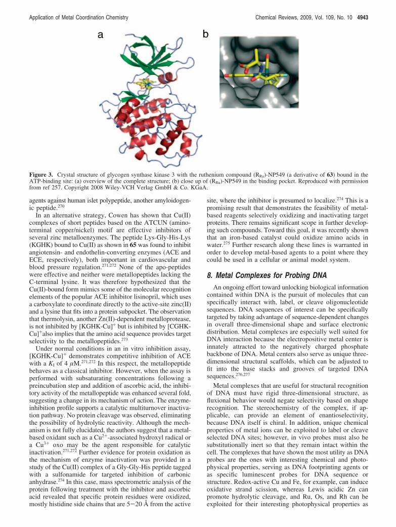
agents against human islet polypeptide, another amyloidogen-ic peptide.270
In an alternative strategy, Cowen has shown that Cu(II)complexes of short peptides based on the ATCUN (amino-terminal copper/nickel) motif are effective inhibitors ofseveral zinc metalloenzymes. The peptide Lys-Gly-His-Lys(KGHK) bound to Cu(II) as shown in 65 was found to inhibitangiotensin- and endothelin-converting enzymes (ACE andECE, respectively), both important in cardiovascular andblood pressure regulation.271,272 None of the apo-peptideswere effective and neither were metallopeptides lacking theC-terminal lysine. It was therefore hypothesized that theCu(II)-bound form mimics some of the molecular recognitionelements of the popular ACE inhibitor lisinopril, which usesa carboxylate to coordinate directly to the active-site zinc(II)and a lysine that fits into a protein subpocket. The observationthat thermolysin, another Zn(II)-dependent metalloprotease,is not inhibited by [KGHK-Cu]+ but is inhibited by [CGHK-Cu]+also implies that the amino acid sequence provides targetselectivity to the metallopeptides.273
Under normal conditions in an in vitro inhibition assay,[KGHK-Cu]+ demonstrates competitive inhibition of ACEwith a KI of 4 µM.271,272 In this respect, the metallopeptidebehaves as a classical inhibitor. However, when the assay isperformed with subsaturating concentrations following apreincubation step and addition of ascorbic acid, the inhibi-tory activity of the metallopeptide was enhanced several fold,suggesting a change in its mechanism of action. The enzyme-inhibition profile supports a catalytic multiturnover inactiva-tion pathway. No protein cleavage was observed, eliminatingthe possibility of hydrolytic reactivity. Although the mech-anism is not fully elucidated, the authors suggest that a metal-based oxidant such as a Cu2+-associated hydroxyl radical ora Cu3+ oxo may be the agent responsible for catalyticinactivation.271,272 Further evidence for protein oxidation asthe mechanism of enzyme inactivation was provided in astudy of the Cu(II) complex of a Gly-Gly-His peptide taggedwith a sulfonamide for targeted inhibition of carbonicanhydrase.274 In this case, mass spectrometric analysis of theprotein following treatment with the inhibitor and ascorbicacid revealed that specific protein residues were oxidized,mostly histidine side chains that are 5-20 Å from the active
site, where the inhibitor is presumed to localize.274 This is apromising result that demonstrates the feasibility of metal-based reagents selectively oxidizing and inactivating targetproteins. There remains significant scope in further develop-ing such compounds. Toward this goal, it was recently shownthat an iron-based catalyst could oxidize amino acids inwater.275 Further research along these lines is warranted inorder to develop metal-based agents to a point where theycould be used in a cellular or animal model system.
8. Metal Complexes for Probing DNAAn ongoing effort toward unlocking biological information
contained within DNA is the pursuit of molecules that canspecifically interact with, label, or cleave oligonucleotidesequences. DNA sequences of interest can be specificallytargeted by taking advantage of sequence-dependent changesin overall three-dimensional shape and surface electronicdistribution. Metal complexes are especially well suited forDNA interaction because the electropositive metal center isinnately attracted to the negatively charged phosphatebackbone of DNA. Metal centers also serve as unique three-dimensional structural scaffolds, which can be adjusted tofit into the base stacks and grooves of targeted DNAsequences.276,277
Metal complexes that are useful for structural recognitionof DNA must have rigid three-dimensional structure, asfluxional behavior would negate selectivity based on shaperecognition. The stereochemistry of the complex, if ap-plicable, can provide an element of enantioselectivity,because DNA itself is chiral. In addition, unique chemicalproperties of metal ions can be exploited to label or cleaveselected DNA sites; however, in vivo probes must also besubstitutionally inert so that they remain intact within thecell. The complexes that have shown the most utility as DNAprobes are the ones with interesting chemical and photo-physical properties, serving as DNA footprinting agents oras specific luminescent probes for DNA sequence orstructure. Redox-active Cu and Fe, for example, can induceoxidative strand scission, whereas Lewis acidic Zn canpromote hydrolytic cleavage, and Ru, Os, and Rh can beexploited for their interesting photophysical properties as
Figure 3. Crystal structure of glycogen synthase kinase 3 with the ruthenium compound (RRu)-NP549 (a derivative of 63) bound in theATP-binding site: (a) overview of the complete structure; (b) close up of (RRu)-NP549 in the binding pocket. Reproduced with permissionfrom ref 257. Copyright 2008 Wiley-VCH Verlag GmbH & Co. KGaA.
Application of Metal Coordination Chemistry Chemical Reviews, 2009, Vol. 109, No. 10 4943
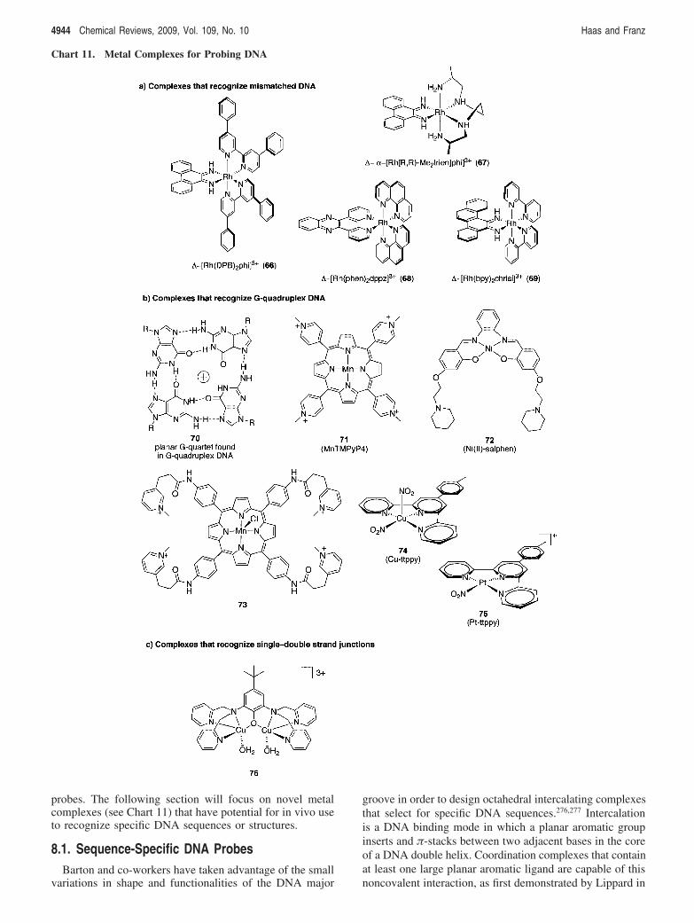
probes. The following section will focus on novel metalcomplexes (see Chart 11) that have potential for in vivo useto recognize specific DNA sequences or structures.
8.1. Sequence-Specific DNA ProbesBarton and co-workers have taken advantage of the small
variations in shape and functionalities of the DNA major
groove in order to design octahedral intercalating complexesthat select for specific DNA sequences.276,277 Intercalationis a DNA binding mode in which a planar aromatic groupinserts and $-stacks between two adjacent bases in the coreof a DNA double helix. Coordination complexes that containat least one large planar aromatic ligand are capable of thisnoncovalent interaction, as first demonstrated by Lippard in
Chart 11. Metal Complexes for Probing DNA
4944 Chemical Reviews, 2009, Vol. 109, No. 10 Haas and Franz

1976 for a square planar platinum complex.278 Octahedralmetal complexes can be designed to bind specific sequencesby using a large planar aromatic intercalating ligand for DNAbinding and ancillary ligands for shape and functional grouprecognition within the major groove. The enantioselectivityof a chiral complex for right-handed B-DNA over left-handedZ-DNA is mostly steric in nature and is achieved throughbulky ancillary ligands. Small ancillary ligands like phenan-throline and bipyridine are nondiscriminatory, whereascomplexes with bulkier ligands like DPB (4,4′-diphenylbi-pyridine) are enantioselective. For example, !-[Rh(DPB)2(phi)] (66, Chart 11) readily intercalates B-DNA atthe sequence 5′-CTCTAGAG-3′, but the "-enantiomer isinactive, even in 1000-fold excess.279,280 On the other hand,the "-enantiomer will bind to left-handed (and less stereo-selective) Z-DNA. It is noteworthy that 66 will also inhibitthe XbaI restriction enzyme, which shares the same recogni-tion site.279,280
Sequence selectivity is also achieved by matching thefunctional group on the ancillary ligands with those posi-tioned in the intercalated major groove. For example, !-R-[Rh((R,R)-Me2trien)(phi)]3+ (67) was designed to recognize5′-TGCA-3′ via hydrogen bonding between the axial aminesand the O6 of guanine, as well as by van der Waals contactsbetween the pendant methyl groups and the methyl groupsof thymine.281 These contacts are highlighted in the crystalstructure shown in Figure 4.
Using both shape and functional group recognition, Bartonhas developed an entire series of site-specific DNA binders.277
In addition to their impressive site-selectivity, these com-plexes employ the useful photophysical properties of Ru andRh to visualize and cleave DNA, respectively. OctahedralRu complexes containing a dppz intercalating ligand show
solvatochromatic luminescence in organic solvents.282-284
One of the best studied examples of these molecular lightswitch complexes is [Ru(phen)2(dppz)]3+ (68). In aqueoussolvent, hydrogen bonding with the endocyclic N of dppzdeactivates the excited state and quenches fluorescence.When DNA is added to solution, however, the complexshows its characteristic luminescence by intercalation intothe base stack, shielding its dppz ligand from any interactionwith the H-bonding solvent. Ru-dppy complexes have goodcellular uptake but so far show little selectivity for DNAover other hydrophobic cellular components.285,286
Rh has been exploited for its photoactivated DNA strandcleavage abilities.287 This reaction is useful because it enablesdirect footprinting of the site of intercalation. Irradiation withshort wavelength light (313-325 nm) in the presence ofdioxygen prompts formation of an intercalating ligand-basedradical that abstracts a hydrogen atom from an adjacentdeoxyribose ring, leading to direct strand scission.287
8.2. Metal Complexes That RecognizeMismatched DNA
A particularly impressive achievement in the area of DNArecognition has been in the selective recognition of mis-matched DNA. Mismatched base pairs are a result of errorsin DNA replication. While cells have mismatch repairmachinery, unchecked mismatches lead to permanent geneticmutations. Almost 80% of hereditary nonpolyposis coloncancers and 15-20% of biopsied solid tumors are linked tomutations in the cell’s mismatch repair machinery.288,289
Barton has extended the application of octahedral Ru andRh coordination complexes into the design of metal coor-dination compounds that are selective for DNA base-pairmismatches. Metal complexes using the expansive 5,6-chrissene quinone (chrisi) ligand, like [Rh(bpy)2(chrisi)2]3+
(69), can bind mismatched base pairs at least 1000 timesstronger than matched base pairs.290 In addition, enantiose-lectivity is observed, with the !-enantiomer binding andphotocleaving well, while the "-enantiomer is almostcompletely inactive toward B-DNA. The rational design ofa mismatch-selective complex is to use a planar aromaticligand that is too wide to fit into a normal DNA majorgroove. The larger ligand cannot intercalate into normalmatched DNA, but in the case of a mismatch, the duplex isthermodynamically destabilized. This destabilization allowsthe large ligand to eject the mismatched bases and replacethem with its own inserting ligand.
A Rh mismatch complex similar to 69 attached to a cell-penetrating polyarginine peptide and fluorophore was testedin cells and shown to localize in the nucleus.291 The questionof whether the complex is specifically binding to DNA invivo is yet to be answered.
8.3. Metal Complexes That RecognizeG-Quadruplex DNA
Certain guanine-rich sequences in DNA are known to formhigher order structures called G-quadruplexes (G4-DNA).These structures are formed by hydrogen bonding betweenguanines of the same or different strands to form a planarG-quartet (70). The resulting structure can be stabilized bythe presence of a monovalent cation bound in the center ofthe tetrad and can be further stabilized by $ stacking withother G-quartets.292,293
Figure 4. Crystal structure of the metallo-intercalator 67 boundto target sequence 5′-TGCA-3′. Note the intercalation of thearomatic phi group, the hydrogen-bonding interactions between theaxial amines and the O6 of guanine, and the contacts between thependant methyl groups of the ligand with the methyl groups ofthymine. Reprinted with permission from ref 281. Copyright 1993American Chemical Society.
Application of Metal Coordination Chemistry Chemical Reviews, 2009, Vol. 109, No. 10 4945
All vertebrate chromosomal DNA ends with telomerescontaining repeats of the sequence d(TTAAGGG), knownto have quadruplex-forming potential. Other sequences withquadruplex-forming potential are overexpressed in promoterregions of diverse organisms. Specifically, 370 000294 suchsequences have been recognized in humans and 40% of allknown promoter regions have at least one.295 The enrichmentof telomeres and promoter regions with quadruplex-formingpotential sequences makes them likely candidates in geneticregulation. Despite their rigorous study in vitro, motivationto study these structures and their biological implicationshas been spurred by recent evidence of their in vivosignificance.293,296-298 Development of new methods not onlyto distinguish G4-DNA from duplex DNA but also todiscriminate between the polymorphs of G4-DNA is neces-sary. Metal complexes are especially promising for G-quadruplex targeting; however, it is important to note thatthese complexes are only in the beginning stages of develop-ment and are not as evolved as metal complexes that bindduplex DNA.
Several metal complexes have been reported to interactwith G-quadruplex with some selectivity. Notable examplesinclude Mn-TMPyP4 (71), which shows nearly 10-foldpreference for G4-DNA over duplex DNA and modesttelomerase inhibition,299 followed by Ni(II)-salphen (72)300
and Mn(II)-porphyrin (73)301 complexes, with 50- and 1000-fold selectivity, respectively.
Square-pyramidal geometry of the metal complex has beenshown to be a major factor in the selectivity for quadruplexover duplex DNA. For example, the simple square pyramidalcomplex, Cu-ttpy (74) is highly selective for G-quadruplexin comparison to square-planar Pt-ttpy (75).302 The ttpy ligandin the Pt complex can stack on the outside of G-quad, aswell as insert into double-stranded DNA. On the other hand,apical ligands of square-pyramidal complexes prevent in-tercalation into duplex DNA without interfering with itsG-quadruplex association.302
Just as the unique properties of metal complexes have beensuccessfully used to probe duplex DNA, the metal complexesdiscussed above show promise for probing quadruplex DNAto help elucidate the potential structures that exist and thesignificance of G-quadruplex structures in live cells.
8.4. Metal Complexes That RecognizeSingle-Double Strand Junctions
Single strand (ss)/double strand (ds) DNA junctions areanother type of structure existing in telomere regions in awide variety of species. Some DNA-binding proteins thatrecognize ss/ds junctions in telomeres are known to havefunctions in telomere maintenance and capping. Otherinstances of ss/ds junctions occur during replication, are amajor component of RNA secondary structure, and areknown to occur at hairpin loops and Holliday junctions.Single/double strand junctions are yet another DNA structurewith implications in disease and represent a potentially richsource of new understanding of in vivo DNA function.
A class of binuclear and trinuclear copper complexes hasdemonstrated exquisite selectivity in oxidizing DNA at ss/ds DNA junctions.303-307 Complexes such as 76 are alsosequence-selective, preferring to oxidize guanine at the n andn + 1 position of the single-stranded DNA directly extendedfrom a duplex region. Site specificity derives in part fromthe coordination of at least one copper to a guanine.Interestingly, efficient cleavage is not observed for ss or ds
DNA alone, nor is cleavage observed for GG-GG mis-matches flanked on both sides by matched duplex DNA.304
The authors predict that the flexibility of the DNA strand isa factor in the ss/ds junction selectivity. The myriad ofpossible conformations of ss DNA may effectively competeagainst its productive association with the metal complex,and duplex DNA may not be flexible enough to accom-modate the required conformation. A ss/ds junction may offerunique proximity of the two single strands yet offer flexibilityto assemble with the copper complex.304 Although these ss/ds targeting complexes have not yet been fully developedfor in vivo application, the potential application of these andsimilar metal complexes that can recognize unique DNAstructures is apparent.
9. Metal-Responsive MRI AgentsMagnetic resonance imaging (MRI) provides three-
dimensional images of opaque biological structures withrelatively high spatial resolution (!10 µM) in a noninvasiveprocedure, making it an increasingly popular imaging modal-ity. Because a MR image derives from the nuclear magneticresonance of water protons, the contrast in the image dependson the local concentration of water and on the longitudinal(T1) and transverse (T2) relaxation times of its protons.308 Aradio-frequency pulse inverts the magnetization vector ofwater protons from their preferential alignment with theexternal magnetic field. The time required for the spins torealign with the field is characterized by T1, which can besignificantly reduced if the spins are in contact with a localparamagnetic center, thereby brightening the image. Coor-dination complexes that leave open coordination sites forwater molecules to access the inner sphere of paramagneticmetal ions (particularly Gd3+ with seven unpaired f electrons,but also high-spin Fe3+ and Mn2+ with five d electrons) aretherefore excellent candidates as agents that enhance MRimages via a T1 mechanism.309
Contrast agents that are currently widely used in clinicalmedicine are nonspecific, extracellular agents. Since theapproval of [Gd(DTPA)(H2O)]2- (known commercially asMagnevist) in 1988, research on designing new Gd coordina-tion complexes as improved MR imaging agents hasincreased dramatically.308 The desire to use MR imaging tounderstand physiological events has sparked a trend in thedesign of “smart” contrast agents that change their relaxivity(and therefore their level of contrast enhancement) inresponse toabiologicalstimulusorchangeinenvironment.310,311
Activateable contrast agents have appeared in the researchliterature that respond to changes in pH,312-315 O2 partialpressure,316 small molecules like glutathione317 and sialicacid,317,318 enzymatic activity,319-322 and specific metal ions.While not used clinically, these kinds of responsive agentsmay become increasingly useful for studying molecularbiology and intracellular chemistry.
9.1. MRI Agents for CalciumThe first metal-responsive probe was introduced by Meade
in 1999.323 The design of DOPTA-Gd (Chart 12, 77) consistsof a BAPTA Ca2+ chelating motif onto which Gd(DOTA)macrocycles are fused. In the absence of Ca2+, the carboxy-late arms of the central BAPTA bind to the Gd3+ centersand occlude H2O from direct coordination (77). Addition ofCa2+ shifts the coordination mode and opens the door forH2O to interact with the Gd3+ centers directly (78) and
4946 Chemical Reviews, 2009, Vol. 109, No. 10 Haas and Franz
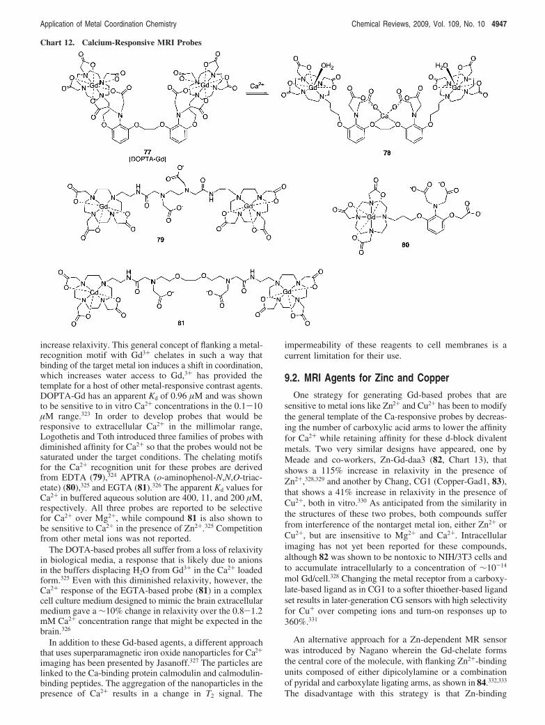
increase relaxivity. This general concept of flanking a metal-recognition motif with Gd3+ chelates in such a way thatbinding of the target metal ion induces a shift in coordination,which increases water access to Gd,3+ has provided thetemplate for a host of other metal-responsive contrast agents.DOPTA-Gd has an apparent Kd of 0.96 µM and was shownto be sensitive to in vitro Ca2+ concentrations in the 0.1-10µM range.323 In order to develop probes that would beresponsive to extracellular Ca2+ in the millimolar range,Logothetis and Toth introduced three families of probes withdiminished affinity for Ca2+ so that the probes would not besaturated under the target conditions. The chelating motifsfor the Ca2+ recognition unit for these probes are derivedfrom EDTA (79),324 APTRA (o-aminophenol-N,N,O-triac-etate) (80),325 and EGTA (81).326 The apparent Kd values forCa2+ in buffered aqueous solution are 400, 11, and 200 µM,respectively. All three probes are reported to be selectivefor Ca2+ over Mg2+, while compound 81 is also shown tobe sensitive to Ca2+ in the presence of Zn2+.325 Competitionfrom other metal ions was not reported.
The DOTA-based probes all suffer from a loss of relaxivityin biological media, a response that is likely due to anionsin the buffers displacing H2O from Gd3+ in the Ca2+ loadedform.325 Even with this diminished relaxivity, however, theCa2+ response of the EGTA-based probe (81) in a complexcell culture medium designed to mimic the brain extracellularmedium gave a !10% change in relaxivity over the 0.8-1.2mM Ca2+ concentration range that might be expected in thebrain.326
In addition to these Gd-based agents, a different approachthat uses superparamagnetic iron oxide nanoparticles for Ca2+
imaging has been presented by Jasanoff.327 The particles arelinked to the Ca-binding protein calmodulin and calmodulin-binding peptides. The aggregation of the nanoparticles in thepresence of Ca2+ results in a change in T2 signal. The
impermeability of these reagents to cell membranes is acurrent limitation for their use.
9.2. MRI Agents for Zinc and CopperOne strategy for generating Gd-based probes that are
sensitive to metal ions like Zn2+ and Cu2+ has been to modifythe general template of the Ca-responsive probes by decreas-ing the number of carboxylic acid arms to lower the affinityfor Ca2+ while retaining affinity for these d-block divalentmetals. Two very similar designs have appeared, one byMeade and co-workers, Zn-Gd-daa3 (82, Chart 13), thatshows a 115% increase in relaxivity in the presence ofZn2+,328,329 and another by Chang, CG1 (Copper-Gad1, 83),that shows a 41% increase in relaxivity in the presence ofCu2+, both in vitro.330 As anticipated from the similarity inthe structures of these two probes, both compounds sufferfrom interference of the nontarget metal ion, either Zn2+ orCu2+, but are insensitive to Mg2+ and Ca2+. Intracellularimaging has not yet been reported for these compounds,although 82 was shown to be nontoxic to NIH/3T3 cells andto accumulate intracellularly to a concentration of !10-14
mol Gd/cell.328 Changing the metal receptor from a carboxy-late-based ligand as in CG1 to a softer thioether-based ligandset results in later-generation CG sensors with high selectivityfor Cu+ over competing ions and turn-on responses up to360%.331
An alternative approach for a Zn-dependent MR sensorwas introduced by Nagano wherein the Gd-chelate formsthe central core of the molecule, with flanking Zn2+-bindingunits composed of either dipicolylamine or a combinationof pyridal and carboxylate ligating arms, as shown in 84.332,333
The disadvantage with this strategy is that Zn-binding
Chart 12. Calcium-Responsive MRI Probes
Application of Metal Coordination Chemistry Chemical Reviews, 2009, Vol. 109, No. 10 4947
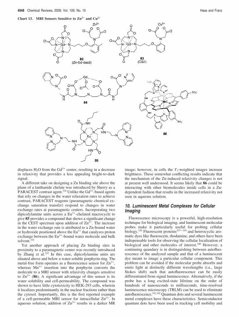
displaces H2O from the Gd3+ center, resulting in a decreasein relaxivity that provides a less appealing bright-to-darksignal.
A different take on designing a Zn binding site above theplane of a lanthanide chelate was introduced by Sherry as aPARACEST contrast agent.334 Unlike the Gd3+-based agentsthat rely on changes in the water relaxation rates to achievecontrast, PARACEST reagents (paramagnetic chemical ex-change saturation transfer) respond to changes in waterexchange rates at paramagnetic centers. Incorporating twodipicolylamine units across a Eu3+-chelated macrocycle togive 85 provides a compound that shows a significant changein the CEST spectrum upon addition of Zn2+. The increasein the water exchange rate is attributed to a Zn-bound wateror hydroxide positioned above the Eu3+ that catalyzes protonexchange between the Eu3+-bound water molecule and bulksolvent.334
Yet another approach of placing Zn binding sites inproximity to a paramagnetic center was recently introducedby Zhang et al.335 In this case, dipicolylamine units aresituated above and below a water-soluble porphyrin ring. Themetal-free form operates as a fluorescence sensor for Zn2+,whereas Mn3+ insertion into the porphyrin converts themolecule to a MRI sensor with relaxivity changes sensitiveto Zn2+ (86). A significant advantage of this sensor is itswater solubility and cell-permeability. The compound wasshown to have little cytotoxicity to HEK-293 cells, whereinit localizes predominantly in the nuclear fractions rather thanthe cytosol. Importantly, this is the first reported exampleof a cell-permeable MRI sensor for intracellular Zn2+. Inaqueous solution, addition of Zn2+ results in a darker MR
image; however, in cells the T1-weighted images increasebrightness. These somewhat conflicting results indicate thatthe mechanism of the Zn-induced relaxivity changes is notat present well understood. It seems likely that 86 could beinteracting with other biomolecules inside cells in a Zn-dependent fashion that results in the increased relaxivity notseen in aqueous solution.
10. Luminescent Metal Complexes for CellularImaging
Fluorescence microscopy is a powerful, high-resolutiontechnique for biological imaging, and luminescent molecularprobes make it particularly useful for probing cellularbiology.336 Fluorescent proteins337-339 and heterocyclic aro-matic dyes like fluoroscein, rhodamine, and others have beenindispensable tools for observing the cellular localization ofbiological and other molecules of interest.340 However, acontinuing quandary is in distinguishing between autofluo-rescence of the analyzed sample and that of a luminescentdye meant to image a particular cellular component. Thisproblem can be avoided if the molecular probe absorbs andemits light at distinctly different wavelengths (i.e., largeStokes shift) such that autofluorescence can be easilydifferentiated from signal luminescence. Alternatively, if theprobe has a long excited-state lifetime on the order ofhundreds of nanoseconds to milliseconds, time-resolvedluminescence microscopy (TRLM) can be used to eliminateautofluorescence.341,342 Quantum dots and several luminescentmetal complexes have these characteristics. Semiconductorquantum dots have been used in tracking cell mobility and
Chart 13. MRI Sensors Sensitive to Zn2+ and Cu2+
4948 Chemical Reviews, 2009, Vol. 109, No. 10 Haas and Franz
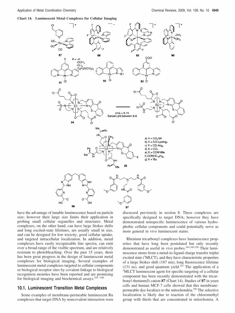
have the advantage of tunable luminescence based on particlesize; however their large size limits their application inprobing small cellular organelles and structures. Metalcomplexes, on the other hand, can have large Stokes shiftsand long excited-state lifetimes, are usually small in size,and can be designed for low toxicity, good cellular uptake,and targeted intracellular localization. In addition, metalcomplexes have easily recognizable line spectra, can emitover a broad range of the visible spectrum, and are relativelyresistant to photobleaching. Over the past 15 years, therehas been great progress in the design of luminescent metalcomplexes for biological imaging. Several examples ofluminescent metal complexes targeted to cellular componentsor biological receptor sites by covalent linkage to biologicalrecognition moieties have been reported and are promisingfor biological imaging and biochemical assays.343-348
10.1. Luminescent Transition Metal ComplexesSome examples of membrane-permeable luminescent Ru
complexes that target DNA by noncovalent interaction were
discussed previously in section 8. These complexes arespecifically designed to target DNA; however they havedemonstrated nonspecific luminescence of various hydro-phobic cellular components and could potentially serve asmore general in vivo luminescent stains.
Rhenium tricarbonyl complexes have luminescence prop-erties that have long been postulated but only recentlydemonstrated as useful in vivo probes.345,349,350 Their lumi-nescence stems from a metal-to-ligand charge transfer tripletexcited state (3MLCT), and they have characteristic propertiesof a large Stokes shift (187 nm), long fluorescence lifetime(131 ns), and good quantum yield.351 The application of a3MLCT luminescent agent for specific targeting of a cellularcomponent has been recently demonstrated with the tricar-bonyl rhenium(I) cation 87 (Chart 14). Studies of 87 in yeastcells and human MCF-7 cells showed that this membrane-permeable dye localizes to the mitochondria.350 The selectivelocalization is likely due to reaction of the chloromethylgroup with thiols that are concentrated in mitochonria. A
Chart 14. Luminescent Metal Complexes for Cellular Imaging
Application of Metal Coordination Chemistry Chemical Reviews, 2009, Vol. 109, No. 10 4949
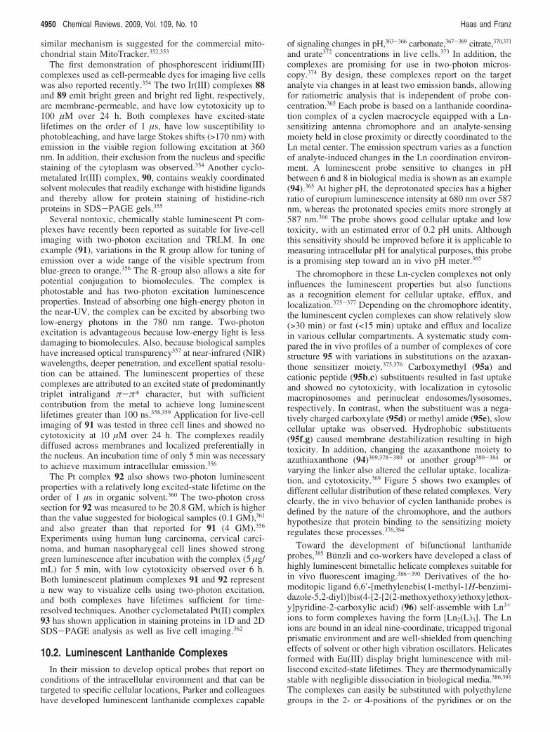
similar mechanism is suggested for the commercial mito-chondrial stain MitoTracker.352,353
The first demonstration of phosphorescent iridium(III)complexes used as cell-permeable dyes for imaging live cellswas also reported recently.354 The two Ir(III) complexes 88and 89 emit bright green and bright red light, respectively,are membrane-permeable, and have low cytotoxicity up to100 µM over 24 h. Both complexes have excited-statelifetimes on the order of 1 µs, have low susceptibility tophotobleaching, and have large Stokes shifts (>170 nm) withemission in the visible region following excitation at 360nm. In addition, their exclusion from the nucleus and specificstaining of the cytoplasm was observed.354 Another cyclo-metalated Ir(III) complex, 90, contains weakly coordinatedsolvent molecules that readily exchange with histidine ligandsand thereby allow for protein staining of histidine-richproteins in SDS-PAGE gels.355
Several nontoxic, chemically stable luminescent Pt com-plexes have recently been reported as suitable for live-cellimaging with two-photon excitation and TRLM. In oneexample (91), variations in the R group allow for tuning ofemission over a wide range of the visible spectrum fromblue-green to orange.356 The R-group also allows a site forpotential conjugation to biomolecules. The complex isphotostable and has two-photon excitation luminescenceproperties. Instead of absorbing one high-energy photon inthe near-UV, the complex can be excited by absorbing twolow-energy photons in the 780 nm range. Two-photonexcitation is advantageous because low-energy light is lessdamaging to biomolecules. Also, because biological sampleshave increased optical transparency357 at near-infrared (NIR)wavelengths, deeper penetration, and excellent spatial resolu-tion can be attained. The luminescent properties of thesecomplexes are attributed to an excited state of predominantlytriplet intraligand $-$* character, but with sufficientcontribution from the metal to achieve long luminescentlifetimes greater than 100 ns.358,359 Application for live-cellimaging of 91 was tested in three cell lines and showed nocytotoxicity at 10 µM over 24 h. The complexes readilydiffused across membranes and localized preferentially inthe nucleus. An incubation time of only 5 min was necessaryto achieve maximum intracellular emission.356
The Pt complex 92 also shows two-photon luminescentproperties with a relatively long excited-state lifetime on theorder of 1 µs in organic solvent.360 The two-photon crosssection for 92 was measured to be 20.8 GM, which is higherthan the value suggested for biological samples (0.1 GM),361
and also greater than that reported for 91 (4 GM).356
Experiments using human lung carcinoma, cervical carci-noma, and human nasopharygeal cell lines showed stronggreen luminescence after incubation with the complex (5 µg/mL) for 5 min, with low cytotoxicity observed over 6 h.Both luminescent platinum complexes 91 and 92 representa new way to visualize cells using two-photon excitation,and both complexes have lifetimes sufficient for time-resolved techniques. Another cyclometalated Pt(II) complex93 has shown application in staining proteins in 1D and 2DSDS-PAGE analysis as well as live cell imaging.362
10.2. Luminescent Lanthanide ComplexesIn their mission to develop optical probes that report on
conditions of the intracellular environment and that can betargeted to specific cellular locations, Parker and colleagueshave developed luminescent lanthanide complexes capable
of signaling changes in pH,363-366 carbonate,367-369 citrate,370,371
and urate372 concentrations in live cells.373 In addition, thecomplexes are promising for use in two-photon micros-copy.374 By design, these complexes report on the targetanalyte via changes in at least two emission bands, allowingfor ratiometric analysis that is independent of probe con-centration.365 Each probe is based on a lanthanide coordina-tion complex of a cyclen macrocycle equipped with a Ln-sensitizing antenna chromophore and an analyte-sensingmoiety held in close proximity or directly coordinated to theLn metal center. The emission spectrum varies as a functionof analyte-induced changes in the Ln coordination environ-ment. A luminescent probe sensitive to changes in pHbetween 6 and 8 in biological media is shown as an example(94).365 At higher pH, the deprotonated species has a higherratio of europium luminescence intensity at 680 nm over 587nm, whereas the protonated species emits more strongly at587 nm.366 The probe shows good cellular uptake and lowtoxicity, with an estimated error of 0.2 pH units. Althoughthis sensitivity should be improved before it is applicable tomeasuring intracellular pH for analytical purposes, this probeis a promising step toward an in vivo pH meter.365
The chromophore in these Ln-cyclen complexes not onlyinfluences the luminescent properties but also functionsas a recognition element for cellular uptake, efflux, andlocalization.375-377 Depending on the chromophore identity,the luminescent cyclen complexes can show relatively slow(>30 min) or fast (<15 min) uptake and efflux and localizein various cellular compartments. A systematic study com-pared the in vivo profiles of a number of complexes of corestructure 95 with variations in substitutions on the azaxan-thone sensitizer moiety.375,376 Carboxymethyl (95a) andcationic peptide (95b,c) substituents resulted in fast uptakeand showed no cytotoxicity, with localization in cytosolicmacropinosomes and perinuclear endosomes/lysosomes,respectively. In contrast, when the substituent was a nega-tively charged carboxylate (95d) or methyl amide (95e), slowcellular uptake was observed. Hydrophobic substituents(95f,g) caused membrane destabilization resulting in hightoxicity. In addition, changing the azaxanthone moiety toazathiaxanthone (94)369,378-380 or another group380-384 orvarying the linker also altered the cellular uptake, localiza-tion, and cytotoxicity.369 Figure 5 shows two examples ofdifferent cellular distribution of these related complexes. Veryclearly, the in vivo behavior of cyclen lanthanide probes isdefined by the nature of the chromophore, and the authorshypothesize that protein binding to the sensitizing moietyregulates these processes.376,384
Toward the development of bifunctional lanthanideprobes,385 Bunzli and co-workers have developed a class ofhighly luminescent bimetallic helicate complexes suitable forin vivo fluorescent imaging.386-390 Derivatives of the ho-moditopic ligand 6,6′-[methylenebis(1-methyl-1H-benzimi-dazole-5,2-diyl)]bis(4-[2-[2(2-methoxyethoxy)ethoxy]ethox-y]pyridine-2-carboxylic acid) (96) self-assemble with Ln3+
ions to form complexes having the form [Ln2(L)3]. The Lnions are bound in an ideal nine-coordinate, tricapped trigonalprismatic environment and are well-shielded from quenchingeffects of solvent or other high vibration oscillators. Helicatesformed with Eu(III) display bright luminescence with mil-lisecond excited-state lifetimes. They are thermodynamicallystable with negligible dissociation in biological media.386,391
The complexes can easily be substituted with polyethylenegroups in the 2- or 4-positions of the pyridines or on the
4950 Chemical Reviews, 2009, Vol. 109, No. 10 Haas and Franz
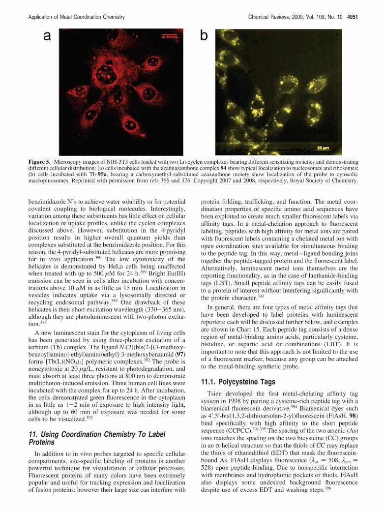
benzimidazole N’s to achieve water solubility or for potentialcovalent coupling to biological molecules. Interestingly,variation among these substituents has little effect on cellularlocalization or uptake profiles, unlike the cyclen complexesdiscussed above. However, substitution in the 4-pyridylposition results in higher overall quantum yields thancomplexes substituted at the benzimidazole position. For thisreason, the 4-pyridyl-substituted helicates are more promisingfor in vivo application.390 The low cytotoxicity of thehelicates is demonstrated by HeLa cells being unaffectedwhen treated with up to 500 µM for 24 h.389 Bright Eu(III)emission can be seen in cells after incubation with concen-trations above 10 µM in as little as 15 min. Localization invesicles indicates uptake via a lysosomally directed orrecycling endosomal pathway.389 One drawback of thesehelicates is their short excitation wavelength (330-365 nm),although they are photoluminescent with two-photon excita-tion.374
A new luminescent stain for the cytoplasm of living cellshas been generated by using three-photon excitation of aterbium (Tb) complex. The ligand N-[2[(bis(2-[(3-methoxy-benzoyl)amino]-ethyl)amino)ethyl]-3-methoxybenzamid (97)forms [Tb(L)(NO3)3] polymeric complexes.392 The probe isnoncytotoxic at 20 µg/L, resistant to photodegradation, andmust absorb at least three photons at 800 nm to demonstratemultiphoton-induced emission. Three human cell lines wereincubated with the complex for up to 24 h. After incubation,the cells demonstrated green fluorescence in the cytoplasmin as little as 1-2 min of exposure to high intensity light,although up to 60 min of exposure was needed for somecells to be visualized.392
11. Using Coordination Chemistry To LabelProteins
In addition to in vivo probes targeted to specific cellularcompartments, site-specific labeling of proteins is anotherpowerful technique for visualization of cellular processes.Fluorescent proteins of many colors have been extremelypopular and useful for tracking expression and localizationof fusion proteins; however their large size can interfere with
protein folding, trafficking, and function. The metal coor-dination properties of specific amino acid sequences havebeen exploited to create much smaller fluorescent labels viaaffinity tags. In a metal-chelation approach to fluorescentlabeling, peptides with high affinity for metal ions are pairedwith fluorescent labels containing a chelated metal ion withopen coordination sites available for simultaneous bindingto the peptide tag. In this way, metal-ligand bonding joinstogether the peptide-tagged protein and the fluorescent label.Alternatively, luminescent metal ions themselves are thereporting functionality, as in the case of lanthanide-bindingtags (LBT). Small peptide affinity tags can be easily fusedto a protein of interest without interfering significantly withthe protein character.393
In general, there are four types of metal affinity tags thathave been developed to label proteins with luminescentreporters; each will be discussed further below, and examplesare shown in Chart 15. Each peptide tag consists of a denseregion of metal-binding amino acids, particularly cysteine,histidine, or aspartic acid or combinations (LBT). It isimportant to note that this approach is not limited to the useof a fluorescent marker, because any group can be attachedto the metal-binding synthetic probe.
11.1. Polycysteine TagsTsien developed the first metal-chelating affinity tag
system in 1998 by pairing a cysteine-rich peptide tag with abiarsenical fluorescein derivative.394 Biarsenical dyes suchas 4′,5′-bis(1,3,2-dithioarsolan-2-yl)fluorescein (FlAsH, 98)bind specifically with high affinity to the short peptidesequence (CCPCC).394,395 The spacing of the two arsenic (As)ions matches the spacing on the two bicysteine (CC) groupsin an R-helical structure so that the thiols of CC may replacethe thiols of ethanedithiol (EDT) that mask the fluorescein-bound As. FlAsH displays fluorescence (%ex ) 508, %em )528) upon peptide binding. Due to nonspecific interactionwith membranes and hydrophobic pockets or thiols, FlAsHalso displays some undesired background fluorescencedespite use of excess EDT and washing steps.396
Figure 5. Microscopy images of NIH-3T3 cells loaded with two Ln-cyclen complexes bearing different sensitizing moieties and demonstratingdifferent cellular distribution: (a) cells incubated with the azathiaxanthone complex 94 show typical localization to nucleosomes and ribosomes;(b) cells incubated with Tb-95a, bearing a carboxymethyl-substituted azaxanthone moiety show localization of the probe to cytosolicmacropinosomes. Reprinted with permission from refs 366 and 376. Copyright 2007 and 2008, respectively, Royal Society of Chemistry.
Application of Metal Coordination Chemistry Chemical Reviews, 2009, Vol. 109, No. 10 4951
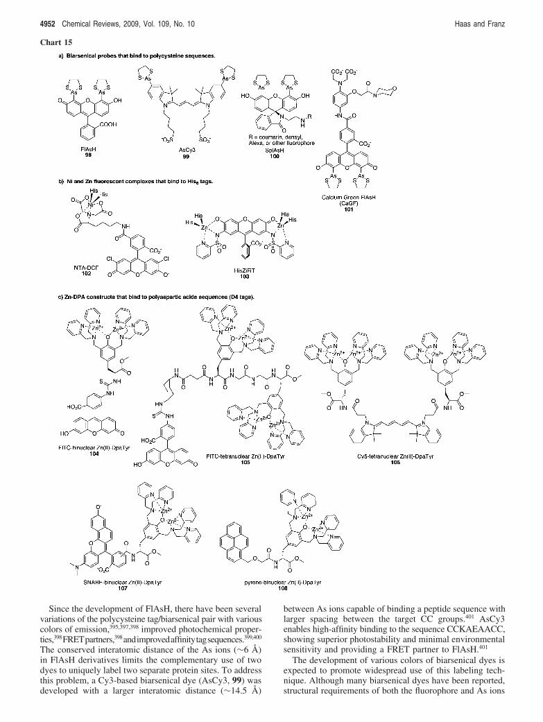
Since the development of FlAsH, there have been severalvariations of the polycysteine tag/biarsenical pair with variouscolors of emission,395,397,398 improved photochemical proper-ties,398FRETpartners,398andimprovedaffinitytagsequences.399,400
The conserved interatomic distance of the As ions (!6 Å)in FlAsH derivatives limits the complementary use of twodyes to uniquely label two separate protein sites. To addressthis problem, a Cy3-based biarsenical dye (AsCy3, 99) wasdeveloped with a larger interatomic distance (!14.5 Å)
between As ions capable of binding a peptide sequence withlarger spacing between the target CC groups.401 AsCy3enables high-affinity binding to the sequence CCKAEAACC,showing superior photostability and minimal environmentalsensitivity and providing a FRET partner to FlAsH.401
The development of various colors of biarsenical dyes isexpected to promote widespread use of this labeling tech-nique. Although many biarsenical dyes have been reported,structural requirements of both the fluorophore and As ions
Chart 15
4952 Chemical Reviews, 2009, Vol. 109, No. 10 Haas and Franz
has made the scope of compatible dyes narrow and difficultto predict. To further improve the applicability of thepolycysteine/biarsenical system, Miller developed a strategyfor recruiting any fluorophore to a polycysteine tag.402 Basedon a modular approach, a fluorescent moiety is separatedfrom the biarsenial targeting moiety in order to remove anyrestrictions on its structure. The fluoroscein-based biarsenicaltargeting moiety, called SplAsH (100), can be convenientlyattached to a variety of fluorophores to create differentcolored emission. In this method, the probe is fluorescentwhether or not it is bound to the peptide tag, but it has theadvantage that any fluorophore can be used.
A recent advance in biarsenical dyes has come in the formof the Ca2+ sensor Calcium Green FlAsH (CaGF, 101),which combines a FlAsH moiety with a Ca2+ chelator. Thebinding of Ca2+ to CaGF results in a 10-fold increase influorescence.403 CaGF-labeled calcium channels have dem-onstrated that CaGF can be used for precise and fast detectionof intracellular Ca2+.403
11.2. Polyhistidine TagsPeptide tags with six or more sequential histidine residues
(His-tag) are known to interact with transition metal com-plexes and are commonly used for purification of expressedprotein by Ni2+-NTA affinity chromatography, where NTAis nitrilotriacetic acid. The affinity of His-tags for metalcomplexes can also be applied to fluorescently label proteinsor other molecules in vitro404,405 and in vivo406-408 byintroducing a Ni-NTA complex where NTA is derivatizedwith a fluorophore.
With the Ni-NTA complex present in solution as opposedto being in large excess on a solid support as is the case forprotein purification, the relatively low affinity between a His-tagged protein and Ni-NTA (Kd # 1-20 µM) becomes alimitation to this approach. Increasing the number of Ni-NTA appendages on the fluorophore increases affinity by 4orders of magnitude in going from a mono-Ni-NTA probeto one with four Ni-NTA groups.404 Increasing the length ofthe His-tag from His6 to His10 also improves affinity.407
In most cases, the use of paramagnetic Ni2+ results inpartial quenching and a decrease in fluorescence. Goldsmithet al., however, have reported that the fluorescence of NTA-DCF (102) is not significantly influenced by the presence ofNi2+.408 This tag does not enter cells either by itself or as itsNi(II) complex; therefore it is useful for imaging extracellularproteins that are expressed with His-tags.408
A strategy for avoiding paramagnetic quenching is toreplace Ni2+ with diamagnetic Zn2+ as the metal. Zn2+, whichis ubiquitous in biological systems, will also help avoid heavymetal toxicity that can be caused by Ni2+. Tsien developeda novel chelating molecule 2′,7′-bis(pyridyl-2-sulfonamido)-4′,5′-dimethylfluorescein (histidine-zinc fluorescent in vivotag, called HisZiFiT, 103), which holds two Zn2+ ions in acoordinately unsaturated environment and has affinity forHis6-tags comparable to bis-NTA-Ni2+ ligands (Kd # 40 nM).HisZiFiT has been used successfully in live cells to labelsurface-exposed His-tagged proteins without compromisingthe integrity of protein structure.409
11.3. Polyaspartate TagsRuns of negatively charged amino acids provide another
target as metal affinity tags. Hamachi and co-workersdeveloped multinuclear Zn(II) luminescent complexes con-
sisting of a fluorescein or Cy5 fluorescent moiety attachedto one (104) or two (105, 106) pendant binuclear Zn-DpaTyrgroups, which have affinity for poly(aspartic acid) se-quences.410 Notable here is the similarity in design of theseconstructs with Zn-DPA constructs mentioned in sections6.5 and 6.6.1 that target phosphate-containing molecules. Theaffinity of the binuclear probes for aspartic acid stretchesincreases by an order of magnitude with each additionalaspartic acid residue up to four (D4 tag). Compared withthe binuclear Zn(II) complex, the tetranuclear Zn(II) com-plexes 105 and 106 have affinity 3 orders of magnitudehigher when paired with a longer tag (D4-G-D4) capable ofinteracting with both Zn-DpaTyr groups.410
The utility of the D4/Zn-complex system was expandedby creating probes capable of ratiometric analysis.411,412
Ratiometric analysis is highly desirable because it helpsreduce background signal due to the unbound fluorophorethereby facilitating precise detection and imaging of theprotein of interest. In the first example, a binuclear Zn-DpaTyr attached to a pH-sensitive fluorophore (seminaph-thorhodafluor (SNARF), 107) takes advantage of the localacidic pH of polyaspartate tags to create an affinity probethat changes emission wavelength upon binding. In bulksolution, the phenolate form of SNARF emits at 586 nm,and upon binding to a D4 tag, protonation of the phenol shiftsemission to 628 nm.411 In a second example, a peptidecontaining two D4 tags attracts two separate binuclear Zn(II)complexes containing pyrene moieties (108) into closeproximity. The 108 monomer shows emission at 379 and407 nm, but when two probe complexes bind to the D4 tagsimultaneously, excimer formation results in emission at 472nm.412
11.4. Covalent Protein Labeling Facilitated byMetal Chelation
A drawback to imaging proteins tagged with metal affinitypeptides is the high background generated by unboundfluorophores and the reversibility of the metal-tag interac-tion. To overcome this problem, covalent labeling strategieshave been developed that take advantage of the peptide tagto direct metal complexes containing a reactive functionalgroup to a protein of interest, as shown in Chart 16. Thereactive group is used to covalently attach the probe therebyachieving site-specific protein labeling without the require-ment of an enzyme. In this way, a binuclear Zn-DpaTyrconstruct 109 bearing a fluorophore and a thiol-reactivechloroacetyl group both targets and reacts with a D4 tagcontaining a cysteine thiol, resulting in a covalently labeledprotein.413 The in vivo utility of this system was demonstratedin Escherichia coli cells with CAAAAAADDDD-taggedmaltose binding protein paired with 109. Labeling in livecells was not as efficient as labeling in vitro, but improve-ments in the affinity between the tag and probe are expectedto improve labeling efficiency.413
Hintersteiner et al. also developed a nonenzymatic covalentlabeling technique, in this case by using a His-tag as theaffinity target.414 Probe 110 consists of a rhodamine reportergroup, a Ni-NTA complex, and a photoactive arylazide cross-linker. After the probe binds to its His-tag target via Nicoordination, UV-light is used to photoactivate the arylazideand form a covalent bond to the protein of interest. Thissystem was applied to label His-tagged proteins on thesurface of living cells, where covalently bound probe
Application of Metal Coordination Chemistry Chemical Reviews, 2009, Vol. 109, No. 10 4953

remained on the cell surface while reversibly bound probewas easily washed away.414
11.5. Lanthanide Binding TagsRather than using a synthetic chelator attached to a
fluorophore to accomplish protein labeling, Imperiali and co-workers developed lanthanide binding tags (LBTs) as geneti-cally encodable peptide sequences optimized for binding Lnions, where the coordinated Ln itself is the luminescentreporter.415 In the proper coordination environment, Ln ionshave very attractive luminescent properties for biologicalapplications, including sharp emission spectra, large Stokesshifts, and millisecond luminescent lifetimes. For luminescentLBTs, Tb(III) is particularly important because its emissioncan be sensitized by native tryptophan or tyrosine chro-mophores. Peptides with high affinity and bright emissionfor Tb(III) were discovered from combinatorial screens ofsequences based on known calcium-binding motifs under 20amino acids in length.415,416 The best sequence to date isFIDTNNDGWIEGDELLLEEG, which binds Tb with anapparent Kd at pH 7.4 of 19 nM and achieves brightluminescence by occluding inner-sphere water molecules thatquench emission.417
The utility of LBT-tagged proteins has so far beendemonstrated in vitro. For example, time-resolved lumines-cencemeasurementswereusedtointerrogatetheprotein-proteininteractions between Src homology 2 (SH2) domains ex-pressed with an LBT and phosphopeptides labeled withchromophores suitable for luminescence energy transfer withthe LBT-bound Tb(III).418 Beyond luminescence applications,
the properties of protein-bound Ln ions enabled by LBTsalso facilitate macromolecular structure determination byboth NMR419-421 and X-ray crystallography.422 Although thechallenge of incorporating Ln ions into tagged proteins in aliving cell remains, the ability to genetically encode a proteinwith a nonintrusive and multifunctional tag is attractive forfuture cellular applications.418
11.6. Unnatural Metal-Binding Amino AcidsLabeling systems that employ metal coordination chem-
istry to unite a synthetic probe with a genetically encodedpeptide tag are applicable to a wide variety of proteins andprovide general tools for in vivo, site-specific labeling ofproteins. These new metal sites are usually limited to the C-or N-terminus of a protein of interest so as not to disruptthe protein’s native structure and function. Schultz demon-strated a new way to introduce metal sites into proteinswithout the use of oligopeptide tags. By incorporating non-natural amino acids directly into prokaryotic and eukaryoticorganisms, chemically and photochemically active groups,including metal-binding centers, can be introduced sitespecifically into proteins.423-425 The metal-binding non-natural amino acid bipyridylalanine (BpyAla, 111) wasincorporated into defined sites in a DNA binding protein todirectly map its binding site on duplex DNA.426 Importantly,introduction of the metal binding site involved replacementof only one amino acid in the entire protein sequence. Inthe case of E. coli catabolite activator protein (CAP), thereplacement of one lysine in the DNA recognition site didnot significantly perturb its interaction with its DNA binding
Chart 16. Constructs Used for Nonenzymatic Covalent Protein Labeling
4954 Chemical Reviews, 2009, Vol. 109, No. 10 Haas and Franz

sequence. CAP-K26Bpy-Ala showed site-specific oxidativecleavage at two different sites on the DNA sequence.426
Because introduction of non-natural amino acids requiresonly simple mutagenesis and because there are numerouspotential metal binding non-natural amino acids, redox-activemetals could be placed almost anywhere in a protein to mapprotein-RNA or protein-protein interfaces.
12. ConclusionsAs discussed in the previous sections, the application of
metal chelating agents, coordination compounds, and orga-nometallic complexes in cell biology offers diverse op-portunities for manipulating biological processes. The ex-amples that have been highlighted here are only representativeof the significant opportunity that metal complexes offer forexploring the chemical biology of living cells. In describingthe principles and trends involved in the design, functionality,and reactivity of inorganic complexes in this context, wehope to inspire further research in the area. In many cases,a creative approach can extend a seemingly simple motifacross several applications. The dipicolylamine chelating unitis one example. This basic motif appears in TPEN, which isused as a chelator to alter cellular metal content, as well asfluorescence and MRI-based Zn(II) sensors, and in constructsthat use their coordinated metal ion to tag proteins, recognizephosphorylated proteins and other phosphate-containingbiomolecules, or bind to specific DNA sequences.
The breadth of these different biological applications forinorganic complexes is exciting, but also presents ongoingchallenges. As these kinds of reagents become more fre-quently used in biological studies, questions of cross reactiv-ity and specificity are likely to increase. The cellularreactivity of any of these complexes will depend on theirdistribution and relative concentration with respect tocompeting metal ions that may induce transmetalation, aswell as endogeneous chelating agents that can compete forthe metal ion or form unanticipated ternary complexes thatmight alter their reactivity. All of these complex equilibria,in addition to factors of stability and metabolism, will affectthe specificity of the reagent for the intended biologicalinteraction. Future development will therefore need to focuson understanding these complex processes. In particular, thisarea will benefit greatly from advances in understandingcellular metal trafficking and regulation.
There is an obvious synergy between inorganic chemicalbiology and the biology of metal trafficking. Small moleculechemical tools that manipulate cellular metal content anddistribution are beneficial for understanding metal traffickingpathways. At the same time, as we better understand thesepathways we can develop better strategies for interceptingthem, whether it be for overcoming a metal deficiency,reducing a metal overload, or hijacking a native metal intoan artificial complex with non-native function, like anenzyme inhibitor or catalytic protein inactivator. Likewise,how do cells handle unnatural metals and metal complexes?For intact metal complexes that are applied to cells ororganisms, at what point does the cellular machinerydifferentiate the cargo from the carrier, and how does thisinfluence where metal complexes are distributed in a cell?Finding answers to these questions will be an excitingjourney into the frontier of inorganic chemistry and cellbiology.
13. Abbreviations4AP 4-aminopyridineACE angiotensis-converting enzymeADP adenosine diphosphateAPTRA aminophenol-N,N,O-triacetateATCUN amino-terminal copper/nickelATP adenosine triphosphateBAPTA (1,2-bis(o-aminophenoxy)ethane-N,N,N′,N′-tetraace-
tic acid)BC bathocuproineBCS bathocuproine disulfonateBPS bathophenanthroline disulfonic acidbpy bipyridineCAP catabolite activator proteinCDK9 cyclin-dependent protein kinase 9CEST chemical exchange saturation transfercod 1,5-cyclooctadieneCORM carbon monoxide releasing moleculeCp* pentamethylcyclopentadienylCTD C-terminal domainCy3 cyanine dyecyclam 1,4,8,11-tetraazacyclotetradecaneDFO desferrioxamineDMSO dimethylsulfoxideDOTA 1,4,7,10-tetraazacyclododecane-1,4,7,10-tetraace-
tic acidDPA dipyridylamineDTPA diethylenetriamine pentaacetic acidECE endothelin-converting enzymeEDT ethanedithiolEDTA ethylene diamine tetraacetic acidEGTA ethylene glycol tetraacetic acidFAD flavin adenine dinucleotideFMN flavin mononucleotideFRET fluorescent resonance energy transferG4-DNA guanidine quadruplex DNAGABA "-amino butyric acidGSK-3 glycogen synthase kinase 3HDAC histone deacetylasesHSAB hard and soft acid and baseIP3 inositol triphosphateIDA iminodiacetic acidLBT lanthanide binding tagLFSE ligand field stabilization energyMLCT metal-to-ligand charge transferMMP metalloproteinaseMNIP 4-methoxy-2-(1H-naptho[2,3-d]imidazole-2-yl)phe-
nolMR magnetic resonanceMRI magnetic resonance imagingNIR near-infraredNMR nuclear magnetic resonanceNTA nitrilotriacetic acidPARACEST paramagnetic chemical exchange saturation trans-
ferPET positron emission tomographyPIH pyridoxal isonicotinoyl hydrazonePPh3 triphenylphosphinepS phosphoserinepSer phosphoserinepy pyridinepY phosphotyrosineResf resorufinRNA ribonucleic acidROS reactive oxygen speciesRSE Roussin’s red salt estersSH2 Src homology 2SIH salicylaldehyde isonicotinoyl hydrazoneSNARF seminaphthorhodafluorSNP sodium nitroprusside
Application of Metal Coordination Chemistry Chemical Reviews, 2009, Vol. 109, No. 10 4955

Sox sulfonamide-oxine amino acidSPECT single photon emission computed tomographySplAsH spirolactam arsenical hairpin binderTPEN N,N,N′,N′-tetrakis-(2-pyridylmethyl)-ethylenedi-
amineTRML time-resolved luminescence microscopy.
14. AcknowledgmentsWe thank all of our current and former group members
for their many contributions, as well as Duke University,the Sloan Foundation, the Camille and Henry DreyfusFoundation, the Parkinson’s Disease Foundation, the USNational Science Foundation (Grant CAREER 0449699), andthe US National Institutes of Health (Grants GM084176 andEY018922) for funding our research. K.L.H. acknowledgesa NSF IGERT Fellowship.
15. References(1) Schreiber, S. L. Nat. Chem. Biol. 2005, 1, 64.(2) Doudna, J. A. Nat. Chem. Biol. 2005, 1, 300.(3) Tomi, K. S. Chem. Biol. Drug Des. 2006, 67, 196.(4) Stockwell, B. R. Nature 2004, 432, 846.(5) Fricker, S. P. Dalton Trans. 2007, 43, 4903.(6) Lippard, S. J. Nat. Chem. Biol. 2006, 2, 504.(7) David, S. S.; Meggers, E. Curr. Opin. Chem. Biol. 2008, 12, 194.(8) Cohen, S. M. Curr. Opin. Chem. Biol. 2007, 11, 115.(9) Biological Inorganic Chemistry: Structure and ReactiVity; Bertini,
I., Gray, H. B., Stiefel, E. I., Valentine, J. S., Eds.; University ScienceBooks: Sausalito, CA, 2007.
(10) Frausto da Silva, J. J. R.; Williams, R. J. P. The Biological Chemistryof the Elements, 2nd ed.; Oxford University Press: Oxford, U.K.,2001.
(11) Lippard, S. J.; Berg, J. M. Principles of Bioinorganic Chemistry;University Science Books: Mill Valley, CA, 1994.
(12) Mann, S. In Biological Inorganic Chemistry: Structure and ReactiVity;Bertini, I., Gray, H. B., Stiefel, E. I., Valentine, J. S., Eds.; UniversityScience Books: Sausalito, CA, 2007; p 79.
(13) Evenas, J.; Malmendal, A.; Forsen, S. Curr. Opin. Chem. Biol. 1998,2, 293.
(14) Pyle, A. J. Biol. Inorg. Chem. 2002, 7, 679.(15) Vinod, K.; Misra, D. E. D. Biopolymers 1998, 48, 113.(16) Winge, D. R. In Biological Inorganic Chemistry: Structure and
ReactiVity; Bertini, I., Gray, H. B., Stiefel, E. I., Valentine, J. S.,Eds.; University Science Books: Sausalito, CA, 2007; p 613.
(17) Frederickson, C. J.; Koh, J.-Y.; Bush, A. I. Nat. ReV. Neurosci. 2005,6, 449.
(18) Chen, P. R.; He, C. Curr. Opin. Chem. Biol. 2008, 12, 214.(19) Pennella, M. A.; Giedroc, D. P. BioMetals 2005, 18, 413.(20) Cowan, J. A. In Biological Inorganic Chemistry: Structure and
ReactiVity; Bertini, I., Gray, H. B., Stiefel, E. I., Valentine, J. S.,Eds.; University Science Books: Sausalito, CA, 2007; p 175.
(21) Thomas, D. D.; Ridnour, L. A.; Isenberg, J. S.; Flores-Santana, W.;Switzer, C. H.; Donzelli, S.; Hussain, P.; Vecoli, C.; Paolocci, N.;Ambs, S.; Colton, C. A.; Harris, C. C.; Roberts, D. D.; Wink, D. A.Free Radical Biol. Med. 2008, 45, 18.
(22) Holm, R. H.; Kennepohl, P.; Solomon, E. I. Chem. ReV. 1996, 96,2239.
(23) Valentine, J. S. In Biological Inorganic Chemistry: Structure andReactiVity; Bertini, I., Gray, H. B., Stiefel, E. I., Valentine, J. S.,Eds.; University Science Books: Sausalito, CA, 2007; p 319.
(24) Ronconi, L.; Sadler, P. J. Coord. Chem. ReV. 2007, 251, 1633.(25) Ang, W. H.; Dyson, P. J. Eur. J. Inorg. Chem. 2006, 2006, 4003.(26) Dyson, P. J.; Sava, G. Dalton Trans. 2006, 16, 1929.(27) Storr, T.; Thompson, K. H.; Orvig, C. Chem. Soc. ReV. 2006, 35,
534.(28) Thompson, K. H.; Orvig, C. Science 2003, 300, 936.(29) Thompson, K. H.; Orvig, C. Dalton Trans. 2006, 6, 761.(30) Wadas, T. J.; Wong, E. H.; Weisman, G. R.; Anderson, C. J. Curr.
Pharm. Des. 2007, 13, 3.(31) Liu, S. Chem. Soc. ReV. 2004, 33, 445.(32) Scott, L. E.; Orvig, C. Chem. ReV. 2009, 109, doi: 10.1021/cr9000176.(33) Hartinger, C. G.; Dyson, P. J. Chem. Soc. ReV. 2009, 38, 391.(34) Domaille, D. W.; Que, E. L.; Chang, C. J. Nat. Chem. Biol. 2008, 4,
168.(35) Kikuchi, K.; Komatsu, K.; Nagano, T. Curr. Opin. Chem. Biol. 2004,
8, 182.(36) Nolan, E. M.; Lippard, S. J. Chem. ReV. 2008, 108, 3443.
(37) Nolan, E. M.; Lippard, S. J. Acc. Chem. Res. 2009, 42, 193.(38) Que, E. L.; Domaille, D. W.; Chang, C. J. Chem. ReV. 2008, 108,
1517.(39) Fahrni, C. Chem. ReV. 2009, 109, doi: 10.1021/cr900223a.(40) Hancock, R. D.; Martell, A. E. Chem. ReV. 1989, 89, 1875.(41) Martell, A. E.; Hancock, R. D.; Motekaitis, R. J. Coord. Chem. ReV.
1994, 133, 39.(42) Martell, A. E.; Hancock, R. D. Metal Complexes in Aqueous Solution;
Plenum Press: New York, 1996.(43) Hancock, R. D.; Melton, D. L.; Harrington, J. M.; McDonald, F. C.;
Gephart, R. T.; Boone, L. L.; Jones, S. B.; Dean, N. E.; Whitehead,J. R.; Cockrell, G. M. Coord. Chem. ReV. 2007, 251, 1678.
(44) Andersen, O. Mini-ReV. Med. Chem. 2004, 4, 11.(45) Sigel, H.; McCormick, D. B. Acc. Chem. Res. 1970, 3, 201.(46) Pearson, R. G. J. Am. Chem. Soc. 1963, 85, 3533.(47) Perrin, D. D.; Dempsey, B. Buffers for pH and Metal Ion Control;
Chapman and Hall: New York, 1979; p 94.(48) Martell, A. E.; Smith, R. M. In NIST Standard Reference Database
46, 6.0 ed.; Motekaitis, R. J., Ed.; NIST: Gaithersburg, MD, 2001.(49) Martell, A. E.; Smith, R. M. Critical Stability Constants; Plenum
Press: New York, 1974; p 1.(50) Miessler, G. L.; Tarr, D. A. Inorganic Chemistry, 3rd ed.; Pearson
Education: Upper Saddle River, N.J., 2004; p 345.(51) Atkins, P.; Overton, T.; Rourke, J.; Weller, M.; Armstrong, F.;
Salvador, P.; Hagerman, M.; Spiro, T.; Stiefel, E. ShriVer & AtkinsInorganic Chemistry, 4th ed.; W. H. Freeman: New York, 2006; p459.
(52) Walling, C. Acc. Chem. Res. 1998, 31, 155.(53) Prousek, J. Pure Appl. Chem. 2007, 79, 2325.(54) Fenton, H. J. H. J. Chem. Soc. Trans 1894, 65, 899.(55) Pierre, J. L.; Fontecave, M. Biometals 1999, 12, 195.(56) Liu, Z. D.; Hider, R. C. Coord. Chem. ReV. 2002, 232, 151.(57) Crumbliss, A. L.; Harrington, J. M. AdV. Inorg. Chem. 2009, 61, in
press.(58) Glickstein, H.; Ben El, R.; Shvartsman, M.; Cabantchik, Z. I. Blood
2005, 106, 3242.(59) Halliwell, B. Biochem. Pharmacol. 1985, 34, 229.(60) Balcerczyk, A.; Sowa, K.; Bartou, G. Biochem. Biophys. Res.
Commun. 2007, 352, 522.(61) Kalinowski, D. S.; Richardson, D. R. Pharmacol. ReV. 2005, 57,
547.(62) Simunek, T.; Boer, C.; Bouwman, R. A.; Vlasblom, R.; Versteilen,
A. M. G.; Sterba, M.; Gersl, V.; Hrdina, R.; Ponka, P.; de Lange,J. J.; Paulus, W. J.; Musters, R. J. P. J. Mol. Cell. Cardiol. 2005, 39,345.
(63) Zhang, A. S.; Sheftel, A. D.; Ponka, P. Blood 2005, 105, 368.(64) Horackova, M.; Ponka, P.; Byczko, Z. CardioVasc. Res. 2000, 47,
529.(65) Richardson, D. R.; Ponka, P. J. Lab. Clin. Med. 1998, 131, 306.(66) Hermes-Lima, M.; Nagy, E.; Ponka, P.; Schulman, H. M. Free
Radical Biol. Med. 1998, 25, 875.(67) Hermes-Lima, M.; Ponka, P.; Schulman, H. M. Biochim. Biophys.
Acta 2000, 1523, 154.(68) Petrat, F.; de Groot, H.; Sustmann, R.; Rauen, U. Biol. Chem. 2002,
383, 489.(69) Esposito, B. P.; Epsztejn, S.; Breuer, W.; Cabantchik, Z. I. Anal.
Biochem. 2002, 304, 1.(70) Richardson, D. R.; Hefter, G. T.; May, P. M.; Webb, J.; Baker, E.
BioMetals 1989, 2, 161.(71) Kovarikova, P.; Mrkvickova, Z.; Klimes, J. J. Pharm. Biomed. Anal.
2008, 47, 360.(72) Rutherford, J. C.; Ojeda, L.; Balk, J.; Muhlenhoff, U.; Lill, R.; Winge,
D. R. J. Biol. Chem. 2005, 280, 10135.(73) Chen, O. S.; Crisp, R. J.; Valachovic, M.; Bard, M.; Winge, D. R.;
Kaplan, J. J. Biol. Chem. 2004, 279, 29513.(74) Blair, D.; Diehl, H. Talanta 1961/2, 7, 163.(75) Cowart, R. E.; Singleton, F. L.; Hind, J. S. Anal. Biochem. 1993,
211, 151.(76) Shakoury-Elizeh, M.; Tiedeman, J.; Rashford, J.; Ferea, T.; Demeter,
J.; Garcia, E.; Rolfes, R.; Brown, P. O.; Botstein, D.; Philpott, C. C.Mol. Biol. Cell 2004, 15, 1233.
(77) Philpott, C. C.; Rashford, J.; Yamaguchi-Iwai, Y.; Rouault, T. A.;Dancis, A.; Klausner, R. D. EMBO J. 1998, 17, 5026.
(78) Courel, M.; Lallet, S.; Camadro, J. M.; Blaiseau, P. L. Mol. Cell.Biol. 2005, 25, 6760.
(79) Berlett, B. S.; Levine, R. L.; Chock, P. B.; Chevion, M.; Stadtman,E. R. Proc. Natl. Acad. Sci. U.S.A. 2001, 98, 451.
(80) Ogra, Y.; Aoyama, M.; Suzuki, K. T. Arch. Biochem. Biophys. 2006,451, 112.
(81) Watanabe, M.; Tezuka, M. J. Health Sci. 2006, 52, 540.(82) Mitsumoto, A.; Kim, K. R.; Oshima, G.; Nakagawa, Y. Biol. Pharm.
Bull. 2001, 24, 336.
4956 Chemical Reviews, 2009, Vol. 109, No. 10 Haas and Franz
(83) Gordge, M. P.; Meyer, D. J.; Hothersall, J.; Neild, G. H.; Payne,N. N.; Noronhadutra, A. Br. J. Pharmacol. 1995, 114, 1083.
(84) Kang, J. H.; Lin, C. J.; Chen, J.; Liu, Q. Chem. Biol. Interact. 2004,148, 115.
(85) Armstrong, C.; Leong, W.; Less, G. J. Brain Res. 2001, 892, 51.(86) Li, Y. B.; Seacat, A.; Kuppusamy, P.; Zweier, J. L.; Yager, J. D.;
Trush, M. A. Mutat. Res. 2002, 518, 123.(87) Lappin, A. G.; Youngblood, M. P.; Margerum, D. W. Inorg. Chem.
1980, 19, 407.(88) Sayre, L. M. Science 1996, 274, 1933.(89) Laggner, H.; Hermann, M.; Gmeiner, B. M. K.; Kapiotis, S. Anal.
Bioanal. Chem. 2006, 385, 959.(90) Mandal, S.; Kazmi, N. H.; Sayre, L. M. Arch. Biochem. Biophys.
2005, 435, 21.(91) Messori, L.; Casini, A.; Gabbiani, C.; Sorace, L.; Muniz-Miranda,
M.; Zatta, P. Dalton Trans. 2007, 21, 2112.(92) Fritsky, I. O.; Kozlowski, H.; Sadler, P. J.; Yefetova, O. P.; Swatek-
Kozlowska, J.; Kalibabchuk, V. A.; Glowiak, T. Dalton Trans. 1998,19, 3269.
(93) Fritsky, I. O.; Kozlowski, H.; Kanderal, O. M.; Haukka, M.; Swiatek-Kozlowska, J.; Gumienna-Kontecka, E.; Meyer, F. Chem. Commun.2006, 39, 4125.
(94) Zatta, P.; Raso, M.; Zambenedetti, P.; Wittkowski, W.; Messori, L.;Piccioli, F.; Mauri, P. L.; Beltramini, M. Cell. Mol. Life Sci. 2005,62, 1502.
(95) Segurado, M.; Lopez-Aragon, R.; Calera, J. A.; Fernandez-Abalos,J. M.; Leal, F. Infect. Immun. 1999, 67, 2377.
(96) Chimienti, F.; Seve, M.; Richard, S.; Mathieu, J.; Favier, A. Biochem.Pharmacol. 2001, 62, 51.
(97) Choi, S. M.; Choi, K. O.; Lee, N.; Oh, M.; Park, H. Biochem. Biophys.Res. Commun. 2006, 343, 1002.
(98) Parat, M. O.; Richard, M. J.; Favier, A.; Amblard, P.; Beani, J. C.J. InVest. Dermatol. 1996, 107, C26.
(99) Seve, M.; Favier, A.; Osman, M.; Hernandez, D.; Vaitaitis, G.; Flores,N. C.; McCord, J. M.; Flores, S. C. Arch. Biochem. Biophys. 1999,361, 165.
(100) Hashemi, M.; Ghavami, S.; Eshraghi, M.; Booy, E. P.; Los, M. Eur.J. Pharmacol. 2007, 557, 9.
(101) Jackson, K. A.; Helston, R. M.; McKay, J. A.; O’Neill, E. D.;Mathers, J. C.; Ford, D. J. Biol. Chem. 2007, 282, 10423.
(102) Anderegg, G.; Hubmann, E.; Podder, N. G.; Wenk, F. HelV. Chim.Acta 1977, 60, 123.
(103) Martell, A. E.; Smith, R. M. Critical Stability Constants; PlenumPress: New York, 1977; p 63.
(104) Sigdel, T. K.; Easton, J. A.; Crowder, M. W. J. Bacteriol. 2006,188, 6709.
(105) Blindauer, C. A.; Razi, M. T.; Parsons, S.; Sadler, P. J. Polyhedron2006, 25, 513.
(106) Charkoudian, L. K.; Pham, D. M.; Franz, K. J. J. Am. Chem. Soc.2006, 128, 12424.
(107) Charkoudian, L. K.; Dentchev, T.; Lukinova, N.; Wolkow, N.;Dunaief, J. L.; Franz, K. J. J. Inorg. Biochem. 2008, 102, 2130.
(108) Charkoudian, L. K.; Pham, D. M.; Kwan, A.; Vangeloff, A.; Franz,K. J. Dalton Trans. 2007, 43, 5031.
(109) Wei, Y.; Guo, M. Angew. Chem., Int. Ed. 2007, 46, 4722.(110) Yiakouvaki, A.; Savovic, J.; Al-Qenaei, A.; Dowden, J.; Pourzand,
C. J. InVest. Dermatol. 2006, 126, 2287.(111) Storr, T.; Merkel, M.; Song-Zhao, G. X.; Scott, L. E.; Green, D. E.;
Bowen, M. L.; Thompson, K. H.; Patrick, B. O.; Schugar, H. J.;Orvig, C. J. Am. Chem. Soc. 2007, 129, 7453.
(112) Schugar, H.; Green, D. E.; Bowen, M. L.; Scott, L. E.; Storr, T.;Bohmerle, K.; Thomas, F.; Allen, D. D.; Lockman, P. R.; Merkel,M.; Thompson, K. H.; Orvig, C. Angew. Chem., Int. Ed. 2007, 46,1716.
(113) Adams, S. R.; Tsien, R. Y. Annu. ReV. Physiol. 1993, 55, 755.(114) Ellis-Davies, G. C. R. Methods Enzymol. 2003, 360, 226.(115) Ellis-Davies, G. C. R. Chem. ReV. 2008, 108, 1603.(116) Adams, S. R.; Kao, J. P. Y.; Grynkiewicz, G.; Minta, A.; Tsien, R. Y.
J. Am. Chem. Soc. 1988, 110, 3212.(117) Ellis-Davies, G. C. R.; Barsotti, R. J. Cell Calcium 2006, 39, 75.(118) Ellis-Davies, G. C. R.; Kaplan, J. H. Proc. Natl. Acad. Sci. U.S.A.
1994, 91, 187.(119) Ciesienski, K. L.; Haas, K. L.; Dickens, M. G.; Tesema, Y. T.; Franz,
K. J. J. Am. Chem. Soc. 2008, 130, 12246.(120) Nitric Oxide: Biology and Pathobiology; Ignarro, L. J., Ed.; Academic
Press: San Diego, CA, 2000.(121) Mann, B. E.; Motterlini, R. Chem. Commun. 2007, 41, 4197.(122) Franz, K. J.; Singh, N.; Lippard, S. J. Angew. Chem., Int. Ed. 2000,
39, 2120.(123) Franz, K. J.; Singh, N.; Spingler, B.; Lippard, S. J. Inorg. Chem.
2000, 39, 4081.(124) Lim, M. H.; Lippard, S. J. Acc. Chem. Res. 2007, 40, 41.(125) Hilderbrand, S. A.; Lippard, S. J. Inorg. Chem. 2004, 43, 4674.
(126) Lim, M. H.; Kuang, C.; Lippard, S. J. ChemBioChem 2006, 7, 1571.(127) Soh, N.; Imato, T.; Kawamura, K.; Maeda, M.; Katayama, Y. Chem.
Commun. 2002, 22, 2650.(128) Hilderbrand, S. A.; Lippard, S. J. Inorg. Chem. 2004, 43, 5294.(129) Lim, M. H.; Lippard, S. J. Inorg. Chem. 2004, 43, 6366.(130) Hilderbrand, S. A.; Lim, M. H.; Lippard, S. J. J. Am. Chem. Soc.
2004, 126, 4972.(131) Lim, M. H.; Lippard, S. J. J. Am. Chem. Soc. 2005, 127, 12170.(132) Lim, M. H.; Lippard, S. J. Inorg. Chem. 2006, 45, 8980.(133) Lim, M. H.; Wong, B. A.; Pitcock, W. H.; Mokshagundam, D.; Baik,
M. H.; Lippard, S. J. J. Am. Chem. Soc. 2006, 128, 14364.(134) Lim, M. H.; Xu, D.; Lippard, S. J. Nat. Chem. Biol. 2006, 2, 375.(135) Ouyang, J.; Hong, H.; Shen, C.; Zhao, Y.; Ouyang, C.; Dong, L.;
Zhu, J.; Guo, Z.; Zeng, K.; Chen, J.; Zhang, C.; Zhang, J. FreeRadical Biol. Med. 2008, 45, 1426.
(136) Khin, C.; Lim, M. D.; Tsuge, K.; Iretskii, A.; Wu, G.; Ford, P. C.Inorg. Chem. 2007, 46, 9323.
(137) Tsuge, K.; DeRosa, F.; Lim, M. D.; Ford, P. C. J. Am. Chem. Soc.2004, 126, 6564.
(138) Shatalin, K.; Gusarov, I.; Avetissova, E.; Shatalina, Y.; McQuade,L. E.; Lippard, S. J.; Nudler, E. Proc. Natl. Acad. Sci. U.S.A. 2008,105, 1009.
(139) Napoli, C.; Ignarro, L. J. Annu. ReV. Pharmacol. Toxicol. 2003, 43,97.
(140) Friederich, J. A.; Butterworth, J. F. Anesth. Analg. 1995, 81, 152.(141) Ford, P. C. Acc. Chem. Res. 2008, 41, 190.(142) Rose, M. J.; Mascharak, P. K. Curr. Opin. Chem. Biol. 2008, 12,
238.(143) Silva, J. J. N.; Osakabe, A. L.; Pavanelli, W. R.; Silva, J. S.; Franco,
D. W. Br. J. Pharmacol. 2007, 152, 112.(144) Pavlos, C. M.; Xu, H.; Toscano, J. P. Curr. Top. Med. Chem. 2005,
5, 635.(145) Rose, M. J.; Mascharak, P. K. Coord. Chem. ReV. 2008, 252, 2093.(146) Ford, P. C.; Wecksler, S. Coord. Chem. ReV. 2005, 249, 1382.(147) Dillinger, S. A. T.; Schmalle, H. W.; Fox, T.; Berke, H. Dalton Trans.
2007, 32, 3562.(148) Chen, Y.-J.; Ku, W.-C.; Feng, L.-T.; Tsai, M.-L.; Hsieh, C.-H.; Hsu,
W.-H.; Liaw, W.-F.; Hung, C.-H.; Chen, Y.-J. J. Am. Chem. Soc.2008, 130, 10929.
(149) Afshar, R. K.; Patra, A. K.; Olmstead, M. M.; Mascharak, P. K. Inorg.Chem. 2004, 43, 5736.
(150) Patra, A. K.; Mascharak, P. K. Inorg. Chem. 2003, 42, 7363.(151) Szundi, I.; Rose, M. J.; Sen, I.; Eroy-Reveles, A. A.; Mascharak,
P. K.; Einarsdottir, O. Photochem. Photobiol. 2006, 82, 1377.(152) Ghosh, K.; Eroy-Reveles, A. A.; Avila, B.; Holman, T. R.; Olmstead,
M. M.; Mascharak, P. K. Inorg. Chem. 2004, 43, 2988.(153) Madhani, M.; Patra, A. K.; Miller, T. W.; Eroy-Reveles, A. A.; Hobbs,
A. J.; Fukuto, J. M.; Mascharak, P. K. J. Med. Chem. 2006, 49, 7325.(154) Greene, S. N.; Richards, N. G. J. Inorg. Chem. 2004, 43, 7030.(155) Works, C. F.; Jocher, C. J.; Bart, G. D.; Bu, X.; Ford, P. C. Inorg.
Chem. 2002, 41, 3728.(156) Eroy-Reveles, A. A.; Leung, Y.; Beavers, C. M.; Olmstead, M. M.;
Mascharak, P. K. J. Am. Chem. Soc. 2008, 130, 4447.(157) Patra, A. K.; Rose, M. J.; Murphy, K. A.; Olmstead, M. M.;
Mascharak, P. K. Inorg. Chem. 2004, 43, 4487.(158) Rose, M. J.; Fry, N. L.; Marlow, R.; Hinck, L.; Mascharak, P. K.
J. Am. Chem. Soc. 2008, 130, 8834.(159) Rose, M. J.; Olmstead, M. M.; Mascharak, P. K. J. Am. Chem. Soc.
2007, 129, 5342.(160) Rose, M. J.; Mascharak, P. K. Chem. Commun. 2008, 33, 3933.(161) Bourassa, J.; DeGraff, W.; Kudo, S.; Wink, D. A.; Mitchell, J. B.;
Ford, P. C. J. Am. Chem. Soc. 1997, 119, 2853.(162) Wecksler, S. R.; Mikhailovsky, A.; Korystov, D.; Buller, F.; Kannan,
R.; Tan, L. S.; Ford, P. C. Inorg. Chem. 2007, 46, 395.(163) Wecksler, S. R.; Hutchinson, J.; Ford, P. C. Inorg. Chem. 2006, 45,
1192.(164) Wecksler, S. R.; Mikhailovsky, A.; Korystov, D.; Ford, P. C. J. Am.
Chem. Soc. 2006, 128, 3831.(165) Wecksler, S.; Mikhailovsky, A.; Ford, P. C. J. Am. Chem. Soc. 2004,
126, 13566.(166) Zheng, Q. D.; Bonoiu, A.; Ohulchanskyy, T. Y.; He, G. S.; Prasad,
P. N. Mol. Pharm. 2008, 5, 389.(167) da Rocha, Z. N.; Marchesi, M. S. P.; Molin, J. C.; Lunardi, C. N.;
Miranda, K. M.; Bendhack, L. M.; Ford, P. C.; da Silva, R. S. DaltonTrans. 2008, 32, 4282.
(168) De Leo, M.; Ford, P. C. J. Am. Chem. Soc. 1999, 121, 1980.(169) DeRosa, F.; Bu, X.; Ford, P. C. Inorg. Chem. 2005, 44, 4157.(170) Neuman, D.; Ostrowski, A. D.; Absalonson, R. O.; Strouse, G. F.;
Ford, P. C. J. Am. Chem. Soc. 2007, 129, 4146.(171) Neuman, D.; Ostrowski, A. D.; Mikhailovsky, A. A.; Absalonson,
R. O.; Strouse, G. F.; Ford, P. C. J. Am. Chem. Soc. 2008, 130, 168.(172) Ryter, S. W.; Alam, J.; Choi, A. M. K. Physiol. ReV. 2006, 86, 583.
Application of Metal Coordination Chemistry Chemical Reviews, 2009, Vol. 109, No. 10 4957

(173) Johnson, T. R.; Mann, B. E.; Clark, J. E.; Foresti, R.; Green, C. J.;Motterlini, R. Angew. Chem., Int. Ed. 2003, 42, 3722.
(174) Mannaioni, P. F.; Vannacci, A.; Masini, E. Inflammation Res. 2006,55, 261.
(175) Alberto, R.; Motterlini, R. Dalton Trans. 2007, 17, 1651.(176) Foresti, R.; Bani-Hani, M.; Motterlini, R. IntensiVe Care Med. 2008,
34, 649.(177) Motterlini, R.; Mann, B. E.; Johnson, T. R.; Clark, J. E.; Foresti, R.;
Green, C. J. Curr. Pharm. Des. 2003, 9, 2525.(178) Motterlini, R.; Clark, J. E.; Foresti, R.; Sarathchandra, P.; Mann,
B. E.; Green, C. J. Circ. Res. 2002, 90, 17.(179) Clark, J. E.; Naughton, P.; Shurey, S.; Green, C. J.; Johnson, T. R.;
Mann, B. E.; Foresti, R.; Motterlini, R. Circ. Res. 2003, 93, 2.(180) Johnson, T. R.; Mann, B. E.; Teasdale, I. P.; Adams, H.; Foresti, R.;
Green, C. J.; Motterlini, R. Dalton Trans. 2007, 15, 1500.(181) Scapens, D.; Adams, H.; Johnson, T. R.; Mann, B. E.; Sawle, P.;
Aqil, R.; Perrior, T.; Motterlini, R. Dalton Trans. 2007, 43, 4962.(182) Fairlamb, I. J. S.; Lynam, J. M.; Moulton, B. E.; Taylor, I. E.; Duhme-
Klair, A. K.; Sawle, P.; Motterlini, R. Dalton Trans. 2007, 33, 3603.(183) Fairlamb, I. J. S.; Duhme-Klair, A. K.; Lynam, J. M.; Moulton, B. E.;
O’Brien, C. T.; Sawle, P.; Hammad, J.; Motterlini, R. Biorg. Med.Chem. Lett. 2006, 16, 995.
(184) Niesel, J.; Pinto, A.; N’Dongo, H. W. P.; Merz, K.; Ott, I.; Gust, R.;Schatzschneider, U. Chem. Commun. 2008, 15, 1798.
(185) Bayly, S.; Beer, P. Struct. Bonding (Berlin) 2008, 129, 45.(186) Tamaru, S.; Hamachi, I. Struct. Bonding (Berlin) 2008, 129, 95.(187) O’Neil, E. J.; Smith, B. D. Coord. Chem. ReV. 2006, 250, 3068.(188) Tobey, S. L.; Anslyn, E. V. J. Am. Chem. Soc. 2003, 125, 14807.(189) Tobey, S. L.; Anslyn, E. V. Org. Lett. 2003, 5, 2029.(190) Aoki, S.; Kagata, D.; Shiro, M.; Takeda, K.; Kimura, E. J. Am. Chem.
Soc. 2004, 126, 13377.(191) Aoki, S.; Kimura, E. J. Am. Chem. Soc. 2000, 122, 4542.(192) Aoki, S.; Zulkefeli, M.; Shiro, M.; Kohsako, M.; Takeda, K.; Kimura,
E. J. Am. Chem. Soc. 2005, 127, 9129.(193) Han, M. S.; Kim, D. H. Angew. Chem., Int. Ed. 2002, 41, 3809.(194) Kim, S. K.; Lee, D. H.; Hong, J.-I.; Yoon, J. Acc. Chem. Res. 2009,
42, 23.(195) Lee, J. H.; Park, J.; Lah, M. S.; Chin, J.; Hong, J.-I. Org. Lett. 2007,
9, 3729.(196) Lee, H. N.; Swamy, K. M. K.; Kim, S. K.; Kwon, J.-Y.; Kim, Y.;
Kim, S.-J.; Yoon, Y. J.; Yoon, J. Org. Lett. 2007, 9, 243.(197) Jang, Y. J.; Jun, E. J.; Lee, Y. J.; Kim, Y. S.; Kim, J. S.; Yoon, J. J.
Org. Chem. 2005, 70, 9603.(198) Ojida, A.; Nonaka, H.; Miyahara, Y.; Tamaru, S. I.; Sada, K.;
Hamachi, I. Angew. Chem., Int. Ed. 2006, 45, 5518.(199) Ojida, A.; Miyahara, Y.; Wongkongkatep, A.; Tamaru, S.; Sada, K.;
Hamachi, I. Chem.sAsian J. 2006, 1, 555.(200) Ojida, A.; Takashima, I.; Kohira, T.; Nonaka, H.; Hamachi, I. J. Am.
Chem. Soc. 2008, 130, 12095.(201) Rhee, H.-W.; Choi, H.-Y.; Han, K.; Hong, J.-I. J. Am. Chem. Soc.
2007, 129, 4524.(202) Hanshaw, R. G.; Lakshmi, C.; Lambert, T. N.; Johnson, J. R.; Smith,
B. D. ChemBioChem 2005, 6, 2214.(203) DiVittorio, K. M.; Leevy, W. M.; O’Neil, E. J.; Johnson, J. R.;
Vakulenko, S.; Morris, J. D.; Rosek, K. D.; Serazin, N.; Hilkert, S.;Hurley, S.; Marquez, M.; Smith, B. D. ChemBioChem 2008, 9, 286.
(204) Leevy, W. M.; Gammon, S. T.; Jiang, H.; Johnson, J. R.; Maxwell,D. J.; Jackson, E. N.; Marquez, M.; Piwnica-Worms, D.; Smith, B. D.J. Am. Chem. Soc. 2006, 128, 16476.
(205) Adams, J. A. Chem. ReV. 2001, 101, 2271.(206) Lawrence, D. S.; Wang, Q. ChemBioChem 2007, 8, 373.(207) Rothman, D. M.; Shults, M. D.; Imperiali, B. Trends Cell Biol. 2005,
15, 502.(208) Sakamoto, T.; Ojida, A.; Hamachi, I. Chem. Commun. 2009, 2, 141.(209) Ojida, A.; Mito-oka, Y.; Inoue, M.; Hamachi, I. J. Am. Chem. Soc.
2002, 124, 6256.(210) Ojida, A.; Mito-oka, Y.; Sada, K.; Hamachi, I. J. Am. Chem. Soc.
2004, 126, 2454.(211) Ojida, A.; Kohira, T.; Hamachi, I. Chem. Lett. 2004, 33, 1024.(212) Ojida, A.; Inoue, M.; Mito-oka, Y.; Hamachi, I. J. Am. Chem. Soc.
2003, 125, 10184.(213) Ojida, A.; Inoue, M.; Mito-oka, Y.; Tsutsumi, H.; Sada, K.; Hamachi,
I. J. Am. Chem. Soc. 2006, 128, 2052.(214) Anai, T.; Nakata, E.; Koshi, Y.; Ojida, A.; Hamachi, I. J. Am. Chem.
Soc. 2007, 129, 6232.(215) Kohira, T.; Honda, K.; Ojida, A.; Hamachi, I. ChemBioChem 2008,
9, 698.(216) Minta, A.; Kao, J. P.; Tsien, R. Y. J. Biol. Chem. 1989, 264, 8171.(217) Chen, C.-A.; Yeh, R.-H.; Lawrence, D. S. J. Am. Chem. Soc. 2002,
124, 3840.(218) Shults, M. D.; Janes, K. A.; Lauffenburger, D. A.; Imperiali, B. Nat.
Methods 2005, 2, 277.(219) Shults, M. D.; Imperiali, B. J. Am. Chem. Soc. 2003, 125, 14248.
(220) Lukovic, E.; Gonzalez-Vera, J. A.; Imperiali, B. J. Am. Chem. Soc.2008, 130, 12821.
(221) Liu, L. L.; Franz, K. J. J. Am. Chem. Soc. 2005, 127, 9662.(222) Liu, L. L.; Franz, K. J. J. Biol. Inorg. Chem. 2007, 12, 234.(223) Balakrishnan, S.; Zondlo, N. J. J. Am. Chem. Soc. 2006, 128, 5590.(224) Tremblay, M. S.; Lee, M.; Sames, D. Org. Lett. 2008, 10, 5.(225) Zayat, L.; Salierno, M.; Etchenique, R. Inorg. Chem. 2006, 45, 1728.(226) Salierno, M.; Fameli, C.; Etchenique, R. Eur. J. Inorg. Chem. 2008,
7, 1125.(227) Zayat, L.; Noval, M. G.; Campi, J.; Calero, C. I.; Calvo, D. J.;
Etchenique, R. ChemBioChem 2007, 8, 2035.(228) Nikolenko, V.; Yuste, R.; Zayat, L.; Baraldo, L. M.; Etchenique, R.
Chem. Commun. 2005, 13, 1752.(229) Zayat, L.; Calero, C.; Albores, P.; Baraldo, L.; Etchenique, R. J. Am.
Chem. Soc. 2003, 125, 882.(230) Hall, M. D.; Failes, T. W.; Yamamoto, N.; Hambley, T. W. Dalton
Trans. 2007, 36, 3983.(231) Ware, D. C.; Brothers, P. J.; Clark, G. R.; Denny, W. A.; Palmer,
B. D.; Wilson, W. R. Dalton Trans. 2000, 6, 925.(232) Teicher, B. A.; Abrams, M. J.; Rosbe, K. W.; Herman, T. S. Cancer
Res. 1990, 50, 6971.(233) Failes, T. W.; Cullinane, C.; Diakos, C. I.; Yamamoto, N.; Lyons,
J. G.; Hambley, T. W. Chem.sEur. J. 2007, 13, 2974.(234) Yamamoto, N.; Danos, S.; Bonnitcha, P. D.; Failes, T. W.; New,
E. J.; Hambley, T. W. J. Biol. Inorg. Chem. 2008, 13, 861.(235) Streu, C.; Meggers, E. Angew. Chem., Int. Ed. 2006, 45, 5645.(236) Fish, R. H.; Jaouen, G. Organometallics 2003, 22, 2166.(237) Riordan, C. G. Dalton Trans. 2009, 22, 4273.(238) Louie, A. Y.; Meade, T. J. Chem. ReV. 1999, 99, 2711.(239) Meggers, E. Chem. Commun. 2009, 9, 1001.(240) Jacobsen, F. E.; Lewis, J. A.; Cohen, S. M. ChemMedChem 2007,
2, 152.(241) Puerta, D. T.; Cohen, S. M. Curr. Top. Med. Chem. 2004, 4, 1551.(242) Puerta, D. T.; Mongan, J.; Tran, B. L.; McCammon, J. A.; Cohen,
S. M. J. Am. Chem. Soc. 2005, 127, 14148.(243) Jacobsen, F. E.; Lewis, J. A.; Cohen, S. M. J. Am. Chem. Soc. 2006,
128, 3156.(244) Agrawal, A.; Romero-Perez, D.; Jacobsen, J. A.; Villarreal, F. J.;
Cohen, S. M. ChemMedChem 2008, 3, 812.(245) Jacobsen, F. E.; Buczynski, M. W.; Dennis, E. A.; Cohen, S. M.
ChemBioChem 2008, 9, 2087.(246) Banerjee, A. L.; Swanson, M.; Roy, B. C.; Jia, X.; Haldar, M. K.;
Mallik, S.; Srivastava, D. K. J. Am. Chem. Soc. 2004, 126, 10875.(247) Banerjee, A. L.; Eiler, D.; Roy, B. C.; Jia, X.; Haldar, M. K.; Mallik,
S.; Srivastava, D. K. Biochemistry 2005, 44, 3211.(248) Jude, K. M.; Banerjee, A. L.; Haldar, M. K.; Manokaran, S.; Roy,
B.; Mallik, S.; Srivastava, D. K.; Christianson, D. W. J. Am. Chem.Soc. 2006, 128, 3011.
(249) De Clercq, E. Nat. ReV. Drug DiscoVery 2003, 2, 581.(250) Gerlach, L. O.; Jakobsen, J. S.; Jensen, K. P.; Rosenkilde, M. R.;
Skerlj, R. T.; Ryde, U.; Bridger, G. J.; Schwartz, T. W. Biochemistry2003, 42, 710.
(251) Paisey, S. J.; Sadler, P. J. Chem. Commun. 2004, 3, 306.(252) Hunter, T. M.; McNae, I. W.; Liang, X. Y.; Bella, J.; Parsons, S.;
Walkinshaw, M. D.; Sadler, P. J. Proc. Natl. Acad. Sci. U.S.A. 2005,102, 2288.
(253) McRobbie, G.; Valks, G. C.; Empson, C. J.; Khan, A.; Silversides,J. D.; Pannecouque, C.; De Clercq, E.; Fiddy, S. G.; Bridgeman,A. J.; Young, N. A.; Archibald, S. J. Dalton Trans. 2007, 43, 5008.
(254) Khan, A.; Silversides, J. D.; Madden, L.; Greenman, J.; Archibald,S. J. Chem. Commun. 2007, 4, 416.
(255) Meggers, E.; Atilla-Gokcumen, G. E.; Bregman, H.; Maksimoska,J.; Mulcahy, S. P.; Pagano, N.; Williams, D. S. Synlett 2007, 8, 1177.
(256) Meggers, E. Curr. Opin. Chem. Biol. 2007, 11, 287.(257) Atilla-Gokcumen, G. E.; Pagano, N.; Streu, C.; Maksimoska, J.;
Filippakopoulos, P.; Knapp, S.; Meggers, E. ChemBioChem 2008,9, 2933.
(258) Atilla-Gokcumen, G. E.; Williams, D. S.; Bregman, H.; Pagano, N.;Meggers, E. ChemBioChem 2006, 7, 1443.
(259) Xie, P.; Williams, D. S.; Atilta-Gokcumen, G. E.; Milk, L.; Xiao,M.; Smalley, K. S. M.; Herlyn, M.; Meggers, E.; Marmorstein, R.ACS Chem. Biol. 2008, 3, 305.
(260) Debreczeni, J. E.; Bullock, A. N.; Atilla, G. E.; Williams, D. S.;Bregman, H.; Knapp, S.; Meggers, E. Angew. Chem., Int. Ed. 2006,45, 1580.
(261) Smalley, K. S. M.; Contractor, R.; Haass, N. K.; Kulp, A. N.; Atilla-Gokcumen, G. E.; Williams, D. S.; Bregman, H.; Flaherty, K. T.;Soengas, M. S.; Meggers, E.; Herlyn, M. Cancer Res. 2007, 67, 209.
(262) Williams, D. S.; Atilla, G. E.; Bregman, H.; Arzoumanian, A.; Klein,P. S.; Meggers, E. Angew. Chem., Int. Ed. 2005, 44, 1984.
(263) Suh, J.; Chei, W. S. Curr. Opin. Chem. Biol. 2008, 12, 207.(264) Cowan, J. A. Pure Appl. Chem. 2008, 80, 1799.(265) Grant, K. B.; Kassai, M. Curr. Org. Chem. 2006, 10, 1035.
4958 Chemical Reviews, 2009, Vol. 109, No. 10 Haas and Franz
(266) Kim, H. M.; Jang, B.; Cheon, Y. E.; Suh, M. P.; Suh, J. J. Biol.Inorg. Chem. 2009, 14, 151.
(267) Chei, W. S.; Suh, J. Prog. Inorg. Chem. 2007, 55, 79.(268) Chae, P. S.; Kim, M. S.; Jeung, C. S.; Du Lee, S.; Park, H.; Lee, S.;
Suh, J. J. Am. Chem. Soc. 2005, 127, 2396.(269) Suh, J.; Yoo, S.; Kim, M.; Jeong, K.; Ahn, J. Y.; Kim, M. S.; Chae,
P. S.; Lee, T. Y.; Lee, J.; Jang, Y. A.; Ko, E. H. Angew. Chem., Int.Ed. 2007, 46, 7064.
(270) Suh, J.; Chei, W. S.; Lee, T. Y.; Kim, M. G.; Yoo, S. H.; Jeong, K.;Ahn, J. Y. J. Biol. Inorg. Chem. 2008, 13, 693.
(271) Gokhale, N. H.; Cowan, J. A. J. Biol. Inorg. Chem. 2006, 11, 937.(272) Gokhale, N. H.; Cowan, J. A. Chem. Commun. 2005, 47, 5916.(273) Gokhale, N. H.; Bradford, S.; Cowan, J. A. J. Biol. Inorg. Chem.
2007, 12, 981.(274) Gokhale, N. H.; Bradford, S.; Cowan, J. A. J. Am. Chem. Soc. 2008,
130, 2388.(275) Ekkati, A. R.; Kodanko, J. J. J. Am. Chem. Soc. 2007, 129, 12390.(276) Erkkila, K. E.; Odom, D. T.; Barton, J. K. Chem. ReV. 1999, 99,
2777.(277) Zeglis, B. M.; Pierre, V. C.; Barton, J. K. Chem. Commun. 2007,
44, 4565.(278) Howegrant, M.; Wu, K. C.; Bauer, W. R.; Lippard, S. J. Biochemistry
1976, 15, 4339.(279) Sitlani, A.; Dupureur, C. M.; Barton, J. K. J. Am. Chem. Soc. 1993,
115, 12589.(280) Sitlani, A.; Barton, J. K. Biochemistry 1994, 33, 12100.(281) Krotz, A. H.; Kuo, L. Y.; Shields, T. P.; Barton, J. K. J. Am. Chem.
Soc. 1993, 115, 3877.(282) Friedman, A. E.; Chambron, J. C.; Sauvage, J. P.; Turro, N. J.; Barton,
J. K. J. Am. Chem. Soc. 1990, 112, 4960.(283) Turro, C.; Bossmann, S. H.; Jenkins, Y.; Barton, J. K.; Turro, N. J.
J. Am. Chem. Soc. 1995, 117, 9026.(284) Olson, E. J. C.; Hu, D.; Hormann, A.; Jonkman, A. M.; Arkin, M. R.;
Stemp, E. D. A.; Barton, J. K.; Barbara, P. F. J. Am. Chem. Soc.1997, 119, 11458.
(285) Puckett, C. A.; Barton, J. K. J. Am. Chem. Soc. 2007, 129, 46.(286) Puckett, C. A.; Barton, J. K. Biochemistry 2008, 47, 11711.(287) Sitlani, A.; Long, E. C.; Pyle, A. M.; Barton, J. K. J. Am. Chem.
Soc. 1992, 114, 2303.(288) Kolodner, R. D. Trends Biochem. Sci. 1995, 20, 397.(289) Arzimanoglou, I. I.; Gilbert, F.; Barber, H. R. K. Cancer 1998, 82,
1808.(290) Boon, E. M.; Kisko, J. L.; Barton, J. K. Methods Enzymol. 2002,
353, 506.(291) Brunner, J.; Barton, J. K. Biochemistry 2006, 45, 12295.(292) Neidle, S.; Balasubramanian, S. Quadruplex Nucleic Acids; Royal
Society of Chemistry: Cambridge, U.K., 2006; p 1.(293) Johnson, J. E.; Smith, J. S.; Kozak, M. L.; Johnson, F. B. Biochimie
2008, 90, 1250.(294) Huppert, J. L.; Balasubramanian, S. Nucleic Acids Res. 2005, 33,
2908.(295) Huppert, J. L.; Balasubramanian, S. Nucleic Acids Res. 2007, 35,
406.(296) Paeschke, K.; Simonsson, T.; Postberg, J.; Rhodes, D.; Lipps, H. J.
Nat. Struct. Mol. Biol. 2005, 12, 847.(297) Fry, M. Front. Biosci. 2007, 12, 4336.(298) Maizels, N. Nat. Struct. Mol. Biol. 2006, 13, 1055.(299) Dixon, I. M.; Lopez, F.; Esteve, J. P.; Tejera, A. M.; Blasco, M. A.;
Pratviel, G.; Meunier, B. ChemBioChem 2005, 6, 123.(300) Reed, J. E.; Arnal, A. A.; Neidle, S.; Vilar, R. J. Am. Chem. Soc.
2006, 128, 5992.(301) Dixon, I. M.; Lopez, F.; Tejera, A. M.; Esteve, J. P.; Blasco, M. A.;
Pratviel, G.; Meunier, B. J. Am. Chem. Soc. 2007, 129, 1502.(302) Bertrand, H.; Monchaud, D.; De Cian, A.; Guillot, R.; Mergny, J. L.;
Teulade-Fichou, M. P. Org. Biomol. Chem. 2007, 5, 2555.(303) Humphreys, K. J.; Karlin, K. D.; Rokita, S. E. J. Am. Chem. Soc.
2002, 124, 8055.(304) Li, L.; Murthy, N. N.; Telser, J.; Zakharov, L. N.; Yap, G. P. A.;
Rheingold, A. L.; Karlin, K. D.; Rokita, S. E. Inorg. Chem. 2006,45, 7144.
(305) Ito, T.; Thyagarajan, S.; Karlin, K. D.; Rokita, S. E. Chem. Commun.2005, 38, 4812.
(306) Humphreys, K. J.; Karlin, K. D.; Rokita, S. E. J. Am. Chem. Soc.2002, 124, 6009.
(307) Li, L.; Karlin, K. D.; Rokita, S. E. J. Am. Chem. Soc. 2005, 127,520.
(308) Caravan, P.; Ellison, J. J.; McMurry, T. J.; Lauffer, R. B. Chem.ReV. 1999, 99, 2293.
(309) Frullano, L.; Meade, T. J. Biol. Inorg. Chem. 2007, 12, 939.(310) Lowe, M. P. Curr. Pharm. Biotechnol. 2004, 5, 519.(311) Jasanoff, A. Curr. Opin. Neurobiol. 2007, 17, 593.(312) Aime, S.; Castelli, D. D.; Terreno, E. Angew. Chem., Int. Ed. 2002,
41, 4334.
(313) Zhang, S.; Wu, K.; Sherry, A. D. Angew. Chem., Int. Ed. 1999, 38,3192.
(314) Garcia-Martin, M. L.; Martinez, G. V.; Raghunand, N.; Sherry, A. D.;Zhang, S.; Gillies, R. J. Magn. Reson. Med. 2006, 55, 309.
(315) Perez-Mayoral, E.; Negri, V.; Soler-Padros, J.; Cerdan, S.; Ballesteros,P. Eur. J. Radiol. 2008, 67, 453.
(316) Aime, S.; Botta, M.; Gianolio, E.; Terreno, E. Angew. Chem., Int.Ed. 2000, 39, 747.
(317) Carrera, C.; Digilio, G.; Baroni, S.; Burgio, D.; Consol, S.; Fedeli,F.; Longo, D.; Mortillaro, A.; Aime, S. Dalton Trans. 2007, 43, 4980.
(318) Frullano, L.; Rohovec, J.; Aime, S.; Maschmeyer, T.; Prata, M. I.;Pedroso de Lima, J. J.; Geraldes, C. F. G. C.; Peters, J. A.Chem.sEur. J. 2004, 10, 5205.
(319) Louie, A. Y.; Huber, M. M.; Ahrens, E. T.; Rothbacher, U.; Moats,R.; Jacobs, R. E.; Fraser, S. E.; Meade, T. J. Nat. Biotechnol. 2000,18, 321.
(320) Lebel, R.; Jastrzebska, B.; Therriault, H.; Cournoyer, M.-M.; McIn-tyre, J. O.; Escher, E.; Neugebauer, W.; Paquette, B.; Lepage, M.Magn. Reson. Med. 2008, 60, 1056.
(321) Giardiello, M.; Lowe, M. P.; Botta, M. Chem. Commun. 2007, 39,4044.
(322) Hanaoka, K.; Kikuchi, K.; Terai, T.; Komatsu, T.; Nagano, T.Chem.sEur. J. 2008, 14, 987.
(323) Li, W.; Fraser, S. E.; Meade, T. J. J. Am. Chem. Soc. 1999, 121,1413.
(324) Mishra, A.; Fouskova, P.; Angelovski, G.; Balogh, E.; Mishra, A. K.;Logothetis, N. K.; Toth, E. Inorg. Chem. 2008, 47, 1370.
(325) Dhingra, K.; Maier, M. E.; Beyerlein, M.; Angelovski, G.; Logothetis,N. K. Chem. Commun. 2008, 29, 3444.
(326) Angelovski, G.; Fouskova, P.; Mamedov, I.; Canals, S.; Toth, E.;Logothetis, N. K. ChemBioChem 2008, 9, 1729.
(327) Atanasijevic, T.; Shusteff, M.; Fam, P.; Jasanoff, A. Proc. Natl. Acad.Sci. U.S.A. 2006, 103, 14707.
(328) Major, J. L.; Parigi, G.; Luchinat, C.; Meade, T. J. Proc. Natl. Acad.Sci. U.S.A. 2007, 104, 13881.
(329) Major, J. L.; Boiteau, R. M.; Meade, T. J. Inorg. Chem. 2008, 47,10788.
(330) Que, E. L.; Chang, C. J. J. Am. Chem. Soc. 2006, 128, 15942.(331) Que, E. L.; Gianolio, E.; Baker, S. L.; Wong, A. P.; Aime, S.; Chang,
C. J. J. Am. Chem. Soc. 2009, 131, 8527.(332) Hanaoka, K.; Kikuchi, K.; Urano, Y.; Nagano, T. J. Chem. Soc.,
Perkin Trans. 2 2001, 9, 1840.(333) Hanaoka, K.; Kikuchi, K.; Urano, Y.; Narazaki, M.; Yokawa, T.;
Sakamoto, S.; Yamaguchi, K.; Nagano, T. Chem. Biol. 2002, 9, 1027.(334) Trokowski, R.; Ren, J.; Krisztian, F.; Sherry, A. D. Angew. Chem.,
Int. Ed. 2005, 44, 6920.(335) Zhang, X. A.; Lovejoy, K. S.; Jasanoff, A.; Lippard, S. J. Proc. Natl.
Acad. Sci. U.S.A. 2007, 104, 10780.(336) Johnsson, N.; Johnsson, K. ACS Chem. Biol. 2007, 2, 31.(337) Shaner, N. C.; Steinbach, P. A.; Tsien, R. Y. Nat. Methods 2005, 2,
905.(338) Lippincott-Schwartz, J.; Patterson, G. H. Science 2003, 300, 87.(339) Festy, F.; Ameer-Beg, S. M.; Ng, T.; Suhling, K. Mol. BioSyst. 2007,
3, 381.(340) Lavis, L. D.; Raines, R. T. ACS Chem. Biol. 2008, 3, 142.(341) Beeby, A.; Botchway, S. W.; Clarkson, I. M.; Faulkner, S.; Parker,
A. W.; Parker, D.; Williams, J. A. G. J. Photochem. Photobiol., B.2000, 57, 83.
(342) Vereb, G.; Jares-Erijman, E.; Selvin, P. R; Jovin, T. M. Biophys. J.1998, 74, 2210.
(343) Manning, H. C.; Goebel, T.; Thompson, R. C.; Price, R. R.; Lee,H.; Bornhop, D. J. Bioconjugate Chem. 2004, 15, 1488.
(344) Lo, K. K. W.; Lee, T. K. M.; Lau, J. S. Y.; Poon, W. L.; Cheng,S. H. Inorg. Chem. 2008, 47, 200.
(345) Lo, K. K. W.; Louie, M. W.; Sze, K. S.; Lau, J. S. Y. Inorg. Chem.2008, 47, 602.
(346) Neugebauer, U.; Pellegrin, Y.; Devocelle, M.; Forster, R. J.; Signac,W.; Moran, N.; Keyes, T. E. Chem. Commun. 2008, 42, 5307.
(347) Petoud, S.; Muller, G.; Moore, E. G.; Xu, J.; Sokolnicki, J.; Riehl,J. P.; Le, U. N.; Cohen, S. M.; Raymond, K. N. J. Am. Chem. Soc.2007, 129, 77.
(348) Moore, E. G.; Samuel, A. P. S.; Raymond, K. N. Acc. Chem. Res.2009, 42, 542.
(349) Amoroso, A. J.; Coogan, M. P.; Dunne, J. E.; Fernandez-Moreira,V.; Hess, J. B.; Hayes, A. J.; Lloyd, D.; Millet, C.; Pope, S. J. A.;Williams, C. Chem. Commun. 2007, 29, 3066.
(350) Amoroso, A. J.; Arthur, R. J.; Coogan, M. P.; Court, J. B.; Fernandez-Moreira, V.; Hayes, A. J.; Lloyd, D.; Millet, C.; Pope, S. J. A. NewJ. Chem. 2008, 32, 1097.
(351) Stufkens, D. J.; Vlcek, A. Coord. Chem. ReV. 1998, 177, 127.(352) Haugland, R. P. The Handbook: A Guide to Fluorescent Probes and
Labelling Technologies, 10th ed.; Molecular Probes: Eugene, OR,2005, p. 562.
Application of Metal Coordination Chemistry Chemical Reviews, 2009, Vol. 109, No. 10 4959
(353) Macho, A.; Decaudin, D.; Castedo, M.; Hirsch, T.; Susin, S. A.;Zamzami, N.; Kroemer, G. Cytometry 1996, 25, 333.
(354) Yu, M.; Zhao, Q.; Shi, L.; Li, F.; Zhou, Z.; Yang, H.; Yi, T.; Huang,C. Chem. Commun. 2008, 18, 2115.
(355) Ma, D.-L.; Wong, W.-L.; Chung, W.-H.; Chan, F.-Y.; So, P.-K.; Lai,T.-S.; Zhou, Z.-Y.; Leung, Y.-C.; Wong, K.-Y. Angew. Chem., Int.Ed. 2008, 47, 3735.
(356) Botchway, S. W.; Charnley, M.; Haycock, J. W.; Parker, A. W.;Rochester, D. L.; Weinstein, J. A.; Williams, J. A. G. Proc. Natl.Acad. Sci. U.S.A. 2008, 105, 16071.
(357) Weissleder, R. Nat. Biotechnol. 2001, 19, 316.(358) Williams, J. A. G. Top. Curr. Chem. 2007, 281, 205.(359) McMillin, D. R.; Moore, J. J. Coord. Chem. ReV. 2002, 229, 113.(360) Koo, C. K.; Wong, K. L.; Man, C. W. Y.; Lam, Y. W.; So, K. Y.;
Tam, H. L.; Tsao, S. W.; Cheah, K. W.; Lau, K. C.; Yang, Y. Y.;Chen, J. C.; Lam, M. H. W. Inorg. Chem. 2009, 48, 872.
(361) Furuta, T.; Wang, S. S. H.; Dantzker, J. L.; Dore, T. M.; Bybee,W. J.; Callaway, E. M.; Denk, W.; Tsien, R. Y. Proc. Natl. Acad.Sci. U.S.A. 1999, 96, 1193.
(362) Wu, P.; Wong, E. L.-M.; Ma, D.-L.; Tong, G. S.-M.; Ng, K.-M.;Che, C.-M. Chem.sEur. J. 2009, 15, 3652.
(363) Parker, D.; Senanayake, K.; Williams, J. A. G. Chem. Commun. 1997,18, 1777.
(364) Parker, D. Coord. Chem. ReV. 2000, 205, 109.(365) Pal, R.; Parker, D. Org. Biomol. Chem. 2008, 6, 1020.(366) Pal, R.; Parker, D. Chem. Commun. 2007, 5, 474.(367) Bretonniere, Y.; Cann, M. J.; Parker, D.; Slater, R. Org. Biomol.
Chem. 2004, 2, 1624.(368) Bretonniere, Y.; Cann, M. J.; Parker, D.; Slater, R. Chem. Commun.
2002, 17, 1930.(369) Murray, B. S.; New, E. J.; Pal, R.; Parker, D. Org. Biomol. Chem.
2008, 6, 2085.(370) Yu, J. H.; Parker, D. Eur. J. Org. Chem. 2005, 4249.(371) Yu, J. H.; Parker, D.; Pal, R.; Poole, R. A.; Cann, M. J. J. Am. Chem.
Soc. 2006, 128, 2294.(372) Poole, R. A.; Kielar, F.; Richardson, S. L.; Stenson, P. A.; Parker,
D. Chem. Commun. 2006, 39, 4084.(373) Montgomery, C. P.; Murray, B. S.; New, E. J.; Pal, R.; Parker, D.
Acc. Chem. Res. 2009, 42, 925.(374) Palsson, L. O.; Pal, R.; Murray, B. S.; Parker, D.; Beeby, A. Dalton
Trans. 2007, 48, 5726.(375) Kielar, F.; Congreve, A.; Law, G.-l.; New, E. J.; Parker, D.; Wong,
K.-L.; Castreno, P.; Mendoza, J. Chem. Commun. 2008, 21, 2435.(376) Kielar, F.; Law, G.-L.; New, E. J.; Parker, D. Org. Biomol. Chem.
2008, 6, 2256.(377) New, E. J.; Parker, D.; Peacock, R. D. Dalton Trans. 2009, 4, 672.(378) Yu, J.; Parker, D.; Pal, R.; Poole, R. A.; Cann, M. J. J. Am. Chem.
Soc. 2006, 128, 2294.(379) Atkinson, P.; Findlay, K. S.; Kielar, F.; Pal, R.; Parker, D.; Poole,
R. A.; Puschmann, H.; Richardson, S. L.; Stenson, P. A.; Thompson,A. L.; Yu, J. H. Org. Biomol. Chem. 2006, 4, 3708.
(380) Poole, R. A.; Montgomery, C. P.; New, E. J.; Congreve, A.; Parker,D.; Bottab, M. Org. Biomol. Chem. 2007, 5, 2055.
(381) Bobba, G.; Frias, J. C.; Parker, D. Chem. Commun. 2002, 8, 890.(382) Frias, J. C.; Bobba, G.; Cann, M. J.; Hutchison, C. J.; Parker, D.
Org. Biomol. Chem. 2003, 1, 905.(383) Bobba, G.; Bretonniere, Y.; Frias, J. C.; Parker, D. Org. Biomol.
Chem. 2003, 1, 1870.(384) Montgomery, C. P.; Parker, D.; Lamarque, L. Chem. Commun. 2007,
37, 3841.(385) Bunzli, J. C. G.; Piguet, C. Chem. ReV. 2002, 102, 1897.(386) Chauvin, A. S.; Comby, S.; Song, B.; Vandevyver, C. D. B.; Thomas,
F.; Bunzli, J. C. G. Chem.sEur. J. 2007, 13, 9515.(387) Claudel-Gillet, S.; Nicolas, J. S.; Chauvin, W. T.; Port, M.; Raynal,
I.; Toth, E.; Ziessel, R. F.; Charbonniere, L. J. Eur. J. Inorg. Chem.2008, 2856.
(388) Bunzli, J. C. G.; Chauvin, A. S.; Vandevyver, C. D. B.; Bo, S.;Comby, S. Ann. N.Y. Acad. Sci. 2008, 1130, 97.
(389) Deiters, E.; Song, B.; Chauvin, A. S.; Vandevyver, C. D. B.; Bunzli,J. C. G. New J. Chem. 2008, 32, 1140.
(390) Chauvin, A. S.; Comby, S.; Song, B.; Vandevyver, C. D. B.; Bunzli,J. C. G. Chem.sEur. J. 2008, 14, 1726.
(391) Elhabiri, M.; Scopelliti, R.; Bunzli, J. C. G.; Piguet, C. J. Am. Chem.Soc. 1999, 121, 10747.
(392) Law, G. L.; Wong, K. L.; Man, C. W. Y.; Wong, W. T.; Tsao, S. W.;Lam, M. H. W.; Lam, P. K. S. J. Am. Chem. Soc. 2008, 130, 3714.
(393) Soh, N. Sensors 2008, 8, 1004.(394) Griffin, B. A.; Adams, S. R.; Tsien, R. Y. Science 1998, 281, 269.(395) Adams, S. R.; Campbell, R. E.; Gross, L. A.; Martin, B. R.; Walkup,
G. K.; Yao, Y.; Llopis, J.; Tsien, R. Y. J. Am. Chem. Soc. 2002,124, 6063.
(396) Stroffekova, K.; Proenza, C.; Beam, K. G. Eur. J. Physiol. 2001,442, 859.
(397) Nakanishi, J.; Nakajima, T.; Sato, M.; Ozawa, T.; Tohda, K.;Umezawa, Y. Anal. Chem. 2001, 73, 2920.
(398) Spagnuolo, C. C.; Vermeij, R. J.; Jares-Erijman, E. A. J. Am. Chem.Soc. 2006, 128, 12040.
(399) Martin, B. R.; Giepmans, B. N. G.; Adams, S. R.; Tsien, R. Y. Nat.Biotechnol. 2005, 23, 1308.
(400) Chen, B. W.; Cao, H. S.; Yan, P.; Mayer, M. U.; Squier, T. C.Bioconjugate Chem. 2007, 18, 1259.
(401) Cao, H. S.; Xiong, Y. J.; Wang, T.; Chen, B. W.; Squier, T. C.;Mayer, M. U. J. Am. Chem. Soc. 2007, 129, 8672.
(402) Bhunia, A. K.; Miller, S. C. ChemBioChem 2007, 8, 1642.(403) Tour, O.; Adams, S. R.; Kerr, R. A.; Meijer, R. M.; Sejnowski, T. J.;
Tsien, R. W.; Tsien, R. Y. Nat. Chem. Biol. 2007, 3, 423.(404) Kapanidis, A. N.; Ebright, Y. W.; Ebright, R. H. J. Am. Chem. Soc.
2001, 123, 12123.(405) Amano, H.; Ohuchi, Y.; Katayama, Y.; Maeda, M. Anal. Sci. 2001,
17 (Suppl), i1469.(406) Boonyarattanakalin, S.; Athavankar, S.; Sun, Q.; Peterson, B. R.
J. Am. Chem. Soc. 2006, 128, 386.(407) Guignet, E. G.; Hovius, R.; Vogel, H. Nat. Biotechnol. 2004, 22,
440.(408) Goldsmith, C. R.; Jaworski, J.; Sheng, M.; Lippard, S. J. J. Am. Chem.
Soc. 2006, 128, 418.(409) Hauser, C. T.; Tsien, R. Y. Proc. Natl. Acad. Sci. U.S.A. 2007, 104,
3693.(410) Ojida, A.; Honda, K.; Shinmi, D.; Kiyonaka, S.; Mori, Y.; Hamachi,
I. J. Am. Chem. Soc. 2006, 128, 10452.(411) Honda, K.; Nakata, E.; Ojida, A.; Hamachi, I. Chem. Commun. 2006,
38, 4024.(412) Honda, K.; Fujishima, S. H.; Ojida, A.; Hamachi, I. ChemBioChem
2007, 8, 1370.(413) Nonaka, H.; Tsukiji, S.; Ojida, A.; Hamachi, I. J. Am. Chem. Soc.
2007, 129, 15777.(414) Hintersteiner, M.; Weidemann, T.; Kimmerlin, T.; Filiz, N.; Buehler,
C.; Auer, M. ChemBioChem 2008, 9, 1391.(415) Franz, K. J.; Nitz, M.; Imperiali, B. ChemBioChem 2003, 4, 265.(416) Nitz, M.; Franz, K. J.; Maglathlin, R. L.; Imperiali, B. ChemBioChem
2003, 4, 272.(417) Nitz, M.; Sherawat, M.; Franz, K. J.; Peisach, E.; Allen, K. N.;
Imperiali, B. Angew. Chem., Int. Ed. 2004, 43, 3682.(418) Sculimbrene, B. R.; Imperiali, B. J. Am. Chem. Soc. 2006, 128, 7346.(419) Martin, L. J.; Hahnke, M. J.; Nitz, M.; Wohnert, J.; Silvaggi, N. R.;
Allen, K. N.; Schwalbe, H.; Imperiali, B. J. Am. Chem. Soc. 2007,129, 7106.
(420) Wohnert, J.; Franz, K. J.; Nitz, M.; Imperiali, B.; Schwalbe, H. J. Am.Chem. Soc. 2003, 125, 13338.
(421) Zhuang, T. D.; Lee, H. S.; Imperiali, B.; Prestegard, J. H. ProteinSci. 2008, 17, 1220.
(422) Silvaggi, N. R.; Martin, L. J.; Schwalbe, H.; Imperiali, B.; Allen,K. N. J. Am. Chem. Soc. 2007, 129, 7114.
(423) Wang, L.; Schultz, P. G. Chem. Commun. 2002, 1, 1.(424) Tippmann, E. M.; Schultz, P. G. Tetrahedron 2007, 63, 6182.(425) Xie, J. M.; Liu, W. S.; Schultz, P. G. Angew. Chem., Int. Ed. 2007,
46, 9239.(426) Lee, H. S.; Schultz, P. G. J. Am. Chem. Soc. 2008, 130, 13194.
CR900134A
4960 Chemical Reviews, 2009, Vol. 109, No. 10 Haas and Franz


