Characterization of the SNAG and SLUG Domains of Snail2 in ...
12
Characterization of the SNAG and SLUG Domains of Snail2 in the Repression of E-Cadherin and EMT Induction: Modulation by Serine 4 Phosphorylation Patricia Molina-Ortiz 1.¤a , Ana Villarejo 1. , Matthew MacPherson 1¤b , Vanesa Santos 1 , Amalia Montes 1 , Serhiy Souchelnytskyi 2 , Francisco Portillo 1 , Amparo Cano 1 * 1 Departamento de Bioquı ´mica, Facultad de Medicina, Universidad Autonoma de Madrid (UAM), Instituto de Investigaciones Biome ´ dicas ‘‘Alberto Sols’’ CSIC-UAM, IdiPAZ, Madrid, Spain, 2 Karolinska Biomics Center, Department of Oncology-Pathology, Karolinska Institutet, Stockholm, Sweden Abstract Snail1 and Snail2, two highly related members of the Snail superfamily, are direct transcriptional repressors of E-cadherin and EMT inducers. Previous comparative gene profiling analyses have revealed important differences in the gene expression pattern regulated by Snail1 and Snail2, indicating functional differences between both factors. The molecular mechanism of Snail1-mediated repression has been elucidated to some extent, but very little is presently known on the repression mediated by Snail2. In the present work, we report on the characterization of Snail2 repression of E-cadherin and its regulation by phosphorylation. Both the N-terminal SNAG and the central SLUG domains of Snail2 are required for efficient repression of the E-cadherin promoter. The co-repressor NCoR interacts with Snail2 through the SNAG domain, while CtBP1 is recruited through the SLUG domain. Interestingly, the SNAG domain is absolutely required for EMT induction while the SLUG domain plays a negative modulation of Snail2 mediated EMT. Additionally, we identify here novel in vivo phosphorylation sites at serine 4 and serine 88 of Snail2 and demonstrate the functional implication of serine 4 in the regulation of Snail2-mediated repressor activity of E-cadherin and in Snail2 induction of EMT. Citation: Molina-Ortiz P, Villarejo A, MacPherson M, Santos V, Montes A, et al. (2012) Characterization of the SNAG and SLUG Domains of Snail2 in the Repression of E-Cadherin and EMT Induction: Modulation by Serine 4 Phosphorylation. PLoS ONE 7(5): e36132. doi:10.1371/journal.pone.0036132 Editor: Guenter Schneider, Technische Universita ¨t Mu ¨ nchen, Germany Received January 30, 2012; Accepted March 26, 2012; Published May 2, 2012 Copyright: ß 2012 Molina-Ortiz et al. This is an open-access article distributed under the terms of the Creative Commons Attribution License, which permits unrestricted use, distribution, and reproduction in any medium, provided the original author and source are credited. Funding: This work was supported by the EU-FP6 (RTN-Epiplast Carcinoma, MRTN-CT-2004-005428), the Spanish Ministry of Science & Innovation (SAF2007- 06351, SAF2010-21143, Consolider Ingenio CDS2007-00017) and Fundacio ´ n Mutua Madrilen ˜ a (FMM2009) to AC. During the realization of this work MMcP was supported by the EU Marie Curie program and PMO was supported by a FPI fellowship from the Spanish Ministry of Science & Innovation. AV is supported by a FPU fellowship from the Spanish Ministry of Education. The funders had no role in study design, data collection and analysis, decision to publish, or preparation of the manuscript. Competing Interests: The authors have declared that no competing interests exist. * E-mail: [email protected] ¤a Current address: Laboratory of Functional Genetics, GIGA Research Centre, Universite ´ de Lie ` ge, Lie ` ge, Belgium ¤b Current address: Division of Pathology and Neuroscience, Ninewells Hospital and Medical School, University of Dundee, Dundee, United Kingdom . These authors contributed equally to this work. Introduction Snail1 and Snail2 belong to the Snail superfamily of zinc finger transcription factors [1] and have emerged as important repressors of E-cadherin and inducers of epithelial to mesenchymal transition (EMT) [2–4]. Vertebrate Snail1 and Snail2 factors share a high degree of homology at the DNA binding C-terminal region, containing four and five C2H2 zinc fingers, respectively, and at the N-terminal region that contains the SNAG transactivation domain [5]. The SNAG domain was originally described as a repressor motif present in several vertebrate zinc finger proteins, including Snail and Gfi factors [6,7]. The minimal SNAG sequence conserved among vertebrate and invertebrate Snail members extends to the first N-terminal 9 amino acids [1]. The SNAG domain is, nevertheless, lacking in Drosophila melanoga- ster Snail (dSnail), the founder member of the Snail family that instead contains a distinct N-terminal region (NT) and two CtBP (C-terminal binding protein) interacting domains (CID) [8]. There are no CID sites in other Snail members, although two highly degenerate CID sites are present in vertebrate Snail2 [1,4,8]. Snail1 and Snail2 present a similar modular organization of nuclear import sequences, distributed among several zinc fingers [9]. However, Snail1 and Snail2 markedly differ in the proline- serine rich central region: Snail1 contains a destruction box and a nuclear export sequence [10,11], whereas Snail2 contains a specific 28 amino-acid sequence (amino acids 96 to 123), called the SLUG domain (SLUG) of unknown function [1,5] (see scheme in Fig. 1A). Previous studies have demonstrated the functional equivalence of Snail1 and Snail2 as EMT inducers and E-cadherin repressors, through interaction with proximal E-boxes or the E-pal element in the human or mouse E-cadherin promoters, respectively [12–16]. They also act on other epithelial genes, such as the tight junction protein claudin1, also through binding to proximal E-boxes on regulatory regions [17]. Nevertheless, significant differences between Snail1 and Snail2 have been observed in their in vitro binding affinity to the E-pal element of the mouse E-cadherin promoter [15], and in their ability to repress E-cadherin in distinct breast carcinoma cells and tumours [14,18,19]. In addition, gene PLoS ONE | www.plosone.org 1 May 2012 | Volume 7 | Issue 5 | e36132
Transcript of Characterization of the SNAG and SLUG Domains of Snail2 in ...

Characterization of the SNAG and SLUG Domains ofSnail2 in the Repression of E-Cadherin and EMTInduction: Modulation by Serine 4 PhosphorylationPatricia Molina-Ortiz1.¤a, Ana Villarejo1., Matthew MacPherson1¤b, Vanesa Santos1, Amalia Montes1,
Serhiy Souchelnytskyi2, Francisco Portillo1, Amparo Cano1*
1 Departamento de Bioquımica, Facultad de Medicina, Universidad Autonoma de Madrid (UAM), Instituto de Investigaciones Biomedicas ‘‘Alberto Sols’’ CSIC-UAM, IdiPAZ,
Madrid, Spain, 2 Karolinska Biomics Center, Department of Oncology-Pathology, Karolinska Institutet, Stockholm, Sweden
Abstract
Snail1 and Snail2, two highly related members of the Snail superfamily, are direct transcriptional repressors of E-cadherinand EMT inducers. Previous comparative gene profiling analyses have revealed important differences in the gene expressionpattern regulated by Snail1 and Snail2, indicating functional differences between both factors. The molecular mechanism ofSnail1-mediated repression has been elucidated to some extent, but very little is presently known on the repressionmediated by Snail2. In the present work, we report on the characterization of Snail2 repression of E-cadherin and itsregulation by phosphorylation. Both the N-terminal SNAG and the central SLUG domains of Snail2 are required for efficientrepression of the E-cadherin promoter. The co-repressor NCoR interacts with Snail2 through the SNAG domain, while CtBP1is recruited through the SLUG domain. Interestingly, the SNAG domain is absolutely required for EMT induction while theSLUG domain plays a negative modulation of Snail2 mediated EMT. Additionally, we identify here novel in vivophosphorylation sites at serine 4 and serine 88 of Snail2 and demonstrate the functional implication of serine 4 in theregulation of Snail2-mediated repressor activity of E-cadherin and in Snail2 induction of EMT.
Citation: Molina-Ortiz P, Villarejo A, MacPherson M, Santos V, Montes A, et al. (2012) Characterization of the SNAG and SLUG Domains of Snail2 in the Repressionof E-Cadherin and EMT Induction: Modulation by Serine 4 Phosphorylation. PLoS ONE 7(5): e36132. doi:10.1371/journal.pone.0036132
Editor: Guenter Schneider, Technische Universitat Munchen, Germany
Received January 30, 2012; Accepted March 26, 2012; Published May 2, 2012
Copyright: � 2012 Molina-Ortiz et al. This is an open-access article distributed under the terms of the Creative Commons Attribution License, which permitsunrestricted use, distribution, and reproduction in any medium, provided the original author and source are credited.
Funding: This work was supported by the EU-FP6 (RTN-Epiplast Carcinoma, MRTN-CT-2004-005428), the Spanish Ministry of Science & Innovation (SAF2007-06351, SAF2010-21143, Consolider Ingenio CDS2007-00017) and Fundacion Mutua Madrilena (FMM2009) to AC. During the realization of this work MMcP wassupported by the EU Marie Curie program and PMO was supported by a FPI fellowship from the Spanish Ministry of Science & Innovation. AV is supported by aFPU fellowship from the Spanish Ministry of Education. The funders had no role in study design, data collection and analysis, decision to publish, or preparation ofthe manuscript.
Competing Interests: The authors have declared that no competing interests exist.
* E-mail: [email protected]
¤a Current address: Laboratory of Functional Genetics, GIGA Research Centre, Universite de Liege, Liege, Belgium¤b Current address: Division of Pathology and Neuroscience, Ninewells Hospital and Medical School, University of Dundee, Dundee, United Kingdom
. These authors contributed equally to this work.
Introduction
Snail1 and Snail2 belong to the Snail superfamily of zinc finger
transcription factors [1] and have emerged as important repressors
of E-cadherin and inducers of epithelial to mesenchymal transition
(EMT) [2–4]. Vertebrate Snail1 and Snail2 factors share a high
degree of homology at the DNA binding C-terminal region,
containing four and five C2H2 zinc fingers, respectively, and at
the N-terminal region that contains the SNAG transactivation
domain [5]. The SNAG domain was originally described as a
repressor motif present in several vertebrate zinc finger proteins,
including Snail and Gfi factors [6,7]. The minimal SNAG
sequence conserved among vertebrate and invertebrate Snail
members extends to the first N-terminal 9 amino acids [1]. The
SNAG domain is, nevertheless, lacking in Drosophila melanoga-
ster Snail (dSnail), the founder member of the Snail family that
instead contains a distinct N-terminal region (NT) and two CtBP
(C-terminal binding protein) interacting domains (CID) [8]. There
are no CID sites in other Snail members, although two highly
degenerate CID sites are present in vertebrate Snail2 [1,4,8].
Snail1 and Snail2 present a similar modular organization of
nuclear import sequences, distributed among several zinc fingers
[9]. However, Snail1 and Snail2 markedly differ in the proline-
serine rich central region: Snail1 contains a destruction box and a
nuclear export sequence [10,11], whereas Snail2 contains a
specific 28 amino-acid sequence (amino acids 96 to 123), called
the SLUG domain (SLUG) of unknown function [1,5] (see scheme
in Fig. 1A).
Previous studies have demonstrated the functional equivalence
of Snail1 and Snail2 as EMT inducers and E-cadherin repressors,
through interaction with proximal E-boxes or the E-pal element in
the human or mouse E-cadherin promoters, respectively [12–16].
They also act on other epithelial genes, such as the tight junction
protein claudin1, also through binding to proximal E-boxes on
regulatory regions [17]. Nevertheless, significant differences
between Snail1 and Snail2 have been observed in their in vitro
binding affinity to the E-pal element of the mouse E-cadherin
promoter [15], and in their ability to repress E-cadherin in distinct
breast carcinoma cells and tumours [14,18,19]. In addition, gene
PLoS ONE | www.plosone.org 1 May 2012 | Volume 7 | Issue 5 | e36132

expression profiling analyses of MDCK cells overexpressing Snail1
or Snail2 demonstrated the regulation of both common and
specific genes by both factors [20], indicating relevant biological
differences between Snail1 and Snail2. Importantly, functional
knockdown studies also revealed a specific role for Snail1 and
Snail2 in the tumorigenic and metastatic behaviour of squamous
carcinoma cells [21]. The biological differences between both
factors are also highlighted by the distinct effect of genetic deletion
of Snail1 or Snail2 genes in embryonic development: Snail1
knockout mice are embryonic lethal [22] while Snail2 knockout
mice are viable [23]. A differential involvement of Snail1 and
Snail2 in neural crest induction and migration during develop-
ment has also been reported in different species [24–26].
The molecular basis for the functional differences between
Snail1 and Snail2 factors remains unexplored to date. Thus, while
the molecular mechanisms involved in Snail1 mediated repression
have been examined in some detail very little is known of the
mechanisms operating in Snail2 repression. Several co-repressors
and epigenetic remodelling complexes are recruited by Snail1
through the SNAG domain, including mSin3A/histone deacety-
lase 1/2 (HDAC1/HDAC2) complexes [27], Polycomb repressor
complex 2 (PRC2) [28], the Ajuba family of Lim proteins and
protein arginine methyl transferase 5 (PRMT5) [29,30], and the
histone lysine specific demethylase 1 (LSD1) [31,32]. In the case of
dSnail1, Ebi1 (the homologue of mammalian TBL1, a component
of the NCoR/SMRT-HDAC3 co-repressor complex) is recruited
through the distinct NT domain and collaborates with CtBP1
recruited through the conserved CIDs [33,34]. Regarding Snail2,
an initial report demonstrated the requirement for an extended
SNAG region (up to 32 N-terminal amino acids) for Snail2
transcriptional repression [8], and more recent studies showed the
participation of the SNAG domain in the recruitment of Ajuba
proteins by Snail2 [29,35]. Interestingly, Snail2 has been described
to repress several genes through recruitment of CtBP1 and
HDAC1 to their proximal promoters in human breast carcinoma
cells [36–38] but the Snail2 regulatory regions involved in those
interactions have yet to be defined. On the other hand, no
functional studies on the specific SLUG domain of Snail2 have
been reported to date.
Post-translational modifications, in particular phosphorylation
events, have been shown to regulate the functional activity of Snail
factors, in particular of Snail1. Both negative and positive
regulation of Snail1 stability and functional activity by different
kinases has been described in different systems [10,11,39–43]. In
contrast, almost nothing is known on post-translational modifica-
tions of Snail2, with exception of its interaction and further
ubiquitinilation with the F-box protein Ppa [26] or with Mdm2
[44] to control its stability.
In the present report, we have analyzed the functional
implication of the SNAG and SLUG domains of Snail2 and
investigated the recruitment of different co-repressors. Both
domains are required for Snail2 transcriptional repression and
for the recruitment of NCoR and CtBP1 co-repressors. In vivo
experiments show the absolute requirement of the minimal SNAG
domain (1–9 amino acids) for EMT induction and its contribution
to Snail2 stability. The influence of Snail2 phosphorylation has
also been investigated, leading to the identification of in vivo
phosphorylation of serines 4 and 88 and to the characterization of
the functional implication of Snail2 serine 4 in modulation of
EMT. These data provide new insights into the molecular
Figure 1. The SNAG and SLUG domains of Snail2 are required for efficient repression of E-cadherin promoter. (A) Schematicrepresentation of the organization of mouse Snail2 protein. Upper, the different domains are represented with a color code: the SNAG domain, 1–9amino acids (orange) the SLUG domain (96 to 123 amino acids) (green) and the five zinc fingers (blue to magenta) constituting the DNA bindingdomain (DBD). Middle and bottom, schematics of the DSNAG and DSLUG mutants with indication of the specific amino acids deleted. (B) Therepressor activity of Snail2-HA wild type and the indicated mutants on the mouse E-cadherin promoter was analyzed on HEK293T cells. Reporterassays were performed with 100 ng of the indicated vectors as described in Material and Methods, and relative luciferase units (RLU) normalized tothe activity obtained in the presence of a void control pcDNA3 vector. Results show the mean of triplicate experiments, performed on quadruplicatesamples, +/2 s.d. *p,0.05; ***p,0.001. Upper insets show western blot controls for equal expression of the different Snail2 forms. (C). Nuclearlocalization of Snail2-HA and the SNAG and DSLUG mutants in transiently transfected HEK293T cells as determined by immunofluorescence analysis.doi:10.1371/journal.pone.0036132.g001
Snail2 Repression Mechanism
PLoS ONE | www.plosone.org 2 May 2012 | Volume 7 | Issue 5 | e36132

mechanisms of Snail2 mediated repression and its regulation that
might help to explain the observed biological and functional
differences of Snail1 and Snail2 factors.
Results
Functional characterization of the SNAG and SLUGdomains of Snail2
We decided to study the function of SNAG and SLUG domains
of Snail2 to further understand the molecular mechanisms of
Snail2 repressor activity. For this purpose, we generated deletion
mutants of both regions of Snail2 (Figure 1A) and analysed their
repression activity on the E-cadherin promoter. It is worth
mentioning that the SNAG region has been considered in different
studies to extend to the first N-terminal 20 amino acids; however,
the minimum conserved SNAG region in vertebrate and
invertebrate Snail proteins consists only of the first 9 amino acids
[1]. Deletion of the minimal SNAG domain fully abolished the
Snail2 repression activity on the E-cadherin promoter (Figure 1B),
in agreement with the function described for the SNAG domain of
Snail1 [13,27,31]. Surprisingly, deletion of the SLUG domain
significantly impaired the repression activity of Snail2 on the E-
cadherin promoter (Figure 1B). The activity of the different mutants
on the E-cadherin promoter was similar when analyzed in
HEK293T (Human Embryonic Kidney Transformed with ade-
novirus) cells or in MDCK (Madin Darby Canine Kidney) cells
(Figure 1B and Figure S1A, C). Similar results were also obtained
for the claudin1 promoter (data not shown). Immunofluorescence
analyses of transiently transfected MDCK and HEK293T cells
revealed no changes in the subcellular localization of Snail2 when
the SNAG or the SLUG domains were deleted (Figure 1C and
Figure S1B, C), excluding alterations in nuclear localization as an
explanation of the deregulated promoter activity exhibited by both
Snail2 deletion mutants. As expected, deletion of both SNAG and
SLUG domains, as represented by the DNt mutant, leads to full
abolition of Snail2 repressive activity on the E-cadherin promoter in
both HEK293T and MDCK cells (Figure S1C). These results
indicate that the minimal SNAG domain is essential for the
repressor activity of Snail2 and that the SLUG domain is required
for an efficient Snail2-mediated repression.
NCoR and CtBP1 are functional co-repressors of Snail2Previous studies have described the interaction of mammalian
Snail1 with mSin3A co-repressor through the SNAG domain [27],
while in the ancestor dSnail the unique NT region interacts with
Ebi co-repressor, the homologue of mammalian TBL1, and
cooperates with CtBP recruited through the two conserved CID
domains [34]. Therefore, we focus our analysis of Snail2 co-
repressors on mSin3A, NCoR and CtBP co-repressors.
To analyze the functional implication of mSin3A, NCoR or
CtBP1 in Snail2 repression, E-cadherin promoter assays were
carried out in the presence of the various co-repressors. Co-
expression of NCoR increased the repression capacity of Snail2
wild type on the E-cadherin promoter (Figure 2A, columns #2)
while co-expression of mSin3A or CtPB1 had no significant effect
on the repression of E-cadherin promoter by Snail2 (Figure 2B and
C, columns #2). We then investigated the role of the SNAG and
SLUG domains in the Snail2 repression activity with different co-
repressors. No change in the activity of either mutant was observed
in the presence of mSin3A (Fig. 2B, columns 3 and 4). The
DSNAG mutant was also mainly inactive in the presence of NCoR
(Fig. 2A, columns 3) but, surprisingly, exhibits a strongly increased
repressor activity in the presence of CtBP1 (Figure 2C, columns
#3). On the other hand, the DSLUG mutant maintained a
moderate repressor activity in the presence or absence of CtBP1
(Figure 2C, columns #4) but exhibited strongly increased
repressor activity in the presence of NCoR (Figure 2A, columns
#4). Collectively, these data support the requirement of the
minimal SNAG domain for NCoR recruitment and/or functional
co-repression and suggest a potential negative modulation of the
SNAG and SLUG domains in the function and/or interaction of
Snail2 with of CtBP1 and NCoR, respectively.
To further understand the function of the SNAG and SLUG
domains in the recruitment of co-repressors, Snail2 interaction
with NCoR and CtBP1 co-repressors was then analysed by co-
immunoprecipitation assays in transiently transfected HEK293T
cells. Results indicated that Snail2 interacts with NCoR and
CtBP1 (Figure 3A, B, left). Deletion of the SNAG domain
completely blocked the Snail2 interaction with NCoR (Figure 3A,
middle), but strongly increased the Snail2 interaction with CtBP1
(Figure 3B, middle, compare to Figure 3B, left). On the other
hand, deletion of the SLUG domain fully abolished the Snail2
interaction with CtBP1 (Figure 3B, right panel) but it did not affect
NCoR interaction (Figure 3A, right). These results indicate that
the SNAG domain of Snail2 is required for recruitment of NCoR
while the SLUG domain appears to be mainly involved in the
recruitment of CtBP1.
Functional characterization of the SNAG and SLUGdomains in Snail2 mediated EMT
To further ascertain the biological relevance of the SNAG and
SLUG domains of Snail2 we analysed the availability of the
corresponding mutants to induce EMT in vivo. To this end, stable
transfectants expressing Snail2-HA wild type and either the
DSNAG-HA or DSLUG-HA mutants were generated in epithelial
MDCK cells. We choose MDCK cells for these studies because
they represent prototypical epithelial cells in which EMT changes
can easily be discerned at both morphological and molecular levels
[12,20,41,45], in contrast to HEK293T that exhibit an epithelioid
phenotype and low cytoplasmic content making more difficult to
distinguish clear phenotypic changes. As previously reported for
untagged Snail2 [15], expression of wild type Snail2-HA in
MDCK cells induced a phenotypic change compatible with an
EMT process as observed from the spindle morphology
(Figure 4Ab), decreased E-cadherin expression, increased expres-
sion of mesenchymal markers, such as vimentin, and reorganiza-
tion of the actin cytoskeleton (Figure 4Ah and 4B, and data not
shown). Strikingly, stable expression of the DSNAG mutant fully
abolished the EMT-inducing activity of Snail2, as MDCK-
DSNAG cells remained fully epithelial both at the phenotypic
level and from marker expression (Figure 4Ac,d,i,j, and 4B). In
contrast, stable expression of the DSLUG mutant induced a full
EMT process, even stronger than that induced by Snail2 wild
type, as ascertained by the marked spindle and more elongated
phenotype of MDCK-DSLUG cells (Figure 4Ae,f), which showed
complete suppression of E-cadherin and increased vimentin
expression (Figure 4Ak,l and 4B). Western blot analysis of the
ectopic Snail2-HA proteins in the various transfectants indicated
high expression levels of the DSLUG mutant and very low or
almost undetectable levels of Snail2-HA wild type or the DSNAG
mutant, respectively (Figure 4C). The low expression level of
Snail2-HA wild type in the stable MDCK transfectants is in
agreement with previous observations [15]. In general, the level of
expression of the various Snail2 mutants is partly coincident with
their ability to induce the EMT program. These observations also
suggested differences in the protein stability of the various Snail2
mutants. To test this possibility, the stability of the different
mutants was analysed by cycloheximide pulse-chase assays. This
Snail2 Repression Mechanism
PLoS ONE | www.plosone.org 3 May 2012 | Volume 7 | Issue 5 | e36132

study showed that, as previously reported in Xenopus [26], Snail2
is a labile protein in human cells with an estimated half-life of
about 6 h in HEK293T cells (Figure 4D). Interestingly, the
DSNAG mutants showed marked decreased protein stability
(estimated half-life: 2 h) while the DSLUG mutant exhibited and
intermediate stability (estimated half-life: 4 h) compared to Snail2
wild type (Figure 4D).
Together, these results indicate that the minimal SNAG
domain, apart from being essential for efficient NCoR co-repressor
recruitment and transcriptional repressor activity, it is absolutely
required for EMT induction and confers stability to Snail2 factor.
Snail2 is phosphorylated in vivo at serines 4 and 88Post-translational modifications of Snail1 have emerged as key
regulatory events that influence Snail1 protein nuclear transloca-
tion, stability and/or repression action [3,46,47]. However, the
potential regulatory role of Snail2’s phosphorylation has not been
investigated to date. To characterize Snail2 phosphorylation we
firstly carried out in vivo phosphorylation studies of Snail2 in
transiently transfected HEK293T cells and performed phospho-
peptide mapping. Phosphorylated Snail2 appears as a main band
(Figure 5A, up) with slightly lower mobility than bulk Snail2 as
ascertained from the parallel Western blot control (Figure 5A,
bottom). In vivo phosphorylated Snail2 was subjected to trypsin
digestion, and the resulting peptides separated in two-dimensions
by electrophoresis and chromatography. The resultant phospho-
peptide map showed a minimum of three distinct phosphopeptides
(Figure 5B). Each phosphopeptide was extracted from the cellulose
plate and subjected to Edmann’s degradation. This analysis
indicated that phosphopeptide 1 was phosphorylated on the 2nd
residue and phosphopeptide 2, most likely, phosphorylated at
position 1 although the signal was much weaker due to difficulties
in extraction and elution (Figure 5C). Phosphopeptide 3 could not
be efficiently eluted to allow reliable sequencing.
Comparison of the phosphorylated residues detected in
phosphopeptides 1 and 2 with all potential tryptic peptides
Figure 2. NCoR and CtBP1 act as functional co-repressors of Snail2 depending on the SNAG and the SLUG domains. The co-repressoraction of NCoR (A), mSin3A (B) and CtBP1 (C) in the Snail2-mediated repression on the mouse E-cadherin promoter was analyzed on HEK293T cells.The E-cadherin promoter activity was determined in the presence of 50 ng of Snail2-HA wild type, the DSNAG or the DSLUG mutants and, whenindicated, in the presence of 50 ng of NCoR, mSin3A, or CtBP1. Reporter assays were performed as described in Material and Methods, and relativeluciferase units (RLU) normalized to the activity obtained in the presence of a void control pcDNA3-HA vector. Results show the mean of at leasttriplicate experiments, performed on quadruplicate samples, +/2 s.d. *p,0.05; **p,0.005; ***p,0.001; n.s, not significant.doi:10.1371/journal.pone.0036132.g002
Snail2 Repression Mechanism
PLoS ONE | www.plosone.org 4 May 2012 | Volume 7 | Issue 5 | e36132

produced by trypsin digestion of Snail2 (Figure 5D), together with
bi-dimensional mobility analysis predictions, strongly suggested
that serine 88 and serine 4 were the phosphorylated residues
present in posphopeptide 1 and 2, respectively (Figure 5C, D).
Interestingly, serine 4 is located inside the repressor SNAG
domain while serine 88 is adjacent to the SLUG domain of Snail2.
Deletion of either the SNAG or SLUG domains abolished the in
vivo phosphorylation potential of Snail2 (Figure 5A), indicating the
requirement of both domains for efficient phosphorylation and
further supporting the participation of serine 4 and serine 88 in
Snail2 phosphorylation.
Functional characterization of serine 4 in Snail2 mediatedEMT
Because the previous observations supported the relevance of
the SNAG domain for Snail2 function, we focused our attention
on the potential involvement of serine 4 in Snail2 mediated EMT.
Non-phosphorylatable (S4A) and phosphomimetic (S4D) mutants
of Snail2 were obtained and analysed for their EMT induction
capacity in MDCK cells as compared to Snail2 wild type. Initially,
we confirmed that both Snail2-S4A and Snail2-S4D mutants are
localized to the cell nuclei when transiently transfected in MDCK
cells (Figure 5E). Interestingly, stable expression of the Snail2-S4A
mutant induced a partial EMT process by which MDCK-S4A
cells acquired a spindle-like morphology at low confluence, similar
to that induced by Snail2 wild type (Figure 6Ab–d, compare to
Figure 6Aa) but maintained high levels of E-cadherin expression
(Figure 6B), partly organized at cell-cell contacts at high cell
density (Figure 6Ah–j), and low to moderate levels of vimentin
expression (Figure 6B). Surprisingly, stable expression of the S4D
mutant induced a remarkable EMT phenotype, manifested by
strong spindle morphology (Figure 6Ae,f), complete absence of E-
cadherin and increased vimentin expression (Figure 6Ak,l and 6B).
Results of E-cadherin promoter activity in the different transfectants
were fully consistent with the observed partial/full EMT
phenotype and E-cadherin levels, showing complete promoter
repression in Snail2-S4D, even stronger than in Snail2 wild type
cells, and moderate activity in Snail2-S4A expressing cells
(Figure 6C). Analyses of the protein levels of the ectopic Snail2
proteins in the stable transfectants showed very low levels of the
Snail2 wild type and Snail2-S4A proteins and higher levels of
Snail2-S4D protein in the corresponding transfectants (Figure 6D).
Cycloheximide pulse-chase assays of the Snail2-S4A and Snail2-
S4D mutants showed that both proteins have similar stability
(estimated half-life: 4 h) slightly lower than Snail2 wild type
(estimated half-life: 6 h) (Figure 6E). Those data discard that
differences in protein stability between Snail2 wild type and the S4
mutants may account for the different protein levels detected in
the corresponding stable transfectants. Instead, they could suggest
that Snail2-S4D is best tolerated by MDCK cells than Snail2 wild
type or S4A mutant, and can provide a cell survival advantage
compared to Snail2 wild type or the Snail2-S4A mutant. Taken
together, the results obtained with the Snail2-serine 4 mutants
indicate that integrity of serine 4 is required for Snail2 to induce
an efficient EMT process that can be further influenced by the
serine 4 phosphorylation status.
Discussion
Snail factors have emerged as essential regulators of physiolog-
ical and pathological EMT processes [1,2,4,46,47]. Studies in
embryogenesis and cell systems have indicated similar but non-
overlapping roles for Snail1 and Snail2 factors in EMT induction
during gastrulation and neural crest formation [16,22–25] as well
Figure 3. The SNAG and SLUG domains of Snail2 are required for recruitment of NCoR and CtBP1 co-repressors. Co-immunoprecipitation analyses of Snail2-HA wild type (left) and the indicated mutants (middle and right) with (A) NCoR-flag and (B) CtBP1-flag aftertransient cotransfection in HEK293T cells. Immunoprecipitates were obtained with anti-HA antibodies or control IgG and sepharose G beads (A) orwith anti-HA affinity matrix (B), followed by Western blot with anti-flag and anti-HA to detect NCoR and Snail2 (A) or CtBP1 and Snail2 (B). Inputs fortransfected NCoR-flag, CtBP1-flag and Snail2-HA (w.t or mutants) are shown in the left lane of each individual panel. Loading controls for IgG Ips areshown in the upper panels; loading control IgG cannot be detected in the case of anti-HA immunoprecipitates (lower panels), because they areretained inside the high affinity anti-HA matrix.doi:10.1371/journal.pone.0036132.g003
Snail2 Repression Mechanism
PLoS ONE | www.plosone.org 5 May 2012 | Volume 7 | Issue 5 | e36132

as in target gene regulation [20]. Furthermore, a distinct role for
both Snail factors has been described in carcinoma cells and
tumours, including colorectal and gynaecological carcinomas and
melanomas [3,4], as well as in the tumorigenic and metastatic
behaviour of squamous carcinoma cells [21]. The molecular bases
underlying the biological differences between Snail1 and Snail2
have so far remained elusive. While several co-repressors and
chromatin remodelling components are recruited by Snail1 [27–
31], almost no information exists on the co-repressor(s) collabo-
rating in Snail2-mediated repression.
Using the E-cadherin promoter as a prototypic model for Snail
factor repression we have examined here the function of the
SNAG and SLUG domains in Snail2-mediated repression and co-
repressors recruitment. Our studies demonstrated that the
minimal SNAG domain is essential for Snail2-mediated repres-
sion, as in Snail1 repression [13,27], and is absolutely required for
EMT induction. On the other hand, the SLUG domain is
required for efficient Snail2-mediated repression. Using interac-
tion and promoter activity assays, we have characterized the
interaction of Snail2 and its functional cooperation with NCoR
and CtBP1 co-repressors. NCoR is recruited to Snail2 through the
SNAG domain while recruitment of CtBP1 depends on an intact
SLUG domain (Figure 3). The SLUG domain also appears to
participate in the regulation of recruitment and/or functional
activity of the SNAG-recruited co-repressors since its deletion
significantly increases the NCoR co-repressor activity (Figure 2A).
Notably, the Snail2/CtBP1 interaction and functional collabora-
tion seem to be hindered by the SNAG domain since active co-
repression by CtBP1 can only be observed following deletion of
the SNAG region (Figure 2C). This suggests that the SLUG
domain may present a cryptic conformation in the native Snail2
protein and that conformational changes as those likely induced by
Figure 4. The SNAG domain is absolutely required for Snail2-mediated EMT while the SLUG domain has a negative modulatoryaction. Snail2-HA wild type and the Snail2-DSNAG or the Snail2-DSLUG mutants were stably transfected into MDCK cells and selected clonesanalyzed for phenotype and EMT markers. (A) Phenotypic characterization of one representative Snail2-HA clone (#3), two representative Snail2-DSNAG clones (#2 and #3) and two representative Snail2-DSLUG clones (#1 and #3) compared to control MDCK-CMV (CMV) cells. Panels a to f,phase contrast images; g to l, immunofluorescence staining for E-cadherin (green). Nuclei are stained with DAPI (blue). Bars, 200 mm. (B) Western blotanalysis of the indicated clones for E-cadherin (upper) and vimentin (middle); migration of the indicated proteins is shown in kDa at the left. (C)Western blot analysis of the indicated clones for Snail2-HA, wt or mutants, detection using anti-HA antibodies. Migration of Snail2-HA wild type(32 kDa) and the DSLUG mutant (26 kDa) is indicated. a -tubulin (55 kDa) was used as loading control in (B) and (C). Similar results were obtained forall isolated clones. (D) The stability of Snail2-HA and the indicated Snail2-HA mutants in HEK293T cells was determined by incubation in the presenceof the translational inhibitor cycloheximide for the indicated time periods. Upper, Western blot analyses of Snail2-HA (wt and mutants) levels of onerepresentative experiment with anti-HA antibodies; a-tubulin was used as a loading control. Bottom, densitometric quantifications of the relativeamount of Snail2-HA and the indicated mutants at the indicated time points. Results show the mean of three independent experiments +/2 s.d.doi:10.1371/journal.pone.0036132.g004
Snail2 Repression Mechanism
PLoS ONE | www.plosone.org 6 May 2012 | Volume 7 | Issue 5 | e36132

SNAG deletion or potential modifications could favour interaction
with CtBP1 or other co-repressors. The present data are in
agreement with previous reports indicating the participation of
CtBP1 in E-cadherin repression [48] and the description of Snail2/
CtBP1/HDAC1 complexes in Snail2 repression of several target
genes, such as BRCA2, VDR and UbcH5c, in breast cancer cells
[36–38], and provide additional information on the SLUG
domain. Whether CtBP1 interacts directly with the SLUG domain
of Snail2 or if such interaction is mediated through intermediary
factors, as previously suggested [49], remains to be established.
Further, the present study indicates that NCoR acts as a functional
co-repressor of Snail2 while mSin3A does not (Figure 2), in
contrast to its observed role in Snail1-mediated repression [27].
These observations provide a rationale for the differences observed
between Snail1 and Snail2 in E-cadherin repression in different
contexts. We speculate that differential expression of distinct co-
repressors (NCoR, mSin3A and/or CtBP1) may influence the
function and determine the apparent prevalent role of Snail1 or
Snail2 under specific contexts, as previously suggested [50]. Future
studies in other cellular models and tumours will be required to
validate this hypothesis.
The present results also demonstrate that the minimal SNAG
domain plays a positive role in Snail2 stability, as previously
reported for the expanded SNAG domain (1–20 amino acids) of
Snail1 [31], further delimiting the SNAG region to the first 9
amino acids as required to confer stability, co-repressor recruit-
ment and EMT induction to Snail proteins. Whether the SNAG
region controls stability by influencing the Snail2 interaction with
Ppa, or other components of the proteasome machinery, remains
to be established, although notably the Ppa interacting region of
Xenopus Slug or that of the ortolog FBXL14 with human Snail1
has been located outside the SNAG domain [26,51]. Our results
also support a negative modulation of the SLUG region in Snail2
function, since the Snail2-DSLUG expressing cells exhibit a
stronger E-cadherin repression and EMT phenotype than the
Snail2-wild type expressing cells (Figure 4). It is thus tempting to
speculate that the SLUG domain of Snail2 confers a conforma-
tional state to Snail2 that hinders the interaction of the SNAG
domain with is cognate co-repressors to promote active repression
and effective EMT induction. This agrees with the reported lower
repression potency of Snail2 compared to Snail1 [15].
Figure 5. Snail2 is phosphorylated in vivo at several potential serine residues. In vivo phosphorylation of Snail2-HA was analyzed aftertransient transfection in HEK293T cells. (A) Autoradiagraphy on immunoprecipitates obtained with anti-HA antibodies from HEK293T cells transfectedwith Snail2-HA wild type, the DSNAG or the DSLUG mutants, labeled with 32P-orthophosphate (up); parallel Western blot analysis with anti-HAantibodies (bottom) of lysates from 32P-labelled cells. (B) Two-dimensional phosphopeptide analysis of immunoprecipitated 32P-Snail2-HA aftertypsin digestion. The main phosphopeptides detected are indicated by circles and numbered 1 to 3. (C) Edmann degradation analyses of theindicated Snail2-32P-phosphopeptides. The amino acid position from the N-terminal of the different peptides is numbered from 1 to 10 above. Thesequence of the assigned peptides with the phosphorylated residue in red is indicated below. (D) The amino acid sequence of Snail2 and the trypticpeptides generated, indicated by arrows, is represented; the different domains of Snail2 are indicated by the same color code as in Figure 1A. (E)Nuclear localization of Snail2-HA and the S4A and S4D mutants in transiently transfected MDCK cells as determined by immunofluorescence andconfocal analysis.doi:10.1371/journal.pone.0036132.g005
Snail2 Repression Mechanism
PLoS ONE | www.plosone.org 7 May 2012 | Volume 7 | Issue 5 | e36132

We may speculate that differential contextual signals influence
the recruitment of active co-repressor complexes to Snail1 and
Snai2 factors, thus influencing the function of both factors in
distinct biological contexts. Post-translational modifications of
mammalian Snail1 have been shown to modulate Snail1 stability
and functional repressor activity. In particular, phosphorylation by
GSK3b or PKD1 plays a negative role [11,40,42] while
phosphorylation by PAK1, CK2, PKC or Lats2, or interaction
with lysyl oxidase-like 2/3 (LOXL2/3) exert a positive effect on
Snai1 functionality [39,41,43,52]. In contrast, almost no informa-
tion exists on post-translational modifications of Snail2 with the
exception of its interaction with and modulation of stability by
Mdm2 [44] and the F-box protein Ppa [26], recently reported to
regulate also the stability of other EMT inducers in Xenopus [53].
We now describe that phosphorylation of Snail2 within the SNAG
domain at serine 4 plays a regulatory action on Snail2 mediated
induction of EMT. The strong EMT program induced by stable
expression of the phosphomimetic Snail2-S4D mutant and the
partial EMT induced by stable expression of the Snail2-S4A
mutant suggests that integrity of serine 4 of Snail2, and thus its
potential phosphorylation, is required for induction of a full EMT
program by Snail2 in in vivo contexts, in agreement with the strict
requirement of the SNAG domain for Snail2-mediated E-cadherin
repression and EMT induction.
Since serine 4 is conserved in the SNAG domain of all
vertebrate Snail factors [1,5], it is highly likely that phosphory-
lation of serine 4 plays a similar regulatory role on Snail1 as well,
although further studies are required to formally support this
Figure 6. Serine 4 modulates Snail2-mediated EMT. Snail2-HA wild type and the Snail2-S4A or the Snail2-S4D mutants were stably transfectedinto MDCK cells and selected clones analyzed for phenotype and EMT markers. (A) Phenotypic characterization of three representative Snail2-S4Aclones (#1, #2 and #5) and two representative Snail2-S4D clones (#2 and #3) compared to Snail2-HA cells (clone #3). Panels a to f, phase contrastimages; g to l, immunofluorescence staining for E-cadherin (green). Nuclei are stained with DAPI (blue). Bars, 200 mm. (B) Western blot analysis of theindicated clones for E-cadherin (upper) and vimentin (middle); migration of the indicated proteins is shown in kDa at the left. (C) E-cadherin promoteractivity in the indicated MDCK clones. Reporter assays were performed as described in Material and Methods, and relative luciferase units (RLU)normalized to the activity obtained in MDCK-Snail2-HA cells. Results show the mean of duplicate experiments, performed on triplicate samples, +/2s.d. (D) Western blot analysis of the indicated clones for Snail2-HA, wt and mutants, detection using anti-HA antibodies. Migration of Snail2-HA wildtype and mutants (32 kDa) is indicated at the left. a-tubulin (55 kDa) was used as loading control in (B) and (D). Similar results were obtained for allisolated clones. (E) The stability of Snail2-HA and the indicated Snail2-HA mutants in HEK293T cells was determined by incubation in the presence ofthe translational inhibitor cycloheximide for the indicated time periods. Upper, Western blot analysis of Snail2-HA levels of one representativeexperiment from Snail2-HA wild type and mutants with anti-HA antibodies; a-tubulin was used as a loading control. Bottom, densitometricquantification of the relative amounts of Snail2-HA and the two mutants at the indicated time points. Results show the mean of three independentexperiments +/2 s.d.doi:10.1371/journal.pone.0036132.g006
Snail2 Repression Mechanism
PLoS ONE | www.plosone.org 8 May 2012 | Volume 7 | Issue 5 | e36132
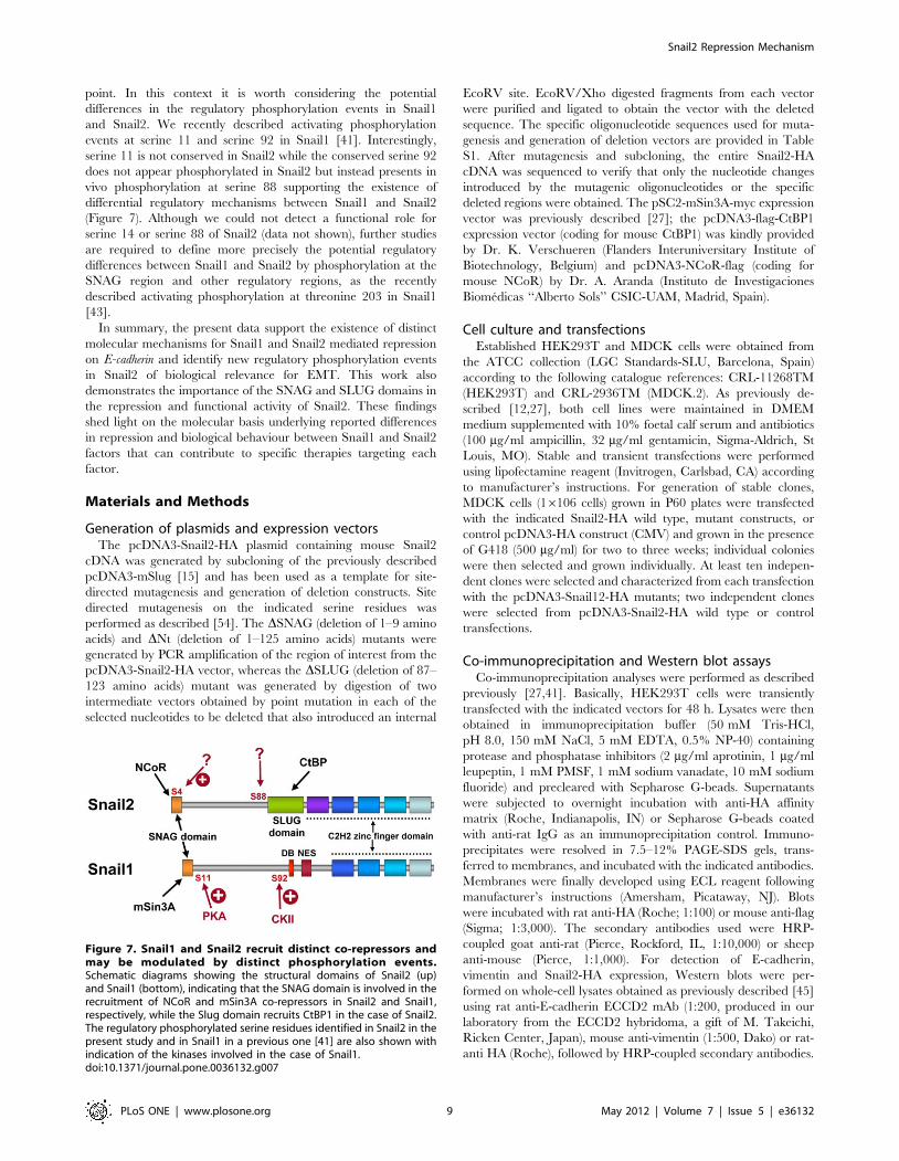
point. In this context it is worth considering the potential
differences in the regulatory phosphorylation events in Snail1
and Snail2. We recently described activating phosphorylation
events at serine 11 and serine 92 in Snail1 [41]. Interestingly,
serine 11 is not conserved in Snail2 while the conserved serine 92
does not appear phosphorylated in Snail2 but instead presents in
vivo phosphorylation at serine 88 supporting the existence of
differential regulatory mechanisms between Snail1 and Snail2
(Figure 7). Although we could not detect a functional role for
serine 14 or serine 88 of Snail2 (data not shown), further studies
are required to define more precisely the potential regulatory
differences between Snail1 and Snail2 by phosphorylation at the
SNAG region and other regulatory regions, as the recently
described activating phosphorylation at threonine 203 in Snail1
[43].
In summary, the present data support the existence of distinct
molecular mechanisms for Snail1 and Snail2 mediated repression
on E-cadherin and identify new regulatory phosphorylation events
in Snail2 of biological relevance for EMT. This work also
demonstrates the importance of the SNAG and SLUG domains in
the repression and functional activity of Snail2. These findings
shed light on the molecular basis underlying reported differences
in repression and biological behaviour between Snail1 and Snail2
factors that can contribute to specific therapies targeting each
factor.
Materials and Methods
Generation of plasmids and expression vectorsThe pcDNA3-Snail2-HA plasmid containing mouse Snail2
cDNA was generated by subcloning of the previously described
pcDNA3-mSlug [15] and has been used as a template for site-
directed mutagenesis and generation of deletion constructs. Site
directed mutagenesis on the indicated serine residues was
performed as described [54]. The DSNAG (deletion of 1–9 amino
acids) and DNt (deletion of 1–125 amino acids) mutants were
generated by PCR amplification of the region of interest from the
pcDNA3-Snail2-HA vector, whereas the DSLUG (deletion of 87–
123 amino acids) mutant was generated by digestion of two
intermediate vectors obtained by point mutation in each of the
selected nucleotides to be deleted that also introduced an internal
EcoRV site. EcoRV/Xho digested fragments from each vector
were purified and ligated to obtain the vector with the deleted
sequence. The specific oligonucleotide sequences used for muta-
genesis and generation of deletion vectors are provided in Table
S1. After mutagenesis and subcloning, the entire Snail2-HA
cDNA was sequenced to verify that only the nucleotide changes
introduced by the mutagenic oligonucleotides or the specific
deleted regions were obtained. The pSC2-mSin3A-myc expression
vector was previously described [27]; the pcDNA3-flag-CtBP1
expression vector (coding for mouse CtBP1) was kindly provided
by Dr. K. Verschueren (Flanders Interuniversitary Institute of
Biotechnology, Belgium) and pcDNA3-NCoR-flag (coding for
mouse NCoR) by Dr. A. Aranda (Instituto de Investigaciones
Biomedicas ‘‘Alberto Sols’’ CSIC-UAM, Madrid, Spain).
Cell culture and transfectionsEstablished HEK293T and MDCK cells were obtained from
the ATCC collection (LGC Standards-SLU, Barcelona, Spain)
according to the following catalogue references: CRL-11268TM
(HEK293T) and CRL-2936TM (MDCK.2). As previously de-
scribed [12,27], both cell lines were maintained in DMEM
medium supplemented with 10% foetal calf serum and antibiotics
(100 mg/ml ampicillin, 32 mg/ml gentamicin, Sigma-Aldrich, St
Louis, MO). Stable and transient transfections were performed
using lipofectamine reagent (Invitrogen, Carlsbad, CA) according
to manufacturer’s instructions. For generation of stable clones,
MDCK cells (16106 cells) grown in P60 plates were transfected
with the indicated Snail2-HA wild type, mutant constructs, or
control pcDNA3-HA construct (CMV) and grown in the presence
of G418 (500 mg/ml) for two to three weeks; individual colonies
were then selected and grown individually. At least ten indepen-
dent clones were selected and characterized from each transfection
with the pcDNA3-Snail12-HA mutants; two independent clones
were selected from pcDNA3-Snail2-HA wild type or control
transfections.
Co-immunoprecipitation and Western blot assaysCo-immunoprecipitation analyses were performed as described
previously [27,41]. Basically, HEK293T cells were transiently
transfected with the indicated vectors for 48 h. Lysates were then
obtained in immunoprecipitation buffer (50 mM Tris-HCl,
pH 8.0, 150 mM NaCl, 5 mM EDTA, 0.5% NP-40) containing
protease and phosphatase inhibitors (2 mg/ml aprotinin, 1 mg/ml
leupeptin, 1 mM PMSF, 1 mM sodium vanadate, 10 mM sodium
fluoride) and precleared with Sepharose G-beads. Supernatants
were subjected to overnight incubation with anti-HA affinity
matrix (Roche, Indianapolis, IN) or Sepharose G-beads coated
with anti-rat IgG as an immunoprecipitation control. Immuno-
precipitates were resolved in 7.5–12% PAGE-SDS gels, trans-
ferred to membranes, and incubated with the indicated antibodies.
Membranes were finally developed using ECL reagent following
manufacturer’s instructions (Amersham, Picataway, NJ). Blots
were incubated with rat anti-HA (Roche; 1:100) or mouse anti-flag
(Sigma; 1:3,000). The secondary antibodies used were HRP-
coupled goat anti-rat (Pierce, Rockford, IL, 1:10,000) or sheep
anti-mouse (Pierce, 1:1,000). For detection of E-cadherin,
vimentin and Snail2-HA expression, Western blots were per-
formed on whole-cell lysates obtained as previously described [45]
using rat anti-E-cadherin ECCD2 mAb (1:200, produced in our
laboratory from the ECCD2 hybridoma, a gift of M. Takeichi,
Ricken Center, Japan), mouse anti-vimentin (1:500, Dako) or rat-
anti HA (Roche), followed by HRP-coupled secondary antibodies.
Figure 7. Snail1 and Snail2 recruit distinct co-repressors andmay be modulated by distinct phosphorylation events.Schematic diagrams showing the structural domains of Snail2 (up)and Snail1 (bottom), indicating that the SNAG domain is involved in therecruitment of NCoR and mSin3A co-repressors in Snail2 and Snail1,respectively, while the Slug domain recruits CtBP1 in the case of Snail2.The regulatory phosphorylated serine residues identified in Snail2 in thepresent study and in Snail1 in a previous one [41] are also shown withindication of the kinases involved in the case of Snail1.doi:10.1371/journal.pone.0036132.g007
Snail2 Repression Mechanism
PLoS ONE | www.plosone.org 9 May 2012 | Volume 7 | Issue 5 | e36132

Protein stability assaysAnalysis of Snail2-HA wild type and the indicated mutant
proteins was performed in the presence of cycloheximide as
previously described [41]. Basically, HEK293T cells were
transiently transfected with the indicated vectors and 24 h later
cells were treated with 20 mM cycloheximide (Sigma-Aldrich) for
the indicated time intervals. Cells were lysed in RIPA buffer (0.1%
SDS, 0.5% sodium deoxycholate, 1% NP-40, 150 mM NaCl,
50 mM Tris-HCl, pH 8.0), containing protease and phosphatase
inhibitors and the expression of Snail2-HA analyzed by Western
blotting as described above. Three independent experiments were
performed; the mean +/2 s.d. of the integrated and normalized
band intensity is represented in the graphs.
ImmunofluorescenceCells grown on coverslips were rinsed with PBS, fixed with cold
(220uC) methanol 3 min, or 3.7% formaldehyde for 20 min at
room temperature and permeabilized using 0.5% (v/v) Triton X-
100. Transiently or stably transfected MDCK or HEK293T cells
were processed for indirect immunofluorescence with rat anti-HA
(1:50) and rat anti-E-cadherin (1:100) antibodies. Alexa-488 or
Alexa-555 (1:800, Molecular Probes, Eugene, OR) were used as
secondary fluorescent antibodies. Nuclei were detected by DAPI
stain. Samples were analysed using a confocal SP2 Spectral Leica
microscope and a Nikon 90i microscope equipped with epifluor-
escence and 663 objective.
Promoter activity assaysPromoter activity assays were basically performed as previously
reported [15,45]. The mouse E-cadherin promoter construct (2178
to +92), upstream of firefly luciferase, was transiently transfected in
24-well plates (200 ng/well) together with pCMV-bGal (Promega,
Madison, WI) (10 ng/well) for transfection efficiency control.
When indicated, co-transfections were carried out in the presence
of the indicated amount of pcDNA3-Snail2-HA wild type,
pcDNA3-Snail2-HA mutants and/or the indicated amounts of
co-repressors (mSin3A, NCoR and/or CtBP1). Luciferase and
bGal activities were measured using the Dual-luciferase b-Glo
Reporter assay kit (Promega) and normalized to the promoter
activity detected in mock transfected (pcDNA3-HA) cells as
described [27,45]. E-cadherin promoter activity in stable MDCK-
Snail2 transfectants was also analyzed as previously described [15].
Reporter assays in the various experimental settings were
performed at least twice and using triplicate samples. The mean
+/2 s.d is represented.
In vivo phosphorylationIn vivo phosphorylation assays were performed as recently
described [41]. HEK293T cells were grown in 30-mm-diameter
plates and transfected with pcDNA3-Snail2-HA or the indicated
Snail2-HA mutants. After 24–36 h, cells were washed three times
with DMEM phosphate-free medium, supplemented with 10%
foetal calf serum, and subsequently incubated in the same medium
containing 1 mCi/ml [32P]orthophosphate (Amersham) for 4 h.
Cells were lysed with ice-cold RIPA buffer containing protease
and phosphatase inhibitors, the resulting lysate centrifuged at
14,0006g for 15 min, and the supernatant incubated overnight at
4uC with 1 mg anti-HA and 25 ml of protein G-Sepharose
(Amersham). Immunoprecipitates were collected and washed four
times with ice-cold RIPA buffer. Precipitated proteins were
resolved by SDS-PAGE and transferred to nitrocellulose mem-
branes before exposure to x-ray film overnight to view radioac-
tively labelled proteins.
Phosphopeptide mapping and Edmann’s degradationPhosphopeptide mapping was performed essentially as de-
scribed [41]. Briefly, phosphorylated Snail2 bands were excised
from the membrane and blocked for 30 min in 0.5% polyvinyl-
pirrolidon K30 (Sigma-Aldrich), 0.6% acetic acid at 37uC. After
several washes with distilled water, bands were each incubated
overnight in 50 mM NH4HCO3, pH 8.0, with 1 mg modified
sequencing grade trypsin (Promega, Madison, WI) at 37uC. The
solution containing eluted peptides was transferred to a fresh tube
and the radioactivity measured to ensure efficient elution. The
peptides were frozen and dried by vacuum evaporation and
oxidized in performic acid for 1 h on ice in the dark. The
oxidization was stopped through dilution in water, the solution was
frozen, and vacuum evaporated. The peptides were then washed
twice in water, finally resuspended in 10 ml electrophoresis
running buffer (2.5% formic acid, 7.5% acetic acid, pH 1.9) and
carefully spotted onto a 206 20-cm cellulose TLC plate (layer
thickness 0.1 mm; cat. no. 5716, Merck, Rahway, NJ). Electro-
phoresis and chromatography was performed following previously
described conditions [41]. The results were observed using a Fuji
FLA-3000 PhosphorImager (Fuji) and the images adjusted using
Adobe Photoshop version 7.0 (San Jose, CA).
For Edmann’s degradation, phosphopeptides were eluted from
the plates in pH 1.9 electrophoresis buffer and lyophilized. The
fractions were then subjected to automated Edmann degradation,
using an Applied Biosystems Gas Phase Sequencer model 470A
(Foster City, CA) as described [41]. Released phenylthiohydantoin
amino acid derivatives from each cycle were spotted onto TLC
plates. The radioactivity in each spot was quantitated by exposure
to a screen and scanning in a FLA-3000 PhosphorImager (Fuji).
Supporting Information
Figure S1 The SNAG and SLUG domains of Snail2 arerequired for efficient repression of E-cadherin promoterin MDCK cells. (A) The repressor activity of Snail2-HA wild
type and the indicated mutants on the mouse E-cadherin promoter
was analyzed on MDCK cells. Reporter assays were performed
with 100 ng of the indicated vectors as described in Material and
Methods, and relative luciferase units (RLU) normalized to the
activity obtained in the presence of a void control pcDNA3 vector.
Results show the mean of triplicate experiments, performed on
quadruplicate samples, +/2 s.d. ***p,0.001. (B) Nuclear
localization of Snail2-HA and the DSNAG and DSLUG mutants
in transiently transfected MDCK cells as determined by confocal
immunofluorescence analysis. (C) Left, Schematic representation
of mouse Snail2 wild type and the indicated deletion mutants:
DSNAG DSLUG and DNt. Right columns, repression activity on
E-cadherin promoter (indicated as maximum and minimum
percentages in independent experiments) and nuclear localization
of the different Snail2-HA proteins in MDCK and HEK293T
cells. N.t., not tested.
(TIF)
Table S1 Sequences of oligonucleotide used for thegeneration of Snail2-HA mutants. Sequences of oligonucle-
tides, forward (Fw) and reversre (Rw), for generation of the
indicated Snail2-HA mutants by PCR are indicated in pairs. For
generation of the DSLUG mutant, two intermediate vectors were
generated, as indicated in Material and Methods section,
containing Ev restriction sites at the indicated positions, corre-
sponding at 87 and 123 amino acids, respectively; the pair of (Fw
and Rw) oligonucleotide sequences used for amplification on each
corresponding fragment, Ev87 and Ev123 are indicated.
(TIF)
Snail2 Repression Mechanism
PLoS ONE | www.plosone.org 10 May 2012 | Volume 7 | Issue 5 | e36132
Acknowledgments
The authors thank Christer Wernstedt for valuable help in the
phosphoaminoacid analysis, and members of A. Cano lab for helpful
discussions.
Author Contributions
Conceived and designed the experiments: AC FP PMO AV. Performed the
experiments: PMO AV MMP VS AM. Analyzed the data: PMO AV SS
MMP FP AC. Contributed reagents/materials/analysis tools: MPO AV
VS. Wrote the paper: AC FP MPO AV.
References
1. Nieto MA (2002) The snail superfamily of zinc-finger transcription factors. Nat
Rev Mol Cell Biol 3: 155–166.
2. Barrallo-Gimeno A, Nieto MA (2005) The Snail genes as inducers of cell
movement and survival: implications in development and cancer. Development
132: 3151–3161.
3. Peinado H, Olmeda D, Cano A (2007) Snail, ZEB, and bHLH factors in tumour
progression: an alliance against the epithelial phenotype? Nat Rev Cancer 7:
415–428.
4. Cobaleda C, Perez-Caro M, Vicente-Duenas C, Sanchez-Garcıa I (2007)
Function of the zinc-finger transcription factor SNAI2 in cancer and
development. Annu Rev Genet 41: 41–61.
5. Manzanares M, Locascio A, Nieto MA (2001) The increasing complexity of the
Snail gene superfamily in metazoan evolution. Trends Genet 17: 78–81.
6. Grimes HL, Chan TO, Zweidler-McKay PA, Tong B, Tsichlis PN (1996) The
Gfi-1 proto-oncoprotein contains a novel transcriptional repressor domain,
SNAG, and inhibits G1 arrest induced by interleukin-2 withdrawal. Mol Cell
Biol 16: 6263–6272.
7. Nakayama H, Scott IC, Cross JC (1998) The transition to endoreduplication in
trophoblast giant cells is regulated by the mSNA zinc finger transcription factor.
Dev Biol 199: 150–163.
8. Hemavathy K, Guru SC, Harris, Chen JD, Ip YT (2000) Human Slug is a
repressor that localizes to sites of active transcription. Mol Cell Biol 20:
5087–5095.
9. Mingot JM, Vega S, Maestro B, Sanz JM, Nieto MA (2009) Characterization of
Snail nuclear import pathways as representatives of C2H2 zinc finger
transcription factors. J Cell Sci 122: 1452–1460.
10. Domınguez D, Montserrat-Sentıs B, Virgos-Soler A, Guaita S, Grueso J, et al.
(2003) Phosphorylation regulates the subcellular location and activity of the snail
transcriptional repressor. Mol Cell Biol 3: 5078–5089.
11. Zhou BP, Deng J, Xia W, Xu J, Li YM, et al. (2004) Dual regulation of Snail by
GSK-3beta-mediated phosphorylation in control of epithelial-mesenchymal
transition. Nat Cell Biol 6: 931–840.
12. Cano A, Perez-Moreno MA, Rodrigo I, Locascio A, Blanco MJ, et al. (2000)
The transcription factor Snail controls epithelial-mesenchymal transitions by
repressing E-cadherin expression. Nat Cell Biol 2: 76–83.
13. Batlle E, Sancho E, Francı C, Domınguez D, Monfar M, et al. (2000) The
transcription factor Snail is a repressor of E-cadherin gene expression in
epithelial tumour cells. Nat Cell Biol 2: 84–89.
14. Hajra KM, Chen DY, Fearon ER (2002) The SLUG zinc-finger protein
represses E-cadherin in breast cancer. Cancer Res 62: 1613–1618.
15. Bolos V, Peinado H, Perez-Moreno MA, Fraga MF, Esteller M, et al. (2003) The
transcription factor Slug represses E-cadherin expression and induces epithelial
to mesenchymal transitions: a comparison with Snail and E47 repressors. J Cell
Sci 116: 499–511.
16. del Barrio MG, Nieto MA (2002) Overexpression of Snail family members
highlights their ability to promote chick neural crest formation. Development
129: 1583–1593.
17. Martınez-Estrada OM, Culleres A, Soriano FX, Peinado H, Bolos V, et al.
(2006) The transcription factors Slug and Snail act as repressors of Claudin-
expression in epithelial cells. Biochem J 394: 449–457.
18. Martin TA, Goyal A, Watkins G, Jiang WG (2005) Expression of the
transcription factors snail, slug, and twist and their clinical significance in
human breast cancer. Ann Surg Oncol 12: 488–496.
19. Come C, Magnino F, Bibeau F, De Santa Barbara P, Becker KF, et al. (2006)
Snail and slug play distinct roles during breast carcinoma progression. Clin
Cancer Res 12: 5395–5402.
20. Moreno-Bueno G, Cubillo E, Sarrio D, Peinado H, Rodriguez-Pinilla SM, et al.
(2006) Genetic profiling of epithelial cells expressing E- cadherin repressors
reveals a distinct role for Snail, Slug, and E47 factors in epithelial mesenchymal
transition. Cancer Res 66: 9543–9556.
21. Olmeda D, Montes A, Moreno-Bueno G, Flores JM, Portillo F, et al. (2008)
Snai1 and Snai2 collaborate on tumor growth and metastasis properties of
mouse skin carcinoma cell lines. Oncogene 27: 4690–4701.
22. Carver EA, Jiang R, Lan Y, Oram KF, Gridley T (2001) The mouse snail gene
encodes a key regulator of the epithelial-mesenchymal transition. Mol Cell Biol
21: 8184–8188.
23. Jiang R, Lan Y, Norton CR, Sundberg JP, Gridley T (1998) The Slug gene is not
essential for mesoderm or neural crest development in mice. Dev Biol 198:
277–285.
24. Aybar MJ, Nieto MA, Mayor R (2003) Snail precedes slug in the genetic cascade
required for the specification and migration of the Xenopus neural crest.
Development 130: 483–494.
25. Sefton M, Sanchez S, Nieto MA (1998) Conserved and divergent roles for
members of the Snail family of transcription factors in the chick and mouse
embryo. Development 125: 3111–3121.
26. Vernon AE, Labonne C (2006) Slug stability is dynamically regulated during
neural crest development by the F-box protein Ppa. Development 133:
3359–3370.
27. Peinado H, Ballestar E, Esteller M, Cano A (2004) Snai1 mediates E-cadherin
repression by recruitment of the Sin3A/histone deacetylase 1 (HDAC 1)/
HDAC2 complex. Mol Cell Biol 24: 306–319.
28. Herranz N, Paisini D, Diaz VM, Franci C, Gutierrez A, et al. (2008) Polycomb
complex 2 is required for E-cadherin repression by the Snail1 transcription
factor. Mol Cell Biol 28: 4772–4781.
29. Langer EM, Feng Y, Zhaoyuan H, Rauscher FJ, 3rd, Kroll KL, et al. (2008)
Ajuba LIM proteins are snail/slug corepressors required for neural crest
development in Xenopus. Dev Cell 14: 424–436.
30. Hou Z, Peng H, Ayyanathan K, Yan KP, Langer EM, et al. (2008) The LIM
protein AJUBA recruits protein arginine methyltransferase 5 to mediate SNAIL-
dependent transcriptional repression. Mol Cell Biol 28: 3198–3207.
31. Lin Y, Wu Y, Li J, Dong C, Ye X, et al. (2010) The SNAG domain of Snail1
functions as a molecular hook for recruiting lysine-specific demethylase 1.
EMBO J 29: 1803–1816.
32. Lin T, Ponn A, Hu X, Law BK, Lu J (2010) Requirement of histone
demethylase LSD1 in Snail1-mediated transcriptional repression during
epitelial-mesenchymal transition. Oncogene 29: 4896–4904.
33. Nibu Y, Zhang H, Bajor E, Barolo S, Small S, et al. (1998) dCtBP mediates
transcriptional repression by Knirps, Kruppel and Snail in the Drosophila
embryo. EMBO J 17: 7009–7020.
34. Qi D, Bergman M, Aihara H, Nibu Y, Mannervik M (2008) Drosophila Ebi
mediates Snail-dependent transcriptional repression through HDAC3-induced
histone deacetylation. EMBO J 27: 898–909.
35. Ayyanathan K, Peng H, Hou Z, Fredericks WJ, Goyal RK, et al. (2007) The
Ajuba LIM domain protein is a corepressor for SNAG domain mediated
repression and participates in nucleocytoplasmic shuttling. Cancer Res 67:
9097–9106.
36. Mittal MK, Myers JN, Misra S, Bailey CK, Chaudhuri G (2008) In vivo binding
to and functional repression of the VDR gene promoter by SLUG in human
breast cells. Biochem Biophys Res Commun 372: 30–34.
37. Mittal MK, Singh K, Misra S, Chaudhuri G (2011) SLUG-induced elevation of
D1 cyclin in breast cancer cells through inhibition of its ubiquitination. J Biol
Chem 286: 469–474.
38. Tripathi MK, Misra S, Khedkar SV, Hamilton N, Irvin-Wilson C, et al. (2005)
Regulation of BRCA2 gene expression by the SLUG repressor protein in human
breast cells. J Biol Chem 280: 17163–17171.
39. Yang Z, Rayala S, Nguyen D, Vadlamudi RK, Chen S, et al. (2005) Pak1
phosphorylation of snail, a master regulator of epithelial-to-mesenchyme
transition, modulates snail’s subcellular localization and functions. Cancer Res
65: 3179–3184.
40. Yook JI, Li XY, Ota I, Fearon ER, Weiss SJ (2005) Wnt-dependent regulation of
the E-cadherin repressor snail. J Biol Chem 280: 11740–11748.
41. MacPherson MR, Molina P, Souchelnytskyi S, Wernstedt C, Martin-Perez J,
et al. (2010) Phosphorylation of serine 11 and serine 92 as new positive
regulators of human Snail1 function: potential involvement of casein kinase-2
and the cAMP-activated kinase protein kinase A. Mol Biol Cell 21: 244–253.
42. Du C, Zhang C, Hassan S, Biswas MH, Balaji KC (2010) Protein kinase D1
suppresses epithelial-to-mesenchymal transition through phosphorylation of
Snail. Cancer Res 70: 7810–7819.
43. Zhang K, Rodriguez-Aznar E, Yabuta N, Owen RJ, Mingot JM, et al. (2011)
Lats kinase potentiates Snail1 activity by promoting nuclear retention upon
phosphorylation. EMBO J 31: 29–43.
44. Wang SP, Wang WL, Chang YL, Wu CT, Chao YC, et al. (2009) p53 controls
cancer cell invasion by inducing the MDM2-mediated degradation of Slug. Nat
Cell Biol 11: 694–704.
45. Moreno-Bueno G, Peinado H, Molina P, Olmeda D, Cubillo E, et al. (2009)
The morphological and molecular features of the epithelial-to-mesenchymal
transition. Nat Protoc 4: 1591–1613.
46. Thiery JP, Acloque H, Huang RY, Nieto MA (2009) Epithelial-mesenchymal
transitions in development and disease. Cell 139: 871–890.
47. Nieto MA (2011) The ins and outs of the epitelial to mesenchymal transition in
health and disease. Annu Rev Cell Dev Biol 10: 347–376.
48. Grooteclaes ML, Frisch SM (2000) Evidence for a function of CtBP in epithelial
gene regulation and anoikis. Oncogene 19: 3823–3828.
49. Bailey CK, Misra S, Mittal MK, Chaudhuri G (2007) Human SLUG does not
directly bind to CtBP1. Biochem Biophys Res Commun 353: 661–664.
Snail2 Repression Mechanism
PLoS ONE | www.plosone.org 11 May 2012 | Volume 7 | Issue 5 | e36132

50. Peinado H, Portillo F, Cano A (2004) Transcriptional regulation of cadherins
during development and carcinogenesis. Int J Dev Biol 48: 365–375.51. Vinas-Castells R, Beltran M, Valls G, Gomez I, Garcıa JM, et al. (2010) The
hypoxia-controlled FBXL14 ubiquitin ligase targets SNAIL1 for proteasome
degradation. J Biol Chem 285: 3794–3805.52. Peinado H, Iglesias-de la Cruz MC, Olmeda D, Csiszar K, Fong KS, et al.
(2005) A molecular role for lysyl oxidase-like 2 enzyme in snail regulation andtumor progression. EMBO J 24: 3446–3458.
53. Lander R, Nordin K, Labonne C (2011) The F-box protein Ppa is a common
regulator of core EMT factors Twist, Snail, Slug and Sip1. J Cell Biol 194:
17–25.
54. Zheng L, Baumann U, Reymond JL (2004) An efficient one-step site-directed
and site-saturation mutagenesis protocol. Nucleic Acid Res 2: e115.
Snail2 Repression Mechanism
PLoS ONE | www.plosone.org 12 May 2012 | Volume 7 | Issue 5 | e36132