Characterization of the Lymantria dispar ... of the Lymantria dispar nucleopolyhedrovirus 25K FP...
11
qo.~'tal..of.. ~"e':eZ. ~Ti,:o!ogz .(~ .99%..7.~:.. ?. 9 .! .3-?.723:.....pr.~nte~..t~. Gr.e.at. Br!!.a.i. n. ........................................................................................................................... Characterization of the Lymantria dispar nucleopolyhedrovirus 25K FP gene David S. Bischoff and James IVl. Slavicek USDA Forest Service, Northeastern Forest Experimental Station, Forest Sciences Laboratory, 359 blain Road, Delaware, OH 4301 5, USA The Lymantria dispar nucleopolyhedrovirus (Ld/V/NPV) gene encoding the 25K FP protein has been cloned and sequenced. The 25KFP 9ene codes for a 217 amino acid protein with a predicted molecular mass of 24870 Da. Expression of the 25K FP protein in a rabbit reticulocyte system generated a 27 kDa protein, in close agreement with the molecular mass predicted from the nucleo- tide sequence. The gene is located between 40"3 and 40.8 map units on the viral genome. It is transcribed in a counterclockwise direction with respect to the circular map at late times during the infection cycle from a consensus baculovirus late promoter. The Ld/V/NPV and Autographa californica nucleopolyhedrovirus (AcMNPV) 25K FP proteins exhibit 52 % amino acid identity with several regions showing greater than 75% identity. Homologues to the Ach/NPV orf59 and orf60 were also identified upstream (with respect to the genome) of the 25K FP gene in Ldh/NPV and exhibit 52% and 45% amino acid identity, respectively. Introduction Nucleopolyhedroviruses (NPV) are members of the Baculoviridae which produce two morphological distinct forms, a budded virus form and a virus form that is occluded into a protein structure termed a polyhedron. Insects are infected by NPVs after ingestion of the polyhedron and release of the occluded virions by dissolution of the polyhedron in the alkaline environment of the insect midgut. Early after infection the budded virus (BV) form is produced, which buds through the plasma membrane and then infects other ceils. Later during the infection cycle the occluded form of the virus is generated (reviewed by Blissard & Rohrmann, I990). During serial passage of NPVs in ceil culture, a class of virus termed few polyhedra (FP) mutants arises at a high frequency and becomes predominant. These mutants have the charac- teristics of altered plaque morphology, production of few polyhedra that contain very few viral nucleocapsids, increased release of BV, defective envelopment of viral nucleo- capsids within the nucleus, and in Lymantria dispar NPV (LdMNPV) a decrease in the percentage of cells that produce polyhedra in comparison to the wild-type (many polyhedra, MP) virus (Hink & Vail, I973; MacKinnon et al., 1974; Authorfor correspondence: James H. Slavicek. Fax + 1 614 368 0152. The GenBank accession number of the nucleotide sequence reported in this paper is U58676. Ramoska & Hink, 1974; Hink & Strauss, 1976; Potter et al., 1976; Fraser & Hink, 1982; Slavicek et al., 1992; Harrison & Summers, 1995; Slavicek et al., 1995). Specific mutations in FP mutants of Autographa californica NPV (AcMNPV) and Galleria mellonella NPV (GmMNPV) have been identified and characterized. The appearance of the FP phenotype during virus passage in cell culture correlates with the presence of DNA insertions/deletions (Fraser et al., 1983; Kumar & Miller, 1987; Cary et aL, 1989) and with the absence of a specific 25 kDa protein (Fraser eta]., 1983). These insertions/deletions range from approximately 0"4-2"8 kbp and occur predominantly within a specific region [36-37 map units (m.u.)] on the AcMNPV and GmM/I'qPV genomes. Analysis of this genomic region has identified a gene coding for a 25 kDa protein that is necessary for the MP phenotype (Beames & Summers, 1988). The 25K FP gene is essential for polyhedron formation and virion occlusion since deletion of this gene is sufficient to eliminate these processes (Beames & Summers, I989). In addition, different mutations in the 25K FP gene generate all phenotypical characteristics of FP mutants (Harrison & Summers, 1995). In contrast to the AcMNPV and GmMNPV FP mutants, DNA insertions and/or deletions do not correlate with the appearance of FP mutants in LdMNPV, as determined by DNA restriction endonuclease analysis (Slavicek eta]., 1995). This suggests that LdMNPV FP mutants may arise through a different mechanism than the formation of FP mutants in AcMNPV. Alternatively, the mechanism of FP mutant for- 91.'- 0001-3823 © 1996 SGM
Transcript of Characterization of the Lymantria dispar ... of the Lymantria dispar nucleopolyhedrovirus 25K FP...

qo.~'tal..of.. ~"e':eZ. ~Ti,:o!ogz .(~ .99%..7.~:.. ?. 9 .! .3-?. 723:.....pr.~nte~..t~. Gr.e.at. Br!!.a.i. n. ...........................................................................................................................
Characterization of the Lymantria dispar nucleopolyhedrovirus 25K FP gene
D a v i d S. B ischof f a n d J a m e s IVl. S lav icek
USDA Forest Service, Northeastern Forest Experimental Station, Forest Sciences Laboratory, 359 blain Road, Delaware, OH 4301 5, USA
The Lymantria dispar nucleopolyhedrovirus (Ld/V/NPV) gene encoding the 25K FP protein has been cloned and sequenced. The 25KFP 9ene codes for a 217 amino acid protein with a predicted molecular mass of 2 4 8 7 0 Da. Expression of the 25K FP protein in a rabbit reticulocyte system generated a 27 kDa protein, in close agreement with the molecular mass predicted from the nucleo- tide sequence. The gene is located between 40"3 and 40.8 map units on the viral genome. It is transcribed in a counterclockwise direction with
respect to the circular map at late times during the infection cycle from a consensus baculovirus late promoter. The Ld/V/NPV and Autographa californica nucleopolyhedrovirus (AcMNPV) 25K FP proteins exhibit 52 % amino acid identity with several regions showing greater than 7 5 % identity. Homologues to the Ach/NPV orf59 and orf60 were also identified upstream (with respect to the genome) of the 25K FP gene in Ldh/NPV and exhibit 5 2 % and 4 5 % amino acid identity, respectively.
In t roduct ion
Nucleopolyhedroviruses (NPV) are members of the Baculoviridae which produce two morphological distinct forms, a budded virus form and a virus form that is occluded into a protein structure termed a polyhedron. Insects are infected by NPVs after ingestion of the polyhedron and release of the occluded virions by dissolution of the polyhedron in the alkaline environment of the insect midgut. Early after infection the budded virus (BV) form is produced, which buds through the plasma membrane and then infects other ceils. Later during the infection cycle the occluded form of the virus is generated (reviewed by Blissard & Rohrmann, I990).
During serial passage of NPVs in ceil culture, a class of virus termed few polyhedra (FP) mutants arises at a high frequency and becomes predominant. These mutants have the charac- teristics of altered plaque morphology, production of few polyhedra that contain very few viral nucleocapsids, increased release of BV, defective envelopment of viral nucleo- capsids within the nucleus, and in Lymantria dispar NPV (LdMNPV) a decrease in the percentage of cells that produce polyhedra in comparison to the wild-type (many polyhedra, MP) virus (Hink & Vail, I973; MacKinnon et al., 1974;
Author for correspondence: James H. Slavicek.
Fax + 1 614 368 0152.
The GenBank accession number of the nucleotide sequence reported in
this paper is U58676.
Ramoska & Hink, 1974; Hink & Strauss, 1976; Potter et al., 1976; Fraser & Hink, 1982; Slavicek et al., 1992; Harrison & Summers, 1995; Slavicek et al., 1995).
Specific mutations in FP mutants of Autographa californica NPV (AcMNPV) and Galleria mellonella NPV (GmMNPV) have been identified and characterized. The appearance of the FP phenotype during virus passage in cell culture correlates with the presence of DNA insertions/deletions (Fraser et al., 1983; Kumar & Miller, 1987; Cary et aL, 1989) and with the absence of a specific 25 kDa protein (Fraser eta]., 1983). These insertions/deletions range from approximately 0"4-2"8 kbp and occur predominantly within a specific region [36-37 map units (m.u.)] on the AcMNPV and GmM/I'qPV genomes. Analysis of this genomic region has identified a gene coding for a 25 kDa protein that is necessary for the MP phenotype (Beames & Summers, 1988). The 25K FP gene is essential for polyhedron formation and virion occlusion since deletion of this gene is sufficient to eliminate these processes (Beames & Summers, I989). In addition, different mutations in the 25K FP gene generate all phenotypical characteristics of FP mutants (Harrison & Summers, 1995).
In contrast to the AcMNPV and GmMNPV FP mutants, DNA insertions and/or deletions do not correlate with the appearance of FP mutants in LdMNPV, as determined by DNA restriction endonuclease analysis (Slavicek eta]., 1995). This suggests that LdMNPV FP mutants may arise through a different mechanism than the formation of FP mutants in AcMNPV. Alternatively, the mechanism of FP mutant for-
91.'- 0001-3823 © 1996 SGM

iiiiiiiiiiiiiii (a)
E P G F N T R L
I I I I ',"',,, M KS A H J Q O I B D C
I I I I I I I I I I I I I Bgl II map
C 2 C 1 2
C77 C 3 8
C 6 4
C 1 5
Map units
97.3 - 23.9
17.4 - 45.6
34.0 - 55.7
50.5 - 69.2
56.6 - 83.5
8 0 . 2 - 4.1
I I 1 0 2 0 4 0
Fig. 1, For legend see facing page,
U E
1
~DB105
)DB104
)DB106
3DB108
~DB122
~DB152
~DB103
I I I 6 0 8 0 1 0 0
map units
34.3 - 39.8 36.7 - 42.5
39.8 - 46.3
39.8 - 42.5
40.2 - 41.3
40.3 - 40.8
42.5 - 46.3
marion in these viruses may be similar; however, mutation(s) within the LdMNPV 25KFP gene may not be readily detectable. As a prerequisite to the molecular analysis of the 25K FP gene in LdMNPV FP mutants, we have identified, cloned and characterized the LdMNPV 25K FP gene.
M e t h o d s • Cells and virus. Lymantria dispar 652Y (Ld652Y) cells were grown as monolayers in Goodwin's IPL-52B (GI) medium supplemented with 6"25 mM-glutamine and 10% fetal bovine serum. Cell cultures were inoculated with either LdMNPV isolate A21-MPV, which produces wild- type polyhedra (Slavicek et al., I996), or the FP mutant A21-2 (Slavicek et al., I995).
• Viral DNA isolation. Non-occluded virus from plaque purified LdMNPV A21-MPV or A21-2 was isolated from infected Ld652Y cells as described previously (Bischoff & Slavicek, 1994), and used as a source of genomic DNA for cosmid library construction and marker rescue experiments. Viral DNA was digested with restriction endonucleases and fractionated on 0'8 % agarose-TBE gels.
• Cosmid construction and subclones. A cosmid library of LdMNPV A21-MPV viral DNA was constructed using the SuperCos 1 cosmid vector (Statagene) following protocols provided with the vector. Viral DNA was partially digested with SaudAI and cloned into the BamHI site of SuperCos I to generate cosmids with LdMNPV inserts that were approximately 40-45 kbp in length. Cosmids containing over- lapping viral DNA were identified by restriction endonuclease analysis. Subclones of the cosmids were constructed in pUCI8 using standard techniques.
• Marker rescue mapping of the A 2 1 - 2 mutation. P6 wells (Coming) were plated with Ld652Y cells (4"5 x 10⁵ cells per well) and the
cells allowed to attach to the bottom for I h. The media was removed, the cells were washed twice with 3 ml of GI medium (without fetal bovine serum) and then covered with 1"5 m[ of medium. Cosmid or plasmid DNA (2'5 ,g) and mutant viral DNA (2"5 ,g) were mixed in a polystyrene tube, diluted to 50 ~1 with water, and then heated to 65 °C for 15 min. An equal volume (50 ~.1) of Lipofectin reagent (BRL) was added to the DNA mixture and incubated at room temperature for 15 min. The Lipofectin-DNA complex (100,1) was added dropwise around the well. After incubation at room temperature for 2 h, the medium was replaced with flesh GI medium supplemented with 6-25 mM- gIutarnine and 10% fetal bovine serum. The cells were incubated at 27 °C for 2 h, Gentamicin (BRL) was added to a final concentration of 10 l~g/ml, and then further incubated at 27 °C for up to 14 days. Cells were viewed with a Nikon Diaphot-TMD inverted microscope at I00 x magnification.
To further localize the A21-2 mutation to the 25K FP gene, a 934 bp region containing the 25K FP gene was amplified from the A21-MPV viral genome by PCR. Primers were designed from the wild-type 25K FP gene sequence (see below) from isolate A21-MPV: 5' primer, GAGC- ACATGACCGTTTCG (complementary to positions 1627-1644 in Fig. 3); 3' primer, GGTAAATCG.AGCACGCTC (positions 711-728 in Fig. 3). PCR reactions (100 ~1) contained buffer (50 mM-KC1, 10 mM-Tris-HCl at pH 9'0, 0'1% Triton X-100), 2"0 mM-MgCI~, 200 ~M-deoxy- nucleotides, 0'5 l~M-primers, 20 ng viral DNA and 2"25 units of Taq DNA polymerase. Thermal cycling was carried out in a Perkin Elmer Cetus thermocycler. After denaturation at 94 °C for 3 min, 35 cycles were performed at 94 °C for I min, 48 °C for I min, and 70 °C for I min. The entire reaction was run-out on a 1% agarose-TBE gel, the 934 bp fragment was excised, and the DNA purified using the GeneClean II kit (Biol01). The fragment was cloned into the TA-cloning vector (Invitrogen) to create pDB152 and used for marker rescue.
Budded virus was recovered from the transfections which rescued the mutant phenotype and plaque purified as previously described (Slavicek et al., 1995). Ld652Y cells (5 x 10 ~ cells per T25 flask) were infected in

Fig. 1. Marker rescue mapping of A21-2 mutation. (a) Location of clones used to map the A21-2 mutation are shown below the LdMNPV Bglll restriction map. Cosmid and plasmid inserts are shown as lines. Inserts that could repair the mutation are shaded. (b--e) Appearance of Ld652Y cells viewed by light microscopy after cotransfection with A21-2 viral DNA and one of the cosmids or plasmids in the A21-MPV genomic library. Cells were viewed with a Nikon Diaphot-TMD inverted microscope at 100 x magnification, A21-2 DNA alone (b) ; A21-2 with C38 (c); A21-2 with C12 (d) ; A21-2 with pDB1 52 (e).
quadruplicate with 5 TCIDs0 units per cell of LdMNPV isolates A21- MPV, A2I-2 or C12-2-1, an MP plaque purified from the C12 trans- fection. The number of polyhedra present per flask was determined at 6 days post-infection (p.i.) as previously described (Slavicek et al., I995).
• S e q u e n c i n g . The sequence of the 25K FP gene region from A2I- MPV was determined on both strands of pDBI08 using the dide- oxynucleotide sequencing method. PIasmid and single-strand M13 DNA
templates were sequenced with the Sequenase version 2.0 DNA sequencing kit (USB) or the fmo[ DNA sequencing system (Promega) using protocols supplied. [~-35S]dATP was supplied by NEN. Sequence analysis was done using the MacVector program (IBI).
• RNA isolation and Northern blot analysis. A2I-MPV infected Ld652Y cells were harvested at various times (p.i.). Cytoplasmic RNA was isoIated as described by Friesen & Miller (1985). Total RNA was
9 1 . ~

separated on I'2 % agarose gels containing formaldehyde and transferred to nitrocellulose. Northern blot analysis was performed as described by Mahmoudi & Lin (I989). A 33base oligonucleotide (positions 1087-1119 in Fig. 3). was end-labelled with [7-a~P]ATP (NEN) and used as a strand-specific probe to detect the 25K FP transcripts.
• Primer extension mapping of transcripts. Primer extension reactions were performed using the method of Crawford & Miller (I988). Total RNA was isolated at 48 h p.i. from Ld652Y cells infected with LdMNPV A2I-MPV. An 18 base oligonucleotide (positions 1489-1506 in Fig. 3) was used in the reactions after being end-labelled with [y-~2P]ATP (NEN). The primer was extended using Moloney murine leukaemia virus (M-MLV) reverse transcriptase. Primer extension products were fractionated on 6% polyacrylamide-8 M-urea gels and visualized by autoradiography.
• In vitro transcription and translation of the 25K FP gene. A 1.8 kbp SstII fragment containing the 25K FP gene was subcloned into pBluescript SK(+) to generate pDB122. The 25KFP protein was expressed from pDB122 with the TNT coupled reticulocyte lysate system and T7 RNA polymerase (Promega) using directions provided with the kit. The expressed protein was labelled by the addition of [35SJmethionine (NEN). Reaction products were analysed by SDS-PAGE and autoradio- graphy.
R e s u l t s a n d D i s c u s s i o n
Cosmid construction
A cosmid library of isolate A2I -MPV was constructed in the SuperCos I vector after partial digestion of LdMNPV
AZI-MPV viral D N A with Sau3AI under conditions that generated fragments approximately 40-45 kbp in length. Cosmid inserts were analysed by restriction endonuclease analysis and comparison with the restriction pattern of LdMNPV clonal isolate 5-6 (RiegeI et aL, 1994). Six cosmids were isolated that contained overlapping viral D N A and spanned the entire length of the viral genome (Fig. la).
Marker re scue of the A 2 1 - 2 mutat ion
Isolate A21-2 exhibits the characteristics of a typical FP mutant (Slavicek et al., 1995). In order to identify the genomic location of the gene mutated in this isolate, marker rescue/ repair experiments were done in which Ld652Y cells were transfected with A2I-2 viral DNA and DNA from each of the cosmids. Focal areas of cells appearing opaque as a consequence of the presence of a large number of polyhedra were only found in transfections with C12 and C77 (Fig. Ia , d). Transfection with the other cosmids generated cells with the A2I-2 polyhedron formation phenotype (Fig. 1 b, c). Since C12 and C77 are overlapping cosmid clones, the mutated gene mapped to the 20 kbp of overlapping D N A (32"7 to 45"1 m.u. on the viral genome) contained within these cosmids.
The 20 kbp of overlapping D N A contained within C12 and C77 was subcloned and the mutation further mapped in marker
(a) E P G F N T R L M K S A H J Q O I B
I , I I I I I polh14")lIeK I I I }1 I I I I , , ~ `% `% %-I)DNApol EGT-)
j . `% `%
d' `%
`%
j . `% .-?
D C I I I
`%
E"
39.~
(b) 1 2
3 4
5 6
Frame
`% `%
`% `%
,%
1.0 kbp
`% `%
`% `%
I I m
42 .5
`%
Fig. 2. Genomic location of LdMNPV off59, off60 and 25KFP genes. (a) Bglll restriction map of the LdMNPV viral genome. The locations of other LdMNPV genes are indicated: polh (polyhedrin; Smith et al., 1988), EGT (ecdysteriod UDP- glucosyltransferase; Riegel et al., 1994), DNA pol (DNA polymerase; Bjornson et al., 1992) and PK (viral protein kinase; Bischoff & Slavicek, 1994). The enlarged map shows the 4.3 kbp BamHI-EcoRI fragment at 39.8-42-5 m.u. on the viral genome. The position of the genes within this fragment and the direction of transcription are indicated. Restriction sites used in subcloning and expression of the gene are shown. (b) ORF analys{s of this fragment in all six reading frames. The black boxes indicate ORFs that are at least 25 amino acids in length and which begin with an ATG start codon. The shaded boxes indicate the ORFs corresponding to LdMNPV off59, orf60 and 25KFP genes.
Fig. 3. Nucleotide sequence of LdMNPV off59 (partial), off60 and 25K FP genes with predicted amino acid sequences. The consensus late promoter sequence (ATAAG) located prior to the 25KFP gene coding region is shaded with the late transcriptional start sites indicated with arrows. A potential polyadenylation signal sequence (AATAAA) after the 25KFP gene coding region is underlined as are oligonucleotides used in transcriptional mapping and primer extension (see text).
91E

98
146
194
247
266
280
297
313
331
347
362
379
396
413
428
442
476
524
572
620
668
716
771
834
888
936
984
1032
1080
1128
1176
1224
1272
1320
1368
1416
1464
1512
1 GGA~ CCA TCT GTT GTT CAA TTT GAC GTA GCG CCG CTC GAC CGC CTT GAA
I W R N N L K V Y R R E V A K F
50 GGC CAA GTG CAA AGC TAT ATT TTC GTC GCC GCC GTC CAG CTT GTG GTA
A L H L A I N E D G G D L K H Y
CTT GCG AAA AGT CTC TAT GAA CAG TCG CCG CGC CCG GGC CGG CAT CTC
K R F T E I F L R R A R A P M E
TTC CTT TAA CAA AAC CTC GTT CAA ATA AAA CAT GAC AAT AGC CTT ATT
E K L L V E N L Y F M V I A K N
ATA TTG AAA TCG CTC GCG CAT ATCGTT AAAGTTTATA TATATGTTA ATTGCGT
Y Q F R E R M (orf59)
TATATTAAAATAGTTTGTG
TATTAAA TAGTTTG
TATATTAAAATAGTTTG
TATATTAAA TAGTTTG
TATATTAAA TAGTTTGTG
TATTAAA TAGTTTGTG
TATTAA TAGTTTGTA
TATTAAAATAGTTTGAC
TGTTAAAATAGTT GAGC
TATTAAAATAGTTTGGC
TATTAGAATAGTTTG
TATTAAA TTGTTTA TACATTAAAA TTA TAT GTC ATC GTC GGA ATT TGT
*** I D D D S N T
TTC ATT ATC AGT GGT GGT GGT GGT GGA AGA GTC CTC CTC CTC CGT GGT
E N D T T T T T S S D E E E T T
GGT GGT GTC GTA ATC GTT GGC GTC CGC GAA CGG CAG CCA CTC GCC GCG
T T D Y D N A D A F P L W E G R
GTC GCA ATA GTA TTT ACG CTT GAC GGC GGT CCA CGC GAT GGC GTC GGC
D C Y Y K R K V A T W A I A D A
CGT CGT CGG CGA GTT GCG CAT TTT GAT GCT TTT ATT GTA AAA TTT TAA
T T P S N R M K I S K N Y F K L
ATA AAT GCG CTT GCC GTT GTA GGG CAA GGC TCG CGT CGA AGT GGG TAA
Y I R K G N Y P L A R T S T P L
ATC GAG CAC GCT CGT AAA TAA CAT ATTCAAATACAATAATCACAACCAATTCAGT
D L V S T F L M (off60) CTTTTATTCGATCCTTAACGCTATTGAGATTCAACTTGAAAGGGTCCGCGTAGAATAATTAAA
GCGAGGT CGTCCGTCCG AGGGTTCGCG CCTCGCTCA AAT AAT GTG GGA GTA ATT *** I I H S Y N
TTT GGC CGG GTC GTC GTC GCC TTC GAA ACT GAT CGC GAC GAC GCT GCT
K A P D D D G E F S I A V V S S
GTC GAT GCT GGC CTC CGC GCG CTG TCC GAC CAA TTT GTG AAT GTC TCG
D I S A E A R Q G V L K H I D R
CTC GTC CTT AAT TAT GTA TAT TTC GCT GCT GTC GTT GCG GCG CGC CAT
E D K I I Y I E S S D N R R A M
GAC GCC GGC CTT GCA CAA CGA CAC GTA CTT GAA GTG CGG CAG CAG GGC
V G A K C L S V Y K F H P L L A
GTC GCG CST CTT CTT CAA CAG CTG TTT GCG CTC GCG ACT GGC GGC CAC
D R T K K L L Q K R E R S A A V
GAA AAT CTT GAC CGG CCC GTC GTA CTC GAC GCC GAG GTC GCG ATT CTT
F I K V P G D Y E V O L D R N K
GAG GCG CAC CTG GCA CGA GCG CGT TTG CCA GTC GCG CGC CGT GGC CGC
L R V Q C S R T Q W D R A T A A
GTC CTT CAA GTG CAC GAG AAT ATA ATT GTT CTT GGT TTC GGA CTC GAG
D K L H V L I Y N N K T E S E L
CAC CGC CTT GAA GTT GAG GCC GAG CAG CGC GCA AAT TTT TTT GAT GTA
V A K F N L G L L A C I K K I Y
ATA GTT TTT CAC TTT ~CG GTC GAA GAA GGG CCG CGT GTC GGC GAG CCC
Y N K V K R D F F P R T D A L G
GTA AAT CTC CAC GCT GCA GTT GAG CGA GTT CTT TTC GAG CCG CTG CAG
Y I E V S C N L S N K E L R Q L
CTT TTG CGC GAG CGC GTC CAG ACT CTC GCT CAC GTT GCG GTC GAT TTC
K Q A L A D L S E S V N R D I E
GGT TTT GAT CAA TTT TTT CAA ACC CSA CAC STT GAT GAG ATC GAA ATC
T K I L K K L G S V N I L D F D
CAT GTC CAT GA CCG CGC GGG CGT GTG TGT GTG TGT GTG CGA GGT TTG
M D M (25K FP) ~ 1559 AATATG TAA ATT AAT TAC TCC ~ TAG TGA GCC GCG AGG CGC GCAG
1608 TCGATCGGAAACCTCGCCCCGAAACGGTCATGTGCTC
Fig. B. Forlegendsee ~cing page.
91;
(a)
Ld/v/NPV 25K FP
AcMNPV 25K FP
i0 20
MDMDFDLINV SGLKKLIKTE
MD :LINV S LK.LI.T:
MDQFEQLINV SLLKSLIDTQ
i0 20
30 40 50 60
ZDRNVSESLD ALAQKLQRLE KNSLNCSVEI YGLADTRPFF
ID NVS:::. ::::KL:RLE ::L. SVEI YG: D:R :
IDENVSDNIK SMSEKLKRLE YDNLTDSVEI YGIHDSR-LN
30 40 50
L~PV 25K FP
Ac/V/NPV 25K FP
7O 8O
DRKVKNYYIK KICALLGLNF
::K::NYY: KICALL:LNF
NKKIRNYYL- KICALLDLNF
6O 70
90 1@O ii0 120
KAVLESETKN NYILVHLKDA ATARDWQTRS CQVRLKNRDL
K V:ES. .: N.I:..L DA : A::WQT:S : RLKN :L
KHVIESSFDK NHIVAKLCDA TRAKEWQTKS RERRLKNFNL
80 90 i00 Ii0
Ld/V/NPV 25K FP
Ac/V/NPV 25K FP
130 140 150 160 170 180
GVEYDGPVKI FVAASRERKQ LLKKTRDALL PHFKYVSLCK AGVMARRNDS SEIYIIKDER
.::YDGPVKI FVAA: E:K LLKKTRDALL P :KY:S:CK .GVM.RR::. S ::I:K:E:
NINYDGPVKI FVAATAEQKL LLKKTRDALL PFYKYISICK NGVMVRRDEK SRVFIVKNEQ * * * * * * * * * * * *
120 130 140 150 160 170
LdMNPV 25K FP
Ac/V/NPV 25K FP
190 200 210
DIHKLVGQRA EASIDSSVVA ISFEGDDDPA KNYSHII
:I: L ::: :::S. V. :FE:::D:. K ::I
NIEYLKANKY -YAFHSDSVD NFESENDSE KMLQNLI
180 190 200 210
(b)
1 50 i00 150 200 I I I I I
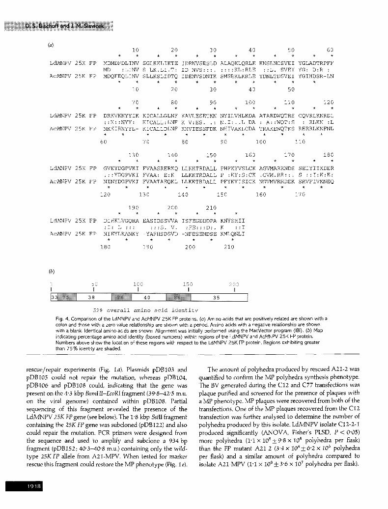
52% o v e r a l l amino ac id i d e n t i t y Fig. 4. Comparison of the LdMNPV and AcMNPV 25K FP proteins. (a) Amino acids that are positively related are shown with a colon and those with a zero value relationship are shown with a period. Amino acids with a negative relationsHp are shown with a blank. Identical amino acids are shown. Alignment was initially performed using the t,'lacVector program (IBI). (b) Map indicating percentage amino acid identity (boxed numbers) within regions of the LdMNPV and AcMNPV 25K FP protein. Numbers above show the location of these regions with respect to the LdMNPV 25K FP protein. Regions exhibiting greater than 75% identity are shaded.
rescue/repair experiments (Fig. Ia). Plasmids pDB103 and pDBI05 could not repair the mutation, whereas pDB104, pDB106 and pDB108 could, indicating that the gene was present on the 4"3 kbp BamHI-EcoRI fragment (39"8-42"5 m.u. on the viral genome) contained within pDB108. Partial sequencing of this fragment revealed the presence of the LdMNPV 25K FP gene (see below). The 1"8 kbp SstlI fragment containing the 25K FP gene was subcloned (pDB122) and also could repair the mutation. PCR primers were designed from the sequence and used to amplify and subclone a 934 bp fragment (pDBI52; 40"3-40"8 m.u.) containing only the wild- type 25K FP allele from A21-MPV. When tested for marker rescue this fragment could restore the MP phenotype (Fig. le).
The amount of polyhedra produced by rescued A2I-2 was quantified to confirm the MP polyhedra synthesis phenotype. The BV generated during the C12 and C77 transfections was plaque purified and screened for the presence of plaques with a MP phenotype. MP plaques were recovered from both of the transfections. One of the MP plaques recovered from the C12 transfection was further analysed to determine the number of polyhedra produced by this isolate. LdMNPV isolate C12-2-1 produced significantly (ANOVA, Fisher's PLSD, P < 0"05) more polyhedra (1"1 x 10s+9"8x 10⁶ polyhedra per flask) than the FP mutant A2I-2 (3"4 x I0~q - 6"2 x I05 polyhedra per flask) and a similar amount of polyhedra compared to isolate A21-MPV (I'1 x 10⁸+ 3"6 x 10⁷ polyhedra per flask).
91~

i0 20 30 40 50 60
LcLMNPV ORF59 MRERFQYNKA IVMFYLNEVL LKEEMPARAR RLFIETFRKY HKLDGGDENI ALHLAFKAVE M: : ::L :E.MP:RA: :LF:ETF KY HK::GGDE:I A:H A KA:E
AcMNPV ORF59 MYQIPDML YNEKMPPRAK KLFVETFTKY HKMNGGDEDI AMHKARKALE
i0 70
Lc~PV ORF59 RRYVKLNNRW I>> :YVK::.
Ac~/NPV ORF59 EKYVKIDTLQ NLGFRAKPPT R
50 60
20 30 40
52% amino acid identity
i0 20 30 40 50 60
L~PV ORF60 MLFTSVLDLP TSTRALPYNG KRIYLKFYNK SIKMRNSPTT ADAIAWTAVK RKYYCDRGEW
LP . :.LPYNG KRI: KFY:: S::. :S. . A. :A AV: :KY G.W
AcMNPV ORF60 MLP KYYQNLPYNG KRIFEKFYDR SLQKYKSTQI ATKLACCAVR KKYMLIDGkW
i0 20 30 40 50
70 80 90
Ldd~NPV ORF60 LPFADANDYD TTTTEEEDSS TTTTTDNETN SDDDI
P :DAN: D TTT ..EDS: T.T.T: .T. .D:
AcMNPV ORF60 QPRPDANNSD TTT-STEDST TDTETEYSTT EDELA
60 7O 8O
45% amino acid identity
Fig. 5. Comparison of the LdMNPV and AcMNPV off59 (partial) and off60 homologues. Amino acids that are positively related are shown with a colon and those with a zero value relationship are shown with a period. Amino acids with a negative relationship are shown with a blank. Identical amino acids are shown.
Identification of the ?.5t( lap gene
The restriction map of the 4"3 kbp BamHI-EcoRI fragment is shown in Fig. 2 (a). Computer analysis of a 1"6 kbp region within this fragment revealed several open reading frames (ORFs) that may encode proteins (Fig. 2 b). The predicted amino acid sequence of these ORFs was compared with other proteins in GenBank at the National Center for Biotechnology Information (NCBI) using the BLAST network service (Altschul et al., I990). This search revealed that three of the ORFs showed homology to genes in AcMNPV. The largest ORF (65i bp in frame 4) is homologous to the AcMNPV 25K FP gene (Beames & Summers, 1989). The LdMNPV 25K FP gene is located at 40"3-40"8 m.u. on the viral genorne and is
transcribed counterclockwise with respect to the circular viral genome. The other two ORFs upstream (with respect to gene location) of the LdMNPV 25K FP gene (213 and 285 bp in flame 5) exhibited homology to AcMNPV orf59 and orf60, respectively, located immediately upstream (with respect to gene location) of the 25K FP gene in AcMNPV (Ayres et al., 1994). Previous reports have indicated that the genes present in the genomes of AcMNPV and LdMNPV are organized in a similar array within short segments; however, overall the organization is not collinear (Bischoff & Slavicek, 1994; Bjomson et al., 1992; Riegel et al., 1994). The 25K FP, orf59 and the orf60 genes in LdMNPV and AcMNPV are at approxi- mately the same location within the viral genomes in terms of
91 c.

(a) (b)
Time p.i. (h) Time p.i. (h) C 0 6 16 24 48 72 C 0 6 16 24 48 72 RNA size (bp)
4-4
~lf @ 2.4
1'77
1.4
0.78
" @ 0.24
Fig. 6. Expression analysis of the LdMNPV 25KFP gene. (a) L. dispar 652Y cells were infected with 10 TCIDso units per cell of LdMNPV A21 -MPV. At the times indicated the cells were harvested, and cytoplasmic RNA was isolated. Total RNA (40 I~g) was separated by formaldehyde-agarose gel electrophoresis, blotted, and probed with a 25K FP strand-specific 32P-labelled oligonucleotide complementary to the nucleotide sequence at positions 1 0 8 7 - 1 1 1 9 (Fig. 3). RNA from uninfected cells was used as a control (lane C). The same Northern blot is shown in (a) shorter exposure and (b) longer exposure. RNA size standards are indicated.
map units (AcMNPV 36"4-37"4 m.u.) and are transcribed in the same direction.
Characteristics of the nucleotide sequence
The nucleotide sequence of the 1644 bp fragment encoding the 25K FP gene, orf59 and orf60 is presented in Fig. 3 with the predicted amino acid (aa) sequences of the proteins encoded by these genes. Only the N-terminal sequence up to the BamHI site of orf59 has been determined.
The 25K FP ORF begins at position 1520 and ends at position 869. This gene could encode a 217 aa protein with a predicted molecular mass of 24870 Da. Late transcriptional start sites are at positions I582 and 1583 as determined by primer extension mapping (see below). These start sites are within the consensus baculovirus late promoter sequence ATAAG (Rohrmann, 1986). A potential polyadenylation signal (AATAAA), beginning at position 778, was identified after the 25K FP gene coding sequences. The size of the transcript predicted from the DNA sequence from the late
promoter to the polyadenylation signal would be approxi- mately 805 or 806 nt [not including a poly(A) tail].
orf60 is located immediately upstream (with respect to gene location) of the 25K FP gene in both LdMNPV and AcMNPV. The ORF could encode a 95 aa protein with a predicted molecular mass of 10 880 Da. The gene begins at position 739 and ends at position 454 (Fig. 3). There is a potential late promoter sequence, TTAAG (positions 784-788), starting 49 bp 5' to the ATG start codon, and a potential alternate polyadenylation signal (AATTAA), beginning at position 242. orf59 begins at position 214. The region of the ORF sequenced could encode 71 aa. There is a TATA box sequence located at positions i91- i96 and a potential late promoter sequence ATAAG beginning at position I92. These promoter elements are located within the ORF but are just before an internal methionine residue from which the homology alignment between the LdMNPV and AcMNPV proteins begin (see below).
Upstream of orf60 is a region (posihons 247-452) with 13 imperfect direct repeats of about 15 bp with the consensus core
92(

G A T C C 48 h kDa
44.0 - -
2
CGAGT G TTT --AXTCc CTc&T
t
0
:: t
>
28.8 - -
18-5 - -
Fig. 7. Fig. 8.
Fig, 7. Determination of the 5' end of the 25K FP transcripts, Primer extension analysis of 25K FP RNAs isolated at 48 h p.i. Total RNA (40 pg) was incubated with an end-labelled 18 base primer complementary to the 25K FP nucleotide sequence at positions 1489-1506 (Fig, 3). After extension of the primer using M-MLV reverse transcriptase, the extension products were fractionated by PAGE and visualized by autoradiography. RNA from uninfected cells was used as a control (lane C), The sequence ladder was generated using the same primer.
Fig. 8. SDS-PAGE analysis after expression of the 25KFP gene under control of the T7 promoter. Autoradiograph of the 25K FP protein expressed in a rabbit reticulocyte transcription and translation system and labelled with [3SS]methionine. Lane 1, pBluescript control plasmid; lane 2, pDB122 expressing the 25K FP protein. Molecular mass standards are indicated.
sequence TATTAAAATAGTTTG. This repeat region is approximately 84 % AT-rich. This region does not show any significant homology to the homologous regions (hrs) of AcMNPV (Ayres et al., 1994) or LdMNPV, nor to the AT-rich Domain II of the LdMNPV hr4 (Pearson & Rohrmann, 1995). These repeats are not present in AcMNPV. There is only 11 bp between the stop codon of off60 and the start codon of orf59 (Ayres et al., 1994) in AcMNPV compared with the 240 bp present in LdMNPV.
Characteristics of the protein s e q u e n c e s
The LdMNPV 25K FP protein shows 52 % aa identity to the AcMNPV protein (Fig. 4a) over the entire length of the proteins. Regions within the proteins that exhibited the highest identity (Fig. 4 b) were from residues 7-26 (75 % aa identity), 66-86 (76%) and 124-151 (86%). The LdM/NPV and AcM/NPV proteins exhibit 78 % similarity when conservative residue changes are taken into consideration.
LdMNPV orf60 and orf59 (first 58 aa) exhibit 52 % and 45 % aa identity (62% and 81% similarity), respectively, to the AcMNPV proteins (Fig. 5). The 25K FP, orf59 and orf60 proteins show no strong homology to any other known protein, nor were any known sequence motifs identified within the predicted aa sequences.
Transcr ipt ional expression analysis of the Ldh /NPV 25K FP gene
A 33 base oligonucleotide was designed from the nucleo- tide sequence (positions 1087-1119 in Fig. 3) and used as a strand-specific probe to characterize the temporal expression of the 25K FP transcript(s). The 251< FP gene is expressed as two late transcripts, of approximately 0"95 and 1"3 kb in length, at high levels, and as a 2"5 kb transcript at lower levels (Fig. 6a, b). The smaller 0"95 kb transcript can be detected as early as 24 h p.i. upon overexposure of the blot (Fig. 6 b). The AcMNPV 25K FP gene is expressed as two late transcripts (0"8 and 1"6 kb).
A strand-specific probe that hybridizes 50 bp 5' to the 25K FP late promoter was used to determine if the 1"3 and 2'5 kb transcripts contained sequences upstream or down- stream of the 25K FP gene coding sequence. This probe did not hybridize to the 1"3 or 2"5 kb transcripts (data not shown), indicating that these transcripts contain sequences 3' to the 25K FP gene. This is consistent with primer extension data in which transcription was found to initiate at only two adjacent residues within the late promoter sequence (see below). The I'3 kb transcript, therefore, contains the 25K FP gene and at least part (if not all) of off60. This transcript may initiate at the 25K FP gene late promoter sequence and terminate at the

potential polyadenylat ion signal 3 ' to the orf60 coding sequence (position 242 in Fig. 3).
Mapping of the 5' ends of the 25K FP t ranscr ipts
Primer extension reactions were carried out using total RNA isolated from L. dispar cells at 48 h p.i. to map the 5" end of 25K FP transcripts (Fig. 7). Transcription initiates at two residues 62 and 63 bp 5" to the 25K FP start codon (see Fig. 3). These start sites are within the consensus baculovirus late promoter sequence ATAAG.
In vitro t ranscr ipt ion and t ranslat ion of the 25KFP gene
The 25K FP gene was expressed from plasmid pDBI22 (containing the I"8 kbp SstlI fragment at 40"2-41"3 m.u.) in a rabbit reticulocyte in vitro transcription and translation system. A protein doublet migrating at approximately 27 kDa was seen after SDS-PAGE and autoradiography (Fig. 8). The observed doublet may be due to translational initiation at another methionine located at the third residue posit ion of the protein (see Fig. 3). The size of the 25K FP protein is predicted to be 25 kDa from the nucleotide sequence. No radiolabelled band was detected from the parent plasmid.
The authors wish to thank Melissa J. Mercer for technical assistance in construction of the cosmid library. We also thank Dr Dan Cullen and Dr Suzanne M. Thiem for critical review of the manuscript. This research was supported by funds from American Cyanamid Company and the U.S. Department of Agriculture, Forest Service, Northeastern Forest Ex- periment Station.
References Altschul, S. F., Gish, W., Miller, W., Myers, E.W. & Lipman, D.G. (1990). Basic local alignment tool. Journal of Molecular Biology 215, 403-410.
Ayres, M. D., Howard, S. C., Kuzio, J., Lopez-Ferber, M. & Possee, R. D. (1994). The complete DNA sequence of Autographa californica nuclear polyhedrosis virus. Virology 202, 586--605.
Beames, B. & Summers, M. D. (1988). Comparison of host cell DNA insertions and altered transcription at the site of insertions in few polyhedra baculovirus mutants. Virology 162, 206-220.
Beames, B. & Summers, M. D. (1989). Location and nucleotide sequence of the 25K protein missing from baculovirus few polyhedra (FP) mutants. Virology 168, 344-353.
Bischoff, D.S. & Slavicek, J.M. (1994). Identification and charac- terization of a protein kinase gene in the Lymantria dispar multi- nucleocapsid nuclear polyhedrosis virus. Journal of Virology 68, 1728-1736.
Bjornson, R.M., Glocker, B. & Rohrmann, G.F. (1992). Charac- terization of the nucleotide sequence of the Lymantria dispar nuclear poly- hedrosis virus DNA polymerase gene region. Journal of General Virology 73, 3177-3183.
Blissard, G. W. & Rohrmann, G. F. (1990). Baculovirus diversity and molecular biology. Annual Review of Entomology 35, 127-155.
Cary, L. C., Goebel, M., Corsaro, B. G., Wang, H. H., Rosen, E. & Fraser, M.J. (1989). Transposon mutagenesis of baculoviruses: analysis of Trichoplusia ni transposon IFP2 insertions within the FP-locus of nuclear polyhedrosis viruses. Virology 172, 156-169.
Crawford, A. M. & Miller, L. K. (1988). Characterization of an early gene accelerating expression of late genes of the baculovirus Autographa californica nuclear polyhedrosis virus. Journal of Virology 62, 2773-2781.
Fraser, M. J. & Hink, W. F. (1982). The isolation and characterization of the MP and FP plaque variants of Galleria mellonella polyhedrosis virus. Virology 117, 366-378.
Fraser, M. J., Smith, G. E. & Summers, M. D. (1983). Acquisition of host cell DNA sequences by baculoviruses: relationship between host DNA insertions and FP mutants of Autographa californica and Galleria mellonella nuclear polyhedrosis viruses. Journal of Virology 47, 287-300.
Frlesen, P. D. & Miller, L K. (1985). Temporal regulation of baculovirus RNA: overlapping early and late transcripts. Journal of Virology 54, 394-400.
Harrison, R. L. & Summers, M. D. (1995). Mutations in the Autographa californica multinucleocapsid nuclear polyhedrosis virus 25 kDa protein gene result in reduced virion occlusion, altered intranuclear envelopment and enhanced virus production. Journal of General Virology 76, 1451-1459.
Hink, W. F. & Vail, P. V. (1973). A plaque assay for titration of alfalfa looper nuclear polyhedrosis virus in a cabbage looper (TN-368) cell line. Journal of Invertebrate Pathology 22, 168-174.
Hink, W. F. & Strauss, E. (1976). Replication and passage of alfalfa looper nuclear polyhedrosis virus plaque variants in cloned cell cultures and larval stages of four host species. Journal of Invertebrate Pathology 27, 49-55.
Kumar, S. & Miller, L. K. (1987). Effects of serial passage of Autographa californica nuclear polyhedrosis virus in cell culture. Virus Research 7, 335-349.
MacKinnon, E.A., Henderson, J.F., Stoltz, D.B. & Faulkner, P. (1974). Morphogenesis of nuclear polyhedrosis virus under conditions of prolonged passage in vitro. Journal of Ultrastructure Research 49, 419-435.
Mahmoudi, M. & Lin, V.K. (1989). Comparison of two different hybridization systems in Northern transfer analysis. BioTechniques 7, 331-333.
Pearson, M. N. & Rohrmann, G. F. (1995). Lymantria dispar nuclear polyhedrosis virus homologous regions: characterization of their ability to function as replication origins. Journal of Virology 69, 213-221.
Potter, K. N., Faulkner, P. & Hink, W. F. (1976). Strain selection during serial passage of Trichoplusia ni nuclear polyhedrosis virus. Journal of Virology 18, 1040-1050.
Ramoska, W. A. & Hink, W. F. (1974). Electron microscope examination of two plaque variants from a nuclear polyhedrosis virus of the alfalfa looper, Autographa californica. Journal of Invertebrate Pathology 23, 197-201.
Riegel, C. I., Lanner-Herrera, C. & Slavicek, J. M. (1994). Identification and characterization of the ecdysteroid UDP-glucosyltransferase gene of the Lymantria dispar multinucteocapsid nuclear polyhedrosis virus. Journal of General Virology 75, 829-838.
Rohrmann, G. F. (1986). Polyhedrin structure.Journal of General Virology 67, 1499-1513.
Slavicek, J. M., Podgewaite, J. & Lanner-Herrera, C. (1992). Properties of two Lymantria dispar nuclear polyhedrosis virus isolates from the
92;

microbial pesticide Gypchek. Journal of Invertebrate Pathology S9, 142-148.
Slavicek, J.M., Hayes-Plazolles, N. & Kelly, M.E. (1995). Rapid formation of few polyhedra mutants of the Lymantria dispar multi- nudeocapsid nuclear polyhedrosis virus during serial passage in cell culture. Biological Control 5, 251-261.
Slavicek, J.M., Mercer, M.J., Kelly, M.E. & Hayes-Plazolles, N.L. (1996). Isolation of a baculovirus variant that exhibits enhanced
..i.iiiii.i. .i i ii iii 6 :ii.iiiiiiiiiiiiiiiiiiiiiiiiiiiiii!ii polyhedra production stability during serial passage in cell culture. Journal of Invertebrate Pathology (in press).
Smith, I. R. L., van Beek, N .A .M. , Podgwaite, J. D. & Wood, H.A. (1988). Physical map and polyhedrin gene sequence of Lymantria dispar nuclear polyhedrosis virus. Gene 71, 97-105.
Received 19 December 1995; Accepted 1 April 1996
92.-