Characterization of the Genome of the Dairy Lactobacillus … · Characterization of the Genome of...
7
Characterization of the Genome of the Dairy Lactobacillus helveticus Bacteriophage AQ113 Miriam Zago, a Erika Scaltriti, b Lia Rossetti, a Alessandro Guffanti, c Angelarita Armiento, b Maria Emanuela Fornasari, a Stefano Grolli, b Domenico Carminati, a Elena Brini, c Paolo Pavan, c Armando Felsani, c Annalisa D’Urzo, d Anna Moles, c Jean-Baptiste Claude, e Rita Grandori, d Roberto Ramoni, b Giorgio Giraffa a,f Consiglio per la Ricerca e la Sperimentazione in Agricoltura, Centro di Ricerca per le Produzioni Foraggere e Lattiero-Casearie (CRA-FLC), Lodi, Italy a ; Department of Veterinary Sciences, University of Parma, Parma, Italy b ; Genomnia srl, Milan, Italy c ; Department of Biotechnology and Biosciences, University of Milano-Bicocca, Milan, Italy d ; Genostar, Montbonnot, France e ; Consiglio per la Ricerca e la Sperimentazione in Agricoltura, Unità di Ricerca per la Maiscoltura (CRA-MAC), Bergamo, Italy f The complete genomic sequence of the dairy Lactobacillus helveticus bacteriophage AQ113 was determined. Phage AQ113 is a Myoviridae bacteriophage with an isometric capsid and a contractile tail. The final assembled consensus sequence revealed a linear, circularly permuted, double-stranded DNA genome with a size of 36,566 bp and a GC content of 37%. Fifty-six open reading frames (ORFs) were predicted, and a putative function was assigned to approximately 90% of them. The AQ113 ge- nome shows functionally related genes clustered together in a genome structure composed of modules for DNA replication/reg- ulation, DNA packaging, head and tail morphogenesis, cell lysis, and lysogeny. The identification of genes involved in the estab- lishment of lysogeny indicates that it may have originated as a temperate phage, even if it was isolated from natural cheese whey starters as a virulent phage, because it is able to propagate in a sensitive host strain. Additionally, we discovered that the AQ113 phage genome is closely related to Lactobacillus gasseri phage KC5a and Lactobacillus johnsonii phage Lj771 genomes. The phylogenetic similarities between L. helveticus phage AQ113 and two phages that belong to gut species confirm a possible common ancestral origin and support the increasing consideration of L. helveticus as a health-promoting organism. L actic acid bacteria (LAB) are important for the food industry because they are used as starter or adjunct cultures for the production of fermented foods. One of the main problems en- countered in food fermentation is the ubiquitous presence of vir- ulent bacteriophages that can alter the quality of fermented prod- ucts or delay the manufacturing processes. In the dairy industry, vast quantities of milk are transformed daily to produce fer- mented dairy products (1–5). Phage infection represents a signif- icant risk to this industry; therefore, phage populations must be kept under control and at a low level on a day-to-day basis (1). Considering the economic impact of phage infections on the dairy industry, the morphology, physiology, and genetics of LAB bacte- riophages have been extensively studied. Most of these studies have concerned phages that infect Lactococcus lactis and Strepto- coccus thermophilus, but the increasing use of LAB, especially Lac- tobacillus spp., as health-promoting agents has led to a growing interest in Lactobacillus phages (2–4). As a matter of fact, phage infections are even more worrying when probiotic bacteria are the target. The manufacturing of certain types of probiotics involves the propagation of single strains as starters, and these strains are often slow growing and, therefore, particularly vulnerable to phage (5). In recent years, probiotic lactobacilli have become in- creasingly important in fermented foods and nutraceuticals; thus, we need to increase our knowledge of the phylogenesis, genomics, and ecology of the phages that infect lactobacilli (6–9). Despite the fact that some phage genomes of Lactobacillus spp. have already been sequenced, most of the sequences available in public data- bases belong to phages that infect Lactococcus spp. or Streptococcus spp. The genomes available for phages of Lactobacillus spp. in- clude those of Lactobacillus plantarum g1e (accession number X98106), LP65 (AY682195), and JL1 (AY236756); Lactobacil- lus gasseri adh (AJ131519) and KC5a (DQ320509); Lactobacil- lus delbrueckii subsp. lactis LL-H (EF455602) and mv4; Lactobacillus casei A2 (AJ251789) and AT3 (AY605066); and Lactobacillus rhamnosus Lc-Nu (AY236756). Surprisingly, no sequences of Lactobacillus helveticus bacteriophages are available, although this species has a recognized role in the starter cultures used for the production of Italian, French, and Swiss cheeses and is increasingly viewed as an emerging probiotic (10–12). Early stud- ies on L. helveticus phages isolated from Emmental starters and cheese whey in French factories were carried out by Sozzi and Maret (13) and Séchaud et al. (14). More recently, Zago et al. demonstrated the presence of L. helveticus phages in natural whey starters for Grana Padano cheese (15, 16). This study had the following objectives: (i) to determine and analyze the complete genome sequence of the L. helveticus phage AQ113; (ii) to eval- uate phage morphology, to identify the structural genes, and to analyze the structural proteins; and (iii) to explore the genomic organization of the phage and to compare it with that of related phages. In this paper, we report the analysis of the complete ge- nome of an L. helveticus bacteriophage and one of the first-ever small genomes to be fully sequenced and assembled ab initio with the SOLiD short-read next-generation sequencing (NGS) tech- nology. Received 26 February 2013 Accepted 25 May 2013 Published ahead of print 31 May 2013 Address correspondence to Miriam Zago, [email protected]. Supplemental material for this article may be found at http://dx.doi.org/10.1128 /AEM.00620-13. Copyright © 2013, American Society for Microbiology. All Rights Reserved. doi:10.1128/AEM.00620-13 4712 aem.asm.org Applied and Environmental Microbiology p. 4712– 4718 August 2013 Volume 79 Number 15 on February 16, 2019 by guest http://aem.asm.org/ Downloaded from
Transcript of Characterization of the Genome of the Dairy Lactobacillus … · Characterization of the Genome of...

Characterization of the Genome of the Dairy Lactobacillus helveticusBacteriophage �AQ113
Miriam Zago,a Erika Scaltriti,b Lia Rossetti,a Alessandro Guffanti,c Angelarita Armiento,b Maria Emanuela Fornasari,a Stefano Grolli,b
Domenico Carminati,a Elena Brini,c Paolo Pavan,c Armando Felsani,c Annalisa D’Urzo,d Anna Moles,c Jean-Baptiste Claude,e
Rita Grandori,d Roberto Ramoni,b Giorgio Giraffaa,f
Consiglio per la Ricerca e la Sperimentazione in Agricoltura, Centro di Ricerca per le Produzioni Foraggere e Lattiero-Casearie (CRA-FLC), Lodi, Italya; Department ofVeterinary Sciences, University of Parma, Parma, Italyb; Genomnia srl, Milan, Italyc; Department of Biotechnology and Biosciences, University of Milano-Bicocca, Milan,Italyd; Genostar, Montbonnot, Francee; Consiglio per la Ricerca e la Sperimentazione in Agricoltura, Unità di Ricerca per la Maiscoltura (CRA-MAC), Bergamo, Italyf
The complete genomic sequence of the dairy Lactobacillus helveticus bacteriophage �AQ113 was determined. Phage �AQ113 isa Myoviridae bacteriophage with an isometric capsid and a contractile tail. The final assembled consensus sequence revealed alinear, circularly permuted, double-stranded DNA genome with a size of 36,566 bp and a G�C content of 37%. Fifty-six openreading frames (ORFs) were predicted, and a putative function was assigned to approximately 90% of them. The �AQ113 ge-nome shows functionally related genes clustered together in a genome structure composed of modules for DNA replication/reg-ulation, DNA packaging, head and tail morphogenesis, cell lysis, and lysogeny. The identification of genes involved in the estab-lishment of lysogeny indicates that it may have originated as a temperate phage, even if it was isolated from natural cheese wheystarters as a virulent phage, because it is able to propagate in a sensitive host strain. Additionally, we discovered that the�AQ113 phage genome is closely related to Lactobacillus gasseri phage KC5a and Lactobacillus johnsonii phage Lj771 genomes.The phylogenetic similarities between L. helveticus phage �AQ113 and two phages that belong to gut species confirm a possiblecommon ancestral origin and support the increasing consideration of L. helveticus as a health-promoting organism.
Lactic acid bacteria (LAB) are important for the food industrybecause they are used as starter or adjunct cultures for the
production of fermented foods. One of the main problems en-countered in food fermentation is the ubiquitous presence of vir-ulent bacteriophages that can alter the quality of fermented prod-ucts or delay the manufacturing processes. In the dairy industry,vast quantities of milk are transformed daily to produce fer-mented dairy products (1–5). Phage infection represents a signif-icant risk to this industry; therefore, phage populations must bekept under control and at a low level on a day-to-day basis (1).Considering the economic impact of phage infections on the dairyindustry, the morphology, physiology, and genetics of LAB bacte-riophages have been extensively studied. Most of these studieshave concerned phages that infect Lactococcus lactis and Strepto-coccus thermophilus, but the increasing use of LAB, especially Lac-tobacillus spp., as health-promoting agents has led to a growinginterest in Lactobacillus phages (2–4). As a matter of fact, phageinfections are even more worrying when probiotic bacteria are thetarget. The manufacturing of certain types of probiotics involvesthe propagation of single strains as starters, and these strains areoften slow growing and, therefore, particularly vulnerable tophage (5). In recent years, probiotic lactobacilli have become in-creasingly important in fermented foods and nutraceuticals; thus,we need to increase our knowledge of the phylogenesis, genomics,and ecology of the phages that infect lactobacilli (6–9). Despite thefact that some phage genomes of Lactobacillus spp. have alreadybeen sequenced, most of the sequences available in public data-bases belong to phages that infect Lactococcus spp. or Streptococcusspp. The genomes available for phages of Lactobacillus spp. in-clude those of Lactobacillus plantarum �g1e (accession numberX98106), �LP65 (AY682195), and �JL1 (AY236756); Lactobacil-lus gasseri �adh (AJ131519) and �KC5a (DQ320509); Lactobacil-lus delbrueckii subsp. lactis �LL-H (EF455602) and �mv4;
Lactobacillus casei �A2 (AJ251789) and �AT3 (AY605066); andLactobacillus rhamnosus �Lc-Nu (AY236756). Surprisingly, nosequences of Lactobacillus helveticus bacteriophages are available,although this species has a recognized role in the starter culturesused for the production of Italian, French, and Swiss cheeses and isincreasingly viewed as an emerging probiotic (10–12). Early stud-ies on L. helveticus phages isolated from Emmental starters andcheese whey in French factories were carried out by Sozzi andMaret (13) and Séchaud et al. (14). More recently, Zago et al.demonstrated the presence of L. helveticus phages in natural wheystarters for Grana Padano cheese (15, 16). This study had thefollowing objectives: (i) to determine and analyze the completegenome sequence of the L. helveticus phage �AQ113; (ii) to eval-uate phage morphology, to identify the structural genes, and toanalyze the structural proteins; and (iii) to explore the genomicorganization of the phage and to compare it with that of relatedphages. In this paper, we report the analysis of the complete ge-nome of an L. helveticus bacteriophage and one of the first-eversmall genomes to be fully sequenced and assembled ab initio withthe SOLiD short-read next-generation sequencing (NGS) tech-nology.
Received 26 February 2013 Accepted 25 May 2013
Published ahead of print 31 May 2013
Address correspondence to Miriam Zago, [email protected].
Supplemental material for this article may be found at http://dx.doi.org/10.1128/AEM.00620-13.
Copyright © 2013, American Society for Microbiology. All Rights Reserved.
doi:10.1128/AEM.00620-13
4712 aem.asm.org Applied and Environmental Microbiology p. 4712–4718 August 2013 Volume 79 Number 15
on February 16, 2019 by guest
http://aem.asm
.org/D
ownloaded from
MATERIALS AND METHODSBacteria and bacteriophage culture conditions. Lactobacillus helveticusphage �AQ113 and its host strain Lh1405 were isolated from a starterculture for Grana Padano cheese (15). Bacterial cells were maintained as afrozen stock at �80°C in the presence of 15% glycerol as cryoprotectiveagent. MRS (Merck, Germany) broth, supplemented with 10 mM CaCl2,was routinely used to grow bacteria and to propagate the phages at 42°C.�AQ113 lysates and stocks were prepared as described by Zago et al. (16).
Electron microscopy (EM). Phage �AQ113 particles were concen-trated from 500 ml of a phage lysate by polyethylene glycol (PEG) precip-itation and resuspended in 500 �l of TMN solution (10 mM Tris-HCl, pH7.7, 10 mM MgSO4, 5 M NaCl) to obtain a titer of 1012 CFU/ml. To avoidfurther loss of phage particles, no additional purification steps were per-formed. Samples of phage suspensions were then negatively stained with2% uranyl acetate and examined in a JEOL JEE 1200 EXII (JEOL, London,United Kingdom) transmission electron microscope at an acceleratingvoltage of 80 kV.
Phage DNA preparation. Phages were propagated in their host in 2liters of MRS-Ca broth. After lysis, the phage particles were filtered and con-centrated by subsequent centrifugations (10,000 � g, 1 h, 4°C; 30,000 � g, 30min, 4°C). The final suspension of approximately 2 ml was treated for 30min with 1 �g/ml DNase I and 1 �g/ml RNase (Sigma-Aldrich, Milan,Italy). After incubation, the phage suspension was centrifuged (8,000 � g,1 h, 4°C). The phage pellet was then resuspended in 500 �l of 20 mMEDTA containing 50 �g/ml proteinase K and 0·5% (wt/vol) SDS. Afterincubation at 56°C for 1 h, DNA was then purified according to the pro-tocol of Brown et al. (17).
Restriction enzyme analysis. Phage DNA was cleaved with the restric-tion enzymes AluI, AvaII, BamHI, HaeIII, HhaI, MspI, PstI, PvuI, andSmaI (New England BioLabs, Hertfordshire, United Kingdom) accordingto the manufacturer’s instructions. The digested DNA fragments wereseparated on a 1.5% (wt/vol) agarose gel and visualized with GelRed so-lution (Biotium, Hayward, CA, USA) staining. A 1-kb Plus DNA ladderand a �-HindIII DNA ladder (Invitrogen, Milan, Italy) were used as DNAmolecular weight markers.
Phage DNA NGS library construction and sequencing. For the se-quence determination, one fragment library (50:50 bp) and two mate-paired sequencing libraries with different insert sizes (MP_1500 library,1.5 kb; MP_650 library, 650 bp) were created from the phage genomicDNA according to the manufacturer’s instructions using the SOLiD frag-ment library construction kit and the SOLiD long mate-paired (LMP)library construction kit (Life Technologies, Monza, Italy), respectively.
Mate-paired library construction. Two aliquots of 12 �g each ofDNA were fragmented by sonication using the Covaris S2 system (Cova-ris, Woburn, MA, USA) to an average size of 650 bp or 1,500 bp. Thefragments were then end repaired and size selected by gel electrophoresis.The size-selected genomic DNA fragments were ligated to long mate-paired (LMP) SOLiD cleaved amplified polymorphic (CAP) adaptors andcircularized with internal adaptors. Due to the phosphorylation state of theadaptors, the procedure results in a nick on each strand when the DNA iscircularized. These nicks were translated by nick translation into the genomicDNA region. The DNA was cut with T7 exonuclease and S1 nuclease on thestrand opposite from the nick, and the DNA mate pair was released. The finalproduct contained two genomic DNA tags derived from the ends of the initialDNA insert flanked by the P1 and P2 adaptors and was amplified by PCR for14 cycles. The two 50-bp mate-paired libraries were checked for size distribu-tion using the Agilent 1000 kit on an Agilent 2100 Bioanalyzer (Agilent Tech-nologies, Inc., Waldbronn, Germany).
Fragment library construction. Six micrograms of DNA was shearedusing the Covaris S2 system (Covaris). The sheared DNA was end repaired,adaptors P1 and P2 were ligated to the end-repaired DNA, and the DNA wassize selected on a gel. The fragment library was obtained by amplifying thenick-translated DNA for 3 cycles. The fragment library dimension waschecked using the Agilent 1000 kit on an Agilent 2100 Bioanalyzer.
Sequencing. Template beads were prepared from both the fragmentand mate-paired libraries according to the manufacturer’s instructionsusing the ePCR kit v.2 and the Bead Enrichment kit (Life Technologies)for SOLiD 3.5. The quality of the libraries was checked using a WorkflowAnalysis kit (Life Technologies) according to the manufacturer’s instruc-tions. A sufficient number of template P2 beads was deposited onto a slideaccording to the manufacturer’s instructions using the Bead Depositionkit (Life Technologies). One quadrant of a slide was run for each libraryusing the SOLiD mate-paired library sequencing kit with Master Mix 50and SOLiD fragment library sequencing kit with Master Mix 50 (LifeTechnologies) using cycled ligation sequencing on a SOLiD 3 Plus system.In this system, five rounds of primers (primers A, B, C, D, and E) are usedto sequence template by ligation of dibase-labeled probes. In particular,each base of the sequencing template is interrogated in two independentligation reactions by two different primers producing sequences in a nu-merical format (Color Space) instead of the standard nucleotide notationnormally associated with pyrosequencing or Sanger sequencing. This en-sures an optimal sequencing quality and the possibility of distinguishingerrors from variants before the actual sequence mapping.
Bioinformatic analysis. Both the fragment and mate-paired librarieswere separately assembled in Color Space with the SOLiD de novo acces-sory tools 2.0; the resulting contigs for each F3 and R3 were separatelyassembled using the Cap3 assembly program (http://seq.cs.iastate.edu)(18). The assemblies were compared pairwise using the EMBOSS Needleprogram, and chimeric contigs were eliminated from further analysis. AnNCBI BLASTX (translated nucleotide query against protein database)search of the remaining contigs was performed against a subset of theNCBI nonredundant protein sequence database to identify the most sim-ilar phages; the same analysis performed with NCBI BLASTN was used tocheck the nucleotide-nucleotide similarity with other phage genomes.The EMBOSS Restrict program was used to produce an in silico restrictionmap and to compare it with the ones obtained in the laboratory. TheGeneMarkS program (http://exon.gatech.edu/genemarks.cgi) (19), com-plemented with BLASTP searches against the NCBI nr database, was usedto create a preliminary list of open reading frames (ORFs) in the assem-bled genome. As a second, complementary approach for comprehensiveORF prediction and functional annotation, we used the Metabolic Path-way Builder 3.10 software. ORFs were predicted ab initio with a Markovmodel, confirmed, and, if necessary, corrected by homology searches(BLAST). Each ORF was functionally annotated according to results fromthe BLAST (Swiss-Prot and nr databases) and HMM (PFAM) searches.
We performed a functional analysis by comparing the predicted ORFsagainst a nonredundant proteome collection of 4,731 proteins that corre-sponded to 96 selected phage genomes retrieved from the NCBI genomedatabase. The search was performed with the NCBI BLASTP software(Blosum45 substitution matrix; gap costs: open, 15; extend, 2). The align-ment program used was Clustalw2 (http://www.ebi.ac.uk) with Blosum45matrix, a gap cost opening penalty of 15, and an extending penalty of 2.The phylogenetic tree was obtained using the Jukes-Cantor algorithm forevaluating genetic distance and neighbor joining for clustering, resam-pling by bootstrap 100 times, and creating a consensus tree. A Mauvegenome alignment of �AQ113 against the two more similar phages L.gasseri �KC5a and Lactobacillus johnsonii �Lj771 was performed (20).
Sanger sequencing. The phage AQ113 DNA sequence was validated inselected regions by Sanger sequencing using an ABI Prism 310 automatedDNA sequencer (Life Technologies) as previously described (21). Theprimer pairs used for the DNA sequencing are listed in Table 1. Theobtained sequences were compared and grouped into clusters accordingto the sequence distance between all pairs. The clusters were aligned aspairs and then collectively as sequence groups to produce the overallalignment. Sequence alignments were performed with the Sequence Nav-igator software using the ClustalW algorithm (Life Technologies).
Analysis of structural proteins. We analyzed purified phage particlesfor structural protein composition using 12% SDS-PAGE according tothe method of Laemmli (22). Concentrated phage particles were prepared
Genome Characterization of L. helveticus Phage �AQ113
August 2013 Volume 79 Number 15 aem.asm.org 4713
on February 16, 2019 by guest
http://aem.asm
.org/D
ownloaded from

from a 2-liter culture of infected host bacteria, as described for the elec-tron microscopy visualization. The phages were lyophilized and resus-pended in 30 �l (final titer, 1012 CFU/ml) of 1� phosphate buffer solution(137 mM NaCl, 2.7 mM KCl, 10 mM sodium phosphate dibasic, 2 mMpotassium phosphate monobasic, pH 7.4). Fifteen microliters of phagesolution was mixed with the same volume of Laemmli sample loadingbuffer that had been prepared with a slightly modified composition (4%SDS, 20% glycerol, 10% 2-mercaptoethanol, 0.004% bromophenol blue,0.125 M Tris HCl, pH 6.8) and boiled for 5 min. After electrophoresis,protein bands were visualized by Coomassie blue staining. Upon bandexcision, the proteins were destained, reduced, alkylated, and digested bytrypsin. The peptides were extracted by the addition of 2 volumes of ace-tonitrile and 5% formic acid and were then lyophilized and desalted byZipTip (Millipore, Billerica, MA), according to the manufacturer’s in-structions.
The peptides were analyzed on a hybrid quadrupole time of flight(Q-TOF) mass spectrometer (Qstar Elite; ABsciex, Framingham, MA)equipped with a nano-electrospray ionization sample source. Metal-coated borosilicate capillaries (Proxeon, Odense, Denmark) with medi-um-length emitter tips of 1-�m internal diameter were used to infuse thesamples. The instrument was calibrated by the standard Renin substratesolution (ABsciex, Framingham, MA) on the molecular ion MH2�
(879.97 Da) and its fragment MH� (110.07 Da).Mass spectrometry (MS) spectra were acquired in the 400-to-2,000
m/z range with an accumulation time of 1.0 s, an ion spray voltage of 1,200V, and a declustering potential of 60 V. The proteins were identified usingBioanalyst (ABsciex, Framingham, MA) software to interrogate a data-base containing the translated open reading frames of the entire bacterio-phage �AQ113 genome with a sequence coverage of at least 36% forpeptide mass fingerprinting and at least 5 peptides matched by tandemMS (MS/MS) data for each protein.
Nucleotide sequence accession number. The nucleotide sequence ofthe phage �AQ113 genome has been deposited in the EMBL databaseunder accession no. HE956704.
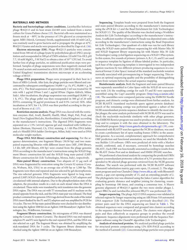
RESULTS AND DISCUSSION�AQ113 genome characterization and proteome analysis. Lac-tobacillus helveticus phage �AQ113 was isolated from a naturalwhey starter culture used for the production of Grana Padano, anItalian hard-cooked cheese (15). Then, its titer was determined(1012 CFU/ml) and it was visualized by electron microscopy (EM).Although phage �AQ113 belongs to the Myoviridae family andhas a short tail, the contractile sheath was not visualized by EM.Additionally, we observed that this phage has an isometric capsid
(diameter, 55.2 � 2.23 nm) with a contractile tail (length,147.25 � 3.47 nm; diameter, 21.7 � 2.45 nm) (Fig. 1). The finalassembled consensus sequence revealed a linear, circularly per-muted, double-stranded DNA genome with a size of 36,566 bpand a G�C content of 37%. The sequence assembly was validatedby a comparative restriction analysis (data not shown). No evi-dence could be found for the presence of cohesive, protrudingends (cos) in the �AQ113 genome, and ligation of its DNA did notalter the restriction patterns (as shown in Fig. 2). Therefore,�AQ113 likely utilizes the pac mechanism of DNA packaging. Thesize of this phage genome is similar to that of the virulent Sipho-viridae phages L. plantarum �phiJL-1 (36,677 bp) and L. rhamno-sus �Lc-Nu (36,466 bp) and the temperate Myoviridae phage L.gasseri �KC5a (38,239 bp) (23). The G�C content is similar tothat (35.3%) of L. gasseri phage �adh (2).
Fifty-six open reading frames (ORFs) were predicted both abinitio and by homology, and a putative function was assigned toapproximately 90% of these ORFs (see Table S1 in the supplemen-tal material). Five ORFs showed no homology to existing se-quences, and at least five others had very low homology matches(�40%). Only 15 ORFs showed homology to annotated phageproteins; the majority of the ORFs showed similarity to sequencesfrom bacterial genomes. As many of these ORFs are typical phagegenes, it may be inferred that the matches in the bacterial genomesrepresented prophages or remnants of prophages. This findingmight indicate that �AQ113 itself has a prophage origin or a close
TABLE 1 Primers used in this study for DNA sequencing
Primer Sequence (5=¡3=) Position in the sequence Amplicon size (bp)
HEAD9 for GACAGAAGTTATTAACTATGCTGAT 4850–4874 1,044HEAD9 rev AGTAGTCTTACCAGAGAAGTCAA 5894–5871SHEATH15 for GGCAATGGTAAGCCAGTATTAACC 7499–7522 812SHEATH15 rev CTTCATAGTTATAAGCCACGCCACT 8311–8288SHEATH16 for GGCAACAACTTTAGAACAGGTT 8910–8931 468SHEATH16 rev CTGAATACCATCAAAAGGTTGAA 9378–9356CDS16 GCTGGTAAGCAAATTATCCAACTCGAT 9232–9258 875CDS19 GGATTGCTAAAAGCGTCAACAACTCTA 10107–10081CDS41 GCACAGTCACTTCTTATAGACAAATT 26894–26919 837CDS43 GTCAATTGGCGTATTGAACACCCTT 27731–27707CDS51 GCTAATAATAATAATGACCAAGCCAC 30922–30948 911CDS52 GTCAATATTAAAGCCACCGCTTCTAT 31833–31808CDS54 GGAGACTACGAATATGTTAGAAAAATA 32966–32992 711CDS56 CCACGCAACGCAATTAATGAAAT 33677–33655CDS57 for GATTCAAATGGACAAGTAGATATTGCA 33888–33915 541CDS57 rev GGGATGCTTAGTTTCTTCAAAATTAA 34429–34405
FIG 1 Transmission electron micrograph of phage �AQ113.
Zago et al.
4714 aem.asm.org Applied and Environmental Microbiology
on February 16, 2019 by guest
http://aem.asm
.org/D
ownloaded from

evolutionary relationship to temperate phages. In fact, this newphage genome is modularly organized, containing DNA packag-ing, head and tail morphogenesis, cell lysis, and DNA replication/regulation modules, as well as elements typically associated withlysogeny controls, such as a repressor, an antirepressor, and aCro-like protein.
The bacteriophage structural proteins were visualized by SDS-PAGE. The high background of the staining is most likely due tointeractions between the Coomassie brilliant blue stain and thePEG that was used for the purification of the phage. Three majorbands were visible (Fig. 3). The smallest band was identified in thebacteriophage genome database as ORF16 (Bioanalyst score 121and 63.7% sequence coverage for the peptide fingerprint data), aputative tail sheath protein (calculated molecular mass, 17,579Da). The intermediate band was identified as ORF9 (Bioanalystscore 228 and 36% sequence coverage for the peptide fingerprintdata), a putative major head protein (calculated molecular mass,40,293 Da). The largest band was identified as ORF15 (Bioanalystscore 516 and 48% sequence coverage for the peptide fingerprintdata), a putative tail sheath protein (calculated molecular mass,52,698 Da). Therefore, on the basis of their putative functions, it isnot surprising that these polypeptides might represent the majorstructural proteins of the bacteriophage.
Genome analysis. The L. helveticus bacteriophage genome wassequenced using the SOLiD system. The sequencing was per-formed at a very high coverage and with two different approaches(fragment and mate pair libraries), the latter at two different insert
sizes (650 bp and 1,500 bp). The high sequence coverage wasdriven by a physical requirement of the sequencing: in one quad,not less than 50 M beads has to be deposited to reach high-qualitydata for a single sample. Moreover, we chose to analyze these threedifferent types of libraries in order to identify the best strategy tosolve the problem of sequencing a very heterogeneous small ge-nome in a single shotgun step. The 50-bp fragment sequence li-brary (FRAG 50) resulted in 69,493,353 reads; the mate pair li-brary with a 650-bp insert (MP_650) resulted in a total of morethan 162 million reads, divided almost equally between the for-ward (F3) and reverse (R3) 50-nucleotide (50-nt) pairs; and themate pair library with a 1,500-bp insert (MP_1500) resulted in anumber of sequences similar to that of the MP_650 library. Intotal, the genome phage sequencing yielded 395,665,425 sequencereads of 50 nt each, for a grand total of 19,783,271,250 nt of se-quence. Preliminary sequence assembly performed in the Se-quencing By Ligation format (Color Space) resulted in three dif-ferent series of contigs: 28 contigs from FRAG 50 with an N50 valueof 2,081 nt, 10 contigs from MP_650 with an N50 value of 32,438nt, and 9 contigs derived from the Color Space assembly ofMP_1500 with an N50 value of 30,033 nt. Thus, sequencing withmate pairs at an insert length of 1,500 bp was identified as the beststrategy to sequence a new phage genome. Further bioinformaticanalysis of these contigs produced a single assembly of 34,928 nt,with extended similarities at the protein level with the L. gasseriphage Kc5a and in good agreement with the results of the restric-tion analysis. Further restriction analysis (Fig. 2) and comparisonwith the restriction profiles developed in silico prompted directSanger sequencing of the contig end from divergent primers (datanot shown). The sequencing revealed a circular genome with anadditional 1.3-kb segment. The genome was then reassembledfrom the start with the addition of the Sanger sequencing results,and we obtained a final contig of 36,307 nucleotides. An in silicorestriction analysis of the new assembly, as well as a quantitativecomparison with the previous restriction pattern and the one ob-tained in the laboratory, confirmed definitive progress toward thefinal genome sequence of 36,566 bp. An ab initio prediction per-formed with two differential methodologies and the functionalannotation by similarity resulted in a total of 56 ORFs that can bedivided into the six functional modules detailed below based on
FIG 2 Restriction analysis of �AQ113 DNA. The phage DNA was digestedwith BglII, HhaI, PstI, HaeIII, MspI, EcoRI, and EcoRV. Lanes M1 and M2,1-kb Plus and �-HindIII DNA ladders; A and B indicate that the digests wereunheated and heated prior to electrophoresis, respectively.
FIG 3 Analysis of the phage �AQ113 structural proteins. After purificationsof �AQ113 by PEG precipitation, the purified phage proteins were separatedby SDS-PAGE. The structural proteins from the main bands were identified byMS and MS/MS (see the text for details). Band 1, sheath tail protein; band 2,major head protein; band 3, sheath tail protein.
Genome Characterization of L. helveticus Phage �AQ113
August 2013 Volume 79 Number 15 aem.asm.org 4715
on February 16, 2019 by guest
http://aem.asm
.org/D
ownloaded from

their putative predicted functions (see Table S1 in the supplemen-tal material): (i) DNA packaging, (ii) head morphogenesis, (iii)tail morphogenesis, (iv) lysis, (v) lysogeny, and (vi) DNA replica-tion/regulation.
(i) Genes encoding proteins involved in phage DNA packag-ing. The products of the genes ORF 1, ORF 3, and ORF 4 mostlikely represent the small and large subunits of the phage termi-nase, found in Lactobacillus vaginalis strain ATCC 49540 and L.gasseri phage �KC5a with a similarity that ranged from 66 to83%. These results suggest that these three proteins are likely tobe involved in phage DNA packaging. The protein encoded byORF 5 shows a similarity of 71% with the portal protein of L.gasseri strain ATCC 33323. The portal protein is the secondcomponent of the bacteriophage DNA packaging. The geneproduct of ORF 2 shares homology (46.4%) with the HNHendonuclease of Bacteroides sp. strain 3_1_33FAA. These en-donucleases can be found as free-standing ORFs between genesor contained within introns. However, it is likely that the HNHhoming endonuclease is involved in DNA packaging because ofthe proximity of ORF 2 to the terminase genes, as has beenobserved for L. plantarum phage �JL-1 (24).
(ii) Genes involved in head morphogenesis. The gene prod-ucts of ORF 6 and ORF 8 show an identity of 54% with L. johnsoniiprophage �Lj771 and of 58.8% with the L. gasseri strain, respec-tively. ORF 9 is similar to the phage major capsid protein of L.gasseri phage �KC5a (71% identity). The presence of this proteinwas confirmed by the analysis of the structural proteins (Fig. 3),and the 41-kDa protein appears to be the major head proteinaccording to the amino acid sequence. The predicted protein en-coded by ORF 10 shares a sequence similarity of 47.3% with thehead-tail connector protein of the bacteriophage SPP1, suggestingthat this ORF product may be involved in phage assembly.
(iii) Genes involved in tail morphogenesis. ORFs 15, 16, 19,21, and 26 are proposed to encode components of the �AQ113tail. They exhibit similarity to tail components of L. gasseri strainsand phage �KC5a. In particular, the ORF 15 and 16 proteins aresimilar to phage sheath proteins that are involved in the creationof a channel for viral genome delivery by driving the tail tubethrough the cell outer membrane (25). The ORF 15 and 16 pro-teins were also observed by SDS-PAGE (Fig. 3) with molecularmasses of 17.5 and 52.7 kDa, respectively. ORF 19 is predicted toencode a tape measure protein, which determines the length of thephage tail during morphogenesis (26). The sequence of ORF 19shows a degree of similarity of 60% with an analogous proteinfrom L. gasseri strain 224-1. The ORF 21 protein was identified asthe minor tail protein, and ORF 26 appears to be a baseplate pro-tein with a high degree of similarity (62.7%) to L. gasseri strain224-1. Kondou et al. (27) showed that the baseplate protein isessential for phage Mu assembly and the generation of viablephages.
(iv) Genes encoding host cell lysis proteins. The protein en-coded by ORF 35 displays similarity to a gene belonging to the lysismodule. In particular, this ORF showed a high degree of identity(88%) with the endolysin (Mur-LH) from the L. helveticus tem-perate phage �-0303 (28). This gene has been detected in other L.helveticus phages with 99% homology (16). Deutsch et al. (28)found a truncated integrase gene downstream from the lys genethat encodes Mur-LH; this genomic organization is similar to thatof other LAB phages (29, 30). However, no integrase genes werefound in the sequence of L. helveticus phage �AQ113. ORF 22
showed homology to a Lys-M-like protein of L. gasseri phage�KC5a (48% of identity), but ORF 22 is not adjacent to the pre-vious endolysin gene.
(v) Genes involved in the lysogeny module. ORFs 40, 43, and44 exhibit similarity to genes involved in the establishment of thelysogeny in other Lactobacillus phages. In particular, ORF 40 isimplicated in the transcription of prophage gene expression (82%similarity), and ORF 43 codes for a Cro-like repressor protein,given its similarity with the Cro repressor of Lactobacillus phageSal2. Cro repressors are antagonistic to cI repressors and thereforefunction to block or terminate lysogeny. ORF 44 appears to be anantirepressor by virtue of its similarity with the prophage antire-pressor protein of Lactobacillus jensenii 269-3 (80.7% identity).This result may suggest an antirepression-mediated inductionsimilar to that observed for a large family of lambdoid prophagesfound in Salmonella genomes. The repressors of these phages arenot cleaved upon induction; rather, they are inactivated by thebinding of small antirepressor proteins. Therefore, the formationof the complex causes the repressor to dissociate from DNA (31).ORF 49 encodes a prophage protein similar to that of Lactobacilluscrispatus strain CTV-05 (74.8% identity). The product of ORF 47shows similarity to the HNH endonuclease of the Lactobacillusrhamnosus strain Lc-Nu (42.8%). The HNH motif was originallyidentified in the subfamily of the HNH homing endonucleasesthat initiate the process of the insertion of mobile genetic elementsinto specific sites (32); therefore, the position of this ORF near thelysogeny module suggests that it may be involved in the excision/integration process of temperate phages, as has been shown forLeuconostoc mesenteroides �1-A4 (33). Overall, the data suggestedthat the virulent phage �AQ113 may have originated from a tem-perate phage.
(vi) Genes involved in DNA replication and regulation. Be-ginning with ORF 51, the remaining genes are related to the rep-lication and regulation of the transcription of phage DNA. ORFs52 and 53 display an identity of 45% with DNA replication pro-teins from L. crispatus strain CTV-05 and L. johnsonii prophageLj771, respectively, suggesting that the two proteins are involvedin DNA synthesis. The predicted proteins of ORFs 54 and 63 showhomology to the putative transcriptional regulators of the L. gas-seri phage �KC5a (73%) and the L. gasseri strain JV-V03 (38%),respectively. ORF 51 contains a conserved domain of the ERF(essential recombination function) superfamily of single-strandDNA annealing proteins that mediate the circularization of linearDNA following infection of the host cell (34). Additionally, ORF56 displays a 55% identity to a single-stranded DNA binding pro-tein of the temperate phage phiNIH1.1. ORF 60 is also in thereplication/regulation module of L. helveticus phage, and it en-codes a protein that belongs to the restriction nuclease superfam-ily and has significant identity (83.6%) to L. gasseri strain JV-V03.
A successful bacteriophage infection requires the regulation ofgene expression, DNA replication, formation of the phage capsid,and the release of the new phage particles from the infected host(35). In most bacteriophages, the genes encoding related biologi-cal functions are clustered together in functional groups or mod-ules, and they are turned on and off in coordination (36). In thecase of �AQ113, the 56 ORFs are divided into six functional mod-ules that also include lysogenic functions. Lysogeny is a peculiartrait in L. helveticus and is considered an important source of vir-ulent phages in dairy factories (14, 37). Despite the fact that nu-merous strategies of phage control have been developed and are
Zago et al.
4716 aem.asm.org Applied and Environmental Microbiology
on February 16, 2019 by guest
http://aem.asm
.org/D
ownloaded from

now used in the dairy industry, the risk of phage infections is stillsignificantly elevated, because virulent bacteriophages are presentand cannot be completely eliminated from food fermentation en-vironments. Phage �AQ113 was isolated from natural cheesewhey starters as a virulent phage because it is able to propagate ina sensitive host strain (15). Nevertheless, the identification ofgenes involved in the establishment of lysogeny (ORFs 40, 43, and44) indicates that it may have originated as a temperate phage.Several virulent Lactobacillus phages have been postulated to havea prophage origin, such as L. delbrueckii phage �LL-H (38) and L.casei phage �S1 (39). This study confirmed the assumption thattemperate phages are one possible source of virulent phages, al-though it is difficult to estimate the extent of their contribution tothe outbreaks of new virulent phages observed in industrial pro-cesses.
Phylogenetic analysis. A phylogenetic analysis was performedwith the ORFs predicted to encode the major head protein (ORF9) and a putative transcriptional regulator (ORF 54). The majorhead protein was one of the structural genes used for phylogeneticstudies because the packaging-head gene cluster has a structuralgene order that is generally very well conserved (40). ORF 54 wasselected using the approach of clustering predicted proteins bysequence similarity. Beginning with all 56 predicted ORFs, wesearched for highly similar sequences against a selection of 96phage genomes by Tblastn (search translated nucleotide targetgenomes using protein queries). We finally selected the two ORFswith the highest similarities that can be found in the highest num-ber of phages in the database. Because viruses are known to begreatly variable, we used a Blosum45 substitution matrix (gapcosts: open, 15; extend, 2) that is recommended for highly diver-gent alignments and contains distances calculated to take intoaccount a greater recombination ratio.
In a few cases, the resulting blast hits were split between twoneighboring high-scoring segment pairs (HSPs), indicating thatthe ORFs had relatively large insertions in that genome. For thesecases, the entire genome sequence was retrieved, translated again,and replaced with the source hits. We thus obtained a new targetproteome that was subjected to a new alignment. We also searchedthe same 56 predicted ORFs against a nonredundant proteomecollection of 4,731 proteins belonging to the same 96 phages re-
trieved from NCBI. The search was performed by BLASTP (Blo-sum45 substitution matrix; gap costs: open, 15; extend, 2).
All of the dendrograms confirmed the similarity of L. helveticus�AQ113 to other Lactobacillus thermophilic phages such as L.gasseri �KC5a and L. johnsonii �Lj771 (see Fig. S1 in the supple-mental material), but Lactobacillus delbrueckii phages were noteven present in the dendrogram, highlighting their lack of simi-larity to L. helveticus �AQ113. Mauve genome alignment revealedmany clusters of genes or colinear blocks that are in the same orderin both �AQ113, L. gasseri �KC5a, and L. johnsonii �Lj771 (Fig.4). The gene order of the different modules was largely colinear,although L. gasseri �KC5a and L. johnsonii �Lj771 were 2 and 4 kblonger than �AQ113, respectively. As expected, the three phagesrevealed a high level of sequence similarity in the packaging andhead and tail morphogenesis modules; however, a greater level ofdiversity was observed for the lysis, lysogeny, and replication/reg-ulation modules. In particular, the modular arrangement of thegenes responsible for packaging and head and tail morphogenesiswas similar to that of the two bacteriophages of L. johnsonii and L.gasseri. This finding suggests that these viruses and the L. helveticusphage �AQ113 have a common ancestry, as shown by Slattery etal. (41) concerning L. helveticus bacteria. It is worth noting that L.helveticus strain DPC4571, isolated from cheddar cheese, shows aremarkable similarity in gene content with intestinal lactobacillisuch as L. acidophilus NCFM, supporting the concept that thesequence differences between the dairy and the gut species may becaused by a relatively small but highly specific gene set (42). Phy-logenetic similarities between L. helveticus phage �AQ113 andtwo phages that belong to gut species confirm a possible commonancestral origin and support the increasing consideration of L.helveticus as a health-promoting organism (43, 44).
In conclusion, L. helveticus phage �AQ113 possesses a mosaicgenome with predicted proteins that have homologies to proteinsfrom a wide range of bacteria. The majority of these are intestinalbacteria; this assumption emphasizes the probiotic characteristicsof L. helveticus species. Furthermore, the fact that many of the�AQ113 genes matched sequences from bacterial genomes high-lights the high propensity to genetic exchange and recombinationbetween bacteriophages and bacteria. �AQ113 shows also a lyso-genic module that underlines its possible origin from a temperate
FIG 4 Comparison of the gene order and orientation of the genomes of L. helveticus �AQ113, L. gasseri �KC5a, and L. johnsonii �Lj771 using Mauve genomealignment software. The figure was generated by the Mauve rearrangement viewer. Each of the three genomes is displayed horizontally with homologous locallycolinear blocks outlined and connected with lines. The height of the similarity profile inside each block corresponds to the average level of conservation in thatregion of the genome sequence.
Genome Characterization of L. helveticus Phage �AQ113
August 2013 Volume 79 Number 15 aem.asm.org 4717
on February 16, 2019 by guest
http://aem.asm
.org/D
ownloaded from

phage. This study may provide new insights into phage geneticsand phage-host interactions in L. helveticus and between Lactoba-cillus phages.
ACKNOWLEDGMENTS
This work was supported by grants from the ASER/COL-MIA project(D.M. MIPAF 16101/7301/08) and Genomnia research and developmentfunds (for NGS and bioinformatic analysis). The strain used in this studybelongs to the ASER/COL-MIA collection.
We thank Maria Luisa Callegari from the University of Piacenza forperforming the electron microscopy.
REFERENCES1. Moineau S, Levesque C. 2005. Control of bacteriophages in industrial
fermentations, p 285–296. In Kutter E, Sulakvelidze A (ed), Bacterio-phages: biology and applications. CRC Press, Boca Raton, FL.
2. Altermann E, Klein JR, Henrich B. 1999. Primary structure and featuresof the genome of the Lactobacillus gasseri temperate bacteriophage �adh.Gene 236:333–346.
3. Brussow H. 2001. Phages of dairy bacteria. Annu. Rev. Microbiol. 55:283–303.
4. Brussow H, Suarez JE. 2006. Lactobacillus phages, p 653– 666. In CalendarR, Abedon ST (ed), The bacteriophages, 2nd ed. Oxford University Press,New York, NY.
5. Mozzi F, Raya RR, Vignolo GM. 2010. Biotechnology of lactic acid bacte-ria—novel applications. Wiley-Blackwell, Blackwell Publishing, Ames, IA.
6. Brussow H, Hendrix RW. 2002. Phage genomics: small is beautiful. Cell108:13–16.
7. Desiere F, Lucchini S, Brüssow H. 1998. Evolution of Streptococcus thermo-philus bacteriophage genomes by modular exchanges followed by point mu-tations and small deletions and insertions. Virology 241:345–356.
8. Desiere F, Mahanivong C, Hillier AJ, Chandry PS, Davidson BE,Brüssow H. 2001. Comparative genomics of lactococcal phages: insightfrom the complete genome sequence of Lactococcus lactis phage BK5-T.Virology 283:240 –252.
9. Hendrix RW. 2003. Bacteriophage genomics. Curr. Opin. Microbiol.6:506 –511.
10. Lazzi C, Rossetti L, Zago M, Neviani E, Giraffa G. 2004. Evaluation ofbacterial communities belonging to natural whey starters for Grana Padanocheese by length-heterogeneity-PCR. J. Appl. Microbiol. 96:481–490.
11. Nakamura Y, Yamamoto N, Sakai K, Okubo A, Yamazaki S, Takano T.1995. Purification and characterization of angiotensin I-converting en-zyme inhibitors from sour milk. J. Dairy Sci. 78:777–783.
12. Rossetti L, Fornasari ME, Gatti M, Lazzi C, Neviani E, Giraffa G. 2008.Grana Padano cheese whey starters: microbial composition and straindistribution. Int. J. Food Microbiol. 127:168 –171.
13. Sozzi T, Maret R. 1975. Isolation and characteristics of Streptococcusthermophilus and Lactobacillus helveticus phages from Emmental starters.Lait 55:269 –288.
14. Séchaud L, Rousseau M, Fayard B, Callegari ML, Quénée P, Accolas JP.1992. Comparative study of 35 bacteriophages of Lactobacillus helveticus:morphology and host range. Appl. Environ. Microbiol. 58:1011–1018.
15. Zago M, Comaschi L, Neviani E, Carminati D. 2005. Investigation on thepresence of bacteriophages in natural whey starters used for the produc-tion of Italian long-ripened cheeses. Milchwissenschaft 60:171–174.
16. Zago M, Rossetti L, Reinheimer J, Carminati D, Giraffa G. 2008.Detection and identification of Lactobacillus helveticus bacteriophages byPCR. J. Dairy Res. 75:196 –201.
17. Brown JCS, Ward LJH, Davey GP. 1994. Rapid isolation and purificationof lactococcal bacteriophage DNA without the use of caesium chloridegradients. Lett. Appl. Microbiol. 18:292–293.
18. Huang X, Madan A. 1999. CAP3: a DNA sequence assembly program.Genome Res. 9:868 – 877.
19. Besemer J, Lomsadze A, Borodovsky M. 2001. GeneMarkS: a self-training method for prediction of gene starts in microbial genomes. Im-plications for finding sequence motifs in regulatory regions. Nucleic AcidsRes. 29:2607–2618.
20. Darling ACE, Mau B, Blattner FR, Perna NT. 2004. Mauve: multiplealignment of conserved genomic sequence with rearrangements. GenomeRes. 14:1394 –1403.
21. Suarez V, Zago M, Giraffa G, Reinheimer J, Quiberoni A. 2009. Evi-
dence for the presence of restriction/modification systems in Lactobacillusdelbrueckii. J. Dairy Res. 76:433– 440.
22. Laemmli UK. 1970. Cleavage of structural proteins during the assembly ofthe head of bacteriophage T4. Nature 227:680 – 685.
23. Villion M, Moineau S. 2009. Bacteriophages of Lactobacillus. Front. Bio-sci. 14:1661–1683.
24. Lu Z, Altermann E, Breidt F, Predki P, Fleming HP, Klaenhammer T.2005. Sequence analysis of the Lactobacillus plantarum bacteriophage �JL-1. Gene 348:45–54.
25. Aksyuk AA, Leiman PG, Kurochkina LP, Shneider MM, KostyuchenkoVA, Mesyanzhinov VV, Rossmann MG. 2009. The tail sheath structure ofbacteriophage T4: a molecular machine for infecting bacteria. EMBO J.28:821– 829.
26. Katsura I. 1987. Determination of bacteriophage lambda tail length by aprotein ruler. Nature 327:73–75.
27. Kondou Y, Kitazawa D, Takeda S, Tsuchiya Y, Yamashita E, MizuguchiM, Kawano K, Tsukihara T. 2005. Structure of the central hub of bacte-riophage Mu baseplate determined by X-ray crystallography of gp44. J.Mol. Biol. 352:976 –985.
28. Deutsch SM, Guezenec S, Piot M, Foster S, Lortal S. 2004. Mur-LH, thebroad-spectrum endolysin of Lactobacillus helveticus temperate bacterio-phage �-0303. Appl. Environ. Microbiol. 70:96 –103.
29. Arendt EK, Daly C, Fitzgerald GF, van de Guchte M. 1994. Molecularcharacterization of lactococcal bacteriophage Tuc2009 and identificationand analysis of genes encoding lysin, a putative holin, and two structuralproteins. Appl. Environ. Microbiol. 60:1875–1883.
30. van Sinderen D, Karsens H, Kok J, Terpstra P, Ruiters MH, Venema G,Nauta A. 1996. Sequence analysis and molecular characterization of thetemperate lactococcal bacteriophage r1t. Mol. Microbiol. 19:1343–1355.
31. Lemire S, Figueroa-Bossi N, Bossi L. 2011. Bacteriophage crosstalk:coordination of prophage induction by trans-acting antirepressors. PLoSGenet. 7:e1002149. doi:10.1371/journal.pgen.1002149.
32. Chevalier BS, Stoddard BL. 2001. Homing endonucleases: structural andfunctional insight into the catalysts of intron/intein mobility. Nucleic Ac-ids Res. 29:3757–3774.
33. Lu Z, Altermann E, Breidt F, Kozyavkin S. 2010. Sequence analysis ofLeuconostoc mesenteroides �1-A4 isolated from an industrial vegetablefermentation. Appl. Environ. Microbiol. 76:1955–1966.
34. Poteete AR. 1982. Location and sequence of the erf gene of phage P22.Virology 119:422– 429.
35. Brøndsted L, Østergaard S, Pedersen M, Hammer K, Vogensen FK.2001. Analysis of the complete DNA sequence of the temperate bacterio-phage TP901-1: evolution, structure, and genome organization of lacto-coccal bacteriophages. Virology 283:93–109.
36. Ptashne M. 2004. A genetic switch: phage lambda revisited, 3rd ed. ColdSpring Harbor Laboratory Press, Cold Spring Harbor, NY.
37. Carminati D, Mazzucotelli L, Giraffa G. 1997. Incidence of induciblebacteriophage in Lactobacillus helveticus strains isolated from naturalwhey starter cultures. J. Dairy Sci. 80:1505–1511.
38. Mikkonen M, Dupont L, Alatossava T, Ritzenthaler P. 1996. Defectivesite-specific integration elements are present in the genome of virulentbacteriophage LL-H of Lactobacillus delbrueckii. Appl. Environ. Micro-biol. 62:1847–1851.
39. Shimizu-Kadota M, Sakurai T, Tsuchida N. 1983. Prophage origin of avirulent phage appearing on fermentations of Lactobacillus casei S-1. Appl.Environ. Microbiol. 45:669 – 674.
40. Brussow H, Desiere F. 2001. Comparative phage genomics and the evolutionof Siphoviridae: insight from dairy phages. Mol. Microbiol. 39:213–222.
41. Slattery L, O’Callaghan J, Fitzgerald GF, Beresford T, Ross RP. 2010.Invited review: Lactobacillus helveticus—a thermophilic dairy starter re-lated to gut bacteria. J. Dairy Sci. 93:4435– 4454.
42. Callanan MP, Kaleta P, O’Callaghan J, O’Sullivan O, Jordan K, McAu-liffe O, Sangrador-Vegas A, Slattery L, Fitzgerald GF, Beresford T, RossRP. 2008. Genome sequence of Lactobacillus helveticus, an organism dis-tinguished by selective gene loss and insertion sequence element expan-sion. J. Bacteriol. 190:727–735.
43. Taverniti V, Guglielmetti S. 2011. The immunomodulatory properties ofprobiotic microorganisms beyond their viability (ghost probiotics: pro-posal of paraprobiotic concept). Genes Nutr. 6:261–274.
44. Vinderola G, Matar C, Perdigón G. 2007. Milk fermentation products ofLactobacillus helveticus R389 activate calcineurin as a signal to promote gutmucosal immunity. BMC Immunol. 8:19. doi:10.1186/1471-2172-8-19.
Zago et al.
4718 aem.asm.org Applied and Environmental Microbiology
on February 16, 2019 by guest
http://aem.asm
.org/D
ownloaded from