Characterization of rrdA, a TetR Family Protein Gene ... · regulators that influence Red...
8
APPLIED AND ENVIRONMENTAL MICROBIOLOGY, Apr. 2009, p. 2158–2165 Vol. 75, No. 7 0099-2240/09/$08.000 doi:10.1128/AEM.02209-08 Copyright © 2009, American Society for Microbiology. All Rights Reserved. Characterization of rrdA, a TetR Family Protein Gene Involved in the Regulation of Secondary Metabolism in Streptomyces coelicolor Xijun Ou, 1 Bo Zhang, 1 Lin Zhang, 1 Guoping Zhao, 1,2,3 * and Xiaoming Ding 1 * State Key Laboratory of Genetic Engineering, Department of Microbiology and Microbial Engineering, School of Life Sciences, Fudan University, Shanghai 200433, China 1 ; Shanghai-MOST Key Laboratory of Health and Disease Genomics, Chinese National Human Genome Centre at Shanghai, Shanghai 201203, China 2 ; and Laboratory of Synthetic Biology, Institute of Plant Physiology and Ecology, Shanghai Institutes for Biological Sciences, Chinese Academy of Sciences, Shanghai 200032, China 3 Received 24 September 2008/Accepted 28 January 2009 Streptomyces not only exhibits complex morphological differentiation but also produces a plethora of sec- ondary metabolites, particularly antibiotics. To improve our general understanding of the complex network of undecylprodigiosin (Red) biosynthesis regulation, we used an in vivo transposition system to identify novel regulators that influence Red production in Streptomyces coelicolor M145. Using this screening system, we obtained 25 Red-deficient mutants. Twenty-four of these mutants had a transposon inserted in the previously described Red biosynthetic gene cluster and produced different amounts of another secondary metabolite, actinorhodin (Act). One mutant was shown to have an insertion in a different region of the chromosome upstream of the previously uncharacterized gene rrdA (regulator of redD, sco1104), which encodes a putative TetR family transcription factor. Compared with wild-type strain M145, the rrdA null mutant exhibited increased Red production and decreased Act production. A high level of rrdA expression resulted in a severe reduction in Red production and Act overproduction. Reverse transcription-PCR analysis showed that RrdA negatively regulated Red production by controlling redD mRNA abundance, while no change was observed at the transcript level of the Act-specific activator gene, actII-orf4. The effects on Act biosynthesis might arise from competition for precursors that are common to both pathways. In addition to its complex morphological differentiation, the gram-positive genus Streptomyces is notable for its ability to produce a wide range of secondary metabolites during its life cycle. These metabolites include the majority of known phar- maceutically important secondary metabolites that exhibit an- tibacterial, anticancer, and immunosuppressive activities (6, 7). The genes responsible for the biosynthesis of secondary me- tabolites are often physically clustered in the genome and coordinately regulated by pathway-specific transcriptional ac- tivators (1, 4, 10, 12, 29, 33). These specific regulators are controlled by various higher-level pleiotropic regulators, and their expression is typically affected by a variety of environ- mental and physiological cues, including the nature and levels of carbon and nitrogen sources and the availability of phos- phate and small signaling molecules, such as ppGpp and -butyrolactone (5). Streptomyces coelicolor A3 (2) has been used for many years as a model organism in morphological and physiological dif- ferentiation studies, particularly in studies of the regulation of antibiotic biosynthesis (7). S. coelicolor produces four antibi- otics: actinorhodin (Act), undecylprodigiosin (Red), methyl- enomycin, and calcium-dependent antibiotic. It has been shown that certain regulators are involved in the pleiotropic control of antibiotic production in S. coelicolor, including AbsA1/A2, AfsR/K, AtrA, and PhoR/P (18, 24, 31, 32). Re- cently, mutational analysis and adventitious overexpression of key regulators in S. coelicolor revealed cross-regulation at the transcriptional level among disparate antibiotic biosynthetic pathways (15). The diversity of these regulatory elements sug- gests that the regulation of antibiotic production is a compli- cated process, and many genes remain to be identified. Red is one of the prodiginine secondary metabolites, and this group of compounds is attracting increasing interest due to its immunosuppressive and anticancer activities (34). In this study, we used an insertion mutagenesis system to identify new genes that regulate Red biosynthesis in S. coelicolor M145. We discovered a novel TetR-like transcription factor gene, rrdA (regulator of redD, sco1104), that was involved in the regula- tion of secondary metabolism. In this study, we also report a link between Red production and Act production that proba- bly occurs at the level of the metabolites utilized that are common to both pathways in S. coelicolor. MATERIALS AND METHODS Bacterial strains, plasmids, and growth conditions. The bacterial strains and plasmids used in this study are listed in Table 1. Escherichia coli DH5 (22) was used for plasmid propagation. E. coli Rosetta-gami (Novagen) was used to express recombinant proteins. Mannitol soy flour (MS) (16) agar was used to generate spores and select Streptomyces exconjugants. YBP agar (2 g yeast extract, 2 g beef extract, 4 g Bacto peptone, 1 g MgSO 4 , 15 g NaCl, 15 g agar, and 10 g glucose in 1 liter [final volume] H 2 O) was used for phenotype screening and RNA preparation. YBP liquid medium was used for quantitative antibiotic assays. YEME (16) was used to cultivate mycelia for genomic DNA preparation. Conjugation of E. coli ET12567/pUZ8002 with Streptomyces was performed as described previously (16). Depending on the requirements, antibiotics were added at the following final concentrations: ampicillin, 50 g ml 1 ; chloram- phenicol, 33 g ml 1 ; kanamycin, 30 g ml 1 ; tetracycline, 12.5 g ml 1 ; thio- strepton, 25 g ml 1 ; and apramycin, 20 g ml 1 . * Corresponding author. Mailing address: Department of Microbi- ology and Microbial Engineering, School of Life Sciences, Fudan Uni- versity, 220 Handan Road, Shanghai 200433, China. Phone: 86 21 65643616. Fax: 86 21 65650149. E-mail for Xiaoming Ding: xmding74 @fudan.edu.cn. E-mail for Guoping Zhao: [email protected]. Published ahead of print on 5 February 2009. 2158 on February 22, 2020 by guest http://aem.asm.org/ Downloaded from
Transcript of Characterization of rrdA, a TetR Family Protein Gene ... · regulators that influence Red...

APPLIED AND ENVIRONMENTAL MICROBIOLOGY, Apr. 2009, p. 2158–2165 Vol. 75, No. 70099-2240/09/$08.00�0 doi:10.1128/AEM.02209-08Copyright © 2009, American Society for Microbiology. All Rights Reserved.
Characterization of rrdA, a TetR Family Protein Gene Involved in theRegulation of Secondary Metabolism in Streptomyces coelicolor�
Xijun Ou,1 Bo Zhang,1 Lin Zhang,1 Guoping Zhao,1,2,3* and Xiaoming Ding1*State Key Laboratory of Genetic Engineering, Department of Microbiology and Microbial Engineering, School of Life Sciences, Fudan University,
Shanghai 200433, China1; Shanghai-MOST Key Laboratory of Health and Disease Genomics, Chinese National Human Genome Centre atShanghai, Shanghai 201203, China2; and Laboratory of Synthetic Biology, Institute of Plant Physiology and Ecology,
Shanghai Institutes for Biological Sciences, Chinese Academy of Sciences, Shanghai 200032, China3
Received 24 September 2008/Accepted 28 January 2009
Streptomyces not only exhibits complex morphological differentiation but also produces a plethora of sec-ondary metabolites, particularly antibiotics. To improve our general understanding of the complex network ofundecylprodigiosin (Red) biosynthesis regulation, we used an in vivo transposition system to identify novelregulators that influence Red production in Streptomyces coelicolor M145. Using this screening system, weobtained 25 Red-deficient mutants. Twenty-four of these mutants had a transposon inserted in the previouslydescribed Red biosynthetic gene cluster and produced different amounts of another secondary metabolite,actinorhodin (Act). One mutant was shown to have an insertion in a different region of the chromosomeupstream of the previously uncharacterized gene rrdA (regulator of redD, sco1104), which encodes a putativeTetR family transcription factor. Compared with wild-type strain M145, the rrdA null mutant exhibitedincreased Red production and decreased Act production. A high level of rrdA expression resulted in a severereduction in Red production and Act overproduction. Reverse transcription-PCR analysis showed that RrdAnegatively regulated Red production by controlling redD mRNA abundance, while no change was observed atthe transcript level of the Act-specific activator gene, actII-orf4. The effects on Act biosynthesis might arise fromcompetition for precursors that are common to both pathways.
In addition to its complex morphological differentiation, thegram-positive genus Streptomyces is notable for its ability toproduce a wide range of secondary metabolites during its lifecycle. These metabolites include the majority of known phar-maceutically important secondary metabolites that exhibit an-tibacterial, anticancer, and immunosuppressive activities (6, 7).The genes responsible for the biosynthesis of secondary me-tabolites are often physically clustered in the genome andcoordinately regulated by pathway-specific transcriptional ac-tivators (1, 4, 10, 12, 29, 33). These specific regulators arecontrolled by various higher-level pleiotropic regulators, andtheir expression is typically affected by a variety of environ-mental and physiological cues, including the nature and levelsof carbon and nitrogen sources and the availability of phos-phate and small signaling molecules, such as ppGpp and�-butyrolactone (5).
Streptomyces coelicolor A3 (2) has been used for many yearsas a model organism in morphological and physiological dif-ferentiation studies, particularly in studies of the regulation ofantibiotic biosynthesis (7). S. coelicolor produces four antibi-otics: actinorhodin (Act), undecylprodigiosin (Red), methyl-enomycin, and calcium-dependent antibiotic. It has beenshown that certain regulators are involved in the pleiotropiccontrol of antibiotic production in S. coelicolor, includingAbsA1/A2, AfsR/K, AtrA, and PhoR/P (18, 24, 31, 32). Re-
cently, mutational analysis and adventitious overexpression ofkey regulators in S. coelicolor revealed cross-regulation at thetranscriptional level among disparate antibiotic biosyntheticpathways (15). The diversity of these regulatory elements sug-gests that the regulation of antibiotic production is a compli-cated process, and many genes remain to be identified.
Red is one of the prodiginine secondary metabolites, andthis group of compounds is attracting increasing interest due toits immunosuppressive and anticancer activities (34). In thisstudy, we used an insertion mutagenesis system to identify newgenes that regulate Red biosynthesis in S. coelicolor M145. Wediscovered a novel TetR-like transcription factor gene, rrdA(regulator of redD, sco1104), that was involved in the regula-tion of secondary metabolism. In this study, we also report alink between Red production and Act production that proba-bly occurs at the level of the metabolites utilized that arecommon to both pathways in S. coelicolor.
MATERIALS AND METHODS
Bacterial strains, plasmids, and growth conditions. The bacterial strains andplasmids used in this study are listed in Table 1. Escherichia coli DH5� (22) wasused for plasmid propagation. E. coli Rosetta-gami (Novagen) was used toexpress recombinant proteins. Mannitol soy flour (MS) (16) agar was used togenerate spores and select Streptomyces exconjugants. YBP agar (2 g yeastextract, 2 g beef extract, 4 g Bacto peptone, 1 g MgSO4, 15 g NaCl, 15 g agar, and10 g glucose in 1 liter [final volume] H2O) was used for phenotype screening andRNA preparation. YBP liquid medium was used for quantitative antibioticassays. YEME (16) was used to cultivate mycelia for genomic DNA preparation.Conjugation of E. coli ET12567/pUZ8002 with Streptomyces was performed asdescribed previously (16). Depending on the requirements, antibiotics wereadded at the following final concentrations: ampicillin, 50 �g ml�1; chloram-phenicol, 33 �g ml�1; kanamycin, 30 �g ml�1; tetracycline, 12.5 �g ml�1; thio-strepton, 25 �g ml�1; and apramycin, 20 �g ml�1.
* Corresponding author. Mailing address: Department of Microbi-ology and Microbial Engineering, School of Life Sciences, Fudan Uni-versity, 220 Handan Road, Shanghai 200433, China. Phone: 86 2165643616. Fax: 86 21 65650149. E-mail for Xiaoming Ding: [email protected]. E-mail for Guoping Zhao: [email protected].
� Published ahead of print on 5 February 2009.
2158
on February 22, 2020 by guest
http://aem.asm
.org/D
ownloaded from

Mutagenesis of S. coelicolor M145. Insertional mutagenesis of M145 wasconducted by in vivo transposition using Tn315 (Table 1) (X. Zhang, Y. Bao,X. Ou, P. Zhou, G. Zhao, and X. Ding, unpublished data) harbored bypFDZ315, which is a conjugative plasmid that does not replicate in Strepto-myces. Tn315, which was derived from insertion element IS204 of Nocardiaasteroides YP21 (35), contains a kanamycin resistance gene, the replicationorigin of pUC plasmids, and an ermE promoter in the terminal inverted
repeats. After plasmid pFDZ315 was introdued into S. coelicolor M145 byconjugation, a single copy of transposon Tn315 was inserted into the chro-mosome, and exconjugants were selected by growth on MS media floodedwith kanamycin (30 �g ml�1). The chromosomal locations of the Tn315insertions were determined by sequencing the transposon-flanking DNA byplasmid rescue. Briefly, genomic DNA from the mutants was isolated anddigested with ApaI, a site for which is not present in Tn315. The DNA was
TABLE 1. Strains, plasmids, and primers used in this study
Strain, plasmid, orprimer Description Reference or source
S. coelicolor strainsM145 Prototroph SCP1� SCP2� 16�rrdA M145 with rrdA gene disrupted This study�rrdA/pFDZ16 �rrdA carrying integrative plasmid pFDZ16 This study�rrdA/pFDZ16*-rrdA �rrdA carrying integrative plasmid pFDZ16*-rrdA This studyWT/pFDZ16 M145 carrying integrative plasmid pFDZ16 This studyWT/pFDZ16-rrdA M145 carrying integrative plasmid pFDZ16-rrdA This study
E. coli strainsDH5� F� recA lacZ�M15 22ET12567 dam dcm hsdS 16Rosetta-gami �ara-leu7697 �lacX74 �phoA(PvuII) phoR araD139 ahpC galE galK rpsL F� �lac� (lacIq) pro�
gor522::Tn10(Tcr) trxB::Kan (DE3)/pRARE (Cmr)Novagen
PlasmidspFDZ315 (Tn315) Conjugative plasmid that does not replicate in Streptomyces strains harboring transposon Tn315, which
uses the transposase of insertion element IS204 as a trans factor and contains a kanamycinresistance gene, origin of pUC plasmids, and ermE promoter in the terminal inverted repeats; Kanr
Ampra
X. Zhang et al., unpublished
pFDZ15 E. coli-Streptomyces integrative shuttle vector; vector pRT802 was cut with SmaI and EcoRV, resultingin a 3.8-kb segment that was subsequently inserted into the NaeI site of pBluescript II KS�(Stratagene), yielding pFDZ15; Kanr Amprb
This study
pFDZ16 E. coli-Streptomyces integrative shuttle vector containing tipA promoter; the 5.4-kb ApaI/BclI fragmentof pFDZ15 was ligated with the 5.0-kb BamHI/ApaI segment of pIJ6021 to obtain pFDZ16; Kanr
Thior Amprc
This study
pFDZ16*-rrdA Derivative obtained from pFDZ16 depletion of PtipA, containing the rrdA gene and its promoter; Kanr
Ampr ThiorThis study
pFDZ16-rrdA Derivative obtained from pFDZ16, containing the rrdA gene located downstream of the tipApromoter; Kanr Ampr Thior
This study
pUCm-T 2.7-kb cloning vector; Ampr Shenggong CompanypBC-AM Donor of aac(3)-IV; the 1.5-kb HindIII/EcoRI fragment of pULVK2A was inserted between the
corresponding sites in vector pBC-SK(�) (Stratagene) to produce pBC-AM; Aprar CmrdThis study
pUC-LR DNA fragment containing the rrdA gene, cloned in pUCm-T; Ampr This studypUC-LAR DNA fragment aac(3)-IV containing apramycin resistance gene, cloned in pUC-LR; Ampr This studypHZ1358 E. coli-Streptomyces shuttle vector; Thior Aprar 26pHZ1358-LAR Derivative obtained from pHZ1358, containing the rrdA gene replacement cassette; Ampr Thior Aprar This studypET28a Expression vector; Kanr NovagenpET28-RrdA RrdA overexpression vector resulting in an N-terminal His6 oligopeptide fusion; Kanr This studypET22a Expression vector; Ampr NovagenpET22-RrdA RrdA overexpression vector resulting in an N-terminal His6 oligopeptide fusion; Ampr Cmr Tcr Kanr This study
PrimersOxj130 5 ATCCATGGCGCATATGTCCCCGCGCAGCGCC 3Oxj131 5 ATAAGCTTACCCGGCCAAGCGGGTCC 3Oxj144 5 TCTAGACCGCCGTTCTGTTCGACTT 3Oxj145 5 GGATCCACATCACCGTCCTGCCCTC 3Oxj161 5 TCTGCCCTCTGACCGCGG 3Oxj162 5 GCCGTCCTCGCGCGTTCT 3Oxj163 5 CTGGCTCCTGGGCGGTCT 3Oxj164 5 ACCAACCCTGGCGTCTCC 3Oxj185 5 CGCCGAAGGAGGAACCGA 3Oxj186 5 CCCGTCATCCACCGAACG 3Oxj187 5 CCCAGGCGCTGGTAGACCT 3Oxj188 5 CCGTCGAGCCGAAAGAGGA 3’Oxj201 5 CCGGAGCCAGCCAAAGATC 3Oxj202 5 GGAGGGCGTTGAGGACGTT 3Oxj203 5 TGCTGACCAAGCCCGAGAA 3Oxj204 5 CGGTGTACGTGGGACCTGAC 3Oxj205 5 TGGTGCTGCTGCTCCTCAG 3Oxj206 5 ATCCAGTCCCGCGTCCAA 3Oxj211 5 GCGCCTCGGTCAATGAAGA 3Oxj212x 5 CGCTTTCCGGGGAAGTAGTAC 3Oxj237 5 CTCTGTCATGGCGCTCATTGA 3Oxj238 5 TTCGCTGCGACGCTCTTT 3
a See reference 35.b See reference 11.c See reference 30.d See reference 17.
VOL. 75, 2009 NOVEL GENE INVOLVED IN Red BIOSYNTHETIC REGULATION 2159
on February 22, 2020 by guest
http://aem.asm
.org/D
ownloaded from
then self-ligated and transformed in E. coli DH5� using a standard protocol(16). Colonies were selected for kanamycin resistance, and the rescued plas-mids were sequenced.
Gene disruption, complementation, and overexpression. Targeted gene re-placement mediated by homologous recombination was used to generate an rrdAnull mutant. A 2.2-kb DNA fragment containing the rrdA gene was amplified byPCR using primers Oxj144 and Oxj145 (Table 1) and cloned into pUCm-T(Shenggong Company, Shanghai, China) by T/A cloning to form pUC-LR. The1.5-kb SmaI fragment containing the aac(3)-IV gene from plasmid pBC-AM(Table 1) was subsequently inserted into EcoNI-linearized pUC-LR, and therrdA gene was separated into 1.1-kb left and 1.1-kb right arms for homologousrecombination to produce pUC-LAR. pUC-LAR was further cut with XbaI/ScaIto generate the 3.7-kb gene replacement cassette LAR. LAR was then insertedbetween the corresponding sites in the Streptomyces-E. coli shuttle vectorpHZ1358 (26), which is a very unstable vector in streptomycetes, yielding theinactivation construct pHZ1358-LAR. This construct was introduced by conju-gation from the donor E. coli ET12567/pUZ8002 into the recipient S. coelicolorM145 (16). The Aprar Thios double-crossover colonies were screened as possiblemutant candidates in which the rrdA gene was disrupted by aac(3)-IV throughhomologous recombination. The mutants were confirmed by PCR using primersOxj163 and Oxj164 (Table 1), which are located outside the homologous recom-bination regions, and this was followed by DNA sequencing.
The DNA fragment encompassing the complete rrdA gene and its possiblepromoter was amplified by PCR using primers Oxj144 and Oxj145. The productwas cut with SacI/XhoI to produce a 1.4-kb fragment that was then insertedbetween the corresponding sites in vector pFDZ16 (Table 1), which is a Strep-tomyces-E. coli single-copy integration shuttle vector. This resulted in pFDZ16*-rrdA. This plasmid was subsequently conjugated with the rrdA null mutant forgenetic complementation from the donor E. coli ET12567/pUZ8002. Exconjugantswere selected by growth on MS media flooded with thiostrepton (25 �g ml�1).
The rrdA gene was amplified by PCR using primers Oxj130 and Oxj131 (Table1), cut with NdeI/HindIII, and inserted between the corresponding sites in vectorpFDZ16, which is a Streptomyces-E. coli single-copy integration shuttle vectorcarrying the tipA promoter. This resulted in pFDZ16-rrdA. This plasmid wasthen conjugated from the donor E. coli ET12567/pUZ8002 into wild-type strainM145 to overexpress rrdA. The exconjugants were selected by growth on MSmedia flooded with thiostrepton (25 �g ml�1).
Quantification of antibiotics. Act and Red were assayed as previously de-scribed (16). Briefly, a culture grown in 40 ml YBP liquid medium was filtered,and the supernatant and pellet were separated. For Act, KOH was added to thesupernatant to a final concentration of 1 M, and the optical density at 640 nm wasdetermined. For Red, the mycelial pellet was dried under a vacuum and ex-tracted with 10 ml methanol (adjusted to pH 2) overnight at room temperature.The optical density at 530 nm was then determined. Measurements were alwaysobtained for three independent cultures.
RT-PCR analysis. Mycelia grown on cellophane disks in YBP medium at 30°Cfor different time periods were scraped, and the RNA was isolated using amodified Kirby mixture. The isolated RNA was subjected to phenol-chloroformextraction and DNase I treatment, as described previously (16). Reverse tran-scription (RT) was performed with a high-fidelity RNA PCR kit (Takara, Japan)used according to the manufacturer’s instructions. The primers used for RT-PCRare shown in Table 1. The following primers were used: for redZ, Oxj203 andOxj204; for redD, Oxj201 and Oxj202; for actII-orf4, Oxj205 and Oxj206; for hrdB,Oxj237 and Oxj238; and for rrdA, Oxj211 and Oxj212x. The PCR conditions wereas follows: 94°C for 30 s, 60°C for 30 s, and 72°C for 30 s. The numbers of PCRcycles used were 27 for redZ, 29 for redD, and 26 for actII-orf4, rrdA, and hrdB.Two independent cultures were used for each condition, and the results werefound to be consistent. RNA that was not reverse transcribed was used as acontrol, and the results obtained with this RNA were negative.
Production and purification of recombinant RrdA and electrophoretic mobil-ity shift assays. A DNA fragment encoding the predicted 233-amino-acid se-quence of RrdA was generated using primers Oxj130 and Oxj131. The PCRfragment was cut with NdeI and HindIII and inserted between the correspondingsites in the expression vector pET28a (Novagen) to generate pET28-RrdA.pET28-RrdA was cut with XbaI/HindIII to produce an 855-bp segment, and thissegment was inserted between the corresponding sites in pET22a (Novagen) toobtain the final RrdA expression vector pET22-RrdA. The recombinant RrdAprotein was tagged at the N terminus with a His6 oligopeptide and was purifiedon an Ni-nitrilotriacetic acid spin column used according to the vendor’s instruc-tions (Qiagen). Electrophoretic mobility shift assays were performed using themethod of Uguru et al. (31). DNA and protein were mixed to obtain a finalvolume of 20 �l in TGEK buffer (10 mM Tris-Cl [pH 7.9], 10% [vol/vol] glycerol,0.1 mM EDTA, 50 mM KCl) at 30°C for 20 min. Samples were run on native 5%
acrylamide-bisacrylamide (80:1) gels at 120 V for 1 h and subsequently visual-ized. The primer pairs used to generate the various redD DNA probes wereOxj161/Oxj162 (402 bp), Oxj185/Oxj186 (408 bp), and Oxj187/Oxj188 (410 bp).
RESULTS AND DISCUSSION
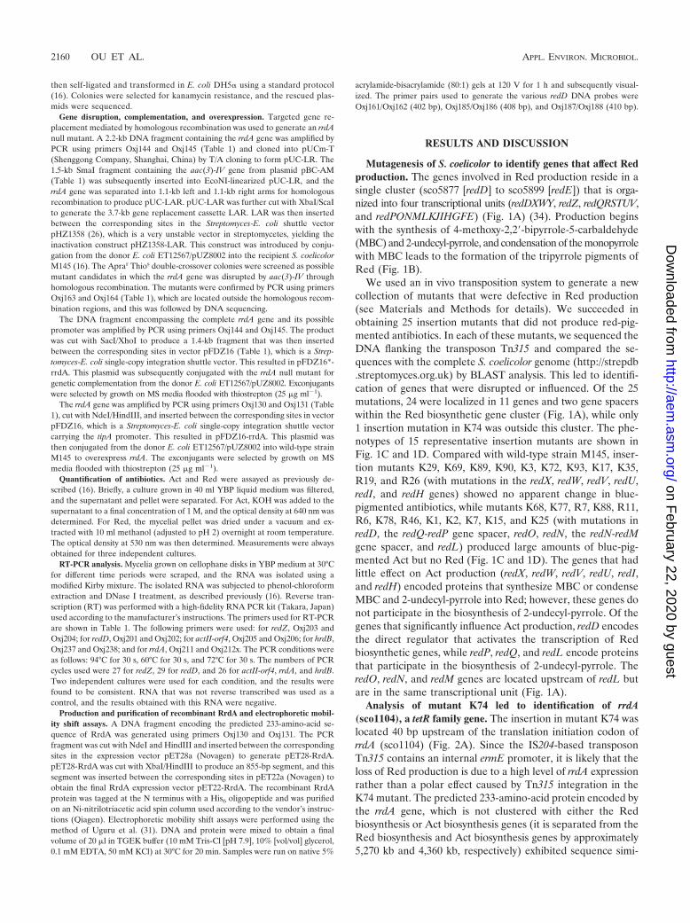
Mutagenesis of S. coelicolor to identify genes that affect Redproduction. The genes involved in Red production reside in asingle cluster (sco5877 [redD] to sco5899 [redE]) that is orga-nized into four transcriptional units (redDXWY, redZ, redQRSTUV,and redPONMLKJIHGFE) (Fig. 1A) (34). Production beginswith the synthesis of 4-methoxy-2,2-bipyrrole-5-carbaldehyde(MBC) and 2-undecyl-pyrrole, and condensation of the monopyrrolewith MBC leads to the formation of the tripyrrole pigments ofRed (Fig. 1B).
We used an in vivo transposition system to generate a newcollection of mutants that were defective in Red production(see Materials and Methods for details). We succeeded inobtaining 25 insertion mutants that did not produce red-pig-mented antibiotics. In each of these mutants, we sequenced theDNA flanking the transposon Tn315 and compared the se-quences with the complete S. coelicolor genome (http://strepdb.streptomyces.org.uk) by BLAST analysis. This led to identifi-cation of genes that were disrupted or influenced. Of the 25mutations, 24 were localized in 11 genes and two gene spacerswithin the Red biosynthetic gene cluster (Fig. 1A), while only1 insertion mutation in K74 was outside this cluster. The phe-notypes of 15 representative insertion mutants are shown inFig. 1C and 1D. Compared with wild-type strain M145, inser-tion mutants K29, K69, K89, K90, K3, K72, K93, K17, K35,R19, and R26 (with mutations in the redX, redW, redV, redU,redI, and redH genes) showed no apparent change in blue-pigmented antibiotics, while mutants K68, K77, R7, K88, R11,R6, K78, R46, K1, K2, K7, K15, and K25 (with mutations inredD, the redQ-redP gene spacer, redO, redN, the redN-redMgene spacer, and redL) produced large amounts of blue-pig-mented Act but no Red (Fig. 1C and 1D). The genes that hadlittle effect on Act production (redX, redW, redV, redU, redI,and redH) encoded proteins that synthesize MBC or condenseMBC and 2-undecyl-pyrrole into Red; however, these genes donot participate in the biosynthesis of 2-undecyl-pyrrole. Of thegenes that significantly influence Act production, redD encodesthe direct regulator that activates the transcription of Redbiosynthetic genes, while redP, redQ, and redL encode proteinsthat participate in the biosynthesis of 2-undecyl-pyrrole. TheredO, redN, and redM genes are located upstream of redL butare in the same transcriptional unit (Fig. 1A).
Analysis of mutant K74 led to identification of rrdA(sco1104), a tetR family gene. The insertion in mutant K74 waslocated 40 bp upstream of the translation initiation codon ofrrdA (sco1104) (Fig. 2A). Since the IS204-based transposonTn315 contains an internal ermE promoter, it is likely that theloss of Red production is due to a high level of rrdA expressionrather than a polar effect caused by Tn315 integration in theK74 mutant. The predicted 233-amino-acid protein encoded bythe rrdA gene, which is not clustered with either the Redbiosynthesis or Act biosynthesis genes (it is separated from theRed biosynthesis and Act biosynthesis genes by approximately5,270 kb and 4,360 kb, respectively) exhibited sequence simi-
2160 OU ET AL. APPL. ENVIRON. MICROBIOL.
on February 22, 2020 by guest
http://aem.asm
.org/D
ownloaded from

FIG. 1. Phenotypes of Red-deficient mutants and locations of the insertions. (A) Locations of the mutations in the Red biosynthetic genecluster. The black arrows indicate genes that are required for Red biosynthesis, while the open arrows indicate genes that are not essential. Thegray arrows indicate the redD and redZ regulatory genes. The bent open arrows indicate mutations that influence only Red production, while thefilled bent arrows indicate mutations that affect both Red production and Act production. The directions of the bent arrows indicate the transcriptorientation of PermE in Tn315 in the mutants. (B) Red biosynthetic pathway. RedU is required for activation of the acyl carrier protein domainsof RedO. The function of RedY is unknown (34). (C) Mutants and wild-type strain M145 were grown on YBP agar at 30°C for 2 days and 5 days.The bottoms of the plates are shown. WT, wild type. (D) Antibiotic production assay. Act production and Red production by the mutants andwild-type strain M145 were assayed in cultures grown in YBP liquid medium at 30°C for 88 h. The bars indicate the averages of three independentdeterminations, and the error bars indicate the standard errors. OD530, optical density at 530 nm; OD640, optical density at 640 nm.
2161
on February 22, 2020 by guest
http://aem.asm
.org/D
ownloaded from
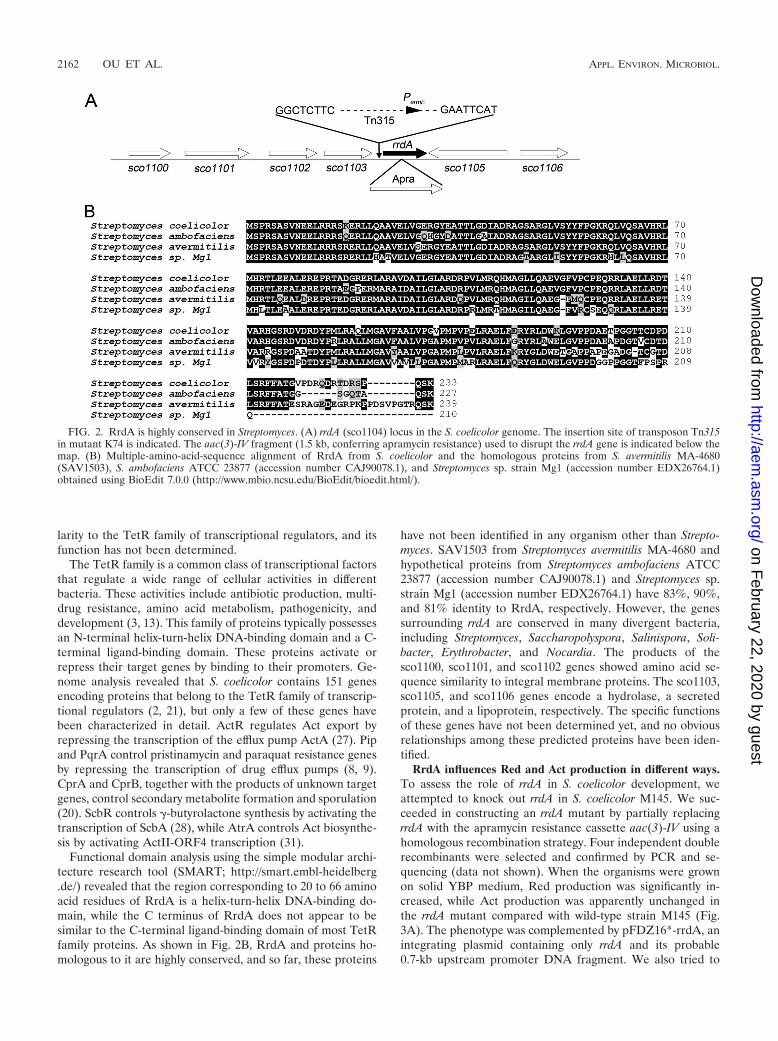
larity to the TetR family of transcriptional regulators, and itsfunction has not been determined.
The TetR family is a common class of transcriptional factorsthat regulate a wide range of cellular activities in differentbacteria. These activities include antibiotic production, multi-drug resistance, amino acid metabolism, pathogenicity, anddevelopment (3, 13). This family of proteins typically possessesan N-terminal helix-turn-helix DNA-binding domain and a C-terminal ligand-binding domain. These proteins activate orrepress their target genes by binding to their promoters. Ge-nome analysis revealed that S. coelicolor contains 151 genesencoding proteins that belong to the TetR family of transcrip-tional regulators (2, 21), but only a few of these genes havebeen characterized in detail. ActR regulates Act export byrepressing the transcription of the efflux pump ActA (27). Pipand PqrA control pristinamycin and paraquat resistance genesby repressing the transcription of drug efflux pumps (8, 9).CprA and CprB, together with the products of unknown targetgenes, control secondary metabolite formation and sporulation(20). ScbR controls �-butyrolactone synthesis by activating thetranscription of ScbA (28), while AtrA controls Act biosynthe-sis by activating ActII-ORF4 transcription (31).
Functional domain analysis using the simple modular archi-tecture research tool (SMART; http://smart.embl-heidelberg.de/) revealed that the region corresponding to 20 to 66 aminoacid residues of RrdA is a helix-turn-helix DNA-binding do-main, while the C terminus of RrdA does not appear to besimilar to the C-terminal ligand-binding domain of most TetRfamily proteins. As shown in Fig. 2B, RrdA and proteins ho-mologous to it are highly conserved, and so far, these proteins
have not been identified in any organism other than Strepto-myces. SAV1503 from Streptomyces avermitilis MA-4680 andhypothetical proteins from Streptomyces ambofaciens ATCC23877 (accession number CAJ90078.1) and Streptomyces sp.strain Mg1 (accession number EDX26764.1) have 83%, 90%,and 81% identity to RrdA, respectively. However, the genessurrounding rrdA are conserved in many divergent bacteria,including Streptomyces, Saccharopolyspora, Salinispora, Soli-bacter, Erythrobacter, and Nocardia. The products of thesco1100, sco1101, and sco1102 genes showed amino acid se-quence similarity to integral membrane proteins. The sco1103,sco1105, and sco1106 genes encode a hydrolase, a secretedprotein, and a lipoprotein, respectively. The specific functionsof these genes have not been determined yet, and no obviousrelationships among these predicted proteins have been iden-tified.
RrdA influences Red and Act production in different ways.To assess the role of rrdA in S. coelicolor development, weattempted to knock out rrdA in S. coelicolor M145. We suc-ceeded in constructing an rrdA mutant by partially replacingrrdA with the apramycin resistance cassette aac(3)-IV using ahomologous recombination strategy. Four independent doublerecombinants were selected and confirmed by PCR and se-quencing (data not shown). When the organisms were grownon solid YBP medium, Red production was significantly in-creased, while Act production was apparently unchanged inthe rrdA mutant compared with wild-type strain M145 (Fig.3A). The phenotype was complemented by pFDZ16*-rrdA, anintegrating plasmid containing only rrdA and its probable0.7-kb upstream promoter DNA fragment. We also tried to
FIG. 2. RrdA is highly conserved in Streptomyces. (A) rrdA (sco1104) locus in the S. coelicolor genome. The insertion site of transposon Tn315in mutant K74 is indicated. The aac(3)-IV fragment (1.5 kb, conferring apramycin resistance) used to disrupt the rrdA gene is indicated below themap. (B) Multiple-amino-acid-sequence alignment of RrdA from S. coelicolor and the homologous proteins from S. avermitilis MA-4680(SAV1503), S. ambofaciens ATCC 23877 (accession number CAJ90078.1), and Streptomyces sp. strain Mg1 (accession number EDX26764.1)obtained using BioEdit 7.0.0 (http://www.mbio.ncsu.edu/BioEdit/bioedit.html/).
2162 OU ET AL. APPL. ENVIRON. MICROBIOL.
on February 22, 2020 by guest
http://aem.asm
.org/D
ownloaded from

overexpress rrdA in wild-type strain M145 by introducingpFDZ16-rrdA, a plasmid containing the PtipA promoter andrrdA coding sequence. Previous studies have reported thatgenes cloned with the tipA promoter appear to be expressed atbasal levels even in the absence of thiostrepton induction (23,30). Transformants of S. coelicolor containing pFDZ16-rrdAproduced no red-pigmented antibiotics either with or withoutthiostrepton induction, so observation of the phenotype and
RT-PCR analysis (see below) of the rrdA high-expressionstrain were performed in the absence of thiostrepton induc-tion. As shown in Fig. 3B, a high level of rrdA expressionresulted in loss of Red production and production of amountsof Act larger than those in M145; thus, the behavior of thismutant was the same as that of the K74 mutant.
Production of Act and Red by the rrdA high-expressionstrain, a null mutant, a complemented strain, and M145 wasmeasured in YBP liquid media at fixed intervals. As shown inFig. 4, the rrdA null mutant produced Red earlier and at muchhigher level than the parent strain did, but the level of Actproduction was lower than that in the parent strain. In the rrdAhigh-expression strain, Red production dramatically decreasedto levels that could barely be detected, and the level of Actproduction was higher than that in M145 on days 3 and 4. Onday 5, the difference in Act production between M145 and therrdA high-expression strain did not appear to be as significantas the difference observed when the organisms were culturedon solid media (Fig. 1A). The slight but notable difference inAct production between liquid media and solid media might beexplained by differences in the developmental stages of therrdA high-expression strain under these two different cultureconditions. These results underline the fact that RrdA is anegative regulator of Red production and has a positive effecton Act production.
RrdA negatively regulates Red production by controlling theabundance of RedD mRNA. The expression of antibiotic bio-synthetic clusters is normally regulated by pathway-specific ac-tivators (4, 10, 29). In S. coelicolor, Act biosynthesis and Redbiosynthesis have been shown to depend on the transcriptionalactivation of the Act and Red biosynthetic clusters by ActII-ORF4 and the RedD and RedZ proteins, respectively. RedD,which is the direct transcriptional activator for the Red bio-synthetic cluster, is RedZ dependent (12, 33). The transcrip-tion of rrdA, actII-orf4, redD, and redZ in M145, an rrdA high-expression strain, and a null mutant was therefore analyzed by
FIG. 3. Phenotypes of the rrdA null mutant and high-expressionstrain. The bottoms of the plates are shown. (A) Colonies of the parentstrain S. coelicolor M145 (WT), the rrdA null mutant (�rrdA), the rrdAmutant harboring the empty vector pFDZ16 (�rrdA/pFDZ16), and thecomplemented strain (�rrdA/pFDZ16*-rrdA) were grown on YBP me-dium at 30°C for 2 days and 5 days. (B) Colonies of the parent strainM145 harboring the empty vector pFDZ16 (WT/pFDZ16) and therrdA high-expression strain (WT/pFDZ16-rrdA) were grown on YBPmedium at 30°C for 2 days and 5 days.
FIG. 4. Antibiotic production by the M145 strain harboring the empty vector pFDZ16, the rrdA null mutant harboring the empty vectorpFDZ16, the complemented strain, and the rrdA high-expression strain. Incubation was carried out in the YBP liquid medium at 30°C. The symbolsindicate the averages of three independent determinations, and the error bars indicate the standard errors. OD530, optical density at 530 nm;OD640, optical density at 640 nm.
VOL. 75, 2009 NOVEL GENE INVOLVED IN Red BIOSYNTHETIC REGULATION 2163
on February 22, 2020 by guest
http://aem.asm
.org/D
ownloaded from

RT-PCR. Total RNA was isolated both from M145 cultures atdifferent stages of development and from mutants grown oncellophane placed on YBP solid medium. As shown in Fig. 5A,the results correlated well with the results of Red production inliquid media or on solid YBP medium. The level of transcrip-tion of redD was slightly higher in the rrdA null mutant than inM145 and significantly lower at 36 to 60 h in the rrdA high-expression strain than in M145. However, at 24 h, redD seemedto be transcribed at the same level in the rrdA high-expressionstrain and the wild type. The levels of transcription of actII-orf4and redZ appeared to be the same as those in M145. The levelof the rrdA transcript peaked at 12 h and then decreased to astable level after 36 h. At this point, M145 became pink, whichindicates the onset of Red production. Since the primer se-quences of rrdA are located in the upstream region of theinsertion site of the aac(3)-IV fragment used for gene disrup-tion, the rrdA transcription profile of the rrdA null mutantcould also be detected, and it was very similar to that of M145,while in the rrdA high-expression strain the rrdA transcriptlevel was higher in all distinct phases during development.These results indicated that RrdA negatively regulated Redproduction by controlling the abundance of RedD mRNA.
Since rrdA encodes a putative TetR family transcription fac-tor, we performed electrophoretic mobility shift assays to ex-amine the binding of the purified recombinant RrdA protein tothree different DNA probes (nucleotides �296 to 115, �94 to315, and 61 to 462 relative to the transcriptional start site ofredD) (19) in TGEK buffer. The effects of Na�, Mg2�, andMn2� on RrdA binding were also tested. Unfortunately, nospecific binding of RrdA to the DNA probes was observedunder any of the test conditions used (data not shown). It ispossible that the activated binding of RrdA to the redD pro-moter requires additional factors or that RrdA indirectly reg-ulates redD transcription and one or more as-yet-unidentifiedproteins may participate in this regulation.
Cross-regulation of Red and Act production might occurthrough a mechanism involving competition for common pre-cursors. Huang et al. reported that deletion of the Red-specificregulator gene redZ caused delayed transcription of the actII-orf4 and Act biosynthesis genes in S. coelicolor (15). RrdA,which upregulates Act production and downregulates Red pro-duction, affects transcription of only the Red-specific regulatorgene redD. We hypothesized that another mechanism may beinvolved in the cross-regulation of Red biosynthesis and Actbiosynthesis. Therefore, we determined the transcriptionallevel of actII-orf4 in the redD null mutant K68. However, nodifference was observed between K68 and wild-type strainM145 (Fig. 5B). The results indicated that the positive effect ofthe redD mutation on Act production was not due to anyalteration at the transcriptional level of actII-orf4. Togetherwith the observation that mutations in Red biosynthesis genesaffect Act production, this suggests that Red production andAct production are interlinked at a nontranscriptional level.
It is known that Act production begins with the synthesis ofa 16-carbon polyketide backbone by a type II polyketide syn-thase complex that uses malonyl coenzyme A (malonyl-CoA)and acetyl-CoA as the precursors (27). These precursors arealso necessary for the production of 2-undecyl-pyrrole, which isan intermediate metabolite in the Red biosynthesis pathway(34). The redD mutation eliminated transcription of most bio-synthetic gene clusters (14, 19), and mutations in the redQ-redPgene spacer, redO, redN, the redN-redM gene spacer, and redLdisrupted or influenced, probably due to a polar effect, theexpression of key enzymes (RedP, RedQ, and RedL) involvedin the synthesis of 2-undecyl-pyrrole. This in turn divertedmalonyl-CoA and acetyl-CoA from Red biosynthesis to Actbiosynthesis. This hypothesis can be used to provide a simpleexplanation for the positive effect of RrdA on Act production.Thus, a decrease in Red production could result in an increasein Act production, and an increase in Red production couldlead to a decrease in Act production due to the levels of themetabolic precursors utilized by both biosynthetic pathways(Fig. 4). A similar effect was also observed for nanchangmycinproduction by Streptomyces nanchangensis. A DNA fragmentdeletion in cluster C of S. nanchangensis resulted in at least athreefold increase in nanchangmycin production because bio-synthesis of the compounds involved similar precursors (26).redX, redW, redV, redU, redI, and redH encode proteins thatparticipate in the biosynthesis of MBC or in the final step ofRed biosynthesis. Liquid chromatography-mass spectrometryanalysis of the redU mutant showed that Red was eliminatedand 2-undecyl-pyrrole accumulated (25). Therefore, disruptionof these genes had little effect on the accumulation of malonyl-CoA and acetyl-CoA and had no obvious influence on produc-tion of blue-pigmented Act.
In conclusion, our study revealed that the nontranscriptionalcross-regulation of Act biosynthesis and Red biosynthesismight involve competition for common precursors. We alsopartially characterized the TetR-like protein gene rrdA andfound that it encodes a novel negative regulator of Red bio-synthesis in S. coelicolor. These observations should be usefulin fermentation engineering of undecylprodigiosin, which is acandidate drug for cancer therapy (34).
FIG. 5. RT-PCR results for wild-type strain M145, the rrdA nullmutant, the rrdA high-expression strain, and redD null mutant K68.(A) Transcriptional levels of the pathway-specific activators, includingredD (primers Oxj201 and Oxj202, 368 bp), redZ (primers Oxj203 andOxj204, 322 bp), actII-orf4 (primers Oxj205 and Oxj206, 320 bp), rrdA[primers Oxj211 and Oxj212x, 166 bp; the primer sequences are lo-cated in the upstream region of the insertion site of the aac(3)-IVfragment used for gene disruption], and hrdB (control) (primersOxj237 and Oxj238, 207 bp). (B) RT-PCR results for the redD nullmutant K68 and wild-type strain M145 for transcriptional detection ofactII-orf4. WT, wild type.
2164 OU ET AL. APPL. ENVIRON. MICROBIOL.
on February 22, 2020 by guest
http://aem.asm
.org/D
ownloaded from
ACKNOWLEDGMENTS
We thank Zixin Deng, Maggie Smith, and Juan F. Martin for pro-viding strains and plasmids.
This work was supported by grants from the National NaturalScience Foundation of China (grants 30600009 and 30830002) andthe China National Basic Research Program (973 program grant2009CB522605).
REFERENCES
1. Aceti, D. J., and W. C. Champness. 1998. Transcriptional regulation ofStreptomyces coelicolor pathway-specific antibiotic regulators by the absA andabsB loci. J. Bacteriol. 180:3100–3106.
2. Bentley, S. D., K. F. Chater, A. M. Cerdeno-Tarraga, G. L. Challis, N. R.Thomson, K. D. James, D. E. Harris, M. A. Quail, H. Kieser, D. Harper, A.Bateman, S. Brown, G. Chandra, C. W. Chen, M. Collins, A. Cronin, A.Fraser, A. Goble, J. Hidalgo, T. Hornsby, S. Howarth, C. H. Huang, T.Kieser, L. Larke, L. Murphy, K. Oliver, S. O’Neil, E. Rabbinowitsch, M. A.Rajandream, K. Rutherford, S. Rutter, K. Seeger, D. Saunders, S. Sharp, R.Squares, S. Squares, K. Taylor, T. Warren, A. Wietzorrek, J. Woodward,B. G. Barrell, J. Parkhill, and D. A. Hopwood. 2002. Complete genomesequence of the model actinomycete Streptomyces coelicolor A3(2). Nature417:141–147.
3. Bertrand, K. P., K. Postle, J. Lewis, V. Wray, and W. S. Reznikoff. 1983.Overlapping divergent promoters control expression of Tn10 tetracyclineresistance. Gene 23:149–156.
4. Bibb, M. 1996. 1995 Colworth Prize Lecture. The regulation of antibioticproduction in Streptomyces coelicolor A3(2). Microbiology 142:1335–1344.
5. Bibb, M. 2005. Regulation of secondary metabolism in streptomycetes. Curr.Opin. Microbiol. 8:208–215.
6. Challis, G. L., and D. A. Hopwood. 2003. Synergy and contingency as drivingforces for the evolution of multiple secondary metabolite production byStreptomyces species. Proc. Natl. Acad. Sci. USA 100(Suppl. 2):14555–14561.
7. Chater, K. F. 1993. Genetics of differentiation in Streptomyces. Annu. Rev.Microbiol. 47:685–713.
8. Cho, Y.-H., E.-J. Kim, H.-J. Chung, J.-H. Choi, K. F. Chater, B.-E. Ahn, J.-H.Shin, and J.-H. Roe. 2003. The pqrAB operon is responsible for paraquatresistance in Streptomyces coelicolor. J. Bacteriol. 185:6756–6763.
9. Folcher, M., R. P. Morris, G. Dale, K. Salah-Bey-Hocini, P. H. Viollier, andC. J. Thompson. 2001. A transcriptional regulator of a pristinamycin resis-tance gene in Streptomyces coelicolor. J. Biol. Chem. 276:1479–1485.
10. Gramajo, H. C., E. Takano, and M. J. Bibb. 1993. Stationary-phase produc-tion of the antibiotic actinorhodin in Streptomyces coelicolor A3(2) is tran-scriptionally regulated. Mol. Microbiol. 7:837–845.
11. Gregory, M. A., R. Till, and M. C. M. Smith. 2003. Integration site forStreptomyces phage BT1 and development of site-specific integrating vec-tors. J. Bacteriol. 185:5320–5323.
12. Guthrie, E., C. Flaxman, J. White, D. Hodgson, M. Bibb, and K. F. Chater.1998. A response-regulator-like activator of antibiotic synthesis from Strep-tomyces coelicolor A3(2) with an amino-terminal domain that lacks a phos-phorylation pocket. Microbiology 144:727–738.
13. Hillen, W., and C. Berens. 1994. Mechanisms underlying expression of Tn10encoded tetracycline resistance. Annu. Rev. Microbiol. 48:345–369.
14. Huang, J., C.-J. Lih, K.-H. Pan, and S. N. Cohen. 2001. Global analysis ofgrowth phase responsive gene expression and regulation of antibiotic bio-synthetic pathways in Streptomyces coelicolor using DNA microarrays. GenesDev. 15:3183–3192.
15. Huang, J., J. Shi, V. Molle, B. Sohlberg, D. Weaver, M. J. Bibb, N. Karoo-nuthaisiri, C. J. Lih, C. M. Kao, M. J. Buttner, and S. N. Cohen. 2005.Cross-regulation among disparate antibiotic biosynthetic pathways of Strep-tomyces coelicolor. Mol. Microbiol. 58:1276–1287.
16. Kieser, T., M. J. Bibb, M. J. Butter, K. F. Chater, and D. A. Hopwood. 2000.Practical Streptomyces genetics. The John Innes Foundation, Norwich,England.
17. Kumar, C. V., J. J. Coque, and J. F. Martin. 1994. Efficient transformationof the cephamycin C producer Nocardia lactamdurans and development ofshuttle and promoter-probe cloning vectors. Appl. Environ. Microbiol. 60:4086–4093.
18. McKenzie, N., and J. Nodwell. 2007. Phosphorylated AbsA2 negatively reg-ulates antibiotic production in Streptomyces coelicolor through interactionswith pathway-specific regulatory gene promoters. J. Bacteriol. 189:5284–5292.
19. Narva, K. E., and J. S. Feitelson. 1990. Nucleotide sequence and transcrip-tional analysis of the redD locus of Streptomyces coelicolor A3(2). J. Bacteriol.172:326–333.
20. Onaka, H., T. Nakagawa, and S. Horinouchi. 1998. Involvement of twoA-factor receptor homologues in Streptomyces coelicolor A3(2) in the regu-lation of secondary metabolism and morphogenesis. Mol. Microbiol. 28:743–753.
21. Ramos, J. L., M. Martinez-Bueno, A. J. Molina-Henares, W. Teran, K.Watanabe, X. Zhang, M. T. Gallegos, R. Brennan, and R. Tobes. 2005. TheTetR family of transcriptional repressors. Microbiol. Mol. Biol. Rev. 69:326–356.
22. Sambrook, J., and D. W. Russell. 2001. Molecular cloning: a laboratory manual,3rd ed. Cold Spring Harbor Laboratory Press, Cold Spring Harbor, NY.
23. Schmitt-John, T., and J. W. Engels. 1992. Promoter constructions for effi-cient secretion expression in Streptomyces lividans. Appl. Microbiol. Biotech-nol. 36:493–498.
24. Sola-Landa, A., R. S. Moura, and J. F. Martin. 2003. The two-componentPhoR-PhoP system controls both primary metabolism and secondary metab-olite biosynthesis in Streptomyces lividans. Proc. Natl. Acad. Sci. USA 100:6133–6138.
25. Stanley, A. E., L. J. Walton, M. K. Zerikly, C. Corre, and G. L. Challis. 2006.Elucidation of the Streptomyces coelicolor pathway to 4-methoxy-2,29-bipyr-role-5-carboxaldehyde, an intermediate in prodiginine biosynthesis. Chem.Commun. 38:3981–3983.
26. Sun, Y., X. Zhou, J. Liu, K. Bao, G. Zhang, G. Tu, T. Kieser, and Z. Deng.2002. ‘Sreptomyces nanchangensis,’ a producer of the insecticidal polyetherantibiotic nanchangmycin and the antiparasitic macrolide meilingmycin, con-tains multiple polyketide gene clusters. Microbiology 148:361–371.
27. Tahlan, K., S. K. Ahn, A. Sing, T. D. Bodnaruk, A. R. Willems, A. R.Davidson, and J. R. Nodwell. 2007. Initiation of actinorhodin export inStreptomyces coelicolor. Mol. Microbiol. 63:951–961.
28. Takano, E., R. Chakraburtty, T. Nihira, Y. Yamada, and M. J. Bibb. 2001. Acomplex role for the gamma-butyrolactone SCB1 in regulating antibioticproduction in Streptomyces coelicolor A3(2). Mol. Microbiol. 41:1015–1028.
29. Takano, E., H. C. Gramajo, E. Strauch, N. Andres, J. White, and M. J. Bibb.1992. Transcriptional regulation of the redD transcriptional activator geneaccounts for growth-phase-dependent production of the antibiotic undecyl-prodigiosin in Streptomyces coelicolor A3(2). Mol. Microbiol. 6:2797–2804.
30. Takano, E., J. White, C. J. Thompson, and M. J. Bibb. 1995. Construction ofthiostrepton-inducible, high-copy-number expression vectors for use in Strep-tomyces spp. Gene 166:133–137.
31. Uguru, G. C., K. E. Stephens, J. A. Stead, J. E. Towle, S. Baumberg, and K. J.McDowall. 2005. Transcriptional activation of the pathway-specific regulatorof the actinorhodin biosynthetic genes in Streptomyces coelicolor. Mol. Mi-crobiol. 58:131–150.
32. Umeyama, T., and S. Horinouchi. 2001. Autophosphorylation of a bacterialserine/threonine kinase, AfsK, is inhibited by KbpA, an AfsK-binding pro-tein. J. Bacteriol. 183:5506–5512.
33. White, J., and M. Bibb. 1997. bldA dependence of undecylprodigiosin pro-duction in Streptomyces coelicolor A3(2) involves a pathway-specific regula-tory cascade. J. Bacteriol. 179:627–633.
34. Williamson, N. R., P. C. Fineran, F. J. Leeper, and G. P. C. Salmond. 2006.The biosynthesis and regulation of bacterial prodiginines. Nat. Rev. Micro-biol. 4:887–899.
35. Yao, W. S., Y. L. Yang, and J. S. Chiao. 1994. IS204: an insertion sequencefrom Nocardia asteroides (mexicana) Yp21. Plasmid 32:262–269.
VOL. 75, 2009 NOVEL GENE INVOLVED IN Red BIOSYNTHETIC REGULATION 2165
on February 22, 2020 by guest
http://aem.asm
.org/D
ownloaded from