Characterization and Comparison of Recombinant Simian Immunodeficiency Virus from Drill
14
JOURNAL OF VIROLOGY, Apr. 2003, p. 4867–4880 Vol. 77, No. 8 0022-538X/03/$08.000 DOI: 10.1128/JVI.77.8.4867–4880.2003 Copyright © 2003, American Society for Microbiology. All Rights Reserved. Characterization and Comparison of Recombinant Simian Immunodeficiency Virus from Drill (Mandrillus leucophaeus) and Mandrill (Mandrillus sphinx) Isolates Jinjie Hu, 1 William M. Switzer, 2 Brian T. Foley, 3 David L. Robertson, 4 Robert M. Goeken, 1 Bette T. Korber, 3 Vanessa M. Hirsch, 1 * and Brigitte E. Beer 1 Laboratory of Molecular Microbiology, National Institute of Allergy and Infectious Diseases, National Institutes of Health, Rockville, Maryland 20852 1 ; HIV and Retrovirology Branch, Division of AIDS, STD, and TB Laboratory Research, National Center for Infectious Diseases, Centers for Disease Control and Prevention, Atlanta, Georgia 30333 2 ; Theoretical Biology and Biophysics, Group T-10, Los Alamos National Laboratory, Los Alamos, New Mexico 87545 3 ; and Department of Zoology, University of Oxford, Oxford OX1 3PS, United Kingdom 4 Received 11 October 2002/Accepted 10 January 2003 Since simian immunodeficiency virus (SIV) was found to be the source of the human AIDS pandemic, a major goal has been to characterize the diversity of SIV strains in the wild and to assess their potential for crossover into humans. In the present study, SIV was isolated from a seropositive drill (Mandrillus leucophaeus) and three seropositive mandrills (Mandrillus sphinx) by using macaque peripheral blood mononuclear cells (PBMC). Full-length sequences were obtained from a drill and mandrill and designated SIVdrl1FAO and SIVmnd5440, respectively. A 182-bp fragment of the pol genes of the two remaining mandrill SIV isolates was also analyzed. Phylogenetic analyses demonstrated that SIVdrl1FAO formed a monophyletic clade with SIVmnd5440 and SIVmndM14, recently designated SIVmnd type 2. Both the SIVdrl and SIVmnd type 2 genomes carried a vpx gene and appeared to share a common ancestor with SIVrcm in the 5 region of the genome and with SIVmndGB1 (type 1) in the 3 region of the genome. A statistically significant recombination breakpoint was detected at the beginning of envelope, suggesting that the viruses were descendents of the same recombinant. Phylogenetic analysis of vpx and vpr genes demonstrated that the vpx genes formed a monophy- letic cluster that grouped with vpr from SIVagm. In addition, both SIVdrl1FAO and SIVmnd5440 replicated in human PBMC and therefore could pose a risk of transmission to the human population. Since human immunodeficiency virus type 1 (HIV-1) and HIV-2 have been shown to originate from simian immunode- ficiency virus (SIV) in African primates (9, 17, 40, 41), char- acterizing these viruses has become a major goal of AIDS research. To date, more than 20 species of SIV-infected Afri- can nonhuman primates have been detected (4, 18, 31, 37, 39); 34 complete sequences of these viruses have been published (30). SIV and HIV are classified as primate lentiviruses (PLVs), and at the present time six lineages, based upon phy- logenetic relationships of full-length genomes, are recognized (5, 12). These six designated lineages are (i) SIVcpz from chimpanzees (Pan troglodytes), including HIV-1 (17, 25, 26, 41, 55); (ii) SIVsm from sooty mangabeys (Cercocebus atys), in- cluding HIV-2 and SIVmac from captive macaques (Macaca spp.) (21, 24, 36); (iii) SIVagm from African green monkeys (members of the Chlorocebus aethiops superspecies) (1, 2, 14– 16, 23, 27, 29, 35); (iv) SIVsyk from Sykes’ monkeys (Cerco- pithecus albogularis); (v) SIVlhoest from L’Hoest monkeys (Cercopithecus lhoesti), including SIVsun from sun-tailed mon- keys (Cercopithecus solatus) and SIVmnd type 1 (GB1) from mandrills (Mandrillus sphinx) (3, 4, 22, 54); and (vi) SIVcol from Colobus monkeys (Colobus guereza) (12). In addition, another novel recombinant SIV from greater spot-nosed mon- keys (Cercopithecus nicitans) was recently described (13). SIVgsn shares the vpu gene of HIV-1 and SIVcpz and is most closely related to these viruses in the envelope but clusters with SIVsyk in Gag and part of Pol. Some SIV strains, such as SIVdrl from drills (Mandrillus leucophaeus), SIVtal from ta- lapoin monkeys (Miopithecus talapoin), SIVmus from mus- tached guenons (Cercopithecus cephus), SIVmon from Mona monkeys (Cercopithecus mona), and SIVdeb from DeBrazza monkeys (Cercopithecus neglectus) have only been partially characterized (11, 38, 39). Sequencing of the entire genome is necessary to classify those viruses into a particular lineage or to designate them as members of a novel lineage. Phylogenetic studies of nonhuman PLVs have suggested that the divergence of viral lineages reflected in some cases the divergence of their host lineages, suggesting host-dependent evolution (1, 4, 44, 45). However, in many cases the differences between phylogenetic relationships of some viruses and pri- mate phylogeny indicated that there have been multiple cross- species transmissions in the past (7, 28, 51). Cross-species transmission of PLVs can have a variety of outcomes in the new host. For example, if the recipient of the transmitted virus is already infected by a PLV, superinfection by other PLVs has the potential for generating recombinant viruses that could have a complex mosaic genome. Thus, some SIV isolates could not be classified into a particular lentivirus lineage because their genome was the result of one or more recombination events between viruses from different lineages. For example, * Corresponding author. Mailing address: Laboratory of Molecular Microbiology, National Institute of Allergy and Infectious Diseases, National Institutes of Health, 12441 Parklawn Dr., Rockville, MD 20852. Phone: (301) 496-2976. Fax: (301) 480-4227. E-mail: vhirsch @nih.gov. 4867 Downloaded from https://journals.asm.org/journal/jvi on 18 November 2021 by 183.214.104.238.
Transcript of Characterization and Comparison of Recombinant Simian Immunodeficiency Virus from Drill
JOURNAL OF VIROLOGY, Apr. 2003, p. 4867–4880 Vol. 77, No. 80022-538X/03/$08.00�0 DOI: 10.1128/JVI.77.8.4867–4880.2003Copyright © 2003, American Society for Microbiology. All Rights Reserved.
Characterization and Comparison of Recombinant SimianImmunodeficiency Virus from Drill (Mandrillus leucophaeus)
and Mandrill (Mandrillus sphinx) IsolatesJinjie Hu,1 William M. Switzer,2 Brian T. Foley,3 David L. Robertson,4 Robert M. Goeken,1
Bette T. Korber,3 Vanessa M. Hirsch,1* and Brigitte E. Beer1
Laboratory of Molecular Microbiology, National Institute of Allergy and Infectious Diseases, National Institutes of Health,Rockville, Maryland 208521; HIV and Retrovirology Branch, Division of AIDS, STD, and TB Laboratory Research,
National Center for Infectious Diseases, Centers for Disease Control and Prevention, Atlanta,Georgia 303332; Theoretical Biology and Biophysics, Group T-10, Los Alamos National Laboratory, Los Alamos,
New Mexico 875453; and Department of Zoology, University of Oxford, Oxford OX1 3PS, United Kingdom4
Received 11 October 2002/Accepted 10 January 2003
Since simian immunodeficiency virus (SIV) was found to be the source of the human AIDS pandemic, amajor goal has been to characterize the diversity of SIV strains in the wild and to assess their potential forcrossover into humans. In the present study, SIV was isolated from a seropositive drill (Mandrillus leucophaeus)and three seropositive mandrills (Mandrillus sphinx) by using macaque peripheral blood mononuclear cells(PBMC). Full-length sequences were obtained from a drill and mandrill and designated SIVdrl1FAO andSIVmnd5440, respectively. A 182-bp fragment of the pol genes of the two remaining mandrill SIV isolates wasalso analyzed. Phylogenetic analyses demonstrated that SIVdrl1FAO formed a monophyletic clade withSIVmnd5440 and SIVmndM14, recently designated SIVmnd type 2. Both the SIVdrl and SIVmnd type 2genomes carried a vpx gene and appeared to share a common ancestor with SIVrcm in the 5� region of thegenome and with SIVmndGB1 (type 1) in the 3� region of the genome. A statistically significant recombinationbreakpoint was detected at the beginning of envelope, suggesting that the viruses were descendents of the samerecombinant. Phylogenetic analysis of vpx and vpr genes demonstrated that the vpx genes formed a monophy-letic cluster that grouped with vpr from SIVagm. In addition, both SIVdrl1FAO and SIVmnd5440 replicated inhuman PBMC and therefore could pose a risk of transmission to the human population.
Since human immunodeficiency virus type 1 (HIV-1) andHIV-2 have been shown to originate from simian immunode-ficiency virus (SIV) in African primates (9, 17, 40, 41), char-acterizing these viruses has become a major goal of AIDSresearch. To date, more than 20 species of SIV-infected Afri-can nonhuman primates have been detected (4, 18, 31, 37, 39);34 complete sequences of these viruses have been published(30). SIV and HIV are classified as primate lentiviruses(PLVs), and at the present time six lineages, based upon phy-logenetic relationships of full-length genomes, are recognized(5, 12). These six designated lineages are (i) SIVcpz fromchimpanzees (Pan troglodytes), including HIV-1 (17, 25, 26, 41,55); (ii) SIVsm from sooty mangabeys (Cercocebus atys), in-cluding HIV-2 and SIVmac from captive macaques (Macacaspp.) (21, 24, 36); (iii) SIVagm from African green monkeys(members of the Chlorocebus aethiops superspecies) (1, 2, 14–16, 23, 27, 29, 35); (iv) SIVsyk from Sykes’ monkeys (Cerco-pithecus albogularis); (v) SIVlhoest from L’Hoest monkeys(Cercopithecus lhoesti), including SIVsun from sun-tailed mon-keys (Cercopithecus solatus) and SIVmnd type 1 (GB1) frommandrills (Mandrillus sphinx) (3, 4, 22, 54); and (vi) SIVcolfrom Colobus monkeys (Colobus guereza) (12). In addition,
another novel recombinant SIV from greater spot-nosed mon-keys (Cercopithecus nicitans) was recently described (13).SIVgsn shares the vpu gene of HIV-1 and SIVcpz and is mostclosely related to these viruses in the envelope but clusters withSIVsyk in Gag and part of Pol. Some SIV strains, such asSIVdrl from drills (Mandrillus leucophaeus), SIVtal from ta-lapoin monkeys (Miopithecus talapoin), SIVmus from mus-tached guenons (Cercopithecus cephus), SIVmon from Monamonkeys (Cercopithecus mona), and SIVdeb from DeBrazzamonkeys (Cercopithecus neglectus) have only been partiallycharacterized (11, 38, 39). Sequencing of the entire genome isnecessary to classify those viruses into a particular lineage or todesignate them as members of a novel lineage.
Phylogenetic studies of nonhuman PLVs have suggested thatthe divergence of viral lineages reflected in some cases thedivergence of their host lineages, suggesting host-dependentevolution (1, 4, 44, 45). However, in many cases the differencesbetween phylogenetic relationships of some viruses and pri-mate phylogeny indicated that there have been multiple cross-species transmissions in the past (7, 28, 51). Cross-speciestransmission of PLVs can have a variety of outcomes in thenew host. For example, if the recipient of the transmitted virusis already infected by a PLV, superinfection by other PLVs hasthe potential for generating recombinant viruses that couldhave a complex mosaic genome. Thus, some SIV isolates couldnot be classified into a particular lentivirus lineage becausetheir genome was the result of one or more recombinationevents between viruses from different lineages. For example,
* Corresponding author. Mailing address: Laboratory of MolecularMicrobiology, National Institute of Allergy and Infectious Diseases,National Institutes of Health, 12441 Parklawn Dr., Rockville, MD20852. Phone: (301) 496-2976. Fax: (301) 480-4227. E-mail: [email protected].
4867
Dow
nloa
ded
from
http
s://j
ourn
als.
asm
.org
/jour
nal/j
vi o
n 18
Nov
embe
r 20
21 b
y 18
3.21
4.10
4.23
8.

SIVagm from sabaeus monkeys was the result of a recombi-nation event between a SIVagm ancestor and another viruspotentially from the SIVsm lineage (27). SIVrcm has beenshown to cluster with different lentivirus lineages in differentregions of the genome, but it is still uncertain whether SIVrcmor one of the potential parental viruses is the recombinant(6, 18). The most recently discovered mosaic virus wasSIVmndM14, which was found to be recombinant betweenSIVrcm and SIVmndGB1 (48).
Based on the commonly accepted hypothesis that HIV-1 andHIV-2 were the result of multiple cross-species transmissionsto humans from chimpanzees and sooty mangabeys, respec-tively (9, 10, 17, 40, 41), it is important to consider the possi-bility of ongoing zoonotic transfer. People in west central Af-rica are still routinely exposed to a large variety of geneticallydivergent SIV stains (20, 39). Indeed, epidemiological surveysidentified a human serum sample from Cameroon that showeda high reactivity directed solely and strongly against theSIVmnd V3 loop, raising the possibility that mandrills mayrepresent a viral reservoir for humans similar to sooty manga-beys in western Africa and chimpanzees in central Africa (48).Moreover, a variety of SIV strains are able to replicate inhuman peripheral blood mononuclear cells (PBMC) and mac-rophages in vitro, indicating the possibility for another cross-over of SIV into the human population (6, 19).
In the present study, we isolated SIV from blood samplesfrom one drill and three mandrills housed in North Americanand European zoos. The highly endangered drill is a large,short-tailed monkey restricted in the wild to several blocks offorest between the Cross river in southeastern Nigeria, theSanaga river in southwestern Cameroon, and the island ofBioko (Equatorial Guinea) (11). The related mandrill is also alarge semiterrestrial primate that lives in the tropical rain for-ests of Cameroon and Gabon (48). Our goal was to isolate andmolecularly characterize SIV from a drill and mandrill andevaluate their in vitro host ranges and coreceptor usages. Forthis purpose, we characterized the full-length genomes of theSIV isolates from the drill (no. 1FAO) and from one of themandrills (no. 5440). We also determined the host range ofSIVdrl1FAO and SIVmnd5440 in permanent cell lines and in
primary PBMC and evaluated the coreceptor usage ofSIVdrl1FAO.
MATERIALS AND METHODS
Primate samples. Fresh blood specimens were obtained from a drill (1FA0)and three mandrills (5440, Pearl, and Gunnite) housed at North American orEuropean zoological parks in accordance with the guidelines of the animal careand use committees at their institutions. The single male drill was in an exhibitthat was exposed to a wild-born drill with an unknown SIV serostatus. Mandrill5440 [female] was captive born to African-born parents with unknown SIVserostatus. The countries of origin of the wild-born animals are not known.Mandrill Gunnite [male] and Pearl [female] were born in captivity to captive-born parents of which the mother was SIV seropositive in both cases. From thefour animals, PBMC and plasma were obtained by standard Ficoll-Hypaquedensity gradient centrifugation and stored in liquid nitrogen until use.
Virus isolation. After being thawed, drill and mandrill PBMC were washedtwice with Hanks’ buffered salt solution and stimulated with 2 �g of phytohe-magglutinin (PHA) (Sigma, St. Louis, Mo.) per ml for 3 days. PBMC fromSIV-negative pig-tailed macaques were separated by Ficoll-Hypaque densitygradient centrifugation of whole blood and also stimulated with 2 �g of PHA perml for 3 days. PBMC blasts from the SIV-positive drill and three SIV-positivemandrills (5440, Pearl, and Gunnite) were cocultivated with 2 � 107 PBMCblasts from pig-tailed macaques and maintained in complete RPMI (RPMI 1640supplemented with 10% heat-inactivated fetal calf serum, 2 mM glutamine, 100U of penicillin per ml, 100 �g of streptomycin per ml, and 10 mM HEPES)supplemented with 5 half-maximal units of human interleukin-2 (Advanced Bio-technologies, Columbia, Md.) per ml. Supernatants were sampled every threedays and subjected to reverse transcriptase (RT) assay. Supernatants were har-vested at the peak of RT activity, filtered through a 0.45-�m-pore-size filter, andcryopreserved as virus stock. Virus stocks were designated SIVdrl1FAO,SIVmnd5440, SIVmndPea, and SIVmndGun.
PCR amplification of the viral genome. PCRs were performed with DNAextracted from PBMC by using phenol-chloroform or from cell lysates as de-scribed previously (49). To generate a small fragment of 194 bp in the RT portionof the pol gene from cell lysate of drill, degenerate primers DR1, DR2, DR4, andDR5 were used as previously described (11). Amplification was performed withan ExTaq kit (Takara, Otsu, Shiga, Japan) with an initial denaturation at 94°Cfor 2 min, followed by 40 cycles of 94°C for 30 s, 55°C for 30 s, and 72°C for 30 s,followed by a final extension of 72°C for 10 min. Primer DR1 was used at 0.8pmol per �l, and DR2, DR4, and DR5 were used at 2.5 pmol per �l. A secondfragment of 182 bp located in the integrase part of the pol gene was generated bynested PCR with primers mpolF1, mpolR1, mpolF2, and mpolR2 (Table 1). Theconditions for PCR amplification were the same as described above.
To amplify the entire SIVdrl1FAO or SIVmnd5440 genomes, genomic DNAwas extracted from infected Molt4clone8 cells by using phenol-chloroform. Twooverlapping fragments, from the primer binding site (PBS) to integrase (4.5 kb)and from integrase to the PBS-gag gene (5.5 and 5.9 kb, respectively), were
TABLE 1. Nested primer pairs and their genomic locations
Primer Sequence (5� to 3�) SpecificityAmplifiedfragment
(bp)Gene Location
(nt)
mpolF1 ATTATAGAACAGGTTAGAGATCAGGCA SIV 233 pol (outer set) 4272–4297a
mpolR1 GGTCCTTTCCACTGTTGATCCCTT 4480–4503a
mpolF2 TTAGAGACAGCAGTTCAAATGGCAGTA 182 pol (inner set) 4304–4330a
mpolR2 TCCCTTCCTTCTCTGTAATAAACCCGA 4459–4485a
drlF1 TGGCGCCCGAACAGGGACTTGA SIVdrl1FAO 4480 PBS 1–22mpolR1 GGTCCTTTCCACTGTTGATTCCTT pol 4480–4503drlF2 GCATACCACAGGAGTTCCCTATAA 5935 pol 4204–4227drlR2 GTTCTTTCGCTACCCAGACAAGAT gag 388–406
mnd5440F1 TGGCGCCCGAACAGGGACTTGA SIVmnd5440 4485 PBS 1–22mnd5440R1 TCCCTTCCTTCTCTGTAATAAACCCGA pol 4459–4485mnd5440F2 GGCTAGAAACAGCAGTGCAAATGG 5457 pol 4302–4325mnd5440R2 GTCCCTTACAGGTGGGTGCGACCTGAA PBS 29–55
a The primer location is in SIVmnd5440.
4868 HU ET AL. J. VIROL.
Dow
nloa
ded
from
http
s://j
ourn
als.
asm
.org
/jour
nal/j
vi o
n 18
Nov
embe
r 20
21 b
y 18
3.21
4.10
4.23
8.

amplified by targeting the integrated genomic as well as unintegrated circularDNA. The primer sequences and location of the expected amplified product aredescribed in Table 1. PCRs were performed in a volume of 50 �l with the ExpandLong Template High Fidelity PCR system (Roche Molecular Biochemicals)under the following conditions: a hot start at 94°C for 3 min; 10 cycles ofdenaturation at 94°C for 20 s, annealing at 58°C for 30 s, and extension at 68°Cfor 5 to 7 min; and 20 cycles with extension at 68°C for 5 to 7 min with anincrement of 20 s per cycle. Amplification was completed by a final extension at72°C for 15 min. The entire 50-�l PCR product was separated on a 0.9% agarosegel. Bands of the correct size were excised, cloned into pCRII-TOPO vector(TOPO TA Cloning kit; Invitrogen, Carlsbad, Calif.) and sequenced by auto-mated fluorescent sequencing (Taq amplification and termination; Perkin-ElmerApplied Biosystems, Warrington, United Kingdom).
Sequence analysis. The sequences of SIVdrl1FAO and SIVmnd5440 werealigned and compared to representatives of the major lentivirus lineages, includ-ing some of those in the HIV sequence database at http://hiv-web.lanl.gov/ALIGN_CURRENT/ALIGN-INDEX.html (“other SIV complete genomeDNA” alignment). For some of the phylogenetic analyses, third-codon positionswere removed. Columns in the alignment, in which gaps had been inserted tomaintain the alignment through regions with insertions and deletions, werestripped prior to the analyses. Protein identities were calculated from the gap-stripped alignment by using BioEdit (http://www.mbio.ncsu.edu/BioEdit/bioedit.html). The gap-stripped alignment was further analyzed with the SIMPLOT
program from Stuart Ray (http://www.med.jhu.edu/deptmed/sray/download),both as nucleotide and as translated amino acid sequences, in order to determinethe regions of the alignment to be analyzed separately by phylogenetic methods.A neighbor-joining phylogenetic tree was built for each genomic region, usingthe PHYLIP, DNADIST, and NEIGHBOR programs (http://evolution.genetic-s.washington.edu/phylip.html) with the F84 model of evolution. The resultingtree was used as input, along with the alignment, to Gary Olsen’s DNArates
FIG. 1. Western blot analysis (HIV-2 blot1.2; Genelabs) of drilland mandrill sera. Sera from mandrills 5440, Pearl, and Gunnite anddrill 1FAO showed cross-reactive antibodies against HIV-2 proteinsEnv gp36 and gp80. Mandrill 5440 and drill 1FAO sera also reactedwith Gag p26.
TABLE 2. Protein sequence identities between SIVdrl1FAO andother PLVs
Virus(accession no.)
% Identity with SIVdrl1FAO in:
Gag Pol Vif Vpx Vpr Tata Revb Env Nef
SIVdrl/Clewley(AJ310481)
92c 93d
SIVmnd5440(AY159322)
83 81 69 70 78 52 57 67 66
SIVmndM14(AF328295)
81 81 70 68 80 52 61 72 63
SIVrcm(AF349680)
73 77 51 57 63 46 39 38 41
SIVcpz (X52154) 59 67 42 49 37 28 36 40SIVsm (L09212) 64 61 44 42 63 39 48 40 43SIVagmSab
(U04005)72 72 49 42 39 39 37 48
SIVagmVer(L40990)
67 66 46 42 44 43 38 44e
SIVlhoest(AF075269)
50 58 35 44 37 50 55 45
SIVsyk (L06042) 60 55 41 25 50 30 38 39SIVcol
(AF301156)47 51 22 28 36 30 29 31
SIVmndGB1(M27470)
54 58 36 40 44 37 63 55
a Based on a 55-amino-acid alignment.b Based on a 46-amino-acid alignment.c Based on a 316-amino-acid alignment.d Based on a 967-amino-acid alignment.e Comparison with SIVagmVer155 (M29975).
TABLE 3. Protein sequence identities between SIVmnd5440 andother PLVs
Virus(accession no.)
% Identity with SIVmnd5440 in:
Gag Pol Vif Vpx Vpr Tata Revb Env Nef
SIVmndM14(AF328295)
93 91 75 83 85 74 76 80 78
SIVdrl1FAO(AY159321)
83 81 69 70 78 52 57 67 66
SIVrcm(AF349680)
70 77 49 63 66 54 30 36 46
SIVcpz (X52154) 56 67 40 52 37 30 35 40SIVsm (L09212) 65 59 40 39 64 43 39 38 48SIVagmSab
(U04005)71 70 47 42 41 43 36 48
SIVagmVer(L40990)
65 65 47 40 54 41 38 43c
SIVlhoest(AF075269)
51 58 34 44 44 50 49 45
SIVsyk (L06042) 60 54 40 25 48 30 35 38SIVcol
(AF301156)47 50 25 27 33 28 30 30
SIVmndGB1(M27470)
53 61 36 44 39 46 58 54
a Based on a 55-amino-acid alignment.b Based on a 46-amino-acid alignment.c Comparison with SIVagmVer155 (M29975).
VOL. 77, 2003 NOVEL SIV DRILL AND MANDRILL ISOLATES 4869
Dow
nloa
ded
from
http
s://j
ourn
als.
asm
.org
/jour
nal/j
vi o
n 18
Nov
embe
r 20
21 b
y 18
3.21
4.10
4.23
8.

program (http://geta.life.uiuc.edu/�gary/programs/DNArates.html) to computesite-specific rates of evolution. These rates were then included in the input to amodified version of fastDNAml written by Tanmoy Bhattacharya (http://ww-w.santafe.edu/�btk/science-paper/bette.html), which computes maximum-likeli-hood trees incorporating site-specific rates of evolution. Bootstrap support (100replicates) for each of the trees was calculated by using the PHYLIP SEQBOOT,DNADIST, NEIGHBOR, and CONSENSE programs. Although the bootstraptrees were computed via the neighbor-joining method, rather than by maximumlikelihood with site-specific rates, the topologies were often identical althoughbranch lengths differed significantly. Trees were edited in TreeTool (ftp://ftp.cme.msu.edu/pub/RDP/programs/TreeTool/).
Infection of chimpanzee PBMC, human PBMC, and human CD4�-T-cell lineswith SIVdrl1FAO or SIVmnd5440. To test the in vitro host range of SIVdrl1FAOand SIVmnd5440, human and chimpanzee PBMC and a panel of CD4� humancells were used. PBMC were separated from heparinized whole blood of HIV-negative humans and SIV- and HIV-negative chimpanzees by Ficoll-Hypaquedensity gradient centrifugation. PBMC were stimulated with 2 �g of PHA (Sig-ma) per ml in complete RPMI 1640 with 10% fetal calf serum, supplementedwith 5 half-maximal units of human interleukin-2 per ml, for 3 days before viralinoculation. The human CD4� cell lines CEMx174, CEM-SS, C8166, H9, Hut78,MT-2, MT-4, Molt4clone8, PM1, SupT1, and U937 were maintained in RPMIcomplete medium. Five million PHA-stimulated PBMC or 2 million humanCD4� T cells were pelleted, and the cell pellet was incubated with 1 ml ofSIVdrl1FAO or SIVmnd5440 (approximately 500,000 cpm of RT activity) for 2 hwith agitation every 30 min. The cells were washed once with medium, 5 ml ofcomplete RPMI was added, and the cells were transferred to T25 flasks (NalgeNunc International, Rochester, N.Y.). Cell culture supernatants were collectedevery 3 days, and the infection of the cells was monitored for up to 6 weeks.
GHOST cell assay. The coreceptor usages of SIVdrl1FAO and SIVmnd5440were determined by using the GHOST cell assay (34). The GHOST cell panelwas obtained through the AIDS Reagent and Reference Program, NationalInstitutes of Health (Rockville, Md.). Cells (2 � 104) from each of the cell lineswere seeded in 24-well plates (Costar, Coring, N.Y.) and cultivated for 2 daysprior to infection. One milliliter of virus stock (corresponding to approximately500,000 cpm of RT activity) was gently added to the cells, and after 4 h ofincubation at 37°C, the cells were washed once with medium, followed by theaddition of 1 ml of fresh medium. Every other day the cells were split 1:2 and 0.5
ml of culture supernatant was collected. The production of SIV Gag p27 antigenwas measured by using the RETRO-TEK SIV-1 p27 antigen enzyme-linkedimmunosorbent assay (ZeptoMetrix Corp., Buffalo, N.Y.) according to the man-ufacturer’s instructions.
Nucleotide sequence accession numbers. The complete genomes ofSIVdrl1FAO and SIVmnd5440 have been submitted to GenBank under acces-sion numbers AY159321 and AY159322.
RESULTS
Identification and isolation of SIVdrl and SIVmnd. Anti-bodies to SIV were detected using a commercially availableHIV-2 Western blot kit (HIV-2 blot1.2; Genelabs, Singapore).Sera of mandrill 5440 and drill FAO cross-reacted with Envgp36, Env gp80, and Gag p26 proteins of HIV-2 (Fig. 1). Viruswas isolated from PBMC of the seropositive drill and from allthree seropositive mandrills (Gunnite, Pearl, and 5440) bycocultivation with PHA-stimulated pig-tailed macaque PBMC.Virus was isolated from the SIV-positive mandrills after 5weeks of coculture, with peak RT activity by 6 weeks postin-fection. Cytopathic effects were not observed during the entireperiod of coculture. A similar strategy was used for isolation ofSIVdrl1FAO, except that isolation only required 1 week ofcoculture.
Genomic organizations of SIVdrl and SIVmnd. Partial polsequences were amplified from three SIV-positive mandrillsand one SIV-positive drill by using degenerate primers as de-tailed in Materials and Methods. Subsequently, the completegenomes of SIVdrl1FAO and SIVmnd5440 were amplifiedin two fragments (4.5 kb and 5.5 to 5.9 kb) by targeting theintegrated genomic as well as unintegrated circular DNA. The
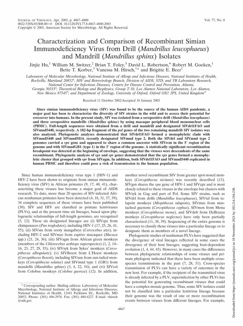
FIG. 2. Similarity plots comparing a 3,450-bp gap-stripped partial gag-pol alignment of SIVdrl1FAO with other SIV isolates from drill andmandrill (A), comparing the full-length genome of SIVdrl1FAO with SIVmnd5440, SIVmndM14, SIVrcm, and representatives of the other majorPLV lineages (B), and comparing the full-length genome of SIVmnd5440 with SIVdrl1FAO, SIVmndM14, SIVrcm, and representatives of theother major PLV lineages (C). The gene boundaries are indicated by arrows. In the Simplots for panels B and C, third-codon positions of thealignment have been removed. The fractional nucleotide sequence difference was calculated for a window size of 300 bp (A) or 400 bp (B and C)moved in steps of 30 bp (A) or 40 bp (B and C) with Hamming distances.
4870 HU ET AL. J. VIROL.
Dow
nloa
ded
from
http
s://j
ourn
als.
asm
.org
/jour
nal/j
vi o
n 18
Nov
embe
r 20
21 b
y 18
3.21
4.10
4.23
8.
PCR fragments were cloned by using the TOPO cloning system(Invitrogen) and sequenced, and the sequences assembledto obtain the full-length genomes, which were compared tothose of other PLVs. The proviral genomes of SIVdrl1FAOand SIVmnd5440 shared their genomic organization withSIVmndM14, SIVrcm, SIVsm, SIVmac, and HIV-2. These vi-ruses possess a vpx gene in addition to vpr but lack a vpu genewhich is present in the SIVcpz/HIV-1 lineage and in SIV fromgreater spot-nosed and mona monkeys (13; J. Clewley, 9thInternational Workshop on HIV Dynamics and Evolution, ab-str. 5, 2002).
SIVdrl1FAO and SIVmnd5440 share the same mosaic struc-ture. We compared protein identities of SIVdrl1FAO andSIVmnd5440 to published full-length sequences of SIV in or-der to estimate their relationship to other PLVs. SIVdrl1FAOwas most closely related to SIVdrl/Clewley (unpublished; Gen-Bank accession no. AJ310481), with identities of 92 and 93% inpartial fragments of Gag and Pol, respectively (Table 2). Next,SIVdrl1FAO was most closely related throughout the entiregenome to SIVmnd5440 and SIVmndM14, with a protein iden-tity of 81 to 83% in Gag and Pol. In comparison to the otherPLVs, SIVdrl1FAO was most closely related in the Gag, Pol,
FIG. 2—Continued.
VOL. 77, 2003 NOVEL SIV DRILL AND MANDRILL ISOLATES 4871
Dow
nloa
ded
from
http
s://j
ourn
als.
asm
.org
/jour
nal/j
vi o
n 18
Nov
embe
r 20
21 b
y 18
3.21
4.10
4.23
8.

FIG. 3. Phylogenetic trees of PLV full-length genomes. (A) Unrooted phylogenetic tree (maximum likelihood with site-specific substitutionrates) of a conserved fragment of the pol gene, including many of the available SIVmnd and SIVdrl sequences. The alignment was 397 nucleotidesafter gap stripping. SIVdrl1FAO and SIVmnd5440 are boxed. (B and C) The primate lentiviral genomes were aligned as described in Materialsand Methods, third-codon positions were removed, and columns containing gaps were stripped from the alignment. (B) Representative tree (max-imum likelihood with site-specific substitution rates) from the 5� part of the genome, spanning bases 651 to 1600 of the alignment. (C) Repre-sentative tree (maximum likelihood with site-specific substitution rates) from the 3� part of the genome, spanning bases 3001 to 4001 of the align-ment. The sequences used are all available at http://hiv-web.lanl.gov by using either the common names or accession numbers (see Materials andMethods). The tree is unrooted. The scale shows the number of substitutions per site.
4872
Dow
nloa
ded
from
http
s://j
ourn
als.
asm
.org
/jour
nal/j
vi o
n 18
Nov
embe
r 20
21 b
y 18
3.21
4.10
4.23
8.

Vif, Vpx, Vpr, and Tat proteins to SIVrcm in Env and Nefto SIVmnd type 1 (SIVmndGB1) and in Rev to SIVlhoest.SIVdrl1FAO was also closely related to SIVagmSAB in Gagand Pol, probably due to the relationship between SIVrcm andSIVagmSAB. SIVdrl1FAO was only distantly related to virusesof the other primate lentivirus lineages.
SIVmnd5440 showed a pattern of genetic relationships sim-ilar to that of SIVdrl1FAO (Table 3). It was closely related toSIVmndM14, with �90% identity in Gag and Pol and anoverall protein identity of at least 74%, indicating thatboth SIV isolates represented the same type of virus. Next,SIVmnd5440 was most closely related to SIVdrl1FAO and also
FIG. 3—Continued.
VOL. 77, 2003 NOVEL SIV DRILL AND MANDRILL ISOLATES 4873
Dow
nloa
ded
from
http
s://j
ourn
als.
asm
.org
/jour
nal/j
vi o
n 18
Nov
embe
r 20
21 b
y 18
3.21
4.10
4.23
8.

to SIVrcm in the 5�part of the genome and to SIVmndGB1 inthe 3� part of the genome. The relationship of SIVdrl1FAOand SIVmnd5440 to SIVrcm from the Gag to the Tat proteinand to SIVmndGB1 in Env and Nef and the overall identity toSIVmndM14 across the entire genome indicated that both nov-el SIV isolates might have been generated by recombination.
In order to more thoroughly investigate the relationship ofSIVdrl1FAO and SIVmnd5440 to other PLVs and to rule outevidence of recombination, similarity plots were constructed.As shown in Fig. 2A, SIVdrl1FAO was compared to par-tial gag-pol sequences of SIVdrl/Clewley, SIVmnd5440,SIVmndM14, SIVmndGB1, and SIVrcm. As expected fromthe amino acid identities, SIVdrl1FAO was most closely re-lated to SIVdrl/Clewley in the gag-pol region. The relationshipbetween the two SIVdrl strains appeared to be similar to therelationship between SIVmnd5440 and SIVmndM14.
We also constructed similarity plots to compare SIVdrl1FAO and SIVmnd5440 to full-length genomes of SIV. Asshown in Fig. 2B, SIVdrl1FAO was most closely related toSIVmnd5440 and SIVmndM14 across the entire genome, in-dicating a common origin of both viruses. As already indicatedby the protein identities, SIVdrl1FAO and SIVmnd5440 weremost closely related to SIVrcm in gag, pol, vif, vpx, and vpr,whereas in env they were most closely related to SIVmndGB1(Fig. 2B and C). This observation confirmed that the genomesof SIVdrl1FAO and SIVmnd5440 were recombinant.
A phylogenetic tree of a small pol fragment (Fig. 3A) dem-onstrated that SIVmnd type 2 and SIVdrl sequences formedmonophyletic clusters, with the exception of SIVmnd2-99CM32 which fell in the SIVdrl cluster. Since the host ofSIVmnd2-99CM32 was a wild-born pet monkey geneticallyconfirmed to be a mandrill by mitochondrial sequencing (50),some cross-species transmission of SIVdrl to a mandrill musthave been occurred in the past. This is particularly interesting,
because the habitats of drills and mandrills are located northand south of the Sanaga River, respectively, and do not over-lap. Therefore, transmission of SIV between drill and mandrillis an unexpected finding.
In order to examine individual regions of the entire genome,phylogenetic trees were constructed by using the maximum-likelihood method (Fig. 3B and C). The third-codon positionsof the alignment were removed, and the alignment was gapstripped. Since phylogenetic trees spanning recombinant se-quences are prohibited, the 4,411-bp remaining nucleotidealignment was divided into seven regions that did not containany obvious recombination breakpoints in any of the alignedsequences (6). Two representative trees, one from the 5� partand one from the 3� part of the genome, are shown in Fig.3B and C, respectively. SIVdrl1FAO, SIVmnd5440, andSIVmndM14 clustered together in all the phylogenetic treesbuilt, and this relationship was supported in 100% of boot-strap replicates. As indicated by the short branch lengths,SIVmnd5440 and SIVmndM14 were extremely closely relatedand SIVdrl1FAO was more distantly related. In the 5� tree,SIVmnd5440, SIVmndM14, and SIVdrl1FAO formed a clusterwith SIVrcm. In the 3� tree, however, SIVmnd5440, SIVmndM14, and SIVdrl1FAO grouped together with SIVmndGB1and therefore clustered with the SIVlhoest lineage. This dis-cordant topology was again indicative of recombination be-tween an ancestor of SIVrcm and of SIVmnd GB1 (type 1).Phylogenetic trees including third-codon positions in the align-ment showed the same topology as trees without third-codonpositions.
Finally, in order to accurately delineate the recombinationpoint where the relative relationship of SIVdrl1FAO orSIVmnd5440 to SIVrcm and SIVmndGB1 changed, we per-formed informative-site analysis and permutation tests (Fig. 4)(42). An informative site is one at which, in a four-sequence
FIG. 4. Informative-site analysis. The recombination breakpoint in SIVdrl1FAO and SIVmnd5440 at which the closest relationship changedfrom SIVrcm to SIVmndGB1 was determined by informative-site analysis of first- and second-codon positions of a nucleotide alignment ofSIVdrl1FAO or SIVmnd5440, SIVrcm, SIVmndGB1, and SIVsyk (outgroup) by surveying the informative sites supporting either of the two rootedphylogenies and maximizing a chi-square value to locate the optimal breakpoint location (33, 42). Statistical significance was assessed byperforming 10,000 permutations and for each maximizing a chi-square value in order to again locate the optimal breakpoint. The P value reflectsthe proportion of permutated informative sites with chi-square values greater than the observed value. Total of 367 and 358 informative sites inSIVdrl1FAO and SIVmnd5440, respectively, were considered. A highly significant breakpoint was found between nt 2944 and 2945 forSIVdrl1FAO and between nt 2939 and 2945 for SIVmnd5440. Both breakpoints were located at the beginning of the envelope.
4874 HU ET AL. J. VIROL.
Dow
nloa
ded
from
http
s://j
ourn
als.
asm
.org
/jour
nal/j
vi o
n 18
Nov
embe
r 20
21 b
y 18
3.21
4.10
4.23
8.

alignment, two sequences share one residue and the two othersshare another residue. The four-sequence alignment consistedof the potentially recombinant sequence (SIVdrl1FAO orSIVmnd5440, respectively), the two parental representativesequences (SIVrcm and SIVmndGB1), and an outgroup(SIVsyk). We considered only informative sites of first- andsecond-codon positions, since the majority of changes in thethird positions are usually saturated in divergent sequencesand do not contribute any reliable phylogenetic information. Atotal of 367 informative sites in SIVdrl1FAO and 358 sites inSIVmnd5440 were considered, and statistically significantbreakpoints at the beginning of the envelope were observed forboth SIVdrl1FAO (between nucleotides [nt] 2944 and 2945 ofthe alignment) and SIVmnd5440 (between nt 2939 and 2945).These data suggest with high probability that SIVdrl1FAO andSIVmnd5440 are descendents of the same recombinant.
Phylogenetic relationship and origin of the vpx gene. Thereare now several highly divergent SIV and HIV-2 strains thatcontain a vpx gene in addition to vpr. Previous studies haveshown that the vpx and vpr genes of HIV-2, SIVsm, and SIV-mac are phylogenetically related (46, 52). Recently, we de-scribed a novel PLV, SIVrcm, with a highly divergent vpx gene;it was only 42% identical on the amino acid level to SIVsmVpx. As shown in Table 2, SIVdrl1FAO Vpx was 70% relatedto SIVmnd5440 and SIVmndM14 Vpx and 57% related toSIVrcm Vpx. Figure 5 depicts an alignment of the major rep-resentatives of Vpx protein sequences, with the consensus se-quence shown on top. Although the majority of the aminoacids differ from each other, there was a highly conservedtyrosine motif in the central part of Vpx (Y-x-x-Y-R-Y-L-x-L).In addition, as already reported previously (32), most of theVpx sequences, except that of SIVrcm, have a proline stretchat the C terminus.
In order to determine the phylogenetic relationships, weincluded all of the vpx sequences in a maximum-likelihood treewith published representatives of vpr sequences to investigatethe origins of the vpx genes (Fig. 6). Despite being highlydivergent, the vpx sequences formed a monophyletic clusterrelative to vpr sequences. This supports a common origin of
vpx genes found in the different vpx-bearing SIV strains andHIV-2, rather than the alternative that multiple vpr genesultimately gave rise to the diverse forms of vpx. TheSIVdrl1FAO/SIVmnd5440 vpx was more closely related toSIVrcm vpx than to SIVsm/HIV-2 vpx. As previously reported(46), the vpx gene cluster was phylogenetically most closelyrelated to SIVagm vpr and did not closely group with SIVsm/HIV-2 vpr or with SIVrcm, SIVmnd, or SIVdrl vpr.
SIVdrl and SIVmnd5440 replicate in human PBMC andhuman T-cell lines but not in chimpanzee PBMC. The repli-cation potentials of SIVdrl1FAO and SIVmnd5440 were testedin a panel of human T-cell lines and human or chimpanzeePBMC. The human CD4� cell lines included CEM-SS,CEMx174, C8166, H9, Hut-78, MT-2, MT-4, Molt4clone8,PM1, SupT1, and U937. The in vitro host ranges ofSIVdrl1FAO and SIVmnd5440 are shown in Table 4. HumanPBMC supported replication of both SIVdrl1FAO andSIVmnd5440 with similar replication kinetics (Fig. 7A; Table4). However, SIVdrl1FAO and SIVmnd5440, like SIVrcm, did
FIG. 5. Alignment of Vpx proteins from SIVmac251, HIV2/Ben, SIVsmPBj6.6, SIVrcmNG411, SIVmndM14, SIVmnd5440, and SIVdrl1FAO.The consensus is shown at the top. Dots indicate amino acid identity, and dashes indicate gaps introduced to optimize the alignment. Twenty-sixconserved amino acids in the consensus sequence are in boldface and underlined.
TABLE 4. Host ranges of SIVdrl1FAO and SIVmnd5440 indifferent cell lines
PBMC or cell lineRT activity of:
SIVdrl1FAO SIVmnd5440
CEMx174 � �CEMss � �C8166 � �H9 � �Hut78 � �MT-2 � �MT-4 � �Molt4 clone8 � �PM1 � �SupT1 � �U937 � �PBMC
Pig-tailed macaque � �Chimpanzee � �Human � �
VOL. 77, 2003 NOVEL SIV DRILL AND MANDRILL ISOLATES 4875
Dow
nloa
ded
from
http
s://j
ourn
als.
asm
.org
/jour
nal/j
vi o
n 18
Nov
embe
r 20
21 b
y 18
3.21
4.10
4.23
8.

FIG. 6. Phylogenetic analysis of vpx and vpr genes. A maximum-likelihood tree (Hasegawa-Kishino-Yano 1985 nucleotide substitution model[20a], codon position site-specific rates, tree bisection-reconnection branch swapping) was built from a vpx and vpr codon-based sequencealignment of 179 nucleotides (after excluding gaps and ambiguous nucleotides). The sequences used are all available from http://hiv-web.lanl.gov.Bootstrap support (100 replicates) was calculated by using the PHYLIP SEQBOOT, DNADIST, NEIGHBOR, and CONSENSE programs. Thetree is unrooted. The different lineages are annotated in boldface.
4876 HU ET AL. J. VIROL.
Dow
nloa
ded
from
http
s://j
ourn
als.
asm
.org
/jour
nal/j
vi o
n 18
Nov
embe
r 20
21 b
y 18
3.21
4.10
4.23
8.

not replicate in chimpanzee PBMC (Fig. 7B). SIVmndGB1served as positive control, since it has been shown previously toreplicate to high titers in chimpanzee PBMC (6). SIVdrl1FAOalso replicated in the CEMx174, CEM-SS, MT-4, Molt4clone8,and SupT1 cell lines whereas SIVmnd5440 replicated inCEMx174, Molt4clone8, and SupT1 cells (Table 4). No cyto-pathic effect was observed following infection of the suscepti-ble cell lines or PBMC.
The coreceptor usage of SIVdrl1FAO differs from thoseof SIVrcm and SIVmndGB1. The coreceptor usage ofSIVdrl1FAO was determined by using the GHOST cell assay.The GHOST cell lines constitute of a HIV-1–HIV-2–SIV in-
dicator cell panel whose individual lines express a specific vi-ral coreceptor molecule in conjunction with human CD4 (34).The coreceptor molecules included CCR1, CCR2B, CCR3,CCR4, CCR5, CCR8, CXCR4, GPR15 (BOB), and CXCR6(STRL33, Bonzo). The production of SIV Gag p27 antigen inthe supernatants collected at different time points post infec-tion was measured. Figure 8 illustrates the time course ofproduction of p27 antigen in the panel of GHOST cell lines.SIVdrl1FAO used CCR5, CXCR6 (STRL33, Bonzo), andGPR15 (BOB) as coreceptors for viral entry. This contrastedto the coreceptor usage of CCR2B and CXCR6 (STRL33,Bonzo) of SIVrcm and CXCR4 of SIVmndGB1 (6, 8, 43).
FIG. 7. Comparative replication kinetics of SIVdrl1FAO and SIVmnd5440 in human and chimpanzee PBMC. PHA-stimulated human PBMC(A) and chimpanzee PBMC (B) were infected with equivalent amounts of virus (measured by RT as described in Material and Methods). Cellsupernatants were collected at 3-day intervals and tested for RT activity. SIVrcmNG411 and SIVmndGB1 were used as positive controls for thereplication in human and chimpanzee PBMC, respectively.
FIG. 8. Evaluation of coreceptor usage of SIVdrl1FAO in the GHOST cell assay. Human osteosarcoma cells transfected with HIV-2 longterminal repeat-green fluorescent protein, CD4, and different HIV coreceptors were infected with SIV or HIV in the presence of Polybrene asindicated. The course of infection of GHOST cell lines was monitored by SIV Gag p27 antigen measurements in culture supernatants with theRETRO-TEK SIV-1 p27 antigen enzyme-linked immunosorbent assay (ZeptoMetrix Corp.) according to the manufacturer’s instructions.
VOL. 77, 2003 NOVEL SIV DRILL AND MANDRILL ISOLATES 4877
Dow
nloa
ded
from
http
s://j
ourn
als.
asm
.org
/jour
nal/j
vi o
n 18
Nov
embe
r 20
21 b
y 18
3.21
4.10
4.23
8.

DISCUSSION
Recent studies (48, 50) have demonstrated that there aretwo different forms of SIV circulating in wild mandrills inAfrica: SIVmnd type 1, represented by SIVmndGB1, andSIVmnd type 2, represented by SIVmndM14. SIVmnd type 1clustered with SIVlhoest and SIVsun, whereas SIVmnd type 2was related to SIVrcm in gag and pol and to SIVlhoest/sun inenv and nef and carried a vpx gene in addition to vpr. In thepresent study, we showed that SIV from drill was most closelyrelated to the recently described SIVmnd type 2 and alsocarried a vpx gene. In addition, we found a novel SIV frommandrill that was more closely related to SIVmndM14 thanSIVdrl1FAO and therefore represented another SIVmnd type2 isolate. Partial sequencing of a small portion of pol of twoother mandrill isolates indicated that these isolates were alsorepresentatives of SIVmnd type 2.
Earlier phylogenetic analyses of SIVmndM14 were sugges-tive of recombination between SIVrcm and SIVmnd type 2,with the breakpoint estimated to be located in the vpr-tatregion (48). Since our full-length sequence of SIVdrl1FAO wasrelated to SIVmndM14, we wished to determine whether (i)SIVdrl was also recombinant between SIVrcm and SIVmndtype 1 and (ii) SIVdrl and SIVmnd type 2 were generated bythe same recombination event. By applying informative-siteanalysis, a highly significant breakpoint was observed at thesame location of the 5�part of the envelope in SIVdrl1FAOand SIVmnd5440 (Fig. 4). These results suggested thatSIVdrl1FAO and SIVmnd5440 are descendents of the samerecombinant. Either drills or mandrills may have been theoriginal host for this type of virus, with the virus subsequentlycrossing the species barrier at some time in the past. Alterna-tively, the recombinant form could have been present in anancestor of drill and mandrill. This scenario is highly unlikely,since SIVmnd type 2 is present only in mandrills in theirnorthern habitat (south of the Sanaga river in Cameroon andnorth of the Ogooue river in Gabon) and not in mandrills intheir southern habitat (south of the Ogooue river in Gabon).Interestingly, the habitat of SIVmnd type 2-infected mandrillsis adjacent to but not overlapping with the drill habitat. Anisolated population of drills also lives offshore on Bioko island(for drill and mandrill geographic distributions, see Fig. 1 inreference 48). The relationship between SIVdrl1FAO from acaptive drill and previously reported SIVdrl sequences fromwild-caught drills (11, 48) indicated that SIVdrl occurs natu-rally in drill populations in the wild. Since the habitats of drilland mandrill are not sympatric, it is not clear how the cross-species transmission between mandrills and drills occurred.However, the present distribution of primates in Africa is likelyto differ from that at the time when the recombination eventoccurred. The presence of recombinant forms in different pri-mate species indicates not only that cross-species transmissionoccurs in wild populations but also that these events are notnecessarily dead ends, since the transmitted forms can spreadwithin the new host population. In addition, the unexpectedfinding of a SIVdrl like sequence (SIVmnd2-99CM32) in amandrill supports the possibility of viral transmission betweenboth species.
Previously we demonstrated that SIV from red-cappedmangabeys carried a highly divergent vpx gene, which was only
42% related to SIVsm vpx at the amino acid level (6).SIVdrl1FAO and SIVmnd5440 were genetically related toSIVrcm and also carried a vpx gene. Therefore, we were inter-ested in investigating the phylogenetic relationships of the vpxand vpr genes. An alignment of all available Vpx protein se-quences revealed a highly conserved tyrosine motif (Y-x-x-Y-R-Y-L-x-L), which is shared with the human acetyl coenzymeA carboxylase 1 (Swiss-Prot accession no. Q13085). As recentlydiscussed (32), most Vpx sequences, except that of SIVrcm,have a proline stretch at the C terminus. Proline-rich se-quences have been shown to provide binding sites for SH3domains. However, since SIVrcm is lacking the proline stretch,it seems to be dispensable for Vpx function.
Phylogenetic analysis of vpx and another accessory gene, vpr,showed that vpx from SIVrcm/drl/mnd-2 and vpx from SIVsm/HIV-2 were phylogenetically related despite being highly di-vergent. The expanded tree with numerous vpx and vpr se-quences also confirmed the earlier observation of Sharp et al.(46) that the vpx gene is phylogenetically related to SIVagm vprand not to SIVsm or SIVrcm vpr. Therefore, the presence ofthe extra gene most probably originated by nonhomologousrecombination rather than by gene duplication as proposed byTristem et al. (52, 53).
In terms of biological properties, SIVdrl1FAO andSIVmnd5440 replicated efficiently in human PBMC, whichcould allow the virus to establish persistent infections in hu-mans. Indeed, epidemiological surveys revealed a case in Cam-eroon of a human infected by a virus serologically related toSIVmnd. This observation suggested that mandrills may rep-resent a viral reservoir for humans, similar to sooty mangabeysin western Africa and chimpanzees in central Africa (48). LikeSIVrcm, SIVdrl1FAO and SIVmnd5440 were not able to in-fect chimpanzee PBMC in our studies (6). Since SIVmndtype 1 (GB1) was able to replicate in chimpanzee PBMC,SIVdrl1FAO and SIVmnd5440 were more similar to SIVrcmin terms of their host range than to SIVmnd type 1, underliningthe importance of the gag and pol genes in establishing pro-ductive infections (47).
In summary, we isolated SIV from one drill and three man-drills and sequenced the full-length genomes of SIVdrl1FAOand SIVmnd5440. Both isolates were genetically most closelyrelated to SIVmndM14, the prototype of SIVmnd type 2. Phy-logenetic and informative-site analyses showed that bothSIVdrl and SIVmnd5440 were recombinant between ancestorsof SIVrcm and SIVmnd type 1, with the recombination pointlocated at the beginning of envelope. These data suggestedwith high probability that SIVdrl and SIVmnd type 2 weregenerated by the same recombination event. Finally, evalua-tion of the biological properties revealed that SIVdrl andSIVmnd5440 were able to replicate in human but not chim-panzee PBMC, indicating a potential risk of transmission uponcontact between humans and infected primate specimens.
ACKNOWLEDGMENTS
The GHOST cell panel was obtained from the NIH AIDS Researchand Reference Reagent Program (provided by Vineet N. KewalRa-mani and Dan R. Littman). We thank Marisa St. Claire and RandyElkins for providing blood samples from chimpanzees, Sonya Whittedfor technical assistance, and the veterinary staffs of the zoos for pro-viding drill and mandrill blood samples.
D.L.R. is supported by a Wellcome Trust Biodiversity Fellowship.
4878 HU ET AL. J. VIROL.
Dow
nloa
ded
from
http
s://j
ourn
als.
asm
.org
/jour
nal/j
vi o
n 18
Nov
embe
r 20
21 b
y 18
3.21
4.10
4.23
8.

REFERENCES
1. Allan, J. S., M. Short, M. E. Taylor, S. Su, V. M. Hirsch, P. R. Johnson, G. M.Shaw, and B. H. Hahn. 1991. Species-specific diversity among simian immu-nodeficiency viruses from African green monkeys. J. Virol. 65:2816–2828.
2. Baier, M., A. Werner, K. Cichutek, C. Garber, C. Mueller, G. Kraus, F. J.Ferdinand, S. Hartung, T. S. Papas, and R. Kurth. 1989. Molecularly clonedsimian immunodeficiency virus SIVagm3 is highly divergent from otherSIVagm isolates and is biologically active in vitro and in vivo. J. Virol.63:5119–5123.
3. Beer, B. E., E. Bailes, G. Dapolito, B. J. Campbell, R. M. Goeken, M. K.Axthelm, P. D. Markham, J. Bernard, D. Zagury, G. Franchini, P. M. Sharp,and V. M. Hirsch. 2000. Patterns of genomic sequence diversity among theirsimian immunodeficiency viruses suggest that L’Hoest monkeys (Cercopithe-cus lhoesti) are a natural lentivirus reservoir. J. Virol. 74:3892–3898.
4. Beer, B. E., E. Bailes, R. Goeken, G. Dapolito, C. Coulibaly, S. G. Norley, R.Kurth, J. P. Gautier, A. Gautier-Hion, D. Vallet, P. M. Sharp, and V. M.Hirsch. 1999. Simian immunodeficiency virus (SIV) from sun-tailed monkeys(Cercopithecus solatus): evidence for host-dependent evolution of SIV withinthe C. lhoesti superspecies. J. Virol. 73:7734–7744.
5. Beer, B. E., E. Bailes, P. M. Sharp, and V. M. Hirsch. 1999. Diversity andevolution of primate lentiviruses, p. 460–474. In C. Kuiken, B. Foley, H.Hahn, P. Marx, et al. (ed.), Human retroviruses and AIDS. Los AlamosNational Laboratory, Los Alamos, N.Mex.
6. Beer, B. E., B. T. Foley, C. L. Kuiken, Z. Tooze, R. M. Goeken, C. R. Brown,J. Hu, M. St Claire, B. T. Korber, and V. M. Hirsch. 2001. Characterizationof novel simian immunodeficiency viruses from red-capped mangabeys fromNigeria (SIVrcmNG409 and -NG411). J. Virol. 75:12014–12027.
7. Bibollet-Ruche, F., A. Galat-Luong, G. Cuny, P. Sarni-Manchado, G. Galat,J. P. Durand, X. Pourrut, and F. Veas. 1996. Simian immunodeficiency virusinfection in a patas monkey (Erythrocebus patas): evidence for cross-speciestransmission from African green monkeys (Cercopithecus aethiops sabaeus)in the wild. J. Gen. Virol. 77:773–781.
8. Chen, Z., D. Kwon, Z. Jin, S. Monard, P. Telfer, M. S. Jones, C. Y. Lu, R. F.Aguilar, D. D. Ho, and P. A. Marx. 1998. Natural infection of a homozygousdelta24 CCR5 red-capped mangabey with an R2b-tropic simian immunode-ficiency virus. J. Exp. Med. 188:2057–2065.
9. Chen, Z., P. Telfer, A. Gettie, P. Reed, L. Zhang, D. D. Ho, and P. A. Marx.1996. Genetic characterization of new West African simian immunodefi-ciency virus SIVsm: geographic clustering of household-derived SIV strainswith human immunodeficiency virus type 2 subtypes and genetically diverseviruses from a single feral sooty mangabey troop. J. Virol. 70:3617–3627.
10. Chen, Z., P. Telfer, P. Reed, L. Zhang, A. Getti, D. D. Ho, and P. A. Marx.1995. Isolation and characterization of the first simian immunodeficiencyvirus from a feral sooty mangabey (Cercocebus atys) in West Africa. J. Med.Primatol. 24:108–115.
11. Clewley, J. P., J. C. M. Lewis, D. W. G. Brown, and E. L. Gadsby. 1998. Anovel simian immunodeficiency virus (SIVdrl) pol sequence from the drillmonkey, Mandrillus leucophaeus. J. Virol. 72:10305–10309.
12. Courgnaud, V., X. Pourrut, F. Bibollet-Ruche, E. Mpoudi-Ngole, A. Bour-geois, E. Delaporte, and M. Peeters. 2001. Characterization of a novel simianimmunodeficiency virus from Guereza Colobus monkeys (Colobus guereza)in Cameroon: a new lineage in the nonhuman primate lentivirus family.J. Virol. 75:857–866.
13. Courgnaud, V., M. Salemi, X. Pourrut, E. Mpoudi-Ngole, B. Abela, P. Auzel,F. Bibollet-Ruche, B. Hahn, A. M. Vandamme, E. Delaporte, and M. Peeters.2002. Characterization of a novel simian immunodeficiency virus with a vpugene from greater spot-nosed monkeys (Cercopithecus nictitans) providesnew insights into simian/human immunodeficiency virus phylogeny. J. Virol.76:8298–8309.
14. Daniel, M. D., Y. Li, Y. M. Naidu, P. J. Durda, D. K. Schmidt, C. D. Troup,D. P. Silva, J. J. MacKey, H. W. Kestler, P. K. Sehgal, et al. 1988. Simianimmunodeficiency virus from African green monkeys. J. Virol. 62:4123–4128.
15. Fomsgaard, A., V. M. Hirsch, J. S. Allan, and P. R. Johnson. 1991. A highlydivergent proviral DNA clone of SIV from a distinct species of African greenmonkey. Virology 182:397–402.
16. Fukasawa, M., T. Miura, A. Hasegawa, S. Morikawa, H. Tsujimoto, K. Miki,T. Kitamura, and M. Hayami. 1988. Sequence of simian immunodeficiencyvirus from African green monkey, a new member of the HIV/SIV group.Nature 333:457–461.
17. Gao, F., E. Bailes, D. L. Robertson, Y. Chen, C. M. Rodenburg, S. F.Michael, L. B. Cummins, L. O. Arthur, M. Peeters, G. M. Shaw, P. M. Sharp,and B. H. Hahn. 1999. Origin of HIV-1 in the chimpanzee Pan troglodytestroglodytes. Nature 397:436–441.
18. Georges-Courbot, M. C., C. Y. Lu, M. Makuwa, P. Telfer, R. Onanga, G.Dubreuil, Z. Chen, S. M. Smith, A. Georges, F. Gao, B. H. Hahn, and P. A.Marx. 1998. Natural infection of a household pet red-capped mangabey(Cercocebus torquatus torquatus) with a new simian immunodeficiency virus.J. Virol. 72:600–608.
19. Grimm, T. A., B. E. Beer, V. M. Hirsch, and K. A. Clouse. Simian immuno-deficiency viruses from multiple lineages infect human macrophages: impli-
cations for cross-species transmission. J. Acquir. Immune Defic. Syndr. Hum.Retrovirol., in press.
20. Hahn, B. H., G. M. Shaw, K. De Cock, and P. M. Sharp. 2000. AIDS as azoonosis: scientific and public health implications. Science 287:607–614.
20a.Hasegawa, M., H. Kishino, and T. Yano. 1985. Dating of the human-apesplitting by a molecular clock of mitochondrial DNA. J. Mol. Evol. 22:160–174.
21. Henderson, L. E., R. E. Benveniste, R. Sowder, T. D. Copeland, A. M.Schultz, and S. Oroszlan. 1988. Molecular characterization of Gag proteinsfrom simian immunodeficiency virus (SIVMne). J. Virol. 62:2587–2595.
22. Hirsch, V. M., B. J. Campbell, E. Bailes, R. Goeken, C. Brown, W. R. Elkins,M. Axthelm, M. Murphey-Corb, and P. M. Sharp. 1999. Characterization ofa novel simian immunodeficiency virus (SIV) from L’Hoest monkeys (Cer-copithecus l’hoesti): implications for the origins of SIVmnd and other primatelentiviruses. J. Virol. 73:1036–1045.
23. Hirsch, V. M., C. McGann, G. Dapolito, S. Goldstein, A. Ogen-Odoi, B.Biryawaho, T. Lakwo, and P. R. Johnson. 1993. Identification of a newsubgroup of SIVagm in tantalus monkeys. Virology 197:426–430.
24. Hirsch, V. M., R. A. Olmsted, M. Murphey-Corb, R. H. Purcell, and P. R.Johnson. 1989. An African primate lentivirus (SIVsm) closely related toHIV-2. Nature 339:389–392.
25. Huet, T., R. Cheynier, A. Meyerhans, G. Roelants, and S. Wain-Hobson.1990. Genetic organization of a chimpanzee lentivirus related to HIV-1.Nature 345:356–359.
26. Janssens, W., K. Fransen, M. Peeters, L. Heyndrickx, J. Motte, L. Bed-jabaga, E. Delaporte, P. Piot, and G. van der Groen. 1994. Phylogeneticanalysis of a new chimpanzee lentivirus SIVcpz-gab2 from a wild-capturedchimpanzee from Gabon. AIDS Res. Hum. Retroviruses 10:1191–1192.
27. Jin, M. J., H. Hui, D. L. Robertson, M. C. Mueller, F. Barre-Sinoussi, V. M.Hirsch, J. S. Allan, G. M. Shaw, P. M. Sharp, and B. H. Hahn. 1994. Mosaicgenome structure of simian immunodeficiency virus from west African greenmonkeys. EMBO J. 13:2935–2947.
28. Jin, M. J., J. Rogers, J. E. Phillips-Conroy, J. S. Allan, R. C. Desrosiers,G. M. Shaw, P. M. Sharp, and B. H. Hahn. 1994. Infection of a yellowbaboon with simian immunodeficiency virus from African green monkeys:evidence for cross-species transmission in the wild. J. Virol. 68:8454–8460.
29. Johnson, P. R., A. Fomsgaard, J. S. Allan, M. Gravell, W. T. London, R. A.Olmsted, and V. M. Hirsch. 1990. Simian immunodeficiency viruses fromAfrican green monkeys display unusual genetic diversity. J. Virol. 64:1086–1092.
30. Kuiken, C. L., B. Foley, B. Hahn, B. T. Korber, F. McCutchan, P. A. Marx,J. W. Mellors, J. I. Mullins, J. Sodroski, and S. Wolinksy. 2000. A compi-lation and analysis of nucleic acid and amino acid sequences. TheoreticalBiology and Biophysics Group, Los Alamos National Laboratory, LosAlamos, N.Mex.
31. Lowenstine, L. J., N. C. Pedersen, J. Higgins, K. C. Pallis, A. Uyeda, P. Marx,N. W. Lerche, R. J. Munn, and M. B. Gardner. 1986. Seroepidemiologicsurvey of captive Old-World primates for antibodies to human and simianretroviruses, and isolation of a lentivirus from sooty mangabeys (Cercocebusatys). Int. J. Cancer 38:563–574.
32. Mahalingam, S., B. Van Tine, M. L. Santiago, F. Gao, G. M. Shaw, and B. H.Hahn. 2001. Functional analysis of the simian immunodeficiency virus Vpxprotein: identification of packaging determinants and a novel nuclear tar-geting domain. J. Virol. 75:362–374.
33. Maynard Smith, J. 1992. Analysing the mosaic structure of genes. J. Mol.Evol. 34:126–129.
34. Morner, A., A. Bjorndal, J. Albert, V. N. Kewalramani, D. R. Littman, R.Inoue, R. Thorstensson, E. M. Fenyo, and E. Bjorling. 1999. Primary humanimmunodeficiency virus type 2 (HIV-2) isolates, like HIV-1 isolates, fre-quently use CCR5 but show promiscuity in coreceptor usage. J. Virol. 73:2343–2349.
35. Muller, M. C., N. K. Saksena, E. Nerrienet, C. Chappey, V. M. Herve, J. P.Durand, P. Legal-Campodonico, M. C. Lang, J. P. Digoutte, A. J. Georges,et al. 1993. Simian immunodeficiency viruses from central and western Af-rica: evidence for a new species-specific lentivirus in tantalus monkeys. J. Vi-rol. 67:1227–1235.
36. Novembre, F. J., V. M. Hirsch, H. M. McClure, P. N. Fultz, and P. R.Johnson. 1992. SIV from stump-tailed macaques: molecular characterizationof a highly transmissible primate lentivirus. Virology 186:783–787.
37. Ohta, Y., T. Masuda, H. Tsujimoto, K. Ishikawa, T. Kodama, S. Morikawa,M. Nakai, S. Honjo, and M. Hayami. 1988. Isolation of simian immunode-ficiency virus from African green monkeys and seroepidemiologic survey ofthe virus in various non-human primates. Int. J. Cancer 41:115–122.
38. Osterhaus, A. D., N. Pedersen, G. van Amerongen, M. T. Frankenhuis, M.Marthas, E. Reay, T. M. Rose, J. Pamungkas, and M. L. Bosch. 1999.Isolation and partial characterization of a lentivirus from talapoin monkeys(Myopithecus talapoin). Virology 260:116–124.
39. Peeters, M., V. Courgnaud, B. Abela, P. Auzel, X. Pourrut, F. Bibollet-Ruche,S. Loul, F. Liegeois, C. Butel, D. Koulagna, E. Mpoudi-Ngole, G. M. Shaw,B. H. Hahn, and E. Delaporte. 2002. Risk to human health from a plethoraof simian immunodeficiency viruses in primate bushmeat. Emerg. Infect. Dis.8:451–457.
VOL. 77, 2003 NOVEL SIV DRILL AND MANDRILL ISOLATES 4879
Dow
nloa
ded
from
http
s://j
ourn
als.
asm
.org
/jour
nal/j
vi o
n 18
Nov
embe
r 20
21 b
y 18
3.21
4.10
4.23
8.

40. Peeters, M., K. Fransen, E. Delaporte, M. Vanden Haesevelde, G. M. Ger-shy-Damet, L. Kestens, G. van der Groen, and P. Piot. 1992. Isolation andcharacterization of a new chimpanzee lentivirus (simian immunodeficiencyvirus isolate cpz-ant) from a wild-captured chimpanzee. AIDS 6:447–451.
41. Peeters, M., C. Honore, T. Huet, L. Bedjabaga, S. Ossari, P. Bussi, R. W.Cooper, and E. Delaporte. 1989. Isolation and partial characterization of anHIV-related virus occurring naturally in chimpanzees in Gabon. AIDS3:625–630.
42. Robertson, D. L., P. M. Sharp, F. E. McCutchan, and B. H. Hahn. 1995.Recombination in HIV-1. Nature 374:124–126.
43. Schols, D., and E. De Clercq. 1998. The simian immunodeficiency virusmnd(GB-1) strain uses CXCR4, not CCR5, as coreceptor for entry in humancells. J. Gen. Virol. 79:2203–2205.
44. Sharp, P. M., E. Bailes, F. Gao, B. E. Beer, V. M. Hirsch, and B. H. Hahn.2000. Origins and evolution of AIDS viruses: estimating the time-scale.Biochem. Soc. Trans. 28:275–282.
45. Sharp, P. M., E. Bailes, D. L. Robertson, F. Gao, and B. H. Hahn. 1999.Origins and evolution of AIDS viruses. Biol. Bull. 196:338–342.
46. Sharp, P. M., E. Bailes, M. Stevenson, M. Emerman, and B. H. Hahn. 1996.Gene acquisition in HIV and SIV. Nature 383:586–587.
47. Shimano, R., R. Inubushi, K. Amano, T. Ogasawa, and A. Adachi. 1998.Gag-Pol region determines the tropism of SIVagm for human cells. VirusGenes 16:137–139.
48. Souquiere, S., F. Bibollet-Ruche, D. L. Robertson, M. Makuwa, C. Apetrei,R. Onanga, C. Kornfeld, J. C. Plantier, F. Gao, K. Abernethy, L. J. White, W.Karesh, P. Telfer, E. J. Wickings, P. Mauclere, P. A. Marx, F. Barre-Sinoussi, B. H. Hahn, M. C. Muller-Trutwin, and F. Simon. 2001. WildMandrillus sphinx are carriers of two types of lentivirus. J. Virol. 75:7086–7096.
49. Switzer, W. M., D. Pieniazek, P. Swanson, H. H. Samdal, V. Soriano, R. F.Khabbaz, J. E. Kaplan, R. B. Lal, and W. Heneine. 1995. Phylogeneticrelationship and geographic distribution of multiple human T-cell lympho-tropic virus type II subtypes. J. Virol. 69:621–632.
50. Takehisa, J., Y. Harada, N. Ndembi, I. Mboudjeka, Y. Taniguchi, C. Ngan-sop, S. Kuate, L. Zekeng, K. Ibuki, T. Shimada, B. Bikandou, Y. Yamaguchi-Kabata, T. Miura, M. Ikeda, H. Ichimura, L. Kaptue, and M. Hayami. 2001.Natural infection of wild-born mandrills (Mandrillus sphinx) with two dif-ferent types of simian immunodeficiency virus. AIDS Res. Hum. Retrovi-ruses 17:1143–1154.
51. Tomonaga, K., J. Katahira, M. Fukasawa, M. A. Hassan, M. Kawamura, H.Akari, T. Miura, T. Goto, M. Nakai, M. Suleman, et al. 1993. Isolation andcharacterization of simian immunodeficiency virus from African white-crowned mangabey monkeys (Cercocebus torquatus lunulatus). Arch. Virol.129:77–92.
52. Tristem, M., C. Marshall, A. Karpas, and F. Hill. 1992. Evolution of theprimate lentiviruses: evidence from vpx and vpr. EMBO J. 11:3405–3412.
53. Tristem, M., A. Purvis, and D. L. Quicke. 1998. Complex evolutionaryhistory of primate lentiviral vpr genes. Virology 240:232–237.
54. Tsujimoto, H., A. Hasegawa, N. Maki, M. Fukasawa, T. Miura, S. Speidel,R. W. Cooper, E. N. Moriyama, T. Gojobori, and M. Hayami. 1989. Sequenceof a novel simian immunodeficiency virus from a wild-caught African man-drill. Nature 341:539–541.
55. Vanden Haesevelde, M. M., M. Peeters, G. Jannes, W. Janssens, G. van derGroen, P. M. Sharp, and E. Saman. 1996. Sequence analysis of a highlydivergent HIV-1-related lentivirus isolated from a wild captured chimpan-zee. Virology 221:346–350.
4880 HU ET AL. J. VIROL.
Dow
nloa
ded
from
http
s://j
ourn
als.
asm
.org
/jour
nal/j
vi o
n 18
Nov
embe
r 20
21 b
y 18
3.21
4.10
4.23
8.