CHARACTERISTICS OF THE PELVIC FLOOR DURING PREGNANCY … › Synapse › Data › PDFData ›...
8
WWW.KJOG.ORG 420 Received: 2011. 1.21. Revised: 2011. 5. 9. Accepted: 2011. 6. 8. Corresponding author: Min Jeong Oh, MD, PhD Department of Obstetrics and Gynecology, Guro Hospital, Korea University Medical Center, 80 Gurodong-gil, Guro-gu, Seoul 152-703, Korea Tel: +82-2-2626-1831 Fax: +82-2-838-1560 E-mail: [email protected] is is an Open Access article distributed under the terms of the Creative Commons Attribution Non-Commercial License (http://creativecommons.org/licenses/ by-nc/3.0/) which permits unrestricted non-commercial use, distribution, and reproduction in any medium, provided the original work is properly cited. Copyright © 2011. Korean Society of Obstetrics and Gynecology The physiological changes occurring during pregnancy and the process of childbirth have a detrimental effect on the structure and function of the muscles, nerves, and fascial tissues (connec- tive tissues) that make up the pelvic floor complex. As the fetus grows, the weight of the fetus and gravid uterus produce various anatomic changes. The levator ani muscle is believed to play an important role in supporting the pelvic organs and maintaining normal pelvic floor function [1-4]. Anatomically, the levator ani muscle is composed of two portions, the lateral supportive iliococcygeus and the central sphincteric puborectalis and pubococcygeus (or pubovisceral mus- cles) [1,2,4]. The levator ani muscle also undergoes hypertrophy during the course of pregnancy [5]. ORIGINAL ARTICLE Korean J Obstet Gynecol 2011;54(8):420-427 http://dx.doi.org/10.5468/KJOG.2011.54.8.420 pISSN 2233-5188 · eISSN 2233-5196 CHARACTERISTICS OF THE PELVIC FLOOR DURING PREGNANCY BY 2D AND 3D ULTRASOUND Hye Ri Hong, MD 1 , Geum Joon Cho, MD 1 , Ae Ra Kang, MD 1 , Hye Mi Jin, MD 1 , Yung Taek Ouh, MD 1 , Min Jeong Oh, MD 1 , Hai Joong Kim, MD 2 Department of Obstetrics and Gynecology, 1 Guro Hospital; 2 Ansan Hospital, Korea University College of Medicine, Seoul, Korea Objective The aim of this study was to evaluate morphological characteristics of the pelvic floor in pregnant women using 2- and 3-dimensional (D)-transperineal ultrasound and compare our findings with findings in non-pregnant women. Methods This case-control study included 40 nulliparous pregnant women at term and 28 nulliparous, non-pregnant women (age-matched). The 2D- and 3D-transperineal ultrasounds were carried out in the semi-supine position, after voiding, at rest and during the Valsalva maneuver. Various biometric parameters related to characteristics of the pelvic floor were measured. Results Satisfactory biometric measurements were obtained in all cases. The mean thickness of the levator ani muscle was significantly greater in pregnant women than in non-pregnant women ( P <0.05). The mean levator hiatus angle and transverse diameter of the levator hiatus were significantly lower in pregnant women than in non-pregnant women ( P <0.05). The anteroposterior diameter of the levator hiatus was not significantly different between pregnant women and non-pregnant women. Conclusion Pregnant women had significantly thicker the levator ani muscles but smaller hiatal areas, as measured by the levator hiatus angle and transverse diameter, than did non-pregnant women. Pregnancy itself may cause morphological changes to the pelvic floor to support the birth canal by closing the lower end of the pelvic cavity as a diaphragm. Further studies are needed to evaluate morphologic changes of the pelvic floor following delivery as measured by 2D and 3D-transperineal ultrasound. Keywords: Ultrasonography; Pregnant; Pelvic floor
Transcript of CHARACTERISTICS OF THE PELVIC FLOOR DURING PREGNANCY … › Synapse › Data › PDFData ›...

WWW.KJOG.ORG420
Received: 2011. 1.21. Revised: 2011. 5. 9. Accepted: 2011. 6. 8.Corresponding author: Min Jeong Oh, MD, PhDDepartment of Obstetrics and Gynecology, Guro Hospital, Korea University Medical Center, 80 Gurodong-gil, Guro-gu, Seoul 152-703, KoreaTel: +82-2-2626-1831 Fax: +82-2-838-1560E-mail: [email protected]
Th is is an Open Access article distributed under the terms of the Creative Commons Attribution Non-Commercial License (http://creativecommons.org/licenses/by-nc/3.0/) which permits unrestricted non-commercial use, distribution, and reproduction in any medium, provided the original work is properly cited.
Copyright © 2011. Korean Society of Obstetrics and Gynecology
The physiological changes occurring during pregnancy and the process of childbirth have a detrimental effect on the structure and function of the muscles, nerves, and fascial tissues (connec-tive tissues) that make up the pelvic fl oor complex. As the fetus grows, the weight of the fetus and gravid uterus produce various anatomic changes. The levator ani muscle is believed to play an important role in supporting the pelvic organs and maintaining normal pelvic fl oor function [1-4]. Anatomically, the levator ani muscle is composed of two portions, the lateral supportive iliococcygeus and the central sphincteric puborectalis and pubococcygeus (or pubovisceral mus-cles) [1,2,4]. The levator ani muscle also undergoes hypertrophy during the course of pregnancy [5].
ORIGINAL ARTICLEKorean J Obstet Gynecol 2011;54(8):420-427http://dx.doi.org/10.5468/KJOG.2011.54.8.420pISSN 2233-5188 · eISSN 2233-5196
CHARACTERISTICS OF THE PELVIC FLOOR DURING PREGNANCY BY 2D AND 3D ULTRASOUND Hye Ri Hong, MD1, Geum Joon Cho, MD1, Ae Ra Kang, MD1, Hye Mi Jin, MD1, Yung Taek Ouh, MD1, Min Jeong Oh, MD1, Hai Joong Kim, MD2 Department of Obstetrics and Gynecology, 1Guro Hospital; 2Ansan Hospital, Korea University College of Medicine, Seoul, Korea
ObjectiveThe aim of this study was to evaluate morphological characteristics of the pelvic fl oor in pregnant women using 2- and 3-dimensional (D)-transperineal ultrasound and compare our fi ndings with fi ndings in non-pregnant women.
Methods This case-control study included 40 nulliparous pregnant women at term and 28 nulliparous, non-pregnant women (age-matched). The 2D- and 3D-transperineal ultrasounds were carried out in the semi-supine position, after voiding, at rest and during the Valsalva maneuver. Various biometric parameters related to characteristics of the pelvic fl oor were measured.
ResultsSatisfactory biometric measurements were obtained in all cases. The mean thickness of the levator ani muscle was signifi cantly greater in pregnant women than in non-pregnant women (P<0.05). The mean levator hiatus angle and transverse diameter of the levator hiatus were signifi cantly lower in pregnant women than in non-pregnant women (P<0.05). The anteroposterior diameter of the levator hiatus was not signifi cantly different between pregnant women and non-pregnant women.
Conclusion Pregnant women had signifi cantly thicker the levator ani muscles but smaller hiatal areas, as measured by the levator hiatus angle and transverse diameter, than did non-pregnant women. Pregnancy itself may cause morphological changes to the pelvic fl oor to support the birth canal by closing the lower end of the pelvic cavity as a diaphragm. Further studies are needed to evaluate morphologic changes of the pelvic fl oor following delivery as measured by 2D and 3D-transperineal ultrasound.
Keywords: Ultrasonography; Pregnant; Pelvic fl oor
WWW.KJOG.ORG 421
Hye Ri Hong, et al. Characteristics of the pelvic fl oor during pregnancy by 2D and 3D ultrasound
Imaging of the pelvic fl oor muscles, to further determine their role in support of the pelvic organs, continence, and childbirth, has been a focus of research in recent years. Changes in the morphol-ogy of the pelvic fl oor following vaginal delivery, incontinence, and prolapse have been demonstrated using magnetic resonance im-aging (MRI) and, more recently, three dimensional (3D) ultrasound [1,6-8]. To date, MRI has been the imaging method of choice due to its superior spatial resolution capabilities and, as shown most recently [9], its ability to identify different muscle groups of the pelvic fl oor. However, recent technological advances in 3D ultra-sound have allowed access to the axial plane and direct imaging of the entire levator hiatus, previously the domain of MRI. Several studies have recently defi ned characteristics of the pelvic fl oor fol-lowing delivery, especially, the effects of delivery mode on the pel-vic fl oor [10,11]. However, there is still a lack of studies describing the effects of pregnancy itself on the pelvic fl oor. The aim of this study was to evaluate morphological characteristic of the pelvic fl oor in pregnant women using 2D- and 3D-transperi-enal ultrasound and compare with non-pregnant women.
Materials and Methods
This was a case-control study of pregnant and non-pregnant women visiting the Guro Hospital, Korea University Medical Cen-ter from November, 2009 to March, 2010. The study population consisted of 40 nulliparous pregnant women at term and 28 age matched nulliparous non-pregnant women.
1. Study participants The case group included pregnant women who had singleton ver-tex fetus. The control group included non-pregnant, age matched women who visited the hospital for routine gynecological check-ups and had no specifi c fi ndings on ultrasound. Women who had a previous gynecologic surgery, especially myomal and adnexal surgery, such as for endometriosis and adhesiolysis or history of fecal or urinary incontinence, were excluded as these conditions may influence pelvic floor function. Women who had a compli-cated pregnancy or had diffi culties due to poor cooperation were also excluded. All subjects provided written informed consent prior to participa-tion in the study, which was approved by the ethical committee at our institution. The patients underwent a semi-structured interview about past history and continence status.
2. Measurements on ultrasoundThe images presented here were obtained by transperineal ultra-sound performed with the same equipment and examiner. The evaluation was performed with low pressure, just suffi cient to ob-tain an image with good resolution.According to the German Association of Urogynecology, there are three methods for the measurement of bladder neck position. The fi rst is the measurement of two distances and the second is the measurement of one distance and one angle. The third is the mea-surement of the height of the bladder neck with a horizontal line is drawn at the lower border of the symphysis. The third method provides reliable results only if the position of the ultrasound probe is stable. The current study used the one distance and one angle method for measurement of the bladder neck position [12].
1) 2-dimensional ultrasound measurements
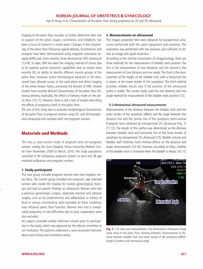
Measurements of the distance between the bladder neck and the lower border of the symphysis (dBNS) and the angle between this distance line and the central line of the symphysis (retro-vesical ß-degree) were obtained by transperineal 2D ultrasound (Fig. 1) [11,12]. The length of the urethra was determined as the distance between bladder neck and horizontal line at the lower border of symphysis by transperineal 2D ultrasound [13]. Bladder volume and bladder wall thickness have minimal effects on the distance and angle measurements [14-16]. However, according to Dietz, mobility of the bladder neck is increased when the bladder is empty [16,17].
Fig. 1. 2-D view and measurements. Two-dimensional ultrasound image (axial view) of the pelvic fl oor showing biometric measurements of dis-tance between bladder neck and lower border of the symphysis (dBNS), length of urethra and retrovesical angle.
WWW.KJOG.ORG422
KJOG Vol. 54, No. 8, 2011
The bladder volume and bladder wall thickness was measured as it related to detrusor overactivity and urinary incontinence [16,18].Bladder volume was measured with three parameters, includ-ing height (H), depth (D), and width (W). These measures were obtained from perpendicular planes (sagittal and transverse) by transperineal 2D ultrasound. In sagittal scanning, height and depth correspond to the greatest superior-inferior measurement and the greatest anterior-posterior measurement, respectively. Thus, the bladder volume (mL)=H × D × W × 0.7. The value of 0.7 is a correction factor for the nonspherical shape of a full bladder. This examination was performed as the bladder neck position is infl uenced by bladder volume. The bladder wall thickness was measured as a hypoechoic layer sandwiched between two hyper-echoic layers, the urothelium and perivesical tissue [19].
2) 3-dimensional ultrasound measurements
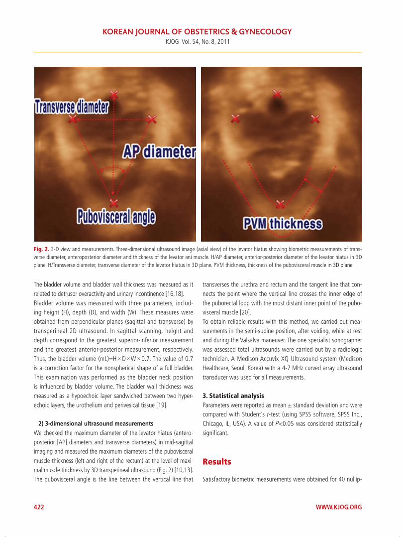
We checked the maximum diameter of the levator hiatus (antero-posterior [AP] diameters and transverse diameters) in mid-sagittal imaging and measured the maximum diameters of the pubovisceral muscle thickness (left and right of the rectum) at the level of maxi-mal muscle thickness by 3D transperineal ultrasound (Fig. 2) [10,13]. The pubovisceral angle is the line between the vertical line that
transverses the urethra and rectum and the tangent line that con-nects the point where the vertical line crosses the inner edge of the puborectal loop with the most distant inner point of the pubo-visceral muscle [20]. To obtain reliable results with this method, we carried out mea-surements in the semi-supine position, after voiding, while at rest and during the Valsalva maneuver. The one specialist sonographer was assessed total ultrasounds were carried out by a radiologic technician. A Medison Accuvix XQ Ultrasound system (Medison Healthcare, Seoul, Korea) with a 4-7 MHz curved array ultrasound transducer was used for all measurements.
3. Statistical analysisParameters were reported as mean ± standard deviation and were compared with Student’s t -test (using SPSS software, SPSS Inc., Chicago, IL, USA). A value of P<0.05 was considered statistically signifi cant.
Results
Satisfactory biometric measurements were obtained for 40 nullip-
Fig. 2. 3-D view and measurements. Three-dimensional ultrasound image (axial view) of the levator hiatus showing biometric measurements of trans-verse diameter, anteroposterior diameter and thickness of the levator ani muscle. H/AP diameter, anterior-posterior diameter of the levator hiatus in 3D plane. H/Transverse diameter, transverse diameter of the levator hiatus in 3D plane. PVM thickness, thickness of the pubovisceral muscle in 3D plane.

WWW.KJOG.ORG 423
Hye Ri Hong, et al. Characteristics of the pelvic fl oor during pregnancy by 2D and 3D ultrasound
arous pregnant women at term and 28 nulliparous non-pregnant women. Table 1 shows basic characteristics of study participants accord-ing to pregnancy status. There were no signifi cant differences in age and height between groups. The mean body mass index (BMI) was higher in the pregnant group compared to the non-pregnant group (29.10 ± 3.27 kg/m² vs. 22.04 ± 3.50 kg/m²; P ≤ 0.001).There was a no difference between pre-pregnant BMI and non-pregnant BMI (22.22 ± 3.00 kg/m² vs. 22.04 ± 3.50 kg/m²; P ≤ 0.537).The characteristics of the pelvic fl oor assessed by 2D ultrasonogra-phy between pregnant and non-pregnant women are summarized in Table 2. The length of urethra was longer in pregnant women at rest and during the valsalva maneuver compared with non-
pregnant women (3.54 ± 0.62 cm vs. 2.70 ± 0.73 cm; P ≤ 0.007) and (3.32 ± 0.57 cm vs. 2.55 ± 0.47 cm; P=0.005), respectively. The distance between the dBNS was shorter in pregnant women at rest and during the valsalva maneuver compared with non-pregnant women (0.68 ± 0.24 cm vs. 1.07 ± 0.30 cm; P=0.002 and 0.49 ± 0.11 cm vs. 0.99 ± 0.38 cm; P ≤ 0.001), respectively. The retro-vesical ß-degree, bladder thickness, and bladder vol-ume were not signifi cantly different between pregnant and non-pregnant women, both at rest and during the valsalva maneuver (P ≥ 0.05). Table 3 shows the characteristics of the levator hiatus obtained by 3D transperineal ultrasonography according to pregnancy status. The anteroposterior diameter of the levator hiatus was not sig-nifi cantly different between pregnant women and non-pregnant
Table 1. Basic Characteristics of study participants pregnant versus non-pregnant women
Parameters Non-pregnant women (n=28) Pregnant women (n=40) P-valuea
Age (yr) 32.18 ± 6.35 33.83 ± 3.56 0.437
Weight (kg) 52.10 ± 9.85 74.37 ± 7.21 < 0.001
Height (m) 159.44 ± 5.71 160.07 ± 5.01 0.689
Body mass index (kg/m2) 22.04 ± 3.50 29.10 ± 3.27 < 0.001 aP-value was calculated by Student’s t test and Chi-square test.
Table 2. Characteristics of the pelvic fl oor stratifi ed on 2-D transperineal ultrasound
Parameters Non-pregnant women (n=28) Pregnant women (n=40) P-valuea
Length of urethra (cm) 2.70 ± 0.73 3.54 ± 0.62 0.007
dBNS (cm) 1.07 ± 0.30 0.68 ± 0.24 0.002
Retro-vesical ß-degree (º) 125.65 ± 16.79 132.43 ± 20.35 0.412
Val/Length of urethra (cm) 2.55 ± 0.47 3.32 ± 0.57 0.005
Val/dBNS (cm) 0.99 ± 0.38 0.49 ± 0.11 < 0.001
Val/retro-vesical ß-degree (º) 135.57 ± 25.83 131.96 ± 20.84 0.694
Bladder thickness (cm) 0.32 ± 0.09 0.35 ± 0.70 0.352
Bladder volume (cm³) 1.05 ± 1.30 0.95 ± 0.58 0.767
dBNS, distance between bladder neck and lower border of the symphysis.aP-value was calculated by Student’s t test and Chi-square test.
Table 3. Characteristics of the pelvic fl oor levator hiatus on 3-D transperineal ultrasound
Parameters Non-pregnant women (n=28) Pregnant women (n=40) P-valuea
H/AP diameter (cm) 4.81 ± 0.65 5.08 ± 0.83 0.423
H/Transverse diameter (cm) 3.97 ± 0.67 3.45 ± 0.49 0.033
Pubovisceral angle (º) 60.67 ± 9.68 51.57 ± 8.52 0.026
Thickness of PVM (cm) 2.77 ± 0.16 1.03 ± 0.15 0.001
H/AP diameter, anterior-posterior diameter of the levator hiatus in 3D plane; H/Transverse diameter, transverse diameter of the levator hiatus in 3D plane; PVM thickness, thickness of the pubovisceral muscle in 3D plane.aP‐value was calculated by Student’s t test and Chi-square test.

WWW.KJOG.ORG424
KJOG Vol. 54, No. 8, 2011
women (P=0.423). The mean thickness of the pubovisceral muscle was greater in pregnant than non-pregnant women (1.03 ± 0.15 cm vs. 0.77 ± 0.16 cm; P=0.001) (Fig. 3). The transverse diam-eter of the levator hiatus and the pubovisceral angle of non-pregnant women were signifi cantly greater than pregnant women. (3.97 ± 0.67 cm vs. 3.45 ± 0.49 cm; P=0.033 and 60.67° ± 9.68° vs. 51.75° ± 8.52°; P= 0.026) (Figs. 4, 5).
Discussion
To our best knowledge, this is the first study to evaluate mor-phological characteristic of the pelvic floor in pregnant women using 2D- and 3D-transperienal ultrasound. Pregnant women had a significantly greater thickness of the pubovisceral muscle and decreased transverse diameter and pubovisceral angle. These changes reflect the fact that pregnant women have a smaller
Fig. 3. Thickness of PVM by 3D ultrasound. The mean the thickness of pubovisceral muscle were greater in pregnant women than non-pregnant (1.03±0.15 vs. 0.77±0.16 cm; P=0.001). Thickness of PVM, thickness of the PuboVisceral Muscle in 3D plane.
Non-pregnant womenTh
ickne
ss o
f PVM
(cm
)
1.4
1.2
1
0.8
0.6
0.4
0.2
0Pregnant women
P-value <0.05
Fig. 4. Transverse diameter by 3D ultrasound. The transverse diameter of levator hiatus is longer non- pregnant women than pregnant women (3.97±0.67 vs. 3.45±0.49 cm; P=0.033). H/Transverse diameter, transverse diameter of the levator hiatus in 3D plane.
Non-pregnant women
H/Tr
ansv
erse
dia
met
er (c
m)
5
4.5
4
3.5
3
2.5
2
1.5
1
0.5
0Pregnant women
P-value <0.05

WWW.KJOG.ORG 425
Hye Ri Hong, et al. Characteristics of the pelvic fl oor during pregnancy by 2D and 3D ultrasound
hiatal area than non-pregnant women. Morphological changes in the levator hiatus may have clinical signifi cance in the subsequent development of urinary incontinence and pelvic organ prolapsed [16,21]. The levator ani muscle is thought to play a signifi cant role in the pathogenesis of these highly prevalent conditions and it is estimated that parous women have an increased lifetime risk (by age 80) of undergoing surgical treatment for one of these condi-tions [22]. Pregnancy and childbirth are frequently cited as major etiological factors, and various obstetric parameters (e.g., length of second-stage labor, birth weight, and mode of delivery) have been demonstrated to be additional risk factors [23,24]. A poten-tial protective effect of Cesarean section could not be verifi ed in long-term studies, suggesting that pregnancy itself (especially the fi rst) causes pathological changes to the pelvic fl oor, regardless of the mode of delivery [24-26]. It has been suggested that the strain of the gravid uterus and hormonal changes during pregnancy lead to connective tissue remodeling and disruption of normal pelvic fl oor function; additional disruption may result during vaginal delivery from traumatic damage, primarily by vacuum or forceps extraction [27].It is interesting to note that the hiatal area at term was decreased. Although the reason for this change is unclear, a possible explana-tion is that during pregnancy, the volume and weight of the uterus increase and the pressure of the uterus and fetal presentation on the pelvic floor also increases [28]. Therefore, the decreased hiatal area may refl ect a compensatory mechanism counteracting these changes. Pregnancy itself may cause morphological changes of the pelvic fl oor to support the birth canal by closing the lower
end of the pelvic cavity as diaphragm. However, these changes may have some detrimental effects in pregnancy. During vaginal delivery, muscles of the levator hiatus are required to deform and stretch markedly. Using computer modeling, based on MRI, it has been found that some parts of the pubovisceral muscle stretch to 3.3 times their resting length during crowning of the head [29]. It is unlikely that the pelvic fl oor presents an absolute barrier to descent and delivery of the presenting part in any but a very small number of women. However, in the context of modern obstetric practice, a less compliant pelvic fl oor may provoke intervention by prolonging the second stage, slowing descent of fetal presentation, and predisposing to fetal distress. Interestingly, in a recent study of pelvic fl oor training in pregnancy, pelvic fl oor exercises were found to decrease the incidence of a prolonged second stage [30]. Therefore, further studies are needed to understand how the changes to the pelvic floor during pregnancy may influence the progress of labor. We found the distance between the bladder neck and the inferior border of the symphysis, both at rest and during the Valsalva maneuver, were significantly decreased in pregnant women compared with non-pregnant women. There was an increase in the length of the urethra, both during at rest and the Valsalva maneuver, in pregnant women. Modification of the anatomic relationship between the bladder and enlarged uterus results in shortening of the distance between the dBNS and increasing the length of the urethra. Recent technological advances in ultrasound imaging have re-sulted in three-dimensional pelvic floor imaging that is able to
Fig. 5. Pubovisceral angle by 3D ultrasound. The pubovisceral angle of non-pregnant women measured was statistically greater than pregnant women (60.67°±9.68° vs. 51.75°±8.52°; P=0.026).
Non-pregnant women
Pubo
visc
eral
ang
le (o )
80
70
60
50
40
30
20
10
0Pregnant women
P-value <0.05

WWW.KJOG.ORG426
KJOG Vol. 54, No. 8, 2011
demonstrate the levator muscle in the axial plane in a manner comparable to MRI [31]. The facility of real-time data capture also permits examination of functional anatomy observed during the Valsalva maneuver and pelvic fl oor muscle contraction. These fac-tors, as well as its relatively low cost and associated high patient compliance, mark ultrasound as an ideal method for further study of the levator in pregnancy [32]. In conclusion, pregnant women had significantly thicker levator ani muscles but smaller hiatal areas, as measured by the leva-tor hiatus angle and transverse diameter, than did non-pregnant women. Pregnancy itself may cause morphological changes of pelvic fl oor to support the birth canal by closing the lower end of the pelvic cavity as a diaphragm.
References
1. Fielding JR. Practical MR imaging of female pelvic fl oor weak-ness. Radiographics 2002;22:295-304.
2. Singh K, Reid WM, Berger LA. Magnetic resonance imaging of normal levator ani anatomy and function. Obstet Gynecol 2002;99:433-8.
3. Walters MD, Newton ER. Pathophysiology and obstetric issues of genuine stress incontinence and pelvic floor dysfunction. In: Walters MD, Karram MM, editors. Urogynecology and re-constructive pelvic surgery. 2nd ed. St. Louis: Mosby; 1999. p.135-44.
4. Maubon A, Aubard Y, Berkane V, Camezind-Vidal MA, Marès P, Rouanet JP. Magnetic resonance imaging of the pelvic fl oor. Abdom Imaging 2003;28:217-25.
5. Cunningham FG, Leveno KJ, Bloom SL, Hauth JC, Rouse DJ. Williams obstetrics. 23rd ed. New York: McGraw-Hill Profes-sional; 2009.
6. DeLancey JO, Kearney R, Chou Q, Speights S, Binno S. The appearance of levator ani muscle abnormalities in magnetic resonance images after vaginal delivery. Obstet Gynecol 2003;101:46-53.
7. Dietz HP, Haylen BT, Broome J. Ultrasound in the quantifi cation of female pelvic organ prolapse. Ultrasound Obstet Gynecol 2001;18:511-4.
8. Dietz HP. Levator function before and after childbirth. Aust N Z J Obstet Gynaecol 2004;44:19-23.
9. Margulies RU, Hsu Y, Kearney R, Stein T, Umek WH, DeLancey JO. Appearance of the levator ani muscle subdivisions in mag-netic resonance images. Obstet Gynecol 2006;107:1064-9.
10. Mørkved S, Salvesen KA, Bø K, Eik-Nes S. Pelvic fl oor muscle strength and thickness in continent and incontinent nullipa-rous pregnant women. Int Urogynecol J Pelvic Floor Dysfunct 2004;15:384-9.
11. Falkert A, Endress E, Weigl M, Seelbach-Göbel B. Three-dimen-sional ultrasound of the pelvic fl oor 2 days after fi rst delivery: influence of constitutional and obstetric factors. Ultrasound Obstet Gynecol 2010;35:583-8.
12. Schaer G, Koelbl H, Voigt R, Merz E, Anthuber C, Niemeyer R, et al. Recommendations of the German Association of Urogyne-cology on functional sonography of the lower female urinary tract. Int Urogynecol J Pelvic Floor Dysfunct 1996;7:105-8.
13. Dietz HP, Shek C, Clarke B. Biometry of the pubovisceral muscle and levator hiatus by three-dimensional pelvic floor ultrasound. Ultrasound Obstet Gynecol 2005;25:580-5.
14. Schaer GN, Koechli OR, Schuessler B, Haller U. Perineal ul-trasound: determination of reliable examination procedures. Ultrasound Obstet Gynecol 1996;7:347-52.
15. Mouritsen L, Bach P. Ultrasonic evaluation of bladder neck position and mobility: the infl uence of urethral catheter, bladder volume, and body position. Neurourol Urodyn 1994;13:637-46.
16. Tunn R, Petri E. Introital and transvaginal ultrasound as the main tool in the assessment of urogenital and pelvic floor dysfunction: an imaging panel and practical approach. Ultra-sound Obstet Gynecol 2003;22:205-13.
17. Dietz HP, Wilson PD. The infl uence of bladder volume on the position and mobility of the urethrovesical junction. Int Urogy-necol J Pelvic Floor Dysfunct 1999;10:3-6.
18. Tunn R, Schaer G, Peschers U, Bader W, Gauruder A, Hanzal E, et al. Updated recommendations on ultrasonography in urogyne-cology. Int Urogynecol J Pelvic Floor Dysfunct 2005;16:236-41.
19. Blatt AH, Titus J, Chan L. Ultrasound measurement of bladder wall thickness in the assessment of voiding dysfunction. J Urol 2008;179:2275-8.
20. Krofta L, Otcenásek M, Kasíková E, Feyereisl J. Pubococcygeus-puborectalis trauma after forceps delivery: evaluation of the levator ani muscle with 3D/4D ultrasound. Int Urogynecol J Pelvic Floor Dysfunct 2009;20:1175-81.
21. Costantini S, Nadalini C, Esposito F, Valenzano MM, Risso D, Lantieri P, et al. Perineal ultrasound evaluation of the ure-throvesical junction angle and urethral mobility in nulliparous women and women following vaginal delivery. Int Urogynecol J Pelvic Floor Dysfunct 2005;16:455-9.
22. Fialkow MF, Newton KM, Lentz GM, Weiss NS. Lifetime risk of surgical management for pelvic organ prolapse or uri-
WWW.KJOG.ORG 427
Hye Ri Hong, et al. Characteristics of the pelvic fl oor during pregnancy by 2D and 3D ultrasound
nary incontinence. Int Urogynecol J Pelvic Floor Dysfunct 2008;19:437-40.
23. Dannecker C, Anthuber C. The effects of childbirth on the pelvic-fl oor. J Perinat Med 2000;28:175-84.
24. Rortveit G, Brown JS, Thom DH, Van Den Eeden SK, Creasman JM, Subak LL. Symptomatic pelvic organ prolapse: prevalence and risk factors in a population-based, racially diverse cohort. Obstet Gynecol 2007;109:1396-403.
25. Viktrup L, Rortveit G, Lose G. Risk of stress urinary inconti-nence twelve years after the fi rst pregnancy and delivery. Ob-stet Gynecol 2006;108:248-54.
26. MacLennan AH, Taylor AW, Wilson DH, Wilson D. The preva-lence of pelvic fl oor disorders and their relationship to gender, age, parity and mode of delivery. BJOG 2000;107:1460-70.
27. Wijma J, Weis Potters AE, de Wolf BT, Tinga DJ, Aarnoudse JG.
Anatomical and functional changes in the lower urinary tract during pregnancy. BJOG 2001;108:726-32.
28. Wang LI, Ying LI. Effect of pregnancy on the pelvic fl oor mus-cle tonus. J Chinese Clin Med 2009;4:385-7.
29. Lien KC, Mooney B, DeLancey JO, Ashton-Miller JA. Levator ani muscle stretch induced by simulated vaginal birth. Obstet Gynecol 2004;103:31-40.
30. Salvesen KA, Mørkved S. Randomised controlled trial of pelvic fl oor muscle training during pregnancy. BMJ 2004;329:378-80.
31. Dietz HP. Ultrasound imaging of the pelvic fl oor. Part II: three-dimensional or volume imaging. Ultrasound Obstet Gynecol 2004;23:615-25.
32. Lanzarone V, Dietz HP. Three-dimensional ultrasound imaging of the levator hiatus in late pregnancy and associations with de-livery outcomes. Aust N Z J Obstet Gynaecol 2007;47:176-80.
2차원, 3차원 초음파를 통한 임신부의 골반저 형태학적 특징
고려대학교 의과대학 1구로병원, 2안산병원 산부인과
홍혜리1, 조금준1, 강애라1, 진혜미1, 오영택1, 오민정1, 김해중2
목적
이 연구의 목적은 2차원 초음파와 3차원 초음파를 사용하여 임신부와 비임신부와 비교를 통해 골반저의 형태학적 특징을 알아보고자 함
이다.
연구방법
본 연구는 40명의 만삭에 가까운 초산부와 28명의 임신을 경험한 적이 없는 여성을 대상으로 시행한 후향연구이다. 모든 대상자는 방광
을 비운 후 즉시 반 누움 자세를 취하게 한 후 2차원 및 3차원 경회음초음파를 시행하였다. 이를 통해 골반저의 특징을 표현할 수 있는 다
양한 변수가 측정되었다.
결과
본 연구에 의해 초산부에 있어 항문거근이 통계학적으로 두터워져 있음을 확인할 수 있었다(P<0.05). 골반거근열공의 각도와 횡단 길이
는 초산부에 있어 통계학적으로 작아지는 것을 확인할 수 있었다(P<0.05). 이에 반해 골반거근열공의 전후방향 길이는 두 군 간에 별다른
차이를 확인할 수 없었다.
결론
이번 연구를 통하여 임신부에 있어 항문거근은 두터워지고 골반거근열공의 각도와 횡단길이는 감소하는 것을 확인할 수 있었다. 이는 임
신 자체가 산도를 좀힘으로 인해 발생한 것으로 여겨진다. 앞으로 분만 후의 골반저의 변화를 평가하기 위한 2차원 및 3차원 경회음초음
파를 통한 연구가 시행되어야 할 것으로 생각된다.
중심단어: 초음파, 임신부, 골반저