Characteristics of Alveolar Inter-Cortical Osteotomy … · 2015-08-10 · Characteristics of...
10
© 2015. Ananian S.G., Zakaryan A.V., Gunko M.V. & Gvetadze S.R. This is a research/review paper, distributed under the terms of the Creative Commons Attribution-Noncommercial 3.0 Unported License http://creativecommons.org/licenses/by-nc/3.0/), permitting all non-commercial use, distribution, and reproduction in any medium, provided the original work is properly cited. Global Journal of Medical Research: J Dentistry and Otolaryngology Volume 15 Issue 2 Version 1.0 Year 2015 Type: Double Blind Peer Reviewed International Research Journal Publisher: Global Journals Inc. (USA) Online ISSN: 2249-4618 & Print ISSN: 0975-5888 Characteristics of Alveolar Inter-Cortical Osteotomy Technique on the Maxilla By Ananian S.G., Zakaryan A.V., Gunko M.V. & Gvetadze S.R. Russian Medical Academy of Postgraduate Education Studies, Russia Abstract- Main statements of inter-cortical osteotomy technique of the alveolar crest with its horizontal atrophy are envisioned. Criteria for patient selection for the technique utilization are presented. Some aspect of prophylaxis against postoperative adverse effects and complications are discussed. Keywords: alveolar crest splitting, vestibular advance-ment of the cortical plate, dental implant, osteoplastic material. GJMR-J Classification: NLMC Code: WU 240 CharacteristicsofAlveolarInterCorticalOsteotomyTechniqueontheMaxilla Strictly as per the compliance and regulations of:
Transcript of Characteristics of Alveolar Inter-Cortical Osteotomy … · 2015-08-10 · Characteristics of...

© 2015. Ananian S.G., Zakaryan A.V., Gunko M.V. & Gvetadze S.R. This is a research/review paper, distributed under the terms of the Creative Commons Attribution-Noncommercial 3.0 Unported License http://creativecommons.org/licenses/by-nc/3.0/), permitting all non-commercial use, distribution, and reproduction in any medium, provided the original work is properly cited.
Global Journal of Medical Research: J Dentistry and Otolaryngology Volume 15 Issue 2 Version 1.0 Year 2015 Type: Double Blind Peer Reviewed International Research Journal Publisher: Global Journals Inc. (USA) Online ISSN: 2249-4618 & Print ISSN: 0975-5888
Characteristics of Alveolar Inter-Cortical Osteotomy Technique on the Maxilla
By Ananian S.G., Zakaryan A.V., Gunko M.V. & Gvetadze S.R. Russian Medical Academy of Postgraduate Education Studies, Russia
Abstract- Main statements of inter-cortical osteotomy technique of the alveolar crest with its horizontal atrophy are envisioned. Criteria for patient selection for the technique utilization are presented. Some aspect of prophylaxis against postoperative adverse effects and complications are discussed.
Keywords: alveolar crest splitting, vestibular advance-ment of the cortical plate, dental implant, osteoplastic material.
GJMR-J Classification: NLMC Code: WU 240
CharacteristicsofAlveolarInterCorticalOsteotomyTechniqueontheMaxilla
Strictly as per the compliance and regulations of:
Characteristics of Alveolar Inter-Cortical Osteotomy Technique on the Maxilla
Ananian S.G. α, Zakaryan A.V. σ, Gunko M.V. ρ & Gvetadze S.R. Ѡ
Abstract- Main statements of inter-cortical osteotomy technique of the alveolar crest with its horizontal atrophy are envisioned. Criteria for patient selection for the technique utilization are presented. Some aspect of prophylaxis against postoperative adverse effects and complications are discussed. Keywords: alveolar crest splitting, vestibular advance-ment of the cortical plate, dental implant, osteoplastic material.
I. INTRODUCTION
mong various techniques of atrophied alveolar crest augmentation, currently a particular place is occupied by transversal expansion obtained by
corticotomies and vestibular advancement of the outer cortical plate in the edentulous region [1, 2, 3, 4, 5, 6].
Its advantages in acquiring long-term results in the field of dental implantation are described in numerous publications [7, 8, 9, 10]. Though it is reported about potential difficulties associated with attaining a primary stability of the dental implantand the optimal thickness of the covering soft tissues or the so-called biological width of the adjacent gingiva, which assumes an existence of a zone (surface) of connective and epithelial tissues attachments to the coronal part of the bony crest [11, 12, 13]. Controversial are the opinions about the structure of the dissected flap which is most favorable for the blood supply of the osteotomized fragment [13, 14], and the rationales for immediate soft-tissue deficit management [15, 16, 17].
In this paper the surgical stages of the alveolar inter-cortical osteotomy (AICO) are presented as well as patient selection principles and aspects of postoperative complications prophylaxis.
After local anesthesia is introduced the operation begins with a development of a compound flap (caudally a full-thickness and cranially – split-thickness) on the outer aspect of the alveolar crest in the site of the future implant placement, after that the inter-cortical osteotomy is utilized for providing the vestibular advancement of the outer cortical plate of alveolar thickness augmentation. Author
α σ
ρ
Ѡ:
Department of Consulting and Diagnostics, Central
Research Institute of Dentistry and Maxillofacial Surgery, Moscow, Russia, Department of Oral and Maxillofacial Surgery, Russian Medical Academy of Postgraduate Education Studies, Moscow, Russia.
e-mail: [email protected]
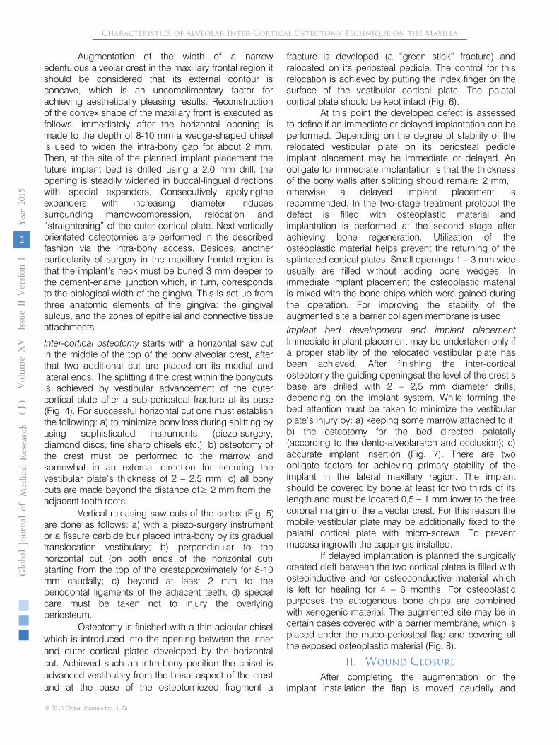
Incision and soft tissue stripping A linear horizontal incision is made: • within the keratinized fixed gingiva; • on the top or the palatal cranial margin of the crest
in the gap between the two neighboring teeth or the two purposed vertical incisions (if the site is edentulous);
• to the bone with the cutting of the periosteum; • at the gingival papillae of the adjacent teeth the
incisions should be performed in intrasulcal and marginal fashion with the gingival margin of the dental crown should be left intact (Fig. 1).
Technical note: an important condition in incision design is to avoid the coincidence of the osteotomy and the suture lines, this decreases the possibility of wound dehiscence and infection.
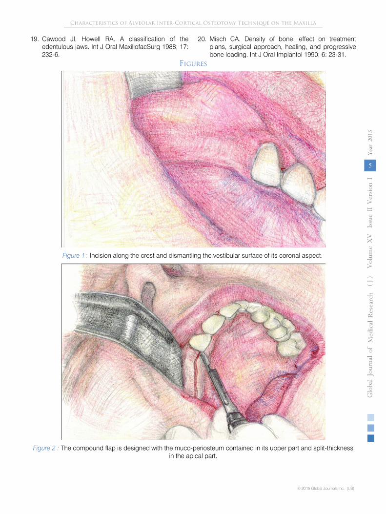
The dissection of the compound flap begins with dismantling of the bony alveolar crest in the extent of 3-4 mm on the vestibular (slightly beyond the muco-gingival junction) and palatal sides, as well as at the projection of the adjacent teeth. At this stage the coronal part of the vestibular flap containing the periosteum is established (Fig. 2). Next the periosteum is cut and the muscular attachments are divided along the whole length of the flap, the tip of the blade should permeate the wound for no more than 2 mm in the direction parallel to the surface of the bone (this is done to elude the undesired perforation of the mucous membrane which is particularly thin in these locations). Further flap elevation is performed: with sharp dissection; over the periosteum, i.e. in the submucous layer; to the level of the upper vestibule holding the scalpel parallel to the mobile mucous membrane. The larger apical part of the compound flap has the split-thickness surface approximately 6-7 mm wide. By keeping the periosteum intact the blood supply of the bone is preserved and the flap mobility is increased, this also helps to avoid placing the verticalmucosal releasing incisions, which is favorable for the trophism of the local soft tissues (Fig. 3).
If augmentation of the fixed gingiva (in cases where its thickness is >2 mm) and/or correction of gingival recession is desired, a bilaminar technique, described by Zucchelli G. (2013) may be utilized. In this technique a coronal advancement of the vestibular flap in conjunction with a free connective tissue graft is employed [18].
A
Globa
l Jo
urna
l of M
edical R
esea
rch
1
Volum
e XV
Issu
e II
Versio
n I
Year
2015
()
J
© 2015 Global Journals Inc. (US)
Augmentation of the width of a narrow edentulous alveolar crest in the maxillary frontal region it should be considered that its external contour is concave, which is an uncomplimentary factor for achieving aesthetically pleasing results. Reconstruction of the convex shape of the maxillary front is executed as follows: immediately after the horizontal opening is made to the depth of 8-10 mm a wedge-shaped chisel is used to widen the intra-bony gap for about 2 mm. Then, at the site of the planned implant placement the future implant bed is drilled using a 2.0 mm drill, the opening is steadily widened in buccal-lingual directions with special expanders. Consecutively applyingthe expanders with increasing diameter induces surrounding marrowcompression, relocation and “straightening” of the outer cortical plate. Next vertically orientated osteotomies are performed in the described fashion via the intra-bony access. Besides, another particularity of surgery in the maxillary frontal region is that the implant’s neck must be buried 3 mm deeper to the cement-enamel junction which, in turn, corresponds to the biological width of the gingiva. This is set up from three anatomic elements of the gingiva: the gingival sulcus, and the zones of epithelial and connective tissue attachments.
Inter-cortical osteotomy starts with a horizontal saw cut in the middle of the top of the bony alveolar crest, after that two additional cut are placed on its medial and lateral ends. The splitting if the crest within the bonycuts is achieved by vestibular advancement of the outer cortical plate after a sub-periosteal fracture at its base (Fig. 4). For successful horizontal cut one must establish the following: a) to minimize bony loss during splitting by using sophisticated instruments (piezo-surgery, diamond discs, fine sharp chisels etc.); b) osteotomy of the crest must be performed to the marrow and somewhat in an external direction for securing the vestibular plate’s thickness of 2 – 2.5 mm; c) all bony cuts are made beyond the distance of ≥ 2 mm from the adjacent tooth roots.
Vertical releasing saw cuts of the cortex (Fig. 5) are done as follows: a) with a piezo-surgery instrument or a fissure carbide bur placed intra-bony by its gradual translocation vestibulary; b) perpendicular to the horizontal cut (on both ends of the horizontal cut) starting from the top of the crestapproximately for 8-10 mm caudally; c) beyond at least 2 mm to the periodontal ligaments of the adjacent teeth; d) special care must be taken not to injury the overlying periosteum.
Osteotomy is finished with a thin acicular chisel which is introduced into the opening between the inner and outer cortical plates developed by the horizontal cut. Achieved such an intra-bony position the chisel is advanced vestibulary from the basal aspect of the crest and at the base of the osteotomiezed fragment a
fracture is developed (a “green stick” fracture) and relocated on its periosteal pedicle. The control for this relocation is achieved by putting the index finger on the surface of the vestibular cortical plate. The palatal cortical plate should be kept intact (Fig. 6).
At this point the developed defect is assessed to define if an immediate or delayed implantation can be performed. Depending on the degree of stability of the relocated vestibular plate on its periosteal pedicle implant placement may be immediate or delayed. An obligate for immediate implantation is that the thickness of the bony walls after splitting should remain ≥ 2 mm, otherwise a delayed implant placement is recommended. In the two-stage treatment protocol the defect is filled with osteoplastic material and implantation is performed at the second stage after achieving bone regeneration. Utilization of the osteoplastic material helps prevent the returning of the splintered cortical plates. Small openings 1 – 3 mm wide usually are filled without adding bone wedges. In immediate implant placement the osteoplastic material is mixed with the bone chips which were gained during the operation. For improving the stability of the augmented site a barrier collagen membrane is used. Implant bed development and implant placement Immediate implant placement may be undertaken only if a proper stability of the relocated vestibular plate has been achieved. After finishing the inter-cortical osteotomy the guiding openingsat the level of the crest’s base are drilled with 2 – 2,5 mm diameter drills, depending on the implant system. While forming the bed attention must be taken to minimize the vestibular plate’s injury by: a) keeping some marrow attached to it; b) the osteotomy for the bed directed palatally (according to the dento-alveolararch and occlusion); c) accurate implant insertion (Fig. 7). There are two obligate factors for achieving primary stability of the implant in the lateral maxillary region. The implant should be covered by bone at least for two thirds of its length and must be located 0,5 – 1 mm lower to the free coronal margin of the alveolar crest. For this reason the mobile vestibular plate may be additionally fixed to the palatal cortical plate with micro-screws. To prevent mucosa ingrowth the cappingis installed.
If delayed implantation is planned the surgically created cleft between the two cortical plates is filled with osteoinductive and /or osteoconductive material which is left for healing for 4 – 6 months. For osteoplastic purposes the autogenous bone chips are combined with xenogenic material. The augmented site may be in certain cases covered with a barrier membrane, which is placed under the muco-periosteal flap and covering all the exposed osteoplastic material (Fig. 8).
II. Wound Closure
After completing the augmentation or the implant installation the flap is moved caudally and
Characteristics of Alveolar Inter-Cortical Osteotomy Technique on the MaxillaGloba
l Jo
urna
l of M
edical R
esea
rch
2
Volum
e XV
Issu
e II
Versio
n I
Year
2015
()
J
© 2015 Global Journals Inc. (US)

without tension sutured to the wound margin with fine thread (5/0 or 6/0 diameter). At this stage the rebasing of the dentures is made to prevent the denture’s contacts with the wound (Fig. 9).
Patients should avoid using the removable dentures before sutures are kept (usually for 10-14 days postoperatively). Later on the dentures must be corrected not press on the surgery site.
III. Criteria for Patient Selection, Advantages and Disadvantages of
aico
The main indication for AICO is the existence of an edentulous site which possesses the following conditions:
1)
moderate atrophy of the residual crest in the horizontal plane*,IV class of Cawood
and Howell
classification (1988) [19] (to obviate small bone chips scabbing during osteotomy);
2)
sufficient vertical dimensions of the residual crest to assure the choice of optimal length of the implant;
3)
bone density of D2 – D4 according to Misch **[20];
4)
the presence of some marrow between the outer and inner cortical plates.
Considering the possibility of damaginglocal
tissue perfusion the procedure should planned in a limited zone, within 1-3 absent teeth, which approves the mobilization of a relative small fragment of bone. Depending on the thickness of the alveolar crest the one-staged or two-staged surgery is performed.
*Minimal preoperative thickness of the apical part of the alveolar crest in cases of delayed implant placement must be no less 3 mm, because for resorption prophylaxis the minimal thickness of the outer cortical plate of the developed intra-bony gap must comprise at least 2 mm.
**In this type of bone density alveolar splitting often can be performed with expanders only without vertical saw cuts.
Indications for delayed implant placement
•
Not possible to ensure the conditions for primary stabilization of the dental implant;
•
the thickness of the vestibular cortical plate after splitting is no less than 1,5 – 2 mm (the significant horizontal resorption of the crest may be noticed consequently to the paradontal pathology, tooth extraction and vestibulary directed teeth roots);
•
insufficient volume of soft tissue available for surgical defect coverage;
•
residual inflammatory process at the apex of the previously extracted tooth.
Delayed implant placement is performed after 4 – 6 months after the first operation.
Indications for immediate implant placement
• Sufficient height of residual bone (no less than 8 – 10 mm);
• enough bone in the basal part of the crest; • the alveolar width within 4 – 5 mm; • primary mechanical stabilization of the advanced
vestibular cortical plate. In the lateral maxillary region if vertical
dimensions of the alveolar crest are no less than 5 mm AICO can be combined by a sinus-lift with a window creation and antral floor augmentation and dental implant placement. This protocol is not utilized if the implant can not be optimally positioned in the palatal bony wall and/or the primary stability of the implant is unattainable.
Hence, AICOwith or without the use of osteoplastic material is performed for augmentation the thickness of an edentulous site of the alveolar crest to develop the conditions for dental implant placement.
IV. Contra-Indications
Utilization of AICO technique is unsuitable in the following conditions: a) vertical crest augmentation at the certain site; b) no marrow between the two cortical plates at the site; c) excessive horizontal atrophy of the crest (thickness less than 3 mm), as it may be a cause of improper implant positioning resulting in difficulties during the prosthetic stage; d) signs of maxillary sinus infection. Advantages and limitations of AICO technique 1. In most cases it helps to achieve widening (for 2 – 5
mm) of a narrow alveolar crest without a need for bone block grafting;
2. The following conditions are accomplished: a) for immediate implant placement (without clinical contra-indications); b) proper osseous regeneration, stable fixation and osseointegration of the installed implant; c) optimal positioning of the dental implant (in the view of future functional loading);
3. High aesthetic result is executed by: a) significant outer advancement of the vestibular cortical plate, possibility for an implant with larger diameter to be installed, new bone formation at the site of implantation and selective modeling of soft tissues; b) immediate increase of keratinized gingiva volume, muco-gingival junction line restoration, and creation of natural soft tissue profile around the neck of the implant and at the site of gingival papillae (if a connective tissue transplant is used);
4. Comparatively little time needed for osseous wound healing (usually take no more 4-6 months);
5. In the two-staged protocol AICO can be combined with a sinus-lift, because outer cortical plate advancement does not lead to significant deterioration of the blood supply to the bone.
Characteristics of Alveolar Inter-Cortical Osteotomy Technique on the Maxilla
Globa
l Jo
urna
l of M
edical R
esea
rch
3
Volum
e XV
Issu
e II
Versio
n I
Year
2015
()
J
© 2015 Global Journals Inc. (US)

The main disadvantage is that only horizontal dimensions can be augmented. Clinicians should also be aware of possible risk associated with technical details of the operation the dislocated bony fragment may experience resorption, in case of complete reattachment of the covering muco-periosteal flap.
Altogether, the use of the compound flap described in cases that allow larger apical parts of the vestibular cortical plate to be attached to periosteum allows to expand the indications for AICO, it means that immediate implant placement is possible if the cortical bony plates possess the thickness of 1 – 1,5 mm. But as far as a durable result is desired one must be sure the gingival thickness is no less 2 mm. Technical difficulty of the surgical interference is among the disadvantages of the method. The most difficult point is the dissection of the compound flap because in the zone of the mobile mucous membrane a risk exists to perforate it. Performance of the three saw cuts with consequent inter-cortical osteotomy is a tedious procedure, especially in сases of incorporated single tooth defects in the lateral mandibular region (because of the alveolar crest inclination and limited space for instrument usage). It is also worth to recommend that magnifying technique (binoculars or a an operating microscope) should be used during 6/0 thickness suture placement.
It must be accepted that for a certain group of patients the alveolar inter-cortical osteotomy is the most realistic treatment option which effectively can lead to optimal results. Deep knowledge of the anatomical relationships of bony and soft tissue structures, and analyzing clinical and radiographic details help to decrease the risks of unforeseen technical difficulties and undesired side effects in the postoperative period.
V. Acknowledgements
No conflict of interest declared among the authors.
References Références Referencias
1. Alfaro F. Bone plasty in dental implantation. Methods’ description and clinical application. Moscow: Azbuka; 2006. (inRussian).
2. Gonzales-Garcia R, Monje F, Moreno C. Alveolar split osteotomy for the treatment of the severe narrow ridge maxillary atrophy: a modified technique. Int J Oral Maxillofac Surg 2011; 40: 57-64.
3. Shcherchkov SV. Characteristic of inter-cortical osteotomy of the alveolar bone in dental implantation within conditions of bone tissue atrophy [dissertation]. Moscow: Central Research Institute of Dentistry and Maxillofacial Surgery, 2013. (in Russian).
4. Elian N, Jalbout Z, Ehrich B et al. A two-stage full-arch ridge expansion technique: review of literature
and clinical guidelines. ImplantDent 2008; 17: 16-22.
5. Jensen J, Sindet-Pederson S, Oliver AJ. Varying treatment strategies for reconstruction of maxillary atrophy with implants: results in 98 patients. J Oral Maxillofac Surg 1994; 52: 210-6.
6. Khoury F, Hensher R. The bony lid approach for the apical root resection of lower molars. Int J Oral Maxillofac Surg 1987; 16: 166-70.
7. Tolstunov L, Hicke B. Horizontal augmentation through the ridge-split procedure: a predictable surgical modality in implant reconstruction. J Oral Implantol 2013; 39: 59-68.
8. Kulakov AA, Losev FF, Gvetadze RS. Dental implantation. Moscow: MIA, 2006. (in Russian).
9. Chiapasco M, Ferrini F, Casentini P, Accardi S, Zaniboni M. Dental implants placed in expanded narrow edentulous ridges with the extension crest device. A 1-3- year multicenter follow-up study. Clin Oral Implants Res 2006; 17: 265-72.
10. Duncan JM, Westwood RM. Ridge widening for the thin maxilla: a clinical report. Int J Oral Maxillofac Implants 1997; 12: 224-7.
11. Cook DR, Mealey BL, Verret RG et al. Relationship between clinical periodontal biotype and labial plate thickness: an in vivo study. Int J Periodontics Restorative Dent 2011; 31: 345-54.
12. Esfahrood ZR, Kadkhodaseh M, Talebi Ardakani MR. Gingival biotype: a review. Gen Dent 2013; 61: 14-7.
13. Fu JH, Yeh CY, Chan HL, Tatarakis N, Leong DJ, Wang HL. Tissue biotype and its relation to the underlying bone morphology. J Periodontol 2010; 81: 569-74.
14. Berglundh T, Lindhe J. Dimension of the periimplant mucosa. Biological width revisited. J Clin Periodontol 1996; 23: 971-3.
15. Scipioni A, Bruschi GB, Calesini G, Bruschi E, De Martino C. Bone regeneration in the edentulous ridge expansion technique: histologic and ultrastructural study of 20 clinical cases. Int J Periodontics Restorative Dent 1999; 19: 269-77.
16. Calesini G., Micarelli C., Coppe S., Scipioni A. Edentulous site enhancement: a regenerative approach to the management of edentulous areas. Part 2: peri-implant tissues. Int J Periodontics Restorative Dent 2009; 29: 49-57.
17. Engelke WG, Diederichs CG, Jacobs HG, Deckwer I. Alveolar reconstruction with splitting osteotomy and micro-fixation of implants. Int J Oral MaxillofacImplants 1997; 12: 310-8.
18. Zucchelli G, Mazzotti C, Mounssif I, Marzadori M, Stefanini M. Esthetic treatment of peri-implant soft tissue defects: a case report of a modified surgical-prosthetic approach. Int J Periodontics Restorative Dent 2013; 33:327-35.
Characteristics of Alveolar Inter-Cortical Osteotomy Technique on the MaxillaGloba
l Jo
urna
l of M
edical R
esea
rch
4
Volum
e XV
Issu
e II
Versio
n I
Year
2015
()
J
© 2015 Global Journals Inc. (US)
19. Cawood JI, Howell RA. A classification of the edentulous jaws. Int J Oral MaxillofacSurg 1988; 17: 232-6.
20. Misch CA. Density of bone: effect on treatment plans, surgical approach, healing, and progressive bone loading. Int J Oral Implantol 1990; 6: 23-31.
Figures
Figure 1: Incision along the crest and dismantling the vestibular surface of its coronal aspect.
Figure 2 : The compound flap is designed with the muco-periosteum contained in its upper part and split-thickness in the apical part.
Characteristics of Alveolar Inter-Cortical Osteotomy Technique on the Maxilla
Globa
l Jo
urna
l of M
edical R
esea
rch
5
Volum
e XV
Issu
e II
Versio
n I
Year
2015
()
J
© 2015 Global Journals Inc. (US)
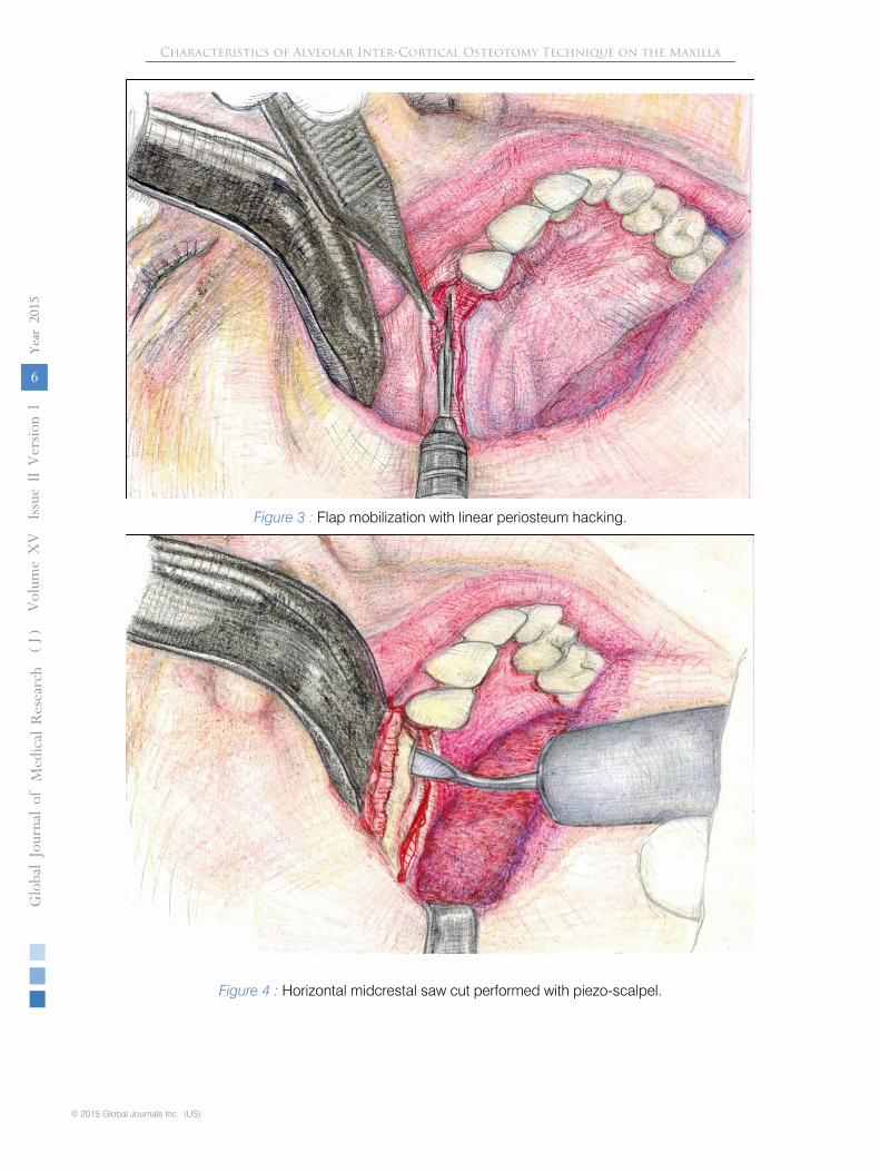
Figure 3 : Flap mobilization with linear periosteum hacking.
Figure 4 : Horizontal midcrestal saw cut performed with piezo-scalpel.
Characteristics of Alveolar Inter-Cortical Osteotomy Technique on the MaxillaGloba
l Jo
urna
l of M
edical R
esea
rch
6
Volum
e XV
Issu
e II
Versio
n I
Year
2015
()
J
© 2015 Global Journals Inc. (US)

Figure 5 : Vertical bone cuts through the cortical layer.
Figure 6 : Fine chisel is introduced in the saw cat for executing the final stage of the osteotomy.
Characteristics of Alveolar Inter-Cortical Osteotomy Technique on the Maxilla
Globa
l Jo
urna
l of M
edical R
esea
rch
7
Volum
e XV
Issu
e II
Versio
n I
Year
2015
()
J
© 2015 Global Journals Inc. (US)

Figure 7 : Dental implant placement.
Figure 8 : Bone deficiency is addressed by filling with osteoplastic material, a bioresorbing barrier membrane is arranged.
Characteristics of Alveolar Inter-Cortical Osteotomy Technique on the MaxillaGloba
l Jo
urna
l of M
edical R
esea
rch
8
Volum
e XV
Issu
e II
Versio
n I
Year
2015
()
J
© 2015 Global Journals Inc. (US)
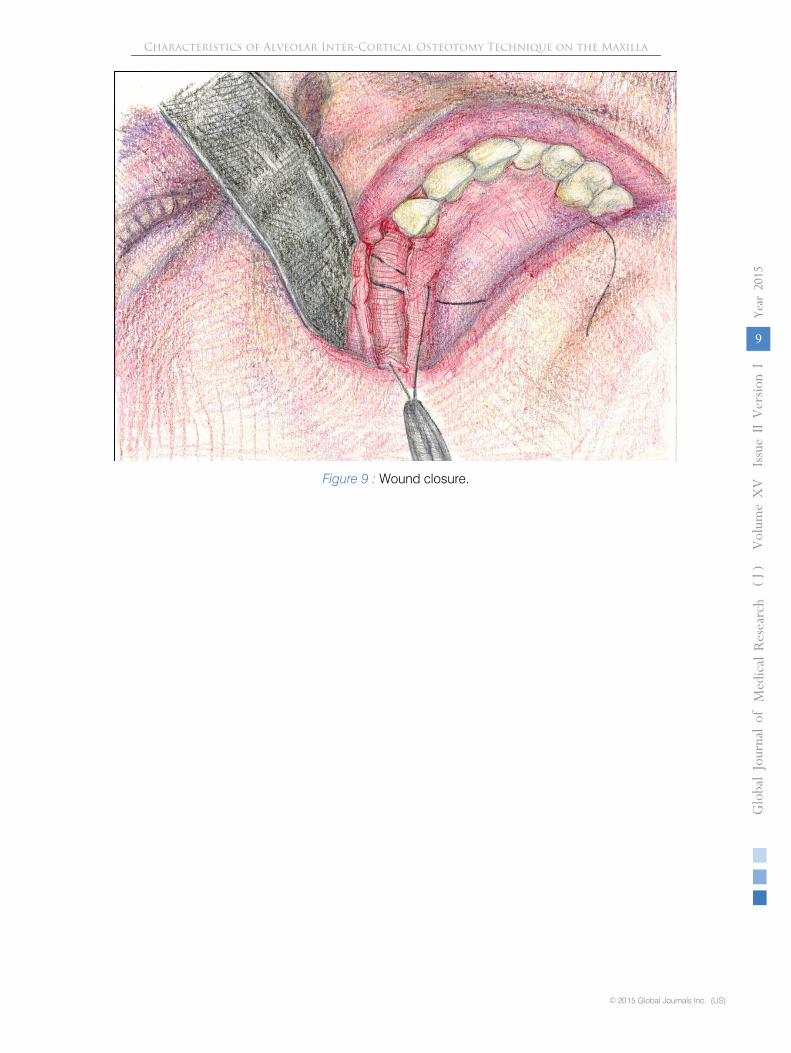
Figure 9 : Wound closure.
Characteristics of Alveolar Inter-Cortical Osteotomy Technique on the Maxilla
Globa
l Jo
urna
l of M
edical R
esea
rch
9
Volum
e XV
Issu
e II
Versio
n I
Year
2015
()
J
© 2015 Global Journals Inc. (US)