Chapters 29 and 30 - Essentials of Immunology
106
Essentials of Immunology Chapters 28 and 29
-
Upload
vinny-tran -
Category
Documents
-
view
37 -
download
3
description
immunology
Transcript of Chapters 29 and 30 - Essentials of Immunology

Essentials of ImmunologyChapters 28 and 29
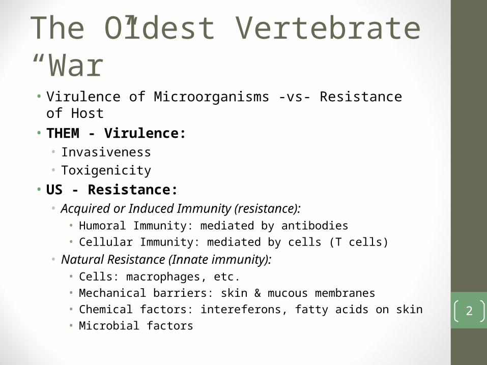
The Oldest Vertebrate “War”• Virulence of Microorganisms -vs- Resistance of Host• THEM - Virulence:• Invasiveness• Toxigenicity
• US - Resistance:• Acquired or Induced Immunity (resistance):
• Humoral Immunity: mediated by antibodies• Cellular Immunity: mediated by cells (T cells)
• Natural Resistance (Innate immunity):• Cells: macrophages, etc.• Mechanical barriers: skin & mucous membranes• Chemical factors: intereferons, fatty acids on skin• Microbial factors
2

Innate Immunity (Natural Resistance) is Non-Specific• Mechanical barriers• Skin: very few microorganisms penetrate• Mucous membranes: mucous traps many airborne bacteria;
coughing/sneezing expel a portion of the microbe-laden mucous into the atmosphere, the rest are carried into the stomach were high acidity and digestive enzymes kill many microbes• Found in nasal-pharyngeal region and the upper respiratory tract and
intestinal epithelium [areas open to outside] as well as genourinary• Much more susceptible to penetration than the unbroken skin• Chemical or physical damage of mucous membranes increases the
chance of infection and disease• Tears: flushing of eyes (contains lysozymes)• Respiratory tract: ciliated epithelium lines the walls; cilia beats
upward, push bacteria, mucous and other particulate material upward to where it is expelled in saliva and nasal secretions
3

Innate Immunity (Natural Resistance)• Chemical factors• Fatty acids: attack Gram-negative on skin surface• Bile salts: in gall bladder, liver, intestines inhibit Gram-positive• Lysozyme: in tears and saliva
• More effective against Gram-positive
• Phagocytins: found in leukocyte• Complement: microbial cell membranes and some cancer cells
can lose physical integrity and lyse in presence of complement• Interferon: active against viruses
4

Innate Immunity (Natural Resistance)• Microbial factors:• Normal flora of the body provides protection by competition
with pathogens• Example: Gram-positive making fatty acids on the skin
• If damaged by antibiotics, the individual may get a fungal infection
• Cellular factors:• Phagocytic cells may engulf microorganisms and destroy it
5

Non-Specific Immunity Phagocytes• On rare occasions, pathogens break through the physical and
chemical defenses one of the first cells that they encounter are class of cells called phagocytes (“cell that eats”)• The primary function of phagocytes is to engulf and destroy
pathogens• Phagocytes can also act as antigen-presenting cells (APCs) and
generate a peptide antigen(s) that will activate specific immune responses
• 3 main phagocytes: neutrophils (polymorphonuclear leukocytes), monocytes and macrophages• Neutrophils: actively motile granulocytes containing large
numbers of lysosomes6
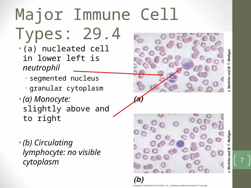
Major Immune Cell Types: 29.4• (a) nucleated cell in
lower left is neutrophil• segmented nucleus• granular cytoplasm
• (a) Monocyte: slightly above and to right
• (b) Circulating lymphocyte: no visible cytoplasm 7

Phagocyte• Toxic oxygen compounds
produced in phagocytes to destroy pathogens
• Toxic compounds include hydrogen peroxide (H2O2), superoxide anions (O2-), hydroxyl radicals (OH), singlet oxygen (1O2), hypochlorous acid (HOCl), and nitric oxide (NO)
8

Phagocytes: 3 Functions
• Phagocytosis: engulfment• Pathogen
Destruction: by the reactive oxygen • Antigen Processing:• Target cell lysis• Inflammation
9

Antigen ProcessingPresentation to TH2 cell to activate B cells plasma cells antibodies
10
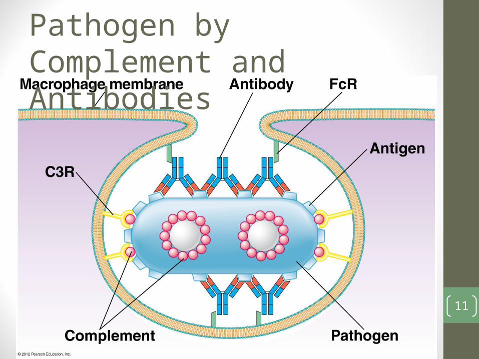
Destruction of Pathogen by Complement and Antibodies
11

Overview of Innate System
12
Induced Immunity (Induced Resistance)• Requires prior contact or exposure to antigen• Humoral Immunity: mediated by antibodies produced by B-
lymphocytes that differentiate into plasma cells that produce antibody
• Cellular Immunity: mediated by cells• Example: cytotoxic T-cells
13

Specific Immune Response• Humoral Immunity: Antibodies• B cells (lymphocytes) plasma cells Antibodies
• Cellular Immunity: T-Cells• Precursor T Lymphocytes antigen reactive/activated T-cells
• T-Cells:• CD4: T-helper (TH1 and TH2): helps or induces an immune response;
associated with MHC Class II• CD8: T-cytotoxic cells; associated with MHC class I
• Characteristics of specific immunity:1. Specificity2. Memory: Capacity to respond more quickly and vigorously after
exposure to an antigen3. Tolerance: acquired inability to make an immune response to
certain antigens
14

15

16

PAMP Interaction with PRR • PRR receptor on surface of
phagocyte• For fast and effective
interaction with a pathogen
• Facilitates recognition of pathogen associated molecular patterns (PAMP)• Pattern recognition
receptors (PRR): membrane bound phagocyte proteins• Interactions activate
phagocyte to ingest and destroy
17

Overview of Immune Response• Pathogens are targeted and destroyed by 3 immune
mechanisms:I. Innate immunity results from interactions between pathogen-
associated molecular patterns (PAMPS) found as cell surface components and pattern recognition receptors (PRRs) found on phagocytes (membrane bound phagocyte proteins)• Does not require previous exposure to pathogen• Mediated by phagocytes• PRRs first observed in Drosophila, called Toll receptors• Toll-like receptors on human phagocytes recognize specific
PAMPS • Ex. TLR-4, a PRR on human phagocytes recognizes & responds to
interactions with LPS, a PAMP in outer membrane of Gram-negative, inducing phagocyte activation and immunity to Gram-negative
18

Overview of Immune Response• Innate immunity continued:
• Interaction of a PAMP with the phagocyte PRR triggers a trans-membrane signal that results in transcription of a number of cell proteins in phagocyte
• This transcriptional activation leads to production of toxic oxygen compounds that lead to pathogen death • H2O2; O2
-; HOCl; NO; OH.; 1O2
• There’s an increase of O2 use which called respiratory burst
19

Inhibition of Phagocytosis• Staphylococcus aureus produces carotenoids that neutralize
singlet oxygen• Mycobacterium tuberculosis uses its cell wall glycolipids to
absorb hydroxyl free radicals and superoxide anions• Others produce leukocidins• Pathogen ingested and produces leukocidin that kills phagocyte
the pathogen is released• Dead phagocytes make up much of material of pus
• Staphylococcus aureus & Streptococcus pyogenes referred to as pyogenic (pus-forming) pathogens
• Streptococcus pneumoniae produces a bacterial capsule which prevents adherence of phagocyte to bacterial cell
• Streptococcus pyogenes produces M-protein that alters the surface of the bacterial cell and inhibits phagocytosis
20

Inflammation• Nonspecific reaction to stimuli such as toxins and pathogens• Causes redness (erythema), swelling (edema), pain, and heat
typically localized to site of infection• Molecular mediators of inflammation include proteins called
cytokines and chemokines produced by various immune cells but especially phagocytes and lymphocytes
• Inflammation is typical outcome of an innate or an adaptive immune response and typically protective
21

Inflammation• An effective response isolates and limits tissue damage• Rapid localization and destruction of pathogen by recruited
neutrophils and macrophages• Sometimes septic shock can occur if reaction is not localized
and spreads through entire circulatory and lymphatic system• Ex: Gram-negative bacteria - even though cleared, LPS interacts
with TLR-4 stimulating production of cytokines that induce systemic responses that can have serious consequences like increased vascular permeability leading to massive efflux of fluids from central vascular tissue: loss of blood pressure, severe edema in tissues, loss of blood volume
22

Overview of Immune Response• Pathogens are targeted and destroyed by 3 immune
mechanisms:I. Innate immunity results from interactions between pathogen-
associated molecular patterns (PAMPS) found as cell surface components and pattern recognition receptors (PRRs - membrane bound phagocyte proteins) found on phagocytes
• Adaptive immunity: due to antigen specific T cells, resulting in two distinct effector pathwaysII. Antibody mediated immunity (humoral immunity): results from
soluble antigen-specific antibody proteins, products of antigen stimulated B lymphocytes plasma cells antibody• TH2
III. Cell-mediated immunity: mediated by antigen specific T cells• TC and TH1
23

Origins of Cells of Immune Response• Lymphoid precursor can generate T and B cells• Myeloid precursor can generate• Monocytes which in turn develop into macrophages or dendritic
cells that are phagocyte cells involved in antigen uptake and display
• Granulocytes which include neutrophils, mast cells, eosinophils, and basophils• Neutrophils are also phagocytes• The others release their granule contents in response to pathogens,
pathogen products, or damaged host cells
24

Cells of Immune Response• Lymphoid Precursor:• Maturation in thymus: T cell• Maturation in bone marrow or fetal liver: B cell
25

Acquired Immunity: Induced • Acquired immunity is a state of altered responsiveness to a
specific substance induced by prior contact with the “foreign” substance• Ex: Contact with Corynebacterium diphtheriae• Anamnestic: has memory therefore repeated contact with
antigen amplifies response• Immunogens: are capable of eliciting an immune response
26

Acquired Immunity: Induced• Characteristics of immunogens:• Usually protein or complex polysaccharides (molecular
complexity) of a microbe, these are on its surface• Nucleic acids, simple polysaccharides, and lipids are typically poor
immunogens because they’re composed of repeating monomers
• High molecular weight: > 10,000 Daltons (10kDa)• Appropriate physical form: large, complex macromolecules in
insoluble or aggregate form (example proteins precipitated by heating) are good immunogens• Insoluble readily taken up by phagocytes, leading to immune
response
• Foreign to responder; not part of self (immunological tolerance to self antigens)
• Note: epitopes on antigen are recognized by antibody27

Antibody• Antibodies are synthesized by plasma cells in humoral
response and are capable of combining with the provoking antigen• 5 classes
• IgG, IgM, IgA, IgE, IgD: different heavy chains• Ex: IgG ( ɣ heavy chains); IgM ( chains)
• Fab & Fc portions• Also subclasses IgG ɣ1 ɣ2 ɣ3 ɣ4 heavy chains
28

More Terms• Immunogen: substance capable of eliciting an immune response• An antigen may or may not be an immunogen
• Antigens: substances that react with antibodies or TCRs (T cell receptors)• May not have actually induced/activated them
• Hapten: good example of antigen but not immunogen• Low molecular weight substance which can not act as an
immunogen by itself• When attached to high molecular weight materials, an antibody
response is made to the carrier (the high molecular weight material), as well as to the hapten complex is immunogenic but not hapten alone
• Next time hapten enters it acts as antigen and can react immediately with those antibodies made to its epitope(s) 29

Epitope• Antibody or TCR does not interact with the antigenic
macromolecule as a whole, but with distinct portions of the molecule called the epitope (formerly called antigenic determinative)• Antigens (most are immunogens) are complex; often >1 epitope
• Antibody specificity is sensitive enough to distinguish between two very similar epitopes• Ex: glucose from galactose which only differ in the orientation of a -OH
group
30
MHC Proteins: Antigen Presentation• MHC: Major Histocompatibility Complex (proteins)• Function as antigen presenting molecules and interact with
both antigen and later TCR (T Cell Receptor)• T cell cannot recognize foreign antigen unless it is presented in
context of an MHC protein• MHC: think of it as a platform that brings degraded antigens and
presents them to the TCR• MHC in humans have differences (polymorphism) which leads
to an issue during organ transplant• Two classes:
1)Class I: found on surface of all nucleated cells2)Class II: found only on surface of B lymphocytes, macrophages,
and dendritic cells, all dedicated APCs (antigen presenting cells)
31

MHC Class I• MHC I proteins are made and assembled
in the ER• Chaperon proteins stabilize MHC I until
antigen is bound• Protein antigens manufactured within
the cell (such as from viruses or tumors) are degraded by the proteasome in the cytoplasm, transported across the ER through a pore formed by TAP proteins
• They then bind to MHC Class I, transported to cell surface to bind with TCR (CD8 coreceptor makes binding stronger)
• TC cell then releases cytokines and cytotoxins to kill the target cell
32
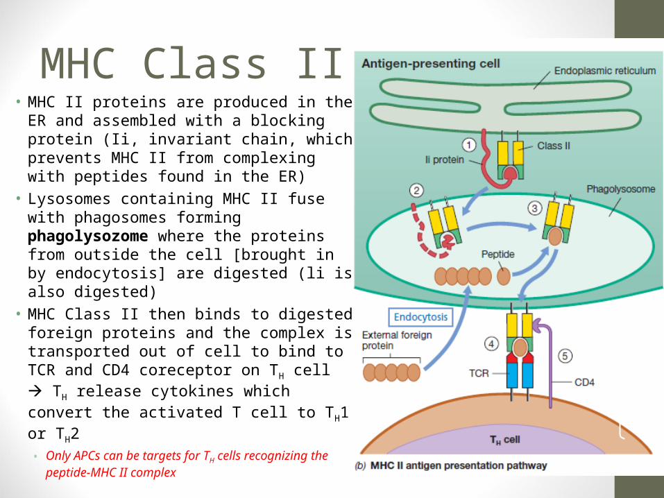
MHC Class II• MHC II proteins are produced in the ER and
assembled with a blocking protein (Ii, invariant chain, which prevents MHC II from complexing with peptides found in the ER)
• Lysosomes containing MHC II fuse with phagosomes forming phagolysozome where the proteins from outside the cell [brought in by endocytosis] are digested (li is also digested)
• MHC Class II then binds to digested foreign proteins and the complex is transported out of cell to bind to TCR and CD4 coreceptor on TH cell TH release cytokines which convert the activated T cell to TH1 or TH2• Only APCs can be targets for TH cells recognizing
the peptide-MHC II complex
33

Comparison MHC Class I & II
34

• Note: Peptide bound by both MHC I and TCR Proteins
35

T Cells: Cytotoxic & Helper• T-Cytotoxic Cells (TC): destroy cells that
display antigens embedded in MHC class I molecules
• Granules migrate to contact site and content is released
• Uses perforin (forms pore to deliver toxic enzymes) and granzymes (proteins) that enter through pore and cause apoptosis
• Intracellular pathogens (such as viruses) hide inside host cells, where they are protected from antibodies• Best killed when host cell sacrificed• Class I MHC found on all nucleated cells
and thus any infected cell can activate TC which kills infected cell
36

37

TH (THelper) Cells: TH1 and TH2• Macrophages: central role as APCs which bind,
process and present antigen to TH cells
• T-inflammatory (TH1) cells are activated by antigens presented on macrophages in the context of MHC Class II protein increased macrophage activity and inflammation
• TH1 cells then produce cytokines that stimulate macrophages to take up and kill certain foreign cells by themselves increased phagocytosis
• Activated macrophages can kill intracellular bacteria that would normally multiply
• TH1 also secrete IL-2 cytokine which is a 2nd signal needed for activation of TC cells• 1st signal was the interaction with MHC Class I
peptides on APC• Thus, TH1 involved in cellular immunity
38
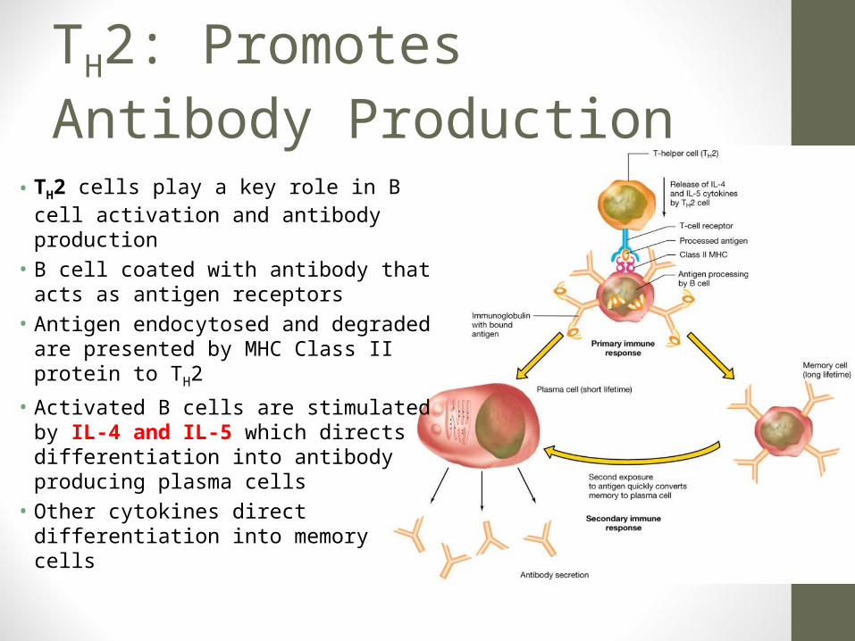
TH2: Promotes Antibody Production
• TH2 cells play a key role in B cell activation and antibody production
• B cell coated with antibody that acts as antigen receptors
• Antigen endocytosed and degraded are presented by MHC Class II protein to TH2
• Activated B cells are stimulated by IL-4 and IL-5 which directs differentiation into antibody producing plasma cells
• Other cytokines direct differentiation into memory cells
39

40

Summary• Intracellular pathogens (such as viruses) generally activate cell
mediated immunity by stimulating cytotoxic T cells (TC)
• Extracellular pathogens tend to activate humoral immunity (TH2)• TH2 activates B cells to make antibody
• TH1 activates macrophages to enhance killing of engulfed pathogen promotes inflammation• Also further stimulates TC cells
41

Antibodies (Immunoglobulins)• Antibodies (Ab)/immunoglobulins (Ig) are protein molecules that are
able to combine with antigenic determinates/epitopes• Found in the serum and in other body fluids such as gastric
secretions and milk• Serum containing antigen-specific antibody is often called antiserum• Comprised of 5 major classes on the basis of their physical,
chemical, and immunological properties• IgG, IgA, IgM, IgD and IgE
• About 80% of the serum antibodies are IgG proteins• Do not directly kill the pathogen• Mark it for destruction (opsonization) • IgM or IgG may attract the complement proteins• IgA blocks pathogen interaction with host cell 42

Opsonization
43

Immunoglobulins• All immunoglobulin classes have
variable domains• VH = variable heavy
• VL = variable light
• Both bind to antigens• All immunoglobulin classes have
constant domains• It is the difference in the amino
acid sequence of the constant domain that defines the class of immunoglobulin molecule• Gamma (), alpha (), mu (),
delta () or epilison ()
• FC crystallizes , Fab does not
44

Papain Reaction with IgG• Papain: nonspecific thiol-endopeptidase• Enzymatically cleaves IgG just above the hinge region to create 2
separate Fab fragments and 1 Fc • Fab fragments bind antigen; antibody-bivalent• Purification scheme shown below
45

Molecular weight/ Serum Antigen- Properties DistrubutionClass/Chain formula mg/mL binding sites
IgG (gamma) 150,000 2(H+L) 13.5 2
Major circulating Ab; 4 subclasses; activate complement
Extracelluar fluid;blood & lymph;
crosses placenta
IgM (mu)
970,000 (pentamer) ; 5[2(H+L)] + J;
175,000 (monomer) ; 2 (H+L)
1.5 0
10 (pentamer), 2 (monomer)
1st Ab to appear after immunization; strong complement
activator
Blood & lymph; monomer is B cell-
surface receptor
IgA (alpha)
150,000 (monomer) ; 2(H+L); 385,000 (secreted dimer) ; 2[2(H+L)+J+SC
3.5 0.05
2 (monomer), 4 (secreted)
Important circulating Ab: -
Major secretory antibody
Secretions (saliva, colostrum, cellular &
blood fluids); monomer in blood
and dimer in secretions
IgD (delta) 180,000, 2(H+L) 0.03 2 Minor circulating AbBlood & lymph; B
lymphocyte surfaces
IgE (epsilon) 190,000; 2(H+L) 0.00005 2
Involved in allergic reactions; C
Helper4 contains mast cell binding
fragment
Blood & lymph; binds to mast cell surfaces
Table 22.2 Properties of human Immunoglobulins
46

IgG Antibody• Most common of the circulating
antibodies• Composed of 4 polypeptide
chains• Interchain disulfide bridges (S-S)
connect the individual chains• A functional IgG molecule consists
of two antigen binding sites• IgG is therefore bivalent and can
bind two identical epitopes• Crosses the placental barrier• Subclasses: gamma 1, gamma 2,
gamma 3, gamma 4 47

IgM Antibody• Usually found as an aggregate of
five immunoglobulin molecules attached by at least one “J” (joining) chain
• Every heavy chain of IgM contains a fourth constant domain (CH4)
• 1st class of immunoglobulin made in a typical immune response to a bacterial infection
• 10 binding sites• Low affinity but high avidity• Monomers on B cell surface 48

IgA Antibody: Secretions• Present in the serum in the monomeric form, but in secretions it is
a dimer• Colostrum (breast milk), mucosal secretions of gastrointestinal, tears,
respiratory and genitourinary tracts• IgA dimer linked by disulfide bridges to J chain protein• Secretory piece wrapped around IgA dimer during secretion. • Total amount produced is twice the amount of IgG in serum
49
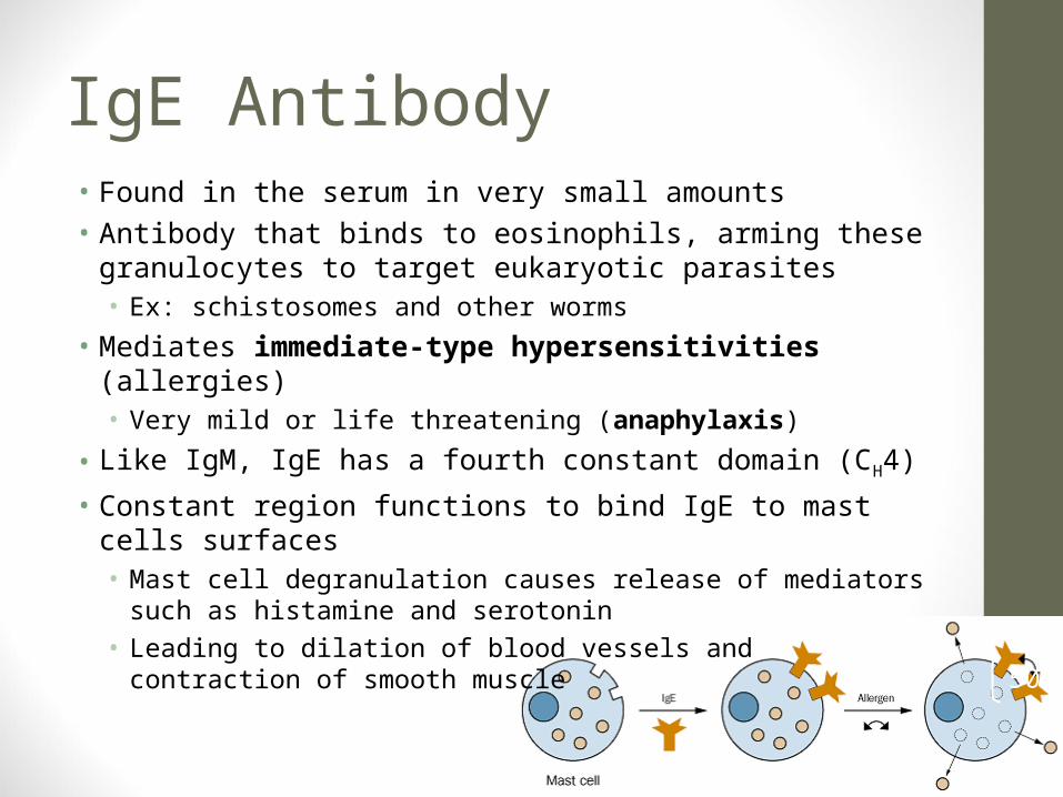
IgE Antibody • Found in the serum in very small amounts• Antibody that binds to eosinophils, arming these granulocytes
to target eukaryotic parasites• Ex: schistosomes and other worms
• Mediates immediate-type hypersensitivities (allergies) • Very mild or life threatening (anaphylaxis)
• Like IgM, IgE has a fourth constant domain (CH4)
• Constant region functions to bind IgE to mast cells surfaces• Mast cell degranulation causes release of mediators such as
histamine and serotonin• Leading to dilation of blood vessels and contraction of smooth
muscle 50

Immediate Hypersensitivity: Type I
51

Types of Hypersensitivity
52

IgD Antibody• Present in the serum in low concentrations• Has no know function• Abundant on the surfaces of B cells and plays a role along with
monomeric IgM in binding antigen to B cells • Especially abundant on memory cells
• "Immunoglobulin D enhances immune surveillance by activating antimicrobial, proinflammatory and B cell-stimulating programs in basophils“• 2009. Nat Immunol
• “Insights into the function of IgD.”• Dev Comp Immunol. 2011 Dec;35(12):1309-16. doi:
10.1016/j.dci.2011.03.002. Epub 2011 Mar 22.• Edholm ES, Bengten E, Wilson M.
53

Summary of Antibody ProductionI. Antigens are spread via lymphatic and blood circulatory
systems to neighboring secondary lymphoid organs [lymph nodes, spleen, or mucosal-associated lympoid tissue (MALT)]
II. Intravenously injected antigen travels via the blood to the spleen, where antibodies are formed
III. Subcutaneously, intradermally, topically, or intraperitoneally introduced antigens are carried by lymphatic system to the nearest lymph node
IV. Antigens introduced to mucosal surfaces (such as the mouth) are delivered to the GALT lining the intestinal tract resulting in antigen-specific IgA antibody production in the gut
54

Lymphatic System• Lymphatic system is part of the immune system,
made up of a network of conduits that carry a clear fluid called lymph
• It includes the lymphoid tissue and lymphatic vessels through which the lymph travels in a one-way system
• Lymph flows only towards the heart• Lymphoid tissue is found in many organs,
particularly the lymph nodes, and in the lymphoid follicles associated with the digestive system such as the tonsils
• The system also includes all the structures dedicated to the circulation and production of lymphocytes, which includes the spleen, thymus, bone marrow and the lymphoid tissue associated with the digestive system
• Movement of leukocytes from blood interstitial space lymphatic vessel blood is known as extravasation
55

Lymphatic System
56Primary lymphoid organs:1.Thymus2.Bone marrow

Antibody Production: Primary Response• Following initial antigen introduction, each antigen-stimulated B cell
(via T cell activation) multiplies and differentiates to form both antibody-secreting plasma cells and memory cells
• Plasma cells are short-lived (< 1 week) but produce large amounts of IgM antibody in this primary antibody response
• Following a latent period, specific antibodies show up in the blood, followed by a gradual increase in antibody titer (quantity), and then a slow fall in the primary response
57

Antibody Production: Secondary Response• Memory cells generated by the initial exposure to antigen may
live for several years• Upon re-exposure to the immunizing agent, memory cells need
no T cell activation quickly transform to plasma cells and begin producing IgG
• The antibody titer rises rapidly to a level 10-100x greater than the titer following the initial exposure• The rise in antibody titer is referred to as the secondary antibody
response (anamnestic)• The secondary response is characterized by a switch from IgM
to IgG production (class switching)• Over time, titer slowly decreases but a later exposure can cause
another secondary response• Basis for booster shots
58

59

60

Recognize Two Divisions of Immune ResponseFrom Nature and Experiments1. Humoral/antibodies Immunity:• B lymphocytes differentiate into plasma cells and produce
antibody• B lymphocytes mature in bone marrow/fetal liver
• Chickens - remove the bursa and they fail to produce antibodies and become susceptible to bacterial or extrinsic invaders
• Humans - Burton’s Agammaglobulinaemia: children fail to produce antibody. Humans do not have a bursa, but instead have GALT (gut associated lymphoid tissue) and MALT (mucosal cells that line external surfaces)
• In both of the above examples, cellular immunity remains essentially intact and they are more susceptible to bacterial infections rather than viral or intrinsic
61

Recognize Two Divisions of Immune ResponseFrom Nature and Experiments2. Cellular Immunity:
• Mediated by cells• Involve lymphocyte processing by thymus and activated T cells• Where antibody mediated (humoral) recognizes substances that
are outside host cells (extrinsic), cell-mediated immune response is more effective in recognizing modified host cells (intrinsic)
• It is important in controlling infections in which pathogens can reproduce within living cells• Ex: viruses, some bacteria like Rickettsia and Chlamydia, and some
parasitic protozoans like typanosomes• By TC and NK cells
• Also important in surveillance/destruction of malignant cells• Phagocytosis (destruction of extrinsic) and antigen presentation
62

Cellular Immunity: Natural Killer (NK) Cells• A fourth cell type (NK cells) are involved in innate immunity1)TH1: CD4; Class II MHC; 2nd signal activating cytotoxic T cells
(cytokines: IFN-γ, IL-2, TNF-α) inflammation increased phagocytosis
2)TH2: CD4; Class II MHC; B-cell helper (cytokines: IL-4, IL-5, IL-6)
3)TC: CD8; Class I MHC; killing virus-infected and cancer cells (cytokines: IFN-γ, TNF)
4)NK: class of lymphoid cells that instead of killing microbes destroys host cells that harbor microbes or have been transformed into cancer cells• Recognize changes in cell surface proteins of compromised cells
and then degranulate to release chemicals that kill them• Innate: reacts early, non-specific
63
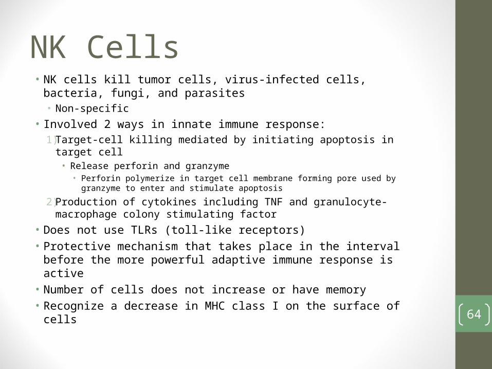
NK Cells• NK cells kill tumor cells, virus-infected cells, bacteria, fungi, and
parasites• Non-specific
• Involved 2 ways in innate immune response:1)Target-cell killing mediated by initiating apoptosis in target cell
• Release perforin and granzyme• Perforin polymerize in target cell membrane forming pore used by granzyme to
enter and stimulate apoptosis
2)Production of cytokines including TNF and granulocyte-macrophage colony stimulating factor
• Does not use TLRs (toll-like receptors)• Protective mechanism that takes place in the interval before the
more powerful adaptive immune response is active • Number of cells does not increase or have memory• Recognize a decrease in MHC class I on the surface of cells
64

Recognize Two Divisions of Immune Response: From Nature and Experiments• Cellular (continued):• Cell mediated immune responses include:
• Surveillance/destruction of malignant cells• Delayed hypersensitivity in response to intracellular bacteria like
Mycobacterium tuberculosis and fungal infections such as Histoplasma capsulatum
• Cytotoxic T lymphocyte (TC) responds to virally infected cells
• Response to tumor cells and tissue grafts by natural killer (NK) cells
65

Recognize Two Divisions of Immune Response: From Nature and Experiments• Cellular (continued): Experiments and nature• Thymectimized mice:
• More susceptible to viral infections or intrinsic parasites like M. tuberculosis
• Increased rate of tumor production• Failure to reject transplanted tissue
• Di George’s Syndrome (humans):• Infants lacking a functioning thymus gland (equivalent to
thymectimized mice above)• 10,000 times greater chance of malignancy but not susceptible to
bacterial “extrinsic” infection66

A Note
• Cellular immunity (TC mediated) and innate NK cells provide an immunosurveillance mechanism to rid the body of old, dead, damaged, or mutated (malignant) cells
• This mechanism is impaired in individuals on immunosupressive drugs, of old age or if chemicals are present (pollutants, drugs, radiation, UV, cigarette smoking, insecticides, etc)
67

68

Natural vs Artificial & Active vs Passive Acquired ImmunityI. Active Immunity: antibodies (Ab) made or T cells activated
in the individual upon contact with antigen (Ag)• Longer lasting memory cells1. Artificial: individual purposely exposed to controlled dose of
harmless Ag to induce artificial active immunity• Process known as vaccination
2. Natural: animals normally develop natural active immunity by acquiring a natural infection that initiates the adaptive immune response
II. Passive: cells or Ab from an immune individual are transferred to a nonimmune individual to prevent or help cure a disease 69

Outcome of Host-Pathogen Interaction• Vaccination: inoculation of host with inactive or weakened
pathogens or pathogen products to stimulate immunity• Attenuated strains: immunocompromised individuals?
• Chemically or physically inactivated strains (formaldehyde treated polio virus: Salk polio vaccine)
• Products of pathogens: some genetically engineered and produced in large quantities• Example: Tetanus toxoid - inactivated exotoxin• Example: Anthrax - protective factor
• New: recombinant vector vaccine• Example: Vaccinia - rabies vaccine used in animals (V-RG)
• New: DNA vaccines• Bacterial plasmid with cloned DNA injected intramuscularly into host
animal TC cells, TH1 cells, Ab made to protein encoded by cloned DNA
70

Attenuation• Attenuated strains have lost virulence• Often they retain immunogenicity, therefore they may be
used for production of vaccines• Especially viral vaccines: measles, mumps
• Laboratory cultivation typically results in a decrease in virulence of pathogens or even a complete loss
• Danger for immunocompromised individuals even though live cells or viruses are generally more effective than immunization with dead or inactivated
71

72

73

Examples of Passive Immunity• Passive: Ab or cells activated in one individual are transferred to
another• Short lived compared to active1. Artificial:
• Tetanus antiserum is given to passively immunize an individual suspected of being exposed to Clostridium tetani due to acute injury in car accident
• Contrast with vaccination (a prophylactic measure) with tetanus toxoid that actively immunizes individual for future encounter with Clostridium tetani exotoxin
• Pooled human gamma globulin given to patients exposed to hepatitis A
2. Natural: transfer occurs as part of a natural process• Placental transfer of IgG from mother to fetus• IgA transferred from mother to baby in breast milk
74

75

Antimicrobial Therapy: In Vivo• Antimicrobial agent is a natural or synthetic chemical that kills or
inhibits growth of microorganisms in vitro and in vivo• Chemotherapeutic agent are those used to treat microbial
disease and now also to prevent proliferation of malignant cells• An in vivo agent includes antibiotics: (Greek for “against life”)• Compounds produced by one species of microbe that can kill or
inhibit growth of other microbes (original definition)• Comprise the vast majority of chemotherapeutic agents• Today the term is commonly used for synthetic and semisynthetic
agents as well as true antibiotics• Synthetic: like sulfonamides• Semisynthetic: different penicillin derivatives
76

Hospital Acquired: Nosocomial Infections• Many hospital patients with noninfectious diseases (cancer
and heart disease) acquire microbial infections that produce disease because they are compromised
• Such health-care associated infections are called nosocomial infections
• Procedures like catheterization, hypodermic injection, spinal puncture, biopsy and surgery unintentionally introduce microbes into patient• Points out the need to control microbes
• Nosocomial are often antibiotic resistant
77

Control of Microorganisms• Physical Agents:• Most widely used is heat
• Chemical Agents:• Called antimicrobial agents
• Kill or Inhibit Growth:• Sterilization: treatment that frees the treated object of all living
organisms, including viruses• Death: defined as irreversible loss of ability to reproduce when
inoculated into an appropriate medium or host
78

Death as a Function of Temperature
• Death from heating is an exponential function• Occurs more rapidly as
the temperature rises
• Effectiveness is measured by the time required to decrease viability by 10x• Maximum temperature• Moist heat is better
than dry heat79

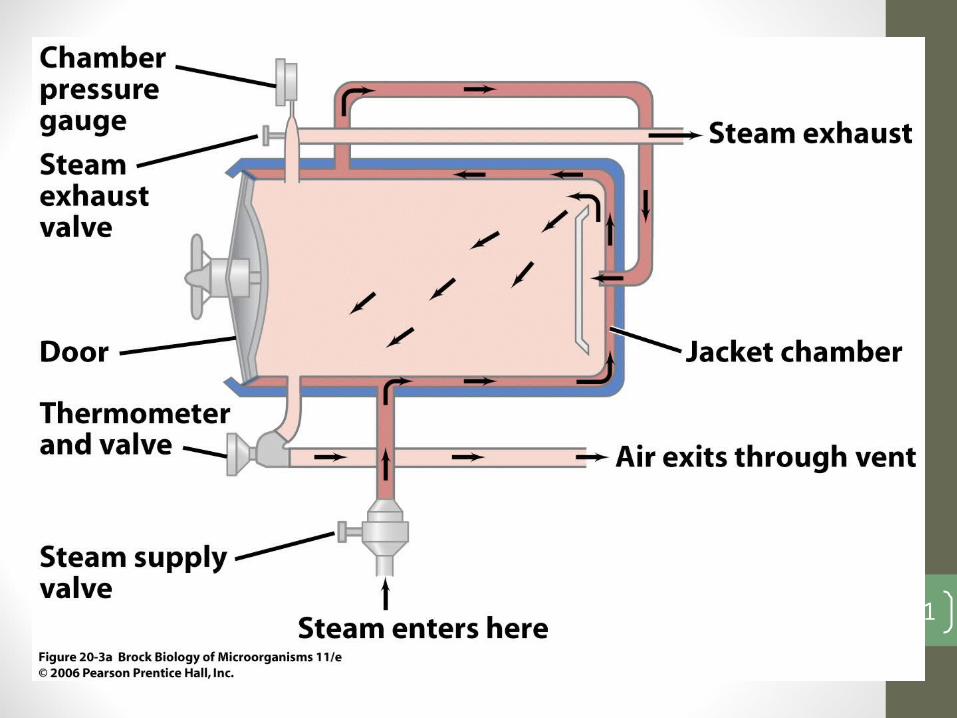
Autoclave• An autoclave permits application of steam heat under
pressure at temperatures above the boiling point of water, resulting in the killing of endospores
• At 15 lbs per sq inch above normal atmospheric pressure water boils at 121°C• At that temperature for 15 minutes, endospores are killed if
volume of liquid not too large• Ex. it takes 30 min for 2L flask
• High sugar and fat concentration in a media increase the resistance of an organism to heat treatment
• High salt concentration may increase or decrease the organism’s resistance to heat 80
81

Pasteurization• Pasteurization does not sterilize liquids but reduces microbial
load, killing most pathogens and inhibiting the growth of spoilage microorganisms• Originally: 30 min at 62°C• Now: Flash Pasteurization: 72°C for 15 sec.
82

Examples of Other Methods• Ultraviolet Light: • Leads to the production of thymine dimers• Most effective at 260 nm
• Ionizing Radiation:• Higher energy and shorter wavelength
• Membrane Filter Sterilization: • You’ve done it in lab!
83

Chemical Control: Antimicrobial Agents• Antimicrobial agent is a natural or synthetic chemical that kills
or inhibits growth of microorganisms• Kills: -cidal (viricidal, fugicidal, bactericidal)
• If kills by lysis: bacteriolytic
• Inhibits growth: -static (bacteristatic)• Two classes: In vitro and In vivo• In vitro:
• Disinfectants: chemicals that kill microorganisms but not necessarily spores
• Sanitizers: reduce to safe levels• Antiseptics or germicides: kill or inhibit growth but nontoxic enough
to be applied to human tissue84

85
• Bacteriostatic – inhibits protein synthesis, if agent concentration decreases the cell can grow again
• Bacteriocidal – kills the cell but does not destroy the cells
• Bacteriolytic – kills the cell but does destroy the cell, leading to release of cellular content

Methods to Evaluate• Agar Zone Diffusion:• Zones of inhibition
• Tube Dilution Test:• MIC: minimum inhibitory concentration (learned in lab)• Vary concentration of agent determines minimum required to
inhibit growth; not turbid• Bactericidal Tests: transfer to medium without agent and test
for growth• Phenol Coefficient Test: involves transfer to fresh medium
• Greatest dilution that kills microorganism at 10 minutes but not 5 to greatest dilution of phenol that kills microorganism in 10 but not 5 minutes
86

87

Kirby-Bauer Disk Susceptibility Test
• After incubating plates, the diameter of zone of inhibition is measured and results compared with a table listing whether a zone size is wide enough to be clinically useful• Larger zone of inhibition does not
always mean more effective• May be different solubility• In vivo versus in vitro
88

Tube Dilution Technique• MIC (Minimum Inhibitory Concentration): lowest
concentration of the agent that completely inhibits the growth of test organism• Bacteristatic Test: only growth inhibition
89

Antimicrobial Agents: In Vivo• Antibiotics: chemical substance produced by one
microorganism that kills or inhibits growth of another• Ex: Penicillin G
• Effective against Gram-positive, are Gram-negative impermeable• Inhibit cell wall synthesis
• Synthetic Antimicrobial Agents: created in hopes of finding a “magic bullet”• Ex: sulfanilamide
• Analog of p-aminobenzoic acid, a nucleic acid precursor• Inhibits nucleic acid synthesis by blocking the synthesis of folic acid• We obtain folic acid from our diet
• Semisynthetic: other penicillin with modified R group90

Ehrlich: Selective Toxicity:• Ability to inhibit or kill
pathogen without adversely affecting host• Ehrlich searched for the “magic
bullet”• Tested large numbers of dyes
that stained bacterial cells• Discovered first effective
antimicrobial agent, Salvarsan, arsenical agent effect in treatment of syphilis
91

Modern Era: Discovery of Penicillin
• Discovery: a great example of Serendipity in Science• Forgotten until Florey rediscovered
his work just before WWII• Originally recognized by Fleming in 1929
Photo of the plate with mold contamination
92

93

94

Mechanism of Action• Transpeptidases bind to penicillin and then cannot catalyze
crosslinking reactio• Cell wall synthesis continues but it cannot be crosslinked and
cannot maintain strength• Pen-transpeptidase complex stimulates autolysins that digest the
wall lysis• Known as PBP
• Cephalosporins: • Beta lactam ring and a six membered additional ring instead of the five ring thiazolidine ring• Semisynthetic and resistant to beta lactamases• Broader spectrum than penicillin
95

Next Discovery: Domagk 1935• Administered a dose of a red dye to his 6 year old daughter
who had a Streptococcal infection that had spread to her lymph nodes under arm• It became so severe that the arm needed amputation• She recovered
• 1928: dye Prontosil did not produce a zone of inhibition on inoculated plate• When tested in a mouse, the prontosil was very effective at
preventing infection• Prontosil sulfanilamide• Great against Gram-positive cocci• First commercially available antibiotic 96

Growth Factor Analogs: Sulfa Drugs
97

Mechanism of Action: Sulfa Drugs• Microbe makes folic acid from precursor para-aminobenzoic acid
(PABA), a vitamin necessary for nucleic acid synthesis• Sulfanilamide resembles PABA in structure, therefore competitive
inhibitor of enzyme that makes folic acid in bacteria• Folic acid is not synthesized by man and instead is obtained in diet• Bacteria do not transport folic acid and therefore sulfa drugs can
inhibit bacterial growth in humans without affecting human host• Today we have some resistant bacterial strains that have gained
ability to transport folic acid
98

Drug Targets
99

Antimicrobial Spectrum of Activity• Spectrum: range of microbes that a given drug affects• Penicillin G: narrow spectrum (only Gram-positive)• Ampicillin: broad spectrum penicillin (inhibits Gram-negative also)• Vancomycin: narrow spectrum
100

Ideal Properties
1. Selective toxicity• Toxic to microbe but not harmful to man and or higher animals• Some not selectively toxic:
• Tetracyclines: taken before adult teeth come in cause black teeth• Chlormaphenicol: implicated in aplastic anemia in children
2. Diffuses to site of infection3. Very few toxic side effects4. Allergic reactions not common5. Broad spectrum
101

Antivirals
102

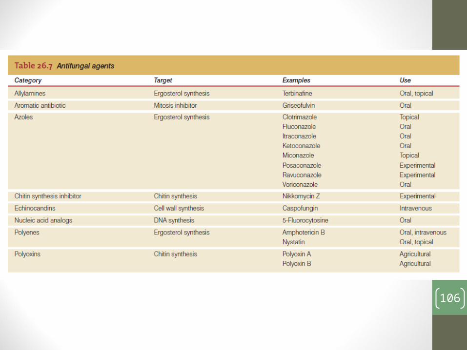
Antifungal Drugs• Fungi are Eukarya, therefore it is difficult to make drugs• Much of their machinery is the same as in animals and
humans• Drugs that affect a metabolic pathway often affect a
correlating pathway in the host cell• This makes the drug toxic
103

104

105
106