Chapter4 Protein Misfolding and Axonal Protection in ...neuroregeneration.org/Inoue Chapter...
13
Chapter 4 Protein Misfolding and Axonal Protection in Neurodegenerative Diseases Haruhisa Inoue, Takayuki Kondo, Ling Lin, Sha Mi, Ole Isacson and Ryosuke Takahashi Abstract Genetically engineered mouse model studies show that neuronal dysfunc- tion caused by protein aggregation/misfolding are reversible, indicating that injured neurons are alive even under disease states. Protein misfolding/aggregation in axons and distal dominant axonal degeneration are observed in a subgroup of degenerative diseases and in certain experimental conditions. Moreover, therapeutic approaches towards axonal protection are effective in neurodegenerative disease mouse models; (a) axonal regeneration, (b) anti-Wallerian degeneration, (c) autophagy enhance- ment, and (d) stabilization of microtubules. These studies demonstrate that axonal protection/functional repair of axons can be general therapeutic interventions for neurodegenerative diseases. 4.1 Neuronal Dysfunction in Neurodegeneration is a Reversible Process It had been believed that neurodegeneration is not reversible. However, recent stud- ies of transgenic mouse models, which express abnormal proteins associated with Alzheimer’s disease, diffuse Lewy body disease, Parkinson’s disease (PD), Hunting- ton’s disease (HD) and tauopathies such as frontotemporal dementia develop distinct disease-related neurological impairments, elegantly show that some neurological deficits of neurodegenerative cascades can be prevented or reversed by removing abnormal proteins, without obvious alteration of the number of neuronal cell bodies [1]. Thus, neurological impairments that are associated with neurodegenerative con- ditions might be caused by neuronal dysfunction to some extent rather than neuronal loss. These studies also demonstrate that symptoms arise from neuronal dysfunction which precedes neuronal death [1]. In HD mice model, as in most of the other triplet repeat diseases, the mutant huntingtin proteins form misfolded nuclear aggregates, H. Inoue (B) Department of Neurology, Kyoto University Graduate School of Medicine, 54 Kawahara-cho Shogoin, Sakyo-ku, Kyoto 606-8507, Japan e-mail: [email protected] J. Ov´ adi, F. Orosz (eds.), Protein Folding and Misfolding: Neurodegenerative Diseases, Focus on Structural Biology 7, DOI 10.1007/978-1-4020-9434-7 4, C Springer Science+Business Media B.V. 2009 97
Transcript of Chapter4 Protein Misfolding and Axonal Protection in ...neuroregeneration.org/Inoue Chapter...
Chapter 4Protein Misfolding and Axonal Protectionin Neurodegenerative Diseases
Haruhisa Inoue, Takayuki Kondo, Ling Lin, Sha Mi, Ole Isacsonand Ryosuke Takahashi
Abstract Genetically engineered mouse model studies show that neuronal dysfunc-tion caused by protein aggregation/misfolding are reversible, indicating that injuredneurons are alive even under disease states. Protein misfolding/aggregation in axonsand distal dominant axonal degeneration are observed in a subgroup of degenerativediseases and in certain experimental conditions. Moreover, therapeutic approachestowards axonal protection are effective in neurodegenerative disease mouse models;(a) axonal regeneration, (b) anti-Wallerian degeneration, (c) autophagy enhance-ment, and (d) stabilization of microtubules. These studies demonstrate that axonalprotection/functional repair of axons can be general therapeutic interventions forneurodegenerative diseases.
4.1 Neuronal Dysfunction in Neurodegenerationis a Reversible Process
It had been believed that neurodegeneration is not reversible. However, recent stud-ies of transgenic mouse models, which express abnormal proteins associated withAlzheimer’s disease, diffuse Lewy body disease, Parkinson’s disease (PD), Hunting-ton’s disease (HD) and tauopathies such as frontotemporal dementia develop distinctdisease-related neurological impairments, elegantly show that some neurologicaldeficits of neurodegenerative cascades can be prevented or reversed by removingabnormal proteins, without obvious alteration of the number of neuronal cell bodies[1]. Thus, neurological impairments that are associated with neurodegenerative con-ditions might be caused by neuronal dysfunction to some extent rather than neuronalloss. These studies also demonstrate that symptoms arise from neuronal dysfunctionwhich precedes neuronal death [1]. In HD mice model, as in most of the other tripletrepeat diseases, the mutant huntingtin proteins form misfolded nuclear aggregates,
H. Inoue (B)Department of Neurology, Kyoto University Graduate School of Medicine, 54 Kawahara-choShogoin, Sakyo-ku, Kyoto 606-8507, Japane-mail: [email protected]
J. Ovadi, F. Orosz (eds.), Protein Folding and Misfolding: Neurodegenerative Diseases,Focus on Structural Biology 7, DOI 10.1007/978-1-4020-9434-7 4,C© Springer Science+Business Media B.V. 2009
97

98 H. Inoue et al.
which are highly insoluble. The double mutant huntingtin transgenic mice, in whichthe bidirectional transgene expression is activated by the removal of doxycycline atbirth, express high levels of both mutant huntingtin and lacZ in the striatum, cortex,and hippocampus [2, 3]. Most of striatal neurons are stained with an anti-huntingtinantibody, showing diffuse nuclear aggregates. By 8 weeks of age, striatal morpho-logical alterations in the mutant huntingtin transgenic mice include a reduced size,reactive gliosis, and a decrease in D1 receptors (a feature seen in HD patients).All of the mice at this age also show a behavioral abnormality common to mousemodels of HD: when suspended by their tails, they clasp their limbs. This behavioralphenotype was aggravated over time. Neuropathological examination demonstratedthe colocalization of various molecular chaperones, ubiquitin, and proteasome sub-units with the aggregated proteins. Surprisingly, abolishing the expression of mutanthuntingtin by Cre-loxP system in mutant huntingtin transgenic mice with neurode-generative phenotype results in either a halt of the disease progression or a fullrecovery from the disease phenotype including pathological changes [2, 3]. Thisobservation indicates that irreversible changes that commit the neurons to persistentdysfunction or death do not necessarily take place in the neurodegenerative process.These observations suggest that therapeutic approaches aiming at elimination ofmisfolded proteins might be effective in treating neuronal dysfunction. Furthermore,the recovery from motor disturbances indicates that plastic changes can occur whenthe toxic insult ceases [3]. Recent studies of mutant huntingtin transgenic mice showthat the neuronal dysfunction may be caused by misfolded mutant huntingtin pro-tein, at synaptosomal proteasome and mitochondria, which seem to trigger viciouscycles of aberrant neuronal activity [4].
4.2 Neuronal Dysfunction Is Not Treatable by Anti-CellDeath Therapy
Although an important role of apoptosis is implicated in neurodegenerative diseases,data from both humans and animal models indicate that neurodegeneration is oftena long-lasting process that finish with cell death only after a prolonged period ofdisease state.
In PD model mice study, although peptide inhibitors of caspases block 1-methyl-4-phenylpyridinium (MPP+)-induced dopaminergic neuronal death, dopaminer-gic neuronal terminals are not rescued [5]. Similarly, adenovirus-mediated trans-gene expression of X-linked inhibitor of apoptosis protein (XIAP) blocks death ofdopaminergic neurons in a N-methyl-4-phenyl-1,2,3,6-tetrahydropyridine (MPTP)-induced PD mouse model, but does not prevent the decrease of dopaminergicterminal markers in the striatum [5]. Moreover the resistance of the dopaminergicneurons in the pro-apoptotic Bax protein knockout mice against MPTP toxicity isaccompanied by a significant, although less prominent, sparing of striatal dopaminecontents [6]. In the superoxide dismutase 1 (SOD1) transgenic mouse models ofamyotrophic lateral sclerosis (ALS) study, we have also shown that overexpression

4 Protein Misfolding and Axonal Protection 99
of XIAP in spinal motor neurons rescues cell bodies of motor neuron withoutinhibition of neuronal dysfunction [7]. Similarly, removal of Bax gene resulted incomplete rescue of cell bodies of motor neurons in ALS model mice, but denervationand axonal degeneration still occurred [8, 9]. Moreover with Bcl-2 transgenic micecrossed with pmn mice to block cellular apoptosis, motor neurons were completelyrescued, but motor axons degenerate to the same extent as in pmn mice with normallevels of Bcl-2, and there is no change in muscle strength or life span [9, 10]. Consis-tent with these findings, although an important role for synaptic caspase activationand apoptosis has been proposed, axonal degeneration after withdrawal of trophicsupport occurs without activation of caspases in contrast to cell death of the cellbody [9, 11]. These studies support the idea that axonal degeneration/dysfunctionmay proceed independently from the molecular events regulating cell death, andthat apoptosis plays a critical role in neuronal cell body death, and neuronal dys-function is not treatable by anti-cell death therapy. Therefore, anti-dysfunctiontherapies which target axonal degeneration/dysfunction are promising for treatmentof neurodegenerative diseases.
4.3 Morphological Aspects of Neuronal DysfunctionCaused by Protein Aggregation/Misfolding in HumanNeurodegenerative Disorder
It is hard to morphologically evaluate the neuronal dysfunction caused by pro-tein aggregation/misfolding in the central nervous system, because neurons possessintricate three-dimensional structure, and are embedded deep in the brain whichprevents accurate observation of cell shape. In contrast, morphological evaluation ofthe peripheral nervous system shows that degeneration of the cardiac sympatheticnerve occurs in PD and diffuse Lewy body disease, both of which are caused byaccumulation of misfolded α-synuclein, and that degeneration of their distal axonsprecedes loss of their neuronal cell bodies in the paravertebral sympathetic ganglia[12]. This interesting observation suggests that distal dominant axonal degenerationprecedes cell death not only in peripheral sympathetic, but central dopaminergicneurons of PD. Moreover, it is implicated that the centripetal degeneration mayrepresent the common pathological process underlying various neurodegenerativedisorders.
In ALS study, there are data supporting the hypothesis that the pathology of ALSstarts with distal axonal degeneration [9]. Neuropathological studies, by quantita-tive morphometry, demonstrate a distal-to-proximal gradient of axonal pathologyin phrenic nerves from ALS patients [9, 13]. Moreover, an autopsy case of anALS patient, who died unexpectedly during a minor surgical procedure, revealedsevere denervation and reinnervation changes demonstrated by electromyography,but there were no detectable changes in the corresponding spinal motor neuronalcell bodies [9, 14]. Threshold tracking, which measures axonal excitability, is analternative electrophysiological technique that demonstrates early abnormalities in
100 H. Inoue et al.
ALS patients. An apparent increase in persistent Na+ current and a decrease in K+conductance is observed in two ALS patients [9, 15, 16], and these changes are moreprominent distally than proximally [9, 17]. Genetical studies of ALS also providefurther evidence for the potential importance of axonal pathology in ALS [9, 18].
4.4 Protein Misfolding and Axonal Degenerationin Experimental Animal Models
Recent studies demonstrate that genetically engineered mice with misfolded proteinaccumulation display axonal degeneration phenotype [19].
One of the excellent examples is the knockout mouse of an essential autophagygene, Atg7, whose alterations have also been observed in several neurodegenerativediseases [20, 21]. Ablation of panneuronal autophagy causes ubiquitin-p62 positiveaggregation in neuronal cell body [20, 21]. Conditional knockout of Atg7 in Purkinjecells initially causes cell-autonomous, progressive dystrophy (manifested by axonalswellings) and degeneration of the axon terminals [22]. Consistent with suppres-sion of autophagy, no autophagosomes are observed in these dystrophic swellings[22]. Axonal dystrophy of mutant Purkinje cells proceeds with little sign of den-dritic or spine atrophy, indicating that axon terminals are much more vulnerable toautophagy impairment than dendrites. This early pathological event in the axonsis followed by Purkinje cell death. Furthermore, ultrastructural analyses of mutantPurkinje cells reveal an accumulation of aberrant membrane structures in the axonaldystrophic swellings, indicating that the autophagic machinery component Atg7 isrequired for membrane trafficking and turnover in the axons, and that impairmentof axonal autophagy as a possible mechanism for axonal degeneration associatedwith neurodegeneration [22]. Accordingly, significant accumulation of ubiquiti-nated proteins is noted in Atg7-deficient brain, but their levels, especially insolubleubiquitinated proteins, are lower than in Atg7-deficient liver, and formation ofthe inclusion is found in restricted groups of neurons. Several ubiquitin-positiveaggregates are recognized in Atg7-deficient brain regions in the presence of mildneuronal loss [22]. Direct degradation of aggregates/misfolded protein by autophagyis contradictory to the recent hypothesis that the generation of protein aggregatesrepresents a protective mechanism [23]. However, the primary targets of autophagyare likely to be diffuse cytosolic proteins, not inclusion bodies themselves, suggest-ing that inclusion body formation in autophagy-deficient cells is an event secondaryto impaired general protein turnover [23]. However, it is still possible that mis-folded proteins in soluble or oligomeric states could be preferentially recognized byautophagosomal membranes, which might also be mediated by ubiquitin–p62–LC3interactions [23, 24].
A recent study also showed that axonal degeneration is relevant to autophagycaused by protein mislocalization [25]. Adaptor protein-4 (AP-4) is a member ofthe adaptor protein complexes, which control vesicular trafficking of membraneproteins. Although AP-4 has been suggested to contribute to basolateral sorting
4 Protein Misfolding and Axonal Protection 101
in epithelial cells, its function in neurons is unknown. A recent study showedthat disruption of the gene encoding the β subunit of AP-4 resulted in increasedaccumulation of axonal autophagosomes, which contained alpha-amino-3-hydroxy-5-methyl-4-isoxazolepropionic acid (AMPA) receptors and transmembrane AMPAreceptor regulatory proteins (TARPs), in axons of hippocampal neurons and cere-bellar Purkinje cells both in vitro and in vivo [25]. AP-4 indirectly associateswith the AMPA receptor via TARPs, and the specific disruption of the interac-tion between AP-4 and TARPs causes the mislocalization of endogenous AMPAreceptors in axons of wild-type neurons. These results indicate that AP-4 may reg-ulate proper somatodendritic-specific distribution of its cargo proteins, includingAMPA receptor-TARP complexes and that protein mislocalization may disturb theautophagic pathway(s) in neurons [25].
4.5 Therapeutic Approaches to Treat Neuronal Dysfunctionby Axonal Protection
4.5.1 Axonal Regeneration
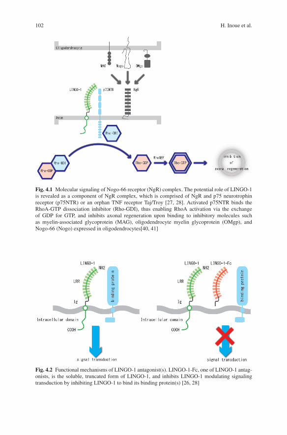
From previous studies showing that neuronal dysfunction, which may morpholog-ically reflect axonal degeneration by misfolded proteins, precedes neuronal celldeath, we hypothesized that axonal regeneration may protect axons from degenera-tion and have therapeutic effects against neuronal dysfunction in neurodegeneration[26]. We have tested this hypothesis using anti-LINGO-1 antagonists in experimen-tal PD models induced by either oxidative (6-hydroxydopamine) or mitochondrial(MPTP) toxicity [25]. LINGO-1 is the nervous system-specific leucine-rich repeatIg-containing protein, and associated with the Nogo-66 receptor (NgR) complexand is endowed with a canonical EGF receptor (EGFR)-like tyrosine phosphory-lation site, playing a critical role as an inhibitor of axonal regeneration (Fig. 4.1)[27, 28]. LINGO-1 antagonists, which block signal transduction of LINGO-1 com-plex (Fig. 4.2) [28], include decoy protein LINGO-1-Fc, Lenti-virus-dominantnegative LINGO-1, and anti-LINGO-1 blocking antibody. We examined the role ofLINGO-1 in cell damage responses of dopaminergic neurons. In LINGO-1 knock-out mice, dopaminergic neuronal survival is increased and behavioral abnormalitiesare reduced compared with wild-type ones. This neuroprotection is accompaniedby increased Akt phosphorylation [26]. Similar in vivo neuroprotective effectson midbrain dopaminergic neurons are obtained in wild-type mice by blockingLINGO-1 activity using LINGO-1-Fc protein which inhibit LINGO-1 function.Neuroprotection and enhanced neurite growth are also demonstrated for midbraindopaminergic neurons in vitro [26]. LINGO-1 antagonists improve dopaminergicneuronal survival in response to MPTP in part by mechanisms that involve activa-tion of the EGFR/Akt signaling pathway through a direct inhibition of the bindingLINGO-1 to EGFR (Fig. 4.3) [26]. LINGO-1 is also upregulated in compromised,probably dysfunctional, neurons in spinal cord injury [29] or kainic acid injection
102 H. Inoue et al.
Fig. 4.1 Molecular signaling of Nogo-66 receptor (NgR) complex. The potential role of LINGO-1is revealed as a component of NgR complex, which is comprised of NgR and p75 neurotrophinreceptor (p75NTR) or an orphan TNF receptor Taj/Troy [27, 28]. Activated p75NTR binds theRhoA-GTP dissociation inhibitor (Rho-GDI), thus enabling RhoA activation via the exchangeof GDP for GTP, and inhibits axonal regeneration upon binding to inhibitory molecules suchas myelin-associated glycoprotein (MAG), oligodendrocyte myelin glycoprotein (OMgp), andNogo-66 (Nogo) expressed in oligodendrocytes[40, 41]
Fig. 4.2 Functional mechanisms of LINGO-1 antagonist(s). LINGO-1-Fc, one of LINGO-1 antag-onists, is the soluble, truncated form of LINGO-1, and inhibits LINGO-1 modulating signalingtransduction by inhibiting LINGO-1 to bind its binding protein(s) [26, 28]

4 Protein Misfolding and Axonal Protection 103
Fig. 4.3 LINGO-1 effect on EGF receptor (EGFR). LINGO-1 binds EGFR, and regulates EGFRexpression level, leading to control axonal regeneration and neuronal survival via phosphorylationof Akt [26, 28]
[30]. We found that LINGO-1 expression is elevated in compromised, dysfunctionalneurons including in the substantia nigra of PD patients compared with age-matchedcontrols and in animal models of PD after neurotoxic lesions [25]. These resultsshow that inhibitory agents of LINGO-1 activity can protect dopaminergic neu-rons from degeneration caused by PD. It is necessary to test whether LINGO-1inhibition of function has protective effects on genetic PD models and/or otherneurodegenerative disease models in the future.
4.5.2 Anti-Wallerian Degeneration
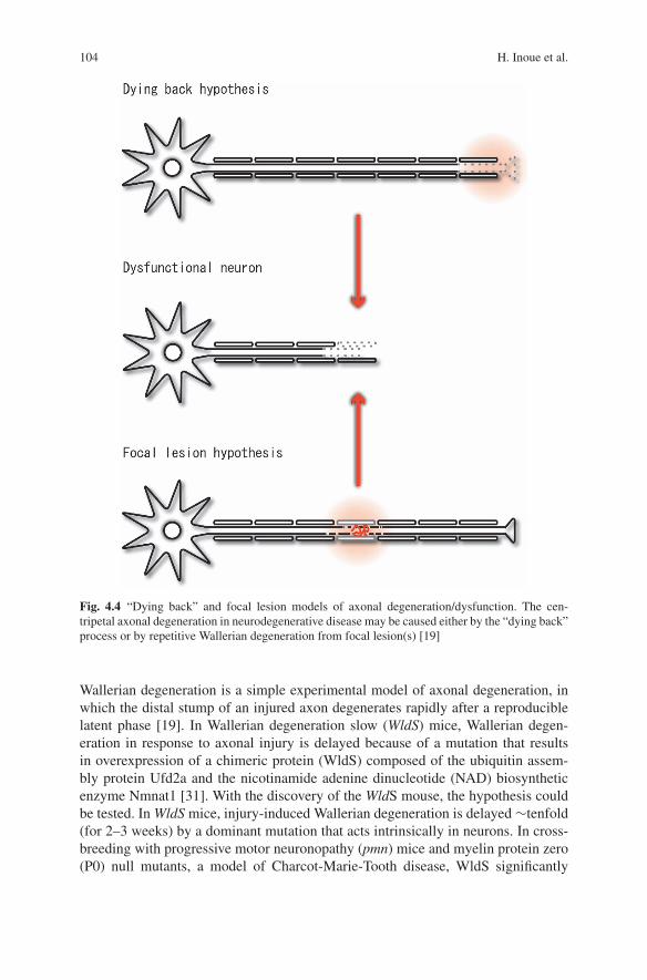
Axonal degeneration in “dying back” disorders seems to be different from Walleriandegeneration which is triggered by focal lesion (Fig. 4.4) [19]. However, appar-ent differences in the directionality of degeneration have been controversial [19].
104 H. Inoue et al.
Fig. 4.4 “Dying back” and focal lesion models of axonal degeneration/dysfunction. The cen-tripetal axonal degeneration in neurodegenerative disease may be caused either by the “dying back”process or by repetitive Wallerian degeneration from focal lesion(s) [19]
Wallerian degeneration is a simple experimental model of axonal degeneration, inwhich the distal stump of an injured axon degenerates rapidly after a reproduciblelatent phase [19]. In Wallerian degeneration slow (WldS) mice, Wallerian degen-eration in response to axonal injury is delayed because of a mutation that resultsin overexpression of a chimeric protein (WldS) composed of the ubiquitin assem-bly protein Ufd2a and the nicotinamide adenine dinucleotide (NAD) biosyntheticenzyme Nmnat1 [31]. With the discovery of the WldS mouse, the hypothesis couldbe tested. In WldS mice, injury-induced Wallerian degeneration is delayed ∼tenfold(for 2–3 weeks) by a dominant mutation that acts intrinsically in neurons. In cross-breeding with progressive motor neuronopathy (pmn) mice and myelin protein zero(P0) null mutants, a model of Charcot-Marie-Tooth disease, WldS significantly

4 Protein Misfolding and Axonal Protection 105
delayed axonal degeneration [19]. In the central nervous system, WldS also protectsagainst both genetic and toxic insults. Some nigrostriatal axons, which degeneratein PD, are spared and remain functional after 6-hydroxydopamine lesions in WldSmice [19, 32]. Axonal spheroids, which are presumably composed by misfoldedprotein(s) accumulation, are reduced in number in the gracile tract of mice withgracile axonal dystrophy (gad), deficient in ubiquitin carboxyterminal hydrolaseL1 (UCHL1) crossed with WldS mice [19, 33]. Not all axonal degeneration isdelayed by WldS. WldS have modest effect on the ALS model mice with protectionof the terminal axon only at its early stage [34, 35]. The failure to protect axonsunder certain circumstances indicates the existence of multiple axonal degenerationmechanisms. WldS may have protective effect(s) in rapidly degenerative or acutedisorders.
4.5.3 Autophagy Enhancement
It is reasonable to assume that autophagy could represent a therapeutic target foraxonal degeneration because the deletion of essential components of autophagycauses axonal degeneration, and relevance(s) of autophagy with degeneration areobserved in several neurodegenerative diseases [23]. Autophagy enhancement bythe regulatory protein kinase complex Target of Rapamycin (TOR) inhibitors suchas rapamycin and its analogue CCI-779 protects against neurodegeneration seen inpolyglutamine disease models in Drosophila and mice [23, 36]. A screened smallmolecule enhancers of rapamycin improve the clearance of mutant huntingtin andα-synuclein, and protect against neurodegeneration in a fruit-fly HD model [23, 37].These results provide us with the evidence supporting autophagy enhancement as atherapeutic strategy against the toxicity of misfolded proteins in neurodegenerativediseases.
Tsc2, also known as tuberin, is a GTPase activating protein that regulates theG protein Rheb, an activator of mTOR (mammalian Target of Rapamycin) [38].Tuberous sclerosis is a single-gene disorder caused by heterozygous mutations inthe TSC1 or TSC2 genes and is frequently associated with mental retardation, autismand epilepsy [38]. Even individuals with tuberous sclerosis and a normal intelli-gence quotient are commonly affected with specific neuropsychological problems,including long-term and working memory deficits [38]. Mice heterozygous for thedeletion of the Tsc2 gene in Tsc2(+/−) mice show deficits in learning and memory[38]. A recent study showed that hyperactive hippocampal signaling led to abnormallong-term potentiation in the CA1 region of the hippocampus and consequently todeficits in hippocampal-dependent learning in TSC mice [38]. Moreover, a brieftreatment with the mTOR inhibitor rapamycin in adult mice rescues not only thesynaptic plasticity, but also the behavioral deficits in this animal model of tuberoussclerosis, demonstrating that treatment with mTOR antagonists ameliorates cogni-tive dysfunction in the TSC mice model [38]. Autophagy may be included in axonand/or synaptic dysfunction in degeneration via the mTOR signaling pathway(s).
106 H. Inoue et al.
4.5.4 Stabilization of Microtubules
In tauopathy model(s), misfolded mutant tau protein causes axonal dysfunction/degeneration [39]. Microtubule-binding drugs can be therapeutically beneficial intauopathy models by functionally substituting for the microtubule-binding pro-tein tau, which is sequestered into inclusions of human tauopathies and transgenicmouse models [39]. Mutant tau transgenic mice treated with paclitaxel (Paxceed)showed that fast axonal transport in spinal axons is restored, and that microtubulenumbers and stable tubulins are increased compared with sham treatment [39].Moreover, Paxceed ameliorated motor impairments in tau transgenic mice [39].Thus, microtubule-stabilizing drugs have therapeutic potential for axonal dysfunc-tion/degeneration, in tauopathies by offsetting losses of tau function that result fromthe sequestration of this microtubule-stabilizing protein into filamentous misfoldedinclusions [39].
4.6 Concluding Remarks
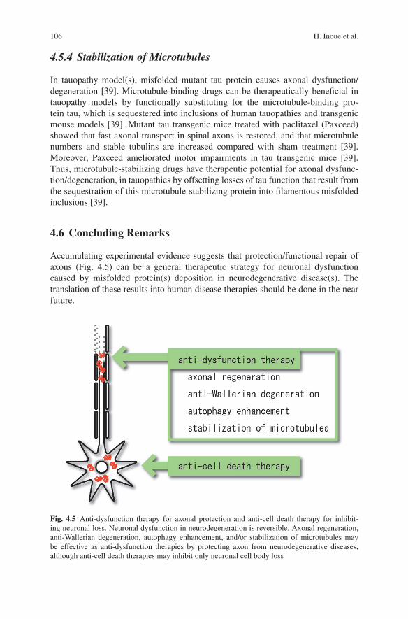
Accumulating experimental evidence suggests that protection/functional repair ofaxons (Fig. 4.5) can be a general therapeutic strategy for neuronal dysfunctioncaused by misfolded protein(s) deposition in neurodegenerative disease(s). Thetranslation of these results into human disease therapies should be done in the nearfuture.
Fig. 4.5 Anti-dysfunction therapy for axonal protection and anti-cell death therapy for inhibit-ing neuronal loss. Neuronal dysfunction in neurodegeneration is reversible. Axonal regeneration,anti-Wallerian degeneration, autophagy enhancement, and/or stabilization of microtubules maybe effective as anti-dysfunction therapies by protecting axon from neurodegenerative diseases,although anti-cell death therapies may inhibit only neuronal cell body loss

4 Protein Misfolding and Axonal Protection 107
Abbreviations
amyotrophic lateral sclerosis (ALS)alpha-amino-3-hydroxy-5-methyl-4-isoxazolepropionic acid (AMPA)adaptor protein-4 (AP-4)EGF receptor (EGFR)Huntington’s disease (HD)N-methyl-4-phenyl-1,2,3,6-tetrahydropyridine (MPTP)mammalian target of rapamycin (mTOR)Parkinson’s disease (PD)progressive motor neuronopathy (pmn)transmembrane AMPA receptor regulatory protein (TARP)leucine-rich repeat Ig-containing protein (LINGO-1)Wallerian degeneration slow (Wlds)
References
1. Palop JJ, Chin J, Mucke L (2006) A network dysfunction perspective on neurodegenerativediseases. Nature 443:768–773
2. Yamamoto A, Lucas JJ, Hen R (2000) Reversal of neuropathology and motor dysfunction ina conditional model of Huntington’s disease. Cell 101:57–66
3. Orr HT, Zoghbi HY. (2000) Reversing neurodegeneration: a promise unfolds. Cell 101:1–44. Wang J, Wang C-E, Orr A, Tydlacka S, Li S-H, Li X-J (2008) Impaired ubiquitin – proteasome
system activity in the synapses of Huntington’s disease mice. J Cell Biol 180:1177–11895. Eberhardt O,Coelln RV, Kugler S, Lindenau J, Rathke-Hartlieb S, Gerhardt E, Haid S,
Isenmann S, Gravel C, Srinivasan A et al. (2000) Protection by synergistic effects ofadenovirus-mediated X chromosome-linked inhibitor of apoptosis and glial cell line-derivedneurotrophic factor gene transfer in the 1-methyl-4-phenyl-1,2,3,6-tetrahydropyridine modelof Parkinson’s disease. J Neurosci 20:9126–9134
6. Vila M, Jackson-Lewis V, Vukosavic S, Djaldetti R, Liberatore G, Offen D, Korsmeyer SJ,Przedborski S (2001) Bax ablation prevents dopaminergic neurodegeneration in the 1-methyl-4-phenyl-1,2,3,6-tetrahydropyridine mouse model of Parkinson’s disease. Proc Natl Acad SciUSA 98:2837–2842
7. Inoue H, Tsukita K, Iwasato T, Suzuki Y, Tomioka M, Tateno M, Nagao M, Kawata A,Saido TC, Miura M et al. (2003) The crucial role of caspase-9 in the disease progressionof a transgenic ALS mouse model. EMBO J 22:6665–6674
8. Gould TW, Buss RR, Vinsant S, Prevette D, Sun W, Knudson CM, Milligan CE, Oppenheim(2006) Complete dissociation of motor neuron death from motor dysfunction by Bax deletionin a mouse model of ALS. J Neurosci 26:8774–8786
9. Fischer LR, Glass JD (2007) Axonal degeneration in motor neuron disease. Neurodegener Dis4:431–442
10. Sagot Y, Vejsada R, Kato A (1997) Clinical and molecular aspects of motoneurone diseases:animal models, neurotrophic factors and Bcl-2 oncoprotein. Trends Pharmacol Sci 18:330–337
11. Finn JT, Weil M, Archer F, Siman R, Srinivasan A, Raff MC (2000) Evidence that Walleriandegeneration and localized axon degeneration induced by local neurotrophin deprivation donot involve caspases. J Neurosci 20:1333–1341
12. Orimo S, Uchihara T, Nakamura A, Mori F, Kakita A, Wakabayashi K, Takahashi H (2008)Axonal α-synuclein aggregates herald centripetal degeneration of cardiac sympathetic nervein Parkinson’s disease. Brain 131:642–650

108 H. Inoue et al.
13. Bradley WG, Good P, Rasool CG, Adelman LS (1983) Morphometric and biochemical studiesof peripheral nerves in amyotrophic lateral sclerosis. Ann Neurol 14:267–277
14. Fischer LR, Culver DG, Tennant P, Davis AA, Wang M, Castellano-Sanchez A, Khan J, PolakMA, Glass JD (2004) Amyotrophic lateral sclerosis is a distal axonopathy: evidence in miceand man. Exp Neurol 185:232–240
15. Kanai K, Kuwabara S, Misawa S, Tamura N, Ogawara K, Nakata M, Sawai S, Hattori T,Bostock H (2006) Altered axonal excitability properties in amyotrophic lateral sclerosis:impaired potassium channel function related to disease stage. Brain 129:953–962
16. Vucic S, Kiernan MC (2006) Axonal excitability properties in amyotrophic lateral sclerosis.Clin Neurophysiol 117:1458–1466
17. Nakata M, Kuwabara S, Kanai K, Misawa S, Tamura N, Sawai S, Hattori T, Bostock H (2006)Distal excitability changes in motor axons in amyotrophic lateral sclerosis. Clin Neurophysiol117:1444–1448
18. Pasinelli P, Brown RH (2006) Molecular biology of amyotrophic lateral sclerosis: insightsfrom genetics. Nat Rev Neurosci 7:710–723
19. Coleman M (2005) Axon degeneration mechanisms: commonality amid diversity. Nat RevNeurosci 6:889–898
20. Hara T, Nakamura K, Matsui M, Yamamoto A, Nakahara Y, Suzuki-Migishima R, YokoyamaM, Mishima K, Saito I, Okano H et al. (2006) Suppression of basal autophagy in neural cellscauses neurodegenerative disease in mice. Nature 441:885–889
21. Komatsu M, Waguri S, Chiba T, Murata S, Iwata J, Tanida I, Ueno T, Koike M, UchiyamaY, Kominami E et al. (2006) Loss of autophagy in the central nervous system causesneurodegeneration in mice. Nature 441:880–884
22. Komatsu M, Wang QJ, Holstein GR, Friedrich VL Jr, Iwata J, Kominami E, Chait BT,Tanaka K, Yue Z. (2007) Essential role for autophagy protein Atg7 in the maintenance ofaxonal homeostasis and the prevention of axonal degeneration. Proc Natl Acad Sci USA 104:14489–14494
23. Mizushima N, Levine B, Cuervo AM, Klionsky DJ (2008) Autophagy fights disease throughcellular self-digestion. Nature 451:1069–1075
24. Komatsu M, Waguri S, Koike M, Sou Y, Ueno T, Hara T, Mizushima N, Iwata J, Ezaki J,Murata S et al. (2007) Homeostatic levels of p62 control cytoplasmic inclusion body formationin autophagy-deficient mice. Cell 131:1149–1163
25. Matsuda S, Miura E, Matsuda K, Kakegawa W, Kohda K, Watanabe M, Yuzaki M (2008)Accumulation of AMPA receptors in autophagosomes in neuronal axons lacking adaptorprotein AP-4. Neuron 57:730–745
26. Inoue H, Lin L, Lee X, Shao Z, Mendes S, Snodgrass-Belt P, Sweigard H, Engber T, Pepin-sky B, Yang L et al. (2007) Inhibition of the leucine-rich repeat protein LINGO-1 enhancessurvival, structure, and function of dopaminergic neurons in Parkinson’s disease models. ProcNatl Acad Sci USA 104:14430–14435
27. Bandtlow C, Dechant G (2004) From cell death to neuronal regeneration, effects of the p75neurotrophin receptor depend on interactions with partner subunits. Sci STKE 235:pe24
28. Mi S, Sandrock A, Miller RH (2008) LINGO-1 and its role in CNS repair. Int J Biochem CellBiol 40:1971–1978
29. Wang J, So K-F, McCoy JM, Pepinsky RB, Mi S, Relton JK (2006) LINGO-1 antagonistpromotes functional recovery and axonal sprouting after spinal cord injury. Mol Cell Neurosci33:311–320
30. Trifunovski A, Josephson A, Ringman A, Brene S, Spenger C, Olson L (2004) Neuronalactivity-induced regulation of Lingo-1. Neuroreport 15:2397–2400
31. Araki T, Sasaki Y, Milbrandt J (2004) Increased nuclear NAD biosynthesis and SIRT1activation prevent axonal degeneration. Science 305:1010–1013
32. Sajadi A, Schneider BL, Aebischer P (2004) WldS-mediated protection of dopaminergic fibersin an animal model of Parkinson disease. Curr Biol 14:326–330
33. Mi W, Beirowski B, Gillingwater TH, Adalbert R, Wagner D, Grumme D, Osaka H, ConfortiL, Arnhold S, Addicks K et al. (2005) The slow Wallerian degeneration gene, WldS, inhibitsaxonal spheroid pathology in gracile axonal dystrophy mice. Brain 128:405–416

4 Protein Misfolding and Axonal Protection 109
34. Fischer LR, Culver DG, Davis AA, Tennant P, Wang M, Coleman M, Asress S, Adalbert R,Alexander GM, Glass JD (2005) The WldS gene modestly prolongs survival in the SOD1G93AfALS mouse. Neurobiol Dis 19:293–300
35. Vande Velde C, Garcia ML, Yin X, Trapp BD, Cleveland DW (2004) The neuroprotectivefactor WldS does not attenuate mutant SOD1-mediated motor neuron disease. NeuromolecularMed 5:193–204
36. Ravikumar B, Vacher C, Berger Z, Davies JE, Luo S, Oroz LG, Scaravilli F, Easton DF, DudenR, O’Kane CJ et al. (2004) Inhibition of mTOR induces autophagy and reduces toxicity ofpolyglutamine expansions in fly and mouse models of Huntington disease. Nat Genet 36:585–595
37. Sarkar S, Perlstein EO, Imarisio S, Pineau S, Cordenier A, Maglathlin RL, Webster JA, LewisTA, O’Kane CJ, Schreiber SL et al. (2007) Small molecules enhance autophagy and reducetoxicity in Huntington’s disease models. Nat Chem Biol 3:331–338
38. Ehninger D, Han S, Shilyansky C, Zhou Y, Li W, Kwiatkowski DJ, Ramesh V, Silva AJ (2008)Reversal of learning deficits in a Tsc2 + /− mouse model of tuberous sclerosis. Nat Med.14:843–848
39. Zhang B, Maiti A, Shively S, Lakhani F, McDonald-Jones G, Bruce J, Lee E B, Xie S X,Joyce S, Li C et al. (2005) Microtubule-binding drugs offset tau sequestration by stabilizingmicrotubules and reversing fast axonal transport deficits in a tauopathy model. Proc Natl AcadSci USA 102:227–231
40. Yamashita T, Tohyama M (2003) The p75 receptor acts as a displacement factor that releaseRho from Rho-GDI. Nat Neurosci. 6:461–467
41. Hasegawa Y, Yamagishi S, Fujitani M, Yamashita T (2004) p75 neurotrophin receptorsignaling in the nervous system. Biotechnol Annu Rev.10:123–149