CHAPTER - Shodhgangashodhganga.inflibnet.ac.in/bitstream/10603/36982/14/14...Chapter 5 131 of...
33
CHAPTER 5 Genetic Polymorphism of PPO in Different Brinjal Cultivars
Transcript of CHAPTER - Shodhgangashodhganga.inflibnet.ac.in/bitstream/10603/36982/14/14...Chapter 5 131 of...

CHAPTER 5
Genetic Polymorphism of
PPO in Different Brinjal
Cultivars

Chapter 5
129
5.1. Introduction
Many cultivars of brinjal (Solanum genus) are available with varying morphological
features such as colours (purple, green, purple with white and green stripes and patches),
shapes (ovoid, obovate, oblong, cylindrical, club shaped), and calyx (spiny, non-spiny)
(Raigón et al., 2008). These cultivars differ in the extent of post-cut browning which
could be due to variations in PPO activity or level of soluble phenolics. The PPO enzyme
causes enzymatic browning in fruits and vegetables and is coded by ppo gene. PPO
activity could vary depending upon the gene sequence or epigenetic factors. Recently, the
PPO gene has been reported to exist in many isoforms and found to be expressed in
different plant parts under stress conditions like wounding (Shetty et al., 2011). Similar
studies were also reported in potato (Newman et al., 1993) and tomato (Thygesen et al.,
1995) and many other fruits and vegetables. The study of the gene sequence and its
analysis in different cultivars would help understand the unique nature of protein
sequence that may have a bearing on the enzyme activity and overall browning. The
molecular studies on ppo gene were undertaken to understand the basics, which could be
helpful in exploring the possibility to inhibit this enzyme in food processing applications.
There are a few reports about cloning and expression of PPO gene in bacteria. None of
these studies have reported enzymatically active recombinant PPO in E.coli (Sullivan et
al., 2004). In the current study, the PPO gene has been cloned and expressed in E.coli
with the aim to produce the protein in high amounts for its further characterization.

Chapter 5
130
5.2. Materials and methods
5.2.1. Chemicals
Chloroform, isoamyl alcohol, ethyl alcohol, agarose, agar powder, Luria broth, plate
count agar were of molecular biology grade and procured from Himedia Laboratories,
Mumbai, Maharashtra, India. The Taq DNA polymerase, dNTP mix, cDNA synthesis kit,
TriPure RNA isolation reagent (containing guanidine thiocyanate and phenol) reagent,
rapid DNA ligation kit were procured from Roche applied sciences, Mannheim,
Germany. The restriction endonuclease (NdeI and XhoI) were procured from New
England Biolabs, Ipswich, MA.
5.2.2. Brinjal
The six major popular cultivars of brinjal (Solanum melongena) ‘Pusa purple long’(V1),
‘Ravaiya’(V2), ‘Azad kranti’(V3), ‘Kalpatharu’(V5), ‘Raveena’(V6), and ‘Anupam’(V7)
(listed in Table 2.1) were procured from a local supplier at Mumbai, India. For cloning
and expression studies of PPO only the cultivar showing maximum activity (‘Kalpatharu’
V5) was used.
5.2.3. DNA isolation
The genomic DNA was isolated using CTAB (cetyl trimethyl ammonium bromide)
method to avoid phenolics interference (Rogers and Bendich, 1994). The whole brinjal
fruit was frozen in liquid N2 and ground to fine powder. An aliquot (100 mg) was added
to sterile 2 mL centrifuge tubes containing 1 mL of preheated (65 ºC) extraction buffer
composed of 0.1 M Tris HCl, pH 8.0; 1.4 M NaCl; 20 mM EDTA; 2%(w/v) CTAB and
1% PVP (polyvinyl polypyrrolidone). Tubes were held for 15 min and then equal volume

Chapter 5
131
of chloroforml/isoamyl alcohol (24:1; v/v) was added, mixed thoroughly and centrifuged
(5810R, Eppendorf, Hamburg, Germany) at 11000 g for 5 min. The aqueous top layer
was mixed gently with 10% CTAB and equal volume of chloroform/isoamyl alcohol
(24:1; v/v) and kept at -20 ºC for 30 min. The mixture was centrifuged (15000 g, 35 min)
and the pellet was dissolved in high salt Tris (10 mM)-EDTA (1 mM) buffer having 1M
NaCl. The precipitation was carried out with absolute ethanol followed by washing with
70% ethanol. The pellet was dried at 65 ºC and dissolved in low salt TE buffer (1 mM
Tris, 0.1 mM EDTA pH 8.0).
5.2.4. RNA isolation
The RNA isolation was performed with guanidine thiocyanate-phenol-chloroform
extraction method using TriPure isolation reagent (Chomczynski, P. 1993). Whole brinjal
fruits procured at early stage of maturation were frozen in liquid nitrogen and ground to
fine powder. An aliquot (100 mg) of this powder was transferred to a sterile 2 mL
centrifuge tube containing 1 ml of readymade TriPure isolation reagent (Roche Applied
Science) (containing guanidine thiocyanate and phenol), thoroughly mixed, and incubated
for 5 min. Later 0.2 ml of chloroform was added and the suspension was centrifuged at
12000 g for 15 min. In a separate vial the aqueous top layer was mixed with isopropanol
(0.5 ml), incubated for 10 min, and later centrifuged at 12000 g for 10 min. The pellet
was resuspended in 70% ethanol (1 ml), precipitated, washed in absolute ethanol, air
dried, and finally solubilized in DEPC treated RNAse free water. This was treated with
DNAse to avoid any DNA contamination. The pure total RNA thus prepared was used for
cDNA synthesis using a cDNA synthesis kit (Roche Applied Sciences).

Chapter 5
132
Table 5.1. The oligonucleotides used for the PCR amplification of PPO gene.
Primer name* Nucleotide sequence
P1. For. end 5’-CACTCCTAAGCCCTCTCAACTTTTC- 3’ (25 bp)
P2. Rev. end 5’-CCAACAGTTCCGTTATCGCC-3’ (20 bp)
P3. For. int. 5’-GTAACTAATGCTCCATGTCCTC-3’ (22 bp)
P4. For. int. 5’-ACCATGGCGTAACTTCAAGCC-3’ (21 bp)
P5. Rev. int. 5’-GAGGACATGGAGCATTAGTTAC-3’ (22 bp)
P6. Rev. int. 5’-GGCTTGAAGTTACGCCATGGT G-3’ (22 bp)
P7. For. end 5'-ATGGCAAGCTTGTCCAATAGTAGTATACAACCC-3'
(33bp)
P8. For. int. 5'-GGGCATGCGTATGCCTGCCATGTTCGATGG -3' (30 bp)
P9. For. int. 5'-GGACTGCCCTTTACGACCAAGTAC GTAACC-3' (31 bp)
P10. For. int. 5'-GATTACGCACCCATGCCAAGACCGTGGCGC-3' (30 bp)
P11. Rev. end 5'-TTAACAATCTGCAAGACTGATCGTCGCACC-3' (30 bp)
P12. Rev. int. 5'-CATTACTGTCCACATTCAGGAACACGTCG-3' (29 bp)
P13. Rev. int. 5'-GCAAGGTTGTACCTGGCACTGTACCAGTCC-3' (30 bp)
P14. Rev. int. 5'-GCTGACGAATACGGAGCTTAGTCATAGAA GGG-3' (32
bp)
P15. For. end 5'-GGAATTCCATATGGCAAGCTTGTGCAATAG-3' (30 bp)
P16. Rev. end 5'-TACTCTCGAGTTAACAATCTGCAAGACTGAT
CGTCGCACC-3' (40 bp)
*For. –Forward, Rev. –Reverse, Int. - internal
5.2.5. Amplification and sequencing of PPO DNA and cDNA
The isolated DNA and synthesized cDNA were subjected to PCR amplification using ppo
gene end primers (P1 and P2, Table 5.1). The amplified fragment was reamplified with
these two and additionally with a set of 4 internal primers (P3, P4, P5, and P6, Table 5.1)
designed from nucleotide sequence of potato PPO gene (Genbank accession no.
U22921.1). The products obtained were subjected to agarose (0.8%) gel electrophoresis
along with molecular weight markers and the amplified fragments were purified by gel

Chapter 5
133
elution method (Gene Elute, Sigma-Aldrich, USA). The amplified fragments were then
subjected to dideoxy nucleotide sequencing using BigDye terminator DNA sequencing kit
(Applied Biosystems, Warrington, England). The sequencing reaction was performed for
25 cycles using sequence specific primers. Later, the reaction mixture was ethanol
precipitated, washed and loaded on to the polyacrylamide gel in the automated
sequencing machine available at Bhabha Atomic Research Centre, Mumbai, India. The
sequence obtained was analysed and submitted to NCBI Genbank. The primers were
again designed from the obtained sequence and were used for PPO amplification and
sequencing in other brinjal cultivars. As indicated in Table 5.1 the forward
oligonucleotides included were P7, P8, P9, and P10 and reverse oligonucleotides were
P11, P12, P13, and P14 (Table 5.1).
5.2.6. Cloning and expression of ppo gene in E.coli
The PPO gene was PCR amplified, restriction digested and inserted in PET-28a vector
plasmid downstream of lacUV5 (IPTG) inducible promoter and transformed into E.coli
BL21 (DE3) cells using methods as detailed in pET expression system manual
(Novagen).
5.2.6.1. PCR amplification of PPO gene for cloning
The genomic DNA sequence of ppo gene was analyzed for the presence of restriction
sites. Forward primers were designed to have NdeI site, whereas, reverse primer with
XhoI site (P15 and P16, Table 5.1). The ppo gene from brinjal fruit (Kalpatharu) was
isolated and PCR amplified using these primers. An aliquot (2 µl) of the amplified
product was checked on agarose gel along with molecular weight markers. The remaining

Chapter 5
134
amplified product was then purified from contaminating PCR reagents using a clean-up
kit (GenElute PCR Clean-up Kit, Sigma-Aldrich, USA).
5.2.6.2. Restriction digestion and DNA isolation
The amplified and purified ppo gene preparation was mixed with pET-28a plasmid vector
in a ratio of 3:1 and further with 40 units of NdeI and XhoI (New England Biolabs) along
with the restriction buffer (4) in a 100 µl reaction mixture, which was incubated at 37 ºC
in a water bath for 2 h. The restriction digested DNA was then extracted from the reaction
mix using phenol: chloroform method (Chomczynski, P. 1993). The mixture was diluted
with H2O (400 µl), mixed with phenol: chloroform (500 µl), and centrifuged at 10000 g
for 5 min. The supernatant was mixed with chloroform (500 µl) and centrifuged at 10000
g for 5 min. The aqueous top layer was mixed with absolute ethanol (1 ml) and 50 µl of
sodium acetate (3 M, pH 5.2), and incubated at -20 ºC for 2 h. Later it was centrifuged at
15000 g for 40 min. The precipitate was then washed with 70% ethanol and centrifuged at
15000 g for 40 min. The pellet was dried and dissolved in sterile water (8 µl).
5.2.6.3. DNA ligation and transformation
The ligation reaction was performed as per the instruction in the kit (Rapid DNA ligation
kit, Fermentas, Ontario, Canada). The restriction digested and purified DNA solution (10
µl) was mixed with 2X ligation buffer (10 µl), and ligase (1 µl), and then incubated at 20
ºC for 5 min. The mixture was then used for transformation of competent E.coli (DH5α)
cells. The competent sells were prepared using CaCl2 method as detailed in Sambrook et
al. (1989). The fresh inoculated culture of E.coli (DH5α) was grown till the absorbance
reached 0.2. The cells were pelleted using a centrifuge (6000g for 10 min). The pellet was
mixed with chilled 10 ml of CaCl2 (50 mM) and incubated on ice for 30 min. Later the
Chapter 5
135
cells were centrifuged (6000g, 10 min, 4 ºC), the pellet was resuspended in chilled CaCl2
(1 ml) and incubated on ice for 2 h. The competent cells (100 µl) were added to the
ligated mix, kept on ice for 10 min, and subjected to heat shock at 42 ºC in a water bath
for 3 min. The preheated (37 ºC) Luria broth (1 ml) was added to the transformation
mixture, which was later incubated at 37 ºC on a shaker for 1 h. The cells were spread-
plated on kanamycin (50 µg/ml)-Luria agar (LA) plates, and observed after 18 h of
incubation at 37 ºC for transformants.
5.2.6.4. Screening of transformants
The screening of transformants was performed using colony PCR with gene specific
primers. A few cells from each cfu were picked using a sterile needle and mixed with
PCR reaction mix (15 µl). After 25 cycles of PCR reaction, the agarose gel
electrophoresis was performed to observe the presence of amplified full size gene (>1.7
kb). Those cfus with full size gene were selected. The plasmid was isolated using a
plasmid miniprep kit (Sigma-Aldrich, USA). The integrity of plasmid was observed using
agarose gel electrophoresis. The plasmid was again transformed into BL 21(DE3) strain
of E. coli using the method detailed above. The transformants were screened on
kanamycin-LA plates.
5.2.6.5. Expression of cloned PPO
A transformant cell having recombinant plasmid was freshly inoculated in Luria broth
having kanamycin (50 µg/ml) and incubated on a shaker (150 rpm) at 37 ºC. IPTG (1
mM) was added at the mid log phase (OD = 0.3 to 0.4). Control E. coli cells without
recombinant ppo and another control without IPTG addition were cultured in parallel.

Chapter 5
136
After 4 h of IPTG addition, the cells were harvested and analyzed for the presence of
recombinant protein using SDS-PAGE as detailed in section 4.2.4.
5.2.7. Purification of PPO from E.coli
The recombinant PPO expression vector contained a histidine tag. Therefore, Ni-
Sepharose column chromatography was used for affinity purification. The recombinant
E.coli culture was grown in Luria broth (20 ml) containing kanamycin (25 µg/ml). An
aliquot (2 ml) of the culture was inoculated fresh in Luria broth (200 ml) having
kanamycin (25 µg/ml) and incubated at 37 ºC at180 rpm till mid log phase (OD600, 0.3-
0.5). The IPTG (500 µM) was then added and the culture was further grown at 37 ºC at
180 rpm for 5 h. The cells were harvested by centrifugation (8000 g, 15 min) and freshly
analyzed or kept at -20 ºC till further use. Upon cell lysis the protein was not found in
supernatant indicating probable formation of inclusion body by the expressed protein.
Hence, the pellet was suspended in 10 ml of inclusion body lysis buffer containing
autoclaved Tris.cl (50 mM, pH 8.0), NaCl (100 mM), EDTA (5 mM), sodium azide
(0.1%) and freshly added PMSF (0.1 mM), DTT (10 mM), Triton X100 (0.5%). The lysis
was performed by sonication for 7 min with 10 sec pulse and 30 sec cooling followed by
centrifugation at 5000 g for 10 min. The pellet containing inclusion body was washed by
suspending in 2 ml of 2X buffer B containing NaH2PO4 (100 mM), Tris.cl (10 mM, pH
8.0) followed by centrifugation at 5000 g for 10 min. The pellet was then dissolved in
buffer B (5 ml) containing urea (8 M) followed by centrifugation at 10000 g for 10 min.
The supernatant was collected and used for Ni column affinity chromatography.
The column material (1 ml) was washed with buffer B (5 ml) containing urea (8 M). The
pH of the flow-through was checked for ensuring protein binding which was found to be

Chapter 5
137
8.0. The supernatant containing dissolved inclusion body was loaded on the column, the
flow-through was reloaded and the final flow-through collected was stored at 4 ºC. The
washing was performed with buffer C (6 ml) containing urea (8 M) and stored at 4 ºC.
The buffer C composition is as that of buffer B, except the pH was 6.3 which was
adjusted with HCl. The elution was performed with the decreasing pH with buffer C (6
ml) and D (6 ml) whose composition is as that of buffer B, but the pH was adjusted to 5.9
and 4.5, respectively. The eluent at each step was collected and stored at 4 ºC. The cell
lysate, flow-through, washing and eluted fractions were analysed using SDS-PAGE for
the presence of overexpressed and pure PPO. The fraction containing pure PPO was
pooled and dialysed with 200 ml of sterile modified refolding buffer (MRB) for proper
folding of the soluble protein. The MRB contained Tris.cl (0.2 M, pH 7.6), L-arginine
(0.8 M), glycerol (20%), and urea (8 M). The protease inhibitor cocktail (1%) or DTT (5
mM) or PMSF (1 mM) was added fresh to the solution. The dialysis with this MRB
solution (100 ml) was performed at ambient temperature overnight for equilibration. The
dialysis was then performed with MRB without urea at 4 ºC for complete removal of
urea. The pure PPO fraction was concentrated by centrifugation (8000 g for 40 min) using
a centricon filter (10 kDa cut off, Millipore, USA) and checked by SDS PAGE and
enzyme assay. For storage purpose the protein fraction was dialyzed against protein
storage buffer [Tris.cl (10 mM pH 7.6), KCl (50 mM), EDTA (0.1 mM), glycerol (50%)
and DTT (1 mM)] using a centricon filter for 99% removal of MRB solution.
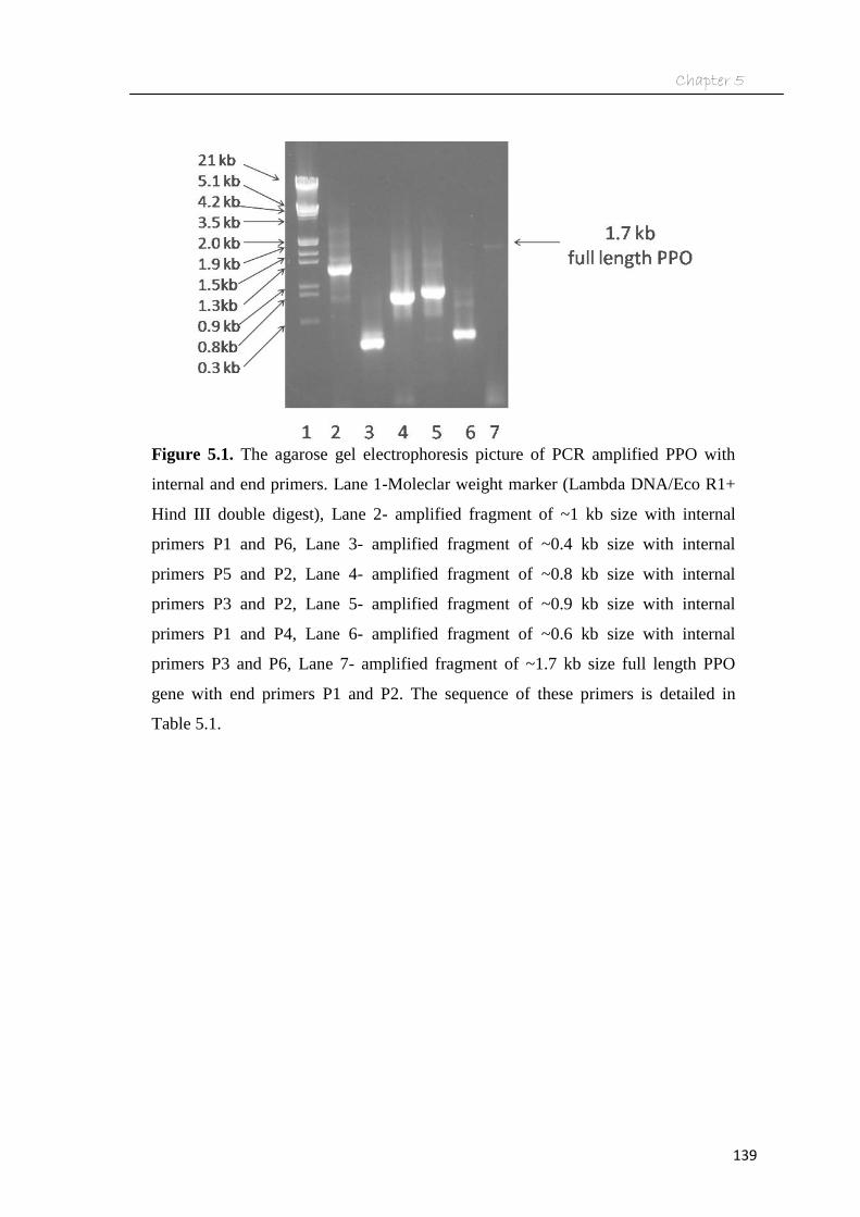
5.3.3. Results
The complimentary DNA (cDNA) prepared from isolated RNA from fruit of ‘Kalpatharu’
(V5) cultivar was amplified with the internal and end primers designed from the potato
PPO gene sequence and shown in Fig. 5.1. The amplification was observed in most of

Chapter 5
138
the cases especially in lanes 2, 3, 4, 5, and 6. However, a faint band was observed with
end primers showing the full length gene of about 1.7 kb (Fig. 5.1). The amplified
fragment was found to be ~1 kb size with internal primers P1 and P6 (Table 5.1). The
amplified fragment of ~0.4 kb size was observed with internal primers P5 and P2. The
internal primers P3 and P2 yielded DNA fragments of ~0.8 kb size. The amplified
fragment of ~0.9 kb size was observed with internal primers P1 and P4. Again the
amplified fragment with internal primers P3 and P6 was found to be ~0.6 kb size. These
fragment sizes were then compared with PPO gene sequence (Genbank accession no.
U22921.1) of potato and found to be matching which indicated its similarity with the
potato PPO. The genomic DNA isolated from brinjal and amplified with the same set of
primers thus produced similar results. The size of amplified product of both cDNA and
genomic DNA was found to be similar (~1.7 kb) as observed in agarose gel
electrophoresis (Fig. 5.2). This indicated that introns were probably absent in brinjal. The
full length DNA and cDNA were sequenced and submitted to Genbank (GQ149349,
GQ149350). The primers were designed from this nucleotide sequence and used for
amplification of the genomic DNA from other cultivars which showed substantial
amplification in five cultivars and poor amplification in ‘Arka navneet’ (V4) and
‘Silki’(V8) cultivars (Fig 5.3).
Chapter 5
139
Figure 5.1. The agarose gel electrophoresis picture of PCR amplified PPO with
internal and end primers. Lane 1-Moleclar weight marker (Lambda DNA/Eco R1+
Hind III double digest), Lane 2- amplified fragment of ~1 kb size with internal
primers P1 and P6, Lane 3- amplified fragment of ~0.4 kb size with internal
primers P5 and P2, Lane 4- amplified fragment of ~0.8 kb size with internal
primers P3 and P2, Lane 5- amplified fragment of ~0.9 kb size with internal
primers P1 and P4, Lane 6- amplified fragment of ~0.6 kb size with internal
primers P3 and P6, Lane 7- amplified fragment of ~1.7 kb size full length PPO
gene with end primers P1 and P2. The sequence of these primers is detailed in
Table 5.1.

Chapter 5
140
Figure 5.2. The agarose gel electrophoresis picture of PCR amplified PPO DNA
and cDNA with the same set of primers. Lane M- molecular weight marker
(Lambda DNA/Eco R1+ Hind III double digest), Lane 1- PCR amplified
genomic DNA, lane 2- PCR amplified cDNA.

Chapter 5
141
Figure 5.3. Agarose gel electrophoresis picture of PCR amplified PPO DNA from
eight cultivars [‘Pusa purple long’ (V1), ‘Ravaiya’ (V2), ‘Azad kranti’ (V3), ‘Arka
navneet’ (V4), ‘Kalpatharu’ (V5), ‘Raveena’ (V6), ‘Anupam’ (V7), ‘Silki’ (V8)] of
brinjal detailed in Table 2.1. M- Molecular weight marker (Lambda DNA/Eco R1+
Hind III double digest). The initial four letter of cultivar name was used as their
abbreviation in figures.

Chapter 5
142
5.3.1. Gene sequence based conceptual translation of PPO protein
The sequence details of PPO from brinjal cultivar ‘Kalpatharu’ (V5) is given in Figure
5.4. The other cultivars showed similar characteristics with minor differences in amino
acid positions. The presence of an 81 amino acid long transit sequence (location 1-81aa)
was observed (Genbank accession no. GQ149349.1), that was also been reported in PPO
from other fruits and vegetables (89 aa in apple, 86 aa in potato and 99 aa in tomato). The
Chloro IP program analysis showed the higher probability of presence of a 40 amino acid
long N-terminal chloroplast targeting sequence with cleavage site between serine (S40)
and Cysteine (C41) residues (Fig. 5.4) (Genbank accession no. GQ149349.1). The same
program also showed the cleavage site for remaining 41 amino acid long transit peptides
at lysine (L81) and alanine (A82). This sequence also showed similarity with other
thylakoid lumen proteins including PPO from other fruits. The protein did not contain a
trans membrane or hydrophobic domain and appeared to be a soluble protein, which ruled
out the possibility of being a thylakoid membrane protein. PPO was found to be a
metalloprotein with active site containing two copper binding regions (A and B), each
coordinated by three histidine (H) residues. One copper (copper A) binds to the
approximate amino acid sequence position between 173-216 and the second one binds to
the amino acid sequence between 349-403 position (Fig. 5.4) (Genbank accession no.
GQ149349.1). Each of these regions contained three histidine residues, which
coordinated with copper (H178, H196, H205 in copper A region and H353, H367, H389
in copper B region).
The x-ray diffraction studies in sweet potato (Ipomoea batatas) showed these histidine
residues in two regions form a trigonal pyramidal coordination sphere. One cysteine

Chapter 5
143
Figure 5.4. Nucleotide sequence based conceptually translated amino acid sequence
of PPO from brinjal (GQ149349.1) and its sequence features. The number indicates
the amino acid position from N-terminal position. The copper binding active regions
are in bold font and underlined. The His residues present in active site involved in
binding to copper are displayed in bigger font.

Chapter 5
144
(C182) present in copper A region is also crucial for active site catalysis and forms a
covalent thioether bridge with histidine (H196). The post translational modifications were
also predicted in PPO. The probable amino acids undergoing phosphorylation in this
protein are predicted to be as many as 13 serine, 9 threonine, and 10 tyrosine. The protein
did not contain any N-myristoylation, N-acetylation, and O-glycosylation sites.
5.3.2. The cultivars with high PPO activity showed maximum sequence similarity
Out of the eight cultivars, PPO gene from only six cultivars could get PCR amplified
using designed primers as discussed in materials and methods (Fig. 5.3). The PPO
nucleotide sequences were submitted to gene bank (Genbank accession numbers V1
(JQ621948), V2 (JQ621949), V3 (JQ621950), V5 (GQ149349.1), (V6 (JQ621951), V7
(JQ621952)). The other two cultivars ‘Arka navneet’ (V4) and ‘Silki’ (V8) could not be
PCR amplified and hence were not sequenced. These cultivars probably have a different
nucleotide sequence in the primer binding site of the designed primers. The amino acid
sequence of the remaining six cultivars has been compared (Fig. 5.5). The PPO nucleotide
sequence was found to contain 1773-1788 bp and hence predicted conceptual protein to
have 590-595 aa, as there was no intron in this gene. With minor differences the initial 81
amino acids were found to be the part of the signal sequence and were mostly identical,
and the active site region was found to be conserved and identical. The differences in
sequence were observed in the region between amino acid position 11 – 17. Valine was
found to be present in place of threonine in V1 (at position 21) and V2 (position 22).

Chapter 5
145
Contd……

Chapter 5
146
Figure 5.5. Multiple sequence alignment of PPO amino acid sequences [Genbank
accession numbers V1- (JQ621948), V2- (JQ621949), V3- (JQ621950), V5-
(GQ149349.1), V6- (JQ621951), V7- (JQ621952)] from six different cultivars of
brinjal and their sequence features. Symbol * below the sequence showed the
positions of exactly matched amino acids. Symbols “:” or “.” indicate high or
moderate matching at that position. The initial four letter of cultivar name was used as
their abbreviation in figures.

Chapter 5
147
Again there was an arginine (~243 aa) in place of glycine in cultivar V6. There was
another replacement of threonine in place of arginine (R) in V1 (430) and V6 (426). A
few single base differences were observed among these cultivars but these differences did
not reflect any difference in the amino acid sequence. However, the region between
amino acid position ~301 to 338 was found to be almost identical in two cultivars V5
(‘Kalpatharu’) and V6 (‘Raveena’) showing maximum PPO activity, whereas, this region
was very different in other four cultivars ‘Pusa purple long’ (V1), ‘Ravaiya’ (V2), ‘Azad
kranti’ (V3), and ‘Anupam’ (V7), which were found to have almost identical sequence in
this region but moderate PPO activity. The amino acid sequence difference in this region
seems to be due to a deletion of adenine base in PPO gene of V5 (at 879 bp position) and
in V6 (at 870 bp position) resulting in appearance of tryptophan (W at ~291 position) in
place of methonine (M) and subsequent change in the reading frame. Two subsequent
deletions at 877 and 881 position in V6, and 886 and 890 position in V5 of adenine and
guanine, respectively, resulted in a single amino acid deletion (asparagine at ~296
position in V5 and 293 position in V6) and the frame shift resumed for subsequent 7
amino acids. Again another deletion of thymine base (at 905 position in V6 and 914
position in V5) resulted in change in frame (from 304 to 309 in V5 and 301 to 306 in V6).
From ~301 to ~338 amino acid position in these two cultivars, the change in reading
frame changed the amino acid sequence till proline-cysteine-phenylalanine (PCL) (Fig.
5.5). Another insertion of thymine (at 1014 position in V5 and 1023 position in V6)
compensated the frameshift and the cultivars again showed identical sequence after ~338
amino acid position (Fig. 5.5).

Chapter 5
148
Contd………

Chapter 5
149
Figure 5.6. Multiple sequence alignment of PPO protein from different fruit and
vegetables. The PPO sequences used were from brinjal (ACR61398.1), potato
(AAA85122.1) tobacco (CAA73103.1), tomato (AAB22610.1), grape (AAB41022.1)
and sweet potato (AAW78869.1). Symbol – indicate the gap introduced for optimal
alignment. Symbol “*” below the sequence showed the positions of exact matching of
amino acids. Symbols “:” or “.” indicates high or moderate matching at that position.

Chapter 5
150
5.3.3. Brinjal PPO gene sequence showed homology with that of potato and tomato
The nucleotide sequence of PCR amplified cDNA and genomic DNA of PPO gene from
brinjal were found to be of similar length and sequence (NCBI Genebank accession no.
GQ149349.1 for gene and GQ149350.1 for mRNA). This indicated that the introns are
missing in the PPO gene of brinjal. Similar observations were also reported in apricot and
other plants (Chevalier et al., 1999; Mayer, 2006). The nucleotide blast search revealed
that the brinjal PPO showed high similarity with potato (86%) and tomato (84%), which
are its closest relatives in Solanaceae family. Further it showed about 80% similarity with
tobacco PPO. The NCBI protein blast results showed about 97% similarity with potato
(AAA85122.1), 96% with tomato (AAB22610.2), 98% with tobacco (ABE96885.1), and
97% with sweet potato (AAW78869.1). NCBI conserved domain database (CDD) search
results showed the presence of domains from three super families. The amino acid region
between 168-247 and 348-380 which included two copper ion (Cu-A and Cu-B) binding
regions, respectively, are similar with common central domain of Tyrosinase; pfam00264
superfamily (CDD 189478). This family includes polyphenol oxidase enzymes from other
plants and hemocyanins. The amino acid region or domain between 386 and 436 showed
homology with PPO1-DWL superfamily pfam12142 (CDD 192942) which was also
annotated as PPO middle domain. This domain family was found in bacteria and
eukaryotes and includes about 50 amino acids along with the presence of conserved DWL
sequence motif which gave the family its name. Again the region between 457-590 was
similar with PPO1-KFDV superfamily pfam12143 (CDD 192943). This domain found in
eukaryotes particularly in plants contain 132-152 amino acids with a highly conserved
sequence motif KFDV from which the name was derived.

Chapter 5
151
(a) (b)
(c)
Figure 5.7. The spread plating of transformant clones (a), streaked, numbered and
grided colonies on a second plate (b), The agarose gel electrophoresis picture of
colony PCR products of transformant clones (c), The plasmid vector specific primers
showed amplification of about 2.0 kb (lanes 2 and 7). The lanes 1 to 14 are represent
colony number from 88 to 101, respectively. The last lane (M) shows the molecular
weight marker (Lambda DNA/Eco R1+ Hind III double digest).

Chapter 5
152
5.3.4. Cloning and expression of polyphenol oxidase in E.coli
The genomic DNA sequence of ppo gene was analyzed and NdeI and Xho I sites were
found to be absent. The PCR of genomic DNA from brinjal fruit (Kalpatharu variety)
with primers having integrated restriction sites (NdeI with forward primer and XhoI site
with reverse primer) resulted in amplification of a single band of about 1.8 kb size. The
size matched with the earlier observed size of PPO gene and confirmed by gene
sequencing. The amplified product was cloned into pET-28a expression vector
downstream of lacUV5 promoter (IPTG inducible). The recombinant plasmid thus
prepared was transformed into E.coli (DH5α) and was selected on a Luria agar-
kanamycin plate (Fig. 5.7a). Around 100 transformants were plated again on a grid plate
(Fig. 5.7b) and screened using colony PCR (Fig 5.7c). Out of these, 6 were found to have
ppo gene insert. The recombinant plasmid was further isolated and transformed to BL 21
and codon (+) strain of E. coli. The BL 21 (DE3) cells were selected on LB kanamycin
plates. The codon (+) recombinant cells were selected on LB-plates containing kanamycin
as well as chloramphenicol.
5.3.5. Overexpression of PPO
The three out of six transformants having recombinant PPO were induced using IPTG and
grown under similar conditions along with E.coli (without PPO insert) and IPTG control
(without IPTG addition). Later cells were lysed, centrifuged (12000 g, 15 min) and
analysed by SDS-PAGE (Fig. 5.8). The lanes 2-8 show the supernatant fractions without
any significant difference of protein expression between control and IPTG induced cells.
The lanes 9-15 show the pellet fraction of protein showing significant presence of
recombinant protein of 66 kDa size in IPTG induced pellet fraction (lanes 9, 11, 13),

Chapter 5
153
Figure 5.8. The SDS PAGE profile of expressed PPO protein in E.coli (BL-21) in
both supernatant and pellet fractions after induction with (+) and without (-) IPTG.
The lanes 2-8 shows the supernatant fractions control and IPTG induced cells. The
lanes 9-15 shows the pellet fractions of control and IPTG induced cells. The over
expression of ~66 protein in IPTG induced pellet fraction (lanes 9, 11, 13), which are
absent in control cells (lanes 10, 12, 14). M – Molecular weight marker. C- control
E.coli (BL-21) cells without the PPO insert.

Chapter 5
154
Figure 5.9. The SDS PAGE profile of the low temperature and low IPTG induction of
recombinant PPO in E.coli. The cells were grown at 20 ºC (lanes 1-3, 7-9) and 37 ºC
(lanes 4-6 and 10-12). The IPTG concentration was 0.1 mM in lanes 1, 4, 7, and 10,
and 1 mM in lanes 2, 5, 8, and 11. Lanes 1-6 supernatant fraction, lanes 7-12 pellet
fraction.

Chapter 5
155
which are absent in control cells (lanes 10, 12, 14) (Fig. 5.8). This has demonstrated the
overexpression of inducible PPO in the pellet lysate fraction which could be due to
improper folding of brinjal PPO protein in E. coli and its aggregation to form inclusion
body. Such inclusion body proteins have been reported to lack enzyme activity and
require solubilization, refolding and purification for regaining activity (Singh and Panda,
2005). The formation of inclusion bodies of recombinant proteins in E. coli is known to
be due to rapid synthesis of recombinant protein and nonavailability of protein folding
machineries (molecular chaperones) for correct folding. This resulted in mis-folded
insoluble aggregates termed as inclusion bodies. In such cases low temperature or low
IPTG induction is used to slow down the rate of translation or expression of the
recombinant protein. The slower translation reported to cause the availability of
molecular chaperones for proper folding. In current study, both low temperature (20 ºC)
incubation as well as low concentration (0.1 mM) IPTG induction of recombinant E. coli
cells was tried but both apparently did not improve the solubility of the protein (Fig. 5.9).
Similar difficulty has earlier been reported in brinjal and red clover by Shetty et al. (2011)
and Sullivan et al. (2004), respectively.
5.3.6. Purification of PPO from E.coli
The inclusion body is normally solubilized by high concentration of urea and refolded by
its slow removal in the presence of oxidizing agents. Though the formation of inclusion
bodies is undesirable, their formation at times becomes advantageous due to the ease of
isolation and purification of the recombinant protein (Singh and Panda, 2005). This
approach was used and the cell lysate was solubilized in urea (8 M), and purified using
Nickel metal affinity chromatography (Fig. 5.10). The figure showed over-expression of

Chapter 5
156
Figure 5.10. The SDS PAGE profile of eluted fractions from Ni Sepharose column
showing elution of soluble PPO from E.coli inclusion body. Lane 1 (cell lysate), lane
2 (lysate supernatant), lanes 3-6 (flow-through fractions), lanes 7-10 (washing
fractions), lanes 11-15 (pH 5.9 eluted fractions), lanes 16-24 (pH 4.5 eluted fractions),
and lane 25 (molecular weight marker).

Chapter 5
157
PPO (lane 1) and its solubilzation in urea (lane 2). The column binding took place as PPO
band was not observed in the flow-through and washing fraction (lanes 3-10). The elution
with buffer D at pH 5.9 did not elute the expressed PPO (lanes 11-15). However, the
expressed PPO eluted with buffer E having pH 4.5 (lanes 22-24). The lane numbers 22-24
showed a single band at 66 kDa and no other bands observed indicated the protein to be
pure. The corresponding fractions were pooled, and dialysed with modified refolding
buffer for removal of urea. Later the volume of dialysed protein was concentrated to 0.5
ml with a centricon filter and the enzyme assay was performed using 4-methyl catechol as
substrate (0.1 M). The purified extract (50 µl) showed very less enzyme activity of about
3 units/ml. This activity was less than 10% of the activity in the crude extract. This
finding indicated that the recombinant protein was solubilized with the loss of enzyme
activity. Similar reports of loss of enzyme activity after expression in E.coli have been
earlier reported in red clover which could be due to the absence of post translational
modification system required for eukaryotic proteins in E.coli.
5.4. Discussion
As reported in other plants, the PPO protein from brinjal too was found to contain
thylakoid lumen targeting sequence and two copper atoms in its active site. The three
histidine residues which interact with copper are present in each of these copper binding
regions. The cysteine amino acid required for catalysis was also found to be present in
copper A binding region. These features include brinjal PPO in type 3 copper proteins.
The other proteins known in this class are tyrosinase and hemocyanins. The type 1 copper
proteins (include proteins like azurin, plastocyanin) were known to have single copper
atom linked to the protein through two histidine and one cysteine residues and are
involved mostly in electron transfer reactions. The type 2 copper proteins (like superoxide

Chapter 5
158
dismutase, dioxygenases) known to be linked to single copper atom and are mostly
involved in oxygenation or oxidation reactions. The crystal structure of PPO has been
reported in sweet potato (Ipomoea batatas) and grenache (Vitis vinifera) (Eicken et al.,
1999; Virador et al., 2010). The catechol oxidase enzyme of sweet potato indicated the
presence of two disulfide bonds (C86-C102 and C101-C189) between cysteine residues in
the secondary structure of the protein along with six α- helix and seven β- sheets (Eicken
et al., 1999). The brinjal PPO showed a moderate similarity in protein sequence with
them. The comparison of sequence in 301-338 amino acid resulting in grouping of these
cultivars into two classes. The four cultivars ‘Pusa purple long’ (V1), ‘Ravaiya’ (V2),
‘Azad kranti’ (V3) and ‘Anupam’ (V7) showed sequence homology in this region. The
‘Kalpatharu’ (V5) and ‘Raveena’ (V6) showed high PPO activity and were found to be
identical but different from the above four cultivars in this region (301-338 amino acid).
Their amino acid sequence in this region was found to be unique and did not match
significantly with published PPO protein in Genbank. The PPO of these two cultivars was
also found to be highly active which indicated a possible role of these sequences (301-338
aa) in the activity. The total amino acid sequence showed higher similarity with other
close members of the solanaceae family including potato and tomato. The similarity was
more in the regions of active site. However, the overall homology was not very high
indicating the uniqueness of PPO sequence in brinjal.
Though two PPO isozymes were detected in the ammonium sulfate fractionation, only a
single gene could be identified and amplified. The PPO gene from brinjal was shown to
contain no introns. The intron less PPO gene has recently been reported in ‘Arka shirish’
cultivar of brinjal (Shetty et al., 2011), as well as in potato, tomato, apricot, tobacco and
few other plants (Thygesen et al., 1995; Shahar et al., 1992; Goldman et al., 1998). The
gene sequence also showed homology with other members of Solanaceae family. The

Chapter 5
159
protein sequence showed conserved domains of tyrosinase and PPO superfamilies. The
N-terminal region showed two stretches of targeting sequence. The first sequence targets
the peptide to chloroplast and the second targets to thylakoid lumen. Inside thylakoid
lumen the peptide is folded to its active form. Similar localization after targeting of PPO
was reported in many other plants including potato, tomato and tobacco (Thygesen et al.,
1995; Shahar et al., 1992; Goldman et al., 1998). The molecular weight was reported to
be 60, 58, and 55 kDa after targeting into the thylakoid lumen in potato, tomato, and
tobacco (Thygesen et al., 1995, Shahar et al., 1992, Goldman et al., 1998). In some
reports the PPO was reported to bind to the thylakoid membrane and have hydrophobic
regions. In such cases the PPO is extracted with addition of detergents. However, brinjal
PPO was analyzed to be soluble without any membrane binding region.
There are reports of cloning PPO, using partial PPO cDNA synthesized from mRNA. The
cDNA was used as a probe and the complete PPO gene was fished out from the cDNA
library using southern hybridization in potato and pokeweed (Hunt et al., 1993; Joy et al.,
1995). In case of apple (Malus pumila) and apricot PPO gene was expressed in E. coli but
the activity of the recombinant protein was not reported (Haruta et al., 1998). The
eukaryotic proteins are known to undergo post-translational modifications including N-
terminal and C-terminal processing along with phoshorylation, N-myristoylation, and O-
glycosylation. Using bioinformatics approach, the occurrence of six potential
myristoylation sites in grape PPO was predicted (Virador et al., 2010). However, in case
of many plants the PPO gene sequence did not indicate the presence of sites for such
modification (Mayer, 2006). In red clover grass leaves PPO cDNA was prepared, cloned
and expressed in E. coli. However, the expressed protein in E.coli formed insoluble
inclusion bodies. Corroborating these findings, in the current study the recombinant

Chapter 5
160
protein was solubilized with the loss of enzyme activity, which was probably due to the
absence of posttranslational modification of expressed eukaryotic proteins in E.coli.