Chapter 7 - Protein Function Introductiontonga.usp.edu/jsnow/chem341/chapter7.pdf · Chapter 7 -...
15
Chapter 7 - Protein Function Introduction We begin the study of protein function and its relationship to protein structure. We’ll restrict the topic to functions of globular proteins. The functions of these and other proteins is dependent on their binding to cellular components. Myoglobin This is a small protein consisting of 1 polypeptide chain (153 residues), whose X-ray structure was the first of its kind (John Kendrew, 1959). It is a compact globular protein whose secondary structure consists predominantly of "- helix (Figure 7-1). - The red structure in Figure 7-1 is the heme group, an example of a prosthetic group, whose function is to bind oxygen (Figure 7-2). The purposes of prosthetic groups like heme are similar to those of coenzymes such as NAD + in that they assist the protein in performing functions that would be impossible without their presence.
Transcript of Chapter 7 - Protein Function Introductiontonga.usp.edu/jsnow/chem341/chapter7.pdf · Chapter 7 -...
Chapter 7 - Protein Function
Introduction
We begin the study of protein function and its relationship to protein
structure. We’ll restrict the topic to functions of globular proteins. The functions
of these and other proteins is dependent on their binding to cellular components.
Myoglobin
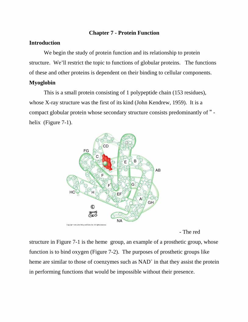
This is a small protein consisting of 1 polypeptide chain (153 residues),
whose X-ray structure was the first of its kind (John Kendrew, 1959). It is a
compact globular protein whose secondary structure consists predominantly of "-
helix (Figure 7-1).
- The red
structure in Figure 7-1 is the heme group, an example of a prosthetic group, whose
function is to bind oxygen (Figure 7-2). The purposes of prosthetic groups like
heme are similar to those of coenzymes such as NAD+ in that they assist the protein
in performing functions that would be impossible without their presence.

Mb + O2 Mb O2
K = [MbO2][Mb][O2]
The central iron atom must be in its +2 oxidation state (Fe++) in order to bind
oxygen. The brownish color of hamburger is due to oxidation of heme iron to its
+3 oxidation state. CO, NO and H2S are toxic because they bind much more
tightly to heme than O2.
Function
In some mammals like whales, myoglobin plays an important role in
oxygen storage in muscle. In most other vertebrates myoglobin facilitates O2
diffusion in muscle by increasing its low concentration (10-4 M) in blood.
Consider the binding of O2 to myoblogin (Mb):

YO 2 = amount of O2 bound
total amount of O2 (free + bound)=
[MbO2]
+ [Mb][MbO2]=
+ [Mb]
[Mb][O2]/K
[Mb][O2]/K
[O2]
K + YO =
[O2] = +
pO2K pO2
where it should be noted that K in this case is a dissociation constant.
Graphically, one can represent the binding of oxygen by defining the fractional
saturation, YO2, as
where [MbO2] was substituted for in terms of K in the last expression. Cancelling
out the common term, [Mb], and multiplying through on top and bottom by K
gives
Figure 7-4 demonstrates
an oxygen binding curve for myoglobin:
Hemoglobin
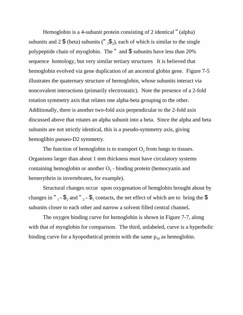
Hemoglobin is a 4-subunit protein consisting of 2 identical "(alpha)
subunits and 2 $ (beta) subunits ("2$2), each of which is similar to the single
polypeptide chain of myoglobin. The " and $ subunits have less than 20%
sequence homology, but very similar tertiary structures It is believed that
hemoglobin evolved via gene duplication of an ancestral globin gene. Figure 7-5
illustrates the quaternary structure of hemoglobin, whose subunits interact via
noncovalent interactions (primarily electrostatic). Note the presence of a 2-fold
rotation symmetry axis that relates one alpha-beta grouping to the other.
Additionally, there is another two-fold axis perpendicular to the 2-fold axis
discussed above that rotates an alpha subunit into a beta. Since the alpha and beta
subunits are not strictly identical, this is a pseudo-symmetry axis, giving
hemoglibin pseueo-D2 symmetry.
The function of hemoglobin is to transport O2 from lungs to tissues.
Organisms larger than about 1 mm thickness must have circulatory systems
containing hemoglobin or another O2 - binding protein (hemocyanin and
hemerythrin in invertebrates, for example).
Structural changes occur upon oxygenation of hemglobin brought about by
changes in "1 - $2 and "2 - $1 contacts, the net effect of which are to bring the $
subunits closer to each other and narrow a solvent filled central channel.
The oxygen binding curve for hemoglobin is shown in Figure 7-7, along
with that of myoglobin for comparison. The third, unlabeled, curve is a hyperbolic
binding curve for a hyopothetical protein with the same p50 as hemoglobin.

Hb (n-1)O2 Hb nO2 O2 +
Note that the shape of the hemoglobin curve is “S” shaped, or sigmoid, in contrast
to the simple hyperbolic shape of the myoglobin curve. This shape curve indicates
that hemoglobin is well-suited for the transport of O2, which must be bound to
hemoglobin in the lungs, but also released in the tissues. Note that myoglobin
would not be well-suited for such a purpose.
A sigmoid curve such as shown for oxygen binding to hemoglobin is
indicative of a general phenomenon known as cooperativity, which refers to an
interaction between binding sites resulting in enhanced activity.
Quantitative Aspects of Cooperative Oxygen Binding
Consider the binding of a single O2 molecule to hemoglobin:
where n = 4, and the corresponding dissociation constant, Kn, where it is assumed
that all the Ki are equal:
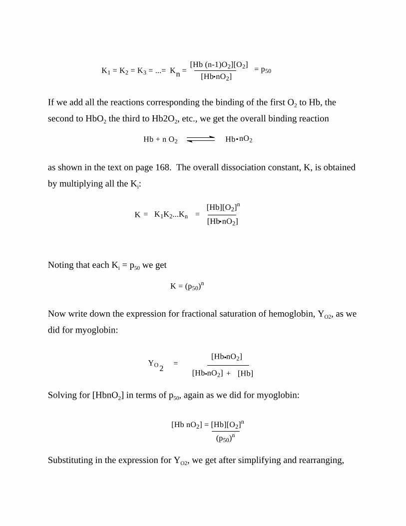
K1 = K2 = K3 = ...= K = n[Hb (n-1)O2][O2]
[Hb nO2]= p50
Hb + n O2 Hb nO2
= K1K2...KnK[Hb][O2]n
[Hb nO2]=
K = (p50)n
YO =[Hb]
[Hb nO2]
[Hb nO2] +2
[Hb nO2] = [Hb][O2]n
(p50)n
If we add all the reactions corresponding the binding of the first O2 to Hb, the
second to HbO2 the third to Hb2O2, etc., we get the overall binding reaction
as shown in the text on page 168. The overall dissociation constant, K, is obtained
by multiplying all the Ki:
Noting that each Ki = p50 we get
Now write down the expression for fractional saturation of hemoglobin, YO2, as we
did for myoglobin:
Solving for [HbnO2] in terms of p50, again as we did for myoglobin:
Substituting in the expression for YO2, we get after simplifying and rearranging,

YO = 2
(pO2)n
+ (p50)n(pO2)n
1 -
YO2YO2
(pO )n
=(p50)n
2
1 -
YO2YO2
log = n log pO2- n log p50
which is the Hill equation. Because of the fact that the denominator contais a sum
of terms with exponents it is convenient to express this equation as
(Because 1 - YO2 = (p50)n/((p50)n + (PO2)n)) which replaces the denominator with a
single term. Taking the log of both sides gives a linear equation:
if we plot log (YO2/(1- YO2 ) vs. log pO2. Such a plot is known as a Hill plot and is
shown in Figure 7-8:

which also shows a Hill plot for myoglobin for comparison. Note that the Hill plot
for hemoglobin is curved, unlike that for myoglobin, which is a straight line
throughout the pO2 range, with a slope of 1. The Hill plot for hemoglobin should
be thought of as being linear with a slope of 1 in the limit of high and low pO2, and
also a connecting portion of the curve with slope > 1. The maximum slope is
called the Hill constant, and is 2.8 - 3.0 for hemoglobin. The above analysis shows
that n should be 4 provided all the O2 bind to or are released from hemoglobin
simultaneously. This would occur if the subunits cooperate with each other fully
in the binding process. That the Hill constant is less than 4 indicates there is not
complete cooperativity.
The Hill Plot yields additional information. The lower asymptote
corresponds to binding of the first O2 at low pO2, whereas the upper asymptote
corresponds to binding the last O2 high pO2. Binding at both limits is not
cooperative (n = 1). Note on the y axis that when Log (YO2/(1- YO2 ) = 0,
(YO2/(1- YO2 ) must be 1, which means that pO2 = p50 at this point, so p50 values can
be determined from the Hill Plot as the pO2 value corresponding to 1 on the y axis.
(compare to determination of p50 for myoglobin). Thus, the upper asymptote yields
a value of 0.3, and the lower asymptote yields a value of 30. Recalling that p50 is
inversely related to binding affinity, it can be concluded that the fourth O2 binds to
hemoglobin with 100 times greater affinity than the first.
Consider the advantage of this to the organism: As a red blood cell
containing hemoglobin enters the lungs, the hemoglobin is largely devoid of O2
because it has delivered the O2 to metabolizing tissue. The release of O2 in the
tissues occurs because the affinity of hemoglobin for O2 has decreased in the
tissues. As the red cells enter the lungs, the increased pO2 results in hemoglobin
starting to bind O2. This results in an increase in cooperativity such that the
affinity of hemoglobin increases markedly, ensuring that hemoglobin will be fully
bound as it leaves the lungs on its way back to the tissues. Thus, hemoglobin
switches from a high-affinity state in the lungs to a low-affinity state in the tissues
as a result of cooperation between subunits. Notice that such cooperativity would
not be possible for myoglobin, which consists of a single polypeptide chain.
Finally, notice that this cooperative effect manifests itself in a sigmoid binding
curve, as shown in Figure 7-7. Initial flatness indicates a low-affintiy state.
However, the subsequent steep rise in the curve indicates that binding becomes
easier as pO2 increases.
The structural events that lead to the cooperative effects for hemoglobin are
well-known and somewhat complicated. The salient features are, first of all, that
cooperativity is only possible for a multiple-subunit protein. Secondly, recall that
the shape, or conformation, of deoxy hemoglobin is different than it is for oxy
hemoglobin. The low relatively low affinity of deoxy-hemoglobin for O2 is due to
the Fe++ of each heme group being slightly out of the plane of the porphyrin ring,
thereby being less accessible to O2, which occupies the sixth coordinate position of
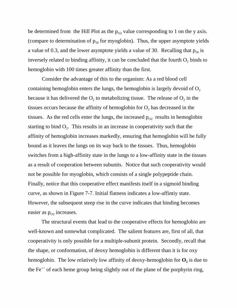
the iron. Upon binding of O2 to any heme group, the iron is forced into a planar
configuration. The histidine residue (His F8) occupying the fifth coordinate
position on the other side of the plane of the porphyrin ring is “dragged” along
with the Fe. This results in a conformational change in that subunit. This
conformational change is communicated to the other subunit via their close
proximity, resulting in the Fe in the remaining heme groups to move closer to
being coplanar with their porphyrin rings. Their affinity for O2 increases
accordingly. The movement of the Fe++ in a single subunit is shown in Figure 7-9.
Thus, hemoglobin switches from a low-affinity (T) to a high affinity (R) state.
Bohr Effect
Consider the metabolism of glucose in the tissues:
C6H12O6 + 6 O2 6 6 CO2 + 6 H20
H2O + CO2 W H2CO3 W H+ + HCO3-
Thus, both CO2 and acid are produced as a result of metabolism. Hb must, in
addition to delivering O2, remove CO2. We have seen that there is a cooperative
change in Hb conformation between a T and R state:
T + 4 O2 W R@4O2
In order to remove CO2 from the tissues and deliver it to the lungs for
removal, it is necessary that CO2 have high affinity for the T state, low for the R
state:
T@CO2 + 4 O2 W R@4O2 + CO2
Note Le Chatelier effects wrt this equilibrium scheme: high CO2 in the tissues
shifts the equilibrium to the left, thus releasing O2 in the tissues, high O2 in the
lungs shifts the equilbrium to the right, thus releasing CO2 for expulsion. The
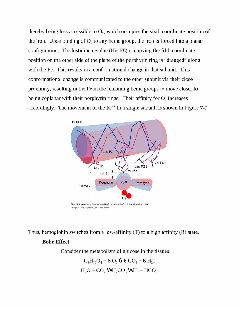
complete Bohr effect includes the effects of H+:
H+@ T@CO2 + 4 O2 W R@4O2 + CO2 + H+
Note also graphical representation (Figure 7-12).

R NH2 R NH C O
O
+ CO2
H+ binds to N-terminal amino groups of the " subunits and the C-terminal His
residues of the $ subunits. In the T state, these residues are located near
negatively charged groups, forming ion pairs with them, and effectively
increasing their pKa’s. Note also that CO2 does not bind to the heme groups;
rather it forms carbamates with N-terminal amino groups:
High altitude adaptation: Effects of bisphosphoglycerate (BPG)
Decreases affinity of Hb for O2 by preferentially binding to the T state:
BPG @H+@ T@CO2 + 4 O2 W R@4O2 + CO2 + H+ + BPG
BPG binds to the central cavity formed at the juncture of the four subunits in the
T state. It is lined with positive charges which interact favorably with the
negative charges of BPG.
Fetal Hb
Increased O2 affinity due to replacement of His residue on $ chain
by Ser ("2(2). Thus, a positively charged side chain (His) is replaced by a
neutral side chain (Ser) near the central cavity, thus decreasing the binding of
BPG in the central cavity, which in turn increases the affinity of fetal Hb for O2
since BPG is a negative allosteric modulator. In order for effective transfer of O2
from mother to fetus to occur, fetal hemoglobin must have a greater affinity for
O2 than adult hemoglobin.

Abnormal Hemoglobins
Nearly 500 variants, 95% of which involve a single amino acid
substitution.
Not all produce clinical symptons (Table 7-1). Some mutations
alter quaternary structure, thereby affecting cooperativity. Some favor oxidation
of Fe(II) to Fe(III). Since a single mutation typically gives rise to such
mutations, only two subunits will be affected; consequently Hb can still bind two
O2's, giving rise to a bluish skin color.
- Sickle cell anemia story: about 100 million people are afflicted
worldwide in tropical regions at any given time.
- In 1945 Pauling hypothesized that the disease was caused by a
single mutation (electrophoresis). Val replaces Glu at $6 position.
Allosteric Proteins
- The cooperativity of O2 binding to hemoglobin provides a model for the
cooperative behavior of other multiple subunit proteins, many of which are
enzymes. The cooperative effects are due to conformational changes within the
subunits, which are then communicated to the other subunits, and are called
allosteric effects.
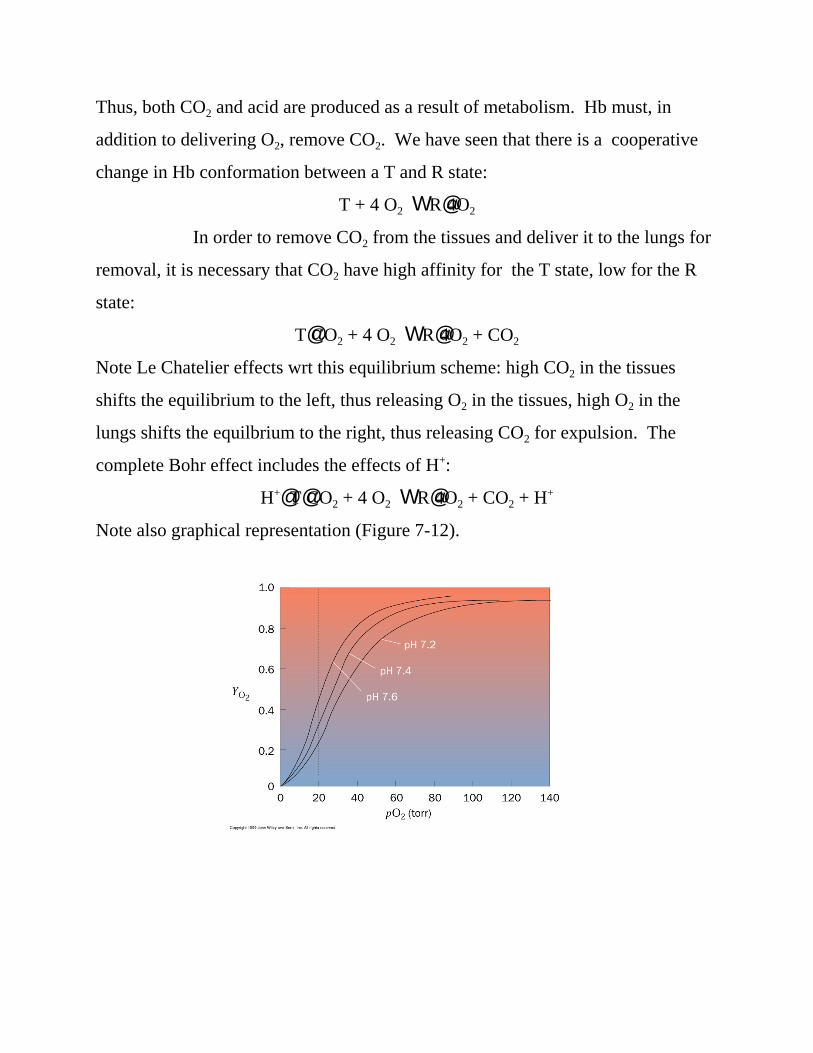
Two models have been developed to explain cooperativity. One of them,
the symmetry model of Monod, Wyman and Changeux, is shown below (Figure
7-16):
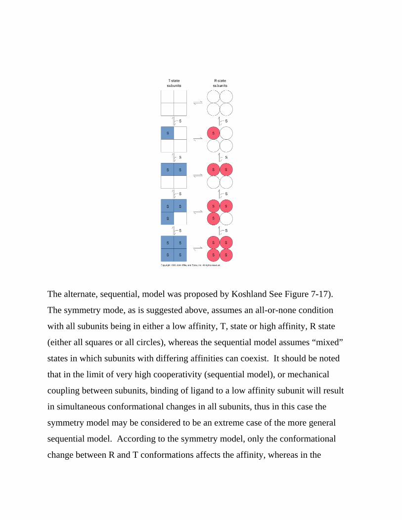
The alternate, sequential, model was proposed by Koshland See Figure 7-17).
The symmetry mode, as is suggested above, assumes an all-or-none condition
with all subunits being in either a low affinity, T, state or high affinity, R state
(either all squares or all circles), whereas the sequential model assumes “mixed”
states in which subunits with differing affinities can coexist. It should be noted
that in the limit of very high cooperativity (sequential model), or mechanical
coupling between subunits, binding of ligand to a low affinity subunit will result
in simultaneous conformational changes in all subunits, thus in this case the
symmetry model may be considered to be an extreme case of the more general
sequential model. According to the symmetry model, only the conformational
change between R and T conformations affects the affinity, whereas in the

sequential model ligand binding induces conformational change in the subunit to
which the ligand binds, and cooperative interactions arise through the influence
of those conformational changes in neighboring subunits. Oxygen binding to
hemoglobin is concerted, thus The T 6 R changes occur simultaneously (or
nearly so) in all subunits, which is consistent with the symmetry mode.
However, ass Figure 7-9 suggests, oxygen binding to the T state induces
conformational changes within the subunit to which oxygen binds, which is
consistent with the sequential model. Thus, oxygen binding to hemoglobin
exhibits features of both the symmetry and sequential models.
Problems: 1, 3, 4, 7, 9, 10