AP Biology Chapter 35 Plant Anatomy AP Biology Basic anatomy root shoot (stem) leaves.

1
Chapter 6
The Shoot System II: The Form and Structure of Leaves
THE FUNCTIONS OF LEAVES
Leaves Are Shaped to Capture Light The Arrangement of Leaf Cells Depends on Their Functions Where Do New Leaves Come From?
LEAF FORM AND SPECIALIZED LEAVES
Leaf Shape May Depend on the Plant's Age and the Environment
ADAPTATIONS FOR ENVIRONMENTAL EXTREMES MODIFICATIONS FOR SPECIAL FUNCTIONS
Leaf Abscission Is the Seasonal Removal of Leaves
SUMMARY
PLANTS, PEOPLE AND THE ENVIRONMENT: Insect-eating Plants ECONOMIC BOTANY: Commercially Important Fibers

2
KEY CONCEPTS 1. Typically, the most important functions of leaves are photosynthesis (creation of chemical energy, in the form of carbohydrate, from sunlight and carbon dioxide) and transpiration (evaporation of water from the leaf surface). Some leaves are modified for specialized functions such as water storage, food storage, protection, and support. 2. A leaf usually consists of a leaf blade and a petiole. Leaves with one leaf blade are called simple leaves; leaves with many blades are called compound leaves and the individual blades are called leaflets. 3. Leaves are composed of epidermis, mesophyll, xylem, and phloem tissues. A waxy cuticle covers the epidermis; the mesophyll is where photosynthesis usually occurs. The xylem conducts water, and the phloem transports sugars. 4. Leaf formation is initiated at the shoot apical meristem. 5. Leaf fall in the autumn involves an active process of cell division and cell breakdown at the abscission layer in the petiole. 6.1 THE FUNCTIONS OF LEAVES Green plants, algae, and a few species of bacteria use sunlight as an energy source. Other inhabitants of the earth obtain energy by consuming--directly or indirectly--what the green plants produce. Photosynthesis is the process used by plants to synthesize sugars and release oxygen; the chloroplasts in plant cells trap light energy and convert it into chemical energy.
Leaves provide large surfaces for the absorption of light and carbon dioxide. Both are the raw materials for photosynthesis. In general, most leaves are thin, so that no cells lie far from the surface. This architecture makes it easy for leaf cells and chloroplasts to absorb light and imbibe carbon dioxide gas. At the same time, however, it promotes the loss of water from plants because any opening that will permit carbon dioxide to pass will also allow water to evaporate. Evaporation, also called transpiration in plants, helps cool the leaf and acts as the driving force for water transport (see Chapter 11); however, excessive evaporation places the plant in danger of dehydration. To a great extent, leaf form (morphology) and anatomy are a compromise between capturing light and carbon dioxide and conserving water. Water-saving structures (for example, a thick waxy cuticle to inhibit evaporation) become important for plants that occupy hot, dry regions, such as rocky cliffs and deserts, where water loss is a great hazard.
In this chapter we will discuss the form and anatomy of leaves, describe their development, and note the modifications that help plants deal with environmental challenges.

3
Leaves Are Shaped to Capture Light The leaves of dicot and monocot plants (defined in Chapter 5) have similar functions, but their basic designs differ. Monocot plants often have leaves with a strap-shaped leafblade, their vascular bundles commonly run parallel through the leaf, and their leaf bases usually wrap around the stem (Fig. 6.1b). Dicot leaves typically are composed of a flat, thin portion (the blade) with the vascular bundles in a netted pattern, and a narrow portion (the petiole) (Fig. 6.1a). The blade is the part that absorbs most of the light; the petiole is designed to hold the blade away from the stem. Not only does this give the blade greater exposure to sunlight, but also it allows the blade to move in the air so that gas exchange can easily take place.
Figure 6.1. Typical dicot (a) and monocot (b) leaves. Dicot leaves usually have netted venation and a midrib. Monocot leaves usually have parallel veins.
THE LEAF BLADE In most dicot and monocot plants, the cells of the leaf blade contain many chloroplasts and perform most of the photosynthesis in the plant. The blade provides a broad, flat surface for capturing light and carbon dioxide. Leaves with a single blade (Fig. 6.2a) are called simple leaves. The form of the blade varies widely among different plants, though. For instance, the leaf edges or margins may be entire (smooth), toothed, or lobed. Some leaves are so deeply divided that the blade forms several separate units, or leaflets (Fig. 6.2b); leaves having this design are called compound leaves. There are two types of compound leaves: A palmately compound leaf has its leaflets diverging from a single point, and a pinnately compound leaf has leaflets arranged along an axis (Fig. 6.2b). These variations in leaves are useful in classifying plants.
Quite often in nature there is a practical reason for differences in structures that have similar functions. For compound leaves, the question naturally arises of how a plant might benefit by having its blade
Figure 6.2. Examples of simple and compound leaves. Red buckeye is palmately compound; black locust is pinnately compound; and honey locust is double pinnately compound.

4
Quite often in nature there is a practical reason for differences in structures that have similar functions. For compound leaves, the question naturally arises how a plant might benefit by having its blade divided into leaflets. Perhaps the most likely answer is that the spaces between leaflets allow better airflow over the leaf surface. This may help cool the leaf (because of increased evaporation) while improving carbon dioxide uptake. However, there may be other adaptive or developmental reasons for these differences.
THE PETIOLE Dicot leaf blades are not directly attached to the stem. Instead, a petiole serves that purpose. The petiole is the narrow base of most dicot leaves. It functions to improve photosynthetic efficiency in several ways. By extending the leaf blade away from the stem, the petiole reduces the extent to which the blade is shaded by other leaves. It also allows the blade to move in response to air currents. This is important because a layer of stagnant air, the boundary layer, forms at the leaf surface. During photosynthesis, the boundary layer becomes depleted of carbon dioxide. Leaf movements as a result of air currents help bring fresh air containing carbon dioxide to the leaf surface and cool the leaf by increasing evaporation. These movements are passive reactions to air movement and not under any kind of mechanical regulation.
Petioles are variable in shape. They may be long or short, cylindrical or flat in cross section. They are usually attached to the base of the leaf blade (Fig. 6.1a). Leaves that lack a petiole are called sessile.
THE SHEATH In most monocot leaves the leaf base typically wraps entirely around the stem to form the sheath (Fig. 6.3). In grasses (for example, corn) the sheath extends almost the entire length of the stem internode (Fig. 6.1b). If you were to carefully examine a leaf of corn or of several other grasses, you would see a small flap of tissue, a ligule, extending upward from the sheath (Fig. 6.3b,c). The function of the ligule is not really understood, but it apparently keeps water and dirt from sifting down between the stem and leaf sheath. In some grass species, such as barley, two additional flaps of leaf tissue, auricles, extend around the stem at the juncture of the sheath and blade (Fig. 6.3c). Figure 6.3. Examples of monocot leaves. (a) Crabgrass (Digitaria sanguinalis). (b) Corn (Zea mays). (c) Barley (Hordeum vulgare).
a
b
c

5
Another interesting characteristic of grass leaves is that they grow from the base
of the leaf sheath. Anyone who has had to cut the lawn in the summer has experienced this phenomenon. Have you ever wondered how the grass blades keep growing week after week? The reason for this is the intercalary meristem of the leaf, a unique feature at the base of grass leaves. The function of this particular meristem is to make new cells at the base of the grass leaf, allowing for continued growth of the mature leaf. The intercalary meristem is not active for the entire life of the leaf and it will eventually stop dividing when the leaf reaches a certain age or length.
LEAF VEINS The internal connections of the leaf to the rest of the plant are through veins. Leaf veins are actually vascular bundles. Vascular bundles (recall from Chapter 4) are composed of xylem (to transport water) and phloem (to transport sugars). The pattern they make in leaves is often quite elaborate (Fig. 6.4). Leaves differ from one another in many ways, including the arrangement of veins. Monocot leaves, for example, have parallel venation, meaning that there are several major veins that run parallel from the base to the tip of the leaf, and minor veins perpendicular to them (Fig. 6.1b, 6.4a). In contrast, many dicot leaves have netted (or reticulate) venation (Fig. 6.4b). In this pattern a major vein, the midvein (also called the midrib) usually runs up the middle of the leaf, and lateral veins branch out from it. The precise pattern of veins differs among species of plants and is sometimes used as an identifying characteristic. Another type, common in ferns and some gymnosperms is called open dichotomous venation. In this pattern the veins have Y-branches with no small veins interconnecting them (Fig. 6.4c). Figure 6.4. Venation patterns in leaves. (a) Parallel venation in a monocot leaf (Orthoclada sp.). (b) Netted venation in a dicot leaf (Acer sp.). (c) Dichotomous venation in Ginkgo biloba. The Arrangement of Leaf Cells Depends on Their Functions A closer look at the cells and tissues of the leaf blade (Fig. 6.5) reveals anatomical structures that aid in photosynthesis, transport of food and water, and the transpiration of gases. The outside surface of the leaf is covered by an epidermis, the ground tissue is chloroplast-filled mesophyll, and the vascular tissue is in the form of vascular bundles
a b c

6
(veins). The petiole has specialized structures that enable it to support the leaf blade and conduct food, water, and minerals. The petiole is also the point at which leaves are shed in the autumn, at the end of the growing season.
THE EPIDERMIS The epidermis covers the entire surface of the blade, petiole, and leaf sheath; it is continuous with the epidermis on the surface of the stem. In most leaves the epidermis is a single layer of cells that consists of epidermal cells, guard cells, subsidiary cells, and trichomes. Epidermal cells are flattened in a cross-sectional view of a leaf (Fig. 6.5). In surface view, epidermal cells often are irregular and sometimes look like puzzle
Figure 6.5. Tissues of a typical leaf blade. (above) Diagram of the upper and lower epidermis, palisade and spongy mesophyll, and a vascular bundle (vein). (left) Leaf blade of pennyroyal (Mentha pulegium). Electron micrograph, X350.

7
pieces (Fig. 6.6a). The outer cell walls of epidermal cells are sometimes thickened, and are usually covered by a waxy layer, the cuticle (Fig. 6.5a). The waxy cuticle inhibits the evaporation of water through the outer epidermal cell wall.
Figure 6.6. (a) Epidermal surface of potato leaf (Solanum nigra), X350. (b) Diagram of a single stomatal apparatus.
Because the cuticle blocks most evaporation, special openings are needed in the epidermis for the controlled exchange of gases. These openings exist as small pores, each lying between two guard cells (Fig. 6.6a,b). Two guard cells plus the pore form a stoma (plural, stomata). In most species of plants the guard cells are surrounded by specialized epidermal cells called subsidiary cells (Fig. 6.6b). These cells typically occur in a recognizable pattern, and they may play a role in the mechanism to open and close the stomatal pore. The term stomatal apparatus describes the guard cells plus their subsidiary cells. The stoma permits the entry of carbon dioxide needed for photosynthesis and the loss of water vapor by transpiration. Transpiration (see Chapter 11) cools the leaf surface by evaporation and, in the process pulls water up from the roots. There are hundreds or even thousands of stomata per square centimeter of leaf area. Usually, there are more stomata on the bottom (abaxial surface) of the leaf than on the top (Table 6.1), probably to minimize water loss due to direct sunlight on the top of the leaf. Stomata occur also in the epidermis of young stems and some flower parts, most likely to insure water movement to these plant parts.
Several different types of trichomes occur as part of epidermal leaf surfaces (Fig. 6.7). Some trichomes are secretory structures. These usually have a stalk and a multicellular head that does the secreting (Fig. 6.7b). The material secreted is often designed to attract pollinators to flowers. The leaves of some desert plants such as saltbush
a
b

8
(Atriplex sp.) have several short hairs. The end cell has a whitish cuticle, and the cells are filled with water. The hairs not only store water but also reflect sunlight and insulate the leaf against the extreme desert heat. Other leaves, such as those of the olive tree (0lea europea), have a mat of branched hairs that act as heat insulators. Leaves modified to eat insects as a food (mainly nitrogen) source also have specialized trichomes.
THE MESOPHYLL The most important function of a leaf is to use sunlight to manufacture carbohydrates. To this end, most of the leaf blade is made up of parenchymal cells containing chloroplasts. Collectively, this is a tissue, called mesophyll. The mesophyll in a typical dicot leaf is organized into two distinct regions (see Fig. 6.5). The palisade mesophyll is usually on the upper (abaxial) surface of the leaf. The individual cells, called palisade parenchyma cells, are packed together and are shaped like columns oriented at right angles to the leaf surface. These cells are tightly packed together to more efficiently absorb sunlight. The spongy mesophyll (usually located on the bottom or abaxial portion of the leaf) is made of spongy parenchyma cells, which are usually irregularly shaped with abundant air spaces between them to allow for more efficient air exchange at their cell surfaces.
Cells of the mesophyll have thin, moist cell walls that allow rapid inward diffusion of carbon dioxide and outward diffusion of oxygen and water vapor (see Fig. 6.5). Just under the stomata there is often an air space called a substomatal chamber (see Fig. 6.6). The cell walls exposed to this space provide the main evaporative surfaces for gas exchange.
Figure 6.7. Trichomes. (a) Branched trichome on the leaf of Aleurites sp. X210. (b) Pointed unicellular hairs from Croton sp. X120.
VEINS The veins consist of vascular bundles that form a network throughout the leaf for transport of water and nutrients. The xylem conducts water and dissolved minerals from roots to leaves, and the phloem transports carbohydrates made via photosynthesis in the mesophyll from leaves to the rest of the plant.
b
a

9
In a typical dicot leaf, a large-diameter midrib (the central vascular bundle) runs the length of the leaf (Fig. 6.1, 6.2). In cross section the xylem is in the upper part of the bundle (Fig. 6.8). A simple way to remember this is that in the stem the xylem is toward the middle, so if a vascular bundle diverged from the stem into the leaf the xylem would end up toward the top of the leaf. Along with the vessel members and tracheids that transport water, xylem often has fibers around the outside of the bundle, which provide mechanical support, and parenchyma cells between the vessel members.
Figure 6.8. A lilac leaf (Syringa vulgaris) in cross section showing the midrib. X93.
Figure 6.9. Diagram of a longitudinal section through a vein ending showing the mesophyll cells, bundle sheath cells, and tracheids.
The phloem makes up the lower part of the bundle. Phloem in flowering plants contains sieve-tube members (STMs, which conduct sugars), companion cells, (which work with the STMs), fibers for support, and parenchyma cells (which load the sugars into the STMs). Most of the smaller vascular bundles have the same basic structure, with the smallest having only a few cells. If you look carefully at Figure 6.4b, you will see that many leaf veins end blindly in the leaf mesophyll. Figure 6.9 shows what a leaf vein ending looks like: often the very tip of the vein is quite simple, with only one or two tracheary elements surrounded by bundle sheath cells. Sometimes a single layerof cells called the bundle sheath surrounds the vascular bundles (Fig. 6.5a, 6.9). The cells in the bundle sheath work to load sugars into the phloem and to unload water and minerals out of the xylem.
The main vascular system difference between many dicot and monocot leaves is that monocot leaves have several main bundles running parallel along the length of the leaf (Fig. 6.1b), rather than a single midrib with branches as you will find in typical dicot leaves. In cross section, leaf vascular bundles have xylem at the top of the bundle and phloem at the bottom. In some grasses a special type of photosynthesis called C4

10
photosynthesis occurs. This type of photosynthesis can operate at lower carbon dioxide concentrations and higher temperatures than regular photosynthesis. The C4 photosynthesis requires more steps to convert carbon dioxide into sugars, and it involves a special role for the bundle sheath cells, as described in Chapter 10. Leaves of C4 plants have enlarged bundle sheath cells because of this (Fig. 6.10).
Where Do New Leaves Come From? Leaves originate from meristems. If you push away the older leaves of a bud, you will see several crescent-shaped bumps on the flanks of the very tip of the shoot apical meristem (SAM). These are the new leaves being formed (Fig. 6.11a). In the early stages of development, they are called leaf primordia (singular, primordium). The initiation of a new leaf involves a complicated set of events. Among them is the movement of some kind of chemical signal from the vascular bundles or perhaps from the already initiated leaf primordia. The location of a new leaf depends on the plant's phyllotaxis (see Chapter 5). The signal will trigger the cells at the initiation site to start dividing. These cells become the leaf primordium (Fig. 6.11b). The shape of the new leaf will be determined by how the cells in the primordium divide and enlarge. 6.2 LEAF FORM AND SPECIALIZED LEAVES Leaf Shape May Depend on the Plant's Age and the Environment Leaves that form during different stages of a plant's life cycle can have different shapes. The first leaves that form are part of the embryo found in seeds. These first seed leaves, or cotyledons, are slightly flattened and often oval-shaped (Fig. 6.12). The cotyledons are
Figure 6.10. C4 leaf anatomy. (a) Corn leaf (Zea mays) cross section. Note the large bundle sheath. X90. (b) Paradermal section (cut parallel to the epidermis) through the leaf at the level of the vascular bundle, showing the large bundle sheath cells. X90.
a
b

11
mostly storage organs, which wither and die during seedling growth, but in some plants they enlarge and conduct photosynthesis. In bean plants the first true leaves are simple, and later-stage leaves are compound (Fig. 6.12). Figure 6.11. the development of leaf primordia. (a) Scanning electron micrograph of shoot apex (SAM) of summer Adonis (Adonis aestivalis) showing newly developing leaf primordial. (b) diagram of a longitudinal section of the shoot apex showing leaf primordia in different states of development.
The phenomenon of different leaf shapes on a single plant is called heterophylly. One kind of heterophylly is related to the age of the plant. In ivy (Hedera helix), age-dependent changes in leaf form are related to the reproductive maturity of the plant. Leaves of juvenile ivy plants have three lobes, the plant is a vine, and juvenile plants do
Figure 6.12. Young bean plants (Phaseolis vulgaris) showing the different leaf shapes that form at different stages in the plant's life: cotyledons, simple first leaves with smooth margins; compound mature leaves.
b
a

12
not flower. An adult ivy plant is upright, the leaves are not lobed, and the plant can produce flowers (Fig. 6.13).
Another kind of heterophylly is induced by the environment. In this case, the environment that the shoot apex is exposed to during leaf development influences the form of the leaf. For example, in certain marsh plants, leaves that develop underwater are thin and have very deep lobes; these are called water leaves (Fig. 6.14). When the shoot tip extends above the water in the summertime, thicker leaves with reduced lobing are formed; these are called air leaves.
Figure 6.14. Effect of environmental conditions on the development of leaves. Buttercup (Ranunculus aquatilis) leaves that form when the shoot tip is under water are thin and more deeply lobed; those that form in the air are thicker and less lobed.
a
b Figure 6.13. Age-dependent leaf shapes in ivy (Hedera helix). (a) Juvenile. (b) Adult.

13
Figure 6.15. Sun and shade leaves. (a) Sun leaves grow in full sun and tend to be thicker, with more palisade layers. (b) shade leaves tend to be thinner and have a larger surface area. X244.
One last example can be seen on several tree species. Leaves that form on bottom
branches, where they are mostly in the shade, tend to be thin and relatively large in surface area; these are shade leaves. The leaves that develop near the top of the same tree and in more direct sunlight, however, tend to be thicker and smaller. These are sun leaves (Fig. 6.15). In each instance of heterophylly, the different leaf designs are adaptive to their environments. 16.3 ADAPTATIONS FOR ENVIRONMENTAL EXTREMES The concept of evolution is discussed in Chapter 18, but the fundamental idea is that some individuals in a plant population will be better suited to the environment in which they grow because of some special characteristics; therefore they potentially produce more offspring and are more “fit.” For example, of the plants growing in a dry climate, those with a thick cuticle to protect against water evaporation might be more successful--that is, more of their progeny will survive. Over time, the individuals best suited for a particular environment will survive to maturity and will propagate new plants that will also be adapted for that particular environment. When researchers have compared plants from different environments, they have found that the species sharing a common environment tend to share specialized structural characteristics.
Plants that grow successfully in dry climates are called xerophytes. These plants tend to have leaves that are designed to conserve water, store water, and insulate against heat. The oleander (Nerium oleander, Fig. 6.16a) and fig (Ficus sp.; Fig. 6.16b) are good examples. The leaves have sunken stomata and a thick cuticle to inhibit water loss, and sometimes multiple layers of epidermis. The jade plant (Crassula argentea) has thick leaves with few air spaces in the mesophyll (Fig. 6.16c).
Another anatomical feature common to xerophytic plants is an abundance of fibers in leaves. These long, rigid cells help support the leaf and keep it from losing its shape when it dries. These fibers often have commercial value.
Plants that grow in moist environments are called hydrophytes. The leaves of such plants tend to be thin, have a thin cuticle, and are often deeply lobed (see Fig. 6.14). Because these leaves always have an abundance of water, they lack characteristics to conserve water.
a
b

14
Leaves from plants in moderate climates are called mesophytes. The typical dicot
leaves we have discussed in this chapter are mesophytes (see Fig. 6.5). 6.4 MODIFICATIONS FOR SPECIAL FUNCTIONS Spines are one of a handful of leaf modifications that help plants protect against predators. Spines of various species of cactus are actually modified leaves (Fig. 6.17a). They are made of cells with hard cell walls. In the black locust, the spines are stipules, which are projections that form at the base of some leaves. Spines are pointed and quite dangerous to potential predators.
Tendrils are modified leaflets that wrap around things and support the shoot. In some plants the entire leaf becomes a tendril (Fig. 6.17b).
Bulbs have very thick leaves sometimes called bulb scales. Bulbs are modified branches with a short, thick stem and short, thick storage leaves. These leaves, or bulb scales, store food and water (see Fig. 5.28d).
Some leaves produce plantlets. In the air-plant (Kalanchoe pinnata, Fig. 6.17c), the leaves have notches along their margins. In the bottom of each notch, a special meristem develops that produces a new plantlet. After these little plantlets grow for a short time, they fall off the leaf and root into the soil. Similar mechanisms occur on other plants. This means of reproducing is a form of vegetative, or asexual reproduction.
Figure 6.16. Xerophytic adaptations help plants survive dry and hot environments. (a) Oleander (Nerium oleander) with sunken stomata. X225. (b) Fig (Ficus sp.) leaf with multiple epidermis that stores water and insulates against solar radiation. X85. (c) Jade plant (Crassula argentea) mesophyll ells are tightly packed with few air spaces to inhibit evaporation. X35.

15
Leaf Abscission Is the Seasonal Removal of Leaves The separation of plant parts from the parent plant is a normal, continually occurring process. Leaves fall, fruits drop, flower parts wither and fall away, and even branch tips or whole branches may separate as a normal part of a plant's life. A familiar example is the fall of leaves from certain woody dicot plants in the autumn. In practically all cases, separation, or abscission, is the result of differentiation in a specialized region known as the abscission zone at the base of the petiole (Fig. 6.18a). Parenchyma cells making up the abscission zone are usually smaller than cells in adjacent tissues, and they may lack lignin in their cell walls. Xylem and phloem cells tend to be shorter in the vascular bundles at the base of the petiole, and fibers are often absent in the abscission zone. These anatomical features make this zone an area of structural weakness (Fig. 6.18b). As the zone gradually weakens, the vascular bundle cells become plugged, and the leaf finally falls. The leaf scar seals over with waxy materials so that pathogens can't enter after the leaf falls off.
Environmental cues, such as cold temperature or short days are important controls for abscission. These cues induce hormonal changes that in turn affect the formation of the abscission zone. The leaves move much of their nutrients back to the stem, then they lose color, fall to the ground and decompose, returning whatever nutrients they still retain back to the soil to be recycled.
Figure 6.17. Leaf modifications. (a) Cactus (Opuntia sp.) spines. (b) Tendrils from leaflets of Trumpet flower (Bignonia capreolata). (c) Plantlets along the leaf margin of air plant (Kalanchoe pinnata).
a b
c

16
Figure 6.18. Leaf abscission. (a) Formation of the abscission zone. (b) Separation within the abscission zone and initiation of periderm.
a b

17
KEY TERMS abscission abscission zone blade bulbs compound leaves heterophylly hydrophytes intercalary meristems leaf primordia (primordium) ligule mesophyll mesophytes netted (reticulate) venation open dichotomous venation palisade mesophyll palisade parenchyma cells
parallel venation petiole photosynthesis sheath simple leaves spines spongy mesophyll spongy parenchyma stoma (stomata) stomatal apparatus substomatal chamber tendrils transpiration trichomes veins xerophytes
SUMMARY 1. The main functions of typical leaves are photosynthesis and transpiration. 2. Leaves of flowering plants are generally flat and have thin blades that are attached to the stem by petioles of sheaths. Veins (vascular bundles) transport food and water and strengthen the leaf blade. Leaf blades may be simple or compound; leaf margins may be entire, toothed, or lobed. 3. A cross section through the blade of a typical leaf shows the following tissues: upper epidermis palisade and spongy mesophyll, vascular bundles, and lower epidermis. Generally, a waxy cuticle coats the epidermis. Guard cells of the epidermis form stomata that control gas exchange. 4. Mesophyll tissue is composed of palisade and spongy parenchyma cells and veins. Chloroplasts found in mesophyll cells convert sunlight to energy stored in carbohydrates during photosynthesis. 5. Modified leaves such as spines, bulbs, and tendrils serve many special functions, such as protecting against herbivores and attachment to a substrate. 6. Young leaves are produced at the shoot apical meristems. 7. Environmental factors such as high light intensity, nutrient deficiency, and too much or too little water can affect leaf form and anatomy. Heterophylly refers to different leaf forms induced by environmental changes--for example, air and water leaves.
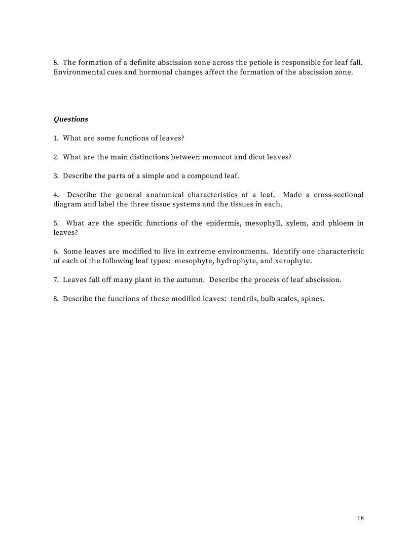
18
8. The formation of a definite abscission zone across the petiole is responsible for leaf fall. Environmental cues and hormonal changes affect the formation of the abscission zone. Questions 1. What are some functions of leaves? 2. What are the main distinctions between monocot and dicot leaves? 3. Describe the parts of a simple and a compound leaf. 4. Describe the general anatomical characteristics of a leaf. Made a cross-sectional diagram and label the three tissue systems and the tissues in each. 5. What are the specific functions of the epidermis, mesophyll, xylem, and phloem in leaves? 6. Some leaves are modified to live in extreme environments. Identify one characteristic of each of the following leaf types: mesophyte, hydrophyte, and xerophyte. 7. Leaves fall off many plant in the autumn. Describe the process of leaf abscission. 8. Describe the functions of these modified leaves: tendrils, bulb scales, spines.

19
PLANTS, PEOPLE AND THE ENVIRONMENT: Insect-eating Plants Insectivorous plants have special mechanisms for snaring and digesting their prey. Some of these involve leaves with elaborate trichomes. The Venus flytrap (Dionaea muscipula), for example, has specialized leaves that look as if they are hinged along the central vein separating the two lobes of the blade (Fig. 1a). On the surface of the lobes are two kinds of trichomes: trigger hairs and secretory hairs. Trigger hairs are long cells with a multicellular base. When an insect moves two or more of these hairs, the trap is triggered, closing the leaf blade and entrapping the insect (Fig. 1b). The insect's struggles push its body against several short secretory hairs with multicellular heads. These hairs secrete digestive enzymes that break down the soft parts of the insect's body into nutrients that the leaf then absorbs. Sundews (Drosera capillaris) capture insects by producing a thick, sticky mucilage from "tentacles" (Fig. 1c), which are multicellular structures much more complex than simple trichomes. When an insect lands on the leaf, it sticks to the mucilage, and the leaf curls to trap the insect. Secreted enzymes than digest the insect body. Pitcher plants secrete substances from glands located along the lip of the pitcher-shaped leaves (Fig. 1d). An insect that lands to feed and happens to crawl to the inside of the "pitcher" is likely to slip down the side. Downward-pointed sharp hairs impede the insect's progress as it tries to crawl out (Fig. 1e). Eventually the insect tires and falls into the digestive fluid at the bottom of the pitcher. The amino acids, lipids, sugars, and minerals, especially nitrogen, released from breaking down the insect's body are used by the plant for growth. Insectivorous plants can survive without capturing and digesting insects, but this highly developed specialization gives them an advantage in nutrient-poor marshes and bogs.
Figure 1. Insectivorous plants that use trichomes on their leaves to attract, capture, or digest insects. (a) Venus fly trap. (b) Venus fly trap that has captured a fly by closing leaf lobes. (c) Sundew captures insects by trapping them in mucilage secreted by "tentacles." (d) Pitcher plant entices insects to land by secreting sweet substances, only to block with sharp hairs (e) the egress of those insects that wander into the leaf cup.
a b c
d e

20
ECONOMIC BOTANY: Commercially Important Fibers People have used fibers from plants for at least 10,000 years. Cotton fibers are known to have been used in Mexico almost 9,000 years ago. Cotton fibers, however, are not really fibers in terms of their cell type; they are epidermal hairs that grow from the cotton seed coat. The kind of fibers discussed here are elongated sclerenchyma fibers joined into long strands by their overlapping ends. Bundles of these fibers can be long and very strong. Archaeological evidence has revealed that Native Americans were using Agave (sisal) to make cords about 10,000 years ago, and Stone Age Europeans were using flax plants (Linum usitatissimum) for linen at about the same time. Where do fibers come from? They tend to form in strands around vascular bundles and within the ground tissues of stems and leaves. Their purpose in the plant is to strengthen stems and leaves so they do not break when moved by the wind. They act like reinforcing bars used in concrete structures. Ancient peoples figured out how to extract these strands and use them for fabrics and ropes.
There are basically two types of commercially important fibers: hard fibers and soft fibers (see the Table). Hard fibers occur in leaves of monocotyledonous plants such as Agave (Fig. 1). Their long, strap-shaped leaves contain thick bundles of fibers, often surrounding the vascular bundles. These fibers are very stiff and often have thick lignified cell walls. Fiber strands are extracted from the leaves, usually by crushing or beating the leaves and then scraping them to expose the fiber bundles. Once exposed, they are pulled and dried. These fibers (called sisal) are woven into coarse fabric or twisted and braided into rope. Another example is Manila hemp (Musa textilis), which is used to make the huge ropes that tie down ships because it resists decay and breakdown by salt water. Some Manila hemp also is used to make lightweight fabric and tea bags. Soft fibers (sometimes called bast fibers) are found mostly in the stems of dicotyledonous plants, where they occur just outside the phloem as part of vascular bundles. Individual soft fiber cells are longer than those of hard fibers. They tend to be rather soft and flexible and are sometimes unlignified. These properties make such fibers valuable for clothing fabric, string, rope, and even cigarette paper and money. Probably the most famous soft fiber is linen from the flax plant, which currently accounts for about 2% of the world's textiles (Fig. 2). The plant, usually about 1 meter in

21
height, is pulled from the soil and bundled to dry. The fibers are extracted by a process called retting. This involves placing the dried plants in a watery bacterial slurry and allowing the bacteria to decay the ground tissues away, leaving the fiber bundles. These are then combed out, removed, and dried (Fig. 3). A newer way to extract them is to pass the undried plants through crushers, comb the bundles free, and dry them. The dried fibers are then spun into yarn, dyed, and woven into fabric. The elegant feel and texture of linen have made it popular for a long time. Its only problem is that it wrinkles easily. Currently, linen fibers are chemically treated, and the yarn is woven with cotton, rayon, or both. This make contemporary fabrics that are more wrinkle-resistant but retain some of the attractive texture of the original. Ramie (Boehmeria nivea) has the longest known fiber cells. They can be up to 550 mm (almost 2 feet) in length and are stronger than cotton fibers. Mercerized ramie, made by treating the fiber bundles with alkali, is among the strongest natural fibers used in clothing. The next time you see a very exotic looking fabric, look at the label. You will probably find that it is linen or ramie.
Figure 1 (right). Agave sisaliana plants foreground are grown as a crop in various places in the world: shown here in Malaysia. The large tree in the background is a baobab (Adonsonia digitata). The swollen trunk of the tree stores water.
Figure 2 (above). Flax plant, Linum usitatissimum. Figure 3 (right). Flax fibers used to make linen are being processed.

22
Figure legends CO http://en.wikipedia.org/wiki/Four-leaf_clover#/media/File:4-leaf_clover.JPG Figure 6.1=Starr and Taggart Figure 6.2=Starr and Taggart Figure 6.3a = examples of moncot leaves- crabgrass –Appeared in Rost 1st ed and originally in Weier 6th Figure 6.3b = examples of moncot leaves- corn – same as 6.3a Figure 6.3 c = examples of moncot leaves-barley – same as 6.3a Figure 6.4 a = venation patterns in leaves- parallel in a monocot – Thomas L. Rost Figure 6.4 b = venation patterns in leaves- netted in a dicot – Thomas L. Rost Figure 6.4c = Thomas L. Rost Figure 6.5a=Starr and Taggart Figure 6.5b=from Lesley Sunell UC, Santa Cruz Figure 6.6 a = features of leaf epidermis- surface of potato leaf – Richard Falk Figure 6.6b=Weier 6th Figure 6.7 a = trichomes- branched (photo) – Richard Falk Figure 6.7 b = trichomes- pointed (photo) – Richard Falk Figure 6.8 = photo of lilac leaf in cross section showing the midrib – appeared in Rost 1st ed and originally from Weier 6th Figure 6.9=Weier 6th Figure 6.10a = C4 leaf anatomy- corn cross section – Thomas L. Rost Figure 6.10 b = C4 leaf anatomy- paradermal section through the leaf – Thomas L. Rost Figure 6.11a=from Joseph Lin, appeared in Rost 1st ed and Weier 6th Figure 6.11b=from Weier 6th and Rost 1st Figure 6.12= Weier 6th & Rost 1st Figure 6.13 a = age-dependent leaves of ivy- juvenile – Thomas L. Rost Figure 6.13 b = age-dependent leaves of ivy- adult– Thomas L. Rost Figure
Figure 6.14=from Weier 6th and Rost 1st
Figure 6.15a&b=Thomas L. Rost Figure 6.16a,b&c= Thomas L. Rost Figure 6.17a,b&c=Rost 1st Figure 6.18a&b=Rost 1st SIDEBAR - PLANTS, PEOPLE, AND THE ENVIRONMENT: Insect-Eating Plants Figure 1a= Venus flytrap open – Thomas L. Rost all of these were in Rost 1st Figure 1b = insectivorous plants that use trichomes- Venus’s-flytrap closed – Thomas L. Rost Figure 1c = insectivorous plants that use trichomes- Sundew Figure 1d = insectivorous plants that use trichomes- Pitcher plant – Thomas L. Rost Figure 1e = insectivorous plants that use trichomes- egress of insects in leaf cup – Thomas L. Rost SIDEBAR – ECONOMIC BOTANY: Commercially Important Fibers Figure 1 = from Tim Metcalf Figure 2 = photo of flax plant – Courtesy of MASTERS OF LINEN/USA. Figure 3 = photo of flax fibers being processed – Courtesy of MASTERS OF LINEN/USA.