
CHAPTER-6 FTIR AND FTR ASSIGNMENTS AND...
45
CHAPTER-6 FTIR AND FTR ASSIGNMENTS AND VIBRATIONAL ANALYSIS OF 2-AMINO PYRIDINE AND 2-AMINO lOLINE
Transcript of CHAPTER-6 FTIR AND FTR ASSIGNMENTS AND...

CHAPTER-6
FTIR AND FTR ASSIGNMENTS ANDVIBRATIONAL ANALYSIS OF
2-AMINO PYRIDINE AND 2-AMINOlOLINE
FTIR AND FTR ASSIGNMENTS AND VIBRATIONAL
ANALYSIS OF 2-AMINO PYRIDINE AND 2-AMINO PICOLINE
1. INTRODUCTION
Pyridine, also called azabenzene or azine is the most benzene like of the heterocyclic
compounds. It has a high resonance energy and its structure resembles to benzene
quite closely. The presence of nitrogen atom in the ring does, of course, represent a
major perturbation and alkylation which has no analogy in benzene. When we
encounter with the properties of pyridine, they resembles to those of tertiary amine
and the aromatic sextet is not involved in these reaction. Distortion of electron
distribution in both it-bonding system and in the ci- bonding system are due to the
influence of nitrogen atom. Nucleophilic substitution not common in benzene is
much easier in pyridines, particularly at the 2- and 4- positions which are activated
by nitrogen. Aminopyridines are obtained from pyridine by replacing the hydrogen
attached to the ring by an amino group. By an appropriate location of the amine in
the pyridine ring either 2- or 3- or 4-aminopyridine shortened by 2AP, 3AP and 4AP
can be obtained.
Heterocyclic àmines (HAs) serve as potent food mutagens, formed in fried, broiled
or grilled meats and increase rates of colon, mammary, prostate and other cancers in
bioassay rodents. Studies of human dietary HA exposure was done to US National
Continuing Survey of Food Intakes by Individual (CSFII) data on meats consumed
and cooking methods showed that the pan-fried meats were the largest source of
115
HAs in the diet and chicken, the largest source of HAs among different meat types
[1]. The heterocyclic amine(HCA), 2-amino-i -methyl-6-phenylimidazo-[4,5-b]-
pyridine (PhIP) forms high levels of adducts in a number of organs particularly
liver, kidney and heart and causes HCA induced carcinogenesis [2]. The exogenous
compounds such as PhIP (HA) may have an important role in the generation of
tumours in Mismatch Repair (MMR) defective individuals. The datas suggest that
PhIP may increase the risk of human carcinogenesis and there by promote
tumourigenesis by mutating growth regulating genes [3]. As a part of a
comprehensive survey of the heterocyclic amine content of foods, hamburgers,
steaks and pork ribs were purchased from restaurants and the results revealed that
the restaurant products are ten fold higher in heterocyclic amine content [4].
Aminopyridines find wide application in pharmacological industry and in analytical
chemical laboratories. They serve as good anesthetic agent and hence used in the
preparation of drugs for certain brain disease particulary 4-Aminopyridine is an
effective medicine in the treatment for Multiple Sclerosis [5].
Aminopyridines are used as anti-convulsants and sodium channel blockers. The
pharmaceutically acceptable salts and pro drugs are used for the treatment of
neuronal damage following global and focal ischemia, for the treatment or
prevention of neuro degenerative conditions such as Amyotrophic Lateral Sclerosis
(ALS) and for the treatment, prevention or amelioration of both acute or chronic
pain, as anti-tinnitus agents, as anti-manic depressants, as local anesthetics as anti
116
arrhythmics and for the treatment or prevention of diabetic neuropathy [6].
The compounds comprising supramolecular complexes of amino pyridine e.g
Melamine is used in Engineering plastics for high temperature applications since
they have little bubble formation during processing [7]. Memantine and Flupirtine-
the derivatives of aminopyridine are extensively used in pharmacology. The
N-methyl-D-aspartate (NMDA) antagonist Memantine - a drug currently used in the
therapy of spasticity and Parkinson's disease. Flupirtine was found to be centrally
acting, non-opiate analgesic agent which additionally possesses anticonvulsant and
muscle-relaxant activity. Also these drugs almost have no clinical side effects, these
may prove useful both in preventing primary infection of brain cells with the HIV-
virus as well as treating the neurological disorders often associated with the immuno
deficiency syndrome such as AIDS related dementia [8].
The non planarity of aminogroup in aminopyridines were investigated by ab initio
methods and the amino group was found to be non-planar in all the systems [9]. As
4-amino pyridine improves the transmission of nerve impulses down damaged
axons, it drastically improves the conditions of patients suffering from spinal-cord
injury [10]. The 3,4-diaminopyridine plays an effective role in the symptomatic
treatment of multiple sclerosis fatigue like 4-aminopyridine [ 11 ] . Even though
4-Aminopyridine is highly toxic to all mammals including humans if dosages are
exceeded agriculturally, it is used as an extremely effective bird poison sold under
the brand name Avitrol [12].
117
3-aminopyridine is used as intermediate for agro chemical pharmaceuticals mainly
Piroxicam, Tenoxicam, Ampiroxicam and dyes [13]. The pink colour defect in
cooked, uncured turkey is a sporadic problem that can result in economic loss and
consumer dissatisfaction. Fourteen ligands were tested in ground turkey samples for
their ability to reduce pink development and 3AP was one of them [14].
2- amino pyridine can be used as intermediate for the synthesis of pharmaceuticals
especially for antihistamines, anti-inflammatory and other drugs [15]. 2AP is used
in the synthesis of biologically useful synthon 2-chloro-5-hydroxy pyridine- a key
component of the non-opioid analgesic agent [16]. Of several weakly basic
compounds tested, 2- Aminopyridine was selected as the most useful UV-active
substance [17]. 2- Amino pyridine and its pharmaceutically acceptable salts are of
useful in disease like septic shock, rheumatism, allergy, parkinsonism,
cardiovascular diseases, obesity and pain [18]. In high performance liquid
chromatography, 2-amino pyridine is used as a fluorometric label [19]. 2AP is used
in the synthesis of 1,3,1 0-triaryl-imidazolo [4,5-e] pyrido [1,2-a] -2,3,4,1 0-tetra
hydopyrimidine-2-thiones which display notable herbicidal activity [20].
Another derivative of interest in this context is 2-amino-4-methyl pyridine or
4-picoline also known as Amino picoline. It is the new radio ligand and was
developed to measure the binding of molecule to the Nos isoenzymes. Various
nitric oxide synthase (Nos) inhibitors were investigated for their affinity and
118

selectivity towards the three human Nos isoenzymes in radio ligand binding
experiments. 2-amino-4-methyl pyridine bound saturably and with high affinity to
human Nos. The datas identified 2-Amino-4-Picoline as a very useful radio label
for the investigation of the substrate binding site of all three isoforms [21]. Two
new one dimensional oxalato-bridged copper (II) complexes with 2-amino-4-methyl
pyridine and 3-hydroxypyridine were synthesized and characterized by FTIR
spectroscopy [22]. 2-amino picoline serves as an effective reagent in chemical
laboratories. In the chelation-assisted hydroacylation of allylic alcohols 2-amino-4-
picoline is used [23].
Derivatives of picoline have potent hypolipidemic effects, antineoplastic and anti-
inflammatory activities in rodents. Some of the derivatives demonstrated more
potent antineospalastic activity against the Ehrlich ascites carcinoma growth
including 2-amino-4-methyl pyridine cyanoborane and 2-amino-pyridine-
cyanoborane. Most of the derivatives showed good activity against murineL1210
lymphoid leukemia, Tmolt3 human leukemia, Uterine HeLaS cells and human
glioma cell growth [24].
A series of 2-amino-5-substituted pyridine derivatives are toxic and exhibit high
molluscidal activity. The most effective compounds were 2-amino-541-
(benzoliazole- 1 -ylmethyl)-3-methyl pyridine and 2-amino-5- [1 -(benzotriazole-
1-yl)nonyl]-3-methyl pyridine etc [25]. 2-aminopicoline is used along with max.
dose of bromide, chloride or perchlorate complex inorder to the protection towards
119

the human red blood cell damage photoinduced by chloroperbenzoic acid(CPBA)
[26] . 2-amino picoline and 2-amino pyridine have very important effects on the
corrosion of mild steel in HCL and these tend to inhibit the corrosion to a
remarkable extent. It was also found that 2-amino methyl pyridine exhibits higher
maximum efficiency than 2AP [27].
This molecule is also very important in medical and agricultural like applications.
When it is mixed with Rifamycin and iodine in CH202 at room temperature and
treated with 20% ascorbic acid gives 4-deoxy-4-methyl pyrido imidazo rifamycin.
This has outstanding antibacterial properties in vitio and in vivo and are very useful
in combating gastrointestinal micobial infections [28]. However its spectral
characteristics are still not fully explored and has been always a source of interest to
spectroscopists.
Gunasekeran et al. [29] carried out a Fourier transformation infrared and Laser
Raman spectroscopic investigation on 2N (benzoylamino) pyridine. They recorded
the FTIR spectrum in the range 4000 cm-1 - 400 cm4 and the Laser Raman spectrum
in the frequency range 4000 cm -1-100 cm'. They analyzed the spectra on the basis
of C symmetry and assigned the observed bands to different modes of vibration.
The assignments were made from the data collected on magnitude and relative
intensities of the observed bands. They compared assignments with the earlier
relevant works.
120

The study of vibrational spectra of substituted pyridines, aminopyridines attract the
attention of many spectroscopists because of their pharmaceutical and agrochemical
applications. Near infrared spectra of 2-, 3- and 4-aminopyridines and their
deuterated analogs were reported by Padhye and Bhujle [30]. They presented an
analysis of combination and overtone bands involving amino group frequencies.
Their spectral analysis shows that the overtones of the bands due to dimmers of
aminopyridine, clearly observed in the fundamental region, have not been observed
in near infrared region.
Baruah et al. [31] presented the Raman and infrared spectra of 2,6-diaminopyridine
in the region 250 cm'-lOOO cm'. Based on the assumption that 2,6-
diaminopyridine belongs to C 2 , point group symmetry, they proposed the
assignment for the prominent vibrational frequencies of the spectrum. Sanyal et al.
[32] presented the results on investigation on the electronic (3 120A°-2900A°) and
infrared absorption (400 cm'- 4000 cm') spectra of 3-amino-2-chloro pyridine. The
vibrational spectra have been analyzed in terms of fundamentals, the combinations
and overtones. They have assigned most of the prominent vibrational bands in the
spectrum. The IR and Raman spectra of mono crystals and polycrystalline samples
of 4-aminopyridine hemiperchiorate have been studied at various temperatures [33].
The far infrared vapour phase spectra of amino pyridine was presented and analyzed
by Kydd et al. [34]. They reported the spectra between 50- and 665 cm' and found
that the far-infrared vapour phase spectra of amino pyridine were dominated by lines
121
due to transitions in the inversion vibration energy levels of the amino group. The
barriers to inversion were determined by them and were shown to correlate
extremely well with the calculated electron density in the amino nitrogen but slightly
differ with the dihedral angle, between the rings and amino group planes.
The JR and Raman spectroscopic investigation of cadmium tetra cyanonickelate
complexes of amino pyridine was reported and was concluded that ring nitrogen and
not the amino nitrogen is involved in complex formation [35]. Kinie Sasaki et al.
[36] recorded and reported the infrared and Raman spectra for Iodine dichloride and
iodine dibromides of 2-, 3- and 4- aminopyridineim solids. Baran et al. [37] have
presented the polarized infrared spectra of 4-aminopyridine hemiperchiorate single
crystal containing structurally asymmetric NHN bridges at room temperature and
liquid nitrogen temperature. They particularly studied the nature of different modes
of vibrations of nitro group.
Carmona et al. [38] reported the vibrational studies on amino pyridines in aqueous
solution by Laser Raman Spectroscopy. They recorded the Raman spectra of 2-, 3-
and 4-aniinopyridine and 3,4-diaminopyridine in water over the frequency range
4000 cm'-300 cm'. They made the vibrational assignments for many of the
observed frequencies on the basis of isotopic frequency shifts, depolarization ratios,
group frequency consideration as well as comparison with accepted assignments for
certain vibrational modes in other compounds with structural similarities. The
assignments of the Raman spectra of mono amino pyridine was made on the
122

assumption that 2- and 3-amino pyridine belongs to planer Cs point group and the
4-amino pyridine possesses C2 symmetry.
After going through the literatures the need for a through vibrational analysis of
aminopyridines is felt, because the understanding of the force field that hold the
molecular structure of these compounds may provide a deeper insight into their
biological actions when they administered as drugs and in environment as
agrochemicals. Hence in the present investigation the FTIR and FT Raman spectra
of all the 2-, 3-, 4- aminopyridine and 3,4-diaminopyridine were recorded and an
assignment of the observed frequencies to the fundamental vibrational modes of the
molecules were made. A normal coordinate analysis of the said compounds was
also carried out since this could not only help the proper assignment of the
vibrational • frequencies but also present a complete picture about the molecular
dynamics of amino-pyridines.
2. EXPERIMENTAL DETAILS
The samples of the compounds 2-amino pyridine and 2-amino-4-methyl pyridine
were obtained from M/s Aldrich Chemicals U.S.A with stated purity 99% and used
as such without further purification to record FTIR and FTR spectra. The FFIR
spectrum of this compound has been recorded in solid phase following the KBr
pellet in the region between 4000 cm-1 - 400 cm' using Bruker IFS 66V spectrometer
with a scanning speed of 30 cm-1 rnin 1 of spectral width 2.0 cm'. The frequencies
for all sharp bands are accurate to ± 1 cm'. The FT Raman spectrum was also
123

recorded in the same instrument with FRA 106 Raman module equipped with
Nd:YAG laser source operating at 1.064 pm line with 200 mW power and the
spectral resolution is 2 cm 1 . The molecular structure of 2-aminopyridine and
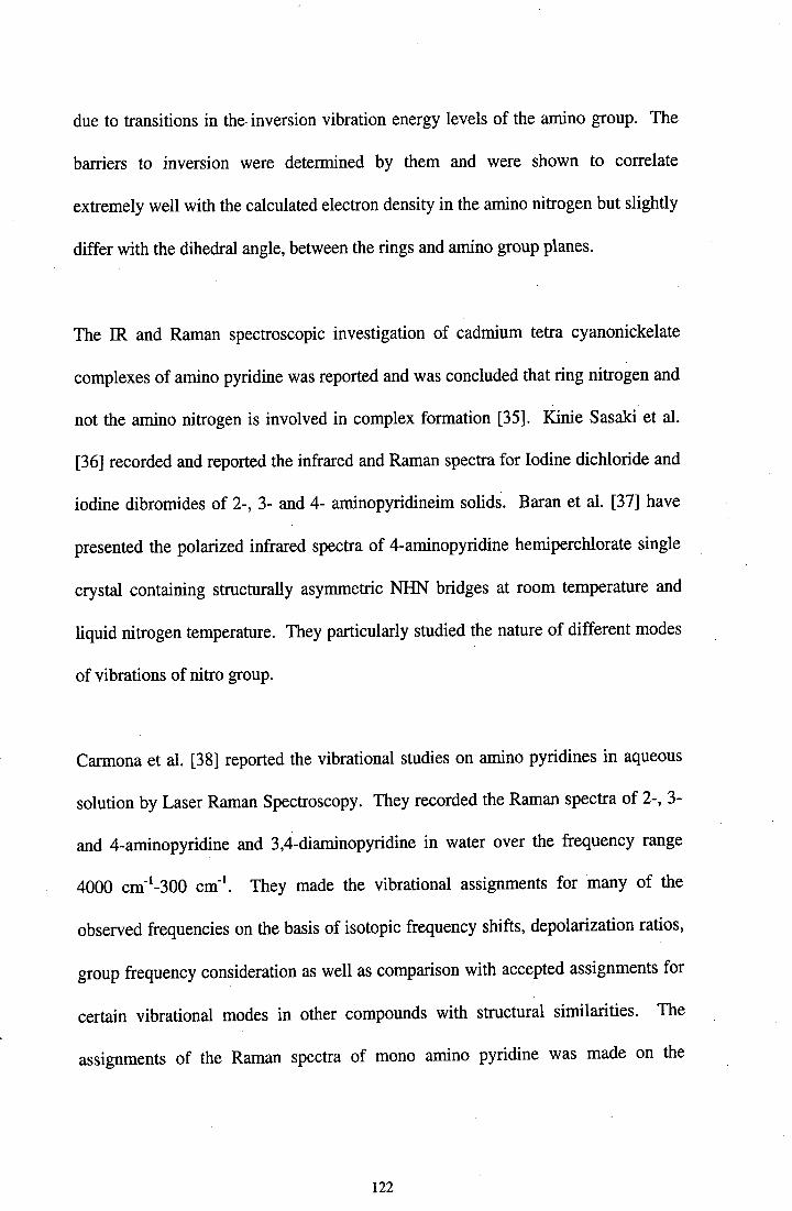
2-amino picoline are shown in fig. 1 and fig. 2. The recorded FTIR and FT Raman
spectra of these compounds are shown in fig 3 and fig. 4 respectively.
3. NORMAL COORDINATE ANALYSIS
Section I
2-AMINO PYRIDINE
The compound, 2-amino pyridine under investigation posses C point group
symmetry by assuming (-NH 2) group as point mass and lies in the plane of the
molecule. C symmetry leads to two types of vibrations namely a' (in-plane) and a"
(out-of-plane) and are distributed into
F vib = 25a+ 8a"
All the vibrations are active both in JR and Raman. The normal co-ordinate analysis
program of Fuhrer et al. [39] was used after suitable modification to calculate
vibrational frequencies and potential energy distribution (PED). This program
follows the Wilson's FG matrix method [40-42] of vibrational analysis in which the
normal co-ordinates are defined with respect to a set of molecular co-ordinates.
JUPAC recommendations were also followed for defining the internal co-ordinates
for the out-of-plane bending vibrations. The structural parameters necessary for the
compound are taken from Sutton table [43] and from the literatures C-C = 1.401 A°,
124

NH2
Fig I. Molecular Structure of 2 - amino pyridine
0000CD000I-
00N00I-
00
CD•1>CL
0CE4
E U
-..
CD
crwD
L)
zLLJw
o--
I-LLz4I—ILr)(IL
000(.400N(.400(.400CD(.400co(.4000(V)
0(.4C',
00C,)
CDC!)
C-H = 1.103 A°, C-NH2 = 1.366 A°, N-H = 0.960 A° and all the ring angles are
1200 . These values are cross checked with molecular modeling program [44]. The
Simplified Valance Force Field (SVFF) was adopted and the force constants were
refined by the damped least square technique. The potential energy distributions are
calculated using the final set of force constants. The SVFF is shown to be very
effective in the normal co-ordinate analysis (NCA) because of the valance force
constants can be transferred between the structurally related molecules, benzene and
pyridine. A salient feature of NCA and force field calculations has been that it could
reproduce the frequencies associated with the skeletal rings as well as the -NH2
group within a reasonable limit (±10 cm') with an acceptable potential energy
distribution.
The potential energy distribution has been calculated in order to check whether the
chosen set of vibrational frequencies contributes the maximum to the potential
energy associated with normal co-ordinate of the molecules. The highest PED
contributions corresponding to each of the observed frequencies are alone listed in
the present work and the PEDs are also listed along with the frequencies in the table.
The close agreement between the observed and calculated frequency confirms the
validity of the present assignment.
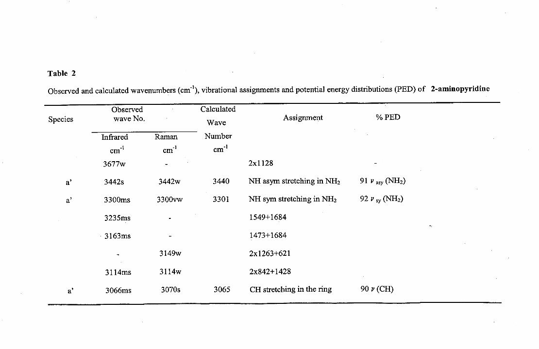
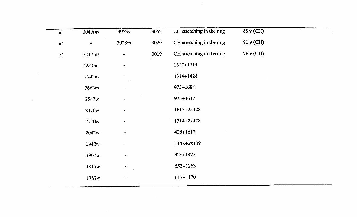
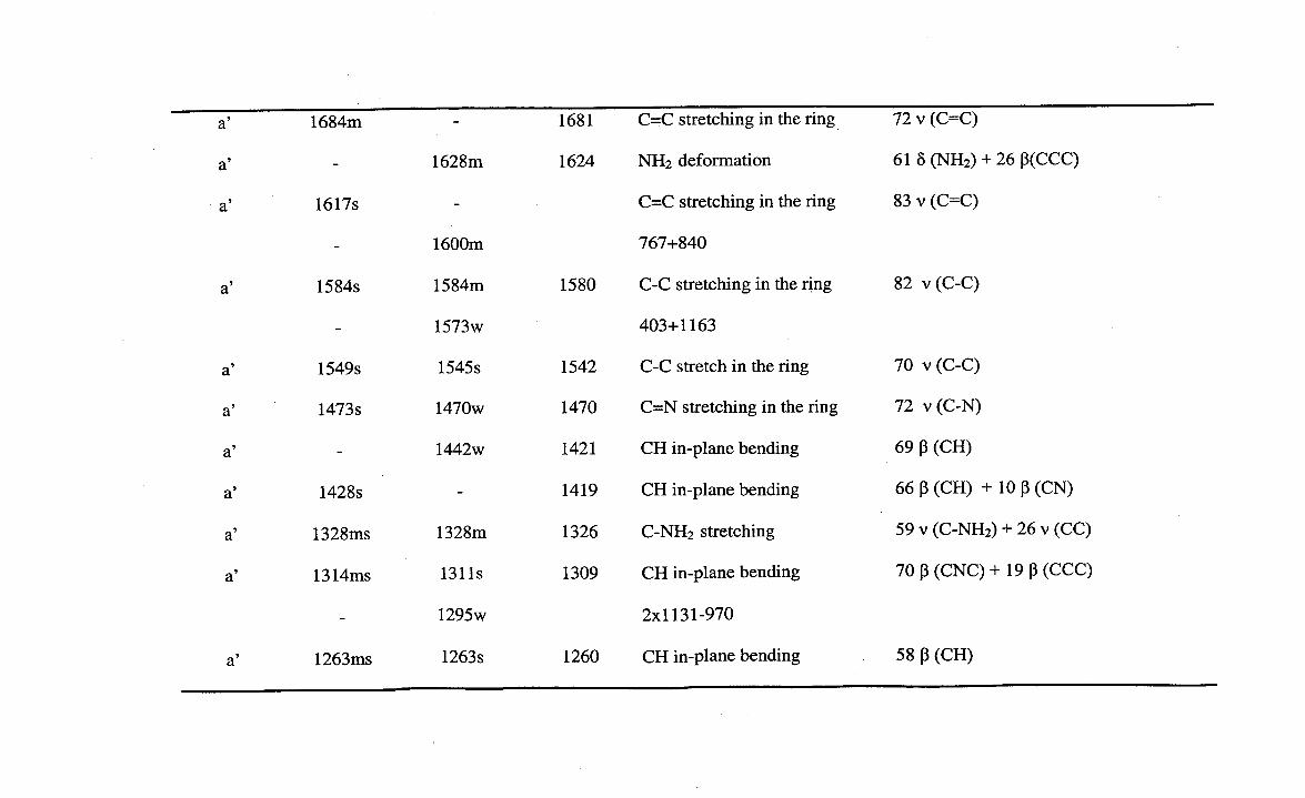
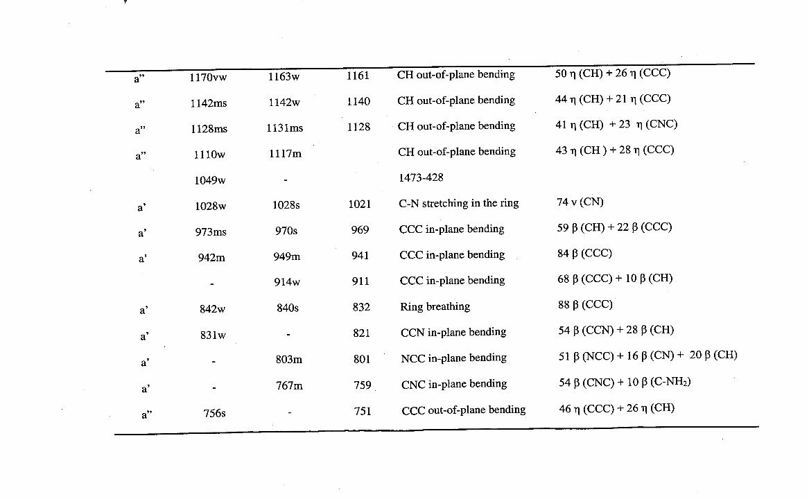
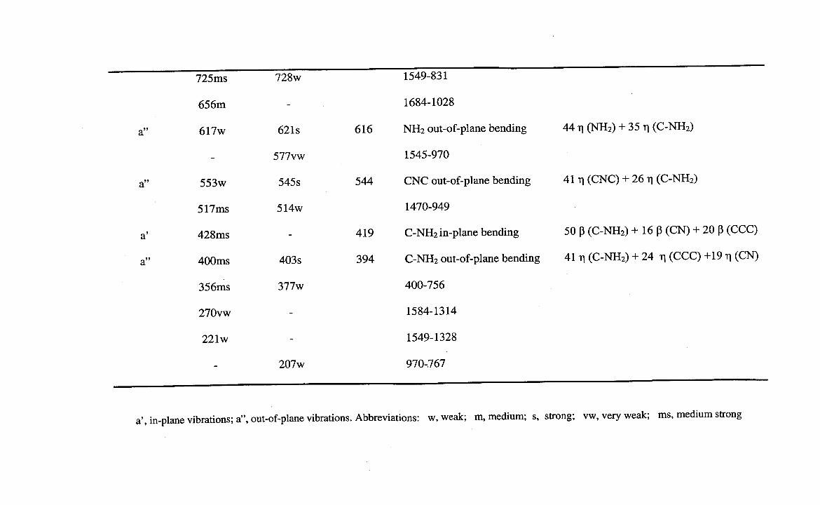
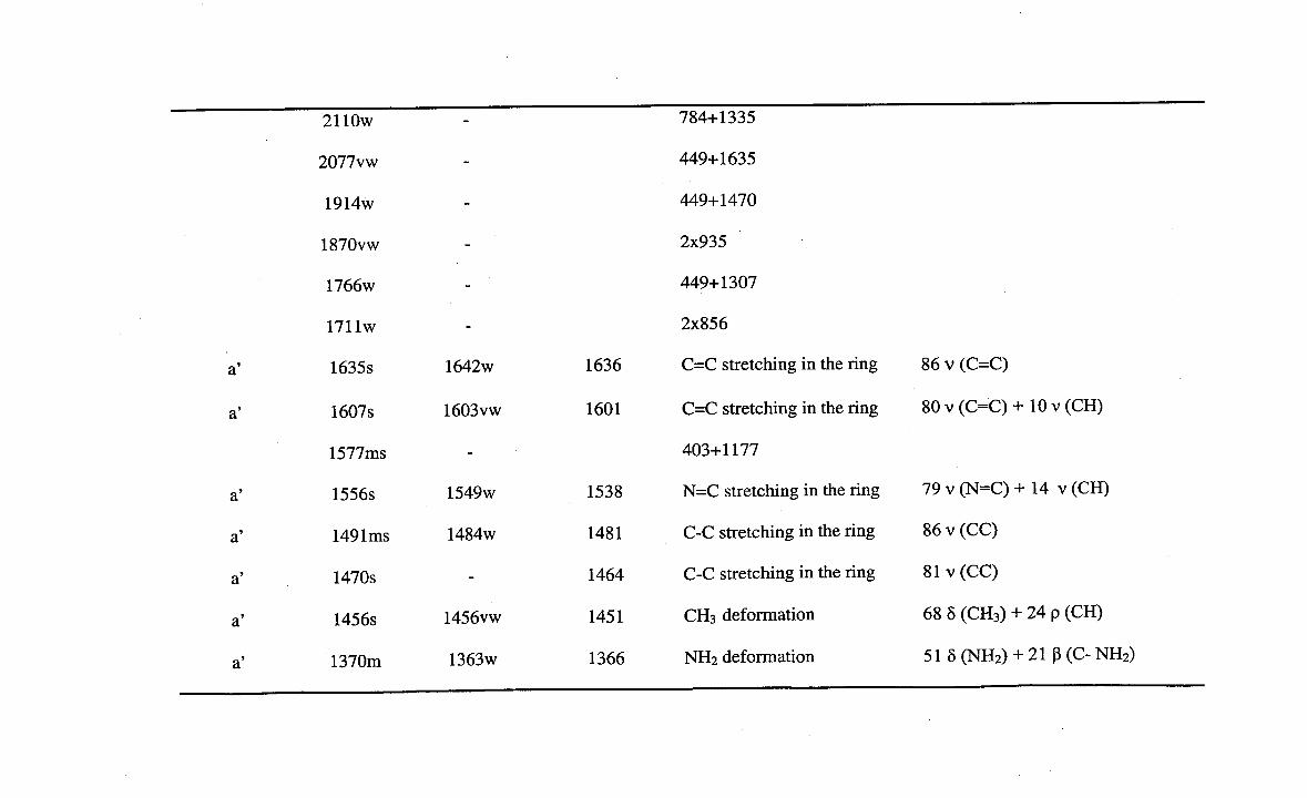
4. VIBRATIONAL ASSIGNMENT
The initial and final set of force constants employed in the present investigation are
given in the table. l. and the observed and calculated frequencies of 2AP along with
125
their relative intensities and probable assignments are presented in table 2.
Assignments have been made on the basis of relative intensities, magnitude of the
frequencies and mainly on the normal co-ordinate calculations as well as literature
data of molecules of similar structure. The vibration of the molecule under study are
divided into two groups: (1) Skeletal vibrations i.e. the vibrations associated with the
ring and (2) Group vibrations due to the substituents. Apart from the assignments to
fundamental vibrations, attempt was also made to assign the overtone and
combination bands. The purity of the normal modes is further confirmed by
calculating the potential energy to each fundamental vibrations. The assignments
pertaining to overtones and combination bands of the samples were not discussed
but listed in the table 2.
4.1 SKELETAL VIBRATIONS
4.1.1 Carbon-carbon vibrations
The ring stretching vibration C-C are very much prominent in the spectrum of
pyridine and its derivatives and are highly characteristic of the aromatic ring itself
[45] . Benzene has two doubly degenerate mode e2g (1596 cm 1 ) and ei (1495 cm')
and two non-degenerate mode b2 (1310 cm-1 ) and aig (995 cm') due to skeletal
stretching of C-C bond. In general the bands around 1650 cm' to 1400 cm' in
benzene derivatives are assigned to skeletal C-C stretching modes [46] . The actual
position are determined not so much by the nature but by the position of the
substituents around the ring [47]. The bands observed at 1684 cm-' and 1617 cm1
in FTIR have been assigned to C=C stretching vibration of 2AP.
126
The bands at 1584 cm' and 1549 cm' in FTIR and 1584 cm -1 and 1545 cm-1 in FTR
have been assigned to C-C stretching vibrations in 2AP. The band at 1028 cm -1 both
in FTIR and FTR has been assigned to C-N stretching. Also C=N stretching
vibration bands are obtained at 1473 cm' in FTIR while 1470 cm-1 in FTR.
The C-C ring breathing aig (995 cm-1 ) and CCC triagonal bending non-degenerate
b1 (1010 cm-1 ) vibrations of benzene give rise to combined modified modes under
Cs symmetry in the present study. The characteristics absorption bands at 842 cm'
in FTIR and 840 cm-1 in FTR belong to ring breathing. These values are in
agreement with the ring breathing mode vibration at around 1000 cm 1 in substituted
benzene [48-49].
In the case of substituted benzene two more in-plane bending vibrations are obtained
from the non-degenerative b1 (1010 cm) and degenerate e2g (606 cm) mode of
benzene [47]. In this present case of 2AP, these are observed at 973 cm 1 and
942 cm' in FTIR and the Raman counterparts are identified at 970 cm 4 and 949 cm4.
The Carbon out-of-plane bending vibrations are derived from the non-degenerate
b2g (703 cm-) and degenerate e21,(404 cm -) modes of benzene. The former is found
constant in substituted benzene [50]. For 2AP this is shifted to 756 cm 1 in FTIR.
4.1.2 carbon-Hydrogen Vibrations
Because of the four C-H bonds in the structure, 2-amino pyridine gives rise to four
127

C-H stretching vibrations. The hetero-aromatic structure shows the presence of C-H
stretching vibration in the region 3000 cm'- 3100 cm' [46]. In this region the bands
are not affected appreciably by the nature of the substituents. In the present work,
the bands observed at 3066 cm', 3049 cm' and 3017 cm_ i in the infrared spectrum
and the corresponding Raman frequencies are observed at 3070 cm', 3053 cm-1 and
3028 cm-1 are assigned to the aromatic C-H stretching vibrations in 2AP. Studies on
the benzene spectrum show that there appear two degenerate e2g (1178 cm-1 ) and
(1037cm 1) and two non-degenerate b2 (1152 cm 1 ) and a2g (1340 cm-1 ) frequencies
involving C-H in-plane vibrations. C-H in-plane bending vibrations lie in the
region1000cm 1 - 1100 cm'. In the light of the above facts, the frequencies
1428 cm 1 , 1314 cm' and 1263 cm-1 in FTIR and 1442 cm', 1311 cm 1 and
1263 cm' in FIR are assigned to C-H in-plane bending vibrations.
The C-H out-of-plane deformation results from b2g (985 cm-') e2 (970 cm1),
eig (820 cm') and a2U(671 cm) modes of benzene and they are expected to occur in
the region 600 cm'- 1000 cm'. The changes in the frequencies of these
deformations from their values in benzene are almost determined exclusively by the
relative position of the sub stituents and are almost independent of their nature [451.
Hence the band 1170 cm - ', 1142 cm 1 , 1128 cm' and 1110 cm' in FTIR and have
almost same counterparts in FIR are assigned to C-H out-of-plane vibrations.
4.1.4 Carbon-Amine vibrations
Carmona et al. [38] assigned C-NH2 stretching absorption in the region 1250 cm1
and 1340 cm' in all the primary aromatic amines. The intensity of the bands
128

appears to be rather variable and probably it is associated with some substituents or
other structural features. Here in this present work, the medium strong bands at
1328 cm 1 in FTIR and in FTR are assigned to C-NH2 stretching vibrations. C-NH2
in-plane bending vibration is assigned at 428 cm 1 in FTIR and C-NH2 out-of-plane
vibration is assigned at 400 cm' in FTIR and 403 cm-1 in Raman.
4.2. GROUP VIBRATION
4.2.1 Amino group
Group vibrations were determined in terms of the motions of the nuclei undergo
during vibrations in the molecules and they appear in fairly constant region of the
spectrum. 2AP possess one NH 2 group and hence one expects one asymmetric and
one symmetric NH stretching in its spectra. It is stated that in amines, the N-H
stretching vibrations occur in the region 3500 cm'-3000 cm 1 [51]. The asymmetric
-NH2 stretching vibration appears from 3500 cm 1 - 3420 cm' and the symmetric
-NH2 stretching is observed in the range 3420 cm 1 to 3340 cm-1 . With reference to
this, the vibrational frequencies observed at 3442 cm -1 both in FTIR and FTR is
assigned to N-H asymmetric stretching vibration and the frequency observed at
3300 cm-1 both in FTIR and FTR is assigned to N-H symmetric stretching
vibrations. These observations agree well with the earlier work [52-531.
Other vibrations of amino group that is deformation has the characteristic frequency
usually located in the region 1650 cm-1 - 1600 cm-1 [47]. Therefore the medium
band in Raman at 1628 cm 1 is assigned to the deformation vibration mode of amino
129
group. Like wise the out-of-plane bending of amino group or wagging, the band
appears between 850 cm-1 and 750 cm-1 . Here the band at 617 cm' in infrared and
at 621cm' in Raman spectra is attributed to the amino wagging mode.
5. POTENTIAL ENERGY DISTRIBUTION
The Potential Energy Distribution (PED) has been calculated to check whether the
chosen set of assignments contribute the most to the potential energy associated with
normal co-ordinates of the molecules. The higher PEDs contribution corresponding
to each of the observed frequency is listed in the table 2.
From the normal coordinate analysis, the calculation of potential energy distribution
of the fundamental vibration modes show that almost all skeletal as well as group
vibration of the compound contribute maximum to the potential energy associated
with the respective bonds. Generally the skeletal carbon stretching vibrations
coupled slightly with the C-H stretching and CCC in-plane bending modes. The C-
H stretching, -NH2 stretching vibrations are considered to be absolutely pure modes
since the PED contribution of these modes are almost 100%. The C-H in-plane and
out-of-plane bending vibrations are also obtained in pure modes. From the NCA, it
is also observed that the maximum number of fundamental vibrations obtained
below 500 cm_ i significantly mixed with the neighboring modes. The RE
calculation determines the reliability and precision of the present spectral
assignments of the fundamental vibrational modes of the compound.
130
SECTION II
2-AMINO 4-METHYL PYRIDINE
2-amino-4-methyl pyridine also posses a C point group symmetry and the (-NH2)
group and (-CH3) group are considered as point masses and lie in the plane of the
molecule. Due to the C 5 symmetry, there are two types of vibrations in-plane (a') and
out-of-plane (a"). They are distributed into
Fvib = 30 a'+ 12 a"
All the vibrations are active both in infrared and Raman. The normal co-ordinate
analysis was used after suitable modification to calculate vibrational frequencies and
potential energy distribution. The structural parameters necessary for the compound
are taken from Sutton table [43] and similar molecules. Both for the in-plane and
out-of-plane vibrations, the simplified general valance force field was adopted and
were refined by the damped least square technique. The potential energy
distributions are calculated using final set of force constants. The SVFF is shown to
be very effective in the normal coordinate analysis because of the valance force
constants can be transferred between the structurally related molecules. The normal
co-ordinate analysis and the force field calculations have a salient feature that they
could reproduce the frequencies associated with the skeletal rings as well as the
(-NH2) and (-CH3) groups within a reasonable limit (±10 cm 1) with acceptable
potential energy distribution.
6. VIBRATIONAL ASSIGNMENTS:
The recorded FTIR and FTR spectrum of 2-amino-4-picoline is shown in fig 4. The
131
. I
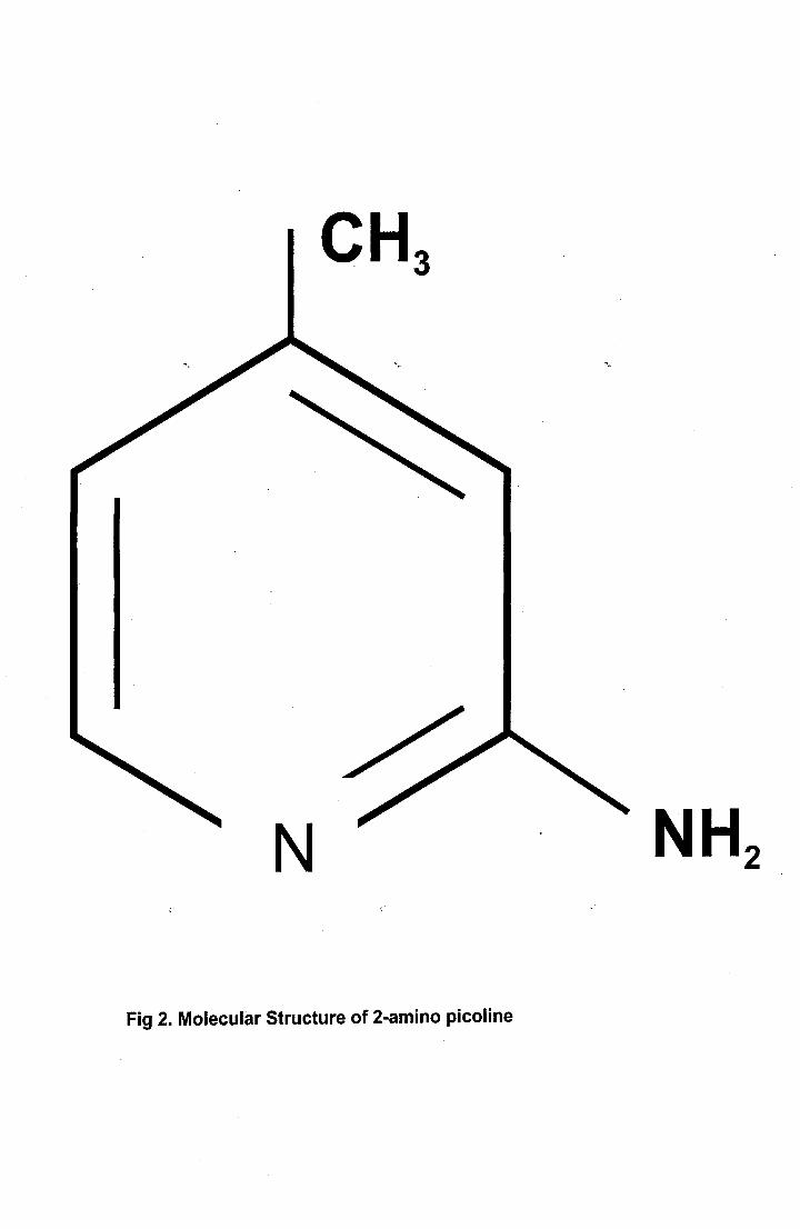
NH2[a
Fig 2. Molecular Structure of 2-amino picoline

N00N00'CDN00
I—ILaz4I—IL0IL
00
— 7 =--:: ^ ^>
. ---: n000N0000CD0.0co
c0CL
NT
E
0 'E
LL
0 W
O000.0
uJ
N(I)
observed and calculated frequencies of 2-amino-4-picoline along with their relative
intensities and probable assignments are presented in table 3. Assignments have
been made on the basis of relative intensities, magnitude of the frequencies and
mainly on the normal co-ordinate calculations as well as literature data of the
molecules of similar structure. The purity of the normal modes is further confirmed
by calculating the potential energy to each fundamental vibration.
6.1 SKELETAL VIBRATIONS
6.1.1 Carbon-Carbon vibrations
The bands around 1400 cm' to 1650 cm-1 in benzene derivatives are assigned to
skeletal C-C bands. The bands observed at 1491 cm' and 1470 cm' in FTIR and
1484 cm' in FTR have been assigned to C-C aromatic stretching vibrations. The
actual position of the bands are determined not so much by the nature of the
substituents but by the position of the substituents in the ring [47]. The bands at
1635 cm 1 and 1607 cm' in FTIR and 1642 cm' and 1603 cm' in FTR are assigned
to C=C stretching vibration of 2-amino-4-picoline. The medium intensity band
obtained at 856 cm-1 in FTIR corresponds to ring breathing vibration which is in line
with Mohan et al. [54].
The carbon in-plane bending vibration are derived from non-degenerate b1
(1010 cm") and degenerate e2g (606 cm') mode of benzene [47] . The degenerate
frequency 606 cm-1 of benzene splits into two when symmetry is reduced. The
magnitude of one of the component remains almost unchanged while the other is
132

reduced substantially [55]. The band obtained at 849 cm -1 and 842 cm-' in FTR
have been assigned to CCC in-plane bending vibration. In this present work of
2-amino-4-picoline, the out-of-plane vibrations have been assigned to 765 cm' in
FTIR and 756 cm-1 both in infra red and Raman.
6.1. 2 Carbon - Hydrogen vibrations
In aromatic compound the C-H stretching vibration generally lie in the region
3000 cm' to 3100 cm-1 which enables quick identification of the band. They will
not be much affected by the nature of the substituents. Because of the four C-H
bonds in the structure of 2-amino-4-picolifle, it gives rise to four C-H stretching
vibrations. In the present study, the bands at 2914 cm', 2842 cm 1 and 2770 cm 1 in
FTIR and 2914 cm', 2863 cm 1 and 2849 cm-1 in FTR have been assigned to C-H
stretching vibrations . Studies on the benzene spectrum show that there appear two
degenerate e2g (1178 cm) and ej u (1037 cm') and two non-degenerate b2
(1152 c1d) and a2g (1340 cm') frequencies involving C-H in-plane vibrations. C-H
in-plane bending vibrations lie in the region 1000 cm -1 -1100 cm'. By keeping the
above facts in account, the frequencies corresponding to 1307 cm 1 , 1270 cm-1 and
1237 cm 1 in FTIR are assigned to C-H in-plane bending vibrations. The
corresponding C-H in-plane bending vibrations obtained in the FTR are 1307 cm',
1266 cm 1 and 1235 cm1.
The C-H out-of-plane deformation results from b2g (985 cm 1 ), e2 (970 cm1),
elg (820 cm 1 ) and a (671 cm-) modes of benzene and they are expected to occur in
133
the region 600 cm'- 1000 cm'. Even if the frequencies are independent of the
nature of the substituents, they are almost determined by the relative position of the
substituents. Hence the bands obtained at 1177 cm 1 and 1128 cm 1 in FTIR and
1184 cm 1 , 1173 cm-1 and 1128 cm-1 in FTR correspond to 3 out-of-plane C-H
bending vibrations.
6.1.3 Carbon-Amine vibrations
The result of normal co-ordinate analysis is used to assign the band observed at
1335 cm -1 to C-NH2 stretching vibration in FTIR and the counterpart in FTR is
obtained at 1331 cm -1 . The C-NH2 in-plane bending modes are observed at
449 cm in FTIR and 442 cm 1 in FTR. The band that is observed at 307 cm4 both
in FTIIR and in Raman has been assigned to C-NH2 out-of-plane bending vibration.
6.1.4 Carbon-Methyl vibrations
The band observed at 977 cm' in infrared is attributed to the C-CH 3 stretching. The
in-plane bending vibration is observed at 703 cm-1 in infrared and the out-of-plane
bending vibrations are attributed to 521 cm' in FTIR and 514 cm' in Raman.
Almost 25% of the C-CH3 stretching, in-plane and out-of-plane vibrations are
contributed by the CC modes of stretching, in-plane and out-of-plane vibrations
respectively.
134
6.2 GROUP VIBRATIONS
6.2.1 Amino group
Group vibrations were determined in terms of the motions of the nuclei in the
molecules, undergo during vibrations and they appear in fairly constant region of the
spectrum. 2 amino-4-picoline possess one NH 2 group and hence one expects one
asymmetric and one symmetric NH stretching in its spectrum. In amines, the N-H
stretching vibrations occur in the region 3500 cm -1 - 3000 cm' [51]. The
asymmetric -NH2 stretching vibration appears from 3500 cm'- 3420 cm' and the
symmetric -NH2 stretching is observed in the range 3420 cm -1 to 3340 cm-1 . With
reference to this, the vibrational frequencies observed at 3435 cm_ I in FTIR and
3431 cm-1 in FTR are assigned to N-H asymmetric stretching vibration and the
frequency observed at 3307 cm' in FTIR is assigned to N-H symmetric stretching
vibrations. These observations agree well with the earlier work [52-53]. The medium
band in infrared at 1370 cm-1 and the weak band at 1363 cm' in Raman are
assigned to the deformation vibration mode of amino group. Like wise the out-of-
plane bending of amino group or the torsional mode of vibration is assigned to the
weak bands observed at 235 cm' both in FTIR and FTR..
6.2.2 Methyl Group
The major spectral changes in the compound, 2-amino-4-picoline is due to the
substituent (-CH 3) group attached with the ring can have the following types of
vibrations. Symmetric and asymmetric stretching, deformation, rocking, wagging
and torsion. Some of these vibrations are observed and are discussed below.
135

The asymmetric and symmetric stretching vibrations of a methyl group usually
occur at about 2965 cm-1 and 2880 cm' respectively. If the C-H bond is adjacent to
an aromatic ring, the C-H stretching frequency and absorption between 3100 cm1
and 3000 cm-1 can be expected. In the light of the above facts the bands at
3135 cm-1 and 3059 cm' in FTIR and 3049 cm-1 in FTR are attributed to C-H
asymmetric stretching in CH 3. Similarly, the bands at 2977 cm -1 in infrared and
2970 cm-1 in Raman are attributed to C-H symmetric stretching in CH3 . And this
C-H stretching modes of vibrations are considered to be absolutely pure modes,
since the PED contribution of these modes are high.
The CH3 in-plane bending vibrations i.e the deformation and rocking mode of
vibrations are assigned for this molecule as follows. The strong band observed at
1456 cm-1 in FTIR and the weak band of the same frequency in Raman are assigned
to the deformation mode of vibration. The medium intensity band at 1335 cm-1 in
FTIR is attributed to the CH 3 rocking mode of vibration.
The next mode of vibration that has to be assigned is the out-of-plane vibrations of
methyl group. The medium/strong intensity band at 403 cm -1 in FTIR and very
weak intensity band at 407 cm' in FTR are assigned to the wagging mode of
vibration. The medium intensity band at 377 cm-1 in infrared has been assigned to
the torsional mode of vibration.
136

7. POTENTIAL ENERGY DISTRIBUTION
The potential energy distribution has been calculated to check whether the chosen
set of assignments contribute maximum to the potential energy associated with
normal co-ordinates of the molecules. The PED contribution corresponding to each
of the observed frequencies are listed in the table 3.
8. CONCLUSION
The above investigation thoroughly analysed the vibrational spectra both infrared
and Raman of 2-amino pyridine and 2-amino-4-picoline. All the vibrational bands
observed in the FT infrared and FT Raman spectra of these compounds are assigned
to the various modes of vibration. Normal co-Ordinate analysis was also carried out
by transferring the force constants from the structurally related molecules and the
calculated frequencies based on NCA are well within the range of observed
frequencies in the spectra of these compounds. PED was also calculated to check
the correctness of the chosen set of force constants, which reveals the purity of the
mode.
137
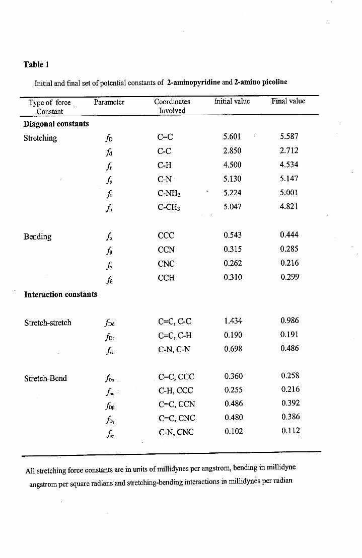
Table 1
Initial and final set of potential constants of 2-aminopyridine and 2-amino picoline
Type of force Parameter Coordinates Initial value Final valueConstant Involved
Diagonal constants
Stretching fD C=C 5.601 5.587
fd C-C
2.850
2.712
Jr C-H
4.500
4.534
Is C-N
5.130
5.147
ft C-NH2 5.224
5.001
A
C-CH3 5.047
4.821
Bending
Ia CCC
0.543
0.444
fp CCN
0.315
0.285
fy CNC
0.262
0.216
A
CCH
0.310
0.299
Interaction constants
Stretch-stretch
fDd C=C, C-C
1.434
0.986
fDr C=C, C-H
0.190
0.191
C-N, C-N
0.698
0.486
Stretch-Bend
IDa C=C, CCC
0.360
0.258
Ja C-H, CCC
0.255
0.216
fDp C=C, CCN
0.486
0.392
fD C=C, CNC
0.480
0.386
A
C-N, CNC
0.102
0.112
All stretching force constants are in units of millidynes per angstrom, bending in millidyne
angstrom per square radians and stretching-bending interactions in millidynes per radian
\00en
IVC
)
CIS
C) ,0
C.)
0
o —It 0
'Cl-en
enen
C)
0en
en
-C
l) C
l) C
l)C
l) C
l)
C)
Cl) E
E
E
Et--
c'0
tnen
- \o
NI t 0 cfl '.0
'. en e
—
— 0
en
en
en
en
en
en
en
C)
C)
C)00Cl)
0II4!Cd
•1
:HIz•;
.EbOECl)
C.)Cl)
C)
bO(1)
—400
E
C14N
Cl) 0
0
00
\0
Cl
0
'.0
+0
—4
'—
.—
Cfl
+
C)
Cl)+
+
'.0
r
-U
)
s.r
-<
N
00—
—4. C'I
(N
0Cl)
0
-—
—4 —
4en e
n e
n 00O\
II
Cl)
C)
-4C.)
C)
r/D
C3d
000
00 —
00
00
00
N
tO
tbi
..
.ti.)
ti)ti.)
ttOb
0000
't 0
0
N N
C-
-N
-
N
f r N
t
m C
C.)C.)
C.)I
00'-
4
<<
-
<N
.0N
0.)0.)
0).—
i-
'O
..O
N
N
's.0N
'crN
'4
+
+ '
+
+'
+
cIDN
It +
+
N
+ N
+
+
+
z
,- - m
cn - -
00t 0
0
NC
n N
N
O
cn N
— N
C.) 0 C
.) —
—4 O
\ O
\ -
1-4 N
t
-
ffl
D
N
C\
CIS
n N
-
C C
Cen
enen
EC
f)00
tr) C
14I
II
II
II
II
II
I
C C
enen
E
EN
C N
en N
C C
N
N
N
N
N
C 00
N
N
't
C -
00C
\N
O
C\ C
\ 0
0 N
N N
N
N
N
N
N
- '- -
—4
I -M
10
•
UUUN+U
C14 U
IIII
u
N —
rnN
\O
00
C?
UN00
N
CA
Ii
001
'.0
ci
•'c!3
Cl) (I)
N—4
Cl)
Cd
Cl)
00
N 0
0
fN
—en
c1r)—
4—
4
_C
•
Cl)
Ecn'.0N—4
U U
—' U
Uc
-N
C\
2+
—_
+ ,;
+U
Z
.U
II
,
zU
U U
U U
U-
c,C
N C
'.0 C
CN
N
'.0 '.0 "
N
U00
0.)0)
.E.E
0)
.—
.-'.-
•bi
0.)0.)
-0.)
0.)
0.)0.)
••0
)C
0)
-0
)o
;-4
0I•
o
•()
C.)c
c
0.)b
—0.)
II
I00
.4
—4
u
U N
UU
Z Z
IIII
'.0C
U Z
U N
U
U U
U U
U U
N
U
C
N C
'-4 Q
\ '.0 C
N
C00 N
00It N
N
-
N C
110
'.0'.0
N
E
E
E
'"
C
't-cfl
'00
C N
00 —
'N
C 0
0 N
N
N '
O\ '.
N N
_4
Em
N
S'—I
- -
— —
.- —
EE
cn N
C
10
ON
ON
00
005
UL) U
ZU
U U
:-:-- m
00
r N
N N
+ +
+ +
U U
U U
---
n
'rIt
C)N+
U
z z
UU
U U
U U
U
-, _,
cell
rn
c00
O
NN
- -
N
N+
+ +
+ +
U U
U Z
U U
UZ
U U
U U
U Z
UU
U U
U U
U, U
U,-./',
-,;.
0L
c
--
:-
00 00
—N
00
O
00kn
'(
'It
t1
LbO
.-
.-
.-
..-.
bO
bJb
bO
a)
a)
-. -
• -
• -
• -
• -
..0
a.))
)
a)
•a)
a)
a)
a)
a)
a)
—I
.0 .0 -
bO
.0 .0 .0 c
cda)
a)
)
)
a.)'1
)
. c
(
mm
CC'
C() - •-1 -
- - - 0
o0
0
000C
d
IM4
CL,
C14
iN
2'
.-.-
•- .0
••
.
o
° 0 c
zU
U u
o Z
U U
UU
() U
U U
Z U
U U
U U
- U
U U
U
U Z
OO
C
00
——
N
.-4 —
C\\C
It N
— _4 —
— —
—
I(
C#)
IE
EE
I C N
00 C
C
\ 00
Ni
NIS
N
N c
n
V4
—
—
—
C C
S
00 00
5
I_
—
''
U Z
U U
U\-,-
-C
\oN
+
+Uc_)
zC.)
•'I I Ii
ti)0)
0)0)
Cd
I—
000
0
'i
00—
N
i C
2
. N
m0
N
\C
C
flN
00.—
iC
'0
Qct
C4
—1
\I
I0
IN
Nc\
'z1-
LflQ
00z N
Z Z
00C
tI
'C
ö ö
C
'fl 'fl N
—
U
'l-l
—CN
—m
IcI
00'
Nin
cn
IN
N N
IN
'C
'fl
lE
E
E
EE
E\C
N
i
N 00 0 'C
C
N—
N
C
'f N
s'C
m
N
NCN
0E
rA 2r.
^cd co
..mc

a)
a)01-IU)
0U)
a)
a)cvj
I4 tiiiI— a)
U)
0 IE
0
U)a)'-40ci)
00
0
caca
>—
O
C
00
00
00
m
en
0 0
0
0•-.
•-
•-bO
bO
. _•4•
•-
•-
•,-4•,-4
o
C)
C)
#•
C)
C)
ci)a)
a)
a)U
),
S
U)
U)
O
,U
)Q
If)U
)
C)+
U)
IfU
)±
ciU
)
Cd
Lf
Cd
Q
U)
z z
4'° — 0
c 0
— 0
C\
Itr C
en en
enen
N
E
'00 C
C)
enN
S
C C
C
\
enN
U)
E
E
E00 5
en
C '
C) If)
1-4-4 C
C C
C)
en N
en
en
en
en

a-,
-,
en
CNr-
00 00
00
tob
b.
.e1)
)
bJtO
en
C
CC
—
u
N
N C
C:)
f_
I.N
L
fl N
00
C
—
.)t)
'- N
C
't
It
O' 00
ON
+ bb +
C
r- +
,- —
+
N
11C
CO)en N
N
++
C
—'
+110 z
It fl N
— N
N
'N
< N
00 C
fl
en
< N
N 0
- 0 0 —
N
N —
C
N
—4
C00 N
N
N N
E
P00NE
I "N
C
N
en
N
en
c\
len
N C
C
't —
t C
t—
00 N
N
O
'r
en
N N
N N
N N
N
N
N N
N
N
N
cq
II
II
N- Q
0
\0—
4 —
4
O\
-It
00—
4 —
4cn
It
—4
C)
—4
+
C) C
)11
11C
) C
)C00
00
C)
CN I
C) C
)——
N N
+
+ +
II
C) C
)
coC
IO
c
.O
—
0
0 —
N 00 00
bbO
..
..
..)
)
..'
0
0bO
boo
.-4—
o
0Ifl L
fl C
N
. N
2 o _
cn c
N
C
Q N
40
, to
n
..0'i
Cfl—
—
4—
tn
110c
c4
C'I
Itt + +
+
C
) +
fl C
) C
) ±
C
) C
) C
)
cnz
GIN
O\
O\
C\
00II
IIII
NIt N
U
C
)
It Z
C
) C
) C
) Z
—4
C"
0C
1-4
—4
00—
It
—4
\0cfl
000
'fl \C
)I r
It
It
Nt
—4 —
4 —
4 —
4 —
4
N'I
N N
"0 '
C \C
C
IN
'C
N
C\ N
tn
It
I —O
IN00
N N
mN
N
.-4
—
,-4
—4
'cr3c
-

00'.0+
-4
+0 0
Zo
0Z
0
0
0 .
-4
m
'.0+
-4
N
'.0
+
+
+
zz N
Qz
0000
00
000
c.
cr.
- m
Q
—
N
C
—
00 Itt C
'II00
N
N
'.0 N
'1N
(( —
to w
bO
to
to
bD 1)-
-a)
a)
a.)a)
a)
a.) a.)
.'1)
a.))
o
-—
—
—
a)
a)
)
— —
0 0 9
a)P
4
0404'
'.0en
II
N
'0 0 +
N
ZN
fl Z
0C
<I
0 0
00000 m
N
00
000N
00 0
0.
O\
00
'r
00
bO
—In
1+
m
bO-4 —
N0
1 10'-4
It
-
N
000
N C
110 c
fl
N '.0
Nm
fl N
N1-4
1-4
-4
4,
0\ C
N N
0\
C'0
-4
N
In0
00
Et
NNaN
'rC
fl 00
Ncn C
'.0 fl 0
0 N
N N
fl N
N
—
— C
1-4
—
1-4
-4
'-4
1-4
II
I
E E
0 In
'0'n
cn In
ONON
00
C/)
Cl)
:E
I N
C
N
N
00 C
N
I cn 0
N
m
N' N
N
00I
) N
N
—
- C
C
o
'0303
Cd_C
C1

CItt
00 C
'.0
'.0
'.0
C'n
n
*t)-4
C
110C
N
00
M 00 fl
t C
It
00 N
N
N
'.0
+z0N+I!
000
0o L) 0
0 -
' 0 0
:-en
— N
NN
++
++
+,-;;
++
'000
G' u
z
00 0
00 0
o
Z0
0 Z
0 0
0000
\_
-,
\__
Co.
Co.
-:-
N O
--1
00
—
00
110O
110C
flfl
"
"
tIO
'-4
Ob1J
O•,
bO)
ti)
0.)0.)
0.)0.)
0.)—
- c
c 0)
C14
C^4
0)0.)
0)0)
0.)
en
enC
)N
0cn.
. •-
0
N
..
CN
00
00
0+
o .- z
00 0
000
0 Z
00
0 Z
OO
ON
'- -
00
0
N
0.)00.)
0
E E
(i It
I I
I I
'.0en
iIt
It
00S
In00
00 r-
>>
'.o en 'n
C
S
-4 C
00C
en C
N
N N
N
N N
'.0
'.0
In
N
•'cv
•'•'

N'-4
C00NN
CCCn
II
II
I
NC
Ni
NCCn
0
Cd
cdD
E(IDt.)
(IDEEa)(ID00a)
+
C'I
0'
z
''0'—I
s::-
9O
O
—
NN
—
C4
++
+
+
+
Z 0 0
0
00
o —
0 C
N
-
flN
toto
O)ti)
to
si.)c)
)—
— —
oo •—
0
ob1tocq
jN
-fl cfl C
o
0
N B
N
C
'C
*,0
00
C?
r
r
--
C"
0 Z
"0 It C
Z
z—
'fl00
erI
Z 00 N
O —
N
N
'—
0
Z
E E
E—
cn
•C
01%
00 0
0
NN
C
00 N
N
N C
cn m
Cfl
•1
I'-)N
REFERENCES
1. Bogen K.T and Keating G.A, J. Esposure-Analysis-and-Environmental-
Epidemiology 11 (2001) 155.
2. Thorgeirsson S.S, Davis C.D, Schut H.A, Adamson R, Snyderwine E.G,
Princess-Takamasu-Symp., 23 (1995) 85
3. Glaab W.E, Kort K.L, Skopek T.R, Cancer-Research, 60 (2000) 4921
4. Knize M.G, Sinha R, Brown E.D, Salmon C.P, Levander O.A, Felton J.S and
Rothman N, J. Agricultural-and-food-chemistry, 46 (1998) 4648.
5. Smith R.C, Emmem H.H, Bertels Mann F.W, Kulig B.M, Van Loenen A.C, and
Polman C.H, Neurology, 44 (1994) 1701.
6. Hogent Camp and Derk, J. Handbook of Am. Chem. Soc. (2001).
7. Loont Jens, Jacobus Antonius, Clauswitz and Kai Uwe, Plastics. Manufacture
and processing (2001)
8. Muller W.E, Pergande G, Ushijima H,Schleger C, Kelve M and Perovic 5,
Prog. Mol.Subcell-Biol., 16 (1996) 44.
9. Bludsky 0, Sponer J, Leszczynski J, Spirko V and Hobza P, J. Chem. Phy. 105
(1998) 11042
10. HYPERLINK http://www.wheelweb.com/aminopyridine.htm
www.wheelweb.com/2-aminopyridine.htm
11. Sheam G.T, Murray N.M.F, Rothwell J.C, Miller D.H and Thompson A.J Brain
121 (1998).
138
12. EXTOXNET, http//ace.ace.orst.eduJinfO/eXOtOXfletIPiPst4T1in0P.htm.
13. www.chemicalland 21.com/arokarhi/lifescience/ago/3- aminopyridine.htm
14. Schwarz S.J, Claus J.R, Wang H, Math Ott N.G, Graham P.P and Fernandes
C.F Poult. Sci., 76 (1997) 1450
15. www.chemicalland 21.com/arokarhi/lifescience/Phar/2- aminopyridine.htm.
16. Krow, Grant R, Xiao, Yushi, Cannon, Kevin, Swan, ScotA, Nickel and Andrew,
Synthetic Communications, 30 (2001) 4093
17. Cheng, Kezhan, Nordmeyer, Francis R, Lamb and John D, J. Capillary.
Electrophoresis, 2 (1995) 279
18. Natsuka, Shunji, Hare and Sumihiro, Methods in Mole. Bio., 76 (1998) 101
19. Araki Y, Andoh A, FujiyamaY, Hata K, Makino J, Okuno T, Nakanura F.
Bamba T, J. Chromatography B: Biomedical Sciences. and Applications, 753
(2001) 209
20. Nizamuddin, Khan, Mukhtar Hussain, Srivastava, ManojKumar, Tiwari and
Shailendra, Indian J Chem. Sec B & Chem. including Medical Chem., 398
(2000) 853.
21. Boer, Rainer, Ulrich, Wolf Rudiger, Klein, Thomas Mirau, bent, Haas, Sabine,
Baur and Ilka, Mole. Phamacology, 58 (2000) 1026.
22. Castillo 0, Luque A, Julve M, Lioret F and Roman P, Inorganica Chimica
Acta, 315 (2001)9.
23. Lee, Dae Yon, Jun, Chul Ho, Moon and Choong Woon, J. Organic Chemistry,
67 (2002) 3945.
139

24. Das M.K, Maiti P,K, Roy S, Mittakanli M, Morse K.W, Hall LH, Arch. Pharm-
Weinheim, 325 (1992) 267
25. Zemity S.R and Radwan M.A, Pesticide Science, 55 (1999) 1203
26. el-Naggar M.M, J. Inorg-Bio. Chem., 65 (1997) 263
27. Ita B.L and Offiony O.E, J. Pure and Appi. Sciences, 6 (2000) 51
28. Cannata, Vincenzo, Tamagnore, gain F, Piani, Silvano, Campana, Manula and
Da Roit, Giovanni. Can., (1986) 30
29. Gunasekaran S, Vardhan R.S and Manoharan K, Indian J. Phys., 67(1993)95.
30. Padhye M.R. and Bhujle V.V, Indian J. Pure and Appi. Phys., 8 (1970) 479
31. Baruah G.D, Ammani AmmaR, Dube P.S and Rai S.N, Indian J. Pure and
Appi. Phys., 8 (1970) 761.
32. Sanyal K.N, Srivastava S.1 and Mrs. Ananda Devi, Indian J. Pure and Appi.
Phys., 21(1983) 56.
33. Grech E, Malarski Z, Sobczyle L, Potier J and Roziere J, J. Mole. Str., 175
(1988) 23
34. Kydd R.A. Spectrochim. Acta 35A (1979) 409
35. Akyoz S, J. Mole. Str., 175 (1988) 365
36. Kimie Sasaki, Ichiro Kuwano and Koyo Aida, J. Inorg. Nuci. Chem., 43 (1981)
485
37. Baron J, Malarski Z, Sobczyle and Erreach B, Spectrochim. Acta, 44A (1988)
993
38. Carmona P, Molina M and Escobar R, Spectrochim. Acta 49A (1993) 1
140
39. Fulirer H, Kartha V.B, Kidd K.L, Kruger P.J and Mantsch H.H, Computer
program for infrared and spectrometry. Normal co-ordinate analysis vol.5,
national research council, ottawa, Canada 1976.
40. Wilson E.B, J. Chem. Phys., 7 (1939) 1047
41. Wilson E.B, J. Chem. Phys., 9 (1947) 76
42. Wilson E.B, Decius J.0 and Cross P.C, Molecular vibrations, MC
Graw Hill, Newyork (1955).
43. Sutton L.E. The interatomic Bond distances and Bond angles in Molecules and
Ions. London Chem-Soc. London (1958)
44. ACD/Chemsketch, Version 3.5, Advanced Chemistry Development Inc. Canada
(1998)
45. Varsanyi G; Vibrational spectra of Benzene derivatives, Akademiai
Kiado, Budapest (1969)
46. George W.O and Mcintyre P.S. Infrared Spectroscopy. John Wiley &
Sons, London (1987)
47. Bellamy L.J, The Infrared Spectra of Complex Molecules, Wiley, New
York 1959.
48. Tripathi R.S, Indian J. Pure Appl. Phys., 11(1973) 277
49. Sharma S.N and Dwivedi C.P, Indian J. Pure and App!. Phys., 13 (1975) 670
50. Green J.H.S, Spectrochim Acta, 18 (1962) 39
51. DilellaD.P and StidhamH.D, J. Raman Spectrosc., 9 (1980) 90
52. Lopez Tocon I, Wolley M-S, Otero J.0 and Marcos J.I, J. Mol.Struct.,
470 (1998) 241
141
53. Arenas J.F, Lopez Tocon I, Otero J.0 and Marcos J.I, J. Mol. Struct., 476
(1999) 139
54. Mohan S, Murugan R and Srinivasan S, Proc.Nat. Acad. Sci. India
62 (1992)154
55. Johri G.K, Prakash V and Srivastava C.L, Indian J. Pure and Appi.
Phys., 14 (1976) 418.
142


