Chapter 5 Cell Respiration
12
Chapter 5 Cellular Respiration & Metabolism 5.1: Glycolysis and the Lactic Acid Pathway Krebs cycle Electron transport CO 2 + H 2 O Lactic acid Capillary Cytoplasm Plasma membrane Interstitial fluid Mitochondrion Glycogen in liver Glucose from digestive tract Glucose in blood plasma Glucose in cell cytoplasm Pyruvic acid Aerobic in skeletal muscle into mitochondrion Respiration Anaerobic Metabolism Glycolysis Glucose from liver Complete combustion of a molecule of glucose requires oxygen and yields ~30 ATP Absence of oxygen, energy is obtained by pathway leading to production of lactic acid Metabolism - (anabolic and catabolic) reactions in body involve energy transformation Catabolic reactions - release energy by breakdown of larger organic molecules break down of glucose, fatty acids, and amino acids serve as primary sources of energy for synthesis of ATP some chem. bonds energy in glucose transferred to energy bonds in ATP, some lost as heat Anabolic reactions - require input of energy and include synthesis of larger energy-storage molecules (glycogen, fat and protein) Energy transfer involves oxidation-reduction reactions. Oxidation - molecule loses election Reduction - molecule accepts the election that was lost final election acceptor is always an oxygen in animal cell Aerobic cell respiration metabolic pathway involving oxygen that converts glucose or fatty acid to carbon dioxide and water Small amount of chem-bond energy of glucose is released at early steps in metabolic pathway, some tissue cells can obtain energy from ATP production in temporary absence of oxygen Glucose undergoes metabolic pathway of glycolysis in cell cytoplasm converting into pyruvic acid Skeletal muscles often convert pyruvic acid into lactic acid under Anaerobic metabolism
-
Upload
aznknight323 -
Category
Documents
-
view
59 -
download
2
description
notes on cell respiration
Transcript of Chapter 5 Cell Respiration

Chapter 5 Cellular Respiration & Metabolism5.1: Glycolysis and the Lactic Acid Pathway
Krebscycle
Electrontransport
CO2 + H2O
Lacticacid
Capillary
Cytoplasm
Plasma membrane
Interstitial fluid
Mitochondrion
Glycogenin liver
Glucosefrom digestive tract
Glucosein blood plasma
Glucosein cell cytoplasm
Pyruvic acid
Aerobic
in skeletal muscle
into mitochondrion
Respiration
Anaerobic
Metabolism
Glycolysis
Glucosefrom liver
Figure 5.1 Overview of energy metabolism using blood glucose. The blood glucose may be obtained from food via the digestive tract, or the liver may produce it from stored glycogen. Plasma glucose enters the cytoplasm of cells, where it can be used for energy by either anaerobic metabolism or aerobic cell respiration. In this schematic diagram, the size of the plasma membrane is greatly exaggerated compared to the size of the other structures and the interstitial (extracellular tissue) fluid.
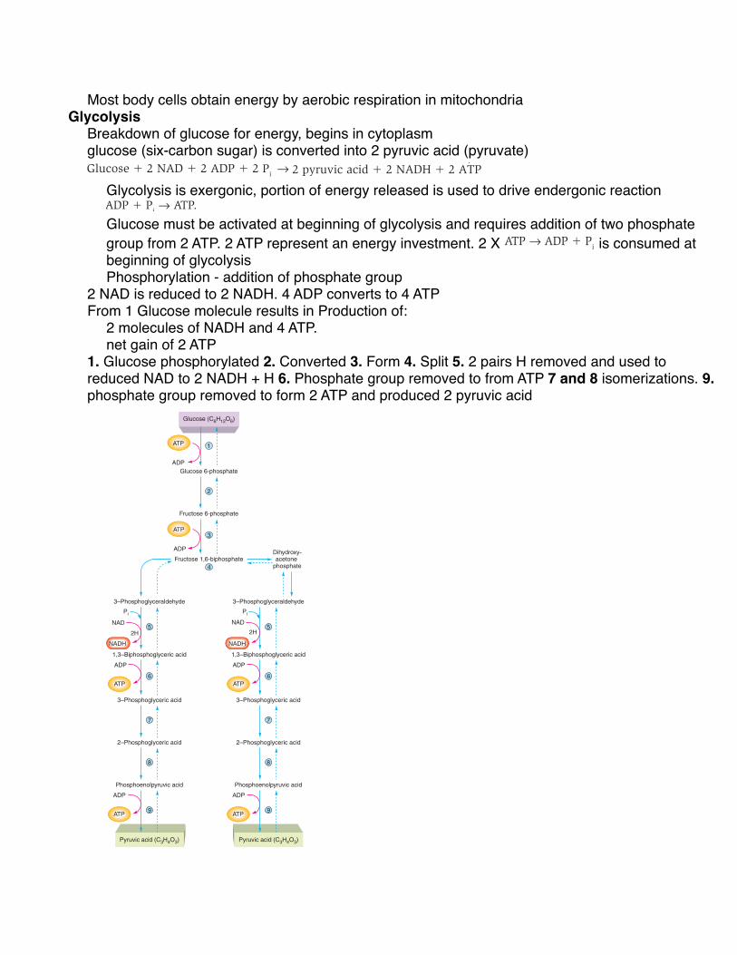
Each pyruvic acid molecule contains 3 carbons, 3 oxy-gens, and 4 hydrogens (see fig. 5.4 ). The number of carbon and oxygen atoms in 1 molecule of glucose—C 6 H 12 O 6 —can thus be accounted for in the 2 pyruvic acid molecules. Because the 2 pyruvic acids together account for only 8 hydrogens, however, it is clear that 4 hydrogen atoms are removed from the intermediates in glycolysis. Each pair of these hydrogen atoms is used to reduce a molecule of NAD. In this process, each pair of hydrogen atoms donates 2 elec-trons to NAD, thereby reducing it. The reduced NAD binds 1 proton from the hydrogen atoms, leaving 1 proton unbound as H + (see chapter 4, fig. 4.17). Starting from 1 glucose mol-ecule, therefore, glycolysis results in the production of 2 mol-ecules of NADH and 2 H + . The H + will follow the NADH in subsequent reactions, so for simplicity we can refer to reduced NAD simply as NADH.
Glycolysis is exergonic, and a portion of the energy that is released is used to drive the endergonic reaction ADP + P i → ATP. At the end of the glycolytic pathway, there is a net gain of 2 ATP molecules per glucose molecule, as indicated in the overall equation for glycolysis:
Glucose + 2 NAD + 2 ADP + 2 Pi →2 pyruvic acid + 2 NADH + 2 ATP
Although the overall equation for glycolysis is exergonic, glucose must be “activated” at the beginning of the pathway before energy can be obtained. This activation requires the addition of two phosphate groups derived from 2 molecules of ATP. Energy from the reaction ATP → ADP + P i is there-fore consumed at the beginning of glycolysis. This is shown as an “up-staircase” in figure 5.2 . Notice that the P i is not shown in these reactions in figure 5.2 ; this is because the phosphate is not released, but instead is added to the inter-mediate molecules of glycolysis. The addition of a phosphate group is known as phosphorylation. Besides being essential for glycolysis, the phosphorylation of glucose (to glucose 6-phosphate) has an important side benefit: it traps the glu-cose within the cell. This is because phosphorylated organic molecules cannot cross plasma membranes.
At later steps in glycolysis, 4 molecules of ATP are pro-duced (and 2 molecules of NAD are reduced) as energy is liberated (the “down-staircase” in fig. 5.2 ). The 2 molecules of ATP used in the beginning, therefore, represent an energy investment; the net gain of 2 ATP and 2 NADH molecules by the end of the pathway represents an energy profit. The overall equation for glycolysis obscures the fact that this is a metabolic pathway consisting of nine separate steps. The individual steps in this pathway are shown in figure 5.3 .
107Cell Respiration and Metabolism
fox78119_ch05_105-127.indd 107fox78119_ch05_105-127.indd 107 25/06/10 9:10 PM25/06/10 9:10 PM
Complete combustion of a molecule of glucose requires oxygen and yields ~30 ATPAbsence of oxygen, energy is obtained by pathway leading to production of lactic acidMetabolism - (anabolic and catabolic) reactions in body involve energy transformation
Catabolic reactions - release energy by breakdown of larger organic moleculesbreak down of glucose, fatty acids, and amino acids serve as primary sources of energy for synthesis of ATPsome chem. bonds energy in glucose transferred to energy bonds in ATP, some lost as heatAnabolic reactions - require input of energy and include synthesis of larger energy-storage molecules (glycogen, fat and protein)Energy transfer involves oxidation-reduction reactions.
Oxidation - molecule loses election Reduction - molecule accepts the election that was lostfinal election acceptor is always an oxygen in animal cell
Aerobic cell respirationmetabolic pathway involving oxygen that converts glucose or fatty acid to carbon dioxide and water
Small amount of chem-bond energy of glucose is released at early steps in metabolic pathway, some tissue cells can obtain energy from ATP production in temporary absence of oxygen Glucose undergoes metabolic pathway of glycolysis in cell cytoplasm converting into pyruvic acidSkeletal muscles often convert pyruvic acid into lactic acid under Anaerobic metabolism
Most body cells obtain energy by aerobic respiration in mitochondriaGlycolysis
Breakdown of glucose for energy, begins in cytoplasm glucose (six-carbon sugar) is converted into 2 pyruvic acid (pyruvate)
Krebscycle
Electrontransport
CO2 + H2O
Lacticacid
Capillary
Cytoplasm
Plasma membrane
Interstitial fluid
Mitochondrion
Glycogenin liver
Glucosefrom digestive tract
Glucosein blood plasma
Glucosein cell cytoplasm
Pyruvic acid
Aerobic
in skeletal muscle
into mitochondrion
Respiration
Anaerobic
Metabolism
Glycolysis
Glucosefrom liver
Figure 5.1 Overview of energy metabolism using blood glucose. The blood glucose may be obtained from food via the digestive tract, or the liver may produce it from stored glycogen. Plasma glucose enters the cytoplasm of cells, where it can be used for energy by either anaerobic metabolism or aerobic cell respiration. In this schematic diagram, the size of the plasma membrane is greatly exaggerated compared to the size of the other structures and the interstitial (extracellular tissue) fluid.
Each pyruvic acid molecule contains 3 carbons, 3 oxy-gens, and 4 hydrogens (see fig. 5.4 ). The number of carbon and oxygen atoms in 1 molecule of glucose—C 6 H 12 O 6 —can thus be accounted for in the 2 pyruvic acid molecules. Because the 2 pyruvic acids together account for only 8 hydrogens, however, it is clear that 4 hydrogen atoms are removed from the intermediates in glycolysis. Each pair of these hydrogen atoms is used to reduce a molecule of NAD. In this process, each pair of hydrogen atoms donates 2 elec-trons to NAD, thereby reducing it. The reduced NAD binds 1 proton from the hydrogen atoms, leaving 1 proton unbound as H + (see chapter 4, fig. 4.17). Starting from 1 glucose mol-ecule, therefore, glycolysis results in the production of 2 mol-ecules of NADH and 2 H + . The H + will follow the NADH in subsequent reactions, so for simplicity we can refer to reduced NAD simply as NADH.
Glycolysis is exergonic, and a portion of the energy that is released is used to drive the endergonic reaction ADP + P i → ATP. At the end of the glycolytic pathway, there is a net gain of 2 ATP molecules per glucose molecule, as indicated in the overall equation for glycolysis:
Glucose + 2 NAD + 2 ADP + 2 Pi →2 pyruvic acid + 2 NADH + 2 ATP
Although the overall equation for glycolysis is exergonic, glucose must be “activated” at the beginning of the pathway before energy can be obtained. This activation requires the addition of two phosphate groups derived from 2 molecules of ATP. Energy from the reaction ATP → ADP + P i is there-fore consumed at the beginning of glycolysis. This is shown as an “up-staircase” in figure 5.2 . Notice that the P i is not shown in these reactions in figure 5.2 ; this is because the phosphate is not released, but instead is added to the inter-mediate molecules of glycolysis. The addition of a phosphate group is known as phosphorylation. Besides being essential for glycolysis, the phosphorylation of glucose (to glucose 6-phosphate) has an important side benefit: it traps the glu-cose within the cell. This is because phosphorylated organic molecules cannot cross plasma membranes.
At later steps in glycolysis, 4 molecules of ATP are pro-duced (and 2 molecules of NAD are reduced) as energy is liberated (the “down-staircase” in fig. 5.2 ). The 2 molecules of ATP used in the beginning, therefore, represent an energy investment; the net gain of 2 ATP and 2 NADH molecules by the end of the pathway represents an energy profit. The overall equation for glycolysis obscures the fact that this is a metabolic pathway consisting of nine separate steps. The individual steps in this pathway are shown in figure 5.3 .
107Cell Respiration and Metabolism
fox78119_ch05_105-127.indd 107fox78119_ch05_105-127.indd 107 25/06/10 9:10 PM25/06/10 9:10 PM
Glycolysis is exergonic, portion of energy released is used to drive endergonic reaction
Krebscycle
Electrontransport
CO2 + H2O
Lacticacid
Capillary
Cytoplasm
Plasma membrane
Interstitial fluid
Mitochondrion
Glycogenin liver
Glucosefrom digestive tract
Glucosein blood plasma
Glucosein cell cytoplasm
Pyruvic acid
Aerobic
in skeletal muscle
into mitochondrion
Respiration
Anaerobic
Metabolism
Glycolysis
Glucosefrom liver
Figure 5.1 Overview of energy metabolism using blood glucose. The blood glucose may be obtained from food via the digestive tract, or the liver may produce it from stored glycogen. Plasma glucose enters the cytoplasm of cells, where it can be used for energy by either anaerobic metabolism or aerobic cell respiration. In this schematic diagram, the size of the plasma membrane is greatly exaggerated compared to the size of the other structures and the interstitial (extracellular tissue) fluid.
Each pyruvic acid molecule contains 3 carbons, 3 oxy-gens, and 4 hydrogens (see fig. 5.4 ). The number of carbon and oxygen atoms in 1 molecule of glucose—C 6 H 12 O 6 —can thus be accounted for in the 2 pyruvic acid molecules. Because the 2 pyruvic acids together account for only 8 hydrogens, however, it is clear that 4 hydrogen atoms are removed from the intermediates in glycolysis. Each pair of these hydrogen atoms is used to reduce a molecule of NAD. In this process, each pair of hydrogen atoms donates 2 elec-trons to NAD, thereby reducing it. The reduced NAD binds 1 proton from the hydrogen atoms, leaving 1 proton unbound as H + (see chapter 4, fig. 4.17). Starting from 1 glucose mol-ecule, therefore, glycolysis results in the production of 2 mol-ecules of NADH and 2 H + . The H + will follow the NADH in subsequent reactions, so for simplicity we can refer to reduced NAD simply as NADH.
Glycolysis is exergonic, and a portion of the energy that is released is used to drive the endergonic reaction ADP + P i → ATP. At the end of the glycolytic pathway, there is a net gain of 2 ATP molecules per glucose molecule, as indicated in the overall equation for glycolysis:
Glucose + 2 NAD + 2 ADP + 2 Pi →2 pyruvic acid + 2 NADH + 2 ATP
Although the overall equation for glycolysis is exergonic, glucose must be “activated” at the beginning of the pathway before energy can be obtained. This activation requires the addition of two phosphate groups derived from 2 molecules of ATP. Energy from the reaction ATP → ADP + P i is there-fore consumed at the beginning of glycolysis. This is shown as an “up-staircase” in figure 5.2 . Notice that the P i is not shown in these reactions in figure 5.2 ; this is because the phosphate is not released, but instead is added to the inter-mediate molecules of glycolysis. The addition of a phosphate group is known as phosphorylation. Besides being essential for glycolysis, the phosphorylation of glucose (to glucose 6-phosphate) has an important side benefit: it traps the glu-cose within the cell. This is because phosphorylated organic molecules cannot cross plasma membranes.
At later steps in glycolysis, 4 molecules of ATP are pro-duced (and 2 molecules of NAD are reduced) as energy is liberated (the “down-staircase” in fig. 5.2 ). The 2 molecules of ATP used in the beginning, therefore, represent an energy investment; the net gain of 2 ATP and 2 NADH molecules by the end of the pathway represents an energy profit. The overall equation for glycolysis obscures the fact that this is a metabolic pathway consisting of nine separate steps. The individual steps in this pathway are shown in figure 5.3 .
107Cell Respiration and Metabolism
fox78119_ch05_105-127.indd 107fox78119_ch05_105-127.indd 107 25/06/10 9:10 PM25/06/10 9:10 PM
Glucose must be activated at beginning of glycolysis and requires addition of two phosphate group from 2 ATP. 2 ATP represent an energy investment. 2 X
Krebscycle
Electrontransport
CO2 + H2O
Lacticacid
Capillary
Cytoplasm
Plasma membrane
Interstitial fluid
Mitochondrion
Glycogenin liver
Glucosefrom digestive tract
Glucosein blood plasma
Glucosein cell cytoplasm
Pyruvic acid
Aerobic
in skeletal muscle
into mitochondrion
Respiration
Anaerobic
Metabolism
Glycolysis
Glucosefrom liver
Figure 5.1 Overview of energy metabolism using blood glucose. The blood glucose may be obtained from food via the digestive tract, or the liver may produce it from stored glycogen. Plasma glucose enters the cytoplasm of cells, where it can be used for energy by either anaerobic metabolism or aerobic cell respiration. In this schematic diagram, the size of the plasma membrane is greatly exaggerated compared to the size of the other structures and the interstitial (extracellular tissue) fluid.
Each pyruvic acid molecule contains 3 carbons, 3 oxy-gens, and 4 hydrogens (see fig. 5.4 ). The number of carbon and oxygen atoms in 1 molecule of glucose—C 6 H 12 O 6 —can thus be accounted for in the 2 pyruvic acid molecules. Because the 2 pyruvic acids together account for only 8 hydrogens, however, it is clear that 4 hydrogen atoms are removed from the intermediates in glycolysis. Each pair of these hydrogen atoms is used to reduce a molecule of NAD. In this process, each pair of hydrogen atoms donates 2 elec-trons to NAD, thereby reducing it. The reduced NAD binds 1 proton from the hydrogen atoms, leaving 1 proton unbound as H + (see chapter 4, fig. 4.17). Starting from 1 glucose mol-ecule, therefore, glycolysis results in the production of 2 mol-ecules of NADH and 2 H + . The H + will follow the NADH in subsequent reactions, so for simplicity we can refer to reduced NAD simply as NADH.
Glycolysis is exergonic, and a portion of the energy that is released is used to drive the endergonic reaction ADP + P i → ATP. At the end of the glycolytic pathway, there is a net gain of 2 ATP molecules per glucose molecule, as indicated in the overall equation for glycolysis:
Glucose + 2 NAD + 2 ADP + 2 Pi →2 pyruvic acid + 2 NADH + 2 ATP
Although the overall equation for glycolysis is exergonic, glucose must be “activated” at the beginning of the pathway before energy can be obtained. This activation requires the addition of two phosphate groups derived from 2 molecules of ATP. Energy from the reaction ATP → ADP + P i is there-fore consumed at the beginning of glycolysis. This is shown as an “up-staircase” in figure 5.2 . Notice that the P i is not shown in these reactions in figure 5.2 ; this is because the phosphate is not released, but instead is added to the inter-mediate molecules of glycolysis. The addition of a phosphate group is known as phosphorylation. Besides being essential for glycolysis, the phosphorylation of glucose (to glucose 6-phosphate) has an important side benefit: it traps the glu-cose within the cell. This is because phosphorylated organic molecules cannot cross plasma membranes.
At later steps in glycolysis, 4 molecules of ATP are pro-duced (and 2 molecules of NAD are reduced) as energy is liberated (the “down-staircase” in fig. 5.2 ). The 2 molecules of ATP used in the beginning, therefore, represent an energy investment; the net gain of 2 ATP and 2 NADH molecules by the end of the pathway represents an energy profit. The overall equation for glycolysis obscures the fact that this is a metabolic pathway consisting of nine separate steps. The individual steps in this pathway are shown in figure 5.3 .
107Cell Respiration and Metabolism
fox78119_ch05_105-127.indd 107fox78119_ch05_105-127.indd 107 25/06/10 9:10 PM25/06/10 9:10 PM
is consumed at beginning of glycolysisPhosphorylation - addition of phosphate group
2 NAD is reduced to 2 NADH. 4 ADP converts to 4 ATPFrom 1 Glucose molecule results in Production of:
2 molecules of NADH and 4 ATP.net gain of 2 ATP
1. Glucose phosphorylated 2. Converted 3. Form 4. Split 5. 2 pairs H removed and used to reduced NAD to 2 NADH + H 6. Phosphate group removed to from ATP 7 and 8 isomerizations. 9. phosphate group removed to form 2 ATP and produced 2 pyruvic acid
Glucose (C6H12O6)
Pyruvic acid (C3H4O3)
ADP
ATP 1
Glucose 6-phosphate
2
Fructose 6-phosphate
ADP
ATP3
Fructose 1,6-biphosphate4
3–Phosphoglyceraldehyde
5
1,3–Biphosphoglyceric acid
Pyruvic acid (C3H4O3)
ADP
ATP
6
3–Phosphoglyceric acid
7
2–Phosphoglyceric acid
8
Phosphoenolpyruvic acid
9
ADP
ATP
NADH
NAD
2H
Pi
3–Phosphoglyceraldehyde
5
1,3–Biphosphoglyceric acid
ADP
ATP
6
3–Phosphoglyceric acid
7
2–Phosphoglyceric acid
8
Phosphoenolpyruvic acid
9
ADP
ATP
NADH
NAD
2H
Pi
Dihydroxy-acetone
phosphate
Figure 5.3 Glycolysis. In glycolysis, 1 glucose is converted into 2 pyruvic acids in nine separate steps. In addition to 2 pyruvic acids, the products of glycolysis include 2 NADH and 4 ATP. Because 2 ATP were used at the beginning, however, the net gain is 2 ATP per glucose. Dashed arrows indicate reverse reactions that may occur under other conditions.
CH C
H O
H
CO
OH
O
OH
NADH + H+ NAD
CH C
H
H
C
OH
H
Lactic acidPyruvic acid
LDH
Figure 5.4 The formation of lactic acid. The addition of 2 hydrogen atoms (colored boxes) from reduced NAD to pyruvic acid produces lactic acid and oxidized NAD. This reaction is catalyzed by lactic acid dehydrogenase (LDH) and is reversible under the proper conditions.
CLIN ICAL APPL ICATION
Ischemia refers to inadequate blood flow to an organ, such that the rate of oxygen delivery is insufficient to maintain aerobic respiration. Inadequate blood flow to the heart, or myocardial ischemia, may occur if the coronary blood flow is occluded by atherosclerosis, a blood clot, or by an artery spasm. People with myocardial ischemia often experience angina pectoris—severe pain in the chest and left (or sometimes, right) arm area. This pain is associated with increased blood levels of lactic acid which are produced by the ischemic heart muscle. If the ischemia is prolonged, the cells may die and produce an area called an infarct. The degree of ischemia and angina can be decreased by vasodilator drugs such as nitroglycerin, which improve blood flow to the heart and also decrease the work of the heart by dilating peripheral blood vessels.
Red blood cells, which lack mitochondria, can use only the lactic acid pathway; therefore (for reasons described in the next section), they cannot use oxygen. This spares the oxygen they carry for delivery to other cells. Except for red blood cells, anaerobic metabolism occurs for only a limited period of time in tissues that have energy requirements in excess of their aerobic ability. Anaerobic metabolism occurs in the skeletal muscles and heart when the ratio of oxy-gen supply to oxygen need (related to the concentration of NADH) falls below a critical level. Anaerobic metabolism is, in a sense, an emergency procedure that provides some ATP until the emergency (oxygen deficiency) has passed.
It should be noted, though, that there is no real “emer-gency” in the case of skeletal muscles, where lactic acid fer-mentation is a normal, daily occurrence that does not harm muscle tissue or the individual. Excessive lactic acid produc-tion by muscles, however, is associated with pain and muscle fatigue. (The metabolism of skeletal muscles is discussed in chapter 12, section 12.4.) In contrast to skeletal muscles, the heart normally respires only aerobically. If anaerobic condi-tions do occur in the heart, a potentially dangerous situation may be present.
109Cell Respiration and Metabolism
fox78119_ch05_105-127.indd 109fox78119_ch05_105-127.indd 109 25/06/10 9:10 PM25/06/10 9:10 PM

Lactic Acid Pathway
Glucose (C6H12O6)
Pyruvic acid (C3H4O3)
ADP
ATP 1
Glucose 6-phosphate
2
Fructose 6-phosphate
ADP
ATP3
Fructose 1,6-biphosphate4
3–Phosphoglyceraldehyde
5
1,3–Biphosphoglyceric acid
Pyruvic acid (C3H4O3)
ADP
ATP
6
3–Phosphoglyceric acid
7
2–Phosphoglyceric acid
8
Phosphoenolpyruvic acid
9
ADP
ATP
NADH
NAD
2H
Pi
3–Phosphoglyceraldehyde
5
1,3–Biphosphoglyceric acid
ADP
ATP
6
3–Phosphoglyceric acid
7
2–Phosphoglyceric acid
8
Phosphoenolpyruvic acid
9
ADP
ATP
NADH
NAD
2H
Pi
Dihydroxy-acetone
phosphate
Figure 5.3 Glycolysis. In glycolysis, 1 glucose is converted into 2 pyruvic acids in nine separate steps. In addition to 2 pyruvic acids, the products of glycolysis include 2 NADH and 4 ATP. Because 2 ATP were used at the beginning, however, the net gain is 2 ATP per glucose. Dashed arrows indicate reverse reactions that may occur under other conditions.
CH C
H O
H
CO
OH
O
OH
NADH + H+ NAD
CH C
H
H
C
OH
H
Lactic acidPyruvic acid
LDH
Figure 5.4 The formation of lactic acid. The addition of 2 hydrogen atoms (colored boxes) from reduced NAD to pyruvic acid produces lactic acid and oxidized NAD. This reaction is catalyzed by lactic acid dehydrogenase (LDH) and is reversible under the proper conditions.
CLIN ICAL APPL ICATION
Ischemia refers to inadequate blood flow to an organ, such that the rate of oxygen delivery is insufficient to maintain aerobic respiration. Inadequate blood flow to the heart, or myocardial ischemia, may occur if the coronary blood flow is occluded by atherosclerosis, a blood clot, or by an artery spasm. People with myocardial ischemia often experience angina pectoris—severe pain in the chest and left (or sometimes, right) arm area. This pain is associated with increased blood levels of lactic acid which are produced by the ischemic heart muscle. If the ischemia is prolonged, the cells may die and produce an area called an infarct. The degree of ischemia and angina can be decreased by vasodilator drugs such as nitroglycerin, which improve blood flow to the heart and also decrease the work of the heart by dilating peripheral blood vessels.
Red blood cells, which lack mitochondria, can use only the lactic acid pathway; therefore (for reasons described in the next section), they cannot use oxygen. This spares the oxygen they carry for delivery to other cells. Except for red blood cells, anaerobic metabolism occurs for only a limited period of time in tissues that have energy requirements in excess of their aerobic ability. Anaerobic metabolism occurs in the skeletal muscles and heart when the ratio of oxy-gen supply to oxygen need (related to the concentration of NADH) falls below a critical level. Anaerobic metabolism is, in a sense, an emergency procedure that provides some ATP until the emergency (oxygen deficiency) has passed.
It should be noted, though, that there is no real “emer-gency” in the case of skeletal muscles, where lactic acid fer-mentation is a normal, daily occurrence that does not harm muscle tissue or the individual. Excessive lactic acid produc-tion by muscles, however, is associated with pain and muscle fatigue. (The metabolism of skeletal muscles is discussed in chapter 12, section 12.4.) In contrast to skeletal muscles, the heart normally respires only aerobically. If anaerobic condi-tions do occur in the heart, a potentially dangerous situation may be present.
109Cell Respiration and Metabolism
fox78119_ch05_105-127.indd 109fox78119_ch05_105-127.indd 109 25/06/10 9:10 PM25/06/10 9:10 PM
When oxygen is not available, NADH + H produced in glycolysis is oxidized in cytoplasm by donating electrons to pyruvic acid result in reformation of NAD and addition of 2 H atoms to pyruvic acidaddition of 2 H atoms to pyruvic acid produces lactic acidAnaerobic metabolism - aka: lactic acid fermentation; glucose converted into lactic acid where oxygen is not used in processorganic molecule is the last electron acceptor in both lactic acid and ethanol productionYields a net gain of 2 ATP per glucose molecule. Cell can survive w/ oxygen as long as sufficient energy can be produced for its need and lactic acid concentrations is not excessiveSurvive longer under anaerobic conditions
Skeletal muscles > cardiac muscle > brain RBCs, lack mitochondria and can only use lactic acid pathwayexcept for RBC, anaerobic metabolism occurs only for a limited period Anaerobic metabolism is an emergency procedure to provide some ATP until emergency has passed Skeletal muscles perform lactic acid fermentation on a daily occurrence that does not harm muscle tissue or individualexcessive lactic acid associates w/ pain and muscle fatigue
Glycogenesis and GlycogenolysisLiver, skeletal muscles, and heart store carbohydrates in form of glycogen, abundance of glucose molecules would exert an osmotic pressure and would draw dangerous amount of water into cellsGlycogenesis - formation of glycogen from glucose
Glucose converts to glucose 6-phosphate which then converts into it’s isomer, glucose 1-phosphate. Glycogen synthase removes phosphate group to polymerize glucose into glycogen
Glycolysis - conversion of glucose into 2 molecules of pyruvic acidGlycogenesis - production of glycogen, mostly in skeletal muscles and liverGlycogenolysis - Hydrolysis (breakdown) of glycogen; yields glucose 6-phosphate for glycolysis, or (in liver only) free glucose that can be secreted into blood
glycogen phosphorlyase catalyzes breakdown of glycogen to glucose 1-phosphate which then converts to glucose 6-phosphate
Gluconeogenesis - production of glucose from noncarbohydrate moleculesLipogenesis - formation of triglycerides (fat), primarily in adipose tissueLipolysis - hydrolysis (breakdown) of triglycerides, primarily in adipose tissueKetogenesis - formation of ketone bodies, four-carbon-long organic acids, from fatty acids, occurs in liverOrganic molecules w/ phosphate groups cannot cross plasma membranes

The liver contain enzyme, glucose 6-phosphatase, able to remove phosphate groups and produce free glucose and able to be transported through plasma membraneLiver can secrete glucose into blood, and thereby supply blood glucose for use by other organs
Blood
Blood
Glucose
Skeletal muscles Liver
Glycogen
1
2
3
9
8
4
7
6
5
Rest
Glucose 6-phosphate
Pyruvic acid
Lactic acid
Exercise
Glycogen
Glucose 6-phosphate
Pyruvic acid
Lactic acid
Figure 5.6 The Cori cycle. During exercise, muscle glycogen serves as a source of glucose 6-phosphate for the lactic acid pathway (steps 1 through 3). This lactic acid is carried by the blood (step 4) to the liver, where it is converted back to glucose 6-phosphate (steps 5 and 6). This is next converted into free glucose (step 7), which can be carried by the blood (step 8) back to the skeletal muscles. During rest, this glucose can be used to restore muscle glycogen (step 9).
1 2
Glucose 6-phosphateGlucose
(blood)
Glucose
(blood)Liver
only
Many
tissues
Fructose 6-phosphate
GLYCOLYSIS
Glucose 1-phosphate
GLYCOGEN
Pi
Pi
Pi
ADP ATP
Figure 5.5 Glycogenesis and glycogenolysis. Blood glucose entering tissue cells is phosphorylated to glucose 6-phosphate. This intermediate can be metabolized for energy in glycolysis, or it can be converted to glycogen ( 1 ) in a process called glycogenesis. Glycogen represents a storage form of carbohydrates that can be used as a source for new glucose 6-phosphate ( 2 ) in a process called glycogenolysis. The liver contains an enzyme that can remove the phosphate from glucose 6-phosphate; liver glycogen thus serves as a source for new blood glucose.
Glucose 6-phosphate in liver cells can then be used as an intermediate for glycogen synthesis, or it can be converted to free glucose that is secreted into the blood. The conversion of noncarbohydrate molecules (not just lactic acid, but also amino acids and glycerol) through pyruvic acid to glucose is an extremely important process called gluconeogenesis. The significance of this process in conditions of fasting will be discussed together with amino acid metabolism (section 5.3).
During exercise, some of the lactic acid produced by skel-etal muscles may be transformed through gluconeogenesis in the liver to blood glucose. This new glucose can serve as an energy source during exercise and can be used after exercise to help replenish the depleted muscle glycogen. This two-way traffic between skeletal muscles and the liver is called the Cori cycle ( fig. 5.6 ). Through the Cori cycle, gluconeo-genesis in the liver allows depleted skeletal muscle glycogen to be restored within 48 hours.
| C H E C K P O I N T
1. Define the term glycolysis in terms of its initial substrates and products. Explain why there is a net gain of 2 molecules of ATP in this process.
2. What are the initial substrates and final products of anaerobic metabolism?
3. Describe the physiological functions of lactic acid fermentation. In which tissue(s) is anaerobic metabolism normal? In which tissue is it abnormal?
4. Describe the pathways by which glucose and glycogen can be interconverted. Explain why only the liver can secrete glucose derived from its stored glycogen.
5. Define the term gluconeogenesis and explain how this process replenishes the glycogen stores of skeletal muscles following exercise.
111Cell Respiration and Metabolism
fox78119_ch05_105-127.indd 111fox78119_ch05_105-127.indd 111 25/06/10 9:10 PM25/06/10 9:10 PM
Cori Cycle
Blood
Blood
Glucose
Skeletal muscles Liver
Glycogen
1
2
3
9
8
4
7
6
5
Rest
Glucose 6-phosphate
Pyruvic acid
Lactic acid
Exercise
Glycogen
Glucose 6-phosphate
Pyruvic acid
Lactic acid
Figure 5.6 The Cori cycle. During exercise, muscle glycogen serves as a source of glucose 6-phosphate for the lactic acid pathway (steps 1 through 3). This lactic acid is carried by the blood (step 4) to the liver, where it is converted back to glucose 6-phosphate (steps 5 and 6). This is next converted into free glucose (step 7), which can be carried by the blood (step 8) back to the skeletal muscles. During rest, this glucose can be used to restore muscle glycogen (step 9).
1 2
Glucose 6-phosphateGlucose
(blood)
Glucose
(blood)Liver
only
Many
tissues
Fructose 6-phosphate
GLYCOLYSIS
Glucose 1-phosphate
GLYCOGEN
Pi
Pi
Pi
ADP ATP
Figure 5.5 Glycogenesis and glycogenolysis. Blood glucose entering tissue cells is phosphorylated to glucose 6-phosphate. This intermediate can be metabolized for energy in glycolysis, or it can be converted to glycogen ( 1 ) in a process called glycogenesis. Glycogen represents a storage form of carbohydrates that can be used as a source for new glucose 6-phosphate ( 2 ) in a process called glycogenolysis. The liver contains an enzyme that can remove the phosphate from glucose 6-phosphate; liver glycogen thus serves as a source for new blood glucose.
Glucose 6-phosphate in liver cells can then be used as an intermediate for glycogen synthesis, or it can be converted to free glucose that is secreted into the blood. The conversion of noncarbohydrate molecules (not just lactic acid, but also amino acids and glycerol) through pyruvic acid to glucose is an extremely important process called gluconeogenesis. The significance of this process in conditions of fasting will be discussed together with amino acid metabolism (section 5.3).
During exercise, some of the lactic acid produced by skel-etal muscles may be transformed through gluconeogenesis in the liver to blood glucose. This new glucose can serve as an energy source during exercise and can be used after exercise to help replenish the depleted muscle glycogen. This two-way traffic between skeletal muscles and the liver is called the Cori cycle ( fig. 5.6 ). Through the Cori cycle, gluconeo-genesis in the liver allows depleted skeletal muscle glycogen to be restored within 48 hours.
| C H E C K P O I N T
1. Define the term glycolysis in terms of its initial substrates and products. Explain why there is a net gain of 2 molecules of ATP in this process.
2. What are the initial substrates and final products of anaerobic metabolism?
3. Describe the physiological functions of lactic acid fermentation. In which tissue(s) is anaerobic metabolism normal? In which tissue is it abnormal?
4. Describe the pathways by which glucose and glycogen can be interconverted. Explain why only the liver can secrete glucose derived from its stored glycogen.
5. Define the term gluconeogenesis and explain how this process replenishes the glycogen stores of skeletal muscles following exercise.
111Cell Respiration and Metabolism
fox78119_ch05_105-127.indd 111fox78119_ch05_105-127.indd 111 25/06/10 9:10 PM25/06/10 9:10 PM
In humans and mammals, lactic acids produced in anaerobic metabolism is eliminated by aerobic respiration of lactic acid to carbon dioxide and water. Lactic acid dehydrogenase (LDH) converts lactic acid to pyruvic acid and NAD is reduced to NADH + H. Gluconeogenesis - conversion of noncarbohydrate molecules (lactic acid, amino acids, glycerol) through pyruvic acid to glucose During exercise, some lactic acid produced by skeletal muscles may be transformed through glucogenogenesis in liver to blood glucose. New glucose can serve as energy source during exercise and after exercise to help replenish depleted muscle glycogenCori cycle - gluconeogenesis in liver allows depleted skeletal muscle glycogen to restore in 48hrs
5.2: Aerobic RespirationGlucose used to formed pyruvic acid by glycolysis and then converted into acetyl coezyme Agenerate large amount of reduced NAD and FAD (NADH and FADH2) and provide electrons for ATP
Pyruvic acid
+
OHO
CH H
H
C O
C
CH H
H
C O + CO2
CoAS
Acetyl coenzyme ACoenzyme A
CoAS
HNADH + H+NAD
Figure 5.7 The formation of acetyl coenzyme A in aerobic respiration. Notice that NAD is reduced to NADH in this process.
5.2 AEROBIC RESPIRATION In the aerobic respiration of glucose, pyruvic acid is formed by glycolysis and then converted into acetyl coenzyme A. This begins a cyclic metabolic pathway called the Krebs cycle. As a result of these pathways, a large amount of reduced NAD and FAD (NADH and FADH 2 ) is generated. These reduced coenzymes pro-vide electrons for a process that drives the formation of ATP.
L E A R N I N G O U T C O M E S
After studying this section, you should be able to:
✔ Describe the aerobic cell respiration of glucose through the Krebs cycle.
✔ Describe the electron transport system and oxidative phosphorylation, explaining the role of oxygen in this process.
✔ Explain how glucose can be produced from glycogen and from noncarbohydrate molecules, and how the liver produces free glucose for secretion.
The aerobic respiration of glucose (C 6 H 12 O 6 ) is given in the following overall equation:
C6H12O6 + O2 → 6 CO2 + 6 H2O
Aerobic respiration is equivalent to combustion in terms of its final products (CO 2 and H 2 O) and in terms of the total amount of energy liberated. In aerobic respiration, however, the energy is released in small, enzymatically controlled oxi-dation reactions, and a portion (38% to 40%) of the energy released is captured in the high-energy bonds of ATP.
The aerobic respiration of glucose begins with glycolysis. Glycolysis in both anaerobic metabolism and aerobic respira-tion results in the production of 2 molecules of pyruvic acid, 2 ATP, and 2 NADH + H + per glucose molecule. In aerobic respiration, however, the electrons in NADH are not donated to pyruvic acid and lactic acid is not formed, as happens in the lactic acid pathway. Instead, the pyruvic acids will move to a different cellular location and undergo a different reac-tion; the NADH produced by glycolysis will eventually be oxidized, but that occurs later in the story.
In aerobic respiration, pyruvic acid leaves the cell cyto-plasm and enters the interior (the matrix) of mitochondria. Once pyruvic acid is inside a mitochondrion, carbon dioxide is enzymatically removed from each three-carbon-long pyruvic acid to form a two-carbon-long organic acid— acetic acid. The enzyme that catalyzes this reaction combines the acetic acid with a coenzyme (derived from the vitamin pantothenic acid) called coenzyme A. The combination thus produced is called acetyl coenzyme A, abbreviated acetyl CoA ( fig. 5.7 ).
Glycolysis converts 1 glucose molecule into 2 molecules of pyruvic acid. Since each pyruvic acid molecule is con-verted into 1 molecule of acetyl CoA and 1 CO 2 , 2 molecules of acetyl CoA and 2 molecules of CO 2 are derived from each glucose. These acetyl CoA molecules serve as substrates for mitochondrial enzymes in the aerobic pathway, while the car-bon dioxide is a waste product that is carried by the blood to the lungs for elimination. It is important to note that the oxy-gen in CO 2 is derived from pyruvic acid, not from oxygen gas.
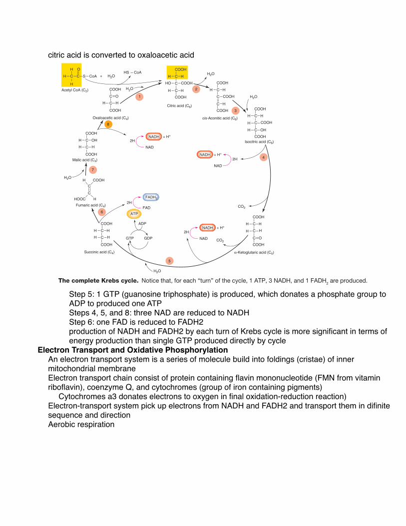
Krebs Cycle Once acetyl CoA has been formed, the acetic acid subunit (2 carbons long) combines with oxaloacetic acid (4 carbons long) to form a molecule of citric acid (6 carbons long). Coen-zyme A acts only as a transporter of acetic acid from one enzyme to another (similar to the transport of hydrogen by NAD). The formation of citric acid begins a cyclic metabolic pathway known as the citric acid cycle, or TCA cycle (for tri-carboxylic acid; citric acid has three carboxylic acid groups). Most commonly, however, this cyclic pathway is called the Krebs cycle, after its principal discoverer, Sir Hans Krebs. A simplified illustration of this pathway is shown in figure 5.8 .
Through a series of reactions involving the elimination of 2 carbons and 4 oxygens (as 2 CO 2 molecules) and the removal of hydrogens, citric acid is eventually converted to oxaloacetic acid, which completes the cyclic metabolic path-way ( fig. 5.9 ). In this process, these events occur:
1. One guanosine triphosphate (GTP) is produced (step 5 of fig. 5.9 ), which donates a phosphate group to ADP to produce one ATP.
2. Three molecules of NAD are reduced to NADH (steps 4, 5, and 8 of fig. 5.9 ).
3. One molecule of FAD is reduced to FADH 2 (step 6).
The production of NADH and FADH 2 by each “turn” of the Krebs cycle is far more significant, in terms of energy production, than the single GTP (converted to ATP) produced directly by the cycle. This is because NADH and FADH 2 even-tually donate their electrons to an energy-transferring process that results in the formation of a large number of ATP.
112 Chapter 5
fox78119_ch05_105-127.indd 112fox78119_ch05_105-127.indd 112 25/06/10 9:10 PM25/06/10 9:10 PM
Energy released is small, and a portion (38-40%) released is captured in high-energy bonds of ATPAerobic respiration of glucose begins w/ glycolysis
Glycolysis in anaerobic metabolism and aerobic respiration results in 2 pyruvic acid, 2 ATP, 2

NADH + H per glucose molecule. In aerobic resp, electrons in NADH are not donated to pyruvic acid and lactic acid is not formed. Pyruvic acids will move to diff. cellular location and undergo diff. rxnIn aerobic resp, pyruvic acid leaves cell cytoplasm, enter interior (matrix) of mitochondria
Once pyruvic acid is inside mitochondrion, carbon dioxide is enzymatically removed from pyruvic acid form acetic acid by combining acetic acid w/ a coenzyme called coenzyme A. This produce acetyl Coenzyme A, Acetyl CoA.
Pyruvic acid
+
OHO
CH H
H
C O
C
CH H
H
C O + CO2
CoAS
Acetyl coenzyme ACoenzyme A
CoAS
HNADH + H+NAD
Figure 5.7 The formation of acetyl coenzyme A in aerobic respiration. Notice that NAD is reduced to NADH in this process.
5.2 AEROBIC RESPIRATION In the aerobic respiration of glucose, pyruvic acid is formed by glycolysis and then converted into acetyl coenzyme A. This begins a cyclic metabolic pathway called the Krebs cycle. As a result of these pathways, a large amount of reduced NAD and FAD (NADH and FADH 2 ) is generated. These reduced coenzymes pro-vide electrons for a process that drives the formation of ATP.
L E A R N I N G O U T C O M E S
After studying this section, you should be able to:
✔ Describe the aerobic cell respiration of glucose through the Krebs cycle.
✔ Describe the electron transport system and oxidative phosphorylation, explaining the role of oxygen in this process.
✔ Explain how glucose can be produced from glycogen and from noncarbohydrate molecules, and how the liver produces free glucose for secretion.
The aerobic respiration of glucose (C 6 H 12 O 6 ) is given in the following overall equation:
C6H12O6 + O2 → 6 CO2 + 6 H2O
Aerobic respiration is equivalent to combustion in terms of its final products (CO 2 and H 2 O) and in terms of the total amount of energy liberated. In aerobic respiration, however, the energy is released in small, enzymatically controlled oxi-dation reactions, and a portion (38% to 40%) of the energy released is captured in the high-energy bonds of ATP.
The aerobic respiration of glucose begins with glycolysis. Glycolysis in both anaerobic metabolism and aerobic respira-tion results in the production of 2 molecules of pyruvic acid, 2 ATP, and 2 NADH + H + per glucose molecule. In aerobic respiration, however, the electrons in NADH are not donated to pyruvic acid and lactic acid is not formed, as happens in the lactic acid pathway. Instead, the pyruvic acids will move to a different cellular location and undergo a different reac-tion; the NADH produced by glycolysis will eventually be oxidized, but that occurs later in the story.
In aerobic respiration, pyruvic acid leaves the cell cyto-plasm and enters the interior (the matrix) of mitochondria. Once pyruvic acid is inside a mitochondrion, carbon dioxide is enzymatically removed from each three-carbon-long pyruvic acid to form a two-carbon-long organic acid— acetic acid. The enzyme that catalyzes this reaction combines the acetic acid with a coenzyme (derived from the vitamin pantothenic acid) called coenzyme A. The combination thus produced is called acetyl coenzyme A, abbreviated acetyl CoA ( fig. 5.7 ).
Glycolysis converts 1 glucose molecule into 2 molecules of pyruvic acid. Since each pyruvic acid molecule is con-verted into 1 molecule of acetyl CoA and 1 CO 2 , 2 molecules of acetyl CoA and 2 molecules of CO 2 are derived from each glucose. These acetyl CoA molecules serve as substrates for mitochondrial enzymes in the aerobic pathway, while the car-bon dioxide is a waste product that is carried by the blood to the lungs for elimination. It is important to note that the oxy-gen in CO 2 is derived from pyruvic acid, not from oxygen gas.
Krebs Cycle Once acetyl CoA has been formed, the acetic acid subunit (2 carbons long) combines with oxaloacetic acid (4 carbons long) to form a molecule of citric acid (6 carbons long). Coen-zyme A acts only as a transporter of acetic acid from one enzyme to another (similar to the transport of hydrogen by NAD). The formation of citric acid begins a cyclic metabolic pathway known as the citric acid cycle, or TCA cycle (for tri-carboxylic acid; citric acid has three carboxylic acid groups). Most commonly, however, this cyclic pathway is called the Krebs cycle, after its principal discoverer, Sir Hans Krebs. A simplified illustration of this pathway is shown in figure 5.8 .
Through a series of reactions involving the elimination of 2 carbons and 4 oxygens (as 2 CO 2 molecules) and the removal of hydrogens, citric acid is eventually converted to oxaloacetic acid, which completes the cyclic metabolic path-way ( fig. 5.9 ). In this process, these events occur:
1. One guanosine triphosphate (GTP) is produced (step 5 of fig. 5.9 ), which donates a phosphate group to ADP to produce one ATP.
2. Three molecules of NAD are reduced to NADH (steps 4, 5, and 8 of fig. 5.9 ).
3. One molecule of FAD is reduced to FADH 2 (step 6).
The production of NADH and FADH 2 by each “turn” of the Krebs cycle is far more significant, in terms of energy production, than the single GTP (converted to ATP) produced directly by the cycle. This is because NADH and FADH 2 even-tually donate their electrons to an energy-transferring process that results in the formation of a large number of ATP.
112 Chapter 5
fox78119_ch05_105-127.indd 112fox78119_ch05_105-127.indd 112 25/06/10 9:10 PM25/06/10 9:10 PM
Glycolysis converts 1 glucose into 2 pyruvic acideach pyruvic acid is converted into 1 acetly CoA and 1 CO2 (2 acetyl CoA and 2 CO2 are derived)Acetyl CoA serve as substrates for mitochondrial enzymes in aerobic pathwayCarbon dioxide is waste produced carried by blood to lungs for elimination Oxygen in CO2 is derived from pyruvic not from oxygen gas
Krebs Cycle
CYTOPLASMC3
C2
C4
C5
C6
Pyruvic acid
Citric acid3 NADH + H+
1 FADH21 ATP
Acetyl CoA
NAD
NADH + H+
CoAMitochondrial matrix
Glycolysis
Oxaloacetic acid
CO2
CO2
CO2
!-Ketoglutaric acid
Krebs cycle
Figure 5.8 A simplified diagram of the Krebs cycle. This diagram shows how the original four-carbon-long oxaloacetic acid is regenerated at the end of the cyclic pathway. Only the numbers of carbon atoms in the Krebs cycle intermediates are shown; the numbers of hydrogens and oxygens are not accounted for in this simplified scheme.
Electron Transport and Oxidative Phosphorylation Built into the foldings, or cristae, of the inner mitochondrial membrane are a series of molecules that serve as an electron-transport system during aerobic respiration. This electron-transport chain of molecules consists of a protein containing flavin mononucleotide (abbreviated FMN and derived from the vitamin riboflavin), coenzyme Q, and a group of iron- containing pigments called cytochromes. The last of these cytochromes is cytochrome a 3 , which donates electrons to oxygen in the final oxidation-reduction reaction (as will be described shortly). These molecules of the electron-transport system are fixed in position within the inner mitochondrial membrane in such a way that they can pick up electrons from NADH and FADH 2 and transport them in a definite sequence and direction.
In aerobic respiration, NADH and FADH 2 become oxi-dized by transferring their pairs of electrons to the electron-transport system of the cristae. It should be noted that the protons (H + ) are not transported together with the electrons;
their fate will be described a little later. The oxidized forms of NAD and FAD are thus regenerated and can continue to “shuttle” electrons from the Krebs cycle to the electron-transport chain. The first molecule of the electron-transport chain in turn becomes reduced when it accepts the electron pair from NADH. When the cytochromes receive a pair of electrons, 2 ferric ions (Fe 3 + ) become reduced to 2 ferrous ions (Fe 2 + ).
The electron-transport chain thus acts as an oxidizing agent for NAD and FAD. Each element in the chain, however, also functions as a reducing agent; one reduced cytochrome transfers its electron pair to the next cytochrome in the chain ( fig. 5.10 ). In this way, the iron ions in each cytochrome alternately become reduced (from Fe 3 + to Fe 2 + ) and oxidized (from Fe 2 + to Fe 3 + ). This is an exergonic process, and the energy derived is used to phosphorylate ADP to ATP. The production of ATP through the coupling of the electron-transport system with the phosphorylation of ADP is appropriately termed oxidative phosphorylation.
The coupling is not 100% efficient between the energy released by electron transport (the “oxidative” part of oxida-tive phosphorylation) and the energy incorporated into the chemical bonds of ATP (the “phosphorylation” part of the term). This difference in energy escapes the body as heat. Metabolic heat production is needed to maintain our internal body temperature.
CLIN ICAL APPL ICATION
Free radicals are molecules with unpaired electrons, in contrast to molecules that are not free radicals because they have two electrons per orbital. A superoxide radical is an oxygen molecule with an extra, unpaired electron. These can be generated in mitochondria through the leakage of electrons from the electron-transport system. Superoxide radicals have some known physiological functions; for example, they are produced in phagocytic white blood cells where they are needed for the destruction of bacteria. However, the production of free radicals and other molecules classified as reactive oxygen species (including the superoxide, hydroxyl, and nitric oxide free radicals) have been implicated in many disease processes, including atherosclerosis (hardening of the arteries—chapter 13, section 13.7). Accordingly, reactive oxygen species have been described as exerting an oxidative stress on the body. Antioxidants are molecules that scavenge free radicals and protect the body from reactive oxygen species.
Coupling of Electron Transport to ATP Production According to the chemiosmotic theory, the electron-transport system, powered by the transport of electrons, pumps protons (H + ) from the mitochondrial matrix
113Cell Respiration and Metabolism
fox78119_ch05_105-127.indd 113fox78119_ch05_105-127.indd 113 25/06/10 9:10 PM25/06/10 9:10 PM
Once acetyl CoA has formed, acetic acid subunit combines w/ oxaloacetic acid to form citric acid. Coenzyme A acts as transporter of acetic acid from one enzyme to another formation of citric acid begins cyclic metabolic pathways known as citric acid cycle or TCA cycle (tricarboxylic acid) aka Krebs cycleReactions involve elimination of 2 carbons and 4 oxygens (as CO2) and removal of hydrogens,
citric acid is converted to oxaloacetic acid
FAD
FADH2
ATP
NADH
NAD
NAD
NADH
GTP GDP
2H
2H
2H
+ H+
+ H+
Oxaloacetic acid (C4)
Acetyl CoA (C2)
Malic acid (C4)
Fumaric acid (C4)
Succinic acid (C4)
Citric acid (C6)
Isocitric acid (C6)
!-Ketoglutaric acid (C5)
cis-Aconitic acid (C6)
NADH
NAD
2H+ H+
1
2
3
4
5
6
7
8
H C C S CoA + H2OCoAHS
H O
H
H
H
H
HHOOC
H
C
C
OH
COOH
COOH
COOH
H H
C
C
O
COOH
COOH
C
C
H
H H
C
C
H
COOH
COOH
H
HO COOH
C
C
H
COOH
H C H
COOH
H
H COOH
C
C
H
COOH
H C OH
COOH
H
H H
C
C
H
COOH
C O
COOH
H
COOH
C
C
H
COOH
C H
COOH
H2O
H2O
H2O
H2O
CO2
CO2
H2O
ADP
Figure 5.9 The complete Krebs cycle. Notice that, for each “turn” of the cycle, 1 ATP, 3 NADH, and 1 FADH2 are produced.
into the space between the inner and outer mito-chondrial membranes. The electron-transport system is grouped into three complexes that serve as proton pumps ( fig. 5.11 ). The first pump (the NADH-coenzyme Q reductase complex) transports 4 H + from the matrix to the intermembrane space for every pair of electrons moved along the electron-transport system. The second pump (the cytochrome c reductase complex) also trans-ports 4 protons into the intermembrane space, and the third pump (the cytochrome c oxidase complex) trans-ports 2 protons into the intermembrane space. As a result, there is a higher concentration of H + in the intermembrane space than in the matrix, favoring the diffusion of H + back out into the matrix. The inner mitochondrial membrane, however, does not permit diffusion of H + , except through structures called respiratory assemblies.
The respiratory assemblies consist of a group of proteins that form a “stem” and a globular subunit. The stem contains a channel through the inner mitochondrial membrane that permits the passage of protons (H + ). The globular subunit, which protrudes into the matrix, contains an ATP synthase enzyme that catalyzes the reaction ADP + Pi → ATP when it is activated by the diffusion of protons through the respira-tory assemblies and into the matrix ( fig. 5.11 ). In this way, phosphorylation (the addition of phosphate to ADP) is cou-pled to oxidation (the transport of electrons) in oxidative phosphorylation.
Function of Oxygen If the last cytochrome remained in a reduced state, it would be unable to accept more electrons. Electron transport would then progress only to the next-to-last cytochrome.
114 Chapter 5
fox78119_ch05_105-127.indd 114fox78119_ch05_105-127.indd 114 25/06/10 9:10 PM25/06/10 9:10 PM
Step 5: 1 GTP (guanosine triphosphate) is produced, which donates a phosphate group to ADP to produced one ATPSteps 4, 5, and 8: three NAD are reduced to NADHStep 6: one FAD is reduced to FADH2production of NADH and FADH2 by each turn of Krebs cycle is more significant in terms of energy production than single GTP produced directly by cycle
Electron Transport and Oxidative PhosphorylationAn electron transport system is a series of molecule build into foldings (cristae) of inner mitochondrial membrane Electron transport chain consist of protein containing flavin mononucleotide (FMN from vitamin riboflavin), coenzyme Q, and cytochromes (group of iron containing pigments)
Cytochromes a3 donates electrons to oxygen in final oxidation-reduction reaction)Electron-transport system pick up electrons from NADH and FADH2 and transport them in difinite sequence and directionAerobic respiration
NADH
FADH2
FAD
Oxidized
CoQ
Reduced
Fe2+
Cytochrome b
Fe3+
Fe2+
Cytochromec1 and c
Fe3+
Fe3+
Cytochrome a
Fe2+
Fe2+
Cytochrome a3
Fe3+
NAD
FMN
FMNH2
2 H+E
lect
ron
ener
gy2 e–
2 e–
2 e–
2 e–
2 H++ O2
H2O
1–2
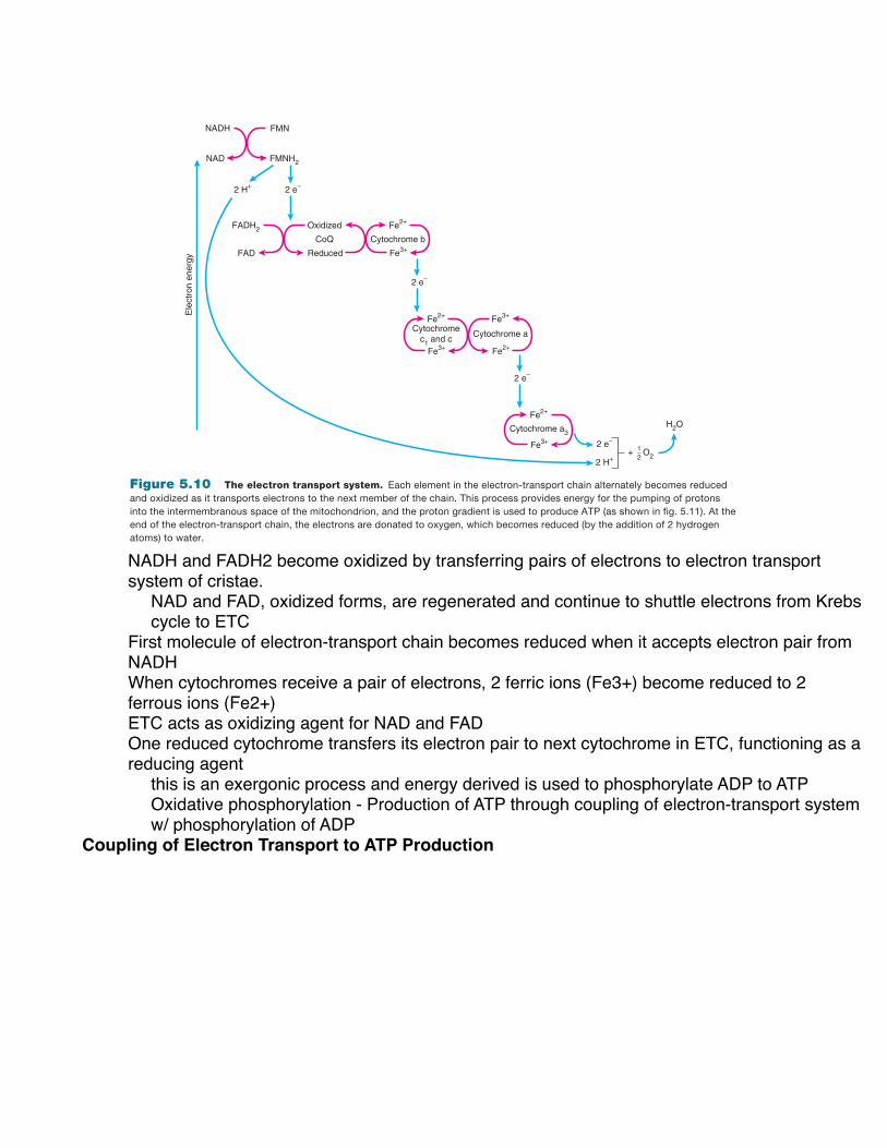
Figure 5.10 The electron transport system. Each element in the electron-transport chain alternately becomes reduced and oxidized as it transports electrons to the next member of the chain. This process provides energy for the pumping of protons into the intermembranous space of the mitochondrion, and the proton gradient is used to produce ATP (as shown in fig. 5.11). At the end of the electron-transport chain, the electrons are donated to oxygen, which becomes reduced (by the addition of 2 hydrogen atoms) to water.
This process would continue until all of the elements of the electron- transport chain remained in the reduced state. At this point, the electron-transport system would stop func-tioning and no ATP could be produced in the mitochondria. With the electron-transport system incapacitated, NADH and FADH 2 could not become oxidized by donating their electrons to the chain and, through inhibition of Krebs cycle enzymes, no more NADH and FADH 2 could be produced in the mito-chondria. The Krebs cycle would stop and only anaerobic metabolism could occur.
Oxygen, from the air we breathe, allows electron trans-port to continue by functioning as the final electron acceptor of the electron-transport chain. This oxidizes cytochrome a 3 , allowing electron transport and oxidative phosphorylation to continue. At the very last step of aerobic respiration, there-fore, oxygen becomes reduced by the 2 electrons that were passed to the chain from NADH and FADH 2 . This reduced oxygen binds 2 protons, and a molecule of water is formed. Because the oxygen atom is part of a molecule of oxygen gas (O 2 ), this last reaction can be shown as follows:
O2 + 4 e– + 4 H+ → 2 H2O
ATP Balance Sheet Overview There are two different methods of ATP formation in cell respiration. One method is the direct (also called substrate-level ) phosphorylation that occurs in glycoly-sis (producing a net gain of 2 ATP) and the Krebs cycle
CLIN ICAL APPL ICATION
Cyanide is a fast-acting lethal poison that produces such symptoms as rapid heart rate, tiredness, seizures, and headache. Cyanide poisoning can result in coma, and ultimately death, in the absence of quick treatment. The reason that cyanide is so deadly is that it has one very specific action: it blocks the transfer of electrons from cytochrome a3 to oxygen. The effects are thus the same as would occur if oxygen were completely removed—aerobic cell respiration and the production of ATP by oxidative phosphorylation comes to a halt.
115Cell Respiration and Metabolism
fox78119_ch05_105-127.indd 115fox78119_ch05_105-127.indd 115 25/06/10 9:10 PM25/06/10 9:10 PM
NADH and FADH2 become oxidized by transferring pairs of electrons to electron transport system of cristae.
NAD and FAD, oxidized forms, are regenerated and continue to shuttle electrons from Krebs cycle to ETC
First molecule of electron-transport chain becomes reduced when it accepts electron pair from NADHWhen cytochromes receive a pair of electrons, 2 ferric ions (Fe3+) become reduced to 2 ferrous ions (Fe2+)ETC acts as oxidizing agent for NAD and FADOne reduced cytochrome transfers its electron pair to next cytochrome in ETC, functioning as a reducing agent
this is an exergonic process and energy derived is used to phosphorylate ADP to ATPOxidative phosphorylation - Production of ATP through coupling of electron-transport system w/ phosphorylation of ADP
Coupling of Electron Transport to ATP Production

1
Outer mitochondrial membrane
Inner mitochondrial membrane
Intermembrane space
First pump
Second pump
Third pump
NADHMatrix
ATP synthase
2 H + 1/2 O2
ADP+Pi
H+
H+
H+H+
e–
2 H+
4 H+
4 H+ NAD+
ATP
H2O
1
2
3
Figure 5.11 The steps of oxidative phosphorylation. (1) Molecules of the electron-transport system function to pump H+ from the matrix to the intermembrane space. (2) This results in a steep H+ gradient between the intermembrane space and the cytoplasm of the cell. (3) The diffusion of H+ through ATP synthase results in the production of ATP.
(producing 1 ATP per cycle). These numbers are certain and constant. In the second method of ATP formation, oxidative phosphorylation, the numbers of ATP mol-ecules produced vary under different conditions and for different kinds of cells. For many years, it was believed that 1 NADH yielded 3 ATP and that 1 FADH 2 yielded 2 ATP by oxidative phosphorylation. This gave a grand total of 36 to 38 molecules of ATP per glucose through cell res-piration ( table 5.2 ). Newer biochemical information, how-ever, suggests that these numbers may be overestimates, because, of the 36 to 38 ATP produced per glucose in the mitochondrion, only 30 to 32 ATP actually enter the cyto-plasm of the cell.
Roughly 3 protons must pass through the respiratory assemblies and activate ATP synthase to produce 1 ATP. How-ever, the newly formed ATP is in the mitochondrial matrix and must be moved into the cytoplasm; this transport also uses the proton gradient and costs 1 more proton. The ATP and H + are transported into the cytoplasm in exchange for
ADP and P i , which are transported into the mitochondrion. Thus, it effectively takes 4 protons to produce 1 ATP that enters the cytoplasm.
To summarize: The theoretical ATP yield is 36 to 38 ATP per glucose. The actual ATP yield, allowing for the costs of transport into the cytoplasm, is about 30 to 32 ATP per glucose. The details of how these numbers are obtained are described in the following section.
Detailed Accounting Each NADH formed in the mitochondrion donates 2 electrons to the electron transport system at the first proton pump (see fig. 5.11 ). The electrons are then passed to the second and third proton pumps, activating each of them in turn until the 2 electrons are ultimately passed to oxygen. The first and second pumps transport 4 protons each, and the third pump transports 2 protons, for a total of 10. Dividing 10 protons by the 4 it takes to produce an ATP (in the cytoplasm) gives
116 Chapter 5
fox78119_ch05_105-127.indd 116fox78119_ch05_105-127.indd 116 25/06/10 9:10 PM25/06/10 9:10 PM
Chemiosmotic theory - electron transport system, powered by transport of electrons, pumps protons (H) from mitochondrial matrix into space between inner and outer mitochondrial mem.Electron-transport system is grouped into three complexes serves as proton pumps.
First pump (NADH-coenzyme Q reductase complex) transports 4H from matrix to inermembrane space for every pair of electrons moved along the electron-transport system. Second pump (cytochrome c reductase complex) transports 4H into intermembrane spaceThird pump (cytochrome c oxidase complex) transports 2H into intermembrane space
results in higher [H] in intermembrane space than in matrix, favoring diffusion of H back into matrix inner mitochondrial membrane does not permit diffusion of H, except through respiratory assemblies
repiratory assemblies consist of group of proteins that forms “stem” and globular subunit. Stem permits passage of protons (H). Globular subunit contain ATP synthase enzyme that catalyzes ADP + P to ATP by diffusion of H through the respiratory assemblies into matrix
Function of OxygenIf last cytochrome remain in reduced state, unable to accept more electrons. Electron transport would then progress only to the next-to-last cytochrome and continue til all elements remain in
reduced state. At this point, electron-transport system would stop functioning and no ATP produced With ETC incapacitated, NADH and FADH2 could not oxidized, and through inhibition of Krebs cycle enzymes, no more NADH and FADH2 could be not be produced in mitochondria. Krebs cycle would stop and only anaerobic metabolism occur. Oxygen allows electron transport to continue by being final electron acceptor in ETC. This oxidizes cytochrome a3, allowing electron transport and oxidative phosphorylation to continue. Last step of aerobic respiration, oxygen become reduced by 2 electrons that were passed to the chain from NADH and FADH2. Oxygen binds 2H, and a mole of water is formed
NADH
FADH2
FAD
Oxidized
CoQ
Reduced
Fe2+
Cytochrome b
Fe3+
Fe2+
Cytochromec1 and c
Fe3+
Fe3+
Cytochrome a
Fe2+
Fe2+
Cytochrome a3
Fe3+
NAD
FMN
FMNH2
2 H+
Ele
ctro
n en
ergy
2 e–
2 e–
2 e–
2 e–
2 H++ O2
H2O
1–2
Figure 5.10 The electron transport system. Each element in the electron-transport chain alternately becomes reduced and oxidized as it transports electrons to the next member of the chain. This process provides energy for the pumping of protons into the intermembranous space of the mitochondrion, and the proton gradient is used to produce ATP (as shown in fig. 5.11). At the end of the electron-transport chain, the electrons are donated to oxygen, which becomes reduced (by the addition of 2 hydrogen atoms) to water.
This process would continue until all of the elements of the electron- transport chain remained in the reduced state. At this point, the electron-transport system would stop func-tioning and no ATP could be produced in the mitochondria. With the electron-transport system incapacitated, NADH and FADH 2 could not become oxidized by donating their electrons to the chain and, through inhibition of Krebs cycle enzymes, no more NADH and FADH 2 could be produced in the mito-chondria. The Krebs cycle would stop and only anaerobic metabolism could occur.
Oxygen, from the air we breathe, allows electron trans-port to continue by functioning as the final electron acceptor of the electron-transport chain. This oxidizes cytochrome a 3 , allowing electron transport and oxidative phosphorylation to continue. At the very last step of aerobic respiration, there-fore, oxygen becomes reduced by the 2 electrons that were passed to the chain from NADH and FADH 2 . This reduced oxygen binds 2 protons, and a molecule of water is formed. Because the oxygen atom is part of a molecule of oxygen gas (O 2 ), this last reaction can be shown as follows:
O2 + 4 e– + 4 H+ → 2 H2O
ATP Balance Sheet Overview There are two different methods of ATP formation in cell respiration. One method is the direct (also called substrate-level ) phosphorylation that occurs in glycoly-sis (producing a net gain of 2 ATP) and the Krebs cycle
CLIN ICAL APPL ICATION
Cyanide is a fast-acting lethal poison that produces such symptoms as rapid heart rate, tiredness, seizures, and headache. Cyanide poisoning can result in coma, and ultimately death, in the absence of quick treatment. The reason that cyanide is so deadly is that it has one very specific action: it blocks the transfer of electrons from cytochrome a3 to oxygen. The effects are thus the same as would occur if oxygen were completely removed—aerobic cell respiration and the production of ATP by oxidative phosphorylation comes to a halt.
115Cell Respiration and Metabolism
fox78119_ch05_105-127.indd 115fox78119_ch05_105-127.indd 115 25/06/10 9:10 PM25/06/10 9:10 PM
ATP Balance Sheet2 different methods of ATP formation in cell respiration
Direct (substrate-level) phosphorylation occurs in glycolysis (producing net gain of 2 ATP) and Krebs cycle (producing 1 ATP per cycle)These numbers are certain and constant
Oxidative phosphorylation ATP produced vary under different conditions and for different kinds of cells. only 30 to 32 ATP actually enter cytoplasm of cell
It takes 4 protons to produce 1 ATP that enters cytoplasm Theoretical ATP yield is 36 to 38 ATP per glucose. Actual ATP yield, allowing for costs of transport into cytoplasm is about 30-32 ATP per glucoseDetail Accounting
Each NADH formed in mitochondrion donates 2 electrons to ETC at first proton pump. Electrons are then passed to second and third proton pumps, activating each until 2 electrons are passed to oxygen. First and second pumps transport 4 proton eachThird pump transports 2 protonsTotal of 10Dividing 10 protons by the 4 it takes to produce an ATP (in the cytoplasm) gives 2.5 ATP produced for every pair of electrons donated by NADHThree molecules of NADH are formed w/ each Krebs cycleOne NADH is also produced when pyruvate is converted into acetyl CoAOne glucose, two Krebs cycle (producing 6 NADH) and 2 pyruvate converted to acetyl CoA (producing 2 NADH) yield 8 NADH8 NADH multiply by 2.5 ATP gives 20 ATPElectrons from FADH2 are donated later in ETC and activate only second and third H pumps. Electrons passed from FADH2 result in pumping of only 6 proton (4 by second and 2 by third)1 ATP is produced for every 4 proton pump, electron derived from FADH2 result in formation of 1.5 ATPEach Krebs cycle produces 1 FADH2 and we get two Krebs cycle from 1 glucose, there are 2 FADH2 that gives 3 ATP23 ATP subtotal from oxidative phosphorylation from only NADH and FADH2 produced in the

mitochondrion. Glycolysis also produces 2 NADH, but cannot directly enter mitochondrion, but can be shuttled in.the 2 NADH is translated into 2 FADH2 when shuttled into mitochondrion and yield 2 X1.5ATP = 3ATP This total to 26 ATP produced by oxidative phosphorylation from glucose. Adding 2 ATP from direct (substrate-level) phosphorylation in glycolysis and 2 ATP directly by two Krebs cycle gives a grand total of 30 ATP produced by aerobic respiration of glucose
2.5 ATP that are produced for every pair of electrons donated by an NADH. (There is no such thing as half an ATP; the decimal fraction simply indicates an average.)
Three molecules of NADH are formed with each Krebs cycle, and 1 NADH is also produced when pyruvate is con-verted into acetyl CoA (see fig. 5.7 ). Starting from 1 glucose, two Krebs cycles (producing 6 NADH) and 2 pyruvates con-verted to acetyl CoA (producing 2 NADH) yield 8 NADH. Multiplying by 2.5 ATP per NADH gives 20 ATP.
Electrons from FADH 2 are donated later in the electron-transport system than those donated by NADH; conse-quently, these electrons activate only the second and third proton pumps. Since the first proton pump is bypassed, the electrons passed from FADH 2 result in the pumping of only 6 protons (4 by the second pump and 2 by the third pump). Because 1 ATP is produced for every 4 protons pumped, electrons derived from FADH 2 result in the formation of 6 ÷ 4 = 1.5 ATP. Each Krebs cycle produces 1 FADH 2 and we get two Krebs cycles from 1 glucose, so there are 2 FADH 2 that give 2 ! 1.5 ATP = 3 ATP.
The 23 ATP subtotal from oxidative phosphorylation we have at this point includes only the NADH and FADH 2 produced in the mitochondrion. Remember that glycolysis, which occurs in the cytoplasm, also produces 2 NADH. These cytoplasmic NADH cannot directly enter the mitochondrion, but there is a process by which their electrons can be “ shuttled” in. The net effect of the most common shuttle is that a molecule of NADH in the cytoplasm is translated into a molecule of FADH 2 in the mitochondrion. The 2 NADH produced in glycolysis, therefore, usually become 2 FADH 2 and yield 2 ! 1.5 ATP = 3 ATP by oxidative phosphoryla-tion. (An alternative pathway, where the cytoplasmic NADH is transformed into mitochondrial NADH and produces 2 ! 2.5 ATP = 5 ATP, is less common; however, this is the dominant pathway in the liver and heart, which are meta-bolically highly active.)
We now have a total of 26 ATP (or, less commonly, 28 ATP) produced by oxidative phosphorylation from glu-cose. We can add the 2 ATP made by direct (substrate-level) phosphorylation in glycolysis and the 2 ATP made directly by the two Krebs cycles to give a grand total of 30 ATP (or, less commonly, 32 ATP) produced by the aerobic respiration of glucose ( table 5.2 ).
Phases of Respiration
ATP Made Directly
Reduced Coenzymes
ATP Made by Oxidative Phosphorylation
Theoretical Yield Actual Yield
Glucose to pyruvate (in cytoplasm)
2 ATP (net gain) 2 NADH, but usually goes into mitochondria as 2 FADH2
If from FADH2:2 ATP (! 2) = 4 ATPor if stays NADH:3 ATP (! 2) = 6 ATP
If from FADH2:1.5 ATP (! 2) = 3 ATPor if stays NADH:2.5 ATP (! 2) = 5 ATP
Pyruvate to acetyl CoA (! 2) None 1 NADH (! 2) = 2 NADH 3 ATP (! 2) = 6 ATP 2.5 ATP (! 2) = 5 ATP
Krebs cycle (! 2) 1 ATP (! 2) = 2 ATP 3 NADH (! 2) = 6 NADH1 FADH2 (! 2) = 2 FADH2
3 ATP (! 6) = 18 ATP2 ATP (! 2) = 4 ATP
2.5 ATP (! 6) = 15 ATP1.5 ATP (! 2) = 3 ATP
Total ATP 4 ATP 32 (or 34) ATP 26 (or 28) ATP
Table 5.2 | ATP Yield per Glucose in Aerobic Respiration
| C H E C K P O I N T
6. Compare the fate of pyruvate in aerobic and anaerobic cell respiration.
7. Draw a simplified Krebs cycle and indicate the high-energy products.
8. Explain how NADH and FADH2 contribute to oxidative phosphorylation.
9. Explain how ATP is produced in oxidative phosphorylation.
5.3 METABOLISM OF LIPIDS AND PROTEINS Triglycerides can be hydrolyzed into glycerol and fatty acids. The latter are of particular importance because they can be converted into numerous molecules of acetyl CoA that can enter Krebs cycles and generate a large amount of ATP. Amino acids derived from proteins also may be used for energy. This involves the removal of the amine group and the conversion of the remaining molecule into either pyruvic acid or one of the Krebs cycle molecules.
117Cell Respiration and Metabolism
fox78119_ch05_105-127.indd 117fox78119_ch05_105-127.indd 117 25/06/10 9:10 PM25/06/10 9:10 PM
5.3 Metabolism of Lipids and ProteinsPyruvic acid and Krebs cycle acids serve as common intermediates in interconversion of glucose, lipids and amino acidsCells do not store extra energy in form of extra ATP
When cellular ATP rise due to more energy (from food) is available than can be immediately used, ATP production is inhibited and glucose is converted into glycogen and fat
Lipid MetabolismGlucose converted into fat
glycolysis occurs and pyruvic acid converted into acetyl CoASome glycoltic intermediates (phosphoglyceraldehyde and dihydroxyacetone phosphate) do not complete conversion to pyruvic acid and acetyle CoA does not enter Krebs cycle. instead they are used to produced variety of lipids, including cholesterol (used in synthesis of bile salts and steroid hormones), ketone bodies, and fatty acidsAcetic acid subunits are joined together to from fatty acid chain.
six acetyl CoA will produce a fatty acid of 12 carbons long.Lipogenesis - formation of fat, occurs in adipose tissue and in liver when concentration of blood glucose is elevated following a meal
White Adipose TissueWhite fat - where most triglycerides in body are stored Lipolysis - lipase enzyme hydrolyze triglycerides into glycerol and free fatty acids to be used as energy sourceglycerol released into blood is mostly taken up by liver which converts into glucose through gluconeogenesisMost significant energy carriers provided by lipolysis are free fatty acids
Brown Adipose Tissue
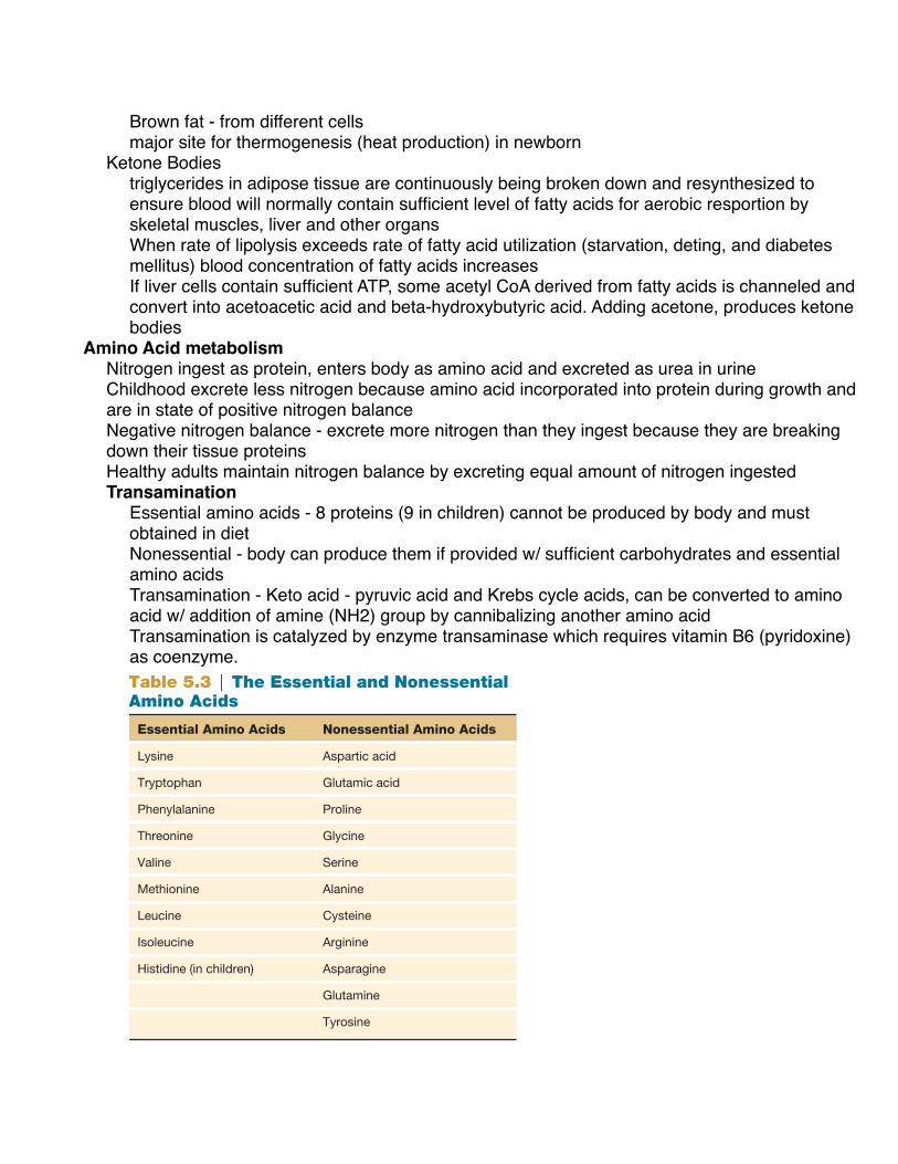
Brown fat - from different cells major site for thermogenesis (heat production) in newborn
Ketone Bodiestriglycerides in adipose tissue are continuously being broken down and resynthesized to ensure blood will normally contain sufficient level of fatty acids for aerobic resportion by skeletal muscles, liver and other organsWhen rate of lipolysis exceeds rate of fatty acid utilization (starvation, deting, and diabetes mellitus) blood concentration of fatty acids increases If liver cells contain sufficient ATP, some acetyl CoA derived from fatty acids is channeled and convert into acetoacetic acid and beta-hydroxybutyric acid. Adding acetone, produces ketone bodies
Amino Acid metabolismNitrogen ingest as protein, enters body as amino acid and excreted as urea in urineChildhood excrete less nitrogen because amino acid incorporated into protein during growth and are in state of positive nitrogen balance Negative nitrogen balance - excrete more nitrogen than they ingest because they are breaking down their tissue proteins Healthy adults maintain nitrogen balance by excreting equal amount of nitrogen ingestedTransamination
Essential amino acids - 8 proteins (9 in children) cannot be produced by body and must obtained in dietNonessential - body can produce them if provided w/ sufficient carbohydrates and essential amino acidsTransamination - Keto acid - pyruvic acid and Krebs cycle acids, can be converted to amino acid w/ addition of amine (NH2) group by cannibalizing another amino acidTransamination is catalyzed by enzyme transaminase which requires vitamin B6 (pyridoxine) as coenzyme.
O
O
HH
O
HOOH
H
HH
H
HO
OH
NH
H
O
H
COH O
C
OC
OH OC
OH
H
HH
O
H
H
H
C
C
C
NO
HH
C
C
H
O
H
HO
C
C
C HH
H
C
CH H
C
C
H
HO
H
O
N H
H
H
O
OC
C
C
C
C
+ +
HO
C
C
C
C
O
O
HH
H
OH OOH
HN
H
HO
H
C
C
C
C
H
H
OH
HO O
C
C
C
C
C
AST
Glutamic acid Oxaloacetic acid
Glutamic acid Pyruvic acid
!-Ketoglutaric acid Aspartic acid
!-Ketoglutaric acid Alanine
ALT+ +
Figure 5.15 Two important transamination reactions. The areas shaded in blue indicate the parts of the molecules that are changed. (AST = aspartate transaminase; ALT = alanine transaminase. The amino acids are identified in boldface.)
!-Ketoglutaric acid
Glutamic acid
H
H
Keto acid
Amino acid NH3 + CO2
N
O
C N
H
H
H
H
Urea
Amino transferUrea cycle in liver
Figure 5.16 Oxidative deamination. Glutamic acid is converted to !-ketoglutaric acid as it donates its amine group to the metabolic pathway that results in the formation of urea.
Transamination An adequate amount of all 20 amino acids is required to build proteins for growth and to replace the proteins that are turned over. However, only 8 of these (9 in children) cannot be produced by the body and must be obtained in the diet. These are the essential amino acids ( table 5.3 ). The remain-ing amino acids are “nonessential” only in the sense that the body can produce them if provided with a sufficient amount of carbohydrates and the essential amino acids.
Pyruvic acid and the Krebs cycle acids are collectively termed keto acids because they have a ketone group; these should not be confused with the ketone bodies (derived from acetyl CoA) discussed in the previous section. Keto acids can be converted to amino acids by the addition of an amine (NH 2 ) group. This amine group is usually obtained by “cannibalizing” another amino acid; in this process, a new amino acid is formed as the one that was cannibalized is converted to a new keto acid. This type of reaction, in which the amine group is transferred from one amino acid to form another, is called transamination ( fig. 5.15 ).
Each transamination reaction is catalyzed by a specific enzyme (a transaminase) that requires vitamin B 6 (pyridox-ine) as a coenzyme. The amine group from glutamic acid, for example, may be transferred to either pyruvic acid or oxalo-acetic acid. The former reaction is catalyzed by the enzyme alanine transaminase (ALT); the latter reaction is catalyzed by aspartate transaminase (AST). These enzyme names reflect the fact that the addition of an amine group to pyruvic acid produces the amino acid alanine; the addition of an amine group to oxaloacetic acid produces the amino acid known as aspartic acid ( fig. 5.15 ).
Oxidative Deamination As shown in figure 5.16 , glutamic acid can be formed through transamination by the combination of an amine group with ! -ketoglutaric acid. Glutamic acid is also produced in the liver from the ammonia that is generated by intestinal bacte-ria and carried to the liver in the hepatic portal vein. Because free ammonia is very toxic, its removal from the blood and incorporation into glutamic acid is an important function of a healthy liver.
If there are more amino acids than are needed for pro-tein synthesis, the amine group from glutamic acid may be removed and excreted as urea in the urine ( fig. 5.16 ). The metabolic pathway that removes amine groups from amino acids—leaving a keto acid and ammonia (which is converted to urea)—is known as oxidative deamination.
Essential Amino Acids Nonessential Amino Acids
Lysine Aspartic acid
Tryptophan Glutamic acid
Phenylalanine Proline
Threonine Glycine
Valine Serine
Methionine Alanine
Leucine Cysteine
Isoleucine Arginine
Histidine (in children) Asparagine
Glutamine
Tyrosine
Table 5.3 | The Essential and Nonessential Amino Acids
121Cell Respiration and Metabolism
fox78119_ch05_105-127.indd 121fox78119_ch05_105-127.indd 121 25/06/10 9:10 PM25/06/10 9:10 PM

Oxidative DeaminationRemoves amine groups from amino acids - leaving a keto acid and ammonia (which is converted to urea)more amino acid than are needed for protein synthesis, amine group from glutamic acid may be removed and excreted as urea in urine
Uses of Different Energy SourcesBlood contains variety of energy sources
glucose and ketone bodies from liver, fatty acids from adipose tissue, and lactic acid and amino acids from muscle
Brain uses blood glucose as its major energy source
Amino acids
Urea
Glucose
Phosphoglyceraldehyde
Pyruvic acid
Acetyl CoA
Glycerol
Fatty acids
Triacylglycerol(triglyceride)
Ketonebodies
C5
C6
C4
Glycogen
Protein
Lactic acid
Krebscycle
Figure 5.18 The interconversion of glycogen, fat, and protein. These simplified metabolic pathways show how glycogen, fat, and protein can be interconverted. Note that while most reactions are reversible, the reaction from pyruvic acid to acetyl CoA is not. This is because a CO2 is removed in the process. (Only plants, in a phase of photosynthesis called the dark reaction, can use CO2 to produce glucose.)
Organ GlucoseFatty Acids
Ketone Bodies
Lactic Acid
Brain + + + – + –
Skeletal muscles (resting)
+ + + + + –
Liver + + + + + + +
Heart + + + + +
Table 5.4 | Relative Importance of Different Molecules in the Blood with Respect to the Energy Requirements of Different OrgansCase Investigation CLUES
Brenda gasped and panted for air longer after her workouts than her teammates did.
■ What is the extra oxygen needed after exercise called, and what functions does it serve?
■ How would a more gradual training program help Brenda to gasp and pant less after her workouts?
| C H E C K P O I N T
10. Construct a flowchart to show the metabolic pathway by which glucose can be converted to fat. Indicate only the major intermediates involved (not all of the steps of glycolysis).
11. Define the terms lipolysis and b -oxidation and explain, in general terms, how fat can be used for energy.
12. Describe transamination and deamination and explain their functional significance.
13. List five blood-borne energy carriers and explain, in general terms, how these are used as sources of energy.
123Cell Respiration and Metabolism
fox78119_ch05_105-127.indd 123fox78119_ch05_105-127.indd 123 25/06/10 9:10 PM25/06/10 9:10 PM