Chapterems.jbpub.com/sanders/paramedic/docs/PPT_Lectures/Chapter_047.… · Chapter 47 Neonatal...
83
9/11/2012 1 1 Chapter 47 Neonatal Care 2 Learning Objectives • Identify risk factors associated with the need for neonatal resuscitation. • Describe physiological adaptations at birth. • Describe pathophysiology and implications of selected genetic anomalies present in some neonates. • Outline the prehospital assessment and management of the neonate. • Describe resuscitation of the distressed neonate. 3 Copyright © 2013 by Jones & Bartlett Learning, LLC, an Ascend Learning Company
Transcript of Chapterems.jbpub.com/sanders/paramedic/docs/PPT_Lectures/Chapter_047.… · Chapter 47 Neonatal...

9/11/2012
1
1
Chapter 47
Neonatal Care
2
Learning Objectives
• Identify risk factors associated with the need for neonatal resuscitation.
• Describe physiological adaptations at birth.
• Describe pathophysiology and implications of selected genetic anomalies present in some neonates.
• Outline the prehospital assessment and management of the neonate.
• Describe resuscitation of the distressed neonate.
3
Copyright © 2013 by Jones & Bartlett Learning, LLC, an Ascend Learning Company

9/11/2012
2
Learning Objectives
• Discuss postresuscitative management and transport.
• Describe signs and symptoms and prehospitalmanagement of specific neonatal resuscitation situations.
• Identify injuries associated with birth.
• Describe appropriate interventions to manage the emotional needs of the neonate’s family.
4
• Vast majority of term newborns require no resuscitation beyond
– Maintenance of temperature
– Suctioning of airway
– Mild stimulation
Risk Factors Associated with the Need for Resuscitation
5
• Incidence of complications increase as birth weight decreases
– Resuscitation required for ≈ 80 percent of 30,000 babies weighing less than 1500 g (3.12 lb) at birth
Risk Factors Associated with the Need for Resuscitation
6
Copyright © 2013 by Jones & Bartlett Learning, LLC, an Ascend Learning Company

9/11/2012
3
• Average term newborn weighs ≈ 3600 g (7.5 lb)
• Birth weight depends on
– Size and racial origin parents• Small parents tend to have small babies
• Asian babies tend to be smaller than Caucasian babies
Risk Factors Associated with the Need for Resuscitation
7
• Newborn boys usually weigh ≈ 8 oz more than baby girls
• Causes of low birth weight
– Premature birth
– Undernourishment in uterus
– Certain maternal factors
• Preeclampsia
• Cigarette smoking during pregnancy
Risk Factors Associated with the Need for Resuscitation
8
• Risk factors may affect need for resuscitation– Antepartum
• Multiple gestation
• Inadequate prenatal care
• Mother’s age (under 16 or over 35)
• History of perinatal morbidity or mortality
• Postterm gestation
• Drugs/medications
• Toxemia, hypertension, diabetes
• Perinatal infections
• Known fetal malformations
Risk Factors Associated with the Need for Resuscitation
9
Copyright © 2013 by Jones & Bartlett Learning, LLC, an Ascend Learning Company

9/11/2012
4
• Risk factors may affect need for resuscitation– Intrapartum
• Premature labor
• Meconium‐stained amniotic fluid
• Rupture of membranes greater than 18 hours before delivery
• Use of narcotics within 4 hours of delivery
• Abnormal presentation
• Prolonged labor or precipitous delivery
• Prolapsed cord
• Bleeding (abruptio placentae)
Risk Factors Associated with the Need for Resuscitation
10
• When any foregoing risk factors present during delivery, paramedic should
– Prepare equipment and drugs that may be needed for neonatal resuscitation
– Advise medical direction of situation to determine appropriate destination hospital
Risk Factors Associated with the Need for Resuscitation
11
Does your ambulance have equipment appropriately sized for
neonatal resuscitation?
12
Copyright © 2013 by Jones & Bartlett Learning, LLC, an Ascend Learning Company

9/11/2012
5
• Newborns make three major physiological adaptations at birth necessary for survival
– Emptying fluids from lungs and beginning ventilation
– Changing circulatory pattern
–Maintaining body temp
Physiological Adaptations at Birth
13
• During vaginal delivery, newborn’s chest usually compressed
– Forces fluid from lungs into mouth and nose
– As chest wall recoils, air drawn into lungs
– First breath = response to chemical changes, changes in temp
Physiological Adaptations at Birth
14
• Cord cut = placental circulation shutdown
• Circulatory system must function on own
– Immediate, permanent closure of pathways that allowed receipt of O2 without using lungs
– Lungs expand with initial breaths, resistance to blood flow in lungs decreases
– Blood begins to be oxygenated
Physiological Adaptations at Birth
15
Copyright © 2013 by Jones & Bartlett Learning, LLC, an Ascend Learning Company
9/11/2012
6
• Permanent brain damage from prolonged hypoxemia
• Causes of hypoxia– Compression of cord
– Difficult L&D
– Maternal hemorrhage
– Airway obstruction
– Hypothermia
– Newborn blood loss
– Immature lungs in premature newborn
– Cardiovascular anomalies also may result in hypoxia
Physiological Adaptations at Birth
16
• Newborns at risk for rapidly developing hypothermia– Should be delivered in warm, draft‐free area when possible
– Risk factor due to newborn's• Larger body surface area• Decreased tissue insulation• Immature temperature regulatory mechanisms
– Cool, wet environment of birth also increases heat loss for newborn
Physiological Adaptations at Birth
17
• Newborns conserve body heat through vasoconstriction and increased metabolism
– This increases risk for hypoxemia, acidosis, bradycardia, and hypoglycemia
Physiological Adaptations at Birth
18
Copyright © 2013 by Jones & Bartlett Learning, LLC, an Ascend Learning Company
9/11/2012
7
• Defects that occur during fetal development
– Develop usually within first trimester
– Present in ≈ 2 percent of all births
– Responsible for 20 percent of all infant deaths
• Congenital anomalies factors in need for neonatal resuscitation
Congenital Anomalies
19
Congenital Anomalies
• Causes
– Heritable
–Maternal infection
– Alcohol or other drug use during pregnancy (teratogens)
– Other factors
20
Anomalies of the Airway
• Choanal atresia is bony or membranous occlusion that blocks passageway between nose and pharynx
– Occurs during fetal development when tissue separating nose and mouth remains after birth
–Most common nose abnormality in newborns
• ≈ 1 in 7,000 live births
– Commonly associated with other congenital anomalies
21
Copyright © 2013 by Jones & Bartlett Learning, LLC, an Ascend Learning Company

9/11/2012
8
• May affect one or both sides of nasal cavity
• May require surgical repair
• Bilateral obstruction can result in serious ventilation problems
– Symptoms
• Chest wall retraction (unless infant is breathing through mouth or crying)
• Dyspnea: may result in cyanosis (unless infant is crying)
• Inability to nurse and breathe at same time
• Persistent one‐sided nasal blockage or discharge
Choanal Atresia
22
23
Tracheoesophageal Fistula
• Abnormal connection between esophagus and trachea
• Congenital disorder occurs in ≈ 1 in every 4,000 live births
• Commonly occurs with esophageal atresia(incomplete formation of esophagus)
24
Copyright © 2013 by Jones & Bartlett Learning, LLC, an Ascend Learning Company

9/11/2012
9
Tracheoesophageal Fistula
• Both require surgical correction– Uncorrected
• Food and fluid in esophagus enter trachea and lungs• Air in trachea enters esophagus
– Symptoms of both disorders in newborn • Copious salivation• Choking• Coughing• Regurgitation during feeding• Cyanosis
25
26
Tracheoesophageal Fistula
• Many infants with tracheoesophageal fistula or esophageal atresia
– Have other congential anomalies
• Heart, kidney, and limb deformities (often together)
• Infants unable to feed properly
• Once diagnosed, early surgery required
27
Copyright © 2013 by Jones & Bartlett Learning, LLC, an Ascend Learning Company

9/11/2012
10
Cleft Lip and Cleft Palate
• Cleft lip
– Incomplete closure of infant’s lip
– Occurs when one or more fissures fail to fuse in embryo
– Visible at birth as vertical, usually off‐center split in upper lip
• Split may extend to nose
28
Cleft Lip and Cleft Palate
• Cleft palate
– Fissure in hard palate of roof of mouth
– Runs along its midline
29
Cleft Lip and Cleft Palate
• Both can occur with other congenital anomalies– Cleft palate
• May involve one or both sides of roof of mouth
• Can extend through hard and soft palates into nasal cavity
– Cleft lip or cleft palate can cause• Nasal deformity
• Difficulty in feeding and speech• Frequent ear infections
30
Copyright © 2013 by Jones & Bartlett Learning, LLC, an Ascend Learning Company

9/11/2012
11
Cleft Lip and Cleft Palate
• Affects 1 in every 2,500 live births
• Both can lead to
– Failure to thrive
–Misaligned teeth
– Difficulties with speech
• Corrected with one or more surgeries, usually beginning in first year of life
31
32
Pierre Robin Syndrome
• Complex of congenital anomalies
– Small mandible
– Tongue that falls back into airway
– Cleft palate
– Additional craniofacial abnormalities and defects of eyes and ears may be present
33
Copyright © 2013 by Jones & Bartlett Learning, LLC, an Ascend Learning Company
9/11/2012
12
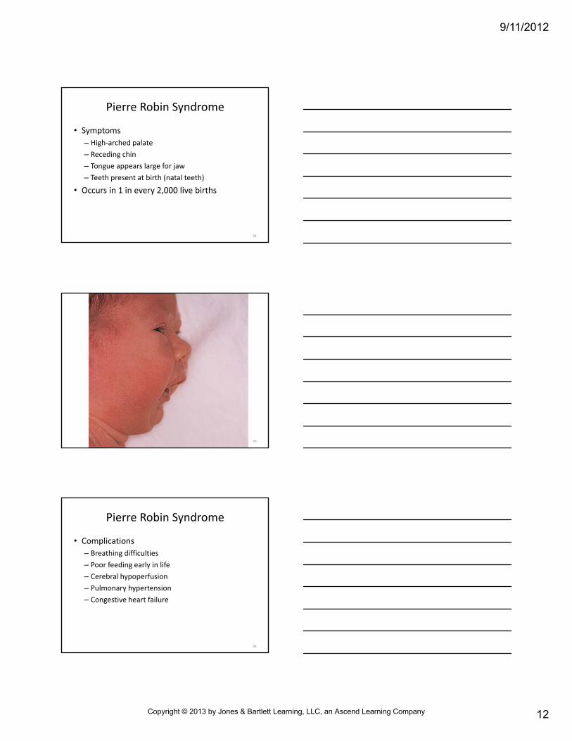
Pierre Robin Syndrome
• Symptoms
– High‐arched palate
– Receding chin
– Tongue appears large for jaw
– Teeth present at birth (natal teeth)
• Occurs in 1 in every 2,000 live births
34
35
Pierre Robin Syndrome
• Complications
– Breathing difficulties
– Poor feeding early in life
– Cerebral hypoperfusion
– Pulmonary hypertension
– Congestive heart failure
36
Copyright © 2013 by Jones & Bartlett Learning, LLC, an Ascend Learning Company
9/11/2012
13
• Death from respiratory failure, secondary to airway obstruction
• Management
– Surgical repair of cleft palate
– Other methods to prevent breathing difficulties and choking
• Most children have some relief as jaw grows and allows more space for tongue
Pierre Robin Syndrome
37
Anomalies of the Heart
• Congenital abnormalities = defects in heart’s structure
– Occur during embryonic development
– Present at birth
38
• Congenital defects most common type of birth defect– Affect 8 of every 1,000 newborns– 50 percent produce hemodynamic effects from altered cardiac function
• Over 35,000 babies in U.S. born with congenital heart defects annually
• ≈ 1 million American adults live with congenital heart abnormality
Anomalies of the Heart
39
Copyright © 2013 by Jones & Bartlett Learning, LLC, an Ascend Learning Company
9/11/2012
14
Anomalies of the Heart
• Causes often unknown
– Heredity• Parent with congenital heart defect more likely to have child with it
• Rare > one child in family born with heart defect
– Children with genetic disorders• 1/2 of all Down syndrome babies
– Smoking during pregnancy
• Septal defects
40
Anomalies of the Heart
• Other possible causes
–Maternal rubella
–Maternal ingestion of alcohol
–Maternal ingestion of drugs or certain medications
41
Anomalies of the Heart
• Congenital defects can involve
– Interior walls
– Valves
– Arteries and veins that carry blood to heart and other body tissues
• Defects can range from
– Simple defects with no sign or symptoms
– To complex defects that are life threatening
42
Copyright © 2013 by Jones & Bartlett Learning, LLC, an Ascend Learning Company
9/11/2012
15
Anomalies of the Heart
• Many types of congenital heart defects
– Left‐to‐right shunt abnormalities
– Valvular
– Single‐ventricle
– Transposition
– Congenital dysrhythmias
43
Anomalies of the Heart
• Prehospital care may be limited to providing comfort measures and transport– In some cases, complete support of vital functions needed
• Infants born with congenital defects often require surgery within first week of life– Additional surgeries may be needed through adulthood
– Medication therapy and long‐term physician care required
44
Left‐to‐Right Shunt
• Most common in patients with congenital heart disease
• Physiological effect– Oxygenated blood shunted from left (systemic) side to right
(pulmonary) side
• Blood oxygenated again ═ redundant circulation
• Increased venous return from lungs to left atrium/left ventricle
– Cardiac output decreased due to
• Volume overload on left ventricle
• Pulmonary circulation
• Classified according to their hemodynamic effects
45
Copyright © 2013 by Jones & Bartlett Learning, LLC, an Ascend Learning Company

9/11/2012
16
Coarctation of the Aorta (CoA)
• Narrowing or constriction of aorta
– Usually in juxta‐ductal part• Just beyond where arteries branch to head and arms
• Close to ductus arteriosus attachment
• Ductus arteriosus normally present in fetus and closes in first hours of life
46
Coarctation of the Aorta (CoA)
• Possibly caused by extra ductal tissue that extends into adjacent aorta
– Results in aortic narrowing as ductal tissue contracts
• May also occur with other cardiac defects
– Typically involves left side of heart
• Causes left ventricle to work harder to generate higher pressure than normal
47
48
Copyright © 2013 by Jones & Bartlett Learning, LLC, an Ascend Learning Company

9/11/2012
17
Coarctation of the Aorta (CoA)
• Narrowing or constriction of aorta
– Usually in juxta‐ductal part• Just beyond where arteries branch to head and arms
• Close to ductus arteriosus attachment
• Ductus arteriosus normally present in fetus and closes in first hours of life
49
• Common in children with some chromosomal abnormalities such as Turner's syndrome
– Usually presents in first month of life
• Suspected when
– Unable to feel (or feels weak) pulses in infant's groin or legs
–When lower body is cyanotic
–Murmur may also be present
Coarctation of the Aorta (CoA)
50
• Patients at increased risk for hypertension, ruptured aorta, aortic aneurysm, stroke
• Care aimed at
– Improving ventricular function
– Improving circulation to lower extremities
• Surgical repair sometimes needed
Coarctation of the Aorta (CoA)
51
Copyright © 2013 by Jones & Bartlett Learning, LLC, an Ascend Learning Company
9/11/2012
18
• “Hole in the heart” may involve atrium or ventricle– Atrial septal defect (ASD) ═ opening between heart's two upper chambers• Blood from left atrium returns via hole to right atrium
• Normal flow = through left ventricle, out aorta, to body
• Many children with ASD have few, if any, symptoms
• Closing defect by surgery in childhood can prevent serious problems later
Septal Defects
52
• Ventricular septal defect (VSD)
– Opening between heart's two lower chambers
• Blood returned into left ventricle flows to right ventricle instead of into aorta
– Heart must pump extra blood
• Overworked
• May become enlarged
• Pulmonary hypertension possible
Septal Defects
53
• Large septal defect may require surgery to close hole
• Hallmark of septal defect = loud heart murmur
Septal Defects
54
Copyright © 2013 by Jones & Bartlett Learning, LLC, an Ascend Learning Company
9/11/2012
19
• Allows blood to mix between pulmonary artery and aorta
• Ductus arteriosus: exists between pulmonary artery and aorta before birth– Normally this closes within few hours of birth
– No closure = blood that should flow through aorta to nourish body returns to lungs
– Common in premature infants; rare in full‐term babies
Patent Ductus Arteriosus (PDA)
55
Patent Ductus Arteriosus (PDA)
• Ductus arteriosus: exists between pulmonary artery and aorta before birth– Large ductus arteriosus = child may
• Tire quickly• Grow slowly• Be prone to pulmonary infection, especially pneumonia
– Small ductus arteriosus = child often seems healthy
• Surgery sometimes needed to close ductusarteriosus and restore normal circulation
56
Truncus Arteriosus
• Rare type of congenital heart disease characterized by
– Large ventricular septal defect over which single great vessel (truncus) arises
– Truncus arises from right and left ventricles, instead of pulmonary artery and aorta
• Carries blood to both body and lungs
• Sits over large opening in wall between pumping chambers (ventricular septal defect)
57
Copyright © 2013 by Jones & Bartlett Learning, LLC, an Ascend Learning Company

9/11/2012
20
Truncus Arteriosus
• Decrease in peripheral vascular resistance at birth
– Causes left to right shunt
– Evidence of congestive heart failure early in life
58
• These children have very high incidence of pulmonary hypertension and vascular disease
–May experience shortness of breath
– Decreased exercise endurance
– Headaches and dizziness
• Surgical repair to close defect and separate blood flow to body from blood flow to lungs
• Long‐term follow‐up care required
Truncus Arteriosus
59
Valvular Defects
• Some defective heart valves occur during embryonic development
• These defects may present
– At birth
– During childhood
– In adulthood
60
Copyright © 2013 by Jones & Bartlett Learning, LLC, an Ascend Learning Company

9/11/2012
21
Valvular Defects
• Common valvular defects
– Stenosis of tricuspid and mitral valve (left side of heart)
– Stenosis of aortic and pulmonic valve (right side of heart)
• Narrowing (caused by valvular lesions) leads to pressure gradient across valve during time blood flows through its opening
61
Valvular Defects
• Mitral valve stenosis results from narrowing of valve orifice when valve open
– High resistance across stenotic mitral valve
• Blood backs up into left atrium, increasing left atrialpressure
• Left atrial pressure becomes greater than left ventricular pressure during diastolic filling
• Associated with diastolic murmur: turbulence occurs as blood flows across stenotic valve
62
Valvular Defects
• Tricuspid valve stenosis
– Similar to mitral valve stenosis
– Pressure and volume changes occur on right side of heart, not left
63
Copyright © 2013 by Jones & Bartlett Learning, LLC, an Ascend Learning Company

9/11/2012
22
Valvular Defects
• Aortic valve stenosis
– Left ventricular pressure much greater than aortic pressure during left ventricular ejection
– Left ventricular pressure greatly increases and aortic pressure slightly decreases
– Pressure gradient across stenotic lesion results from
• Increased resistance (related to narrowing of valve opening)
• Turbulence distal to valve (associated with systolic murmur)
64
Valvular Defects
• Pulmonic valve stenosis
– Similar to aortic valve stenosis except changes in pressure on right side of heart
• Increased resistance to outflow
• Elevates right ventricular pressure and limits pulmonary blood flow
65
Valvular Defects
• Accounts for ≈ 8 to 12 percent of congenital defects in children
• May occur without associated congenital abnormalities
• Most often with other structural abnormalities of heart
66
Copyright © 2013 by Jones & Bartlett Learning, LLC, an Ascend Learning Company
9/11/2012
23
Single‐Ventricle Defects
• Complex and rare
– Occur in embryonic stage when a ventricle underdeveloped
–Most common types
• Tricuspid atresia
• Pulmonary atresia
• Hypoplastic left heart syndrome
67
Single‐Ventricle Defects
• Prehospital setting: follow standard resuscitation procedures with infants/children
– End‐tidal CO2 measurements maybe unreliable indicator of CPR quality
• Rapid change in pulmonary blood flow doesn't always reflect cardiac output during CPR
• Modifications to patient care provided in in‐hospital setting
68
Tricuspid Atresia
• Absence or abnormal development of tricuspid valve
– Prevents normal flow of blood from right atrium to right ventricle
– Results in small, underdeveloped right ventricle
• Ultimately, blood cannot enter lungs for oxygenation
69
Copyright © 2013 by Jones & Bartlett Learning, LLC, an Ascend Learning Company

9/11/2012
24
Tricuspid Atresia
• Patient survival depends on atrial septal defect and usually ventricular septal defect– No pathway = atrial septal defect must be present to maintain blood
flow
– Undeveloped right ventricle = blood must be pumped into pulmonary arteries via ventricular septal defect
– Patent ductus arteriosus also usually formed to increase pulmonary flow
– Cyanosis/shortness of breath usually present until surgical repair
• Surgery required to repair connection between arteries to body and arteries to lungs
70
Pulmonary Atresia
• No pulmonary valve exists
• No blood flow from right ventricle into pulmonary artery then to lungs
– Right ventricle and tricuspid valve also often poorly developed
71
• Opening in atrial septum lets blood exit right atrium– Low‐O2 blood mixes with O2‐rich blood in left atrium
– Left ventricle pumps O2‐poor mixture into aorta and out to body• Infant appears cyanotic• Patent ductus arteriosus often only source of blood flow to lung
• Surgical repair required
Pulmonary Atresia
72
Copyright © 2013 by Jones & Bartlett Learning, LLC, an Ascend Learning Company

9/11/2012
25
Hypoplastic Left Heart Syndrome (HLHS)
• Heart’s left side (aorta, aortic valve, left ventricle, mitral valve) underdeveloped
• Blood from lungs must flow through opening in wall between atria (atrial septal defect)
• Right ventricle pumps blood into pulmonary artery– Blood reaches aorta through patent ductusarteriosus
• Surgical repair required
73
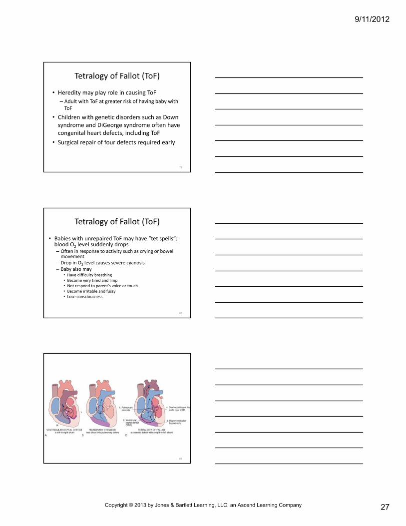
Tetralogy of Fallot (ToF)
• Rare congenital heart defect affects ≈ 5 of every 10,000 babies
• Involves four heart defects
– A large ventricular septal defect (VSD)
– Pulmonary stenosis
– Right ventricular hypertrophy
– An overriding aorta
74
Tetralogy of Fallot (ToF)
• VSD = O2‐rich blood from left ventricle mixes with O2‐poor blood from right ventricle
• Pulmonary stenosis: heart works harder to pump blood though narrowed pulmonary valve
– Causes right ventricular hypertrophy
75
Copyright © 2013 by Jones & Bartlett Learning, LLC, an Ascend Learning Company

9/11/2012
26
Tetralogy of Fallot (ToF)
• ToF: overriding aorta between left and right ventricles, directly over VSD
– In healthy hearts, aorta attached to left ventricle
– O2‐poor blood from right ventricle flows into aorta, not into pulmonary artery
76
Tetralogy of Fallot (ToF)
• Together, these four defects prevent blood from reaching lungs for oxygenation
– O2‐poor blood flows to body, resulting in cyanosis
• Other signs and symptoms: heart murmur, delayed growth and development, clubbing of fingers
77
Tetralogy of Fallot (ToF)
• Cause of defect often unknown
• Contributing factors that may occur during pregnancy– German measles (rubella) and some other viral illnesses
– Poor nutrition – Overuse of alcohol – Age (over 40) – Diabetes
78
Copyright © 2013 by Jones & Bartlett Learning, LLC, an Ascend Learning Company
9/11/2012
27
Tetralogy of Fallot (ToF)
• Heredity may play role in causing ToF
– Adult with ToF at greater risk of having baby with ToF
• Children with genetic disorders such as Down syndrome and DiGeorge syndrome often have congenital heart defects, including ToF
• Surgical repair of four defects required early
79
• Babies with unrepaired ToF may have “tet spells“: blood O2 level suddenly drops – Often in response to activity such as crying or bowel movement
– Drop in O2 level causes severe cyanosis– Baby also may
• Have difficulty breathing • Become very tired and limp • Not respond to parent's voice or touch • Become irritable and fussy • Lose consciousness
Tetralogy of Fallot (ToF)
80
81
Copyright © 2013 by Jones & Bartlett Learning, LLC, an Ascend Learning Company
9/11/2012
28
• Healthy heart = aorta and pulmonary artery properly aligned with appropriate ventricle
– If reversed (transposed), aorta arises from right ventricle, pulmonary artery from left
• Defect known as transposition of the great arteries (TGA)
Transposition of the Great Arteries
82
• TGA = systemic and pulmonary circulations being in parallel rather than in series
– O2‐poor blood returning from body to right atrium, right ventricle pumped to aorta and to body
– O2‐rich blood returning from lungs to left atrium and ventricle sent back to lungs via pulmonary artery
Transposition of the Great Arteries
83
• Patient survival before surgical repair depends on presence of an ASD, a VSD, or PDA
• If untreated, over 50 percent of infants with TGA die in first month of life; 90 percent in first year
Transposition of the Great Arteries
84
Copyright © 2013 by Jones & Bartlett Learning, LLC, an Ascend Learning Company
9/11/2012
29
Total Anomalous Pulmonary Venous Return (TAPVR )
• Congenital heart defect in which four pulmonary veins that bring oxygen‐rich blood back to heart from lungs are not properly attached to left atrium
– Improperly attached to another area (usually superior vena cava)
– With this defect, O2‐rich blood that should return to left atrium, left ventricle, aorta, and then body instead mixes with the O2‐poor blood flowing into right side of heart
– Blood circles to and from lungs and never to body
85
Total Anomalous Pulmonary Venous Return (TAPVR)
• To survive before corrective surgery, large ASD or patent foramen ovale (passage between left and right atria) must exist to allow oxygenated blood to flow to left side of heart and rest of body – Infants with TAPVR may appear critically ill with following symptoms • Lethargy • Poor feeding • Rapid breathing • Poor growth • Frequent respiratory infections • Cyanosis
86
Congenital Dysrhythmias
• Over 800,000 adults with CHD living in U.S.
– ≈ 45 percent considered to have mild CHD: atrialseptal defect, valvar pulmonary stenosis
– 40 percent classified as having moderate disease: ToF
– 15 percent considered to have complex disease: single ventricle, TGA
• Dysrhythmias develop within any of these but incidence increases in moderate and severe
87
Copyright © 2013 by Jones & Bartlett Learning, LLC, an Ascend Learning Company
9/11/2012
30
Congenital Dysrhythmias
• Malfunctions in embryonic development responsible for CHDs can directly impact development of heart’s conduction systems
– Especially displacement of AV node and bundle of His
88
Congenital Dysrhythmias
• These patients more vulnerable to dysrhythmias
– Atrial fibrillation
– Atrial flutter
– Reentry tachycardias
– Heart blocks
• Standard of care = follow same treatment guidelines as for other patients
89
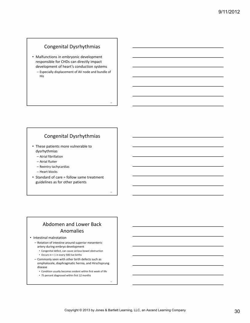
Abdomen and Lower Back Anomalies
• Intestinal malrotation
– Rotation of intestine around superior mesenteric artery during embryo development
• Congenital defect, can cause serious bowel obstruction
• Occurs in ≈ 1 in every 500 live births
– Commonly seen with other birth defects such as omphalocele, diaphragmatic hernia, and Hirschsprungdisease
• Condition usually becomes evident within first week of life
• 75 percent diagnosed within first 12 months
90
Copyright © 2013 by Jones & Bartlett Learning, LLC, an Ascend Learning Company
9/11/2012
31
• May be acute or chronic– Primary presenting sign in acute malrotation: emesis filled with bile
– Features of chronic malrotation
– Recurrent bouts of abdominal pain and diarrhea (alternating with constipation)
– Intolerance of solid food
– Jaundice
– Lower GI bleeding
– Gastroesophageal reflux
Abdomen and Lower Back Anomalies
91
• If symptoms persist
– Shock, including poor perfusion
– Decreased urine output
– Hypotension
• Requires surgical repair
Abdomen and Lower Back Anomalies
92
Pyloric Stenosis
• Narrowing of pylorus (opening from stomach into small intestine)
• Most common cause of intestinal obstruction in infancy
• Narrowing occurs from muscles around pylorus that have grown too large
93
Copyright © 2013 by Jones & Bartlett Learning, LLC, an Ascend Learning Company
9/11/2012
32
Pyloric Stenosis
• Diagnosis usually made when infant presents history of progressive, forceful vomiting
– Usually begins within second/third week of life
– Parents may complain baby “spitting up”
– Often try different formula in formula‐fed babies without change being noted
94
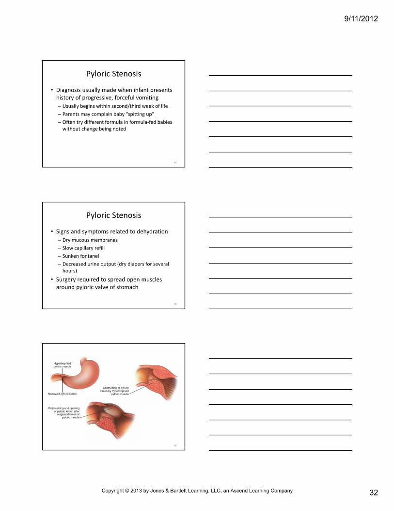
Pyloric Stenosis
• Signs and symptoms related to dehydration
– Dry mucous membranes
– Slow capillary refill
– Sunken fontanel
– Decreased urine output (dry diapers for several hours)
• Surgery required to spread open muscles around pyloric valve of stomach
95
96
Copyright © 2013 by Jones & Bartlett Learning, LLC, an Ascend Learning Company

9/11/2012
33
Diaphragmatic Hernia
• Caused by malformation of diaphragm during fetal development
– Affects 1 in every 2,500 to 5,000 live births
–May occur on right, left, or both sides, but left most common
97
Diaphragmatic Hernia
• Results from defect (hole) in diaphragm muscle– Opening allows abdominal organs (stomach, bowel, liver, and spleen) to enter chest cavity
– Lung on affected side does not develop normally (pulmonary hypoplasia)• = reduced lung capacity
– Other organ damage possible, including heart
– Significant prenatal shift in mediastinum may indicate some degree of pulmonary hypoplasia on contralateral side• Elevate infant’s head and thorax to assist downward displacement of abdominal organs and help improve ventilation
98
99
Copyright © 2013 by Jones & Bartlett Learning, LLC, an Ascend Learning Company

9/11/2012
34
Diaphragmatic Hernia
• Symptoms
– Respiratory distress: shortly after birth
– Cyanosis unresponsive to ventilations
– Tachypnea
– Tachycardia
100
Diaphragmatic Hernia
• Infant born may have irregular chest wall movement, displaced heart sounds, absent breath sounds on affected side, bowel sounds in chest cavity
• Abdomen may be scaphoid (flat) and feel “full” on palpation
• Medical direction may recommend orogastric tube with ↓ periodic suc on
• Tracheal intubation may be needed
101
Diaphragmatic Hernia
• Use of bag device/aggressive positive pressure ventilation may cause gastric distention, worsened condition
– Bag device contraindicated
• Surgery required to repair hernia and place abdominal organs in normal location
• If condition diagnosed during pregnancy, fetal surgery may be indicated
102
Copyright © 2013 by Jones & Bartlett Learning, LLC, an Ascend Learning Company

9/11/2012
35
Omphalocele
• Hernia in which infant’s intestines or other abdominal organs protrude through umbilicus
• During fetal development, abdominal wall (umbilical ring) muscles do not close properly
– Leads to intestines outside umbilical cord
– Affects 1 in every 5,000 to 6,000 live births
– 25 to 40 percent of infants with this will have another congenital anomaly
• Usually first seen on prenatal ultrasound
103
104
Omphalocele
• Most quite visible at birth
– Small contains only small section of intestines
– Large contains liver and spleen
• Protruding organs usually covered by thin sac of peritoneum (intact omphalocele)
105
Copyright © 2013 by Jones & Bartlett Learning, LLC, an Ascend Learning Company

9/11/2012
36
Omphalocele
• Umbilical vessels usually present within sac
• Protect and cover exposed tissue with moist, sterile gauze pads
• Rupture (nonintact omphalocele) may occur immediately before or during delivery
106
Omphalocele
• Usually successfully repaired with surgery
• Surgery
– Small omphalocele‐exposed tissues covered & held with special plastic pouch (silo)
• Over time, silo squeezes exposed tissue back into infant’s abdomen
• Surgical repair of abdominal muscles may be needed as well
– Larger omphalocele with intact sac, often delayed
• Tissue eventually covered by surrounding skin, after which surgery performed to improve cosmetic outcome
107
• Accomplish initial assessment and management within ≈ 60 seconds ("Golden Minute")
• These steps enable immediate recognition of infant needing resuscitation
– Also leads to efficient, effective emergency care delivery
Assessment and Management of Neonate
108
Copyright © 2013 by Jones & Bartlett Learning, LLC, an Ascend Learning Company

9/11/2012
37
• Initial steps– Provide warmth by drying baby to prevent heat loss, avoid hypothermia
– Position head in sniffing position to open airway– Clear airway if necessary with bulb syringe or suction catheter
– Provide tactile stimulation to initiate breathing if necessary
– Further evaluate
Assessment and Management of Neonate
109
110
• All newborns limited in ability to conserve heat, so are at risk for developing hypothermia
– Immediately after delivery, dry infant’s body and head
• Prevents evaporative heat loss, metabolic derangements from cold stress
• Provides gentle stimulation, which may initiate respirations
Prevent Heat Loss and Avoid Hypothermia
111
Copyright © 2013 by Jones & Bartlett Learning, LLC, an Ascend Learning Company

9/11/2012
38
• Remove any wet coverings from infant and cover w/ dry wrappings
• Majority of heat loss prevented by covering newborn’s head
– Head accounts for 20 percent of newborn’s BSA
Prevent Heat Loss and Avoid Hypothermia
112
What other measures can you take to warm the infant?
113
Clear the Airway
• After drying and covering newborn, establish open airway– Place infant supine, with head in sniffing position– Take care to prevent hyperextension or underextension to avoid compromised airway
– Blanket or towel under infant’s shoulders to elevate torso 3/4 to 1 inch helps maintains correct position
– Once properly positioned, most newborns breathe without difficulty
114
Copyright © 2013 by Jones & Bartlett Learning, LLC, an Ascend Learning Company
9/11/2012
39
115
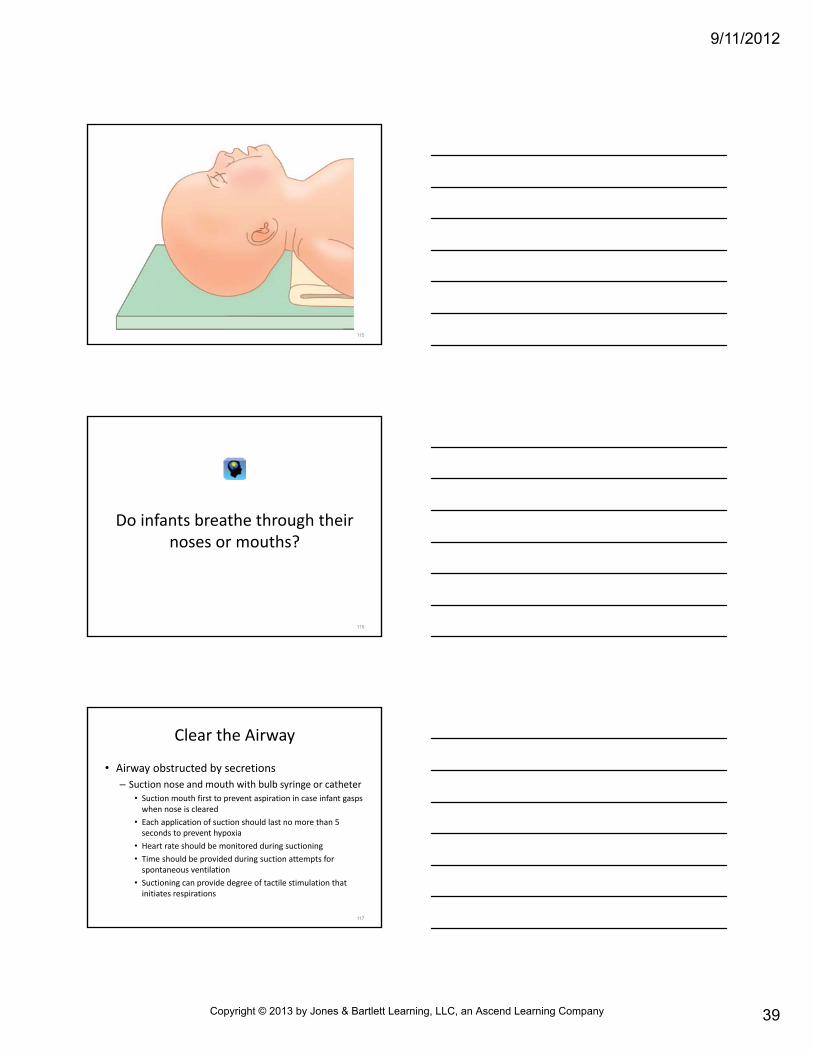
Do infants breathe through their noses or mouths?
116
Clear the Airway
• Airway obstructed by secretions
– Suction nose and mouth with bulb syringe or catheter
• Suction mouth first to prevent aspiration in case infant gasps when nose is cleared
• Each application of suction should last no more than 5 seconds to prevent hypoxia
• Heart rate should be monitored during suctioning
• Time should be provided during suction attempts for spontaneous ventilation
• Suctioning can provide degree of tactile stimulation that initiates respirations
117
Copyright © 2013 by Jones & Bartlett Learning, LLC, an Ascend Learning Company
9/11/2012
40
Meconium Staining
• Presence of fetal stool in amniotic fluid
– Occurs in utero or intrapartum and in ≈ 12 percent of all deliveries
–More common in post‐term and small‐for‐gestational‐age newborns
– Also common in infants who develop fetal distress during L&D
118
Meconium Staining
• Associated with
– Increased perinatal mortality
– Hypoxemia
– Aspiration pneumonia
– Pneumothorax
– Pulmonary hypertension
119
Meconium Staining
• Appearance depends on amount of meconiumparticles and amniotic fluid– Slightly yellow or light green, thin and watery– Thick, dark green or black, pea‐soup appearance– Thick meconium in amniotic fluid
• Chance particles will be aspirated into infant’s mouth
• Potentially aspirated into trachea and lungs• Can lead to partial or complete obstruction of airways
• Death can result from hypoxia, hypercapnia, and acidosis
120
Copyright © 2013 by Jones & Bartlett Learning, LLC, an Ascend Learning Company

9/11/2012
41
121
Meconium Staining
• Meconium (or evidence of infection) observed– If vigorous (strong respiratory efforts, good muscle tone, heart rate greater than 100 beats/min) • No special care of airway required
– If not vigorous (absent or depressed respirations, decreased muscle tone, heart rate less than 100 beats/min)• Endotracheal intubation and endotracheal suctioning immediately after birth
• Because presence can be determined only after membranes have ruptured, critical to have airway equipment available
122
Meconium Staining
• Emergency care
– Prepare necessary equipment
• Intubation equipment
• Bulb syringe
• DeLee suction
• 12 F or 14 F suction catheter
• Portable suction and irrigation solution
• Gauze pads
• Infant bag device
123
Copyright © 2013 by Jones & Bartlett Learning, LLC, an Ascend Learning Company

9/11/2012
42
Meconium Staining
• Emergency care
– Intubation equipment
• Padding for patient positioning
• Stethoscope
• # 0 and # 1 laryngoscope blades
• ET tubes (2.5, 3.0, 3.5, 4.0)
• Stylet
• Meconium aspirator
• Oxygen tubing
124
Meconium Staining
• After delivery, clear infant’s airway and thoroughly suction nose, mouth, pharynx
– Remove residual meconium in hypopharynx by suction under direct visualization
125
• Quickly intubate trachea– Suction to proximal end of ET tube using DeLee device while withdrawing tube
– During intubation/suction• Aim 100 percent oxygen toward infant’s face
• Monitor fetal heart rate for bradycardia
• If bradycardia develops, ventilate using bag device after suctioning to prevent persistent bradycardia and hypoxia
– Repeat intubation‐suction‐extubation cycle until no further meconium obtained• Do not ventilate b/w intubations
Meconium Staining
126
Copyright © 2013 by Jones & Bartlett Learning, LLC, an Ascend Learning Company
9/11/2012
43
Meconium Staining
• After tracheal suction complete, resuscitative measures as needed
• If respirations adequate, manage airway in normal fashion
• Medical direction may recommend 8 F orogastric tube aspirated with syringe and left open to air to prevent aspiration of gastric contents after resuscitation complete
127
128
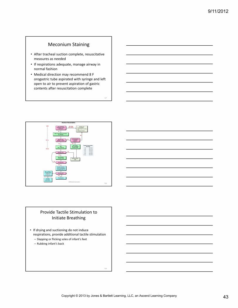
• If drying and suctioning do not induce respirations, provide additional tactile stimulation
– Slapping or flicking soles of infant’s feet
– Rubbing infant’s back
Provide Tactile Stimulation to Initiate Breathing
129
Copyright © 2013 by Jones & Bartlett Learning, LLC, an Ascend Learning Company

9/11/2012
44
Further Evaluate the Infant
• Drying and positioning necessary in every infant at birth– They open airway and initiate breathing
• To further evaluate infant, paramedic should– Observe and evaluate infant’s respirations
• If normal (e.g., crying), continue evaluation
– Place neonatal pulse oximetry probe on right upper extremity (wrist or medial surface of palm) to monitor oxygen saturation
130
Further Evaluate the Infant
• To further evaluate infant, paramedic should– Evaluate heart rate by stethoscope or palpation of pulse in base of umbilical cord• Greater than 100 beats/min, continue evaluation
– Evaluate infant’s color• Peripheral cyanosis (acrocyanosis) common first few minutes of life, does not indicate hypoxemia
– If color is normal ("pinking up") and SpO2 readings are within normal ranges, continue evaluation with Apgar score
131
Apgar Score
• Enables rapid evaluation of newborn’s condition at specific intervals after birth
– Routinely assessed at 1 and 5 minutes
– Should not be used alone in determining need for resuscitation
– APGAR evaluates appearance, pulse rate, grimace, activity, and respirations
• Score of 7 to 10: Normal
• 4 to 6: Moderately distressed; give oxygen and stimulation
• Less than 4: Severely distressed; resuscitation required
132
Copyright © 2013 by Jones & Bartlett Learning, LLC, an Ascend Learning Company
9/11/2012
45
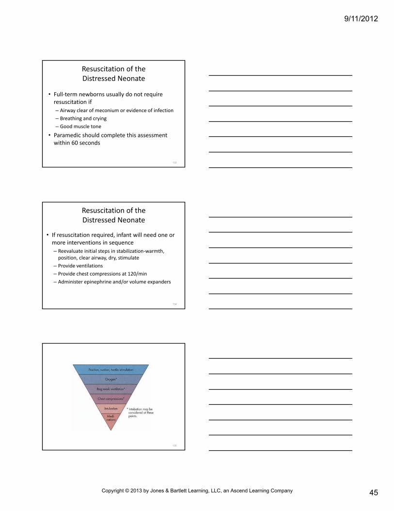
• Full‐term newborns usually do not require resuscitation if
– Airway clear of meconium or evidence of infection
– Breathing and crying
– Good muscle tone
• Paramedic should complete this assessment within 60 seconds
Resuscitation of the Distressed Neonate
133
• If resuscitation required, infant will need one or more interventions in sequence
– Reevaluate initial steps in stabilization‐warmth, position, clear airway, dry, stimulate
– Provide ventilations
– Provide chest compressions at 120/min
– Administer epinephrine and/or volume expanders
Resuscitation of the Distressed Neonate
134
135
Copyright © 2013 by Jones & Bartlett Learning, LLC, an Ascend Learning Company
9/11/2012
46
• Need to progress in sequence based on
– Simultaneous assessment of infant’s respirations, heart rate, oximetry readings
• ≈ 30 seconds to complete each step; to reevaluate; and to decide whether to progress to next sequenced step in resuscitation
Resuscitation of the Distressed Neonate
136
• Paramedic should ensure infant is dry and warm
– Cold stress can increase oxygen consumption and impede effective breathing
– Hypothermia associated with perinatal respiratory depression
Reevaluate Initial Steps in Stabilization
137
• Additional methods to warm a newborn
– Covering in plastic wrap (food‐grade, heat‐resistant plastic)
– Placing baby skin‐to‐skin with mother and covering both with blanket
• Avoid hyperthermia
– Goal is to achieve and maintain normal body temp for newborn
Reevaluate Initial Steps in Stabilization
138
Copyright © 2013 by Jones & Bartlett Learning, LLC, an Ascend Learning Company

9/11/2012
47
• Properly position (or reposition) head and neck to ensure open airway
• Paramedic can make other attempts at stimulation to initiate breathing
Reevaluate Initial Steps in Stabilization
139
• Breathing, but color and SpO2 don't improve within 90 seconds or if central cyanosis– Give supplemental oxygen
• Free‐flow oxygen mixed with air through face mask and flow‐inflating bag, oxygen mask, or a hand cupped around oxygen tubing (2 inches from infant’s nose)
– Oxygen therapy guided by pulse oximetry, continued until target SpO2 range achieved
– Titrate oxygen delivery to maintain SpO2 in target range• If baby is bradycardic (heart rate less than 60 beats/min) after 90 seconds of resuscitation, increase oxygen concentration to 100 percent until recovery of normal heart rate
Reevaluate Initial Steps in Stabilization
140
Provide Ventilations
• Newborn should have respirations sufficient to maintain
– Target SpO2
– Heart rate greater than 100 beats/min
• Gasping and apnea indicate need for assisted ventilations
• Increased or decreased heart rate provides clues of improvement or deterioration
141
Copyright © 2013 by Jones & Bartlett Learning, LLC, an Ascend Learning Company
9/11/2012
48
Provide Ventilations
• Inadequate respirations or heart rate less than 100 beats/min 30 seconds after initial steps– Initiate positive‐pressure ventilation– Provide assisted ventilations @ 40 to 60 breaths/min until heart rate is greater than 100 beats/min
• Assisted ventilations can be delivered with– Flow‐inflating bag– Self‐inflating bag– T‐piece
142
Provide Ventilations
• Medical direction may recommend continuous positive airway pressure (CPAP) to infants breathing spontaneously, but with difficulty, following birth
• Positive‐end expiratory pressure (PEEP) also in supporting ventilations
– Use of CPAP or PEEP should be guided by medical direction
143
Endotracheal Intubation
• Indicated at several points during neonatal resuscitation
– Tracheal suctioning of meconium required
– Bag‐mask ventilation ineffective or prolonged
–When chest compressions performed
– Special resuscitation circumstance, such as congenital diaphragmatic hernia, or extremely low birth weight (under 1000 g)
144
Copyright © 2013 by Jones & Bartlett Learning, LLC, an Ascend Learning Company

9/11/2012
49
Endotracheal Intubation
• Before considering intubation or pharmacological therapy, reevaluate
– Is chest movement adequate?
• Check chest expansion and auscultate for bilateral breath sounds
• Is bag mask seal tight? Turn large mask upside down for better fit
• Is airway blocked? Reassess head position, reexamine airway for secretions
• Is adequate ventilatory pressure being used? May need to disable bag mask pop‐off valve for ↑ inspiratory pressures
• Is air in stomach interfering with chest expansion? Consider nasogastric or orogastric decompression per protocol
145
Endotracheal Intubation
• Before considering intubation or pharmacological therapy, reevaluate
– Are target SpO2 measurements within normal range?
• Is oxygen tubing attached to bag and flowmeter?
• If using self‐inflating bag, is oxygen reservoir attached?
146
• Prompt increase in heart rate and chest wall movement after intubation and intermittent plus pressure ventilation best indicators of correct tube placement
– Verify tube placement visually during intubation and by primary and secondary confirmation methods
Endotracheal Intubation
147
Copyright © 2013 by Jones & Bartlett Learning, LLC, an Ascend Learning Company

9/11/2012
50
• Exhaled CO2 recommended method for confirmation of tube placement in infants with adequate cardiac output
– False‐negative readings can occur if infant has poor or absent pulmonary flow
• Laryngeal mask airway (LMA) may be used to establish airway if bag‐mask ventilation ineffective or tracheal intubation failed
Endotracheal Intubation
148
Provide Chest Compressions
• Indicated if heart rate is under 60 beats/min after 30 seconds ventilation with supplemental oxygen
• Ensure assisted ventilations effective before initiating chest compressions
149
Provide Chest Compressions
• Coordinate compressions with ventilations to avoid simultaneous delivery
• Deliver compressions and ventilations at ratio of 31 at rate of 120/min
– This will achieve ≈ 90 compressions and 30 breaths/min
150
Copyright © 2013 by Jones & Bartlett Learning, LLC, an Ascend Learning Company
9/11/2012
51
Why would the compressions be initiated when the infant still has a
pulse?
151
Provide Chest Compressions
• Two thumb‐encircling hands compression preferred for full‐term newly born and older infants
– Preterm infants and infants small for gestational age = chest compressions using two fingers
– Performed on lower 1/3 of sternum
– Depth of compression ≈ 1/3 of anterior‐posterior diameter of chest and sufficiently deep to generate palpable pulse
– Frequently reassess respirations, heart rate, and oxygenation
– Coordinated compressions and ventilations until spontaneous heart rate equals or is above 60 beats/min
152
• Drugs rarely indicated in resuscitation of newly born– Administer only if heart rate remains below 60 beats/min despite ventilation with 100 percent oxygen and effective chest compressions
• Drug therapy may include epinephrine and volume expanders
• Rarely, buffers, a narcotic antagonist, or vasopressors useful
Administer Epinephrine and/or Volume Expanders
153
Copyright © 2013 by Jones & Bartlett Learning, LLC, an Ascend Learning Company

9/11/2012
52
• Give all drugs intravenously as soon as venous access obtained
– Epinephrine may be administered via ET tube while obtaining IV access, but safety and efficacy not established
• Concentration of epinephrine for either route is 110,000 (0.1 mg/mL)
Administer Epinephrine and/or Volume Expanders
154
• Give all drugs intravenously as soon as venous access obtained– Consider volume expanders when blood loss suspected
• Also consider if infant is in shock (pale skin, poor perfusion, weak pulse) and has not responded to other resuscitative measures
• Evaluate end organ perfusion by comparing central vs. peripheral pulses and through capillary refill tests
• Give volume expanders slowly and with caution when resuscitating preemies
• Recommended dose 10 mL/kg, which may need to be repeated
• Rapid infusion of large volumes associated with intraventricularhemorrhage
Administer Epinephrine and/or Volume Expanders
155
Routes of Drug Administration
• IV route preferred in newborn, but other methods to consider
– Endotracheal route
– Intraosseous (IO) route
• During CPR or treatment of severe shock, establish IO access when venous access cannot be rapidly achieved
156
Copyright © 2013 by Jones & Bartlett Learning, LLC, an Ascend Learning Company
9/11/2012
53
Postresuscitation Care
• Three most common complications of postresuscitation period
– Endotracheal tube migration (including dislodgment)
– Tube occlusion by mucus or meconium
– Pneumothorax
157
Postresuscitation Care
• Suspect these complications in presence of
– Decreased chest wall movement
– Diminished breath sounds
– Return of bradycardia
– Unilateral decrease in chest expansion
– Altered intensity to pitch of breath sounds
– Increased resistance to hand ventilation
158
Postresuscitation Care
• Corrective management in field
– Adjustment of endotracheal tube (exhaled CO2
devices recommended for monitoring l tube placement)
– Reintubation
– Suction
– Needle decompression to manage suspected pneumothorax must be guided carefully by medical direction
159
Copyright © 2013 by Jones & Bartlett Learning, LLC, an Ascend Learning Company
9/11/2012
54
How much movement would it take to dislodge the ET tube from a neonate?
160
• ITH (33.5°C to 34.5°C) can decrease mortality and decrease neurological complications in some who have return of spontaneous circulation (RSOC) following cardiac arrest
• Protocols for treatment in neonates ≥36 weeks gestation– Cooling within 6 hours following birth
• Cooling continued 72 hours, after which infant rewarmed over at least 4 hours
Induced Therapeutic Hypothermia
161
Induced Therapeutic Hypothermia
• Induced therapeutic hypothermia not a prehospital consideration in neonatal resuscitation
• Procedure instituted in hospital setting
162
Copyright © 2013 by Jones & Bartlett Learning, LLC, an Ascend Learning Company
9/11/2012
55
Neonatal Transport
• Important to maintain infant’s body temperature and prevent hypothermia during transport
• Critical to maintain oxygen levels and support ventilations
163
Neonatal Transport
• Initial prehospital phase transport strategies
– Provide warm ambulance
– Free‐flow oxygen (warmed if available)
– Cover baby’s head
– Apply warm blankets to prevent hypothermic complications
164
Neonatal Transport
• Specialized transport equipment such as isolettes and radiant heating units often used for interhospital transfers
– These require special training
– Neonatal transport teams of paramedics, nurses, respiratory therapists, and physicians are part of well‐organized regional referral systems throughout U.S.
165
Copyright © 2013 by Jones & Bartlett Learning, LLC, an Ascend Learning Company

9/11/2012
56
166
Specific Situations
• May call for advanced life support for neonate– Situations include
• Apnea• Bradycardia• Prematurity
• Respiratory distress and cyanosis
• Hypovolemia
• Seizures
• Fever• Hypothermia
• Hypoglycemia
• Vomiting and diarrhea
• Common birth injuries
167
Specific Situations
• While providing advanced life support, consider emotional needs of mother and family
• When possible, explain what is being done and why procedure is necessary
168
Copyright © 2013 by Jones & Bartlett Learning, LLC, an Ascend Learning Company

9/11/2012
57
Apnea • Absence of spontaneous respirations
– Primary apnea self‐limited condition controlled by PCO2 levels
• Common event immediately after birth
– Secondary apnea exceeds 20 seconds without spontaneous breathing occurring
• Can lead to hypoxemia and bradycardia
• Common in preterm infant
• Often results from hypoxia or hypothermia
• May also be caused by maternal use of narcotics or CNS depressants, prolonged or difficult L&D, airway and respiratory muscle weakness, septicemia, metabolic disorders, CNS disorders
169
Apnea
• Emergency care for prolonged apnea begins with stimulating infant to breathe– Flick soles of feet– Rub back– If needed, use bag device (with disabled pop‐off valve) while applying least amount of pressure that produces adequate chest rise
• Suction secretions from airway and maintain body temp to prevent hypothermia
170
Apnea
• Endotracheal intubation and circulatory support required if central cyanosis persists despite adequate ventilations
171
Copyright © 2013 by Jones & Bartlett Learning, LLC, an Ascend Learning Company

9/11/2012
58
Apnea
• Appropriate drug therapy in managing some infants with prolonged apnea– Dextrose (10 percent in H2O) if hypoglycemia confirmed
– Naloxone: reversal of respiratory depression in newborn whose mother received narcotics within 4 hours of delivery• Do not give narcotic antagonists if infant's mother is drug abuser
• May induce drug withdrawal in neonate
• Apnea treated early and aggressively normally results in good outcome
172
Bradycardia
• Heart rate less than 100 beats/min
– In neonate, most commonly caused by hypoxia
– Also may result from
• Increased intracranial pressure
• Hypothyroidism
• Acidosis
• Congenital AV nodal block in infants of mothers with systemic lupus erythematosa
173
Bradycardia
• Other risk factors
– Prolonged suctioning
– Use of airway or invasive procedures during resuscitation that may cause vagal stimulation (e.g., inadequately secured ET tube movement)
• Minimal risk to life in neonates if corrected quickly
174
Copyright © 2013 by Jones & Bartlett Learning, LLC, an Ascend Learning Company

9/11/2012
59
Bradycardia
• Initial management: assess for upper airway obstruction
– Possible causes of obstruction• Airway secretions
• Foreign body
• Position of tongue or soft tissues of neck
175
Bradycardia
• Prehospital care to improve ventilation may include
– Airway positioning
– Suction
– Positive pressure ventilation with supplemental oxygen
– Tracheal intubation
176
Bradycardia
• Monitor ventilatory and circulatory status closely
– This determines need for more advanced life support measures
• Measures may include chest compressions and drug therapy
177
Copyright © 2013 by Jones & Bartlett Learning, LLC, an Ascend Learning Company

9/11/2012
60
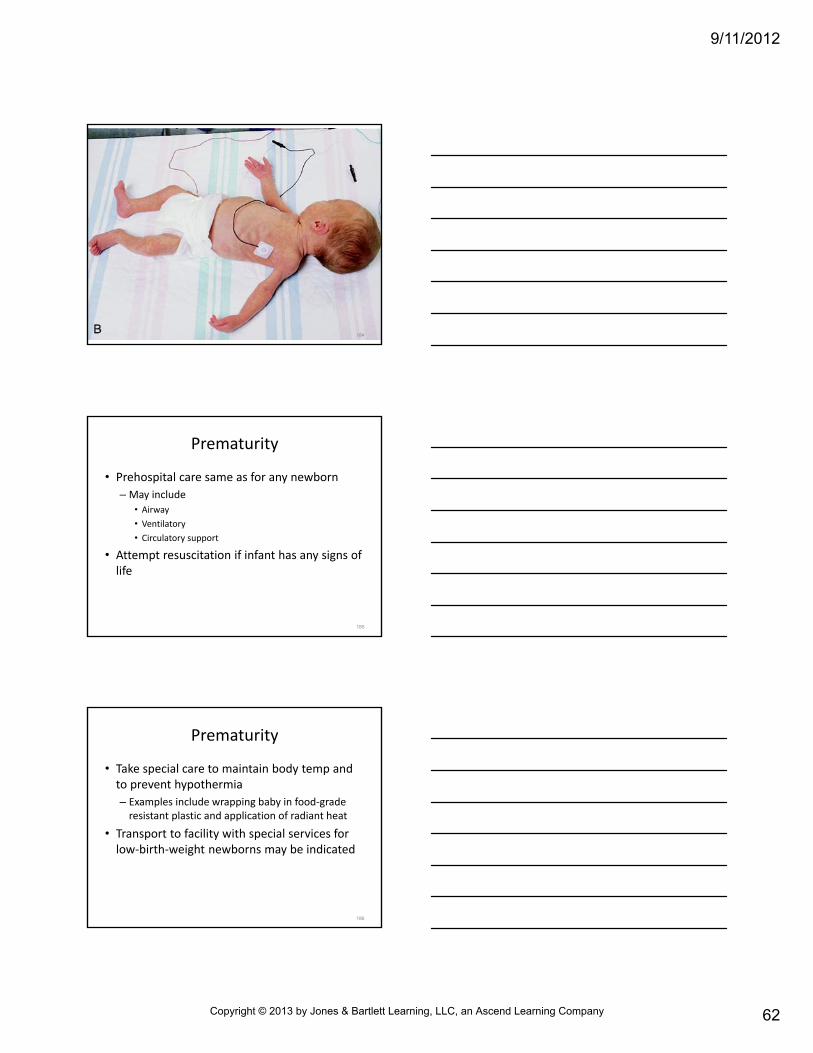
Prematurity
• Premature infant (preemie) refers to baby born before 37 weeks of gestation
–Weight often b/w 0.6 to 2.2 kg (1.5 to 5 lbs)
– Healthy premies: infants who weigh over 1700 g have survivability and outcome ≈ that of full‐term infants
–Mortality rate decreases weekly with gestation beyond onset of viability (currently ≈ 23 to 24 wks)
178
Prematurity
• Premature infant (preemie) refers to baby born before 37 weeks of gestation
– Premies have increased risk for
• Respiratory depression
• Hypothermia
• Brain injury from hypoxemia
• Also especially vulnerable to changes in blood pressure, intraventricular hemorrhage, and fluctuations in serum chemistry
179
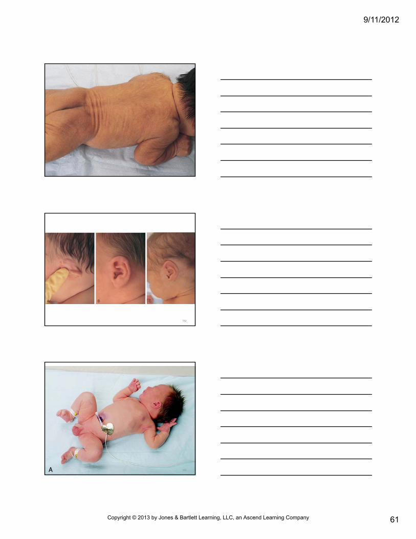
Prematurity
• Degree of immaturity determines how infant appears physically, but most have
– Large trunk
– Short extremities
– Less subcutaneous fat than full‐term infants
– Skin that appears translucent
– Telltale physical findings include lanugo, ear characteristics, and muscle tone
180
Copyright © 2013 by Jones & Bartlett Learning, LLC, an Ascend Learning Company
9/11/2012
61
181
182
183
Copyright © 2013 by Jones & Bartlett Learning, LLC, an Ascend Learning Company
9/11/2012
62
184
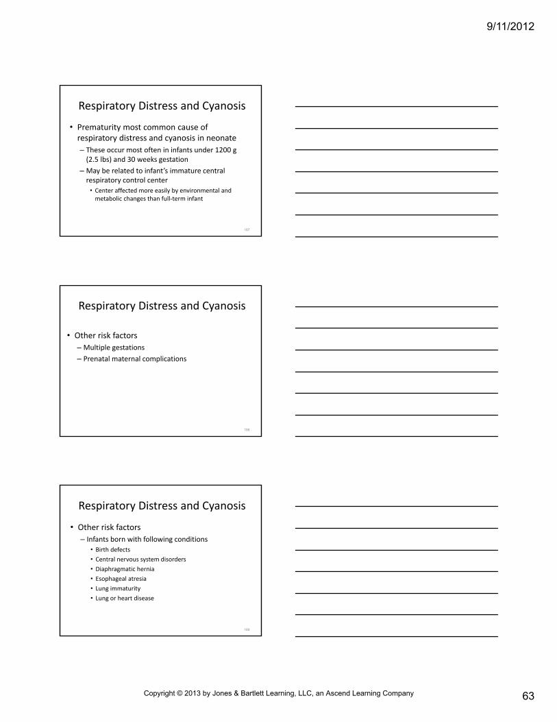
Prematurity
• Prehospital care same as for any newborn
–May include
• Airway
• Ventilatory
• Circulatory support
• Attempt resuscitation if infant has any signs of life
185
Prematurity
• Take special care to maintain body temp and to prevent hypothermia
– Examples include wrapping baby in food‐grade resistant plastic and application of radiant heat
• Transport to facility with special services for low‐birth‐weight newborns may be indicated
186
Copyright © 2013 by Jones & Bartlett Learning, LLC, an Ascend Learning Company
9/11/2012
63
Respiratory Distress and Cyanosis
• Prematurity most common cause of respiratory distress and cyanosis in neonate
– These occur most often in infants under 1200 g (2.5 lbs) and 30 weeks gestation
–May be related to infant’s immature central respiratory control center
• Center affected more easily by environmental and metabolic changes than full‐term infant
187
• Other risk factors
–Multiple gestations
– Prenatal maternal complications
Respiratory Distress and Cyanosis
188
• Other risk factors
– Infants born with following conditions• Birth defects
• Central nervous system disorders
• Diaphragmatic hernia
• Esophageal atresia
• Lung immaturity
• Lung or heart disease
Respiratory Distress and Cyanosis
189
Copyright © 2013 by Jones & Bartlett Learning, LLC, an Ascend Learning Company

9/11/2012
64
• Other risk factors
– Infants born with following conditions• Meconium or amniotic fluid aspiration
• Metabolic acidosis
• Mucous obstruction of nasal passages
• Pneumonia
• Primary pulmonary hypertension
• Shock and sepsis
• Tracheoesophageal fistula
Respiratory Distress and Cyanosis
190
• Respiratory distress and cyanosis can lead to cardiac arrest in neonate– Take immediate action to improve breathing and support respirations
– Assessment findings may include• Tachypnea
• Paradoxical breathing
• Intercostal retractions
• Nasal flaring
• Expiratory grunting
• Central cyanosis
Respiratory Distress and Cyanosis
191
• Generally managed with
– Stimulation
– Positioning of airway
– Prevention of heat loss and hypothermia
– Oxygenation and ventilation
– Suction and intubation with ventilatory support (if needed)
Respiratory Distress and Cyanosis
192
Copyright © 2013 by Jones & Bartlett Learning, LLC, an Ascend Learning Company

9/11/2012
65
Hypovolemia
• May result from dehydration, hemorrhage, trauma, sepsis
• May be associated with myocardial dysfunction– Signs and symptoms
• Mottled or pale color
• Cool skin• Tachycardia• Diminished peripheral pulses
• Delayed capillary refill despite normal ambient temp
193
Hypovolemia
• Shock may be present despite normal BP– Prompt, effective treatment of compensated shock• May prevent development of hypotension (decompensated shock) and associated high morbidity and mortality
• Prehospital care always directed at
– Adequate airway, ventilatory, and circulatory support (plus control of external hemorrhage)
– Providing rapid transport to appropriate facility
194
Hypovolemia
• Signs of hypovolemia
– Give fluid bolus (10 mL/kg over 5 to 10 minutes of isotonic crystalloid) immediately after obtaining IV access
– Reassess the infant
– If signs of shock persist, give second 10‐mL/kg bolus
– Further boluses given as needed and under medical direction
195
Copyright © 2013 by Jones & Bartlett Learning, LLC, an Ascend Learning Company

9/11/2012
66
Seizures
• Occur in small percent of newborns
– Usually sign of underlying abnormality
• Prolonged or frequent may result in metabolic changes and cardiopulmonary difficulties
196
Types of Seizures
• Seizures in neonates usually fragmented, not well sustained
– Classifications• Subtle
• Tonic
• Multifocal
• Focal clonic
• Myoclonic
197
Types of Seizures
• Subtle
– Eye deviation
– Blinking
– Sucking
– Swimming movements of arms
– Peddling movements of legs
– Apnea may be present
198
Copyright © 2013 by Jones & Bartlett Learning, LLC, an Ascend Learning Company
9/11/2012
67
Types of Seizures • Tonic – Extension of limbs– Less often, flexion of upper extremities and extension of lower extremities
– Tonic more common in premature infants, especially with intraventricular hemorrhage
• Multifocal– Clonic activity in one extremity that may migrate randomly to another area of body
– Mainly occurs in full‐term infants
199
Types of Seizures
• Focal clonic
– Clonic, localized jerking
– Occur in full‐term and premature newborns
• Myoclonic
– Flexion and jerking of upper or lower extremities
–May occur singly or in series of repetitive jerking cycles
200
Emergency Care for Neonatal Seizures
• Provide airway, ventilatory, and circulatory support
– Maintain infant’s body temp
– Drug therapy that may be prescribed by medical direction
• Dextrose to treat hypoglycemia
• Anticonvulsant agents
• Benzodiazepines (for status epilepticus)
– Seizure activity always considered pathological
– Rapid transport for physician evaluation needed
201
Copyright © 2013 by Jones & Bartlett Learning, LLC, an Ascend Learning Company

9/11/2012
68
Fever
• Described as rectal temperature over 100.4°F (38.0°C)– Often response to acute viral or bacterial infection–May result from change in infant’s limited ability to control body temp
– Effect of dehydration
• ↑ in core temp ↑ oxygen demands and ↑ glucose metabolism– These may lead to metabolic acidosis
202
Fever
• Assessment findings may include
–Mental status changes (e.g., irritability and lethargy)
– Decreased intake
– Rashes and petechia
–Warm or hot skin
203
Fever
• Prehospital care for febrile infants mainly supportive
• As a rule, cooling procedures and use of antipyretics delayed until arrival at hospital
204
Copyright © 2013 by Jones & Bartlett Learning, LLC, an Ascend Learning Company
9/11/2012
69
Hypothermia
• Febrile seizures usually affect children b/w 6 months to 5 yrs of age
– Generally not concern in caring for neonate
• All febrile neonates require immediate transport for physician evaluation
– Presume systemic sepsis until proved otherwise
205
Hypothermia
• Core body temp lower than 95°F (35°C)
• May result from
– Decrease in heat production
– Increase in heat loss through• Evaporation
• Conduction
• Convection
• Radiation
– Combination of both
206
Hypothermia
• Neonates sensitive to effects of hypothermia because of their increased surface‐to‐volume ratio– Especially true when they are wet– Associated increase in metabolic demand to maintain body temp can cause• Metabolic acidosis
• Pulmonary hypertension
• Hypoxemia
• Hypothermia may be sign of sepsis in neonate
207
Copyright © 2013 by Jones & Bartlett Learning, LLC, an Ascend Learning Company

9/11/2012
70
• Assessment findings may include– Pale color
– Cool skin (especially extremities)
– Respiratory distress
– Apnea
– Bradycardia
– Central cyanosis
– Acrocyanosis (cyanosis of extremities)
– Irritability (initially)
– Lethargy (in late stage)
– Absence of shivering (variable)
Hypothermia
208
• Prehospital care may include
– Provision of basic and advanced cardiac life support
• Rapid transport to appropriate facility
– Other therapeutic measures
• Ensure infant is dry and warm
• Warm hands before touching newborn
• Possible administration of dextrose to treat hypoglycemia
• Possible IV therapy with warm fluids
• Transport in heated ambulance: 76°F to 80°F (24°C to 26.5°C)
Hypothermia
209
Hypoglycemia
• Blood glucose measurement less than 40 mg/dL
• Condition determined by blood glucose screening on all sick infants
• May be due to inadequate glucose intake or increased use of glucose
210
Copyright © 2013 by Jones & Bartlett Learning, LLC, an Ascend Learning Company

9/11/2012
71
Hypoglycemia
• Risk factors
– Asphyxia
– Toxemia
– Being smaller twin
– CNS hemorrhage
– Sepsis
211
• Assessment findings may include– Twitching or seizure– Limpness– Lethargy– Irritability– Eye rolling– High‐pitched crying– Apnea– Irregular respirations– Cyanosis (possibly)
Hypoglycemia
212
Hypoglycemia
• Prehospital care directed at
– Ensuring adequate airway, ventilatory, and circulatory support
–Maintaining body temp
– Rapid transport
– IV administration of dextrose 10 percent per medical direction
213
Copyright © 2013 by Jones & Bartlett Learning, LLC, an Ascend Learning Company

9/11/2012
72
Hypoglycemia
• Check glucose level again if infant fails to respond to initial resuscitative measures
• Transport immediately to medical facility
– All infants who do not respond normally to dextrose
– Hypoglycemic infants who fail to respond to dextrose
214
Vomiting and Diarrhea
• Occasional, not unusual in neonate
– Vomiting mucus (may be streaked with blood) common in first few hours of life
– 5 to 6 stools/day normal, especially if breast‐feeding
• Consider persistent vomiting and/or diarrhea as warning signs of serious illness
215
Vomiting
• Persistent vomiting in first 24 hours suggests
– Obstruction in upper digestive tract
– Increased intracranial pressure
• Non‐bile‐stained fluid = obstruction at or above first portion of duodenum
–May indicate gastroesophageal reflux
• Bile‐stained vomit may result from obstruction ↓ opening of bile duct
216
Copyright © 2013 by Jones & Bartlett Learning, LLC, an Ascend Learning Company

9/11/2012
73
Vomiting
• Dark blood usually sign of life‐threatening illness
• Assessment findings may include– Distended stomach
– Signs of infection– Dehydration– Increased intracranial pressure
• Also consider that vomiting may be result of drug withdrawal (from mother’s drug use)
217
Vomiting
• Prehospital care
– Maintaining airway clear of vomit and ensure adequate oxygenation
• In severe cases, medical direction may advise IV fluid therapy started before transport
– Fluid therapy treats dehydration and any bradycardia from vagal stimulation
• Transport infants on their sides to help prevent aspiration
218
Diarrhea
• If persistent, serious dehydration and electrolyte imbalance– Often associated with bacterial or viral infection
– Other possible causes• Bacterial enteritis (Clostridium difficile, salmonella, shigella)
• Cystic fibrosis
• Lactose intolerance
• Neonatal abstinence syndrome (drug withdrawal)
• Phototherapy (treatment for hyperbilirubinemia and jaundice)
• Thyrotoxicosis
• Viral gastroenteritis (rotavirus)
219
Copyright © 2013 by Jones & Bartlett Learning, LLC, an Ascend Learning Company

9/11/2012
74
Diarrhea
• Assessment findings often include
– Presence of loose stools
– Decreased urinary output
– Signs of dehydration
• Treatment
– Support infant’s vital functions
– IV fluid therapy (per medical direction)
– Rapid transport to receiving hospital
220
Common Birth Injuries
• Significant birth injury accounts for less than 2 percent of neonatal deaths and stillbirths in U.S.
• Average of 6 to 8 injuries in 1,000 live births
• Larger infants generally more susceptible to birth trauma
– Greater rates reported for infants who weigh over 4500 g
221
Common Birth Injuries
• Uncontrolled, explosive delivery greatest risk factor
• Cranial injuries may include–Molding of head
– Overriding of parietal bones– Soft tissue injuries from forceps delivery
– Subconjunctival and retinal hemorrhage
– Subperiosteal hemorrhage
– Skull fracture
222
Copyright © 2013 by Jones & Bartlett Learning, LLC, an Ascend Learning Company

9/11/2012
75
Common Birth Injuries
• Intracranial hemorrhage can occur from trauma or asphyxia
• Spine/spinal cord injury can result from strong traction or lateral pull during delivery
223
Common Birth Injuries
• Other birth injuries
– Peripheral nerve injury
– Liver or spleen injury
– Adrenal hemorrhage
– Clavicle or extremity fracture
– Brain or soft tissue injury from hypoxia‐ischemia
224
Common Birth Injuries
• Assessment findings vary by nature of injury and may include
– Diffuse, sometimes ecchymotic, edematous swelling of soft tissues of scalp
– Paralysis• Below level of spinal cord injury
• Of upper arm with or without paralysis of forearm
• Of diaphragm
225
Copyright © 2013 by Jones & Bartlett Learning, LLC, an Ascend Learning Company

9/11/2012
76
Common Birth Injuries
• Assessment findings vary by nature of injury and may include–Movement on only one side of face when newborn cries
– Inability to move arm freely on same side of fractured clavicle
– Lack of spontaneous movement of injured extremity
– Hypoxia– Shock
226
Common Birth Injuries
• Prehospital care: support vital functions
– Ensure adequate oxygenation, ventilation, and circulatory support
– Administer fluid or drug therapy (if indicated)
• These are high‐risk newborns and require rapid transport to proper medical facility
227
Neonatal Resuscitation, Postresuscitation, and Stabilization
• Neonate’s heart generally healthy and strong
– Conduction system disorders most often result of hypoxemia and respiratory arrest
– Outcome poor if interventions not initiated quickly
– Likelihood for brain and organ damage greater in infants requiring resuscitation
• Continually assess and monitor respiratory distress for treatable causes
228
Copyright © 2013 by Jones & Bartlett Learning, LLC, an Ascend Learning Company
9/11/2012
77
• Asystole and pulseless cardiac arrest uncommon in neonate
– Like bradycardia, usually result of hypoxia
• Cardiac arrest also caused by
– Primary and secondary apnea
– Unresolved bradycardia
– Persistent fetal circulation (persistent pulmonary hypertension)
Neonatal Resuscitation, Postresuscitation, and Stabilization
229
• Assessment findings may include
– Peripheral cyanosis
– Inadequate respiratory effort
– Ineffective or absent heart rate
Neonatal Resuscitation, Postresuscitation, and Stabilization
230
• Risk factors associated with cardiac arrest in newborn
– Congenital malformations
– Congenital neuromuscular disease
– Drugs administered or taken by mother
– Intrapartum hypoxemia
– Intrauterine asphyxia
Neonatal Resuscitation, Postresuscitation, and Stabilization
231
Copyright © 2013 by Jones & Bartlett Learning, LLC, an Ascend Learning Company

9/11/2012
78
• Emergency care for asystole or pulseless arrest described earlier in chapter and includes
– Airway, ventilatory, and circulatory support
– Pharmacological therapy
– Rapid transport to appropriate medical facility
Neonatal Resuscitation, Postresuscitation, and Stabilization
232
How will you feel if you deliver a critically ill or dead infant?
233
• Paramedic must be aware of feelings and reactions of parents, siblings, family members, caregivers while providing emergency care to ill or injured child
• Keep those at scene abreast of all procedures and inform family members of necessity of procedures
Psychological and Emotional Support
234
Copyright © 2013 by Jones & Bartlett Learning, LLC, an Ascend Learning Company
9/11/2012
79
• As a rule, never discuss infant’s chances of survival with parent or family member
• Do not give false hope about infant’s condition
• Assure family everything that can be done is being done
Psychological and Emotional Support
235
• Also assure family that baby will receive best possible care during transport and at hospital
• Hospital will have support personnel who can assist family members and loved ones
Psychological and Emotional Support
236
Summary
• Low birth weight and a variety of antepartum and intrapartum risk factors affect need for resuscitation
• Some of the more common congenital anomalies include choanal atresia, tracheobronchial fistula and atresia, Pierre Robin syndrome, cleft lip and cleft palate, congenital heart anomalies, pyloric stenosis, diaphragmatic hernia, omphalocele, and spina bifida
237
Copyright © 2013 by Jones & Bartlett Learning, LLC, an Ascend Learning Company
9/11/2012
80
Summary
• At birth, newborns make three major physiological adaptations necessary for survival: (1) emptying fluids from their lungs and beginning ventilation, (2) changing their circulatory pattern, and (3) maintaining body temperature
• Initial steps of neonatal resuscitation (except for those born through meconium) are to prevent heat loss, clear airway by positioning and suctioning, provide tactile stimulation and initiate breathing if necessary, and further evaluate infant
238
Summary
• If neonatal resuscitation is needed, the paramedic should reevaluate initial steps of stabilization (warm, position, clear airway, dry, stimulate, reposition)
– If there is no change, resuscitation proceeds in 30‐second increments with ventilation, chest compressions, and, if needed, administration of epinephrine
239
Summary
• Three most common complications during postresuscitation period are endotrachealtube position change (including dislodgment), tube occlusion by mucus or meconium, and pneumothorax
– During transport of neonate, important to maintain body temperature, oxygen administration, ventilatory support
240
Copyright © 2013 by Jones & Bartlett Learning, LLC, an Ascend Learning Company

9/11/2012
81
Summary
• Specific situations that may require advanced life support for neonate include meconiumstaining, apnea, diaphragmatic hernia, bradycardia, premature infants, respiratory distress and cyanosis, hypovolemia, seizures, fever, hypothermia, hypoglycemia, vomiting and diarrhea, and birth injuries
241
Summary
• Primary apnea is common immediately after birth and is self‐limiting
– Secondary apnea is a pause in breathing over 20 seconds
• Bradycardia is a heart rate less than 100 beats/min
–Most often caused by hypoxia
242
Summary
• Premature infants have an increased risk of respiratory suppression, hypothermia, and head and brain injury
– In addition to low birth weight, various antepartum and intrapartum risk factors may affect need for resuscitation
• Prematurity is most common cause of respiratory distress in neonates
243
Copyright © 2013 by Jones & Bartlett Learning, LLC, an Ascend Learning Company
9/11/2012
82
Summary
• Hypovolemia in infants may result from dehydration, hemorrhage, trauma, or sepsis
• Seizures in the newborn signal an underlying abnormality
• Temperature over 100.4°F (38.0°C) is a fever and often signals viral or bacterial infection
244
Summary
• Hypothermia is a core body temperature under 95°F (35°C)
– Increases metabolic demand and can cause metabolic acidosis, pulmonary hypertension, and hypoxemia
• Blood glucose less than 40 mg/dL indicates hypothermia in neonates
245
Summary
• Injuries at birth may include cranial trauma, intracranial hemorrhage or brain injury, spine and spinal cord injury, peripheral nerve injury, spleen or liver injuries, fractures, and soft‐tissue injury
• Paramedic should be aware of the normal feelings and reactions of parents, siblings, other family members, and caregivers while providing emergency care to an ill or injured child
246
Copyright © 2013 by Jones & Bartlett Learning, LLC, an Ascend Learning Company

9/11/2012
83
Questions?
247
Copyright © 2013 by Jones & Bartlett Learning, LLC, an Ascend Learning Company