![Plant tissues [2015]](https://static.fdocuments.us/doc/165x107/55c30ee7bb61ebd9738b4723/plant-tissues-2015.jpg)
Chapter 4 The Organization Of The Plant Body · Chapter 4 The Organization Of The Plant Body PLANT...
28
1 Chapter 4 The Organization Of The Plant Body PLANT CELLS AND TISSUES There Are Three Types of Simple Tissues: Parenchyma, Collenchyma, and Sclerenchyma Complex Tissues Make Up the Plant's Vascular System and Outer Covering Secretory Tissues Produce and Secrete Materials MERISTEMS: WHERE CELLS DIVIDE What Is a Meristem? What Are the Categories of Meristems, and How Do They Differ? SUMMARY PLANTS, PEOPLE, AND THE ENVIRONMENT: Plant Anatomists as Detectives? ECONOMIC BOTANY: Music from Plants
Transcript of Chapter 4 The Organization Of The Plant Body · Chapter 4 The Organization Of The Plant Body PLANT...

1
Chapter 4
The Organization Of The Plant Body
PLANT CELLS AND TISSUES
There Are Three Types of Simple Tissues: Parenchyma, Collenchyma, and Sclerenchyma Complex Tissues Make Up the Plant's Vascular System and Outer Covering Secretory Tissues Produce and Secrete Materials
MERISTEMS: WHERE CELLS DIVIDE
What Is a Meristem? What Are the Categories of Meristems, and How Do They Differ?
SUMMARY
PLANTS, PEOPLE, AND THE ENVIRONMENT: Plant Anatomists as Detectives?
ECONOMIC BOTANY: Music from Plants

2
KEY CONCEPTS 1. The plant body is composed of individual cells that are organized into aggregates of cells called tissues. The cells of each tissue function as a unit. 2. Simple tissues are composed of cells that are all of the same type. Complex tissues are composed of more than one cell type. Tissues may function as structural supports, protective coverings, or transporters of water and nutrients. 3. Meristems are the sites of cell division and differentiation in the plant body. A hierarchy of meristems exist in the plant body, each with a specific role in plant development. 4.1 ORGANIZATION OF THE PLANT BODY The next time you are outside, notice the amazing variation in the forms that plants take. Despite the big differences between them, they all have evolved a basically common mechanism for development and a similar internal body plan. This chapter and the next few chapters examine the internal structure of vascular plants. These plants include ferns, cone-bearing plants (gymnosperms) like pine trees, and flowering plants (angiosperms) like rose bushes and grasses. The plant body of most vascular plants consists of an aboveground part, the shoot system, which includes stems, leaves, buds, flowers, and fruits, and a belowground part, the root system, composed of main roots and branches (Fig. 4.1). This plant body is constructed from millions of tiny cells, each having a characteristic shape and function. This chapter will examine several different cell types, tissues (aggregates of cells), and their origins from unique parts of the plant body called meristems. 4.2 PLANT CELLS AND TISSUES Around each plant cell is a cell wall. Living cells filled with water exert force (turgor pressure) against their walls, making each cell a rigid box. Plant cells are glued to each other by a material called pectin, and collectively they form a very strong yet flexible plant body. Plants consist of many different types of cells that are organized into aggregates called tissues. Tissues are derived from specialized groups of dividing cells called meristems. Meristems are the source of cells and tissues and therefore they are not strictly speaking tissues themselves. The organs of the plant, leaves, stems, roots, and flower parts are composed of tissues arranged in different patterns. Tissues in the plant body are made up of both living and dead cells. The dead cells, often with thick, strong cell walls, are retained as strengthening cells. Knock on your wood table right now, and you'll see firsthand how strong and hard these dead cells are. The main tissues of plants may be grouped into three systems (Fig. 4.1). The ground tissue system is the most extensive, at least in leaves (mesophyll) and young green stems (pith and cortex). The vascular tissue system contains two types of conducting tissues that distribute water and solutes (xylem) and sugars (phloem) through the plant body. The dermal tissue system (epidermis and periderm) covers and protects the plant surface.

3
Figure 4.1. The plant body, shown here as a tomato plant, consists of the shoot system (leaves, buds, stems, flowers and fruits) and the root system (roots). Each organ is made up of cells organized into tissue systems: dermal, vascular and ground. One way the vegetative organs (leaves, stems and roots) differ from each other is in the distribution of the tissues. Some of the tissues are composed mostly of a single cell type; these are called simple tissues. Tissues made from aggregates of different cell types are called complex tissues. Tissues, simple or complex, act together as a unit to accomplish a collective function and are derived from meristems. Table 4.1 lists the plant tissues described in this chapter and their cell types.
shoot tip (terminal bud)
axillary bud young leaf
flower
petiole
leaflet
leaf
seeds (inside fruit)
EPIDERMIS
hypocotyl
withered cotyledon
root hairs lateral root
primary root
SHOOT SYSTEM ROOT SYSTEM
GROUND TISSUES
VASCULAR TISSUES
node internode
node
node internode
root tip root cap

4
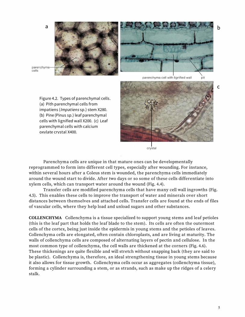
There Are Three Types of Simple Tissues: Parenchyma, Collenchyma, and Sclerenchyma The simple tissues are made of cells that are the workhorse cells of the plant body. They do the photosynthesis, load things in and out of the vascular system, hold up the weight of the plant, store things, and generally conduct the important business and housekeeping chores needed to keep the plant body healthy and functioning. PARENCHYMA Parenchyma cells are usually somewhat spherical or elongated, but they may have diverse shapes (Fig. 4.2). They usually have a thin primary cell wall, but they may have a secondary wall, which is sometimes lignified (Fig. 4.2b). Lignin is a polymer that is embedded between cell wall cellulose molecules. It renders the wall impermeable to water, so that water movement occurs only through openings in the cell wall called pits. Lignin is also quite hard and strong, making lignified cells rigid and supportive even when the cells are dead.
Living parenchyma cells found in all plant organs perform the basic metabolic functions of cells: respiration, photosynthesis, storage, and secretion. Parenchyma cells usually live for one to two years but have been known to live hundreds of years in some plants, such as cactus. Crystals of many different shapes and sizes, usually made of calcium oxalate, are commonly found inside the vacuoles of parenchyma cells (Fig. 4.2c). They may be involved in regulating the pH of cells by crystallizing oxalic acid (which is a way of taking it out of solution).
Parenchyma cells may occur as aggregates forming parenchyma tissue; the cortex and pith of stems and roots and the mesophyll of leaves are composed of parenchyma tissue (Fig. 4.3a-c). The cortex is the region between the plant's epidermal and vascular tissues in most stems and roots. The pith is usually composed of storage parenchyma cells and lies at the center of many stems, inside the cylinder of vascular tissues. In some stems, such as corn (Fig. 4.3d), the vascular tissue is dispersed in bundles. In this case the parenchyma tissue making up the bulk of the stem is simply called ground tissue, and the terms cortex and pith are not used. The mesophyll makes up the bulk of most leaves and is the site of most photosynthesis and water storage in leaves (Fig. 4.3c). Sometimes the air spaces are very large, especially in the stems and leaves of plants that grow in wet places.
5
Parenchyma cells are unique in that mature ones can be developmentally reprogrammed to form into different cell types, especially after wounding. For instance, within several hours after a Coleus stem is wounded, the parenchyma cells immediately around the wound start to divide. After two days or so some of these cells differentiate into xylem cells, which can transport water around the wound (Fig. 4.4). Transfer cells are modified parenchyma cells that have many cell wall ingrowths (Fig. 4.5). This enables these cells to improve the transport of water and minerals over short distances between themselves and attached cells. Transfer cells are found at the ends of files of vascular cells, where they help load and unload sugars and other substances. COLLENCHYMA Collenchyma is a tissue specialized to support young stems and leaf petioles (this is the leaf part that holds the leaf blade to the stem). Its cells are often the outermost cells of the cortex, being just inside the epidermis in young stems and the petioles of leaves. Collenchyma cells are elongated, often contain chloroplasts, and are living at maturity. The walls of collenchyma cells are composed of alternating layers of pectin and cellulose. In the most common type of collenchyma, the cell walls are thickened at the corners (Fig. 4.6). These thickenings are quite flexible and will stretch without snapping back (they are said to be plastic). Collenchyma is, therefore, an ideal strengthening tissue in young stems because it also allows for tissue growth. Collenchyma cells occur as aggregates (collenchyma tissue), forming a cylinder surrounding a stem, or as strands, such as make up the ridges of a celery stalk.
Figure 4.2. Types of parenchymal cells. (a) Pith parenchymal cells from impatiens (Impatiens sp.) stem X280. (b) Pine (Pinus sp.) leaf parenchymal cells with lignified wall X200. (c) Leaf parenchymal cells with calcium oxylate crystal X400.
a
c
b

6
Figure 4.3. Cross sections of vegetative organs showing the distribution of parenchyma in the ground tissue (cortex and pit in the stem, cortex In the root, mesophyll in the leaf). (a) Clover (Trifolium sp.) is a typical stem with cortex and pith. X35 (b) Buttercup root (Ranunculus sp.) has cortex. X80 (c) Lilac leaf (Syringa vulgaris) has vascular bundles embedded in mesophyll. X75 (d) Corn stem (Zea mays) shows vascular bundles scattered in the ground tissue. X16
Figure 4.4 (left). Redifferentiation of parenchymal cells. This Coleus stem shows the regeneration of xylem vessel members around a wound. Figure 4.5 (right). Transfer cell from a Funaria sporophyte, showing ingrowths of the cell wall.

7
SCLERENCHYMA The cells making sclerenchyma tissue are rigid and function to support the weight of a plant organ. There are two types of sclerenchyma cells: fibers and sclereids. These cells tend to have thick, lignified secondary cell walls. They are dead at maturity. Fibers can occur in aggregates forming a continuous cylinder around stems; they may connect end-to-end to form multicellular strands acting like strengthening cables like re-bar in concrete; or they can form a component of vascular tissues. They are long, narrow cells with thick, pitted cell walls and tapered ends (Fig. 4.7a, b). Fibers are sometimes very elastic and can be stretched to a degree, but they will snap back to their original lengths.
a b
c d
Figure 4.6 (left). Section of marigold (Caledula officinalis) stem showing pink-stained collenchyma cells. X170. Figure 4.7 (below). Schlerenchyma. (a) Section of geranium (Pelargonium sp.) stem showing fiber clusters. X200 (b) Macerated cells of tulip tree (Liriodendron tulipifera) wood, showing long, thin fibers. X100 (c) Stone cells, a type of schlereid, from pear (Pyrus sp.) fruit. X200 (d) Star-shaped schlereid from water lily (Nymphia sp.) leaf.

8
Sclereids sometimes occur as sheets (an example being the hard outer layer of some seed coats), but they usually occur in small clusters or as solitary cells. Sclereids have many striking shapes, from elaborately branched cells, to star-shaped cells, to the simple stone cells that give a gritty texture to pear fruits (Fig. 4.7c, d). Sclereid cell walls are often thicker than the walls of fibers. Complex Tissues Make Up the Plant's Vascular System and Outer Covering THE VASCULAR SYSTEM: XYLEM The vascular system consists of an interconnected network of cells that traverse the entire body of the plant (Fig. 4.1). All cells of the plant require minerals and water, which are absorbed by the roots and transported by the xylem. Sugars are manufactured in the leaves and transported by the phloem. The xylem is a complex tissue made up of different kinds of cells that work together to transport water and dissolved minerals. The cell types found in xylem are, the water-conducting cells--tracheids and vessel members (the latter join together end to end to make vessels); fibers, for strength and support; and parenchyma cells, which help load minerals in and out of the vessel members and tracheids. In leaves and young stems the xylem is found in discrete bundles called vascular bundles (Fig. 4.3a, 4.8a), and in young roots the xylem occurs in groups of cells at or near the center of the root known as the vascular cylinder (Fig. 4.3b). Xylem in those locations is called primary xylem. It is formed in the root and shoot apex very early in organ development (this will be discussed later in the chapter). Xylem that forms later in the development of stems and roots is organized in cylindrical patterns and is called secondary xylem (Fig. 4.8b). Usually, leaves have only primary xylem. Figure 4.8. Xylem tissue and cells. (left) Cross-section of primary vascular bundle of a sunflower (Heleanthus annuus). X72 (right) Cross-section of a 1-year-old basswood (Tilia americana) stem. X69.

9
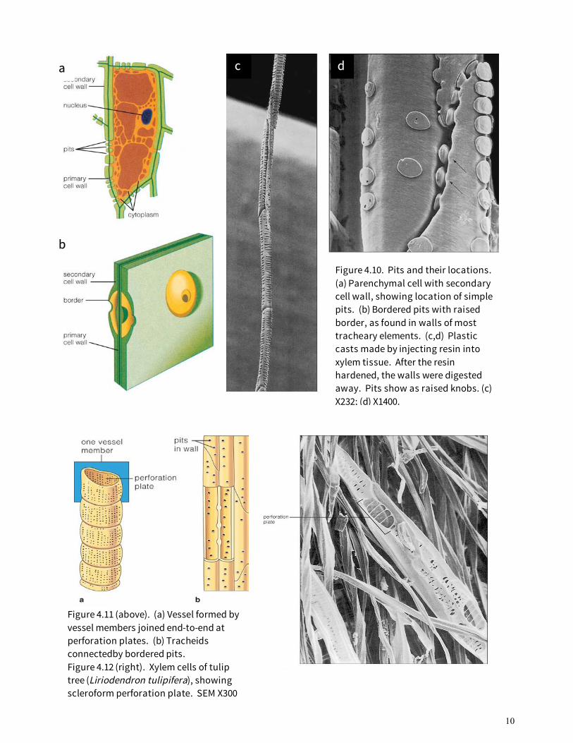
Parenchyma cells are the only living cells found in xylem. They usually have a thin primary cell wall, but in secondary xylem they often have a lignified secondary wall. Fibers function to support xylem tissue and hold it rigid, rather like a steel rod would be used to hold up a plastic water pipe. Vessel members and tracheids have many structural and functional characteristics in common, so the term tracheary element is used to refer to them generally. Tracheary elements are not living at maturity. Before the cells die their cell wall becomes thickened with cellulose and lignin, and then the protoplast degenerates. The secondary cell wall, deposited after the cell stops enlarging, forms in one of several patterns: annular (ring shaped), spiral, scalariform (ladder like), reticulate (forming a network), or pitted (Fig. 4.9). These patterns relate to the timing of their formation. Tracheary elements that form and function in primary xylem of organs that are still elongating have annular or spiral secondary cell walls. Such cells are able to stretch as the surrounding cells elongate and they can still function in water transport. Vessel members that form after organ elongation has ended (in late-forming primary xylem or secondary xylem) tend to have pitted cell walls. These cells are quite rigid and very strong but are unable to stretch.
Since lignified secondary cell walls are impermeable to water, the only way that water can be exchanged between cells is through tiny openings called pits. A pit is not actually a hole in the cell wall; the primary cell wall remains intact, rather like a loose membrane across the pit opening. There are two types of pits: simple pits and bordered pits. Simple pits (Fig. 4.10a) are openings in the secondary walls of fibers and lignified parenchyma cells. Bordered pits occur in tracheids, vessel members, and some fibers (Fig. 4.10b-d). These pits have an expanded border of secondary wall that extends over a small pit chamber. Tracheids and vessel members have some structural and functional characteristics in common, but there are also differences (Fig. 4.11). A vessel member is a cell with an oblique, pointed, or transverse end. The ends of mature vessel members are partially or completely digested away during their development to form a perforation plate. There are several different types of perforation plates; simple and scalariform are the most common (Fig. 4.12). A vessel is a series of vessel members connected end to end (see Fig. 4.11a). Vessels are often several centimeters long, and in some vines and trees they may be many meters in
Figure 4.9. Different patterns of secondary cells walls in tracheary elements. Vessel members that form in early primary xylem have rings or spirals, allowing cells to stretch as the organ grows. The "annular" cell on the left has stretched; the one on the right has not. Xylem cells that form after growth is finished tend to have "pitted" cell walls. "Spiral", "scalariform", and "reticulate" are intermediate forms.
10
Figure 4.11 (above). (a) Vessel formed by vessel members joined end-to-end at perforation plates. (b) Tracheids connectedby bordered pits. Figure 4.12 (right). Xylem cells of tulip tree (Liriodendron tulipifera), showing scleroform perforation plate. SEM X300
Figure 4.10. Pits and their locations. (a) Parenchymal cell with secondary cell wall, showing location of simple pits. (b) Bordered pits with raised border, as found in walls of most tracheary elements. (c,d) Plastic casts made by injecting resin into xylem tissue. After the resin hardened, the walls were digested away. Pits show as raised knobs. (c) X232; (d) X1400.
a
b
c d
11
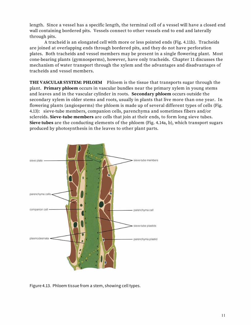
length. Since a vessel has a specific length, the terminal cell of a vessel will have a closed end wall containing bordered pits. Vessels connect to other vessels end to end and laterally through pits. A tracheid is an elongated cell with more or less pointed ends (Fig. 4.11b). Tracheids are joined at overlapping ends through bordered pits, and they do not have perforation plates. Both tracheids and vessel members may be present in a single flowering plant. Most cone-bearing plants (gymnosperms), however, have only tracheids. Chapter 11 discusses the mechanism of water transport through the xylem and the advantages and disadvantages of tracheids and vessel members. THE VASCULAR SYSTEM: PHLOEM Phloem is the tissue that transports sugar through the plant. Primary phloem occurs in vascular bundles near the primary xylem in young stems and leaves and in the vascular cylinder in roots. Secondary phloem occurs outside the secondary xylem in older stems and roots, usually in plants that live more than one year. In flowering plants (angiosperms) the phloem is made up of several different types of cells (Fig. 4.13): sieve-tube members, companion cells, parenchyma and sometimes fibers and/or sclereids. Sieve-tube members are cells that join at their ends, to form long sieve tubes. Sieve tubes are the conducting elements of the phloem (Fig. 4.14a, b), which transport sugars produced by photosynthesis in the leaves to other plant parts.
Figure 4.13. Phloem tissue from a stem, showing cell types.

12
Figure 4.14. Phloem. (a) Elm (Ulmus sp.) tangential longitudinal section showing sieve-tube members and parenchymal cells in secondary phloem. X375 (b) Cross-section of primary phloem of cucumber (Cucurbita pepo) stem. X130. (c) Pine (Pinus sp.) tangential longitudinal section of secondary phloem showing sieve cells and blue-stained callose in sieve areas. X228. A young sieve-tube member contains a nucleus, plastids, mitochondria, and Golgi stacks. As the cell matures, its nucleus disintegrates, the plastids and mitochondria shrink, and the cytoplasm becomes reduced to a thin peripheral layer. The central part of the cell becomes occupied by a mass of dense material. This mass, which was called slime in the early literature, can be seen with the light microscope. It is called P-protein (Fig. 4.15a); it is actually composed of a complex of proteins. When the sieve-tube member is mature, one or more companion cells lie connected to it by plasmodesmata (Fig. 4.13). Since companion cells have a nucleus and a full complement of organelles, it is thought that they regulate the metabolism of their adjacent sieve-tube member. Companion cells are also known to play an important role in the mechanism of loading and unloading the phloem. The walls of mature sieve-tube members contain aggregates of small pores called sieve areas. One or more sieve areas on the end wall of a sieve-tube member is called a sieve plate (Fig. 4.13, 4.15a). The end walls of two connecting sieve-tube members are thickened, and strands of cytoplasm and P-protein pass through pores adjoining them. Sieve-tube members live and function from one to three years except in a few trees such as palms, where they live much longer.
In many studies of the structure of mature sieve-tube members, a carbohydrate known as callose was seen to surround the margins of the pores in the sieve areas (Fig. 4.15a). In some instances, protein is also collected at the sieve plate. It has been demonstrated that callose can form very rapidly in response to aging, wounding, and other stresses (Fig. 4.15b), and the result is to limit the loss of cell sap from injured cells.
a
b
c

13
In gymnosperms and ferns, sieve cells rather than sieve-tube members are the conducting elements in the phloem. These cells are quite long, with tapered ends (Fig. 4.14c). They have sieve areas but no sieve plates at their ends. Sieve cells apparently function similarly to sieve tubes and usually lack nuclei at maturity. Adjacent albuminous cells are short, living cells that act as companion cells to these sieve cells. Phloem fibers are usually long, tapered cells with lignified cell walls. Mature fibers from the ramie plant (Boehmeria nivea) have been reported up to 55 cm long. Such fibers are valued for their commercial uses, especially for rope and fabric. Phloem parenchyma cells are usually living cells that function in phloem loading and unloading. THE PLANT'S OUTER COVERING: EPIDERMIS The epidermis is the outer covering of the plant. It is a complex tissue composed of epidermal cells, guard cells, and trichomes (hairs) of various types. The epidermis is usually one layer of cells, but it may be as many as five or six layers in the leaves of some succulent plants and in the aerial roots of certain orchids. The epidermis protects the inner tissues both from drying and from infection by some pathogens. It also regulates the movement of water and gases out of and into the plant. Epidermal cells are the main cell type making up the epidermis. These cells are living, lack chloroplasts, are usually somewhat elongate, and often have walls with irregular contours (Fig. 4.16). The outer walls of epidermal cells are often thicker than the inner and sidewalls. The outer epidermal wall is coated with a waxy substance (cutin) forming an impermeable layer called the cuticle (Fig. 4.17). All parts of the plant, except the tip of the shoot apex and the root cap, have a cuticle. In roots, the cuticle is often very thin (and has been reported to be totally lacking on the surface of root hairs).
Figure 4.15. P-protein and callose. (a) Beet (Beta vulgaris) petiole sieve-tube members with P-protein and callose surrounding the pores at the sieve plate. X3000 (b) Beet petiole 1 hour after stress by exposure to cold temperature. X10,200.
a b

14
Figure 4.16. Epidermis and epidermal cells. (a) Upper leaf surface of Shepard's purse (Capsella bursa-pastoris). X70 (b) Higher magnificaiton showing epidermal cells, star-shaped hairs, and stomata. X350.
Young stems, leaves, flower parts, and even some roots in exceptional instances have specialized epidermal cells called guard cells. Between each pair of guard cells is a small opening, or pore, through which gases enter and leave the underlying tissues. Two guard cells plus the pore constitute one stoma (plural, stomata; Fig. 4.18). Guard cells differ from other epidermal cells by their crescent shape and the fact that they contain chloroplasts. Another type of epidermal cell, the subsidiary cell, forms in close association with guard cells and functions in stomatal opening and closing. The physiological role of stomata and the mechanism of their opening and closing will be discussed in Chapter 11. Trichomes are epidermal outgrowths and may be a single cell or multicellular (Fig. 4.19). In roots, for example, root hairs are extensions of single epidermal cells that increase the root surface area in contact with soil water. In some leaves, very elaborate multicellular trichomes may form (Fig. 4.16b). Trichomes will be discussed again in the following Chapters.
Figure 4.17. Cross section of leaf of pincushion tree (Hakea sp.). Note the thick cuticle and small channels crossing the cuticle. X184.

15
THE PLANTS OUTER COVERING: PERIDERM The periderm is a protective layer that forms in older stems and roots after those organs expand and the epidermis splits and is lost. It is a secondary tissue. This tissue is several cell layers deep and is composed of phellem (cork) cells on the outside, a layer of dividing cells (phellogen or cork cambium), and the phelloderm toward the inside (Fig. 4.20). Phellem cells are dead at maturity, and have a waxy substance (suberin) embedded in their cell walls. Phelloderm cells live longer than phellem cells and are parenchymalike.
Figure 4.20. Diagram of periderm, showing the layers: phellum, cork cambium, and phelloderm.
Figure 4.19. (above) Glandular hairs of tobacco (Nicotiana tabacum). X22. The thickened bulb-like end of this trichome secretes sticky material.
Figure 4.18 (left). Stomata. (a) surface view of Iris sp. leaf, showing several stomata composed of two guard cells surrounding a pore. X415 (b) Diagram of stomatal apparatus consisting of two guard cells and surrounding subsidiary cells such as would be found in leaves of Sedum.
a
b

16
Secretory Tissues Produce and Secrete Materials Secretory structures occur mostly in leaves and stems. These may be composed of single secretory cells or very complex multicellular structures (Fig. 4.19). Some trichomes, for example, may secrete materials out of the plant to attract insect pollinators. They may also form inside the plant body and secrete materials within the plant; cells called laticifers (Fig. 4.21), for example, secrete latex, which discourages herbivores from eating the plant. They also form complex ducts inside wood of trees. These interesting structures will be discussed again with leaves and stems (Chapters 5 and 6). Table 4.2 contains a summary of the structure and function of the common cell types found in seed plants.
Figure 4.21. Laticifer cells in spurge (Euphorbia sp.) stems. Longitudinal section X 20.

17
MERISTEMS: WHERE CELLS DIVIDE We now know the tissues and cell types making up the vascular plant, but how do these tissue and cells come to be? Do plant cells specialize and group into tissues in the same manner that animal cells do? The answer is, largely, no. The growth patterns of animals and plants differ in one very significant way. Animal cells divide mostly during the embryo stage of development. After the animal reaches adult size, cells divide only in the bone marrow (to produce new blood cells), in the intestinal lining (to repair wounded tissues), and in the epithelial layers (to form new skin, nails, and hair). In vascular plants, an entirely different process exists: Cell division continues through the whole lifetime of the plant, but it occurs in special regions of the plant body called meristems (from the Greek word meaning "to divide"). What Is a Meristem? A meristem is a site in the plant body where new cells form and the complex processes of growth and differentiation are initiated. Growth means the irreversible increase in size that comes from both cell division and cell enlargement. Cell differentiation refers to the changes that a cell undergoes structurally and biochemically so that it can perform a specialized function. Since cells and tissues are derived from meristems, we do not consider meristems themselves to be tissues. There are different categories of meristems, each with a specific function. Shoot and root apical meristems are at the tips of branches and roots (Fig. 4.22); they are the ultimate sources of all cells in the plant. Primary meristems, the next level of meristems, originate in apical meristems and produce, or more correctly, differentiate into, the primary tissues. The secondary meristems produce the secondary tissues. These categories of
18
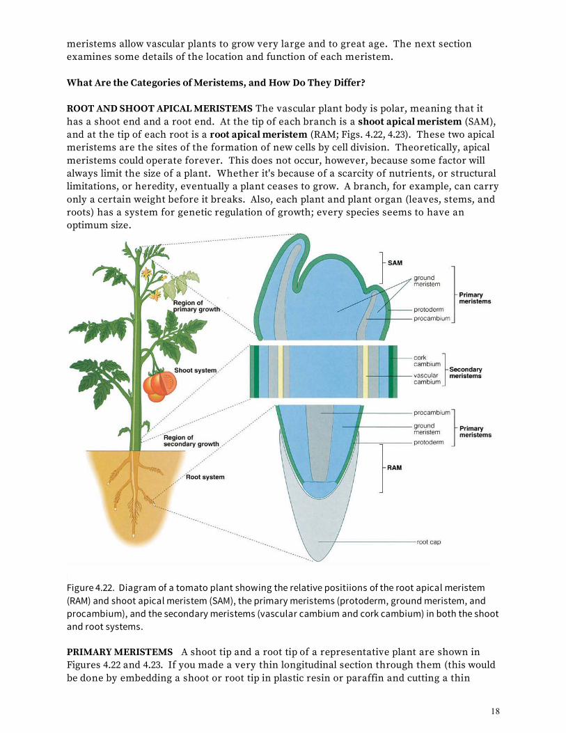
meristems allow vascular plants to grow very large and to great age. The next section examines some details of the location and function of each meristem. What Are the Categories of Meristems, and How Do They Differ? ROOT AND SHOOT APICAL MERISTEMS The vascular plant body is polar, meaning that it has a shoot end and a root end. At the tip of each branch is a shoot apical meristem (SAM), and at the tip of each root is a root apical meristem (RAM; Figs. 4.22, 4.23). These two apical meristems are the sites of the formation of new cells by cell division. Theoretically, apical meristems could operate forever. This does not occur, however, because some factor will always limit the size of a plant. Whether it's because of a scarcity of nutrients, or structural limitations, or heredity, eventually a plant ceases to grow. A branch, for example, can carry only a certain weight before it breaks. Also, each plant and plant organ (leaves, stems, and roots) has a system for genetic regulation of growth; every species seems to have an optimum size.
Figure 4.22. Diagram of a tomato plant showing the relative positiions of the root apical meristem (RAM) and shoot apical meristem (SAM), the primary meristems (protoderm, ground meristem, and procambium), and the secondary meristems (vascular cambium and cork cambium) in both the shoot and root systems. PRIMARY MERISTEMS A shoot tip and a root tip of a representative plant are shown in Figures 4.22 and 4.23. If you made a very thin longitudinal section through them (this would be done by embedding a shoot or root tip in plastic resin or paraffin and cutting a thin

19
section), you would see that the cells of the SAM and RAM and those just basal to them (toward the more mature cells) are small, with a relatively dense protoplasts (Fig. 4.23). Cells with these characteristics are usually capable of dividing and so are referred to as meristematic cells. Cells immediately basal to the SAM are ordered into distinct files of cells (Figs. 4.22, 4.23). These newly ordered cells are still meristematic (they can divide); they are in a sense the embryonic stages of the tissues. These groups of cells are called the primary meristems, and they have two roles: to form the primary tissues and to elongate the root and shoot. There are three primary meristems: protoderm, procambium, and ground meristem (Fig. 4.22). The cells of the protoderm differentiate into the epidermis. The procambium cells differentiate into the cells of the primary xylem and primary phloem. The ground meristem differentiates into the cells of the pith and cortex of stems and roots and the mesophyll of leaves .
c
Figure 4.23. Shoot and root tips. (a) Photograph showing the position of the shoot apex. The shoot apical meristem (SAM) is surrounded by several small leaves. (b) Median longitudinal section through the SAM of a Coleus blumei plant. X49 (c) Photograph of Epiphyllum phyllanthus primary root tip. Scale bar = 2 mm. The root apical meristem is at the very tip, just inside the root cap. (d) Median longitudinal section through RAM of an Arabidopsis thaliana root tip. X426
a
b
d

20
The primary meristems near the tips of the roots and shoots are the site of most elongation. They produce new cells, which then enlarge mostly by elongation. However, in many plants the branch or root continues to increase in girth as well. This increase in girth requires lateral growth, which involves the formation and activity of the next category of meristems, called secondary meristems. SECONDARY MERISTEMS The secondary meristems are responsible for cell division, initiation of cell differentiation, and growth in a lateral direction, thereby increasing the thickness and circumference of stems and roots. The wood in trees, for example, is really secondary growth resulting from the activity of secondary meristems. Not all plants have secondary meristems. There are thousands of species that grow only one season and usually lack secondary growth. Leaves also usually lack secondary growth. The bodies of many plants have two secondary meristems: the vascular cambium and the cork cambium (Fig. 4.22). Vascular cambium differentiates into secondary xylem and secondary phloem, and the cork cambium into the periderm (Fig. 4.24). These will be discussed at length in the chapters on stems and roots, which follow. OTHER MERISTEMS There are several other meristems. In stems, as an example, an intercalary meristem occurs within the stem to regulate its elongation. As leaves develop, there are leaf-specific meristems that regulate leaf shape. The intercalary meristem at the base of grass leaves allows the leaf to continue to grow after being grazed or mowed. Other meristems are involved in forming buds and roots in unusual places, such as at the base of trees, and also in the repair of wounds. We will discuss some of these other meristems in the following chapters.
Figure 4.24. Summary of meristems and the tissues they generate.
21
KEY TERMS albuminous cells callose cell differentiation companion cells cork cambium cortex dermal tissue system epidermal cells epidermis fibers ground meristem ground tissue system guard cells meristem mesophyll parenchyma tissue periderm phellem
phelloderm phellogen pith pits P-protein primary meristem primary phloem primary xylem procambium protoderm root apical meristem root system sclereids sclerenchyma secondary meristem secondary phloem secondary xylem shoot apical meristem )
shoot system sieve areas sieve cells sieve plate sieve tubes sieve-tube members stoma (stomata) subsidiary cell tracheary element tracheids trichomes vascular bundles vascular cambium vascular cylinder vascular tissue system vessel members vessels xylem
SUMMARY 1. The vascular plant body is organized into a shoot system (stems, leaves, buds, flowers, and fruits) and a root system. Flowering plants (angiosperms), cone-bearing plants (gymnosperms), and ferns are all vascular plants. 2. Cells in the plant body are organized into the ground, vascular, and dermal tissue systems. There are several different cell types making up the plant body. These cells are organized into aggregates called tissues. 3. The simple tissues are parenchyma, collenchyma, and sclerenchyma. (Refer to Tables 4.1 and 4.2 and Figure 4.24 for summaries of cell types, tissues, and meristems.) 4. The complex tissues are epidermis, xylem, phloem, and periderm. 5. Secretory structures are specialized to secrete various substances within and outside the plant body. 6. Each cell originates from a cell that was once meristematic. The process whereby a cell changes into a mature cell is called differentiation. 7. Meristems are the sites of cell division, cell elongation, and the beginning of cell differentiation. S. Apical meristems occur at the tips of stems and roots and are the ultimate source of all cells in the shoot and root systems.

22
9. Primary meristems (protoderm, procambium, and ground meristem) form the primary tissues (epidermis, primary xylem and phloem, and pith and cortex). 10. Secondary meristems (vascular cambium and cork cambium) form the secondary tissues (secondary xylem and secondary phloem, and phellem and phelloderm, respectively). Questions 1. What are the tissues found in the plant body? How are they organized in each vegetative organ? 2. What are the features, cell types, and functions of each of the tissues listed below? epidermis periderm xylem phloem
parenchyma collenchyma sclerenchyma (fibers and sclereids)
3. Define the following xylem terms: vessel member vessel
tracheid bordered pit
4. Define the following phloem terms: sieve-tube member sieve tube
companion cell sieve plate
5. Describe the function of the following meristems: root and shoot apical meristems primary meristems: protoderm, ground meristem, procambium secondary meristems: vascular cambium, cork cambium 6. Each tissue in the plant body has a different function. Describe these functions and discuss how the tissues and cells communicate with each other.

23
PLANTS, PEOPLE, AND THE ENVIRONMENT: Plant Anatomists as Detectives?
FBI agents, police, lawyers, medical and veterinary doctors, agricultural extension specialists, and members of the public have all had occasion to contact me over the past several years concerning the identification of plant material. Usually this material was chewed, digested, burned, dried, or otherwise distorted, so that the identity of the plant or plant organ was no longer apparent. Yet every plant species contains cell types with special structures, shapes, sizes, and staining reactions--characteristics that make it possible to identify the plant. Regardless of the kind of case, similar microtechnique tools and methods are used, and the application of basic knowledge of plant anatomy is required.
Plant anatomists have been involved in many legal cases, but mostly we are asked for opinions dealing with medical or agricultural problems. For example, agricultural extension specialists need help in identifying root specimens pulled from clogged sewers. Veterinarians who suspect that sick animals have eaten poisonous plants will call plant anatomists. Anatomists have also worked with anthropologists interested in ancient basketry or the identification of wood and charcoal pieces found in ancient fire pits. Probably the most famous legal case involving a plant anatomist was the Lindbergh kidnapping. In 1932 the infant son of celebrated aviator Charles Lindbergh and his wife, Anne Morrow Lindbergh, a noted writer and poet, was kidnapped out of a second-story nursery. The kidnapper sent a ransom note, but the baby was later found dead. The only evidence left at the crime scene was a crude wooden ladder leaning against the second-story window. The police and FBI gave the ladder to the Forest Products Laboratory in Wisconsin for analysis. A plant anatomist there, Arthur Koehler, was able to specifically identify the wood used. After a man named Bruno Hauptmann was arrested for the crime, police found that several boards were missing from the floor of the attic in his house. Pieces of this wood were sent to scientists at the Forest Products USDA Laboratory in Wisconsin, and they positively identified the wood as a match to the ladder. This testimony was used to convict Hauptmann, who was later executed. Most cases are less poignant. In 1984 I was contacted by the Sheriff's office in Calaveras County, California (the part of California's gold country made famous by Mark Twain's story of the jumping frog contest). In this instance, a man was suspected of growing marijuana (Cannabis sativa) in an elaborate hydroponic setup in his attic. The suspect was warned of an impending raid just before the sheriff arrived. He hid the stems

24
of the plants outside his house and tried to burn the stem stumps and roots in his fireplace. The sheriff arrested him and took the partially burned material as evidence. The suspected grower reportedly told a sheriff's deputy that he would get off because it was not possible to prove that he was burning marijuana in his fireplace. I examined the material taken as evidence and compared it with known specimens of marijuana from herbarium sheets and from identified marijuana stems. The charred evidence and the known specimens showed similar wood anatomy. After I testified in a pretrial hearing that, based on the comparison, the partially burned material was most likely marijuana, the defendant accepted a plea bargain. The case was never brought to trial. Several of my cases have come from the San Diego Zoo. One of them involved a group of Hanuman langur monkeys, a rare Asian species. For two years the monkeys had been fed mostly Acacia leaves. Suddenly three monkeys died after bouts of weight loss, diarrhea, and vomiting. An autopsy revealed intestinal lesions and plugging with masses of fibrous plant material. I examined some of this material, along with samples of Acacia leaf browse and other plant materials within reach of the monkey enclosure. Microscopic examination of this material revealed partially digested vascular strands with attached thick-walled cells and small epidermal fragments. By making polarized light images and comparing them to known specimens, I was able to identify the material as Acacia leaf vascular bundles. The keepers changed the monkeys' feed. The point of these stories is that the study of plant anatomy has uses far beyond just knowing what is inside the plant. Basic information and a few simple tools--such as a razor blade, some common dyes, and a microscope--can go a long way toward solving criminal and medical puzzles. So study hard; you never know when your knowledge of plant anatomy might come in handy. Thomas L. Rost, Department of Plant Biology, University of California, Davis

25
ECONOMIC BOTANY: Music from Plants The direct relation between plants and music is obvious if one thinks about it even a little. The earliest musical instruments were probably drums made of grooved or hollowed logs, and to this day drums are usually made of wood cylinders, typically with skins or synthetic materials stretched over them. The bodies of ancient stringed instruments, such as lyres and harps, were made of wood. Tambourines, pan pipes, whistles, recorders, early flutes, shawms, accordions, organs, harpsichords, and pianos were, and still are, made largely of wood. Bodies, bells, mouthpieces, backs, sides, fronts, bridges, or fingerboards of clarinets, English horns, oboes, bassoons, violins, violas, cellos, contrabasses ("stand-up basses"), xylophones, and guitars are made of various kinds of wood carefully chosen for their acoustic and other physical properties. But wood is not the only plant material that contributes to music making. Many of you have made a sort of whistle using your hands and a blade of grass. Paper, most of which is made of various kinds of plant fibers, can be used to made a kazoo. The oldest types of traditional flutes and whistles were made of various types of cane, a cousin of bamboo. For 2,000 to 3,000 years, the preferred type of cane has traditionally been Arundo donax (giant cane). The hollow stems from this species have always been the best for making reeds for woodwind instruments, such as clarinets (Fig. 1), saxophones, oboes, and bassoons, and for not-so-obvious examples, such as the Chinese sheng (perhaps the oldest reed instrument), Turkish zurna, Egyptian mizmar, Vietnamese ding tac ta (played by inhaling), Scot and Irish bagpipes, hornpipes, krummhorns, concertinas, bandoneons, small organs, some harmonicas, melodicas, and many others. Anyone who has played the clarinet or other reed instrument for any length of time knows that getting a reed that produces just the right sound quality is a challenge. Players who cut their own
Figure 1. A clarinet reed is a piece of Arundo donax stem that has been carved or machine milled to an appropriate shape and clamped to the mouthpiece of the instrument. Air from the player's lungs causes the reed to vibrate.
reeds can tell you that the raw material is highly variable in its playing properties. Commercially, clarinet reeds are made from quartered pieces of Arundo donax internodes that are milled into the classic reed shape (Fig. 2). In the past some commercially made reeds often were discarded because they did not sound quite right when used with a particular instrument.

26
When I started the project described below a considerable traditional lore had developed among players and people who manufactured reeds as to how to select and process them.
In 1992, as part of an effort to improve their products, Rico International, an American reed-making company, asked me to research whether there were aspects of the anatomy of A. donax plants that predisposed them to be especially good or bad for clarinet reeds. We asked several skilled symphonic performers to play a number of clarinet reeds and to identify the best and the worst among them. Each reed was randomly given a numeric code identifier, and the reeds were sent to me for analysis. I had no idea which reeds played well.
Figure 2. Traditionally, internodes of A. donax were quartered and then carved into the characteristic reed shape. This process is now done commercially by milling machines.
Thin sections (slices) of each reed were carefully cut. The sections were treated with stains to increase the contrast of the cells and tissues in the reed, and photographs were then taken of each section using a precision photomicroscope. For each reed I recorded twenty-three anatomic characteristics (for example, the ratio of vascular to ground tissue, the size of vascular bundles, the shape of vascular bundles, the proportion of vascular bundles that was bundle sheath tissue, the length and diameter of parenchyma cells, the thinkness of parenchyma cell walls, and so on). The combined data were statistically analyzed. I found that the coded reeds clustered into two groups when several characteristics of the vascular system were considered in combination. The statistical conclusion for any partiular clarinet reed matched the professional players' assessments 92% of the time for good reeds and 85% of the time for bad
Figure 3. The best sounding reeds have vascular bundles with thick bundle sheaths of fiber cells that completely surround each bundle. The more of the vacular bundles that look like this, the better.
reeds. My analysis showed that reeds made better music when their vascular bundles were more or less lined up, uniformly distributed through the tissue, and had thick, fibrous bundle sheaths that completely surrounded the vascular bundles (Fig 3). Because my study was

27
done for a private company it was never published, but another group of scientists in Australia, which included a clarinet player, did a similar study. Using similar methods, they came to the same conclusions that I did. It was satisfying to know that other scientists had confirmed my results. Clarinet reed manufacturers can now use these data to help them select the best canes for new reeds. It is clear to me that science can be used to help artists get better results. Dr. Daniel K. Gladish, Botany Department, Miami University, Hamilton, Ohio

28
Figure credits CO. D.D. Brandon, University of California, Davis. Figure 4.1. Daniel K. Gladish, University of Miami, Ohio Figure 4.2(a-c). Thomas L. Rost. Figure 4.3(a-d). Thomas L. Rost. Figure 4.4. Thomas L. Rost. Figure 4.5. Used by permission from Gunning BES, 'Transfer cells and their roles in transport of solutes in plants" Science Progress Oxford 977, 64:539-568. Photo taken by Dr. Alan J. Browning, Australia National University. Figure 4.6. Thomas L. Rost. Figure 4.7(a-c) Thomas L. Rost. (d) Leslie Sunell, University of California, Santa Cruz. Figure 4.8(a-b). Thomas L. Rost. Figure 4.10(c-d). Steve Mauseth, University of Texas. Figure 4.12. Sonia Cook, University of California, Davis. Figure 4.14(a-c). Thomas L. Rost. Figure 4.15(a-b). Vincent R. Franceschi, Washington State University. Figure 4.16(a-b). Richard H. Falk, Virginia Commonwealth University. Figure 4.17. Thomas L. Rost. Figure 4.18(a). Thomas L. Rost. Figure 4.19. Thomas L. Rost. Figure 4.21. Thomas L. Rost. Figure 4.23 (a). Jacob Maentz. (c) Svetlana Shishkova and Joseph Dubrovsky, UNAM, México. (b, d) Thomas L. Rost Economic Botany/Music: Figures 1, 2 and 3. Daniel K. Gladish, Miami University, Ohio